CN113520272B - Endoscopic catheter-multimode optical imaging coupling detection system - Google Patents
Endoscopic catheter-multimode optical imaging coupling detection systemDownload PDFInfo
- Publication number
- CN113520272B CN113520272BCN202110723621.2ACN202110723621ACN113520272BCN 113520272 BCN113520272 BCN 113520272BCN 202110723621 ACN202110723621 ACN 202110723621ACN 113520272 BCN113520272 BCN 113520272B
- Authority
- CN
- China
- Prior art keywords
- optical
- optical fiber
- imaging
- imaging device
- image
- Prior art date
- Legal status (The legal status is an assumption and is not a legal conclusion. Google has not performed a legal analysis and makes no representation as to the accuracy of the status listed.)
- Active
Links
Images

Classifications
- A—HUMAN NECESSITIES
- A61—MEDICAL OR VETERINARY SCIENCE; HYGIENE
- A61B—DIAGNOSIS; SURGERY; IDENTIFICATION
- A61B1/00—Instruments for performing medical examinations of the interior of cavities or tubes of the body by visual or photographical inspection, e.g. endoscopes; Illuminating arrangements therefor
- A61B1/00112—Connection or coupling means
- A61B1/00121—Connectors, fasteners and adapters, e.g. on the endoscope handle
- A61B1/00126—Connectors, fasteners and adapters, e.g. on the endoscope handle optical, e.g. for light supply cables
- A—HUMAN NECESSITIES
- A61—MEDICAL OR VETERINARY SCIENCE; HYGIENE
- A61B—DIAGNOSIS; SURGERY; IDENTIFICATION
- A61B1/00—Instruments for performing medical examinations of the interior of cavities or tubes of the body by visual or photographical inspection, e.g. endoscopes; Illuminating arrangements therefor
- A61B1/00002—Operational features of endoscopes
- A61B1/00004—Operational features of endoscopes characterised by electronic signal processing
- A61B1/00009—Operational features of endoscopes characterised by electronic signal processing of image signals during a use of endoscope
- A—HUMAN NECESSITIES
- A61—MEDICAL OR VETERINARY SCIENCE; HYGIENE
- A61B—DIAGNOSIS; SURGERY; IDENTIFICATION
- A61B1/00—Instruments for performing medical examinations of the interior of cavities or tubes of the body by visual or photographical inspection, e.g. endoscopes; Illuminating arrangements therefor
- A61B1/04—Instruments for performing medical examinations of the interior of cavities or tubes of the body by visual or photographical inspection, e.g. endoscopes; Illuminating arrangements therefor combined with photographic or television appliances
- A61B1/043—Instruments for performing medical examinations of the interior of cavities or tubes of the body by visual or photographical inspection, e.g. endoscopes; Illuminating arrangements therefor combined with photographic or television appliances for fluorescence imaging
Landscapes
- Health & Medical Sciences (AREA)
- Life Sciences & Earth Sciences (AREA)
- Surgery (AREA)
- Engineering & Computer Science (AREA)
- Biomedical Technology (AREA)
- Molecular Biology (AREA)
- Pathology (AREA)
- Radiology & Medical Imaging (AREA)
- Nuclear Medicine, Radiotherapy & Molecular Imaging (AREA)
- Biophysics (AREA)
- Physics & Mathematics (AREA)
- Heart & Thoracic Surgery (AREA)
- Medical Informatics (AREA)
- Optics & Photonics (AREA)
- Animal Behavior & Ethology (AREA)
- General Health & Medical Sciences (AREA)
- Public Health (AREA)
- Veterinary Medicine (AREA)
- Signal Processing (AREA)
- Endoscopes (AREA)
- Investigating, Analyzing Materials By Fluorescence Or Luminescence (AREA)
Abstract
Description
Translated fromChinese技术领域technical field
本发明涉及生物医学工程领域,尤其是涉及一种内窥导管-多模态光学成像耦合检测系统。The invention relates to the field of biomedical engineering, in particular to an endoscopic catheter-multimodal optical imaging coupling detection system.
背景技术Background technique
结直肠癌是一种常见的癌症,约占全球所有癌症病例的10%。传统上结肠镜检查是结直肠癌诊断和分类的金标准,结肠镜检查可视化结肠和直肠粘膜上异常生长的息肉组织。除了结直肠癌筛查之外,医生还利用结肠镜检查以微创方式切除小息肉,并对较大的息肉或肿瘤进行活检以进行进一步诊断。但结肠镜只能提供直肠壁的表面形态,而不能观察与结直肠癌高度相关的异常层结构和皮下微血管。并且结肠镜对小息肉的检出率较低,影响结直肠癌诊断的准确性。Colorectal cancer is a common cancer, accounting for approximately 10% of all cancer cases worldwide. Traditionally the gold standard for the diagnosis and classification of colorectal cancer, colonoscopy visualizes abnormally growing polyp tissue on the mucosa of the colon and rectum. In addition to colorectal cancer screening, doctors use colonoscopy to minimally invasively remove small polyps and to biopsy larger polyps or tumors for further diagnosis. However, colonoscopy can only provide the surface morphology of the rectal wall, but cannot observe the abnormal layer structure and subcutaneous microvessels that are highly related to colorectal cancer. Moreover, the detection rate of small polyps by colonoscopy is low, which affects the accuracy of diagnosis of colorectal cancer.
近些年来,新成像技术的不断发展为疾病的早期诊断提供了新的手段。In recent years, the continuous development of new imaging technologies has provided new means for early diagnosis of diseases.
光学相干层析成像(Optical Coherence Tomography,OCT)具有高分辨率,无接触,无损伤几个方面的特点。内窥OCT(Endoscopic OCT,E-OCT)作为OCT技术的重要分支,通过探头将光引导至待测器官组织处,可以克服光穿透深度有限的弱点,从而获得人体内器官深度高分辨的层析图像,通过组织形态学的研究,实现疾病的早期治疗。Optical coherence tomography (OCT) has the characteristics of high resolution, no contact, and no damage. Endoscopic OCT (Endoscopic OCT, E-OCT), as an important branch of OCT technology, guides light to the organ tissue to be measured through the probe, which can overcome the weakness of limited light penetration depth, so as to obtain high-resolution layers of human internal organs. Analyze images, and realize early treatment of diseases through the study of histomorphology.
荧光成像(Fluorescence Imaging)是荧光物质被激发后所发射的荧光信号的强度在一定范围内与荧光素的量呈线性关系。荧光是一种特异性高的分子,结合荧光成像和OCT就可以同时分析组织的结构和成分,可为直肠早期病变提供精准的影像手段。Fluorescence imaging (Fluorescence Imaging) is that the intensity of the fluorescent signal emitted by the excited fluorescent substance is linearly related to the amount of fluorescein within a certain range. Fluorescence is a highly specific molecule. Combining fluorescence imaging and OCT can simultaneously analyze the structure and composition of tissues, which can provide accurate imaging methods for early rectal lesions.
如何将光学相干层析成像和荧光成像进行结合,构建一种新的检测系统,是目前亟需解决的技术问题。How to combine optical coherence tomography and fluorescence imaging to construct a new detection system is a technical problem that needs to be solved urgently.
美国专利US 2016/0242737 Al提出了一种能够对胆管或胰腺进行超声/OCT成像的多模态系统。该系统通过内窥探头检测组织上异常的结构以诊断出病因。但该专利并未使用光学相干层析血流成像对组织的微血管进行成像。微血管的形态变化有助于在早期诊断出直肠病变,让患者得到及时的治疗。US Patent US 2016/0242737 Al proposes a multimodal system capable of ultrasound/OCT imaging of the bile duct or pancreas. The system uses an endoscopic probe to detect abnormal structures on tissue to diagnose the cause. But the patent does not use optical coherence tomography to image the microvessels of tissue. The morphological changes of microvessels are helpful for early diagnosis of rectal lesions, so that patients can receive timely treatment.
发明内容Contents of the invention
本发明的目的就是为了克服上述现有技术存在的缺陷而提供一种内窥导管-多模态光学成像耦合检测系统,结合光学相干层析成像和近红外荧光成像组成多模态光学成像系统可视化组织形态和血管结构来弥补传统结肠镜检查的局限性。The purpose of the present invention is to provide an endoscopic catheter-multimodal optical imaging coupling detection system in order to overcome the above-mentioned defects in the prior art, and combine optical coherence tomography and near-infrared fluorescence imaging to form a multimodal optical imaging system for visualization Tissue morphology and vascular structure to compensate for the limitations of traditional colonoscopy.
本发明的目的可以通过以下技术方案来实现:The purpose of the present invention can be achieved through the following technical solutions:
本发明的目的是保护一种内窥导管-多模态光学成像耦合检测系统,包括内窥探头、光纤、电机组件、波分复用器、OCT成像装置、荧光成像装置、图像处理装置,其中具体地:The purpose of the present invention is to protect an endoscopic catheter-multimodal optical imaging coupling detection system, including an endoscopic probe, an optical fiber, a motor assembly, a wavelength division multiplexer, an OCT imaging device, a fluorescence imaging device, and an image processing device, wherein specifically:
内窥探头,包括套管,所述套管的始端内设有光学聚焦模块;The endoscopic probe includes a sleeve, and an optical focusing module is arranged in the beginning of the sleeve;
光纤,所述光纤的始端接入所述套管的末端,且使得光纤的始端与所述光学聚焦模块光通信连接;an optical fiber, the beginning end of the optical fiber is connected to the end of the sleeve, and the beginning end of the optical fiber is optically connected to the optical focusing module;
电机组件,与所述光纤的末端连接,所述电机组件能够驱动光纤进行旋转,并能对光纤进行收拉,以此实现内窥探头的旋转和回拉位移;The motor assembly is connected to the end of the optical fiber, the motor assembly can drive the optical fiber to rotate, and can pull the optical fiber, so as to realize the rotation and pull-back displacement of the endoscopic probe;
波分复用器,其第一端口与所述光纤的末端光通信连接,a wavelength division multiplexer, the first port of which is optically connected to the end of the optical fiber,
OCT成像装置,其输入端与所述波分复用器的第二端口光通信连接;OCT imaging device, its input terminal is optically connected to the second port of the wavelength division multiplexer;
荧光成像装置,其输入端与所述波分复用器的第二端口光通信连接;A fluorescence imaging device, the input end of which is optically connected to the second port of the wavelength division multiplexer;
图像处理装置,分别与OCT成像装置、荧光成像装置的输出端电连接,图像处理装置能够指令波分复用器和图像处理装置输出特定规格的光信号,同时图像处理装置能够将OCT成像装置、荧光成像装置输出的图像信息进行图像配准,之后生成待测区域的多模态影像。The image processing device is electrically connected to the output ends of the OCT imaging device and the fluorescence imaging device respectively. The image processing device can instruct the wavelength division multiplexer and the image processing device to output an optical signal of a specific specification. At the same time, the image processing device can combine the OCT imaging device, The image information output by the fluorescence imaging device is subjected to image registration, and then a multimodal image of the region to be tested is generated.
进一步地,所述OCT成像装置包括光学相干成像样品臂光源和第一CCD传感器。Further, the OCT imaging device includes an optical coherent imaging sample arm light source and a first CCD sensor.
进一步地,所述荧光成像装置包括荧光激励光源和第二CCD传感器。Further, the fluorescence imaging device includes a fluorescence excitation light source and a second CCD sensor.
进一步地,所述波分复用器能够将光学相干成像样品臂光源和荧光激励光源整合到光纤中,并通过光学聚焦模块输出至待测区域;Further, the wavelength division multiplexer can integrate the optical coherent imaging sample arm light source and the fluorescence excitation light source into the optical fiber, and output them to the area to be measured through the optical focusing module;
所述波分复用器还能够将光学聚焦模块接收的光反馈信号分配至所述第一CCD传感器和第二CCD传感器。The wavelength division multiplexer can also distribute the optical feedback signal received by the optical focusing module to the first CCD sensor and the second CCD sensor.
进一步地,所述光学聚焦模块通过固定环固定于所述套管中。Further, the optical focusing module is fixed in the sleeve through a fixing ring.
进一步地,所述光学聚焦模块包括梯度折射率透镜和三角棱镜,三角棱镜将光纤传输的信号以90°角的方向照射在待测区域。Further, the optical focusing module includes a gradient index lens and a triangular prism, and the triangular prism irradiates the signal transmitted by the optical fiber to the area to be measured at an angle of 90°.
进一步地,所述电机组件驱动内窥探头旋转,以此采集待测区域的二维图像,电机组件驱动电机驱动内窥探头回拉,以此得到待测区域的三维图像。Further, the motor assembly drives the endoscopic probe to rotate to collect a two-dimensional image of the area to be measured, and the motor assembly drives the motor to drive the endoscopic probe to pull back to obtain a three-dimensional image of the area to be measured.
进一步地,所述光学相干成像样品臂光源为VECSEL光源,所述荧光激励光源采用半导体可调激光的680-750nm波段。Further, the optical coherent imaging sample arm light source is a VECSEL light source, and the fluorescence excitation light source adopts the 680-750nm wavelength band of semiconductor tunable laser.
进一步地,所述图像配准过程中,使用位于套管上的基准标记,根据检测到的标记位置对横截面图像进行重复采样,并使采集的图像每一条A-line向像素点对应于配准图像的角度位置,在连续的旋转扫描中校正OCT图像的抖动和拉伸。Further, in the image registration process, the cross-sectional image is repeatedly sampled according to the detected mark position using the fiducial mark located on the sleeve, and each A-line direction pixel point of the collected image corresponds to the registration The angular position of the aligned images is corrected for jitter and stretching of the OCT images in successive rotational scans.
进一步地,所述图像配准过程中,还使用基于灰度的多帧配准和图像加权方法消除组织运动造成的伪影。Further, in the image registration process, the grayscale-based multi-frame registration and image weighting methods are also used to eliminate artifacts caused by tissue movement.
进一步地,电机组件包括第一旋转电机和第二旋转电机,所述第一旋转电机仅驱动光纤进行旋转实现内窥探头的旋转,所述第二旋转电机的输出轴与第一旋转电机连接,第二旋转电机输出的扭转力能够驱动光纤收卷,实现内窥探头的回拉。Further, the motor assembly includes a first rotating motor and a second rotating motor, the first rotating motor only drives the optical fiber to rotate to realize the rotation of the endoscopic probe, the output shaft of the second rotating motor is connected to the first rotating motor, The torsional force output by the second rotating motor can drive the optical fiber to be wound up, so as to realize the pull-back of the endoscopic probe.
与现有技术相比,本发明具有以下技术优势:Compared with the prior art, the present invention has the following technical advantages:
1.本发明的多模态光学成像系统设有成像原理不同的第一光学成像装置,即光学相干成像装置和第二光学成像装置,即荧光成像装置,其中光学相干成像技术相比于其他技术具有非侵入、高分辨率、可在体检测生物组织内部微结构,与内窥技术结合之后的OCT内窥成像技术可直接对生物组织进行成像,能够完成对组织的高精度扫描;荧光成像是荧光物质被激发后所发射的荧光信号的强度在一定范围内与荧光素的量呈线性关系;多模态系统将充分利用深度组织成像能力、OCT的高分辨组织成像能力以及荧光分子靶向成像的高灵敏度和特异性,实现实时可视化的多模态成像。1. The multimodal optical imaging system of the present invention is provided with a first optical imaging device with different imaging principles, i.e. an optical coherent imaging device and a second optical imaging device, i.e. a fluorescence imaging device, wherein the optical coherent imaging technology is compared to other technologies It is non-invasive, high-resolution, and can detect the internal microstructure of biological tissues in vivo. The OCT endoscopic imaging technology combined with endoscopic technology can directly image biological tissues and can complete high-precision scanning of tissues; fluorescence imaging is The intensity of the fluorescent signal emitted after the fluorescent substance is excited is linearly related to the amount of fluorescein within a certain range; the multimodal system will make full use of deep tissue imaging capabilities, high-resolution tissue imaging capabilities of OCT, and fluorescent molecular targeted imaging High sensitivity and specificity for multimodal imaging with real-time visualization.
2.本发明的多模态内窥系统改善了单模态系统的单一性,不稳定性和成像分辨率差的特点,多模态内窥探头的镜头通过光纤可将光信号—电信号进行相互转换,具有综合在体、断层成像、高分辨率和成像深度几个方面的临床特点,综合平衡了这一系列问题,适合对于体内早期疾病的诊断,可更准确的检测出小息肉的位置和数量,为早期病变等提供精准的影像手段,有利于病人可提前治疗并提高治愈率。2. The multi-modal endoscopic system of the present invention improves the singleness of the single-mode system, the characteristics of instability and poor imaging resolution, and the lens of the multi-modal endoscopic probe can perform optical signal-electrical signal through the optical fiber. Mutually converted, it has the clinical characteristics of comprehensive in vivo, tomographic imaging, high resolution and imaging depth, comprehensively balances this series of problems, and is suitable for the diagnosis of early diseases in vivo, and can detect the location of small polyps more accurately And quantity, provide accurate imaging means for early lesions, etc., which will help patients to be treated in advance and improve the cure rate.
3.本发明的光学相干成像装置、荧光成像装置分别与图像处理装置信号连接,且利用波分复用器相结合共同构成光学成像系统,可结合各种成像装置的优点获取组织器官的医学影像,有助于捕获更准确的组织器官的层析图。3. The optical coherent imaging device and the fluorescence imaging device of the present invention are respectively connected to the signal of the image processing device, and are combined with a wavelength division multiplexer to form an optical imaging system, which can combine the advantages of various imaging devices to obtain medical images of tissues and organs , which helps to capture more accurate tomograms of tissues and organs.
附图说明Description of drawings
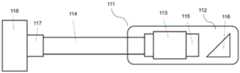
图1为本发明实施例中多模态内窥导管装置的结构示意图;FIG. 1 is a schematic structural view of a multimodal endoscopic catheter device in an embodiment of the present invention;
图2为本发明实施例中多模态光学成像装置的结构框图;2 is a structural block diagram of a multi-modal optical imaging device in an embodiment of the present invention;
图3为本发明实施例中图像配准算法框图。Fig. 3 is a block diagram of an image registration algorithm in an embodiment of the present invention.
图中:111、内窥探头,112、套管,113、固定环,114、光纤,115、梯度折射率透镜,116、三角棱镜,117、电机接口,118、驱动电机,201、图像处理装置,202、OCT成像装置,203、荧光成像装置,204、波分复用器。In the figure: 111, endoscopic probe, 112, sleeve tube, 113, fixed ring, 114, optical fiber, 115, gradient index lens, 116, triangular prism, 117, motor interface, 118, drive motor, 201, image processing device , 202, an OCT imaging device, 203, a fluorescence imaging device, 204, a wavelength division multiplexer.
具体实施方式Detailed ways
下面结合附图和具体实施例对本发明进行详细说明。The present invention will be described in detail below in conjunction with the accompanying drawings and specific embodiments.
本技术方案借助多模态成像系统,近红外荧光成像可用于快速识别可疑病变,光学相干断层扫描内窥检查有助于将表层结构的细节可视化,以便进一步诊断,从而更好地分期和诊断肠道疾病。本技术方案结合光学相干层析成像和近红外荧光成像组成多模态光学成像系统可视化组织形态和血管结构来弥补传统结肠镜检查的局限性。With the help of a multimodal imaging system, near-infrared fluorescence imaging can be used to quickly identify suspicious lesions, and optical coherence tomography endoscopy can help visualize the details of superficial structures for further diagnosis, thereby better staging and diagnosing bowel disease. disease. This technical solution combines optical coherence tomography and near-infrared fluorescence imaging to form a multi-modal optical imaging system to visualize tissue morphology and vascular structure to make up for the limitations of traditional colonoscopy.
本发明中多模态光学成像系统包含图像处理装置、OCT成像装置、荧光成像装置和波分复用器;内窥导管装置包含套管、驱动电机和内窥探头,用于插入组织内部进行成像。导管装置具有综合在体、断层成像、高分辨率和成像深度几个方面的临床特点。具体实施如下:The multimodal optical imaging system in the present invention includes an image processing device, an OCT imaging device, a fluorescence imaging device, and a wavelength division multiplexer; the endoscopic catheter device includes a sleeve, a driving motor, and an endoscopic probe, which are used to insert into the tissue for imaging . The catheter device has the clinical characteristics of comprehensive in vivo, tomographic imaging, high resolution and imaging depth. The specific implementation is as follows:
内窥导管-多模态光学成像耦合检测系统包括内窥探头111、光纤114、电机组件、波分复用器204、OCT成像装置202、荧光成像装置203、图像处理装置201,其中具体地:The endoscopic catheter-multimodal optical imaging coupling detection system includes an
内窥探头111包括套管112,所述套管112的始端内设有光学聚焦模块;The
光纤114的始端接入所述套管112的末端,且使得光纤114的始端与所述光学聚焦模块光通信连接;The beginning end of the
电机组件与所述光纤114的末端连接,所述电机组件能够驱动光纤114进行旋转,并能对光纤114进行收拉,以此实现内窥探头111的旋转和回拉位移;The motor assembly is connected to the end of the
波分复用器204第一端口与所述光纤114的末端光通信连接,The first port of the
OCT成像装置202输入端与所述波分复用器204的第二端口光通信连接;The input end of the
荧光成像装置203输入端与所述波分复用器204的第二端口光通信连接;The input end of the
图像处理装置201分别与OCT成像装置202、荧光成像装置的输出端电连接203,图像处理装置201能够指令波分复用器204和图像处理装置201输出特定规格的光信号,同时图像处理装置201能够将OCT成像装置202、荧光成像装置203输出的图像信息进行图像配准,之后生成待测区域的多模态影像。The
OCT成像装置202包括光学相干成像样品臂光源和第一CCD传感器。荧光成像装置203包括荧光激励光源和第二CCD传感器。波分复用器204能够将光学相干成像样品臂光源和荧光激励光源整合到光纤114中,并通过光学聚焦模块输出至待测区域;波分复用器204还能够将光学聚焦模块接收的光反馈信号分配至所述第一CCD传感器和第二CCD传感器。The
光学聚焦模块由梯度折射率透镜115和三角棱镜116组成。该三角棱镜上设置有镀金反射膜,且与水平面成45°角,以使该镀金反射膜将传输来的光信号以90°角的方向照射在样本组织上。光学聚焦模块与光纤连接用于将光纤传输过来的光信号输出至样本组织,并用来接收样本组织所反射的光反馈信号,再通过光纤传输至图像处理装置。The optical focusing module is composed of a
内窥探头通过光纤与电机接口117连接。驱动电机118接收到微处理器发出电信号,使得驱动电机118的端部转动,以此按一定转速带动内窥探头旋转,生成样品组织的二维图像,微处理器根据执行的程序,指令驱动电机118接收到回拉信号,带动内窥探头在样品组织回拉从而生成三维图像。The endoscopic probe is connected with the
从上述技术方案来看,本实施例提供了一种内窥导管装置,能够采集样品组织的三维图像,有利于结直肠癌的诊断。From the above technical solutions, the present embodiment provides an endoscopic catheter device capable of acquiring a three-dimensional image of sample tissue, which is beneficial to the diagnosis of colorectal cancer.
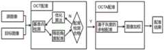
图2为一种多模态光学成像系统框图。如图2所示,本实施例结合OCT成像装置202和荧光成像装置203后与图像处理装置201连接,以获得样品组织的多模态图像。波分复用器204用于将光学相干成像样品臂光源和荧光激励光源整合到同一单模宽带光纤光路。图像处理装置201接收到反馈信号后使用配准算法对数据进行预处理,配准完毕后使用光学多普勒层析算法对样品组织进行血流成像。Fig. 2 is a block diagram of a multi-modal optical imaging system. As shown in FIG. 2 , in this embodiment, an
具体实施时图像处理装置201包括微处理器、RAM和ROM,其中ROM中预存储有本技术方案中所用到的算法程序及其他匹配的辅助程序。During specific implementation, the
OCT成像装置202与梯度折射率透镜通过光纤连接,用于根据控制信号输出光信号,所述的光信号通过聚焦模块到达待测样品组织,并接收采集的待测样品组织反馈的光信号。The
荧光成像装置利用波分复用器与OCT成像观测装置构成光学成像系统,用于将光学相干成像样品臂光源和荧光激励光源整合到同一单模宽带光纤光路。The fluorescence imaging device uses a wavelength division multiplexer and an OCT imaging observation device to form an optical imaging system, which is used to integrate the optical coherent imaging sample arm light source and the fluorescence excitation light source into the same single-mode broadband optical fiber optical path.
图像配准:Image registration:
组织的生理运动以及内窥导管回撤遇到弯曲组织会加剧旋转不均匀性,此时会产生大量伪影和形变,导致无法准确计算血流成像。在光学相干层析血流成像之前需要预先使用图像配准算法处理,算法流程如图3所示。该方法使用位于导管上的基准标记,根据检测到的标记位置对横截面图像进行重复采样,并使采集的图像每一条A-line向像素点对应于配准图像的角度位置,在连续的旋转扫描中校正OCT图像的抖动和拉伸。为了量化校正算法的性能,在每次旋转中使用两个基准点进行矫正计算,并使用另外两个基准点进行测量。校正后角度标准偏差降低到1毫弧度,相当于旋转稳定性提高了15倍以上。矫正后背景噪声和沿拉回方向的垂直条纹噪声明显减少并且可以通过图像观察到直肠规则的凹坑结构。The rotational inhomogeneity is exacerbated by the physiological movement of the tissue and the retraction of the endoscopic catheter to encounter curved tissue, which can produce a large number of artifacts and deformations, making it impossible to accurately calculate the blood flow image. Before optical coherence tomography blood flow imaging, an image registration algorithm needs to be used in advance, and the algorithm flow is shown in Figure 3. This method uses fiducial markers located on the catheter to repeatedly sample cross-sectional images according to the detected marker positions, and makes each A-line pixel point of the acquired image correspond to the angular position of the registration image, in continuous rotation Correct jitter and stretching of OCT images during scanning. To quantify the performance of the correction algorithm, two fiducials are used for the correction calculation and two other fiducials are used for the measurement in each rotation. The corrected angular standard deviation was reduced to 1 milliradian, which equates to more than a 15-fold improvement in rotational stability. After correction, the background noise and the vertical streak noise along the pullback direction were significantly reduced and the regular pit structure of the rectum could be observed through the image.
本技术方案还使用基于灰度的多帧配准和图像加权方法消除组织运动造成的伪影,经过OCT和OCTA配准后输出结果。The technical solution also uses grayscale-based multi-frame registration and image weighting methods to eliminate artifacts caused by tissue movement, and outputs results after OCT and OCTA registration.
光学相干层析血流成像:Optical coherence tomography:
光学相干层析血流成像(Optical Coherence Tomography Angiography,OCTA)是OCT的扩展技术,利用不同b-scan图像之间的相位差对小血管进行成像。谱域OCT系统中,线阵CCD探测到的实干涉光谱信号I(λ)经图像采集卡采集后,先经过光谱校正、减直流项和重新采样,得到关于波矢k的干涉信号I(k)。对进行傅立叶变换后得到是与样品深度位置z有关的复解析信号


其中T为相邻两次A扫描的时间间隔,即CCD的积分时间。p(f)是多普勒功率谱。Where T is the time interval between two adjacent A scans, that is, the integration time of the CCD. p(f) is the Doppler power spectrum.
作为优选方案,在本多模态光学成像系统中,OCT成像装置202采用高速VECSEL光源,可实现长距离成像,光源发出的光通过多模光纤传输至三角棱镜再90°折射到样品上。采用半导体可调激光的680-750nm波段作为激发光源,实现对荧光分子成像。对双模态光路部分的集成则根据OCT成像装置202和荧光成像装置203不同波长的情况选取波分复用器204将光学相干成像样品臂光源和荧光激励光源整合到同一单模宽带光纤光路,这种全光纤光路设计保证了双模态光路系统紧凑和稳定。As a preferred solution, in this multimodal optical imaging system, the
本发明的多模态光学成像系统设有成像原理不同的光学相干成像装置和荧光成像装置,光学相干成像技术相比于其他技术具有非侵入、高分辨率、可在体检测生物组织内部微结构,与内窥技术结合之后的OCT内窥成像技术可直接对生物组织进行成像,能够完成对组织的高精度扫描,对早期癌变进行早期诊断。The multimodal optical imaging system of the present invention is equipped with an optical coherent imaging device and a fluorescence imaging device with different imaging principles. Compared with other technologies, the optical coherent imaging technology has non-invasive, high-resolution, and can detect the internal microstructure of biological tissues in vivo. , OCT endoscopic imaging technology combined with endoscopic technology can directly image biological tissues, can complete high-precision scanning of tissues, and perform early diagnosis of early cancers.
本发明结合荧光成像技术和OCT/OCTA成像技术可实现分析组织的结构和成分,可为直肠早期病变等提供精准的影像手段,相比单一模态的成像技术的单一性,不稳定性和成像分辨率差的特点,多模态的成像技术更适合对于体内早期疾病的诊断,综合在体、断层成像、高分辨率和成像深度几个方面的临床特点。The present invention combines fluorescence imaging technology and OCT/OCTA imaging technology to realize the analysis of tissue structure and composition, and can provide accurate imaging means for early rectal lesions, etc. Due to the characteristics of poor resolution, multi-modal imaging technology is more suitable for the diagnosis of early diseases in vivo, and integrates the clinical characteristics of in vivo, tomographic imaging, high resolution and imaging depth.
上述的对实施例的描述是为便于该技术领域的普通技术人员能理解和使用发明。熟悉本领域技术的人员显然可以容易地对这些实施例做出各种修改,并把在此说明的一般原理应用到其他实施例中而不必经过创造性的劳动。因此,本发明不限于上述实施例,本领域技术人员根据本发明的揭示,不脱离本发明范畴所做出的改进和修改都应该在本发明的保护范围之内。The above descriptions of the embodiments are for those of ordinary skill in the art to understand and use the invention. It is obvious that those skilled in the art can easily make various modifications to these embodiments, and apply the general principles described here to other embodiments without creative effort. Therefore, the present invention is not limited to the above-mentioned embodiments. Improvements and modifications made by those skilled in the art according to the disclosure of the present invention without departing from the scope of the present invention should fall within the protection scope of the present invention.
Claims (8)
Priority Applications (1)
| Application Number | Priority Date | Filing Date | Title |
|---|---|---|---|
| CN202110723621.2ACN113520272B (en) | 2021-06-29 | 2021-06-29 | Endoscopic catheter-multimode optical imaging coupling detection system |
Applications Claiming Priority (1)
| Application Number | Priority Date | Filing Date | Title |
|---|---|---|---|
| CN202110723621.2ACN113520272B (en) | 2021-06-29 | 2021-06-29 | Endoscopic catheter-multimode optical imaging coupling detection system |
Publications (2)
| Publication Number | Publication Date |
|---|---|
| CN113520272A CN113520272A (en) | 2021-10-22 |
| CN113520272Btrue CN113520272B (en) | 2023-06-27 |
Family
ID=78126080
Family Applications (1)
| Application Number | Title | Priority Date | Filing Date |
|---|---|---|---|
| CN202110723621.2AActiveCN113520272B (en) | 2021-06-29 | 2021-06-29 | Endoscopic catheter-multimode optical imaging coupling detection system |
Country Status (1)
| Country | Link |
|---|---|
| CN (1) | CN113520272B (en) |
Families Citing this family (5)
| Publication number | Priority date | Publication date | Assignee | Title |
|---|---|---|---|---|
| CN115089118A (en)* | 2022-06-20 | 2022-09-23 | 暨南大学附属第一医院(广州华侨医院) | A dual-modality fusion vascular imaging system |
| CN115568827B (en)* | 2022-10-14 | 2025-08-22 | 中山大学附属第一医院 | OCT-based fluorescence imaging catheter and system |
| CN115919372A (en)* | 2023-01-28 | 2023-04-07 | 深圳先进技术研究院 | A multi-modal endoscopic imaging system based on short-wave near-infrared fluorescence |
| CN116509305A (en)* | 2023-04-28 | 2023-08-01 | 天津恒宇医疗科技有限公司 | Endoscopic OCTA Imaging Method and Imaging System Based on Pause, Stepping and Rotating Scanning |
| CN118680505B (en)* | 2024-08-26 | 2025-01-28 | 浙江大学 | Esophageal endoscopy diagnosis and treatment system and storage medium |
Citations (4)
| Publication number | Priority date | Publication date | Assignee | Title |
|---|---|---|---|---|
| CN101730498A (en)* | 2006-09-12 | 2010-06-09 | 卡尔斯特里姆保健公司 | Low coherence dental optical coherence tomography imaging |
| CN102178526A (en)* | 2011-05-12 | 2011-09-14 | 上海交通大学医学院附属新华医院 | Ultrasonic and nuclear magnetic resonance image fusion transluminal registration device and method |
| CN103222846A (en)* | 2007-01-19 | 2013-07-31 | 桑尼布鲁克健康科学中心 | Scanning mechanisms for imaging probe |
| WO2016180291A1 (en)* | 2015-05-08 | 2016-11-17 | 南京微创医学科技有限公司 | Endoscopic oct microprobe, oct imaging system, and use method |
Family Cites Families (8)
| Publication number | Priority date | Publication date | Assignee | Title |
|---|---|---|---|---|
| CN201085617Y (en)* | 2007-10-25 | 2008-07-16 | 浙江大学 | Single fiber endoscopic system integrating optical coherence tomography and laser-induced fluorescence optical spectrum |
| US8285368B2 (en)* | 2009-07-10 | 2012-10-09 | The Regents Of The University Of California | Endoscopic long range fourier domain optical coherence tomography (LR-FD-OCT) |
| US10231706B2 (en)* | 2013-03-14 | 2019-03-19 | The Regents Of The University Of California | Integrated multimodality intravascular imaging system that combines optical coherence tomography, ultrasound imaging, and acoustic radiation force optical coherence elastography |
| WO2015054243A1 (en)* | 2013-10-07 | 2015-04-16 | Van Dam, Jacques | Integrated ultrasound, oct, pa and/or florescence imaging endoscope for diagnosing cancers in gastrointestinal, respiratory, and urogenital tracts |
| CN104523233B (en)* | 2014-12-29 | 2017-02-22 | 浙江大学 | Capillary optical imaging and jitter compensating method based on complex number mutual correlation |
| CN107411708A (en)* | 2017-05-22 | 2017-12-01 | 上海交通大学 | A kind of optical coherence tomography and photoacoustic imaging bimodal endoscope |
| CN108618758A (en)* | 2018-04-27 | 2018-10-09 | 华南师范大学 | Intravascular photoacoustic-optical coherence tomography-near infrared light multi-modality imaging apparatus and method |
| CN111419149A (en)* | 2020-03-13 | 2020-07-17 | 上海应用技术大学 | A multimodal endoscope and endoscope imaging system |
- 2021
- 2021-06-29CNCN202110723621.2Apatent/CN113520272B/enactiveActive
Patent Citations (4)
| Publication number | Priority date | Publication date | Assignee | Title |
|---|---|---|---|---|
| CN101730498A (en)* | 2006-09-12 | 2010-06-09 | 卡尔斯特里姆保健公司 | Low coherence dental optical coherence tomography imaging |
| CN103222846A (en)* | 2007-01-19 | 2013-07-31 | 桑尼布鲁克健康科学中心 | Scanning mechanisms for imaging probe |
| CN102178526A (en)* | 2011-05-12 | 2011-09-14 | 上海交通大学医学院附属新华医院 | Ultrasonic and nuclear magnetic resonance image fusion transluminal registration device and method |
| WO2016180291A1 (en)* | 2015-05-08 | 2016-11-17 | 南京微创医学科技有限公司 | Endoscopic oct microprobe, oct imaging system, and use method |
Also Published As
| Publication number | Publication date |
|---|---|
| CN113520272A (en) | 2021-10-22 |
Similar Documents
| Publication | Publication Date | Title |
|---|---|---|
| CN113520272B (en) | Endoscopic catheter-multimode optical imaging coupling detection system | |
| Tsai et al. | Optical coherence tomography in gastroenterology: a review and future outlook | |
| Bouma et al. | Optical coherence tomography | |
| JP7069236B2 (en) | How to control the behavior of the imaging system and the system to acquire the image | |
| Tsai et al. | Endoscopic optical coherence tomography for clinical gastroenterology | |
| Zagaynova et al. | Endoscopic OCT with forward‐looking probe: clinical studies in urology and gastroenterology | |
| Yuan et al. | Co-registered optical coherence tomography and fluorescence molecular imaging for simultaneous morphological and molecular imaging | |
| US10076248B2 (en) | Hybrid catheter system | |
| CN105105717B (en) | Endoscopic OCT imaging system adopting optical path difference external compensation common-path interference probe | |
| Jung et al. | Modern trends in imaging V: optical coherence tomography for rapid tissue screening and directed histological sectioning | |
| US12076118B2 (en) | Devices, systems, and methods for detecting external elastic lamina (EEL) from intravascular OCT images | |
| CN114053553B (en) | Automatic pullback triggering method for intracoronary imaging device or system using blood clearance | |
| US20220040402A1 (en) | Methods and systems for automatic pullback trigger | |
| Wang et al. | Clinical applications of optical coherence tomography in urology | |
| Zheng et al. | Confocal endomicroscopic imaging of normal and neoplastic human tongue tissue using ALA-induced-PPIX fluorescence: a preliminary study | |
| CN104305955B (en) | Wide-field optical coherence tomography electronic colposcopy imaging system | |
| CN104523216B (en) | A hysteroscopy system based on optical coherence tomography and its implementation method | |
| JP7470761B2 (en) | Fluorescence calibration based on manual lumen detection | |
| Jung et al. | Optical coherence tomography for rapid tissue screening and directed histological sectioning | |
| US10905341B2 (en) | Cancer invasiveness diagnosis system | |
| CN204379228U (en) | A hysteroscopy system based on optical coherence tomography | |
| Li et al. | Optical coherence tomography technology for diagnosis of diseases in organs | |
| US20210282642A1 (en) | Scanning mechanism for multimodality intravascular and endoscopic imaging catheters | |
| Abouei | Optimization of multimodal OCT for early cancer detection and diagnosis | |
| Yuan et al. | Combining optical coherence tomography with fluorescence molecular imaging: towards simultaneous morphology and molecular imaging |
Legal Events
| Date | Code | Title | Description |
|---|---|---|---|
| PB01 | Publication | ||
| PB01 | Publication | ||
| SE01 | Entry into force of request for substantive examination | ||
| SE01 | Entry into force of request for substantive examination | ||
| GR01 | Patent grant | ||
| GR01 | Patent grant |