CN113427019B - Method for manufacturing composite material and metal bone implant with structural function - Google Patents
Method for manufacturing composite material and metal bone implant with structural functionDownload PDFInfo
- Publication number
- CN113427019B CN113427019BCN202110692786.8ACN202110692786ACN113427019BCN 113427019 BCN113427019 BCN 113427019BCN 202110692786 ACN202110692786 ACN 202110692786ACN 113427019 BCN113427019 BCN 113427019B
- Authority
- CN
- China
- Prior art keywords
- bone
- powder
- layer
- scanning
- implant
- Prior art date
- Legal status (The legal status is an assumption and is not a legal conclusion. Google has not performed a legal analysis and makes no representation as to the accuracy of the status listed.)
- Active
Links
Images

Classifications
- B—PERFORMING OPERATIONS; TRANSPORTING
- B22—CASTING; POWDER METALLURGY
- B22F—WORKING METALLIC POWDER; MANUFACTURE OF ARTICLES FROM METALLIC POWDER; MAKING METALLIC POWDER; APPARATUS OR DEVICES SPECIALLY ADAPTED FOR METALLIC POWDER
- B22F10/00—Additive manufacturing of workpieces or articles from metallic powder
- B22F10/20—Direct sintering or melting
- B22F10/28—Powder bed fusion, e.g. selective laser melting [SLM] or electron beam melting [EBM]
- A—HUMAN NECESSITIES
- A61—MEDICAL OR VETERINARY SCIENCE; HYGIENE
- A61L—METHODS OR APPARATUS FOR STERILISING MATERIALS OR OBJECTS IN GENERAL; DISINFECTION, STERILISATION OR DEODORISATION OF AIR; CHEMICAL ASPECTS OF BANDAGES, DRESSINGS, ABSORBENT PADS OR SURGICAL ARTICLES; MATERIALS FOR BANDAGES, DRESSINGS, ABSORBENT PADS OR SURGICAL ARTICLES
- A61L27/00—Materials for grafts or prostheses or for coating grafts or prostheses
- A61L27/02—Inorganic materials
- A61L27/04—Metals or alloys
- B—PERFORMING OPERATIONS; TRANSPORTING
- B22—CASTING; POWDER METALLURGY
- B22F—WORKING METALLIC POWDER; MANUFACTURE OF ARTICLES FROM METALLIC POWDER; MAKING METALLIC POWDER; APPARATUS OR DEVICES SPECIALLY ADAPTED FOR METALLIC POWDER
- B22F5/00—Manufacture of workpieces or articles from metallic powder characterised by the special shape of the product
- B—PERFORMING OPERATIONS; TRANSPORTING
- B33—ADDITIVE MANUFACTURING TECHNOLOGY
- B33Y—ADDITIVE MANUFACTURING, i.e. MANUFACTURING OF THREE-DIMENSIONAL [3-D] OBJECTS BY ADDITIVE DEPOSITION, ADDITIVE AGGLOMERATION OR ADDITIVE LAYERING, e.g. BY 3-D PRINTING, STEREOLITHOGRAPHY OR SELECTIVE LASER SINTERING
- B33Y10/00—Processes of additive manufacturing
- B—PERFORMING OPERATIONS; TRANSPORTING
- B33—ADDITIVE MANUFACTURING TECHNOLOGY
- B33Y—ADDITIVE MANUFACTURING, i.e. MANUFACTURING OF THREE-DIMENSIONAL [3-D] OBJECTS BY ADDITIVE DEPOSITION, ADDITIVE AGGLOMERATION OR ADDITIVE LAYERING, e.g. BY 3-D PRINTING, STEREOLITHOGRAPHY OR SELECTIVE LASER SINTERING
- B33Y80/00—Products made by additive manufacturing
- Y—GENERAL TAGGING OF NEW TECHNOLOGICAL DEVELOPMENTS; GENERAL TAGGING OF CROSS-SECTIONAL TECHNOLOGIES SPANNING OVER SEVERAL SECTIONS OF THE IPC; TECHNICAL SUBJECTS COVERED BY FORMER USPC CROSS-REFERENCE ART COLLECTIONS [XRACs] AND DIGESTS
- Y02—TECHNOLOGIES OR APPLICATIONS FOR MITIGATION OR ADAPTATION AGAINST CLIMATE CHANGE
- Y02P—CLIMATE CHANGE MITIGATION TECHNOLOGIES IN THE PRODUCTION OR PROCESSING OF GOODS
- Y02P10/00—Technologies related to metal processing
- Y02P10/25—Process efficiency
Landscapes
- Chemical & Material Sciences (AREA)
- Engineering & Computer Science (AREA)
- Manufacturing & Machinery (AREA)
- Materials Engineering (AREA)
- Health & Medical Sciences (AREA)
- Oral & Maxillofacial Surgery (AREA)
- Veterinary Medicine (AREA)
- Epidemiology (AREA)
- Life Sciences & Earth Sciences (AREA)
- Animal Behavior & Ethology (AREA)
- General Health & Medical Sciences (AREA)
- Public Health (AREA)
- Transplantation (AREA)
- Medicinal Chemistry (AREA)
- Dermatology (AREA)
- Inorganic Chemistry (AREA)
- Mechanical Engineering (AREA)
- Physics & Mathematics (AREA)
- Plasma & Fusion (AREA)
- Prostheses (AREA)
- Materials For Medical Uses (AREA)
Abstract
Translated fromChinese
Description
Translated fromChinese技术领域technical field
本发明属于医疗器械领域,尤其涉及一种复合材料和结构功能的金属骨植入物的制造方法。The invention belongs to the field of medical devices, in particular to a method for manufacturing a composite material and a metal bone implant with structural functions.
背景技术Background technique
骨骼是一种有活力的组织,始终处于一种变化、更新的状态,破骨细胞吸收骨,成骨细胞生成新骨,在应力刺激下,形成并维持最佳力学结构,兼具最大强度和最小质量。全世界每年有数以千万的患者由于创伤、肿瘤切除和关节翻修等原因造成骨缺损。大范围的骨缺损导致骨骼难以自我修复,成为骨科临床的棘手问题。为了解决上述问题,骨科植入物一般被用来替代、支撑缺失和损坏的骨骼以及关节。这些植入物在矫形、固定身体姿态、恢复正常骨骼功能等方面起着重要的辅助作用。而理想的金属骨植入物应具有下列特点:第一,植入物外形应与骨缺损的解剖形态贴合,提供可靠的固定支撑和应力传导;第二,植入物内部应由相互连通的孔隙构成,提供营养输送通道和骨长入空间,同时避免应力遮挡;第三,很多骨缺损修复只需要初期固定,随着骨重建和功能恢复,植入物应逐渐降解消失。Bone is a dynamic tissue that is always in a state of change and renewal. Osteoclasts absorb bone, and osteoblasts generate new bone. Under stress stimulation, they form and maintain an optimal mechanical structure with maximum strength and minimum mass. Every year, tens of millions of patients suffer from bone defects worldwide due to trauma, tumor resection, and joint revision. Large-scale bone defects make it difficult for bones to repair themselves, which has become a thorny problem in orthopedics. In order to solve the above problems, orthopedic implants are generally used to replace and support missing and damaged bones and joints. These implants play an important auxiliary role in orthopedics, fixing body posture, and restoring normal bone function. An ideal metal bone implant should have the following characteristics: first, the shape of the implant should conform to the anatomical shape of the bone defect, providing reliable fixed support and stress conduction; second, the interior of the implant should be interconnected. The pores are formed to provide nutrient delivery channels and bone ingrowth into the space, while avoiding stress shielding; third, many bone defect repairs only require initial fixation, and the implants should gradually degrade and disappear with bone reconstruction and functional recovery.
增材制造(3D打印)能够高效精确地赋予金属与骨骼贴合的个性化外形以及内部连通微结构,被认为是金属骨植入物的理想制造方法,电子束或激光粉末床熔融制造的钛合金、CoCr合金等金属骨植入物已经成功应用于临床。然而,钛合金等生物惰性金属永久占据了骨骼生长空间,难以实现受损骨骼的整体重建,尤其是不符合青少年生长发育的特点,如果进行二次手术取出又给病人造成新的组织创伤和经济负担。在骨骼和肌肉环境下使用的可降解金属主要为镁合金和锌合金,这两种材料均具有良好的生物相容性和促生骨能力。Additive manufacturing (3D printing) can efficiently and accurately give metal and bone a personalized shape and internal connected microstructure, and is considered to be an ideal manufacturing method for metal bone implants. Titanium produced by electron beam or laser powder bed fusion Alloy, CoCr alloy and other metal bone implants have been successfully used clinically. However, biologically inert metals such as titanium alloys permanently occupy the bone growth space, and it is difficult to achieve the overall reconstruction of damaged bones, especially not in line with the characteristics of adolescent growth and development. If a second operation is performed to remove it, it will cause new tissue trauma and economical damage to the patient. burden. Degradable metals used in bone and muscle environments are mainly magnesium alloys and zinc alloys, both of which have good biocompatibility and bone-promoting ability.
增材制造可降解金属骨植入物满足了骨缺损修复的结构和降解需求,然而目前对可降解金属在人体内的降解行为研究数据很少,如何将降解行为和骨重建过程进行匹配尚无可靠的解决方案。如果完全使用可降解金属,一旦发生因降解过快导致承载失效,后果不堪设想。此外,镁合金和锌合金的力学性能较钛合金和不锈钢等目前广泛使用的骨固定金属材料尚有较大差距。Additive manufacturing of degradable metal bone implants meets the structural and degradation requirements of bone defect repair. However, there are few research data on the degradation behavior of degradable metals in the human body. How to match the degradation behavior with the bone reconstruction process is unclear. reliable solution. If all degradable metals are used, once the bearing fails due to rapid degradation, the consequences will be disastrous. In addition, the mechanical properties of magnesium alloys and zinc alloys still have a large gap compared with titanium alloys and stainless steels, which are currently widely used in bone fixation metal materials.
综上所述,如何满足骨愈合过程中对植入物材料承载的功能需求,够刺激骨细胞生长的同时也为骨骼的生长提供空间,提升骨重建效果,已经成为亟需解决的问题。To sum up, how to meet the functional requirements of implant materials in the process of bone healing, to stimulate the growth of bone cells and provide space for bone growth and improve the effect of bone reconstruction has become an urgent problem to be solved.
发明内容Contents of the invention
为了克服现有技术存在的一系列缺陷,本发明的目的在于针对上述问题,提供一种复合材料和结构功能的金属骨植入物的制造方法,包括以下步骤:In order to overcome a series of deficiencies in the prior art, the object of the present invention is to provide a kind of composite material and the manufacturing method of the metallic bone implant of structural function, comprise the following steps:
S10,建立宏观外形与目标骨缺损的解剖形态相符的填充骨植入物三维模型;S10, establishing a three-dimensional model of the bone-filling implant whose macroscopic shape matches the anatomical shape of the target bone defect;
S20,将骨植入物拆分为具有承载或降解特性的不同的功能区域,分别采用相应合金进行植入物材料设计;S20, split the bone implant into different functional areas with load-bearing or degradable properties, and use corresponding alloys for implant material design;
S30,对不同功能区域的三维模型进行剖分,根据骨植入物材料特性设置打印参数,得到各截面的轮廓打印数据,并将打印数据输入激光粉末床熔融打印机;S30, subdividing the three-dimensional model of different functional areas, setting printing parameters according to the material characteristics of the bone implant, obtaining contour printing data of each section, and inputting the printing data into a laser powder bed fusion printer;
S40,基于打印数据分别打印不同功能区域零件,合金粉末采用逐层送粉、逐层激光扫描熔化凝固的方式打印成形,得到一种复合材料和结构功能的金属骨植入物。S40, based on the printing data, the parts of different functional areas are printed separately, and the alloy powder is printed and formed by layer-by-layer powder feeding, layer-by-layer laser scanning, melting and solidification, and a metal bone implant with composite materials and structural functions is obtained.
优选的,骨植入物拆分为钛合金内固定骨板功能区及镁合金骨内填充物功能区,或者拆分为钛合金内固定骨板功能区、镁合金骨内填充物功能区和锌合金骨内填充物功能区,根据体外降解实验和力学行为的仿真模拟,以及骨的生理结构需求设计每个功能区域的几何形貌,且每个区域均以三重周期极小曲面结构为单元。Preferably, the bone implant is split into a titanium alloy internal fixation bone plate functional area and a magnesium alloy intraosseous filler functional area, or is split into a titanium alloy internal fixation bone plate functional area, a magnesium alloy intraosseous filler functional area and Zinc alloy intraosseous filling functional area, according to in vitro degradation experiments and simulation of mechanical behavior, as well as the physiological structure requirements of bone design the geometric shape of each functional area, and each area is based on a triple periodic minimal surface structure as a unit .
优选的,在步骤S20中,钛合金材质采用Ti6Al4V、纯钛或者其他获得临床许可的钛合金,镁合金材质采用Mg-Zn-Ca、WE43、纯镁或者其他获得临床许可的镁合金;锌合金采用Zn-Li-Mg、纯锌或者其他锌合金。Preferably, in step S20, the titanium alloy is made of Ti6Al4V, pure titanium or other clinically approved titanium alloys, the magnesium alloy is made of Mg-Zn-Ca, WE43, pure magnesium or other clinically approved magnesium alloys; zinc alloy Use Zn-Li-Mg, pure zinc or other zinc alloys.
优选的,在步骤S20中,还包括采用具有不同孔隙率的孔隙单元进行植入物内部结构设计,其中,孔隙单元尺寸取1~2mm,孔隙直径取0.4~1mm,单元结构孔隙率为60~90%,所述孔隙单元为三重周期极小曲面结构。Preferably, in step S20, it also includes the use of pore units with different porosities to design the internal structure of the implant, wherein the size of the pore units is 1-2 mm, the diameter of the pores is 0.4-1 mm, and the porosity of the unit structure is 60-2 mm. 90%, the pore unit is a triple periodic minimal curved surface structure.
优选的,在步骤S20中,孔隙单元的强度和刚度通过孔隙率调整,利用多孔结构特性突破基体材料的性能制约,获得与骨结构力学性能匹配的金属多孔结构。Preferably, in step S20, the strength and stiffness of the pore units are adjusted through the porosity, and the properties of the porous structure are used to break through the performance constraints of the matrix material to obtain a metal porous structure that matches the mechanical properties of the bone structure.
优选的,在步骤S40中,基于打印数据,通过逐层送粉和逐层激光扫描熔化打印的步骤包括:Preferably, in step S40, based on the printing data, the steps of layer-by-layer powder feeding and layer-by-layer laser scanning fusion printing include:
S41,合金粉末预置在打印机的粉舱中,刮刀预置在焦平面上,用保护气体清洗和预热粉舱;S41, the alloy powder is preset in the powder cabin of the printer, the scraper is preset on the focal plane, and the powder cabin is cleaned and preheated with protective gas;
S42,根据打印数据,对合金粉末逐层进行扫描熔化成形。S42, according to the printing data, the alloy powder is scanned and melted layer by layer.
优选的,在步骤S40中,S41中,使用保护气体洗气将粉舱氧含量控制在800ppm以下,预热温度为100~200℃。Preferably, in step S40 and step S41, the oxygen content in the powder cabin is controlled below 800ppm by using protective gas scrubbing, and the preheating temperature is 100-200°C.
优选的,在步骤S40中,S42打印过程完成后,还包括以下步骤:Preferably, in step S40, after the printing process of S42 is completed, the following steps are also included:
待舱内温度冷却至室温后,打开舱门,将舱内粉末回收、筛分,放入真空袋中备用;取出打印件和基板,用线切割或小型锯将零件与基板分离,用压缩空气将零件表面的粉末清理干净;零件在氩气气氛中用加热至200~500℃,保温1~2小时。After the temperature in the cabin cools down to room temperature, open the hatch, recover and sieve the powder in the cabin, and put it in a vacuum bag for later use; take out the printed parts and the substrate, separate the parts from the substrate with wire cutting or a small saw, and use compressed air to Clean up the powder on the surface of the part; heat the part to 200-500°C in an argon atmosphere and keep it warm for 1-2 hours.
优选的,在步骤S40中,在打印过程中,使用循环送风系统将多余的粉末吹除。Preferably, in step S40, during the printing process, a circulating air supply system is used to blow off excess powder.
优选的,在步骤S40中,在步骤S40中,所采用的激光粉末床熔融打印机的激光扫描功率50~500W,扫描速度200~2000mm/s,层厚0.01~0.05mm,扫描间距0.05~0.1mm,激光光斑直径0.05~0.07mm,所使用的合金粉末平均粒径为0.02~0.04mm。Preferably, in step S40, in step S40, the laser scanning power of the laser powder bed fusion printer used is 50-500W, the scanning speed is 200-2000mm/s, the layer thickness is 0.01-0.05mm, and the scanning distance is 0.05-0.1mm , the laser spot diameter is 0.05-0.07mm, and the average grain size of the alloy powder used is 0.02-0.04mm.
与现有技术相比,本发明具备以下有益效果:Compared with the prior art, the present invention has the following beneficial effects:
1)本发明充分发挥不同材料的承载、降解和促生骨特性,其中,钛合金用于骨的固定,确保骨重建过程中的安全承载;锌合金和镁合金用于骨内填充,其降解本身及降解产物有利骨的整体重建和愈合;锌合金的降解速率较慢,镁合金的降解速率较快,根据骨重建的需求进行选择;各材料对应的功能区域具有定制化的形状及几何尺寸,可以满足患者的个性化需求;1) The present invention gives full play to the load-bearing, degradation and bone-promoting characteristics of different materials, among which, titanium alloy is used for bone fixation to ensure safe bearing in the process of bone reconstruction; zinc alloy and magnesium alloy are used for intraosseous filling, and their degradation Its own and its degradation products are beneficial to the overall reconstruction and healing of bone; the degradation rate of zinc alloy is relatively slow, while the degradation rate of magnesium alloy is relatively fast, so choose according to the needs of bone reconstruction; the functional areas corresponding to each material have customized shapes and geometric dimensions , to meet the individual needs of patients;
2)本发明采用可编程参数化的方法生成单元和孔隙尺寸可控的孔隙单元填充各功能区域,孔隙单元的强度和刚度通过孔隙率调整,利用多孔结构特性突破基体材料的性能制约,获得与骨结构力学性能匹配的金属多孔结构,顺畅的应力传导和刺激大大提升了骨重建效果。2) The present invention uses a programmable parameterization method to generate units and pore units with controllable pore size to fill each functional area. The strength and stiffness of the pore units are adjusted through the porosity, and the porous structure characteristics are used to break through the performance constraints of the matrix material. The metal porous structure with matching mechanical properties of bone structure, smooth stress conduction and stimulation greatly improves the effect of bone reconstruction.
附图说明Description of drawings
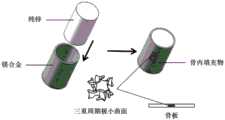
图1为本发明的优选实施例1的骨植入物的设计及制造示意图;Fig. 1 is the design and manufacture schematic diagram of the bone implant of preferred embodiment 1 of the present invention;
图2为本发明的优选实施例2的骨植入物的设计示意图。Fig. 2 is a schematic diagram of the design of the bone implant in the preferred embodiment 2 of the present invention.
具体实施方式Detailed ways
为使本发明实施的目的、技术方案和优点更加清楚,下面将结合本发明实施例中的附图,对本发明实施例中的技术方案进行更加详细的描述。在附图中,自始至终相同或类似的标号表示相同或类似的元件或具有相同或类似功能的元件。所描述的实施例是本发明一部分实施例,而不是全部的实施例。In order to make the objectives, technical solutions and advantages of the present invention clearer, the technical solutions in the embodiments of the present invention will be described in more detail below in conjunction with the drawings in the embodiments of the present invention. In the drawings, the same or similar reference numerals denote the same or similar elements or elements having the same or similar functions throughout. The described embodiments are some, but not all, embodiments of the invention.
基于本发明中的实施例,本领域普通技术人员在没有做出创造性劳动前提下所获得的所有其他实施例,都属于本发明保护的范围。Based on the embodiments of the present invention, all other embodiments obtained by persons of ordinary skill in the art without making creative efforts belong to the protection scope of the present invention.
下面通过参考附图描述的实施例以及方位性的词语均是示例性的,旨在用于解释本发明,而不能理解为对本发明的限制。The embodiments and directional words described below by referring to the figures are exemplary and are intended to explain the present invention, but should not be construed as limiting the present invention.
本发明的一个宽泛实施例中,一种复合材料和结构功能的金属骨植入物的制造方法,包括以下步骤:In one broad embodiment of the invention, a method of manufacturing a composite material and structurally functional metal bone implant, comprising the steps of:
S10,建立宏观外形与目标骨缺损的解剖形态相符的填充骨植入物三维模型;S10, establishing a three-dimensional model of the bone-filling implant whose macroscopic shape matches the anatomical shape of the target bone defect;
S20,将骨植入物拆分为具有承载或降解特性的不同的功能区域,分别采用相应合金进行植入物材料设计;S20, split the bone implant into different functional areas with load-bearing or degradable properties, and use corresponding alloys for implant material design;
S30,对不同功能区域的三维模型进行剖分,根据骨植入物材料特性设置打印参数,得到各截面的轮廓打印数据,并将打印数据输入激光粉末床熔融打印机;S30, subdividing the three-dimensional model of different functional areas, setting printing parameters according to the material characteristics of the bone implant, obtaining contour printing data of each section, and inputting the printing data into a laser powder bed fusion printer;
S40,基于打印数据分别打印不同功能区域零件,合金粉末采用逐层送粉、逐层激光扫描熔化凝固的方式打印成形,得到一种复合材料和结构功能的金属骨植入物。S40, based on the printing data, the parts of different functional areas are printed separately, and the alloy powder is printed and formed by layer-by-layer powder feeding, layer-by-layer laser scanning, melting and solidification, and a metal bone implant with composite materials and structural functions is obtained.
优选的,骨植入物拆分为钛合金内固定骨板功能区及镁合金骨内填充物功能区,或者拆分为钛合金内固定骨板功能区、镁合金骨内填充物功能区和锌合金骨内填充物功能区,根据体外降解实验和力学行为的仿真模拟,以及骨的生理结构需求设计每个功能区域的几何形貌,且每个区域均以三重周期极小曲面结构为单元。Preferably, the bone implant is split into a titanium alloy internal fixation bone plate functional area and a magnesium alloy intraosseous filler functional area, or is split into a titanium alloy internal fixation bone plate functional area, a magnesium alloy intraosseous filler functional area and Zinc alloy intraosseous filling functional area, according to in vitro degradation experiments and simulation of mechanical behavior, as well as the physiological structure requirements of bone design the geometric shape of each functional area, and each area is based on a triple periodic minimal surface structure as a unit .
优选的,在步骤S20中,钛合金材质采用Ti6Al4V、纯钛或者其他获得临床许可的钛合金,镁合金材质采用Mg-Zn-Ca、WE43、纯镁或者其他获得临床许可的镁合金;锌合金采用Zn-Li-Mg、纯锌或者其他锌合金。所使用的钛合金、镁合金和锌合金均优先考虑获得临床使用许可的材料,锌合金目前尚无获得临床使用许可的牌号,可参考使用Zn-Li-Mg或纯锌,或者未来获得临床使用许可的其他锌合金。Preferably, in step S20, the titanium alloy is made of Ti6Al4V, pure titanium or other clinically approved titanium alloys, the magnesium alloy is made of Mg-Zn-Ca, WE43, pure magnesium or other clinically approved magnesium alloys; zinc alloy Use Zn-Li-Mg, pure zinc or other zinc alloys. The titanium alloys, magnesium alloys and zinc alloys used are given priority to materials that have obtained clinical use approval. Zinc alloys currently have no grades that have obtained clinical use approval. You can refer to the use of Zn-Li-Mg or pure zinc, or obtain clinical use in the future Other zinc alloys permitted.
优选的,在步骤S20中,还包括采用具有不同孔隙率的孔隙单元进行植入物内部结构设计,其中,孔隙单元尺寸取1~2mm,孔隙直径取0.4~1mm,单元结构孔隙率为60~90%,所述孔隙单元为三重周期极小曲面结构。Preferably, in step S20, it also includes the use of pore units with different porosities to design the internal structure of the implant, wherein the size of the pore units is 1-2 mm, the diameter of the pores is 0.4-1 mm, and the porosity of the unit structure is 60-2 mm. 90%, the pore unit is a triple periodic minimal curved surface structure.
优选的,周期性多孔单元结构采用可编程参数化的方法生成,可编程参数化的方法使用三重周期极小曲面法,并采用Matlab和Python实现,其中,孔隙单元的强度和刚度通过孔隙率调整,利用多孔结构特性突破基体材料的性能制约,获得与骨结构力学性能匹配的金属多孔结构,以便顺畅的应力传导和刺激大大提升了骨重建效果。Preferably, the periodic porous unit structure is generated by a programmable parameterization method, and the programmable parameterized method uses the triple periodic minimal surface method, and is realized by Matlab and Python, wherein the strength and stiffness of the pore unit are adjusted by the porosity , use the porous structure characteristics to break through the performance constraints of the matrix material, and obtain a metal porous structure that matches the mechanical properties of the bone structure, so that the smooth stress conduction and stimulation can greatly improve the bone reconstruction effect.
优选的,在步骤S40中,基于打印数据,通过逐层送粉和逐层激光扫描熔化打印的步骤包括:Preferably, in step S40, based on the printing data, the steps of layer-by-layer powder feeding and layer-by-layer laser scanning fusion printing include:
S41,合金粉末预置在打印机的粉舱中,刮刀预置在焦平面上,用保护气体清洗和预热粉舱;S41, the alloy powder is preset in the powder cabin of the printer, the scraper is preset on the focal plane, and the powder cabin is cleaned and preheated with protective gas;
S42,根据打印数据,对合金粉末逐层进行扫描熔化成形。S42, according to the printing data, the alloy powder is scanned and melted layer by layer.
优选的,S41中,使用保护气体洗气将粉舱氧含量控制在800ppm以下,预热温度为100~200℃。Preferably, in S41, the oxygen content in the powder cabin is controlled below 800ppm by using protective gas scrubbing, and the preheating temperature is 100-200°C.
优选的,S42打印过程完成后,还包括以下步骤:Preferably, after the S42 printing process is completed, the following steps are also included:
待舱内温度冷却至室温后,打开舱门,将舱内粉末回收、筛分,放入真空袋中备用;取出打印件和基板,用线切割或小型锯将零件与基板分离,用压缩空气将零件表面的粉末清理干净;零件在氩气气氛中用加热至200~500℃,保温1~2小时。After the temperature in the cabin cools down to room temperature, open the hatch, recover and sieve the powder in the cabin, and put it in a vacuum bag for later use; take out the printed parts and the substrate, separate the parts from the substrate with wire cutting or a small saw, and use compressed air to Clean up the powder on the surface of the part; heat the part to 200-500°C in an argon atmosphere and keep it warm for 1-2 hours.
优选的,在打印过程中,循环送风系统可以将多余的粉末吹除。Preferably, during the printing process, the circulating air supply system can blow off excess powder.
优选的,S30中,所采用的激光粉末床熔融打印机的激光扫描功率50~500W,扫描速度200~2000mm/s,层厚0.01~0.05mm,扫描间距0.05~0.1mm,激光光斑直径0.05~0.07mm,所使用的合金粉末平均粒径为0.02~0.04mm。Preferably, in S30, the laser scanning power of the laser powder bed fusion printer used is 50-500W, the scanning speed is 200-2000mm/s, the layer thickness is 0.01-0.05mm, the scanning distance is 0.05-0.1mm, and the laser spot diameter is 0.05-0.07 mm, the average particle size of the alloy powder used is 0.02-0.04mm.
下面结合附图,列举本发明的优选实施例,对本发明作进一步的详细说明。The preferred embodiments of the present invention will be listed below in conjunction with the accompanying drawings, and the present invention will be further described in detail.
优选实施例1Preferred Embodiment 1
本优选实施例设计并制造了由Ti-6Al-4V-5Cu钛合金和Mg-5.2Zn-0.5Zr(ZK60)镁合金组成的复合材料和结构功能的金属骨植入物,以用于修复股骨骨折造成的骨缺损,请参阅图1。采用的ZK60镁合金粉末粒径为15~43μm,Ti-6Al-4V-5Cu钛合金粉末粒径为15~45μm,具体操作方式如下:The present preferred embodiment designs and manufactures the metallic bone implant of composite material and structural function by Ti-6Al-4V-5Cu titanium alloy and Mg-5.2Zn-0.5Zr (ZK60) magnesium alloy, in order to be used for repairing femur Bone defect due to fracture, see Figure 1. The particle size of the ZK60 magnesium alloy powder used is 15-43 μm, and the particle size of the Ti-6Al-4V-5Cu titanium alloy powder is 15-45 μm. The specific operation method is as follows:
(1)对患者缺损骨进行CT扫描,并将数据导入计算机,建立用于修复骨缺损植入物的三维模型;(1) Carry out CT scanning on the patient's defect bone, and import the data into the computer to establish a three-dimensional model of the implant for repairing the bone defect;
(2)根据骨的生理结构及功能需求,将植入物拆分为钛合金内固定骨板功能区及镁合金骨内填充物功能区,并建立两个功能区域零件的三维模型;钛合金骨板满足了植入物材料的力学性能需求,可降解的镁合金骨内填充物可以刺激骨细胞的生长,有助于骨细胞的爬升以及对植入物的包覆,同时可以为骨骼的生长提供空间;钛合金骨板长120mm,厚度2mm,宽20mm;镁合金骨内填充物具有符合骨解剖形态的定制化形态,长90mm,外直径400mm;每个区域均以三重周期极小曲面结构为单元,孔隙单元尺寸取2mm,孔隙直径取0.5mm,单元结构孔隙率为70%,满足植入物与骨力学性能的匹配;(2) According to the physiological structure and functional requirements of the bone, the implant was divided into the functional area of the titanium alloy internal fixation bone plate and the functional area of the magnesium alloy bone filler, and the three-dimensional models of the parts in the two functional areas were established; The bone plate meets the mechanical performance requirements of the implant material, and the degradable magnesium alloy intraosseous filler can stimulate the growth of bone cells, help the climbing of bone cells and wrap the implant, and at the same time provide for bone Provide space for growth; the titanium alloy bone plate is 120mm long, 2mm thick, and 20mm wide; the magnesium alloy intraosseous filling has a customized shape that conforms to the anatomical shape of the bone, with a length of 90mm and an outer diameter of 400mm; each area has a triple periodic minimal curved surface The structure is a unit, the size of the pore unit is 2mm, the diameter of the pore is 0.5mm, and the porosity of the unit structure is 70%, which meets the matching of the mechanical properties of the implant and the bone;
(3)分别设置两功能区域零件的打印参数;ZK60镁合金骨内填充物区域零件扫描功率为80W,扫描速度为400mm/s,扫描间距为0.07mm,层厚为0.02mm,激光光斑直径为60μm,层间的扫描方向旋转90°,扫描路径为“之”字形路径;Ti-6Al-4V-5Cu钛合金内固定骨板区域零件扫描功率为260W,扫描速度为1500mm/s,扫描间距为0.045mm,铺粉厚度为0.03mm,激光光斑直径为70μm,层间的扫描方向旋转90°,扫描路径为“之”字形路径;(3) Set the printing parameters of the parts in the two functional areas respectively; the scanning power of the parts in the ZK60 magnesium alloy intraosseous filler area is 80W, the scanning speed is 400mm/s, the scanning distance is 0.07mm, the layer thickness is 0.02mm, and the laser spot diameter is 60μm, the scanning direction between layers is rotated by 90°, and the scanning path is a "zigzag" path; the scanning power of parts in the Ti-6Al-4V-5Cu titanium alloy internal fixation bone plate area is 260W, the scanning speed is 1500mm/s, and the scanning distance is 0.045mm, the powder coating thickness is 0.03mm, the laser spot diameter is 70μm, the scanning direction between layers is rotated 90°, and the scanning path is a zigzag path;
(4)将两功能区域零件模型导出为stl格式,并使用剖分软件对模型分别进行剖分,具体剖分基于步骤(3)的打印参数;将剖分后的工程文件导入到打印机上准备打印;(4) Export the part model of the two functional areas into stl format, and use the subdivision software to subdivide the model respectively. The specific subdivision is based on the printing parameters in step (3); import the subdivided project file to the printer for preparation Print;
(5)将Ti-6Al-4V-5Cu钛合金粉末和Mg-5.2Zn-0.5Zr(ZK60)镁合金粉末分别预置在粉末床熔化增材打印机的粉仓中,调整刮刀位置使其置于打印机的焦平面上,采用氩气进行洗气,当含氧量低于50ppm时对基板进行150℃的预热,预热完成后启动循环送风系统并开始打印程序;(5) Preset Ti-6Al-4V-5Cu titanium alloy powder and Mg-5.2Zn-0.5Zr (ZK60) magnesium alloy powder in the powder bin of the powder bed melting additive printer respectively, and adjust the position of the scraper so that it is placed On the focal plane of the printer, argon gas is used for scrubbing. When the oxygen content is lower than 50ppm, the substrate is preheated at 150°C. After the preheating is completed, the circulating air supply system is started and the printing process is started;
(6)打印结束后,待打印舱内温度冷却至室温后打开舱门并取出打印零件和基板,并通过切割机将零件与基板分离;用压缩空气吹除零件表面的残余粉末,之后将零件浸泡在酒精中采用25kHz的频率进行20分钟的清洗,进一步去除表面附着的粉末,最后用吹风机吹干零件即可,得到了复合材料和结构功能的金属骨植入物。(6) After printing, after the temperature in the printing cabin cools down to room temperature, open the cabin door and take out the printed parts and substrate, and separate the parts from the substrate by a cutting machine; use compressed air to blow off the residual powder on the surface of the parts, and then the parts Soak in alcohol and wash at a frequency of 25kHz for 20 minutes to further remove the powder attached to the surface, and finally blow dry the parts with a hair dryer, and a metal bone implant with composite materials and structural functions is obtained.
优选实施例2Preferred embodiment 2
本优选实施例设计并制造了由Ti-6Al-4V(TC4钛合金)、Mg-5.2Zn-0.5Zr(ZK60镁合金)及Zn组成的复合材料和结构功能的金属骨植入物,以用于修复股骨骨肿瘤治疗造成的大段骨缺损,请参阅图2。采用的ZK60镁合金粉末粒径为15~43μm,TC4钛合金粉末粒径为14~56μm,Zn粉末粒径为15~45μm具体操作方式如下:This preferred embodiment designs and manufactures the metal bone implant of composite material and structural function by Ti-6Al-4V (TC4 titanium alloy), Mg-5.2Zn-0.5Zr (ZK60 magnesium alloy) and Zn, to use For the repair of large bone defects caused by the treatment of femoral bone tumors, please refer to Figure 2. The particle size of ZK60 magnesium alloy powder used is 15-43 μm, the particle size of TC4 titanium alloy powder is 14-56 μm, and the particle size of Zn powder is 15-45 μm. The specific operation method is as follows:
(1)对患者缺损骨进行CT扫描,并将数据导入计算机,建立用于修复骨缺损植入物的三维模型;(1) Carry out CT scanning on the patient's defect bone, and import the data into the computer to establish a three-dimensional model of the implant for repairing the bone defect;
(2)根据骨的生理结构及功能需求,将植入物拆分为钛合金内固定骨板功能区、镁合金骨内填充物功能区及锌合金骨内填充物功能区,并建立三个功能区域零件的三维模型;钛合金区域满足骨愈合过程中对植入物承载的功能需求,镁合金区域可以刺激骨细胞的生长,同时也可以为骨骼的生长提供空间,有助于骨愈合;可降解的锌合金具有较慢的腐蚀速率,可以避免因镁合金腐蚀过快而导致填充物过早失去功能形态的失效发生;钛合金骨板长120mm,厚度2mm,宽20mm;镁合金骨内填充物为圆筒结构,外直径400mm,厚度50mm,长80mm;锌合金骨内填充物为圆柱结构,直径150mm,长80mm。每个区域均以三重周期极小曲面结构为单元,孔隙单元尺寸取2mm,孔隙直径取0.5mm,单元结构孔隙率为70%,满足植入物与骨力学性能的匹配;(2) According to the physiological structure and functional requirements of the bone, the implant was divided into titanium alloy internal fixation bone plate functional area, magnesium alloy intraosseous filling functional area and zinc alloy intraosseous filling functional area, and three functional areas were established. The three-dimensional model of the parts in the functional area; the titanium alloy area meets the functional requirements of the implant during bone healing, and the magnesium alloy area can stimulate the growth of bone cells, and at the same time provide space for bone growth, which is conducive to bone healing; The degradable zinc alloy has a slow corrosion rate, which can avoid the premature failure of the filler due to the rapid corrosion of the magnesium alloy; the titanium alloy bone plate is 120mm long, 2mm thick, and 20mm wide; the magnesium alloy intraosseous The filler has a cylindrical structure with an outer diameter of 400 mm, a thickness of 50 mm, and a length of 80 mm; the zinc alloy intraosseous filler has a cylindrical structure with a diameter of 150 mm and a length of 80 mm. Each region is based on a triple periodic minimal surface structure, the pore unit size is 2mm, the pore diameter is 0.5mm, and the porosity of the unit structure is 70%, which meets the matching of implant and bone mechanical properties;
(3)分别设置三功能区域零件的打印参数:ZK60镁合金区域零件扫描功率为80W,扫描速度为400mm/s,扫描间距为0.07mm,层厚为0.02mm,激光光斑直径为60μm,层间的扫描方向旋转90°,扫描路径为“之”字形路径;TC4钛合金区域零件扫描功率为300W,扫描速度为1500mm/s,扫描间距为0.05mm,层厚为0.1mm,光斑直径取70μm,层间的扫描方向旋转90°,扫描路径为“之”字形路径;Zn区域零件扫描功率为100W,扫描速度为500mm/s,扫描间距为0.07mm,层厚为0.03mm,激光光斑直径为60μm,层间的扫描方向旋转90°,扫描路径为“之”字形路径。(3) Set the printing parameters of the parts in the three functional areas respectively: the scanning power of the parts in the ZK60 magnesium alloy area is 80W, the scanning speed is 400mm/s, the scanning distance is 0.07mm, the layer thickness is 0.02mm, the laser spot diameter is 60μm, and the interlayer The scanning direction is rotated 90°, and the scanning path is a "zigzag" path; the scanning power of TC4 titanium alloy area parts is 300W, the scanning speed is 1500mm/s, the scanning distance is 0.05mm, the layer thickness is 0.1mm, and the spot diameter is 70μm. The scanning direction between layers is rotated by 90°, and the scanning path is a "zigzag" path; the scanning power of Zn area parts is 100W, the scanning speed is 500mm/s, the scanning distance is 0.07mm, the layer thickness is 0.03mm, and the laser spot diameter is 60μm , the scanning direction between layers is rotated by 90°, and the scanning path is a zigzag path.
(4)将三功能区域零件模型导出为stl格式,并使用与打印机配合的剖分软件对模型分别进行剖分,具体剖分基于步骤(3)的打印参数;将剖分后的工程文件导入到打印机上准备打印;(4) Export the part model of the three-functional area into stl format, and use the subdivision software that cooperates with the printer to subdivide the model respectively, and the specific subdivision is based on the printing parameters in step (3); import the subdivided project file Go to the printer and prepare to print;
(5)将Ti-6Al-4V(TC4钛合金)粉末、Mg-5.2Zn-0.5Zr(ZK60)镁合金粉末和Zn粉末分别预置在粉末床熔化增材打印机的粉仓中,调整刮刀位置使其置于打印机的焦平面上,采用氩气进行洗气,当含氧量低于50ppm时对基板进行200℃的预热,预热完成后启动循环送风系统并开始打印程序;(5) Preset Ti-6Al-4V (TC4 titanium alloy) powder, Mg-5.2Zn-0.5Zr (ZK60) magnesium alloy powder and Zn powder in the powder bin of the powder bed melting additive printer respectively, and adjust the position of the scraper Place it on the focal plane of the printer, use argon gas for scrubbing, and preheat the substrate at 200°C when the oxygen content is lower than 50ppm. After the preheating is completed, start the circulating air supply system and start the printing process;
(6)打印结束后,待打印舱内温度冷却至室温后打开舱门并取出打印零件和基板,并通过切割机将零件与基板分离;用压缩空气吹除零件表面的残余粉末,之后将零件浸泡在酒精中采用25kHz的频率进行20分钟的清洗,进一步去除表面附着的粉末,最后用吹风机吹干零件即可,将镁合金、锌合金骨内填充功能区进行装配,得到了复合材料和结构功能的金属骨植入物。(6) After printing, after the temperature in the printing cabin cools down to room temperature, open the cabin door and take out the printed parts and substrate, and separate the parts from the substrate by a cutting machine; use compressed air to blow off the residual powder on the surface of the parts, and then the parts Soak in alcohol and clean with a frequency of 25kHz for 20 minutes to further remove the powder attached to the surface, and finally use a hair dryer to dry the parts. The magnesium alloy and zinc alloy bone filling functional areas are assembled to obtain composite materials and structures. Functional metal bone implants.
最后需要指出的是:以上实施例仅用以说明本发明的技术方案,而非对其限制。尽管参照前述实施例对本发明进行了详细的说明,本领域的普通技术人员应当理解:其依然可以对前述各实施例所记载的技术方案进行修改,或者对其中部分技术特征进行等同替换;而这些修改或者替换,并不使相应技术方案的本质脱离本发明各实施例技术方案的精神和范围。Finally, it should be pointed out that the above embodiments are only used to illustrate the technical solutions of the present invention, rather than to limit them. Although the present invention has been described in detail with reference to the aforementioned embodiments, those skilled in the art should understand that: they can still modify the technical solutions described in the aforementioned embodiments, or perform equivalent replacements for some of the technical features; and these The modification or replacement does not make the essence of the corresponding technical solutions deviate from the spirit and scope of the technical solutions of the various embodiments of the present invention.
Claims (6)
Priority Applications (1)
| Application Number | Priority Date | Filing Date | Title |
|---|---|---|---|
| CN202110692786.8ACN113427019B (en) | 2021-06-22 | 2021-06-22 | Method for manufacturing composite material and metal bone implant with structural function |
Applications Claiming Priority (1)
| Application Number | Priority Date | Filing Date | Title |
|---|---|---|---|
| CN202110692786.8ACN113427019B (en) | 2021-06-22 | 2021-06-22 | Method for manufacturing composite material and metal bone implant with structural function |
Publications (2)
| Publication Number | Publication Date |
|---|---|
| CN113427019A CN113427019A (en) | 2021-09-24 |
| CN113427019Btrue CN113427019B (en) | 2023-03-10 |
Family
ID=77757082
Family Applications (1)
| Application Number | Title | Priority Date | Filing Date |
|---|---|---|---|
| CN202110692786.8AActiveCN113427019B (en) | 2021-06-22 | 2021-06-22 | Method for manufacturing composite material and metal bone implant with structural function |
Country Status (1)
| Country | Link |
|---|---|
| CN (1) | CN113427019B (en) |
Families Citing this family (11)
| Publication number | Priority date | Publication date | Assignee | Title |
|---|---|---|---|---|
| CN115737906A (en)* | 2022-10-10 | 2023-03-07 | 北京科技大学 | Controllable degradable bone filling material and additive manufacturing method thereof |
| CN115591015B (en)* | 2022-10-25 | 2024-01-26 | 季华实验室 | Degradable metal/polymer composite bone fracture plate and preparation method thereof |
| CN115533122A (en)* | 2022-12-01 | 2022-12-30 | 四川工程职业技术学院 | Iron-based alloy body and forming method and application thereof |
| CN116212104A (en)* | 2023-03-28 | 2023-06-06 | 北京航空航天大学 | Porous zinc alloy bracket, preparation method and application thereof |
| CN117047127A (en)* | 2023-08-23 | 2023-11-14 | 北京科技大学 | High-compression strong-plasticity zinc alloy material and additive manufacturing method thereof |
| CN117206544B (en)* | 2023-11-09 | 2024-02-20 | 四川工程职业技术学院 | Laser selective melting forming method for Zn-Cu-Mn-Mg alloy porous structure |
| CN117226118B (en)* | 2023-11-15 | 2024-02-09 | 西安赛隆增材技术股份有限公司 | Additive manufacturing method of zirconium-niobium alloy implant |
| CN118023540A (en)* | 2024-01-31 | 2024-05-14 | 华南理工大学 | 3D printing porous metal implant without sticky powder rough surface and manufacturing method thereof |
| CN118773470A (en)* | 2024-06-17 | 2024-10-15 | 广东省科学院新材料研究所 | A ternary zinc alloy porous structure material and its preparation method and application |
| CN118699335B (en)* | 2024-08-29 | 2024-10-29 | 中北大学 | Extrusion casting method of intelligent metal part embedded with sensor |
| CN119952077A (en)* | 2025-02-25 | 2025-05-09 | 广州湘龙高新材料科技股份有限公司 | A zinc alloy 3D printing method for bone defect filling |
Citations (8)
| Publication number | Priority date | Publication date | Assignee | Title |
|---|---|---|---|---|
| CN103908328A (en)* | 2013-01-06 | 2014-07-09 | 香港中文大学 | bone implant |
| CN108277386A (en)* | 2018-03-23 | 2018-07-13 | 北京大学 | A kind of Zn-Li-Mg systems kirsite and the preparation method and application thereof |
| CN109998660A (en)* | 2019-04-09 | 2019-07-12 | 南通罗伯特医疗科技有限公司 | Degradable magnesium kirsite bone plate and its increasing material manufacturing device and method |
| CN110129642A (en)* | 2019-04-15 | 2019-08-16 | 中山市环顺机械科技有限公司 | A kind of low modulus artificial bone and preparation method thereof |
| JP2019530588A (en)* | 2016-09-08 | 2019-10-24 | 科能三維技術(医療)有限公司Koln 3D Technology (Medical) Limited | Manufacturing apparatus and manufacturing method of solid artificial bone |
| KR102040453B1 (en)* | 2018-05-31 | 2019-11-27 | (주)메디쎄이 | Customized implant for long bones of the limbs using 3D printing |
| CN112045185A (en)* | 2020-08-24 | 2020-12-08 | 清华大学 | Method for preparing functionally graded material based on selective laser melting technology, computer-readable storage medium and electronic device |
| CN212853732U (en)* | 2020-08-27 | 2021-04-02 | 上海交通大学 | Degradable magnesium net for reconstruction of personalized alveolar bone defect |
Family Cites Families (2)
| Publication number | Priority date | Publication date | Assignee | Title |
|---|---|---|---|---|
| US9707058B2 (en)* | 2009-07-10 | 2017-07-18 | Zimmer Dental, Inc. | Patient-specific implants with improved osseointegration |
| CN112617995B (en)* | 2020-12-11 | 2022-03-25 | 中国人民解放军总医院 | Fracture repair device for realizing transition from mechanical fixation (AO) to biological fixation (BO) |
- 2021
- 2021-06-22CNCN202110692786.8Apatent/CN113427019B/enactiveActive
Patent Citations (8)
| Publication number | Priority date | Publication date | Assignee | Title |
|---|---|---|---|---|
| CN103908328A (en)* | 2013-01-06 | 2014-07-09 | 香港中文大学 | bone implant |
| JP2019530588A (en)* | 2016-09-08 | 2019-10-24 | 科能三維技術(医療)有限公司Koln 3D Technology (Medical) Limited | Manufacturing apparatus and manufacturing method of solid artificial bone |
| CN108277386A (en)* | 2018-03-23 | 2018-07-13 | 北京大学 | A kind of Zn-Li-Mg systems kirsite and the preparation method and application thereof |
| KR102040453B1 (en)* | 2018-05-31 | 2019-11-27 | (주)메디쎄이 | Customized implant for long bones of the limbs using 3D printing |
| CN109998660A (en)* | 2019-04-09 | 2019-07-12 | 南通罗伯特医疗科技有限公司 | Degradable magnesium kirsite bone plate and its increasing material manufacturing device and method |
| CN110129642A (en)* | 2019-04-15 | 2019-08-16 | 中山市环顺机械科技有限公司 | A kind of low modulus artificial bone and preparation method thereof |
| CN112045185A (en)* | 2020-08-24 | 2020-12-08 | 清华大学 | Method for preparing functionally graded material based on selective laser melting technology, computer-readable storage medium and electronic device |
| CN212853732U (en)* | 2020-08-27 | 2021-04-02 | 上海交通大学 | Degradable magnesium net for reconstruction of personalized alveolar bone defect |
Non-Patent Citations (3)
| Title |
|---|
| 基于三周期极小曲面和等参单元法的骨支架建模方法研究;张壮雅,赵珂,杨晓峰,段明德;《机械设计与制造》;20171130(第11期);正文第2页* |
| 尹浜兆,秦瑜,温鹏,郑玉峰,田耘.激光粉末床熔融制备金属骨植入物.《中国激光》.2020,第47卷(第11期),* |
| 激光粉末床熔融制备金属骨植入物;尹浜兆,秦瑜,温鹏,郑玉峰,田耘;《中国激光》;20201130;第47卷(第11期);正文第2-4,7-9页* |
Also Published As
| Publication number | Publication date |
|---|---|
| CN113427019A (en) | 2021-09-24 |
Similar Documents
| Publication | Publication Date | Title |
|---|---|---|
| CN113427019B (en) | Method for manufacturing composite material and metal bone implant with structural function | |
| Gao et al. | Additive manufacturing technique-designed metallic porous implants for clinical application in orthopedics | |
| Attarilar et al. | 3D printing technologies in metallic implants: a thematic review on the techniques and procedures | |
| Tamayo et al. | Additive manufacturing of Ti6Al4V alloy via electron beam melting for the development of implants for the biomedical industry | |
| CN100588379C (en) | Preparation method of artificial joint prosthesis with locally controllable porous structure | |
| Moiduddin et al. | Structural and mechanical characterization of custom design cranial implant created using additive manufacturing | |
| Sing et al. | Laser and electron‐beam powder‐bed additive manufacturing of metallic implants: A review on processes, materials and designs | |
| Majumdar et al. | Additive manufacturing of titanium alloys for orthopedic applications: a materials science viewpoint | |
| Amaya-Rivas et al. | Future trends of additive manufacturing in medical applications: An overview | |
| Munir et al. | Selective laser melting in biomedical manufacturing | |
| US8992825B2 (en) | Rapid manufacturing of porous metal prostheses | |
| Munir et al. | Metallic scaffolds manufactured by selective laser melting for biomedical applications | |
| Liang et al. | Recent advances in 3D printing of biodegradable metals for orthopaedic applications | |
| Pesode et al. | Additive manufacturing of metallic biomaterials and its biocompatibility | |
| Syam et al. | Rapid prototyping and rapid manufacturing in medicine and dentistry: This paper presents an overview of recent developments in the field of rapid prototyping and rapid manufacturing with special emphasis in medicine and dentistry | |
| Pesode et al. | Additive manufacturing of metallic biomaterials: sustainability aspect, opportunity, and challenges | |
| Liu et al. | Additive manufacturing techniques and their biomedical applications | |
| Cong et al. | Innovative 3D printing technologies and advanced materials revolutionizing orthopedic surgery: Current applications and future directions | |
| Zaharin et al. | Additive manufacturing technology for biomedical components: a review | |
| CN204636623U (en) | 3D metal embedded bone trabecula intervertebral fusion device | |
| CN115634311A (en) | Multi-structure cartilage repair implant and preparation method thereof | |
| Katsuura et al. | Additive manufacturing for metal applications in orthopaedic surgery | |
| Verma et al. | A review on the microstructural and biomedical properties of implants manufactured using additive manufacturing | |
| ZHENG et al. | Additively manufactured biodegrabable metal implants | |
| Sercombe et al. | Additive manufacturing of cp-Ti, Ti-6Al-4V and Ti2448 |
Legal Events
| Date | Code | Title | Description |
|---|---|---|---|
| PB01 | Publication | ||
| PB01 | Publication | ||
| SE01 | Entry into force of request for substantive examination | ||
| SE01 | Entry into force of request for substantive examination | ||
| GR01 | Patent grant | ||
| GR01 | Patent grant |