CN113420826B - Liver focus image processing system and image processing method - Google Patents
Liver focus image processing system and image processing methodDownload PDFInfo
- Publication number
- CN113420826B CN113420826BCN202110770193.9ACN202110770193ACN113420826BCN 113420826 BCN113420826 BCN 113420826BCN 202110770193 ACN202110770193 ACN 202110770193ACN 113420826 BCN113420826 BCN 113420826B
- Authority
- CN
- China
- Prior art keywords
- liver
- image
- information
- focus
- lesion
- Prior art date
- Legal status (The legal status is an assumption and is not a legal conclusion. Google has not performed a legal analysis and makes no representation as to the accuracy of the status listed.)
- Active
Links
Images


Classifications
- G—PHYSICS
- G06—COMPUTING OR CALCULATING; COUNTING
- G06F—ELECTRIC DIGITAL DATA PROCESSING
- G06F18/00—Pattern recognition
- G06F18/20—Analysing
- G06F18/21—Design or setup of recognition systems or techniques; Extraction of features in feature space; Blind source separation
- G06F18/214—Generating training patterns; Bootstrap methods, e.g. bagging or boosting
- G—PHYSICS
- G06—COMPUTING OR CALCULATING; COUNTING
- G06T—IMAGE DATA PROCESSING OR GENERATION, IN GENERAL
- G06T7/00—Image analysis
- G06T7/0002—Inspection of images, e.g. flaw detection
- G06T7/0012—Biomedical image inspection
- G—PHYSICS
- G06—COMPUTING OR CALCULATING; COUNTING
- G06T—IMAGE DATA PROCESSING OR GENERATION, IN GENERAL
- G06T2207/00—Indexing scheme for image analysis or image enhancement
- G06T2207/10—Image acquisition modality
- G06T2207/10072—Tomographic images
- G06T2207/10081—Computed x-ray tomography [CT]
- G—PHYSICS
- G06—COMPUTING OR CALCULATING; COUNTING
- G06T—IMAGE DATA PROCESSING OR GENERATION, IN GENERAL
- G06T2207/00—Indexing scheme for image analysis or image enhancement
- G06T2207/20—Special algorithmic details
- G06T2207/20081—Training; Learning
- G—PHYSICS
- G06—COMPUTING OR CALCULATING; COUNTING
- G06T—IMAGE DATA PROCESSING OR GENERATION, IN GENERAL
- G06T2207/00—Indexing scheme for image analysis or image enhancement
- G06T2207/30—Subject of image; Context of image processing
- G06T2207/30004—Biomedical image processing
- G06T2207/30056—Liver; Hepatic
Landscapes
- Engineering & Computer Science (AREA)
- Theoretical Computer Science (AREA)
- Computer Vision & Pattern Recognition (AREA)
- Data Mining & Analysis (AREA)
- General Physics & Mathematics (AREA)
- Physics & Mathematics (AREA)
- General Engineering & Computer Science (AREA)
- Health & Medical Sciences (AREA)
- Evolutionary Computation (AREA)
- Bioinformatics & Computational Biology (AREA)
- Bioinformatics & Cheminformatics (AREA)
- Artificial Intelligence (AREA)
- Life Sciences & Earth Sciences (AREA)
- Evolutionary Biology (AREA)
- General Health & Medical Sciences (AREA)
- Medical Informatics (AREA)
- Nuclear Medicine, Radiotherapy & Molecular Imaging (AREA)
- Radiology & Medical Imaging (AREA)
- Quality & Reliability (AREA)
- Apparatus For Radiation Diagnosis (AREA)
- Image Analysis (AREA)
- Medical Treatment And Welfare Office Work (AREA)
Abstract
Description
Translated fromChinese技术领域technical field
本发明涉及图像处理技术领域,尤其涉及一种人工智能辅助肝脏病灶图像处理系统及图像处理方法。The invention relates to the technical field of image processing, in particular to an artificial intelligence-assisted liver lesion image processing system and an image processing method.
背景技术Background technique
目前,在人工智能的各大产业中,医疗加人工智能方向已然成为瞩目焦点、备受关注。人工智能和医疗的结合具有非凡的发展潜力和想象空间。计算机视觉和医学影像一直联系非常紧密,在AI技术大发展的推动下,市场上涌现了大量的影像诊断产品,如肺结节检测、骨关节骨折和骨龄检测、脑出血和脑肿瘤检测、眼底病筛查、心血管检查,甚至最近的新冠肺炎检测等等。经过几十年的发展,人工智能的主要技术,卷积神经网络能训练更深的网络带来更高的准确度,其中FCN,U-net等神经网络更是让语义分割任务中目标实体显得更为清楚。现在的医学影像人工智能产品大多以此类算法为核心,即分割出病变实体然后分析病变情况。尤其是U-net,将常见的卷积神经网络变成U型结构让左右对应网络层桥接。其深度依然是整个网络的层数,保证能够学习到更好的类别特征。医学CT影像是经过物理和化学的方式形成的特殊图像,它们一般都是基于Hu值的数字信息,映射到计算机数字图像上是灰度图片。各个器官或病变因为吸收物理光线能力的不同或者化学剂的影响而表现密度上的不同,视觉上因此能够分辨人体内部结构。但是它们给人的感觉还是朦胧不清的,而U-net特别适合这种图像的处理。At present, among the major industries of artificial intelligence, the direction of medical treatment plus artificial intelligence has become the focus of attention and has attracted much attention. The combination of artificial intelligence and medical treatment has extraordinary development potential and imagination space. Computer vision and medical imaging have always been closely linked. Driven by the rapid development of AI technology, a large number of imaging diagnostic products have emerged in the market, such as lung nodule detection, bone and joint fracture and bone age detection, cerebral hemorrhage and brain tumor detection, fundus Disease screenings, cardiovascular exams, and even a recent COVID-19 test, to name a few. After decades of development, the main technology of artificial intelligence, convolutional neural network can train deeper networks to bring higher accuracy. Among them, neural networks such as FCN and U-net make the target entity in the semantic segmentation task appear more obvious. for clarity. Most of the current medical imaging artificial intelligence products are based on such algorithms, that is, segmenting the lesion entity and then analyzing the lesion. Especially U-net, which turns the common convolutional neural network into a U-shaped structure to bridge the left and right corresponding network layers. Its depth is still the number of layers in the entire network, ensuring that better class features can be learned. Medical CT images are special images formed by physical and chemical methods. They are generally digital information based on Hu values, which are grayscale images mapped to computer digital images. Each organ or lesion exhibits different density due to the difference in the ability to absorb physical light or the influence of chemical agents, so it is possible to visually distinguish the internal structure of the human body. But they still feel hazy, and U-net is especially suitable for this kind of image processing.
肝病诊断主要通过影像、生化检测指标、病理切片等技术确诊。然而,肝脏病变在早期器质性变化并不明显,肉眼识别难度很大,并且每个患者影像数量从200到2000张左右不等,对医生诊断提出了很大挑战。肝脏影像表现尤甚复杂,首先肝脏病灶种类多,有肿瘤、囊肿、血管瘤等等,它们的影像在密度表现上有差别但是不大;其次它们形态各异且有大有小,不是固定形态;再次病灶和肝脏正常部分往往密度阴影程度很接近,给识别任务造成困难;最后是肝脏密度阴影表现复杂,一些形变如肝裂造成了密度阴影很接近病灶阴影;另外血管在肝脏中穿过造成挤压等也给影像密度表现造成了错觉;最后仪器设备的差异和操作带来的影像密度表现不一定是恒定的。这些复杂因素造成了算法在识别上的困难。人工智能算法的成功是基于大数据的,这些复杂情况更为数据的多样性和数量带来了挑战。据了解,目前国内还没有相关成熟技术公布。通过上述分析,现有技术存在的问题及缺陷为:目前国内外还没有相关成熟的基于深度学习的人工智能算法技术解决肝脏微小病灶、模糊病灶、弥漫病灶、异质病灶、边界病灶等图像分割上的困难。由于缺乏大量带标注的、差异显著的肝脏影像数据,目前没有基于深度学习开发的肝脏影像分析系统在市面上应用。传统的方法在小数据集上依靠特征工程进行病变识别由于受到病灶异质性的影响效果远没有基于深度学习的好。The diagnosis of liver disease is mainly confirmed by imaging, biochemical detection indicators, pathological sections and other techniques. However, the early organic changes of liver lesions are not obvious, it is very difficult to identify with the naked eye, and the number of images per patient ranges from 200 to 2,000, which poses a great challenge for doctors to diagnose. The imaging manifestations of the liver are particularly complex. First, there are many types of liver lesions, including tumors, cysts, hemangiomas, etc., and their imaging density is different but not large; secondly, they have different shapes and sizes, not fixed shapes. ; Again, the density shadow of the lesion and the normal part of the liver is often very close, which makes the identification task difficult; finally, the liver density shadow is complex, and some deformations such as liver fissure cause the density shadow to be very close to the lesion shadow; Squeeze, etc. also creates an illusion of image density performance; in the end, the image density performance caused by differences in equipment and operations is not necessarily constant. These complex factors make it difficult for algorithms to recognize. The success of artificial intelligence algorithms is based on big data, and these complexities create challenges with the diversity and quantity of data. It is understood that there is currently no relevant mature technology announced in China. Through the above analysis, the existing problems and defects of the existing technology are: At present, there is no relevant mature deep learning-based artificial intelligence algorithm technology at home and abroad to solve the image segmentation of small liver lesions, fuzzy lesions, diffuse lesions, heterogeneous lesions, border lesions, etc. difficulties on. Due to the lack of a large number of annotated and significantly different liver image data, there is currently no liver image analysis system developed based on deep learning in the market. Traditional methods relying on feature engineering for lesion identification on small datasets are far less effective than those based on deep learning due to the influence of lesion heterogeneity.
解决以上问题及缺陷的难度为:算法优化方面,第一点,肝脏及占位差异小,难以识别,需要放大数据的差异性,让网络一开始更鲜明的学习图像特征;第二点,多数病灶较小,对模型不敏感,需要放大病灶的敏感性,保证精确学习小病灶特征;第三点,病灶多样且类别不平衡,需要通过图像增强解决;第四点,大量在肝脏边界上的病灶不能通过传统的方法解决,需要融入边界信息。基于此本研究提出五种不同的改进方法:一是放大病灶增强微小病灶的识别能力;二是增强病灶和正常组织的对比度,降低模糊、弥漫病灶的识别难度;三是融合多个特异性模型保证病灶识别的准确度和完整性;四是采用计算机敏感的数据处理方式保障有效信息的保留;五是对数据进行多样式的增强,尤其是小病灶嵌入,提高算法的准确度。另外,数据集的持续扩展为算法优化增加动力,用与算法相互反馈的系统不断拓展其规模和准确度。The difficulty of solving the above problems and defects is as follows: in terms of algorithm optimization, the first point is that the difference between the liver and the space is small, which is difficult to identify. It is necessary to enlarge the difference of data, so that the network can learn image features more clearly at the beginning; second point, most of the The lesions are small and insensitive to the model, so it is necessary to enlarge the sensitivity of the lesions to ensure accurate learning of the characteristics of small lesions; the third point, the lesions are diverse and unbalanced, which needs to be solved by image enhancement; the fourth point, a large number of lesions on the liver boundary. Lesions cannot be resolved by traditional methods and need to incorporate boundary information. Based on this, this study proposes five different improvement methods: one is to enlarge the lesion to enhance the ability to identify tiny lesions; the other is to enhance the contrast between lesions and normal tissues to reduce the difficulty of identifying fuzzy and diffuse lesions; the third is to integrate multiple specific models To ensure the accuracy and integrity of lesion identification; fourth, to use computer-sensitive data processing methods to ensure the retention of valid information; fifth, to enhance the data in multiple styles, especially the embedding of small lesions, to improve the accuracy of the algorithm. In addition, the continuous expansion of the data set adds momentum to the optimization of the algorithm, and the system continuously expands its scale and accuracy with a system that interacts with the algorithm.
解决以上问题及缺陷的意义为:一是快速准确定位肝脏及肝脏病变位置,减轻医务人员工作负担;二是深入分析肝脏及病变形态,将肉眼不可解释的图像信息转化为医生可理解的专业信息,便于医疗决策;三是综合不同部位、不同时期的图像信息进行综合研判,辅助疑难病变的诊断。The significance of solving the above problems and defects is: firstly, to quickly and accurately locate the location of the liver and liver lesions, reducing the workload of medical staff; secondly, to deeply analyze the liver and lesion morphology, and convert the image information that cannot be explained by the naked eye into professional information that doctors can understand. , which is convenient for medical decision-making; the third is to synthesize image information of different parts and different periods for comprehensive research and judgment to assist in the diagnosis of difficult lesions.
发明内容SUMMARY OF THE INVENTION
本发明的目的就在于为了解决上述问题而提供一种肝脏病灶图像处理系统及图像处理方法。The purpose of the present invention is to provide a liver lesion image processing system and an image processing method in order to solve the above problems.
本发明通过以下技术方案来实现上述目的:The present invention realizes above-mentioned purpose through following technical scheme:
本发明提出一种肝脏病灶图像处理系统,包括用于获取做过腹部CT的检查者基本信息和CT图像信息的图像获取模块、用于将图像传输给算法服务器进行计算的图像信息处理模块、用于将信息和计算结果写入MySQL和MongoDB数据库建立RESTful接口将数据暴露的数据库存储模块和用于前端网页展示肝脏和可能病灶信息的信息展示模块;所述信息和图像获取模块的信息输出端与所述图像信息处理模块的信息输入端连接,所述算法服务器将计算结果回传至所述图像信息处理模块,所述图像信息处理模块的数据输出端与所述数据库存储模块连接,所述数据库存储模块的显示信号输出端与所述信息展示模块连接。The present invention provides a liver lesion image processing system, which includes an image acquisition module for acquiring basic information and CT image information of an examiner who has undergone abdominal CT, an image information processing module for transmitting the image to an algorithm server for calculation, and a A database storage module for writing information and calculation results into MySQL and MongoDB databases and establishing a RESTful interface to expose data and an information display module for displaying liver and possible lesion information on front-end web pages; the information output end of the information and image acquisition module is connected to The information input end of the image information processing module is connected, the algorithm server returns the calculation result to the image information processing module, the data output end of the image information processing module is connected with the database storage module, and the database The display signal output end of the storage module is connected with the information display module.
本发明所述肝脏病灶图像处理系统的图像处理方法,包括以下步骤:The image processing method of the liver lesion image processing system according to the present invention includes the following steps:
S1:信息和图像获取模块从安全端口获取做过腹部CT的检查者的基本信息和CT图像;S1: The information and image acquisition module acquires the basic information and CT images of the examiner who has done abdominal CT from the safety port;
S2:图像信息处理模块将图像传输给算法服务器进行计算,算法服务器再将结果传回给信息处理模块;S2: The image information processing module transmits the image to the algorithm server for calculation, and the algorithm server sends the result back to the information processing module;
S3:数据库存储模块将信息和计算结果写入MySQL和MongoDB数据库,建立RESTful接口将数据暴露;S3: The database storage module writes information and calculation results into MySQL and MongoDB databases, and establishes a RESTful interface to expose the data;
S4:最后信息展示模块将前端网页进行展示肝脏和可能的病灶信息。S4: The final information display module displays the liver and possible lesion information on the front-end web page.
所述步骤S1中基本信息主要是患者在医疗信息系统里的唯一标示码、姓名、性别、年龄,CT图像是原始的DICOM格式。The basic information in the step S1 is mainly the unique identification code, name, gender, and age of the patient in the medical information system, and the CT image is in the original DICOM format.
所述步骤S2中图像信息处理模块通过gRPC将图像传输给算法服务器,算法服务器先将存储Hu值的DICOM格式的图像根据肝脏、肝占位不同任务经过800-4000窗宽截取后转化为高精度矩阵,然后将该矩阵及其转换后的图像灰度值经过直方图统计,并通过改进的z-score算法进行校正,获得增强差异后的图像,肝脏病灶图像处理方法采用两阶段病灶识别和独立病灶识别融合方法,两阶段病灶识别是利用经过百万级别影像训练后的肝脏人工智能识别模型获得肝脏图像,包括肝占位图像,两阶段病灶识别准确的分割肝脏内部病灶,独立病灶识别作为补充,识别和发现边界病灶。对肝脏图像进一步通过直方图均衡化、伪影消除等技术获得校正后的肝脏图像,以其为掩码取出肝脏区域的高精度矩阵,进而对肝脏部分通过反射、平移等变换,获得待分析肝占位图像,再利用精细设计的微小病灶、模糊病灶增强分割和常规病灶分割等已充分训练好的肝占位识别模型获得肝占位分割结果,然后通过反转获得原始图像对应的肝占位区域;独立病灶识别只在原始图像上直接进行肝占位识别,图像处理过程同上述两阶段处理方法,识别出肝脏边缘病灶,进而对肝占位区域的影像进行分析,获得其病变类型、大小、密度、形态等信息,最后算法服务器再将结果传回给信息处理服务器。In the step S2, the image information processing module transmits the image to the algorithm server through gRPC, and the algorithm server first converts the image in the DICOM format storing the Hu value into a high-precision image after 800-4000 window width interception according to different tasks of liver and liver occupancy. Then the matrix and its converted image gray value are processed by histogram statistics, and corrected by the improved z-score algorithm to obtain the image after enhanced difference. The liver lesion image processing method adopts two-stage lesion identification and independent The fusion method of lesion recognition, two-stage lesion recognition is to obtain liver images, including liver mass images, using the liver artificial intelligence recognition model trained on millions of images. , identification and detection of border lesions. The liver image is further obtained through histogram equalization, artifact removal and other techniques to obtain the corrected liver image, which is used as a mask to extract the high-precision matrix of the liver area, and then the liver part is transformed by reflection, translation, etc., to obtain the liver to be analyzed. Occupancy images, and then use the well-trained liver mass recognition models such as finely designed tiny lesions, enhanced segmentation of fuzzy lesions, and conventional lesion segmentation to obtain liver mass segmentation results, and then obtain the liver mass corresponding to the original image by inversion. The independent lesion recognition only directly performs liver mass recognition on the original image. The image processing process is the same as the above two-stage processing method, to identify the liver edge lesions, and then analyze the image of the liver mass region to obtain the type and size of the lesions. , density, shape and other information, and finally the algorithm server sends the result back to the information processing server.
所述步骤S3中数据库存储模块将检查者信息和计算结果写入MySQL和MongoDB数据库,包括肝脏和肝占位的位置JSON格式信息、肝脏形态、肝占位形态、肝占位类型、肝占位大小、肝占位密度、肝占位分期,然后建立RESTful接口将数据暴露。In the step S3, the database storage module writes the examiner information and the calculation results into the MySQL and MongoDB databases, including the location of the liver and liver occupancy in JSON format, liver shape, liver occupancy shape, liver occupancy type, and liver occupancy. Size, liver mass density, liver mass stage, and then establish a RESTful interface to expose the data.
所述步骤4中前端网页在原始肝脏DICOM格式的基础上高亮展示肝脏轮廓和可能的肝占位轮廓,并且详细展示肝脏和肝占位的形态、大小、类型信息,并能够通过鼠标键盘输入设备连续查看、放大、缩小、度量影像操作。In the
所述肝脏病灶图像处理方法将表现不好的数据集收集,通过专家校正后放入的数据集再进行训练。采用计算机敏感的信息保真的高精度图像转换,即基本保持原始数据蕴含信息,而不采用肉眼敏感的图像转换。对于单个图像,根据对应矩阵上病变和肝脏数值直方图分布进行统计学分析,构建图像的自适应增强方法,并根据梯度信息、超分辨率融合等,增强模糊边界的分辨率。通过反射、平移、旋转提高病变样本在数据上的占比,肝脏病灶数据随机嵌入改变正负样本的比例。使用u-net基础架构网络,通过调整基本计算模块结构、损失函数等更加准确的识别肝脏和肝脏病变。本发明肝脏病灶图像处理方法改变传统识别病灶的低效且类型有限的问题,转而采用识别肝脏的方法然后反转的策略提高准确度。肝脏病灶识别方法通过病灶大小分布模型对肝占位分割结果进行后处理,排除假阳性。肝脏病灶图像处理方法采用放大和缩小相融合的分析技术,有效发现5毫米以下微小病灶。肝脏病灶图像处理方法通过肝脏识别、肝脏病变识别、病变分析三步策略,提升识别和分析的准确性。The liver lesion image processing method collects data sets with poor performance, and then performs training on the data sets put in after being corrected by experts. High-precision image conversion using computer-sensitive information fidelity, that is, basically keeping the information contained in the original data, without using the image conversion sensitive to the naked eye. For a single image, statistical analysis is performed according to the distribution of histograms of lesions and liver values on the corresponding matrix, an adaptive enhancement method for the image is constructed, and the resolution of the blurred boundary is enhanced according to gradient information, super-resolution fusion, etc. Through reflection, translation, and rotation, the proportion of lesion samples in the data is increased, and the random embedding of liver lesion data changes the proportion of positive and negative samples. Using the u-net infrastructure network, the liver and liver lesions can be more accurately identified by adjusting the basic computing module structure, loss function, etc. The liver lesion image processing method of the present invention changes the problems of inefficiency and limited types of the traditional lesion identification, and adopts the method of identifying the liver and then inverting the strategy to improve the accuracy. The liver lesion identification method uses the lesion size distribution model to post-process the results of liver mass segmentation to exclude false positives. The image processing method of liver lesions adopts the analysis technology of zoom-in and zoom-out, which can effectively find tiny lesions under 5 mm. The liver lesion image processing method improves the accuracy of identification and analysis through a three-step strategy of liver identification, liver lesion identification, and lesion analysis.
本发明的有益效果在于:The beneficial effects of the present invention are:
本发明是一种肝脏病灶图像处理系统及图像处理方法,与现有技术相比,本发明经过一系列调研和技术尝试,开发出了肝脏病变影像辅助检测AI,即通过人工智能算法将肝脏中的病变标注出来并检测出其属于哪种病变。本发明的系统算法在现有经典神经网络u-net架构的基础上,在不断反馈增大的肝脏病灶数据集的支持上,经过模型全面改进,能够识别并分割平扫和增强肝脏CT中的微小病灶、模糊病灶、弥漫病灶、边缘上病灶、异质型病灶等,并将识别肝脏的准确度提高到98%,而肝脏病灶(癌症、肿瘤、囊肿等)识别准确度提高到97%。在此基础上,结合本院医疗信息系统开发了一套自动识别肝脏和肝占位的人工智能系统。据调查所知,国内目前还没有类似的产品在任何一家医院成功部署。The present invention is a liver lesion image processing system and an image processing method. Compared with the prior art, the present invention has developed a liver lesion image auxiliary detection AI through a series of investigations and technical attempts. The lesions are annotated and detected to which lesion they belong. On the basis of the existing classical neural network u-net architecture, the system algorithm of the present invention is supported by the continuously feeding back and increasing liver lesion data set, and through the comprehensive improvement of the model, it can identify and segment plain scan and enhanced liver CT images. Small lesions, fuzzy lesions, diffuse lesions, lesions on the margins, heterogeneous lesions, etc., and the accuracy of identifying the liver was increased to 98%, while the accuracy of liver lesions (cancer, tumor, cyst, etc.) was increased to 97%. On this basis, combined with the medical information system of the hospital, an artificial intelligence system for automatic identification of liver and liver occupancy was developed. According to the survey, no similar product has been successfully deployed in any hospital in China.
附图说明Description of drawings
图1是本发明实施例提供的肝脏病灶图像处理系统的结构示意图;FIG. 1 is a schematic structural diagram of a liver lesion image processing system provided by an embodiment of the present invention;
图中:1、信息和图像获取模块;2、图像信息处理模块;3、数据库存储模块;4、信息展示模块。In the figure: 1. Information and image acquisition module; 2. Image information processing module; 3. Database storage module; 4. Information display module.
图2是本发明实施例提供的肝脏病灶图像处理方法流程图。FIG. 2 is a flowchart of a method for processing a liver lesion image provided by an embodiment of the present invention.
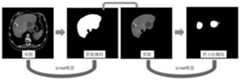
图3是本发明实施例提供的肝脏病灶图像处理系统整体架构示意图。FIG. 3 is a schematic diagram of the overall architecture of a liver lesion image processing system provided by an embodiment of the present invention.
图4是本发明实施例提供的肝脏病灶图像处理系统功能示意图。FIG. 4 is a functional schematic diagram of a liver lesion image processing system provided by an embodiment of the present invention.
图5是本发明实施例提供的肝脏病灶图像数据处理流程图。FIG. 5 is a flowchart of liver lesion image data processing provided by an embodiment of the present invention.
图6是本发明实施例提供的两级级联u-net架构识别肝脏病变模型示意图。FIG. 6 is a schematic diagram of a liver lesion recognition model provided by a two-level cascaded u-net architecture according to an embodiment of the present invention.
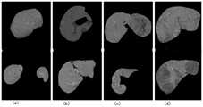
图7是本发明实施例提供的第一阶段肝脏识别模型对不同复杂场景下的肝脏分割结果示意图;7 is a schematic diagram of a liver segmentation result under different complex scenarios by a first-stage liver identification model provided by an embodiment of the present invention;
图中:(a)肝脏被其它器官截断成两部分;(b)含有肝裂的情形;(c)含有弥漫型肝病情形;(d)肝脏内含有非肝脏区域。In the figure: (a) the liver is truncated into two parts by other organs; (b) the case with liver fissure; (c) the case with diffuse liver disease; (d) the non-liver region in the liver.
图8是本发明实施例提供的第二阶段肝占位识别模型对不同复杂病灶的分割结果示意图。FIG. 8 is a schematic diagram of a segmentation result of different complex lesions by a second-stage liver occupancy recognition model provided by an embodiment of the present invention.
图中:(a)微小型病灶分割实例;(b)边缘型病灶分割实例;(c)异质型病灶分割实例;(d)弥漫型病灶分割实例。In the figure: (a) Segmentation example of tiny lesions; (b) Segmentation example of marginal lesions; (c) Segmentation example of heterogeneous lesions; (d) Segmentation example of diffuse lesions.
图9是本发明实施例提供的肝脏病灶图像处理方法采用两阶段病灶识别和独立病灶识别融合技术,识别肝脏内部和边缘病灶系统展示结果示意图。FIG. 9 is a schematic diagram showing results of a system for identifying internal and peripheral lesions of the liver using the two-stage lesion identification and independent lesion identification fusion technology provided by the liver lesion image processing method according to the embodiment of the present invention.
图10是本发明实施例提供的系统对接示意图。FIG. 10 is a schematic diagram of system docking provided by an embodiment of the present invention.
图11是本发明实施例提供的病变分析结果展示示意图。FIG. 11 is a schematic diagram showing a lesion analysis result according to an embodiment of the present invention.
具体实施方式Detailed ways
下面结合附图对本发明作进一步说明:The present invention will be further described below in conjunction with the accompanying drawings:
如图1所示:本发明提供的肝脏病灶图像处理系统包括:As shown in Figure 1: The liver lesion image processing system provided by the present invention includes:
信息和图像获取模块1,用于获取做过腹部CT的检查者的基本信息和CT图像。The information and
图像信息处理模块2,用于将图像传输给算法服务器进行计算,算法服务器再将结果传回给信息处理服务器。The image
数据库存储模块3,用于将信息和计算结果写入MySQL和MongoDB数据库,建立RESTful接口将数据暴露出来。The
信息展示模块4,用于前端网页进而展示肝脏和可能的病灶信息。The
如图2所示,本发明提供的肝脏病灶图像处理方法包括以下主要步骤:As shown in FIG. 2 , the method for processing a liver lesion image provided by the present invention includes the following main steps:
S1:信息处理服务器通过安全端口从医疗信息系统(如PACS、RIS等)获取做过腹部CT的检查者的基本信息和DICOM格式的CT图像;S1: The information processing server obtains the basic information of the examiner who has done abdominal CT and the CT image in DICOM format from the medical information system (such as PACS, RIS, etc.) through the secure port;
S2:通过gRPC,信息处理服务器将图像传输给算法服务器进行计算,获得肝脏、肝脏病变位置以及病变的类型、形态等分析结构,算法服务器再将结果传回给信息处理服务器;S2: Through gRPC, the information processing server transmits the image to the algorithm server for calculation, and obtains the analysis structure of the liver, the location of the liver lesion, and the type and shape of the lesion, and the algorithm server sends the result back to the information processing server;
S3:信息处理服务器将检查者信息和计算结果写入MySQL和MongoDB数据库,建立RESTful接口将数据暴露出来,前端网页进而展示肝脏和可能的病灶信息。S3: The information processing server writes the examiner's information and calculation results into MySQL and MongoDB databases, establishes a RESTful interface to expose the data, and the front-end web page displays information on the liver and possible lesions.
本发明是基于自改进算法的肝脏病灶图像处理AI分析系统,并且携带算法数据集持续反馈增长系统。基于医院的信息系统,开发了该系统。The present invention is a liver lesion image processing AI analysis system based on a self-improving algorithm, and carries a continuous feedback growth system of the algorithm data set. Based on the hospital's information system, the system was developed.
本发明的系统主体算法基于团队的自研算法,在保证医院数据安全性的基础上,通过常见的计算机技术构造了一系列的系统。首先信息处理服务器从安全端口获取即时的做过腹部CT的检查者的基本信息和CT图像;然后通过gRPC,信息处理服务器将图像传输给有GPU具有快速计算的算法服务器进行计算,算法服务器再将结果传回给信息处理服务器;信息处理服务器将检查者信息和计算结果写入MySQL和MongoDB数据库,这里会建立一个RESTful接口将数据暴露出来,前端网页进而展示肝脏和可能的病灶信息,系统整体架构如图3所示。系统功能包含:CT图像转换、数据增强、模型训练、肝脏和肝占位识别、各类信息存储、患者信息展示与交互、影像中肝脏和肝占位描画与结构化描述等,如图4所示。The main algorithm of the system of the present invention is based on the self-developed algorithm of the team, and on the basis of ensuring the security of hospital data, a series of systems are constructed through common computer technology. First, the information processing server obtains the basic information and CT images of the examiner who has undergone abdominal CT in real time from the secure port; then through gRPC, the information processing server transmits the images to the algorithm server with GPU with fast computing for calculation, and the algorithm server The results are sent back to the information processing server; the information processing server writes the examiner information and calculation results into MySQL and MongoDB databases, where a RESTful interface will be established to expose the data, and the front-end web page will then display the liver and possible lesion information, and the overall system architecture As shown in Figure 3. System functions include: CT image conversion, data enhancement, model training, liver and liver occupancy recognition, various information storage, patient information display and interaction, liver and liver occupancy description and structured description in images, etc., as shown in Figure 4. Show.
本发明的系统识别结果反馈机制,可以将一些表现不好的数据集会收集起来,通过改进的标注软件,专业标注团队会改进结果并放入数据集再进行训练。算法的核心如图5所示。为了提高算法性能,本发明提出了一系列先进技术,包括DICOM格式数据经过大窗宽截取后转化为高精度矩阵、涵盖像素级灰度值的图像自适应增强方法、反射模式的图像增强及结果重构、逆向肝脏病变位置识别、两阶段病灶识别和独立病灶识别融合技术分割边缘病灶、肝占位假阳性过滤策略等。The system identification result feedback mechanism of the present invention can collect some data sets with poor performance, and through the improved labeling software, the professional labeling team will improve the results and put them into the data sets for training. The core of the algorithm is shown in Figure 5. In order to improve the performance of the algorithm, the present invention proposes a series of advanced technologies, including the conversion of DICOM format data into a high-precision matrix after being intercepted by a large window width, an image adaptive enhancement method covering pixel-level gray values, and reflection mode image enhancement and results. Reconstruction, reverse liver lesion location identification, two-stage lesion identification and independent lesion identification fusion technology to segment marginal lesions, liver mass-occupying false-positive filtering strategy, etc.
本发明检测肝脏病变采用两阶段病灶识别和独立病灶识别融合技术,其中两阶段病灶识别如图6所示,算法框架采用U-net架构,首先将肝脏部分提取出来,再识别该肝脏区域内的病变部分。在算法创新上本发明改进并发现使用了多种方法。The detection of liver lesions in the present invention adopts the fusion technology of two-stage lesion identification and independent lesion identification, wherein the two-stage lesion identification is shown in Figure 6, and the algorithm framework adopts the U-net architecture. First, the liver part is extracted, and then the diseased part. The present invention improves on algorithmic innovation and finds that various methods are used.
(1)DICOM格式数据转化为高精度矩阵。(1) The DICOM format data is converted into a high-precision matrix.
肝脏CT原始DICOM图像存储的是Hu值,经过大窗宽(800<窗宽<4000)截取后转化为高精度矩阵,该矩阵对计算机敏感而非人眼敏感。该方法保证原始有效信息最大化的保留,同时去除噪声和干扰数据。模型训练以转换后的矩阵为基础。The original DICOM image of liver CT stores the Hu value, which is converted into a high-precision matrix after being intercepted by a large window width (800<window width<4000), which is sensitive to the computer rather than the human eye. This method guarantees maximum retention of the original valid information while removing noise and interfering data. Model training is based on the transformed matrix.
(2)涵盖像素级灰度值的图像自适增强方法。(2) Image adaptive enhancement method covering pixel-level gray value.
对于单个图片,根据图像上病变和肝脏像素直方图分布,建立统计方法,自动构建像素级别的自适应增强方法,作用于对应的高精矩阵,能够自动将病变跟肝脏的差异增强,便于识别。For a single image, a statistical method is established according to the histogram distribution of lesions and liver pixels on the image, and a pixel-level adaptive enhancement method is automatically constructed, acting on the corresponding high-precision matrix, which can automatically enhance the difference between the lesion and the liver for easy identification.
令f(x,y)表示在图像任意坐标(x,y)处的像素值,g(x,y)表示该坐标处的相应的增强像素值,则有Let f(x, y) represent the pixel value at any coordinate (x, y) of the image, and g(x, y) represent the corresponding enhanced pixel value at this coordinate, then we have

其中,k是局部范围内核的大小,

(3)样本增强(3) Sample enhancement
针对病变部分样本在图像上普遍占比较小,造成正样本数目较少,本发明提出两个解决方法:从输入数据入手,通过反射、平移、旋转等手段提高病变样本在数据上的占比,这样可以明显增强模型对病灶部分的敏感性;从识别类别出发,改变正负样本的比例。在负样本中大量增加正样本,使得类别相对较平衡。In view of the fact that the samples of the lesions generally occupy a small proportion in the image, resulting in a small number of positive samples, the present invention proposes two solutions: starting from the input data, and increasing the proportion of the lesion samples in the data by means of reflection, translation, rotation, etc., In this way, the sensitivity of the model to the lesion part can be significantly enhanced; starting from the identification category, the proportion of positive and negative samples can be changed. A large number of positive samples are added to the negative samples, so that the categories are relatively balanced.
(4)局部对比度增强技术分割微小病灶和模糊病灶(4) Local contrast enhancement technology to segment tiny lesions and fuzzy lesions
针对微小病灶和较模糊病灶难以识别和分割的问题,本发明构造多种对比度增强算法,包括在目标检测后的局部区域重建超分辨率图像,在保证算法效率的同时进而增大待检测目标;利用滤波后的图像计算梯度的策略突出目标区域对比度差异,便于后续分割。Aiming at the problem of difficulty in identifying and segmenting tiny lesions and relatively vague lesions, the present invention constructs a variety of contrast enhancement algorithms, including reconstructing super-resolution images in local areas after target detection, so as to increase the target to be detected while ensuring the efficiency of the algorithm; The strategy of using the filtered image to calculate the gradient highlights the contrast difference of the target area, which is convenient for subsequent segmentation.
令f(x,y)表示在图像任意坐标(x,y)处的像素值,g(x,y)表示该坐标处对应超分辨率重建后的像素值,则有Let f(x, y) represent the pixel value at any coordinate (x, y) of the image, and g(x, y) represent the pixel value corresponding to the super-resolution reconstruction at this coordinate, then we have
g(x,y)=σ(W*f(x,y)+B)L#(2)g(x,y)=σ(W*f(x,y)+B)L #(2)
其中W是神经网络参数矩阵,B是网络偏置矩阵,L是网络层数。W和B通过损失函数后向传播学习,其中损失函数定义为:where W is the neural network parameter matrix, B is the network bias matrix, and L is the number of network layers. W and B are learned by back-propagating a loss function, where the loss function is defined as:

式中poolingk(gi(x,y))是将位置为(x,y)、核大小为k的区域下采样为一个值,Z,M,N三个值分别代表批大小、图片宽和高。采用poolingk(·)函数下采样的原因是超分辨率重建后的图片大小跟原始输入之间存在k倍差距。In the formula, poolingk (gi (x, y)) is to downsample the region with position (x, y) and kernel size k as a value, and the three values of Z, M, and N represent batch size, image width, respectively. and high. The reason for downsampling with the poolingk ( ) function is that there is a k-fold difference between the size of the reconstructed image after super-resolution and the original input.
对于图片滤波后的梯度增强,则采用如下公式进行计算:For the gradient enhancement after image filtering, the following formula is used to calculate:
g(x,y)=H(f(x,y)-γfLP(x,y))#(4)g(x,y)=H(f(x,y)-γfLP (x,y))#(4)
式中fLP(x,y)是对f(x,y)在频域上的低通滤波,通过以下公式计算:where fLP (x, y) is the low-pass filtering of f (x, y) in the frequency domain, which is calculated by the following formula:

dis(x,y)是任意像素(x,y)到中心点(x0,y0)的距离,d0是以点(x0,y0)为中心的局部区域半径。H(g(x,y))是以(x,y)为中心的局部区域数值的二阶导数。dis(x,y) is the distance from any pixel (x,y) to the center point (x0 , y0 ), and d0 is the local area radius centered at the point (x0 , y0 ). H(g(x,y)) is the second derivative of the local region value centered at (x,y).
通过上述增强后的图像和原始图像一起以多通道的形式输入到神经网络进行模型训练,从而提高局部病灶识别的准确度。该处理方式不等同于对原始图像经过神经网络对原始图像的非线性变换得到的目标图像,因为以上操作都是在原始图像的局部进行,而不是全局进行的变换。The above-mentioned enhanced image and the original image are input to the neural network in the form of multi-channel for model training, thereby improving the accuracy of local lesion identification. This processing method is not equivalent to the target image obtained by the non-linear transformation of the original image through the neural network, because the above operations are performed locally in the original image, not the global transformation.
(5)病变位置重构(5) Reconstruction of lesion location
样本增强后的图像结果往往有多个,为了达到最佳识别效果,本发明通过多数投票机制将不同位置的判定结果映射到相同位置进行综合判定,降低单次识别造成的偶然误差,即There are often multiple image results after sample enhancement. In order to achieve the best recognition effect, the present invention maps the judgment results of different positions to the same position for comprehensive judgment through the majority voting mechanism, so as to reduce the accidental error caused by a single recognition, that is,

式中wi是第i个模型的权重,I(·)是指示函数。wherewi is the weight of the ith model, and I(·) is the indicator function.
(6)病变逆向分析(6) Reverse analysis of lesions
针对病变位置、形态、大小、类型等复杂多样,而实际训练数据在某些类型上可能较少不能充分训练的缺陷,本发明采用逆向思维,确定正常肝脏的位置,然后反转获得病变的位置,大大提高发现病变位置的准确性和敏感度。Aiming at the defect that the location, shape, size, and type of the lesion are complex and diverse, and the actual training data may be less and cannot be fully trained in some types, the present invention adopts reverse thinking to determine the location of the normal liver, and then reversely obtains the location of the lesion , greatly improving the accuracy and sensitivity of finding the location of the lesion.
(7)两阶段病灶识别和独立病灶识别融合技术分割边缘病灶(7) Two-stage lesion identification and independent lesion identification fusion technology to segment marginal lesions
两阶段病灶识别是在肝脏分割的基础上进行病灶识别,该方法可以非常准确的识别肝脏内部病灶,但是对于肝脏边沿病灶无能为力;独立病灶识别可以发现边界病灶,但是识别准确度较两阶段算法有所下降。融合这两个算法可以非常准确的识别肝脏内部以及边界上的病灶。融合通过以下公式实现:Two-stage lesion identification is based on liver segmentation. This method can identify the internal liver lesions very accurately, but it cannot do anything for the border lesions of the liver; independent lesion identification can find boundary lesions, but the recognition accuracy is lower than that of the two-stage algorithm. dropped. The fusion of these two algorithms can very accurately identify the lesions inside and on the border of the liver. Fusion is achieved by the following formula:

式中f2(x,y)和f1(x,y)分别代表两阶段模型和单阶段模型在图像位置(x,y)处生成的分割概率值,C0为病灶质心到最近的肝脏边界的距离,K为病灶的大小(取长和宽中的最大值)。In the formula, f2 (x, y) and f1 (x, y) represent the segmentation probability values generated by the two-stage model and the single-stage model at the image position (x, y), respectively, and C0 is the centroid of the lesion to the nearest liver The distance of the boundary, K is the size of the lesion (take the maximum of the length and width).
(8)肝占位假阳性后处理策略(8) Post-processing strategies for false-positive liver mass
针对少量肝脏病变的错误分割,利用识别的肝占位在其前后连续几张影像切片上具有相同或相近的特征进行过滤,排除识别结果中的假阳性。For the wrong segmentation of a small number of liver lesions, the identified liver mass has the same or similar features on several consecutive image slices before and after it for filtering to exclude false positives in the identification results.
令P(X)表示病灶X=<x1,x2,…,xn>的联合概率分布,a(xi)为第i张病灶xi的图像面积,则Let P(X) denote the joint probability distribution of the lesion X=<x1 ,x2 ,...,xn >, and a(xi ) be the image area of thei -th lesion xi, then

其中pn(xi)表示xi的概率,计算公式为where pn (xi ) represents the probability of xi , and the calculation formula is

式中


式中μ和δ分别为长度为n的所有病灶块联合概率分布的均值和方差。P(X)分布在2δ范围内定义为正常的病灶,否则为假阳性。where μ and δ are the mean and variance of the joint probability distribution of all lesion blocks of length n, respectively. A normal lesion was defined as a P(X) distribution within the 2δ range, otherwise it was a false positive.
本发明自项目实施以来累积收集肝病患者CT影像超过5100例,通过影像学专家和模型多次迭代标注肝脏和肝病病变影像超过60万张,肝脏识别准确度超过98%(DICE),肝脏病变位置识别准确度超过97%(DICE),肝脏病变类型判别准确度超过96%。Since the implementation of the project, the present invention has accumulated more than 5,100 CT images of liver disease patients, and iteratively labeled more than 600,000 images of liver and liver disease lesions through imaging experts and models. The liver recognition accuracy exceeds 98% (DICE), and the location of liver lesions The recognition accuracy exceeds 97% (DICE), and the liver lesion type discrimination accuracy exceeds 96%.
为了体现本发明对肝脏、肝占位的识别效果和准确度,进一步结合以下三个实施例进行说明:In order to reflect the recognition effect and accuracy of the present invention for liver and liver space occupancy, the following three embodiments are further described:
实施例一,模型第一阶段对于CT影像中肝脏的识别。随机挑选模型在复杂场景下对肝脏的分割结果,如图7所示,模型能够很好的处理识别难度非常大的各类情形,如:图7(a)原图肝脏被其它器官截断成两部分;图7(b)肝脏中含有肝裂的情形;图7(c)肝脏中含有弥漫型病变的情形;图7(d)肝脏内含有非肝脏区域。Example 1: The first stage of the model is for the identification of the liver in the CT image. The segmentation results of the liver by the randomly selected model in complex scenes are shown in Figure 7. The model can handle various situations that are very difficult to identify, such as: Figure 7(a) The original liver is truncated into two parts by other organs. Part; Fig. 7(b) liver with fissures; Fig. 7(c) liver with diffuse lesions; Fig. 7(d) liver with non-liver areas.
实施例二,模型第二阶段对于肝脏图像中肝占位的识别。随机挑选模型在不同肝脏病变下对肝占位的分割结果,如图8所示,模型能够很好的处理识别难度大、隐秘性强的各类情形,主要包含:图8(a)微小型病灶;图8(b)边缘型病灶;图8(c)异质型病灶;图8(d)弥漫型病灶。Embodiment 2: The second stage of the model is for the identification of liver occupancy in liver images. Figure 8 shows the segmentation results of the liver occupancy by the randomly selected model under different liver lesions. The model can well handle various situations with high identification difficulty and strong concealment, mainly including: Figure 8(a) Miniature Lesions; Figure 8(b) Marginal lesions; Figure 8(c) Heterogeneous lesions; Figure 8(d) Diffuse lesions.
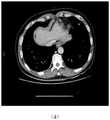
实施例三,两阶段病灶识别和独立病灶识别融合技术的肝占位识别效果。系统应用效果,如图9所示,两阶段病灶识别方法在肝脏分割的基础上识别病灶,可以非常准确的识别肝脏内部病灶,但是容易遗漏肝脏边沿病灶;独立病灶识别可以识别超出肝脏分割区域以外的边界病灶,融合这两个算法对于各类情形的肝占位识别效果非常显著。Embodiment 3: The liver mass recognition effect of the fusion technology of two-stage lesion recognition and independent lesion recognition. The application effect of the system is shown in Figure 9. The two-stage lesion identification method identifies lesions on the basis of liver segmentation, which can identify lesions within the liver very accurately, but it is easy to miss the border lesions of the liver; independent lesion identification can identify beyond the liver segmentation area. The fusion of these two algorithms has a very significant effect on the identification of liver mass in various situations.
本发明专利对应的肝脏影像AI系统自2019年7月在十堰市某大型三甲医院影像科上线测试运行,系统稳定、高效,反响非凡,系统主要呈现效果如图10、11所示。The liver imaging AI system corresponding to the patent of the present invention has been online and tested in the imaging department of a large tertiary hospital in Shiyan City since July 2019. The system is stable, efficient, and has an extraordinary response. The main effects of the system are shown in Figures 10 and 11.
以上显示和描述了本发明的基本原理和主要特征及本发明的优点。本行业的技术人员应该了解,本发明不受上述实施例的限制,上述实施例和说明书中描述的只是说明本发明的原理,在不脱离本发明精神和范围的前提下,本发明还会有各种变化和改进,这些变化和改进都落入要求保护的本发明范围内。本发明要求保护范围由所附的权利要求书及其等效物界定。The foregoing has shown and described the basic principles and main features of the present invention and the advantages of the present invention. Those skilled in the art should understand that the present invention is not limited by the above-mentioned embodiments, and the descriptions in the above-mentioned embodiments and the description are only to illustrate the principle of the present invention. Without departing from the spirit and scope of the present invention, the present invention will have Various changes and modifications fall within the scope of the claimed invention. The claimed scope of the present invention is defined by the appended claims and their equivalents.
Claims (1)


Applications Claiming Priority (2)
| Application Number | Priority Date | Filing Date | Title |
|---|---|---|---|
| CN202010674223.1ACN111950595A (en) | 2020-07-14 | 2020-07-14 | Liver lesion image processing method, system, storage medium, program, terminal |
| CN2020106742231 | 2020-07-14 |
Publications (2)
| Publication Number | Publication Date |
|---|---|
| CN113420826A CN113420826A (en) | 2021-09-21 |
| CN113420826Btrue CN113420826B (en) | 2022-08-23 |
Family
ID=73341100
Family Applications (2)
| Application Number | Title | Priority Date | Filing Date |
|---|---|---|---|
| CN202010674223.1AWithdrawnCN111950595A (en) | 2020-07-14 | 2020-07-14 | Liver lesion image processing method, system, storage medium, program, terminal |
| CN202110770193.9AActiveCN113420826B (en) | 2020-07-14 | 2021-07-07 | Liver focus image processing system and image processing method |
Family Applications Before (1)
| Application Number | Title | Priority Date | Filing Date |
|---|---|---|---|
| CN202010674223.1AWithdrawnCN111950595A (en) | 2020-07-14 | 2020-07-14 | Liver lesion image processing method, system, storage medium, program, terminal |
Country Status (1)
| Country | Link |
|---|---|
| CN (2) | CN111950595A (en) |
Families Citing this family (8)
| Publication number | Priority date | Publication date | Assignee | Title |
|---|---|---|---|---|
| CN112330731B (en)* | 2020-11-30 | 2024-12-31 | 深圳开立生物医疗科技股份有限公司 | Image processing device, method, equipment, ultrasound system and readable storage medium |
| CN112735568A (en)* | 2021-01-26 | 2021-04-30 | 杭州联众医疗科技股份有限公司 | Artificial intelligence auxiliary diagnosis platform based on medical image and clinical requirements |
| CN112967291B (en)* | 2021-03-01 | 2021-11-16 | 北京安德医智科技有限公司 | Image processing method and device, electronic equipment and storage medium |
| CN113229936A (en)* | 2021-05-06 | 2021-08-10 | 卫飞鹏 | Method and system for improving liver intervention target positioning accuracy |
| CN115512184B (en)* | 2022-10-20 | 2025-08-05 | 北京工业大学 | A plug-and-play low-light image enhancement method for end-to-end object detection in low-light conditions |
| CN116309593B (en)* | 2023-05-23 | 2023-09-12 | 天津市中西医结合医院(天津市南开医院) | Liver puncture biopsy B ultrasonic image processing method and system based on mathematical model |
| CN116759052B (en)* | 2023-06-20 | 2024-04-05 | 华平祥晟(上海)医疗科技有限公司 | Image storage management system and method based on big data |
| CN118170458A (en)* | 2024-03-19 | 2024-06-11 | 中山仰视科技有限公司 | Intelligent window width and window position selection system |
Family Cites Families (9)
| Publication number | Priority date | Publication date | Assignee | Title |
|---|---|---|---|---|
| US10127659B2 (en)* | 2016-11-23 | 2018-11-13 | General Electric Company | Deep learning medical systems and methods for image acquisition |
| CN108171709A (en)* | 2018-01-30 | 2018-06-15 | 北京青燕祥云科技有限公司 | Detection method, device and the realization device of Liver masses focal area |
| CN108805858A (en)* | 2018-04-10 | 2018-11-13 | 燕山大学 | Hepatopathy CT image computers assistant diagnosis system based on data mining and method |
| CN108875734B (en)* | 2018-05-23 | 2021-07-23 | 平安科技(深圳)有限公司 | Liver canceration positioning method, device and storage medium |
| US10621728B2 (en)* | 2018-06-26 | 2020-04-14 | Sony Corporation | Internal organ localization in computed tomography (CT) images |
| CN109447969B (en)* | 2018-10-29 | 2021-08-10 | 北京青燕祥云科技有限公司 | Liver occupation lesion identification method and device and implementation device |
| CN110009599A (en)* | 2019-02-01 | 2019-07-12 | 腾讯科技(深圳)有限公司 | Liver masses detection method, device, equipment and storage medium |
| CN110085288A (en)* | 2019-04-19 | 2019-08-02 | 四川大学华西医院 | A kind of liver and gall surgical department Internet-based treatment information sharing system and sharing method |
| CN111402268B (en)* | 2020-03-16 | 2023-05-23 | 苏州科技大学 | A Method for Segmenting the Liver and Its Lesions in Medical Images |
- 2020
- 2020-07-14CNCN202010674223.1Apatent/CN111950595A/ennot_activeWithdrawn
- 2021
- 2021-07-07CNCN202110770193.9Apatent/CN113420826B/enactiveActive
Also Published As
| Publication number | Publication date |
|---|---|
| CN111950595A (en) | 2020-11-17 |
| CN113420826A (en) | 2021-09-21 |
Similar Documents
| Publication | Publication Date | Title |
|---|---|---|
| CN113420826B (en) | Liver focus image processing system and image processing method | |
| Su et al. | Lung nodule detection based on faster R-CNN framework | |
| Saikumar et al. | A novel implementation heart diagnosis system based on random forest machine learning technique. | |
| CN107909585B (en) | A method for segmentation of vascular media and intima from intravascular ultrasound images | |
| CN110310281A (en) | A method for detection and segmentation of lung nodules in virtual medicine based on Mask-RCNN deep learning | |
| CN110853111B (en) | Medical image processing system, model training method and training device | |
| Rajee et al. | Gender classification on digital dental x-ray images using deep convolutional neural network | |
| Ye et al. | Medical image diagnosis of prostate tumor based on PSP-Net+ VGG16 deep learning network | |
| CN111429474A (en) | Mammary gland DCE-MRI image focus segmentation model establishment and segmentation method based on mixed convolution | |
| CN110188792A (en) | The characteristics of image acquisition methods of prostate MRI 3-D image | |
| Zhou et al. | Interpreting medical images | |
| CN112508884A (en) | Comprehensive detection device and method for cancerous region | |
| Wang et al. | Automatic measurement of fetal head circumference using a novel GCN-assisted deep convolutional network | |
| Fu et al. | Deep‐Learning‐Based CT Imaging in the Quantitative Evaluation of Chronic Kidney Diseases | |
| CN114663445A (en) | A 3D Cardiac Image Segmentation Method Based on Multiscale Edge Perception | |
| CN113223704B (en) | Auxiliary diagnosis method for computed tomography aortic aneurysm based on deep learning | |
| CN115330600A (en) | Lung CT image super-resolution method based on improved SRGAN | |
| Kumar et al. | A Novel Approach for Breast Cancer Detection by Mammograms | |
| Wang et al. | Multimodal parallel attention network for medical image segmentation | |
| Neha | Kidney localization and stone segmentation from a ct scan image | |
| CN111383222A (en) | An intelligent diagnosis system of intervertebral disc MRI image based on deep learning | |
| CN115375632A (en) | Lung nodule intelligent detection system and method based on CenterNet model | |
| Lan et al. | Breast mass lesion area detection method based on an improved YOLOv8 model. | |
| Nemri et al. | Automatic segmentation of echocardiographic images using a shifted windows vision transformer architecture | |
| CN112862786B (en) | CTA image data processing method, device and storage medium |
Legal Events
| Date | Code | Title | Description |
|---|---|---|---|
| PB01 | Publication | ||
| PB01 | Publication | ||
| SE01 | Entry into force of request for substantive examination | ||
| SE01 | Entry into force of request for substantive examination | ||
| GR01 | Patent grant | ||
| GR01 | Patent grant |