CN113244175B - Immune vesicle maytansine conjugate as well as preparation method and application thereof - Google Patents
Immune vesicle maytansine conjugate as well as preparation method and application thereofDownload PDFInfo
- Publication number
- CN113244175B CN113244175BCN202110561582.0ACN202110561582ACN113244175BCN 113244175 BCN113244175 BCN 113244175BCN 202110561582 ACN202110561582 ACN 202110561582ACN 113244175 BCN113244175 BCN 113244175B
- Authority
- CN
- China
- Prior art keywords
- maytansine
- dar
- vesicle
- immune
- conjugate
- Prior art date
- Legal status (The legal status is an assumption and is not a legal conclusion. Google has not performed a legal analysis and makes no representation as to the accuracy of the status listed.)
- Active
Links
- WKPWGQKGSOKKOO-RSFHAFMBSA-NmaytansineChemical compoundCO[C@@H]([C@@]1(O)C[C@](OC(=O)N1)([C@H]([C@@H]1O[C@@]1(C)[C@@H](OC(=O)[C@H](C)N(C)C(C)=O)CC(=O)N1C)C)[H])\C=C\C=C(C)\CC2=CC(OC)=C(Cl)C1=C2WKPWGQKGSOKKOO-RSFHAFMBSA-N0.000titleclaimsabstractdescription41
- 229930126263MaytansineNatural products0.000titleclaimsabstractdescription38
- 238000002360preparation methodMethods0.000titleclaimsabstractdescription22
- 229920000642polymerPolymers0.000claimsabstractdescription46
- 239000003814drugSubstances0.000claimsabstractdescription21
- 229940079593drugDrugs0.000claimsabstractdescription18
- 238000006243chemical reactionMethods0.000claimsabstractdescription8
- 206010035226Plasma cell myelomaDiseases0.000claimsdescription23
- 208000034578Multiple myelomasDiseases0.000claimsdescription16
- 230000002209hydrophobic effectEffects0.000claimsdescription12
- 238000000034methodMethods0.000claimsdescription12
- 208000024893Acute lymphoblastic leukemiaDiseases0.000claimsdescription8
- 208000014697Acute lymphocytic leukaemiaDiseases0.000claimsdescription8
- 208000006664Precursor Cell Lymphoblastic Leukemia-LymphomaDiseases0.000claimsdescription8
- 239000012528membraneSubstances0.000claimsdescription7
- 239000002994raw materialSubstances0.000claimsdescription6
- 125000000524functional groupChemical group0.000claimsdescription5
- 229960002204daratumumabDrugs0.000claimsdescription4
- 239000002904solventSubstances0.000claimsdescription4
- 208000002250Hematologic NeoplasmsDiseases0.000claimsdescription3
- 230000008878couplingEffects0.000claimsdescription3
- 238000010168coupling processMethods0.000claimsdescription3
- 238000005859coupling reactionMethods0.000claimsdescription3
- VSWDORGPIHIGNW-UHFFFAOYSA-NPyrrolidine dithiocarbamic acidChemical compoundSC(=S)N1CCCC1VSWDORGPIHIGNW-UHFFFAOYSA-N0.000claimsdescription2
- 238000007306functionalization reactionMethods0.000claimsdescription2
- 208000019691hematopoietic and lymphoid cell neoplasmDiseases0.000claimsdescription2
- ZUHQCDZJPTXVCU-UHFFFAOYSA-NC1#CCCC2=CC=CC=C2C2=CC=CC=C21Chemical compoundC1#CCCC2=CC=CC=C2C2=CC=CC=C21ZUHQCDZJPTXVCU-UHFFFAOYSA-N0.000claims1
- 206010066476Haematological malignancyDiseases0.000claims1
- OXFKPZMRAPEFSB-UHFFFAOYSA-N[S].S[S]Chemical group[S].S[S]OXFKPZMRAPEFSB-UHFFFAOYSA-N0.000claims1
- 238000011282treatmentMethods0.000abstractdescription27
- 206010028980NeoplasmDiseases0.000abstractdescription15
- 230000000259anti-tumor effectEffects0.000abstractdescription10
- 231100000331toxicToxicity0.000abstractdescription9
- 230000002588toxic effectEffects0.000abstractdescription9
- 229910052717sulfurInorganic materials0.000abstractdescription7
- 239000011593sulfurSubstances0.000abstractdescription7
- 231100000419toxicityToxicity0.000abstractdescription7
- 230000001988toxicityEffects0.000abstractdescription7
- 201000005787hematologic cancerDiseases0.000abstractdescription4
- 208000024200hematopoietic and lymphoid system neoplasmDiseases0.000abstractdescription4
- 230000003211malignant effectEffects0.000abstractdescription3
- 230000003389potentiating effectEffects0.000abstractdescription2
- 210000004027cellAnatomy0.000description57
- 241000699670Mus sp.Species0.000description49
- 239000000562conjugateSubstances0.000description25
- 230000004083survival effectEffects0.000description17
- ZMXDDKWLCZADIW-UHFFFAOYSA-NN,N-DimethylformamideChemical compoundCN(C)C=OZMXDDKWLCZADIW-UHFFFAOYSA-N0.000description15
- 238000002474experimental methodMethods0.000description14
- 239000002245particleSubstances0.000description14
- 230000037396body weightEffects0.000description13
- WEVYAHXRMPXWCK-UHFFFAOYSA-NAcetonitrileChemical compoundCC#NWEVYAHXRMPXWCK-UHFFFAOYSA-N0.000description12
- 239000002202Polyethylene glycolSubstances0.000description11
- 229920001223polyethylene glycolPolymers0.000description11
- 239000000243solutionSubstances0.000description11
- 230000008685targetingEffects0.000description10
- 241000699666Mus <mouse, genus>Species0.000description8
- 230000002441reversible effectEffects0.000description8
- 238000009826distributionMethods0.000description7
- 230000000694effectsEffects0.000description7
- 238000011068loading methodMethods0.000description7
- 201000000050myeloid neoplasmDiseases0.000description7
- 229940049595antibody-drug conjugateDrugs0.000description6
- 229920006037cross link polymerPolymers0.000description6
- 231100000135cytotoxicityToxicity0.000description6
- 230000003013cytotoxicityEffects0.000description6
- 230000003247decreasing effectEffects0.000description6
- 230000012202endocytosisEffects0.000description6
- 238000004128high performance liquid chromatographyMethods0.000description6
- 210000003462veinAnatomy0.000description6
- 238000002835absorbanceMethods0.000description5
- 239000000611antibody drug conjugateSubstances0.000description5
- 239000007924injectionSubstances0.000description5
- 238000002347injectionMethods0.000description5
- 229920000575polymersomePolymers0.000description5
- 230000001225therapeutic effectEffects0.000description5
- 238000000108ultra-filtrationMethods0.000description5
- 210000004369bloodAnatomy0.000description4
- 239000008280bloodSubstances0.000description4
- 210000000988bone and boneAnatomy0.000description4
- 230000008859changeEffects0.000description4
- 238000011503in vivo imagingMethods0.000description4
- 230000001965increasing effectEffects0.000description4
- 238000011081inoculationMethods0.000description4
- 230000007774longtermEffects0.000description4
- 238000001840matrix-assisted laser desorption--ionisation time-of-flight mass spectrometryMethods0.000description4
- 230000009467reductionEffects0.000description4
- 238000003756stirringMethods0.000description4
- IAZDPXIOMUYVGZ-UHFFFAOYSA-NDimethylsulphoxideChemical compoundCS(C)=OIAZDPXIOMUYVGZ-UHFFFAOYSA-N0.000description3
- PEDCQBHIVMGVHV-UHFFFAOYSA-NGlycerineChemical compoundOCC(O)COPEDCQBHIVMGVHV-UHFFFAOYSA-N0.000description3
- 108010087230SincalideProteins0.000description3
- 208000035896Twin-reversed arterial perfusion sequenceDiseases0.000description3
- 238000004458analytical methodMethods0.000description3
- 210000001185bone marrowAnatomy0.000description3
- 238000010609cell counting kit-8 assayMethods0.000description3
- 238000004132cross linkingMethods0.000description3
- 230000034994deathEffects0.000description3
- 238000001514detection methodMethods0.000description3
- 238000000684flow cytometryMethods0.000description3
- 210000003958hematopoietic stem cellAnatomy0.000description3
- 238000003384imaging methodMethods0.000description3
- 238000001727in vivoMethods0.000description3
- 238000011534incubationMethods0.000description3
- 210000002414legAnatomy0.000description3
- 210000001930leg boneAnatomy0.000description3
- 210000003141lower extremityAnatomy0.000description3
- 231100000682maximum tolerated doseToxicity0.000description3
- 229910052757nitrogenInorganic materials0.000description3
- 230000008569processEffects0.000description3
- 230000035755proliferationEffects0.000description3
- 108090000623proteins and genesProteins0.000description3
- 102000004169proteins and genesHuman genes0.000description3
- IZTQOLKUZKXIRV-YRVFCXMDSA-NsincalideChemical compoundC([C@@H](C(=O)N[C@@H](CCSC)C(=O)NCC(=O)N[C@@H](CC=1C2=CC=CC=C2NC=1)C(=O)N[C@@H](CCSC)C(=O)N[C@@H](CC(O)=O)C(=O)N[C@@H](CC=1C=CC=CC=1)C(N)=O)NC(=O)[C@@H](N)CC(O)=O)C1=CC=C(OS(O)(=O)=O)C=C1IZTQOLKUZKXIRV-YRVFCXMDSA-N0.000description3
- 238000010186stainingMethods0.000description3
- 238000003860storageMethods0.000description3
- 238000012360testing methodMethods0.000description3
- 210000004881tumor cellAnatomy0.000description3
- 230000004580weight lossEffects0.000description3
- QRZUPJILJVGUFF-UHFFFAOYSA-N2,8-dibenzylcyclooctan-1-oneChemical compoundC1CCCCC(CC=2C=CC=CC=2)C(=O)C1CC1=CC=CC=C1QRZUPJILJVGUFF-UHFFFAOYSA-N0.000description2
- FWBHETKCLVMNFS-UHFFFAOYSA-N4',6-Diamino-2-phenylindolChemical compoundC1=CC(C(=N)N)=CC=C1C1=CC2=CC=C(C(N)=N)C=C2N1FWBHETKCLVMNFS-UHFFFAOYSA-N0.000description2
- 102100031585ADP-ribosyl cyclase/cyclic ADP-ribose hydrolase 1Human genes0.000description2
- IJGRMHOSHXDMSA-UHFFFAOYSA-NAtomic nitrogenChemical compoundN#NIJGRMHOSHXDMSA-UHFFFAOYSA-N0.000description2
- BWGNESOTFCXPMA-UHFFFAOYSA-NDihydrogen disulfideChemical compoundSSBWGNESOTFCXPMA-UHFFFAOYSA-N0.000description2
- 101000777636Homo sapiens ADP-ribosyl cyclase/cyclic ADP-ribose hydrolase 1Proteins0.000description2
- 238000012449Kunming mouseMethods0.000description2
- 241001465754MetazoaSpecies0.000description2
- NQTADLQHYWFPDB-UHFFFAOYSA-NN-HydroxysuccinimideChemical compoundON1C(=O)CCC1=ONQTADLQHYWFPDB-UHFFFAOYSA-N0.000description2
- 229930040373ParaformaldehydeNatural products0.000description2
- 206010033799ParalysisDiseases0.000description2
- -1PolyethylenePolymers0.000description2
- 238000007112amidation reactionMethods0.000description2
- 230000008901benefitEffects0.000description2
- 230000037237body shapeEffects0.000description2
- 239000006285cell suspensionSubstances0.000description2
- 238000005119centrifugationMethods0.000description2
- 238000002983circular dichroismMethods0.000description2
- 238000010276constructionMethods0.000description2
- 238000010586diagramMethods0.000description2
- 238000000502dialysisMethods0.000description2
- 238000005538encapsulationMethods0.000description2
- 238000000338in vitroMethods0.000description2
- 238000011065in-situ storageMethods0.000description2
- 238000012986modificationMethods0.000description2
- 238000012544monitoring processMethods0.000description2
- 239000003960organic solventSubstances0.000description2
- 210000002997osteoclastAnatomy0.000description2
- 229920002866paraformaldehydePolymers0.000description2
- 239000004417polycarbonateSubstances0.000description2
- 229920000515polycarbonatePolymers0.000description2
- PCMORTLOPMLEFB-ONEGZZNKSA-Nsinapic acidChemical compoundCOC1=CC(\C=C\C(O)=O)=CC(OC)=C1OPCMORTLOPMLEFB-ONEGZZNKSA-N0.000description2
- PCMORTLOPMLEFB-UHFFFAOYSA-Nsinapinic acidNatural productsCOC1=CC(C=CC(O)=O)=CC(OC)=C1OPCMORTLOPMLEFB-UHFFFAOYSA-N0.000description2
- 150000003384small moleculesChemical class0.000description2
- 239000000126substanceSubstances0.000description2
- 239000006228supernatantSubstances0.000description2
- 239000003053toxinSubstances0.000description2
- 231100000765toxinToxicity0.000description2
- 230000001960triggered effectEffects0.000description2
- 238000000870ultraviolet spectroscopyMethods0.000description2
- MUZIZEZCKKMZRT-UHFFFAOYSA-N1,2-dithiolaneChemical groupC1CSSC1MUZIZEZCKKMZRT-UHFFFAOYSA-N0.000description1
- IMLSAISZLJGWPP-UHFFFAOYSA-N1,3-dithiolaneChemical compoundC1CSCS1IMLSAISZLJGWPP-UHFFFAOYSA-N0.000description1
- BUOYTFVLNZIELF-UHFFFAOYSA-N2-phenyl-1h-indole-4,6-dicarboximidamideChemical compoundN1C2=CC(C(=N)N)=CC(C(N)=N)=C2C=C1C1=CC=CC=C1BUOYTFVLNZIELF-UHFFFAOYSA-N0.000description1
- 208000031261Acute myeloid leukaemiaDiseases0.000description1
- 208000025321B-lymphoblastic leukemia/lymphomaDiseases0.000description1
- 108700004676Bence JonesProteins0.000description1
- 208000031648Body Weight ChangesDiseases0.000description1
- 101100298998Caenorhabditis elegans pbs-3 geneProteins0.000description1
- BVKZGUZCCUSVTD-UHFFFAOYSA-LCarbonateChemical compound[O-]C([O-])=OBVKZGUZCCUSVTD-UHFFFAOYSA-L0.000description1
- 208000006069Corneal OpacityDiseases0.000description1
- CMSMOCZEIVJLDB-UHFFFAOYSA-NCyclophosphamideChemical compoundClCCN(CCCl)P1(=O)NCCCO1CMSMOCZEIVJLDB-UHFFFAOYSA-N0.000description1
- 238000008157ELISA kitMethods0.000description1
- LFQSCWFLJHTTHZ-UHFFFAOYSA-NEthanolChemical compoundCCOLFQSCWFLJHTTHZ-UHFFFAOYSA-N0.000description1
- 206010059024Gastrointestinal toxicityDiseases0.000description1
- 108060003951ImmunoglobulinProteins0.000description1
- PIWKPBJCKXDKJR-UHFFFAOYSA-NIsofluraneChemical compoundFC(F)OC(Cl)C(F)(F)FPIWKPBJCKXDKJR-UHFFFAOYSA-N0.000description1
- 101710085938Matrix proteinProteins0.000description1
- 101710127721Membrane proteinProteins0.000description1
- 206010027646MiosisDiseases0.000description1
- 208000033776Myeloid Acute LeukemiaDiseases0.000description1
- 206010029350NeurotoxicityDiseases0.000description1
- 208000003076OsteolysisDiseases0.000description1
- 239000004698PolyethyleneSubstances0.000description1
- 108010039918PolylysineProteins0.000description1
- 241000700159RattusSpecies0.000description1
- 206010044221Toxic encephalopathyDiseases0.000description1
- 102000004243TubulinHuman genes0.000description1
- 108090000704TubulinProteins0.000description1
- 102000001742Tumor Suppressor ProteinsHuman genes0.000description1
- 108010040002Tumor Suppressor ProteinsProteins0.000description1
- 230000002159abnormal effectEffects0.000description1
- 230000005856abnormalityEffects0.000description1
- 230000009471actionEffects0.000description1
- 230000002776aggregationEffects0.000description1
- 238000004220aggregationMethods0.000description1
- 125000003277amino groupChemical group0.000description1
- 239000003963antioxidant agentSubstances0.000description1
- 230000003078antioxidant effectEffects0.000description1
- 235000006708antioxidantsNutrition0.000description1
- 230000006907apoptotic processEffects0.000description1
- 150000001540azidesChemical class0.000description1
- 229920002988biodegradable polymerPolymers0.000description1
- 239000004621biodegradable polymerSubstances0.000description1
- 230000029918bioluminescenceEffects0.000description1
- 238000005415bioluminescenceMethods0.000description1
- 230000015572biosynthetic processEffects0.000description1
- 230000000903blocking effectEffects0.000description1
- 238000009534blood testMethods0.000description1
- 230000004579body weight changeEffects0.000description1
- 239000000872bufferSubstances0.000description1
- 238000004364calculation methodMethods0.000description1
- 201000011510cancerDiseases0.000description1
- 230000022131cell cycleEffects0.000description1
- 230000004663cell proliferationEffects0.000description1
- 230000030570cellular localizationEffects0.000description1
- 239000003153chemical reaction reagentSubstances0.000description1
- 238000001553co-assemblyMethods0.000description1
- 239000012141concentrateSubstances0.000description1
- 238000001218confocal laser scanning microscopyMethods0.000description1
- 238000007796conventional methodMethods0.000description1
- 238000012258culturingMethods0.000description1
- 229960004397cyclophosphamideDrugs0.000description1
- 230000007547defectEffects0.000description1
- 229920006237degradable polymerPolymers0.000description1
- 238000011161developmentMethods0.000description1
- 201000010099diseaseDiseases0.000description1
- 208000037265diseases, disorders, signs and symptomsDiseases0.000description1
- 239000006185dispersionSubstances0.000description1
- VHJLVAABSRFDPM-QWWZWVQMSA-NdithiothreitolChemical compoundSC[C@@H](O)[C@H](O)CSVHJLVAABSRFDPM-QWWZWVQMSA-N0.000description1
- 238000012377drug deliveryMethods0.000description1
- 238000002296dynamic light scatteringMethods0.000description1
- 230000002121endocytic effectEffects0.000description1
- 230000002708enhancing effectEffects0.000description1
- 210000000887faceAnatomy0.000description1
- 238000000799fluorescence microscopyMethods0.000description1
- 238000001943fluorescence-activated cell sortingMethods0.000description1
- 231100000414gastrointestinal toxicityToxicity0.000description1
- 230000003862health statusEffects0.000description1
- 238000007490hematoxylin and eosin (H&E) stainingMethods0.000description1
- 102000018358immunoglobulinHuman genes0.000description1
- 229940027941immunoglobulin gDrugs0.000description1
- 230000001939inductive effectEffects0.000description1
- 230000008595infiltrationEffects0.000description1
- 238000001764infiltrationMethods0.000description1
- 239000003999initiatorSubstances0.000description1
- 230000003834intracellular effectEffects0.000description1
- 239000007928intraperitoneal injectionSubstances0.000description1
- 229960002725isofluraneDrugs0.000description1
- 230000002147killing effectEffects0.000description1
- 125000000686lactone groupChemical group0.000description1
- 231100001231less toxicToxicity0.000description1
- 208000032839leukemiaDiseases0.000description1
- 239000003446ligandSubstances0.000description1
- AGBQKNBQESQNJD-UHFFFAOYSA-MlipoateChemical compound[O-]C(=O)CCCCC1CCSS1AGBQKNBQESQNJD-UHFFFAOYSA-M0.000description1
- 235000019136lipoic acidNutrition0.000description1
- 208000029791lytic metastatic bone lesionDiseases0.000description1
- 238000004519manufacturing processMethods0.000description1
- 238000004949mass spectrometryMethods0.000description1
- 238000005259measurementMethods0.000description1
- 238000000386microscopyMethods0.000description1
- 230000003547miosisEffects0.000description1
- 230000011278mitosisEffects0.000description1
- 239000011259mixed solutionSubstances0.000description1
- 238000002156mixingMethods0.000description1
- 239000000203mixtureSubstances0.000description1
- 238000010172mouse modelMethods0.000description1
- 238000001728nano-filtrationMethods0.000description1
- 239000002539nanocarrierSubstances0.000description1
- 239000002105nanoparticleSubstances0.000description1
- 238000003333near-infrared imagingMethods0.000description1
- 230000007135neurotoxicityEffects0.000description1
- 231100000228neurotoxicityToxicity0.000description1
- 210000000056organAnatomy0.000description1
- 210000000963osteoblastAnatomy0.000description1
- 238000012261overproductionMethods0.000description1
- 239000008055phosphate buffer solutionSubstances0.000description1
- 210000004180plasmocyteAnatomy0.000description1
- 229920000573polyethylenePolymers0.000description1
- 229920000656polylysinePolymers0.000description1
- 235000010482polyoxyethylene sorbitan monooleateNutrition0.000description1
- 229920000053polysorbate 80Polymers0.000description1
- 208000017426precursor B-cell acute lymphoblastic leukemiaDiseases0.000description1
- 229940002612prodrugDrugs0.000description1
- 239000000651prodrugSubstances0.000description1
- 239000000047productSubstances0.000description1
- 238000004393prognosisMethods0.000description1
- 230000002035prolonged effectEffects0.000description1
- 210000001747pupilAnatomy0.000description1
- 238000009666routine testMethods0.000description1
- 210000003625skullAnatomy0.000description1
- 229940126586small molecule drugDrugs0.000description1
- 238000006557surface reactionMethods0.000description1
- 208000024891symptomDiseases0.000description1
- 231100000057systemic toxicityToxicity0.000description1
- 238000002560therapeutic procedureMethods0.000description1
- 229960002663thioctic acidDrugs0.000description1
- 125000003396thiol groupChemical group[H]S*0.000description1
- 210000002303tibiaAnatomy0.000description1
- 238000001269time-of-flight mass spectrometryMethods0.000description1
- 238000002054transplantationMethods0.000description1
- 229960001612trastuzumab emtansineDrugs0.000description1
- 230000004614tumor growthEffects0.000description1
- 229910021642ultra pure waterInorganic materials0.000description1
- 239000012498ultrapure waterSubstances0.000description1
- 238000002211ultraviolet spectrumMethods0.000description1
- 210000000689upper legAnatomy0.000description1
- 230000007332vesicle formationEffects0.000description1
- XLYOFNOQVPJJNP-UHFFFAOYSA-NwaterSubstancesOXLYOFNOQVPJJNP-UHFFFAOYSA-N0.000description1
- 230000003442weekly effectEffects0.000description1
Images


Classifications
- A—HUMAN NECESSITIES
- A61—MEDICAL OR VETERINARY SCIENCE; HYGIENE
- A61K—PREPARATIONS FOR MEDICAL, DENTAL OR TOILETRY PURPOSES
- A61K31/00—Medicinal preparations containing organic active ingredients
- A61K31/33—Heterocyclic compounds
- A61K31/395—Heterocyclic compounds having nitrogen as a ring hetero atom, e.g. guanethidine or rifamycins
- A61K31/535—Heterocyclic compounds having nitrogen as a ring hetero atom, e.g. guanethidine or rifamycins having six-membered rings with at least one nitrogen and one oxygen as the ring hetero atoms, e.g. 1,2-oxazines
- A61K31/537—Heterocyclic compounds having nitrogen as a ring hetero atom, e.g. guanethidine or rifamycins having six-membered rings with at least one nitrogen and one oxygen as the ring hetero atoms, e.g. 1,2-oxazines spiro-condensed or forming part of bridged ring systems
- A—HUMAN NECESSITIES
- A61—MEDICAL OR VETERINARY SCIENCE; HYGIENE
- A61K—PREPARATIONS FOR MEDICAL, DENTAL OR TOILETRY PURPOSES
- A61K47/00—Medicinal preparations characterised by the non-active ingredients used, e.g. carriers or inert additives; Targeting or modifying agents chemically bound to the active ingredient
- A61K47/50—Medicinal preparations characterised by the non-active ingredients used, e.g. carriers or inert additives; Targeting or modifying agents chemically bound to the active ingredient the non-active ingredient being chemically bound to the active ingredient, e.g. polymer-drug conjugates
- A61K47/51—Medicinal preparations characterised by the non-active ingredients used, e.g. carriers or inert additives; Targeting or modifying agents chemically bound to the active ingredient the non-active ingredient being chemically bound to the active ingredient, e.g. polymer-drug conjugates the non-active ingredient being a modifying agent
- A61K47/56—Medicinal preparations characterised by the non-active ingredients used, e.g. carriers or inert additives; Targeting or modifying agents chemically bound to the active ingredient the non-active ingredient being chemically bound to the active ingredient, e.g. polymer-drug conjugates the non-active ingredient being a modifying agent the modifying agent being an organic macromolecular compound, e.g. an oligomeric, polymeric or dendrimeric molecule
- A61K47/59—Medicinal preparations characterised by the non-active ingredients used, e.g. carriers or inert additives; Targeting or modifying agents chemically bound to the active ingredient the non-active ingredient being chemically bound to the active ingredient, e.g. polymer-drug conjugates the non-active ingredient being a modifying agent the modifying agent being an organic macromolecular compound, e.g. an oligomeric, polymeric or dendrimeric molecule obtained otherwise than by reactions only involving carbon-to-carbon unsaturated bonds, e.g. polyureas or polyurethanes
- A61K47/60—Medicinal preparations characterised by the non-active ingredients used, e.g. carriers or inert additives; Targeting or modifying agents chemically bound to the active ingredient the non-active ingredient being chemically bound to the active ingredient, e.g. polymer-drug conjugates the non-active ingredient being a modifying agent the modifying agent being an organic macromolecular compound, e.g. an oligomeric, polymeric or dendrimeric molecule obtained otherwise than by reactions only involving carbon-to-carbon unsaturated bonds, e.g. polyureas or polyurethanes the organic macromolecular compound being a polyoxyalkylene oligomer, polymer or dendrimer, e.g. PEG, PPG, PEO or polyglycerol
- A—HUMAN NECESSITIES
- A61—MEDICAL OR VETERINARY SCIENCE; HYGIENE
- A61K—PREPARATIONS FOR MEDICAL, DENTAL OR TOILETRY PURPOSES
- A61K47/00—Medicinal preparations characterised by the non-active ingredients used, e.g. carriers or inert additives; Targeting or modifying agents chemically bound to the active ingredient
- A61K47/50—Medicinal preparations characterised by the non-active ingredients used, e.g. carriers or inert additives; Targeting or modifying agents chemically bound to the active ingredient the non-active ingredient being chemically bound to the active ingredient, e.g. polymer-drug conjugates
- A61K47/51—Medicinal preparations characterised by the non-active ingredients used, e.g. carriers or inert additives; Targeting or modifying agents chemically bound to the active ingredient the non-active ingredient being chemically bound to the active ingredient, e.g. polymer-drug conjugates the non-active ingredient being a modifying agent
- A61K47/68—Medicinal preparations characterised by the non-active ingredients used, e.g. carriers or inert additives; Targeting or modifying agents chemically bound to the active ingredient the non-active ingredient being chemically bound to the active ingredient, e.g. polymer-drug conjugates the non-active ingredient being a modifying agent the modifying agent being an antibody, an immunoglobulin or a fragment thereof, e.g. an Fc-fragment
- A61K47/6801—Drug-antibody or immunoglobulin conjugates defined by the pharmacologically or therapeutically active agent
- A61K47/6803—Drugs conjugated to an antibody or immunoglobulin, e.g. cisplatin-antibody conjugates
- A—HUMAN NECESSITIES
- A61—MEDICAL OR VETERINARY SCIENCE; HYGIENE
- A61K—PREPARATIONS FOR MEDICAL, DENTAL OR TOILETRY PURPOSES
- A61K47/00—Medicinal preparations characterised by the non-active ingredients used, e.g. carriers or inert additives; Targeting or modifying agents chemically bound to the active ingredient
- A61K47/50—Medicinal preparations characterised by the non-active ingredients used, e.g. carriers or inert additives; Targeting or modifying agents chemically bound to the active ingredient the non-active ingredient being chemically bound to the active ingredient, e.g. polymer-drug conjugates
- A61K47/51—Medicinal preparations characterised by the non-active ingredients used, e.g. carriers or inert additives; Targeting or modifying agents chemically bound to the active ingredient the non-active ingredient being chemically bound to the active ingredient, e.g. polymer-drug conjugates the non-active ingredient being a modifying agent
- A61K47/68—Medicinal preparations characterised by the non-active ingredients used, e.g. carriers or inert additives; Targeting or modifying agents chemically bound to the active ingredient the non-active ingredient being chemically bound to the active ingredient, e.g. polymer-drug conjugates the non-active ingredient being a modifying agent the modifying agent being an antibody, an immunoglobulin or a fragment thereof, e.g. an Fc-fragment
- A61K47/6835—Medicinal preparations characterised by the non-active ingredients used, e.g. carriers or inert additives; Targeting or modifying agents chemically bound to the active ingredient the non-active ingredient being chemically bound to the active ingredient, e.g. polymer-drug conjugates the non-active ingredient being a modifying agent the modifying agent being an antibody, an immunoglobulin or a fragment thereof, e.g. an Fc-fragment the modifying agent being an antibody or an immunoglobulin bearing at least one antigen-binding site
- A61K47/6849—Medicinal preparations characterised by the non-active ingredients used, e.g. carriers or inert additives; Targeting or modifying agents chemically bound to the active ingredient the non-active ingredient being chemically bound to the active ingredient, e.g. polymer-drug conjugates the non-active ingredient being a modifying agent the modifying agent being an antibody, an immunoglobulin or a fragment thereof, e.g. an Fc-fragment the modifying agent being an antibody or an immunoglobulin bearing at least one antigen-binding site the antibody targeting a receptor, a cell surface antigen or a cell surface determinant
- A—HUMAN NECESSITIES
- A61—MEDICAL OR VETERINARY SCIENCE; HYGIENE
- A61K—PREPARATIONS FOR MEDICAL, DENTAL OR TOILETRY PURPOSES
- A61K9/00—Medicinal preparations characterised by special physical form
- A61K9/10—Dispersions; Emulsions
- A61K9/127—Synthetic bilayered vehicles, e.g. liposomes or liposomes with cholesterol as the only non-phosphatidyl surfactant
- A61K9/1271—Non-conventional liposomes, e.g. PEGylated liposomes or liposomes coated or grafted with polymers
- A61K9/1273—Polymersomes; Liposomes with polymerisable or polymerised bilayer-forming substances
- A—HUMAN NECESSITIES
- A61—MEDICAL OR VETERINARY SCIENCE; HYGIENE
- A61P—SPECIFIC THERAPEUTIC ACTIVITY OF CHEMICAL COMPOUNDS OR MEDICINAL PREPARATIONS
- A61P35/00—Antineoplastic agents
- B—PERFORMING OPERATIONS; TRANSPORTING
- B82—NANOTECHNOLOGY
- B82Y—SPECIFIC USES OR APPLICATIONS OF NANOSTRUCTURES; MEASUREMENT OR ANALYSIS OF NANOSTRUCTURES; MANUFACTURE OR TREATMENT OF NANOSTRUCTURES
- B82Y30/00—Nanotechnology for materials or surface science, e.g. nanocomposites
- B—PERFORMING OPERATIONS; TRANSPORTING
- B82—NANOTECHNOLOGY
- B82Y—SPECIFIC USES OR APPLICATIONS OF NANOSTRUCTURES; MEASUREMENT OR ANALYSIS OF NANOSTRUCTURES; MANUFACTURE OR TREATMENT OF NANOSTRUCTURES
- B82Y40/00—Manufacture or treatment of nanostructures
- B—PERFORMING OPERATIONS; TRANSPORTING
- B82—NANOTECHNOLOGY
- B82Y—SPECIFIC USES OR APPLICATIONS OF NANOSTRUCTURES; MEASUREMENT OR ANALYSIS OF NANOSTRUCTURES; MANUFACTURE OR TREATMENT OF NANOSTRUCTURES
- B82Y5/00—Nanobiotechnology or nanomedicine, e.g. protein engineering or drug delivery
Landscapes
- Health & Medical Sciences (AREA)
- Chemical & Material Sciences (AREA)
- Engineering & Computer Science (AREA)
- Life Sciences & Earth Sciences (AREA)
- General Health & Medical Sciences (AREA)
- Medicinal Chemistry (AREA)
- Pharmacology & Pharmacy (AREA)
- Nanotechnology (AREA)
- Animal Behavior & Ethology (AREA)
- Veterinary Medicine (AREA)
- Public Health (AREA)
- Bioinformatics & Cheminformatics (AREA)
- Epidemiology (AREA)
- Crystallography & Structural Chemistry (AREA)
- Condensed Matter Physics & Semiconductors (AREA)
- Physics & Mathematics (AREA)
- Immunology (AREA)
- General Physics & Mathematics (AREA)
- General Chemical & Material Sciences (AREA)
- Dispersion Chemistry (AREA)
- Nuclear Medicine, Radiotherapy & Molecular Imaging (AREA)
- Cell Biology (AREA)
- Materials Engineering (AREA)
- Organic Chemistry (AREA)
- Chemical Kinetics & Catalysis (AREA)
- Composite Materials (AREA)
- Manufacturing & Machinery (AREA)
- Biophysics (AREA)
- Biotechnology (AREA)
- General Engineering & Computer Science (AREA)
- Medical Informatics (AREA)
- Molecular Biology (AREA)
- Medicinal Preparation (AREA)
Abstract
Description
Translated fromChinese技术领域technical field
本发明属于聚合物纳米药物技术领域,具体涉及一种单抗导向的可逆交联聚合物囊泡美登素偶联物及其制备方法与在肿瘤靶向治疗中的应用。The invention belongs to the technical field of polymer nano-medicines, and in particular relates to a monoclonal antibody-directed reversible cross-linked polymer vesicle maytansinoid conjugate, a preparation method thereof, and an application in tumor targeting therapy.
背景技术Background technique
美登素(DM1)是一种植物来源的大环内酯结构的疏水药物,主要通过作用于微管蛋白,阻止其在细胞有丝分裂中的形成,从而阻滞细胞周期,诱导产生细胞凋亡。美登素因其在临床前实验中出色的抗肿瘤效果而被广泛研究,但在早期临床实验中,美登素表现出较强的胃肠毒性和神经毒性,治疗窗口较小,无法单独使用。由于其强毒性,近年来美登素被广泛研究用于抗体药物偶联物(ADC)的毒素弹头,目前有一款Ado-transtuzumabemtansine(Kadcyla®)获得FDA批准,且有多款处于不同阶段的临床实验中。ADC 虽然可以改善DM1的循环时间,降低其系统毒性,依然面临着药物含量低、抗体用量大、使用成本高等问题。此外,除了ADC以外,DM1靶向递送的研究非常有限,这主要归因于其强毒性,需要纳米载体具备稳定包裹及肿瘤特异性的药物递送性能。因此,如何实现DM1的高效稳定装载及肿瘤特异性的靶向递送,同时提高药物负载量降低治疗成本,至关重要。Maytansine (DM1) is a plant-derived hydrophobic drug with a macrocyclic lactone structure. It mainly acts on tubulin to prevent its formation in cell mitosis, thereby blocking the cell cycle and inducing apoptosis. Maytansine has been widely studied due to its excellent anti-tumor effect in preclinical experiments, but in early clinical trials, maytansine showed strong gastrointestinal toxicity and neurotoxicity, and the therapeutic window was small, so it cannot be used alone. Due to its strong toxicity, maytansine has been widely studied in recent years for the toxin warhead of antibody-drug conjugates (ADCs). At present, one Ado-transtuzumabemtansine (Kadcyla®) has been approved by the FDA, and several clinical trials are in different stages. In experiment. Although ADC can improve the circulation time of DM1 and reduce its systemic toxicity, it still faces problems such as low drug content, large antibody dosage, and high cost of use. In addition, except for ADCs, studies on targeted delivery of DM1 are very limited, mainly due to its strong toxicity, requiring nanocarriers with stable encapsulation and tumor-specific drug delivery properties. Therefore, how to achieve efficient and stable loading of DM1 and tumor-specific targeted delivery, while increasing drug loading and reducing treatment costs, is very important.
发明内容Contents of the invention
本发明的目的是公开免疫囊泡美登素偶联物及其制备方法与应用,具体为一种单抗导向的可逆交联聚合物囊泡美登素偶联物及其制备方法和应用。The purpose of the present invention is to disclose the immune vesicle maytansine conjugate and its preparation method and application, specifically a monoclonal antibody-directed reversible cross-linked polymer vesicle maytansine conjugate and its preparation method and application.
为达到上述发明目的,本发明采用如下技术方案:In order to achieve the above-mentioned purpose of the invention, the present invention adopts the following technical solutions:
一种免疫囊泡美登素偶联物,由美登素、两亲性嵌段聚合物、官能团化两亲性嵌段聚合物、单抗制备;所述两亲性嵌段聚合物为PEG-P(TMC-DTC) 、PEG-P(LA-DTC)或者PEG-P(CL-DTC)。An immune vesicle maytansine conjugate prepared from maytansine, an amphiphilic block polymer, a functionalized amphiphilic block polymer, and a monoclonal antibody; the amphiphilic block polymer is PEG- P(TMC-DTC), PEG-P(LA-DTC) or PEG-P(CL-DTC).
本发明的两亲性嵌段聚合物为现有聚合物,比如两亲性嵌段聚合物PEG-P(TMC-DTC)具有如下化学结构式:The amphiphilic block polymer of the present invention is an existing polymer, such as the amphiphilic block polymer PEG-P (TMC-DTC) has the following chemical structural formula:

两亲性嵌段聚合物表示为PEG-P(TMC-DTC),与结构式单元对应;所述两亲性嵌段聚合物中,PEG的分子量为3000~8000 Da;P(TMC-DTC)的分子量为PEG分子量的2.0~6.0倍;PDTC的分子量为P(TMC-DTC)分子量的10%~30%。The amphiphilic block polymer is expressed as PEG-P (TMC-DTC), which corresponds to the structural formula unit; in the amphiphilic block polymer, the molecular weight of PEG is 3000-8000 Da; the molecular weight of P(TMC-DTC) The molecular weight is 2.0 to 6.0 times that of PEG; the molecular weight of PDTC is 10% to 30% of that of P(TMC-DTC).
本发明中,所述单抗为靶向CD38的单抗,如达雷木单抗(Dar)、艾沙妥昔单抗(Isa)或其它靶向CD38的单抗。In the present invention, the monoclonal antibody is a monoclonal antibody targeting CD38, such as daratumumab (Dar), isartuximab (Isa) or other monoclonal antibodies targeting CD38.
上述免疫囊泡美登素偶联物的制备方法为,以美登素、两亲性嵌段聚合物、官能团化两亲性嵌段聚合物、单抗为原料,通过溶剂置换法制备免疫囊泡美登素偶联物。优选的,将官能团化两亲性嵌段聚合物与两亲性嵌段聚合物共组装、交联,同时通过巯基-硫硫交换反应偶联美登素(DM1),然后与单抗反应,制备免疫囊泡美登素偶联物。The preparation method of the above-mentioned immune vesicle maytansinoid conjugate is as follows: using maytansine, amphiphilic block polymer, functionalized amphiphilic block polymer, and monoclonal antibody as raw materials, the immune vesicle is prepared by a solvent replacement method. Bubble maytansine conjugates. Preferably, the functionalized amphiphilic block polymer is co-assembled and cross-linked with the amphiphilic block polymer, and maytansine (DM1) is coupled through a sulfhydryl-sulfur sulfur exchange reaction, and then reacted with a monoclonal antibody, Preparation of immunovesicle maytansine conjugates.
本发明公开了上述免疫囊泡美登素偶联物在制备纳米药物中的应用;纳米药物为抗肿瘤药物。所述肿瘤优选为恶性血液肿瘤,具体为多发性骨髓瘤、急性淋系白血病或者急性髓系白血病。The invention discloses the application of the immune vesicle maytansinoid conjugate in the preparation of nanometer medicine; the nanometer medicine is an antitumor medicine. The tumor is preferably a hematologic malignancy, specifically multiple myeloma, acute lymphoid leukemia or acute myeloid leukemia.
本发明的免疫囊泡美登素(DM1)偶联物,由两亲性嵌段聚合物组装并交联得到,其具有对称膜结构,外壳为聚乙二醇(PEG),膜层为可逆交联的疏水聚碳酸酯,疏水连段上的双硫戊环可以与DM1的巯基发生巯基-硫硫交换反应,从而实现DM1的高效稳定装载。The immune vesicle maytansine (DM1) conjugate of the present invention is obtained by assembling and cross-linking amphiphilic block polymers. It has a symmetrical membrane structure, the outer shell is polyethylene glycol (PEG), and the membrane layer is reversible Cross-linked hydrophobic polycarbonate, the dithiolane ring on the hydrophobic link can undergo a sulfhydryl-sulfur sulfur exchange reaction with the sulfhydryl group of DM1, thereby achieving efficient and stable loading of DM1.
本发明采用两亲性嵌段聚合物先与官能团化两亲性嵌段聚合物作为原料制备聚合物囊泡美登素偶联物,然后再连接单抗,得到免疫囊泡美登素偶联物。官能团来自PEG引发剂,得到的聚合物PEG端带有可反应性官能团,比如叠氮(N3)或N-羟基琥珀酰亚胺(NHS),官能团化两亲性嵌段聚合物可以为N3-PEG-P(TMC-DTC)、NHS-PEG-P(TMC-DTC)。The invention adopts the amphiphilic block polymer and the functionalized amphiphilic block polymer as raw materials to prepare the polymer vesicle maytansine conjugate, and then connects the monoclonal antibody to obtain the immune vesicle maytansine conjugate things. The functional group comes from the PEG initiator, and the resulting polymer has a reactive functional group at the PEG end, such as azide (N3 ) or N-hydroxysuccinimide (NHS), and the functionalized amphiphilic block polymer can be N3 - PEG-P(TMC-DTC), NHS-PEG-P(TMC-DTC).
本发明的免疫囊泡美登素偶联物的制备方法可以如下:The preparation method of the immune vesicle maytansinoid conjugate of the present invention can be as follows:
(1)在PEG-P(TMC-DTC)的PEG端引入N3或者NHS等官能团,得到官能化PEG-P(TMC-DTC);(1 ) Introduce functional groups such as N3 or NHS at the PEG end of PEG-P (TMC-DTC) to obtain functionalized PEG-P (TMC-DTC);
(2)以美登素(DM1)、PEG-P(TMC-DTC)和官能化PEG-P(TMC-DTC)为原料,通过溶剂置换法制备表面含有可反应性官能团的、偶联DM1的、可逆交联、可降解聚合物囊泡,进而与单抗反应制备免疫囊泡美登素偶联物。(2) Using maytansine (DM1), PEG-P (TMC-DTC) and functionalized PEG-P (TMC-DTC) as raw materials, the surface containing reactive functional groups and coupling DM1 were prepared by solvent replacement method , reversible cross-linking, degradable polymer vesicles, and then react with monoclonal antibodies to prepare immune vesicle maytansine conjugates.
本发明公开了上述免疫囊泡美登素偶联物的制备方法中:将PEG-P(TMC-DTC)的DMF溶液和官能化PEG-P(TMC-DTC)如N3-PEG-P(TMC-DTC)的DMF溶液混合均匀后,注入含有DM1的PB溶液中,均匀分散后透析即可得到表面含有N3的可逆交联聚合物囊泡DM1偶联物。通过二苯并环辛炔修饰的单抗,如达雷木单抗(Dar)、艾沙妥昔单抗(Isa)或其它靶向CD38的单抗与叠氮官能化的DM1囊泡(N3-Ps-DM1)发生张力触动的点击化学反应,可在温和条件下制备得到单抗导向的免疫囊泡DM1偶联物(Ab-Ps-DM1)。采用同样的方法,通过单抗与NHS官能化的聚合物囊泡DM1偶联物发生酰胺化反应也可简单制备得到Ab-Ps-DM1。The invention discloses the preparation method of the above immune vesicle maytansinoid conjugate: the DMF solution of PEG-P (TMC-DTC) and functionalized PEG-P (TMC-DTC) such as N3 -PEG-P ( After the DMF solution of TMC-DTC) was mixed uniformly, it was injected into the PB solution containing DM1, and after uniform dispersion, it was dialyzed to obtain the reversibly cross-linked polymer vesicle DM1 conjugate containing Non the surface. Dibenzocyclooctyne-modified mAbs such as daratumumab (Dar), isartuximab (Isa) or other CD38-targeting mAbs combined with azide-functionalized DM1 vesicles (N3 -Ps-DM1) undergoes a tension-triggered click chemical reaction, and can prepare a monoclonal antibody-directed immune vesicle DM1 conjugate (Ab-Ps-DM1) under mild conditions. Using the same method, the Ab-Ps-DM1 can also be simply prepared by the amidation reaction of the monoclonal antibody and the NHS-functionalized polymer vesicle DM1 conjugate.
本发明通过化学键合(双硫键),在双硫交联的疏水囊泡膜中偶联毒性小分子DM1,可避免在输送过程中泄漏而造成的损失和毒副作用,并在体内还原剂谷胱甘肽(GSH)的作用下,双硫键迅速断裂释放DM1,有效杀伤肿瘤细胞。The present invention couples the toxic small molecule DM1 in the disulfide-crosslinked hydrophobic vesicle membrane through chemical bonding (disulfide bond), which can avoid the loss and toxic side effects caused by leakage during the delivery process, and reduce the DM1 in the body. Under the action of GSH, the disulfide bond is rapidly broken to release DM1, which effectively kills tumor cells.
本发明中的聚合物囊泡为还原敏感可逆交联、细胞内可解交联且生物可降解的聚合物囊泡;所述聚合物为PEG-P(TMC-DTC),其中疏水嵌段的TMC与DTC呈无规排列。囊泡膜为可逆交联的生物可降解且相容性好的PTMC,侧链的双硫戊烷结构类似人体天然的抗氧化剂硫辛酸,可自发形成还原敏感的可逆交联,不但可保证药物在血液中的稳定长循环,还可实现细胞内快速解交联,快速释放药物到靶细胞内。The polymer vesicles in the present invention are reduction-sensitive reversible cross-linked, intracellularly reversible cross-linked and biodegradable polymer vesicles; the polymer is PEG-P (TMC-DTC), wherein the hydrophobic block TMC and DTC are randomly arranged. The vesicle membrane is a reversibly cross-linked biodegradable and well-compatible PTMC. The dithiopentane structure of the side chain is similar to the human body's natural antioxidant lipoic acid, which can spontaneously form a reduction-sensitive reversible cross-link. The stable and long-term circulation in the blood can also realize the rapid decrosslinking in the cells and the rapid release of drugs into the target cells.
与现有技术相比,本发明具有如下优点:Compared with prior art, the present invention has following advantage:
1. 本发明设计了新的单抗导向的聚合物囊泡美登素偶联物用于强毒性小分子药物DM1的高效稳定装载及肿瘤靶向递送,解决了DM1本身毒性大,无法使用的难题;囊泡膜为可逆交联的生物可降解且生物相容性好的PTMC,侧链的双硫戊烷可提供还原敏感的可逆交联,不但可保证药物在血液中的长循环,还可在细胞内快速解交联,释放药物到靶细胞内;外壳为PEG同时具有单抗分子,可特异性结合癌细胞;囊泡的小尺寸以及肿瘤特异性靶向使得囊泡可特异性输送DM1至肿瘤细胞内。1. The present invention designs a new monoclonal antibody-directed polymer vesicle maytansine conjugate for efficient and stable loading of the highly toxic small molecule drug DM1 and targeted tumor delivery, which solves the problem of DM1 itself being highly toxic and unusable difficult problem; the vesicle membrane is reversibly cross-linked biodegradable and biocompatible PTMC, and the dithiopentane in the side chain can provide reduction-sensitive reversible cross-linking, which not only ensures the long-term circulation of the drug in the blood, but also It can quickly uncrosslink in the cell and release the drug into the target cell; the shell is PEG and has a monoclonal antibody molecule, which can specifically bind cancer cells; the small size of the vesicle and tumor-specific targeting enable the vesicle to be specifically delivered DM1 into tumor cells.
2. 本发明公开的免疫囊泡美登素偶联物在体内外具有显著的抗肿瘤效果,聚合物生物相容性好,可通过还原敏感的双硫键偶联强毒性DM1,具有高效稳定的包载效果。2. The immune vesicle maytansinoid conjugate disclosed in the present invention has significant anti-tumor effects in vivo and in vitro, the polymer has good biocompatibility, and can be coupled with highly toxic DM1 through a reduction-sensitive disulfide bond, which has high efficiency and stability The loading effect of .
3. 本发明的免疫囊泡美登素偶联物规避了现有以DM1为毒素弹头的抗体药物偶联物药物负载量低、抗体用量大,生产成本高及药物可能泄漏等缺陷。3. The immune vesicle maytansinoid conjugate of the present invention avoids the defects of the existing antibody-drug conjugates with DM1 as the toxin warhead, such as low drug load, large antibody dosage, high production cost and possible drug leakage.
4. 本发明的免疫囊泡美登素偶联物有机结合了双硫交联聚合物囊泡和抗体药物偶联物的优点,有望用于高效及特异性靶向递送DM1至肿瘤细胞,实现强效的肿瘤抑制。4. The immune vesicle maytansinoid conjugate of the present invention organically combines the advantages of disulfide cross-linked polymer vesicles and antibody-drug conjugates, and is expected to be used for efficient and specific targeted delivery of DM1 to tumor cells, realizing Potent tumor suppressor.
附图说明Description of drawings
图1A为游离DM1、N3-Ps-DM1 及其在10 mM DTT 处理12 h 后的HPLC 图;1B为实施例二例中Dar与Dar-DBCO的MALDI-TOF-MS图。Figure 1A is the HPLC chart of free DM1, N3-Ps-DM1 and their treatment with 10 mM DTT for 12 h; 1B is the MALDI-TOF-MS chart of Dar and Dar-DBCO in Example 2.
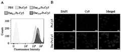
图2为实施例三中不同单抗密度的Dar-Ps-Cy5在697和MV4-11细胞中的内吞情况。Figure 2 shows the endocytosis of Dar-Ps-Cy5 with different monoclonal antibody densities in 697 and MV4-11 cells in Example 3.
图3为实施例三中不同单抗密度的Dar-Ps-Cy5在LP-1细胞中的内吞情况。Figure 3 shows the endocytosis of Dar-Ps-Cy5 with different monoclonal antibody densities in LP-1 cells in Example 3.
图4为实施例四中不同单抗密度的Dar-Ps-DM1、Ps-DM1和游离DM1在697、LP-1、MV4-11和L929细胞中的毒性。Fig. 4 shows the toxicity of Dar-Ps-DM1, Ps-DM1 and free DM1 with different monoclonal antibody densities in Example 4 in 697, LP-1, MV4-11 and L929 cells.
图5为实施例四中不同单抗密度的空囊泡Dar-Ps、Ps和Dar在LP-1细胞中的毒性。Figure 5 shows the toxicity of empty vesicles Dar-Ps, Ps and Dar with different monoclonal antibody densities in Example 4 in LP-1 cells.
图6为实施例五中Dar-Ps-DM1和游离DM1单剂量处理的昆明鼠的体重变化和生存曲线图。Fig. 6 is a graph showing body weight changes and survival curves of Kunming mice treated with a single dose of Dar-Ps-DM1 and free DM1 in Example 5.
图7为实施例五中Dar-Ps-DM1和游离DM1单剂量处理的昆明鼠的血常规测试图。Fig. 7 is a blood routine test chart of Kunming rats treated with a single dose of Dar-Ps-DM1 and free DM1 in Example 5.
图8为实施例七中Dar-Ps-DM1和Ps-DM1在原位LP-1-Luc多发性骨髓瘤模型中的生物分布图。Fig. 8 is a biodistribution diagram of Dar-Ps-DM1 and Ps-DM1 in the orthotopic LP-1-Luc multiple myeloma model in Example 7.
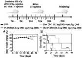
图9为实施例八中原位697急性淋系白血病小鼠移植模型的构建和治疗工作流程图以及Dar-Ps-DM1对其的治疗效果图。9 is a flow chart of the construction and treatment of the orthotopic 697 acute lymphoid leukemia mouse transplantation model in Example 8 and the therapeutic effect of Dar-Ps-DM1 on it.
图10为实施例九中Dar-Ps-DM1对原位LP-1-Luc多发性骨髓瘤模型的治疗流程和治疗效果图。Fig. 10 is a diagram of the treatment process and therapeutic effect of Dar-Ps-DM1 on the orthotopic LP-1-Luc multiple myeloma model in Example 9.
图11为实施例九中原位LP-1-Luc多发性骨髓瘤模型经不同治疗后的Luc信号变化、生存期和体重变化图。Fig. 11 is a graph showing changes in Luc signal, survival period and body weight of the orthotopic LP-1-Luc multiple myeloma model in Example 9 after different treatments.
图12为实施例九中原位LP-1-Luc多发性骨髓瘤模型经不同治疗后的BJP和IgG水平图。FIG. 12 is a graph showing the BJP and IgG levels of the orthotopic LP-1-Luc multiple myeloma model in Example 9 after different treatments.
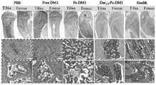
图13为实施例九中原位LP-1-Luc多发性骨髓瘤模型经不同治疗后的腿骨(股骨和胫骨)切片图。Fig. 13 is a section view of leg bones (femur and tibia) of the orthotopic LP-1-Luc multiple myeloma model in Example 9 after different treatments.
图14为实施例九中原位LP-1-Luc多发性骨髓瘤模型经不同治疗后的腿骨TRAP染色图。FIG. 14 is a TRAP staining image of the leg bone of the orthotopic LP-1-Luc multiple myeloma model in Example 9 after different treatments.
具体实施方式Detailed ways
本发明免疫囊泡DM1偶联物,由两亲性嵌段聚合物/官能化两亲性嵌段聚合物自组装、自交联的同时通过巯基硫硫交换反应偶联DM1,并后修饰单抗得到;所述嵌段聚合物的分子链包括依次连接的亲水链段及疏水链段;所述亲水链段为聚乙二醇(PEG),分子量为3000-8000 Da;所述疏水链段为聚碳酸酯链段,分子量为亲水链段分子量的2.1-5.7倍。The immune vesicle DM1 conjugate of the present invention is self-assembled and self-crosslinked by amphiphilic block polymer/functionalized amphiphilic block polymer, and coupled with DM1 through thiol-sulfur-sulfur exchange reaction at the same time, and post-modified single Anti-obtained; the molecular chain of the block polymer includes a hydrophilic segment and a hydrophobic segment connected in sequence; the hydrophilic segment is polyethylene glycol (PEG) with a molecular weight of 3000-8000 Da; the hydrophobic The segment is a polycarbonate segment, and the molecular weight is 2.1-5.7 times that of the hydrophilic segment.
聚乙二醇-b-聚(三亚甲基碳酸酯-co-二硫戊环三亚甲基碳酸酯)(PEG-P(TMC-DTC),Mn = 5.0-(15.1-2.0) kg/mol,Mw/Mn = 1.1)和叠氮官能化的聚合物N3-PEG-P(TMC-DTC)(Mn = 7.9-(15.1-2.0) kg/mol,Mw/Mn = 1.1)为现有产品,根据申请人已经公开的专利或者文献可常规获取。达雷木单抗(Dar,21.7 mg/mL,分子量:148 kDa,上海赛迈生物科技有限公司),NHS-OEG4-DBCO(97%,BroadPharm),美登素(DM1,99.4%,苏州博瑞生物医药有限公司),3-(4,5-二甲基噻唑-2)-2,5-二苯基四氮唑溴盐(MTT,北京索莱宝科技有限公司),4,6-二脒基-2-苯基吲哚(DAPI,碧云天),二硫苏糖醇(DTT,99%,Merck),CCK-8 试剂盒(苏州福麦斯生物科技有限公司),免疫球蛋白G 试剂盒(IgG,上海广锐生物有限公司),本周蛋白BJP 试剂盒(Bence-Jones 蛋白,上海广锐生物有限公司),不同截留分子量(MWCO)的超滤管(Millipore),透析袋(MWCO:3.5 kDa,西安优博生物科技有限公司)购买后直接使用。N,N-二甲基甲酰胺(DMF)、无水乙醇、乙腈(ACN)等试剂均从国药集团化学试剂有限公司购买并直接使用。Polyethylene glycol-b -poly(trimethylene carbonate-co -dithiolantrimethylene carbonate) (PEG-P(TMC-DTC),M n = 5.0-(15.1-2.0) kg/mol ,M w/M n = 1.1) and the azide-functionalized polymer N3-PEG-P(TMC-DTC) (M n = 7.9-(15.1-2.0) kg/mol,M w/M n = 1.1) As an existing product, it can be routinely obtained according to the patents or documents that the applicant has published. Daratumumab (Dar, 21.7 mg/mL, molecular weight: 148 kDa, Shanghai Saimai Biotechnology Co., Ltd.), NHS-OEG4-DBCO (97%, BroadPharm), maytansine (DM1, 99.4%, Suzhou Bo Ruibio Pharmaceutical Co., Ltd.), 3-(4,5-dimethylthiazole-2)-2,5-diphenyltetrazolium bromide salt (MTT, Beijing Solaibao Technology Co., Ltd.), 4,6- Diamidino-2-phenylindole (DAPI, Beyont), dithiothreitol (DTT, 99%, Merck), CCK-8 kit (Suzhou Formes Biotechnology Co., Ltd.), immunoglobulin G kit (IgG, Shanghai Guangrui Biological Co., Ltd.), this week’s protein BJP kit (Bence-Jones protein, Shanghai Guangrui Biological Co., Ltd.), ultrafiltration tubes (Millipore) with different molecular weight cut-offs (MWCO), dialysis bags (MWCO: 3.5 kDa, Xi'an Youbo Biotechnology Co., Ltd.) was purchased and used directly. Reagents such as N,N-dimethylformamide (DMF), absolute ethanol, and acetonitrile (ACN) were purchased from Sinopharm Chemical Reagent Co., Ltd. and used directly.
聚合物囊泡的粒径及粒径分布采用英国马尔文公司的633 nm He-Ne 背散射激光光源的Zeta-sizer Nano-ZS 动态光散射仪(DLS,Malvern 公司)测定。紫外光谱由HITACHIUH5300 双光束紫外可见分光光度计(UV-vis)测定。细胞毒性通过酶标仪(ThermoMultiskan FC)测定570 nm 和450 nm 波长下的吸光值分析得到。聚合物囊泡在细胞中的内吞及细胞内定位采用共聚焦激光扫描显微镜(CLSM,Leica,TCS SP5)和流式细胞仪(BD FACS Calibur)测定。大分子质谱采用芥子酸(SA)基质辅助激光解吸电离飞行时间质谱(MALDI-TOF-MS,Bruker)测定。小鼠活体成像采用近红外成像系统(Caliper IVISLuminaII,Ex 643 nm,Em 668 nm)采集图像并采用Living image 软件分析。H&E 通过倒置显微镜(Eclipse Ci-L,Nikon)拍摄并采用ImageJ 软件分析。DM1 的浓度通过HPLC(Waters1525)测定,检测波长为252 nm,流动相为乙腈和水的混合溶液(v/v =60/40),流速为1.0mL/min,温度为30 ºC。单抗的浓度及接枝率由HPLC(SEC 柱)测定,波长为214 nm,流动相为乙腈和磷酸缓冲溶液(PB,150 mM)的混合液(v/v =10/90)。The particle size and particle size distribution of the polymersomes were measured using a Zeta-sizer Nano-ZS dynamic light scattering instrument (DLS, Malvern Company) with a 633 nm He-Ne backscattering laser source from Malvern Company, UK. The ultraviolet spectrum was measured by a HITACHI UH5300 double-beam ultraviolet-visible spectrophotometer (UV-vis). Cytotoxicity was analyzed by measuring the absorbance at 570 nm and 450 nm wavelengths with a microplate reader (ThermoMultiskan FC). The endocytosis and intracellular localization of polymersomes in cells were determined by confocal laser scanning microscopy (CLSM, Leica, TCS SP5) and flow cytometry (BD FACS Calibur). Macromolecular mass spectrometry was determined by sinapinic acid (SA) matrix-assisted laser desorption ionization time-of-flight mass spectrometry (MALDI-TOF-MS, Bruker). In vivo imaging of mice was collected by a near-infrared imaging system (Caliper IVIS LuminaII, Ex 643 nm, Em 668 nm) and analyzed by Living image software. H&E was taken by an inverted microscope (Eclipse Ci-L, Nikon) and analyzed by ImageJ software. The concentration of DM1 was determined by HPLC (Waters1525), the detection wavelength was 252 nm, the mobile phase was a mixed solution of acetonitrile and water (v/v =60/40), the flow rate was 1.0 mL/min, and the temperature was 30 ºC. The concentration and grafting rate of monoclonal antibody were determined by HPLC (SEC column) with a wavelength of 214 nm, and the mobile phase was a mixture of acetonitrile and phosphate buffer solution (PB, 150 mM) (v/v =10/90).
本发明涉及的原料为现有市售原料,具体的制备方法以及测试方法为本领域常规技术;下面结合实施例和附图对本发明作进一步描述。The raw materials involved in the present invention are existing commercially available raw materials, and the specific preparation methods and testing methods are conventional techniques in the field; the present invention will be further described below in conjunction with the examples and accompanying drawings.
实施例一 可逆交联且生物可降解的囊泡美登素偶联物(N3-Ps-DM1)的制备Example 1 Preparation of reversibly cross-linked and biodegradable vesicular maytansine conjugate (N3 -Ps-DM1)
N3-Ps-DM1通过溶剂置换法制备,其中疏水链段部分的二硫戊环与DM1通过巯基-硫硫交换作用进行化学键合,将药物偶联在囊泡的疏水膜中。N3-Ps-DM1由N3-PEG-P(TMC-DTC)和PEG-P(TMC-DTC)共组装的同时偶联DM1而得到,其中N3-PEG-P(TMC-DTC)的含量为1~10 wt.%。具体地,以含有2 wt.% N3-PEG-P(TMC-DTC)的N3-Ps-DM1的制备为例,称取0.72 mgN3-PEG-P(TMC-DTC)和35.28 mg PEG-P(TMC-DTC)溶解于DMF中(聚合物总浓度为40 mg/mL);将0.4 mL DM1的DMF溶液(10 mg/mL)加入到7.7 mL PB(pH 7.4,10 mM)中混合均匀,在搅拌下向其中注入0.9 mL聚合物溶液,常规搅拌3分钟后,置于37 ºC静置12小时。用PB(pH7.4,10 mM)透析(MWCO = 3.5 kDa)6小时除去有机溶剂后,采用纳滤系统除去游离的DM1即得到N3官能团修饰的Ps-DM1,后续称为Ps-DM1。其中DM1的理论载药量设定为10 wt.%,研究发现所得N3-Ps-DM1的粒径为47 nm,粒径分布~0.13(表1)。通过高效液相色谱法测定其在252 nm波长下的吸光值计算得到DM1的包封率为63.5%,载药量是6.6 wt.%。N3 -Ps-DM1 is prepared by a solvent replacement method, wherein the dithiolane of the hydrophobic segment is chemically bonded to DM1 through sulfhydryl-sulfur-sulfur exchange, and the drug is coupled in the hydrophobic membrane of the vesicle. N3 -Ps-DM1 is obtained by co-assembling N3 -PEG-P(TMC-DTC) and PEG-P(TMC-DTC) and coupling DM1 at the same time, wherein N3 -PEG-P(TMC-DTC) The content is 1~10 wt.%. Specifically, taking the preparation of N3 -Ps-DM1 containing 2 wt.% N3 -PEG-P (TMC-DTC) as an example, weigh 0.72 mg N3 -PEG-P (TMC-DTC) and 35.28 mg PEG -P(TMC-DTC) was dissolved in DMF (
N3-Ps由N3-PEG-P(TMC-DTC)和PEG-P(TMC-DTC)根据上述方法共组装得到。N3 -Ps was co-assembled from N3 -PEG-P(TMC-DTC) and PEG-P(TMC-DTC) according to the above method.
实施例二 单抗导向的免疫囊泡美登素偶联物(Ab-Ps-DM1)的制备Example 2 Preparation of mAb-directed immune vesicle maytansine conjugate (Ab-Ps-DM1)
Ab-Ps-DM1通过在叠氮官能化的聚合物囊泡DM1纳米药物(N3-Ps-DM1)表面后修饰二苯并环辛炔官能化的单抗(Ab-DBCO)得到。首先,为了高效地键合单抗,采用切向流装置将N3-Ps-DM1由4 mg/mL浓缩到19.8 mg/mL。浓缩后N3-Ps-DM1的粒径为48 nm,PDI为0.16。N3-Ps-DM1 的长期储存稳定性通过监测其在4 ºC 放置180天期间的粒径、粒径分布及药物泄漏量进行表征,浓缩后N3-Ps-DM1在4 ºC 储存180 天期间,N3-Ps-DM1的粒径和粒径分布变化不大(3 nm以内),且没有DM1泄漏,表明其具有最佳的长期储存稳定性。在加入10 mMDTT 处理后,DM1以游离的形式快速释放出来(图1A)。另外研究了N3-Ps-DM1在37 ºC,含0.3% 吐温80的PBS中的释放情况,2天后没有检测到游离的DM1,具有优异的稳定性。现有技术公开了FA-PLA-TPGS载美登素纳米粒子FA-DM1-NPs,无论线性还是星形,都具有大于120nm的粒径,且5天内DM1释放达到40%。与通常需要多步制备的聚合物前药纳米体系相比,本发明N3-Ps-DM1制备简单,在囊泡形成过程中,一步法稳定键合DM1,尤其是得到的载药囊泡具有优异的稳定性,为靶向配体后修饰和DM1的特异性靶向递送提供了一个简单稳定的纳米平台。Ab-Ps-DM1 was obtained by post-modification of dibenzocyclooctyne-functionalized monoclonal antibody (Ab-DBCO) on the surface of azide-functionalized polymersome DM1 nanomedicine (N3 -Ps-DM1). First, in order to efficiently bind monoclonal antibodies, N3 -Ps-DM1 was concentrated from 4 mg/mL to 19.8 mg/mL using a tangential flow device. The particle diameter of N3 -Ps-DM1 after concentration is 48 nm, and the PDI is 0.16. The long-term storage stability of N3-Ps-DM1 was characterized by monitoring its particle size, particle size distribution and drug leakage during storage at 4 ºC for 180 days. -Ps-DM1 exhibited little change in particle size and particle size distribution (within 3 nm), and no DM1 leakage, indicating its best long-term storage stability. After treatment with 10 mMDTT, DM1 was rapidly released in the free form (Fig. 1A). In addition, the release of N3 -Ps-DM1 at 37 ºC in PBS containing 0.3
Ab-DBCO通过小分子NHS-OEG4-DBCO与单抗上的氨基发生酰胺化反应制备得到,其中DBCO的官能化度可通过改变Ab与NHS-OEG4-DBCO的摩尔比进行调节。以DBCO官能化Dar单抗(Dar-DBCO)的制备为例,取4 mg Dar(21.7 mg/mL),用pH 8.5的缓冲液将其稀释至10mg/mL,在振荡下向其中加入3倍摩尔当量的NHS-OEG4-DBCO的DMSO溶液(5 mg/mL),氮气下,置于25 ºC,100 rpm摇床中反应过夜。随后用超滤管离心(MWCO = 10 kDa,3000 rpm)除去未反应的NHS-OEG4-DBCO,并用PBS(pH 7.4,10 mM)洗涤超滤两次,得到Dar-DBCO,以上反应示意如下:Ab-DBCO is prepared by the amidation reaction between small molecule NHS-OEG4 -DBCO and the amino group on the monoclonal antibody, wherein the functionalization degree of DBCO can be adjusted by changing the molar ratio of Ab to NHS-OEG4 -DBCO. Taking the preparation of DBCO functionalized Dar monoclonal antibody (Dar-DBCO) as an example, take 4 mg Dar (21.7 mg/mL), dilute it to 10 mg/mL with pH 8.5 buffer, add 3 times to it under shaking The molar equivalent of NHS-OEG4 -DBCO in DMSO (5 mg/mL) was reacted overnight in a shaker at 25 ºC and 100 rpm under nitrogen. Then use an ultrafiltration tube to centrifuge (MWCO = 10 kDa, 3000 rpm) to remove unreacted NHS-OEG4 -DBCO, and wash the ultrafiltration twice with PBS (pH 7.4, 10 mM) to obtain Dar-DBCO. The above reaction is shown below :

飞行时间质谱(MALDI-TOF-MS)测得此投料摩尔比下每个Dar上修饰了1.4个DBCO(附图1B),表示为Dar-DBCO1.4。后续均采用Dar-DBCO1.4及修饰有1.5-2个DBCO的其它单抗进行实验。Time-of-flight mass spectrometry (MALDI-TOF-MS) measured that 1.4 DBCOs were modified on each Dar at this feed molar ratio (Fig. 1B), expressed as Dar-DBCO1.4 . In the subsequent experiments, Dar-DBCO1.4 and other monoclonal antibodies modified with 1.5-2 DBCOs were used for experiments.
通过N3-Ps-DM1表面的N3与Dar-DBCO之间发生张力触动的点击化学反应可简单制备得到Dar-Ps-DM1,Dar的表面密度可通过改变投料比进行调节。设定Dar-DBCO与N3的摩尔比分别为0.5∶1,1∶1和2∶1,即在50.5 μL N3-Ps-DM1(19.8 mg/mL)中分别加入9.2、18.4和36.8 μL的Dar-DBCO溶液(6.84 mg/mL),然后在25 ºC、100 rpm摇床中反应过夜。采用超滤管离心(MWCO = 300 kDa,3000 rpm)除去未键合的Dar-DBCO,并用PBS(pH 7.4,1×)洗涤两次,同时收集Dar-Ps-DM1和下清以测定Dar的键合量。下清中未键合的Dar-DBCO通过HPLC测定,从而计算出每毫克聚合物囊泡表面Dar的含量分别为27.3、64.1和108.0 μg,根据多角度激光光散射测得的聚合物囊泡的绝对重均分子量(1.54×10⁷ g/mol)和聚集数(635个)计算可知每个Dar-Ps-DM1表面分别键合有2.6、6.2和10.5个Dar(表1)。随着Dar密度的增加,Dar-Ps-DM1的粒径略有增加(53-56 nm),粒径分布较窄(PDI:0.21-0.25)。Dar-Ps-DM1 can be easily prepared by tension-triggered click chemical reaction between N3 on the surface of N3 -Ps-DM1 and Dar-DBCO, and the surface density of Dar can be adjusted by changing the feeding ratio. Set the molar ratio of Dar-DBCO to N3 to be 0.5:1, 1:1 and 2:1 respectively, that is, add 9.2, 18.4 and 36.8 μL of N3- Ps-DM1 (19.8 mg/mL) to 50.5 μL Dar-DBCO solution (6.84 mg/mL), and then react overnight at 25 ºC, 100 rpm in a shaker. Unbonded Dar-DBCO was removed by ultrafiltration tube centrifugation (MWCO = 300 kDa, 3000 rpm), and washed twice with PBS (pH 7.4, 1×), while Dar-Ps-DM1 and the supernatant were collected to determine the concentration of Dar. Amount of bonding. The unbonded Dar-DBCO in the supernatant was determined by HPLC, and the Dar content per mg of the polymersome surface was calculated to be 27.3, 64.1 and 108.0 μg, respectively. The calculation of the absolute weight average molecular weight (1.54×10⁷ g/mol) and the number of aggregations (635) shows that each Dar-Ps-DM1 surface has 2.6, 6.2 and 10.5 Dar bonded respectively (Table 1). With the increase of Dar density, the particle size of Dar-Ps-DM1 increased slightly (53-56 nm), and the particle size distribution was narrower (PDI: 0.21-0.25).

a由HPLC测得;b由DLS测得。a Measured by HPLC;b Measured by DLS.
其它单抗导向的聚合物囊泡DM1偶联物,如Isa-Ps-DM1、Anti-BCMA-Ps-DM1的制备方法均与Dar-Ps-DM1类似。其粒径在40-60 nm之间,粒径分布较窄(PDI:0.10-0.30),每个囊泡表面单抗的个数为1-12个。The preparation methods of other mAb-directed polymer vesicle DM1 conjugates, such as Isa-Ps-DM1 and Anti-BCMA-Ps-DM1, are similar to Dar-Ps-DM1. The particle size is between 40-60 nm, the particle size distribution is narrow (PDI: 0.10-0.30), and the number of monoclonal antibodies on the surface of each vesicle is 1-12.
Dar-IPs的制备方法与Dar-IPs-DM1 类似,通过Dar-DBCO与叠氮官能化的空囊泡(N3-Ps)反应得到,最终分散于超纯水中。Dar-IPs表面Dar的二级结构通过圆二色谱(CD)测定,以同浓度的游离Dar作为对照。The preparation method of Dar-IPs is similar to Dar-IPs-DM1, obtained by reacting Dar-DBCO with azide-functionalized empty vesicles (N3-Ps), and finally dispersed in ultrapure water. The secondary structure of Dar on the surface of Dar-IPs was determined by circular dichroism (CD), and free Dar at the same concentration was used as a control.
实施例三 Dar-Ps-DM1的细胞内吞行为Example 3 Endocytosis of Dar-Ps-DM1
由于DM1本身无荧光,采用Cy5标记聚合物囊泡(Dar-Ps-Cy5)并通过流式细胞仪和激光共聚焦显微镜(CLSM)研究不同Dar密度的Dar-Ps-Cy5在697急性淋系白血病细胞、LP-1多发性骨髓瘤细胞及MV4-11白血病细胞中的摄取情况。Ps-Cy5通过2 wt.% N3-PEG-P(TMC-DTC)、20 wt.% PEG-P(TMC-DTC)-Cy5和78 wt.% PEG-P(TMC-DTC)的聚合物共组装制备得到。具体如下:称取0.32 mg N3-PEG-P(TMC-DTC)、3.2 mg PEG-P(TMC-DTC)-Cy5、12.48 mgPEG-P(TMC-DTC)溶解于DMF中(40 mg/mL),将三种聚合物混合均匀后,在搅拌下注入至3.6mL PB(pH 7.4,10 mM)中。搅拌3分钟后,用PB(pH 7.4,10 mM)透析(MWCO = 1 kDa)除去有机溶剂,并浓缩至聚合物浓度为17 mg/mL。Dar-Ps-Cy5的制备方法如同Dar-Ps-DM1类似,设定Dar-DBCO与N3的摩尔比分别为0.5:1,1:1和2:1,以1:1为例,即在100 μL Ps-Cy5(17 mg/mL)中加入36.9 μL的Dar-DBCO溶液(5.5 mg/mL),然后在25 ºC、100 rpm摇床中反应过夜。采用超滤管离心(MWCO = 300 kDa,3000 rpm)除去未键合的Dar-DBCO,并用PBS(pH 7.4,1×)洗涤两次。Since DM1 itself has no fluorescence, Cy5-labeled polymersomes (Dar-Ps-Cy5) were used to study the effect of Dar-Ps-Cy5 with different Dar densities on 697 acute lymphoid leukemia by flow cytometry and confocal laser microscopy (CLSM). cells, LP-1 multiple myeloma cells and MV4-11 leukemia cells. Ps-Cy5 passed through a polymer of 2 wt.% N3 -PEG-P(TMC-DTC), 20 wt.% PEG-P(TMC-DTC)-Cy5 and 78 wt.% PEG-P(TMC-DTC) Prepared by co-assembly. The details are as follows: Weigh 0.32 mg N3 -PEG-P(TMC-DTC), 3.2 mg PEG-P(TMC-DTC)-Cy5, 12.48 mg PEG-P(TMC-DTC) and dissolve them in DMF (40 mg/mL ), after mixing the three polymers evenly, inject them into 3.6mL PB (pH 7.4, 10 mM) under stirring. After stirring for 3 min, dialyze (MWCO = 1 kDa) against PB (pH 7.4, 10 mM) to remove the organic solvent and concentrate to a polymer concentration of 17 mg/mL. The preparation method of Dar-Ps-Cy5 is similar to that of Dar-Ps-DM1, and the molar ratios of Dar-DBCO and N3 are set to 0.5:1, 1:1 and 2:1 respectively, taking 1:1 as an example, that is, in Add 36.9 μL of Dar-DBCO solution (5.5 mg/mL) to 100 μL of Ps-Cy5 (17 mg/mL), and react overnight at 25 ºC, 100 rpm on a shaker. Unbound Dar-DBCO was removed by ultrafiltration (MWCO = 300 kDa, 3000 rpm) and washed twice with PBS (pH 7.4, 1×).
流式实验中,首先将697细胞(5×10⁵个/孔)、LP-1细胞(2×105个/孔)或MV4-11细胞悬液(5×105个/孔)铺在6孔板中,置于培养箱孵育12小时后,每孔加入200 μL Dar-Ps-Cy5和Ps-Cy5(Cy5孔内浓度为2.0 μg/mL),用PBS组作为对照。继续孵育4小时后,离心(800rpm,5分钟)收集细胞,并用PBS清洗两次,最后用500 μL PBS分散并置于流式管中进行测定。测试结果显示,Dar-Ps-Cy5在697和LP-1细胞中的内吞量明显高于Ps-Cy5,其中与Dar6.2-Ps-Cy5孵育的细胞具有最高的荧光强度,其在697和LP-1细胞中的荧光强度分别是Ps-Cy5对照组的5.7倍(附图2A)和3.1倍(附图3A)。然而,在MV4-11细胞中,Dar-Ps-Cy5的摄取量与Ps-Cy5相比无明显区别(附图2B)。In the flow cytometry experiment,
随后采用CLSM进一步研究了Dar6.2-Ps-Cy5和Ps-Cy5在LP-1细胞中的内吞行为。具体实验步骤如下,将多聚赖氨酸(300 μL,0.1 mg/mL)预处理的小圆片置于24孔板中,并加入LP-1细胞悬液(3×105个/孔),于培养箱中培养24小时后,分别加入100 μL Dar6.2-Ps-Cy5和Ps-Cy5(Cy5孔内浓度为40 μg/mL)。继续孵育4小时后小心移去培养基,用PBS洗3次,接着用4%多聚甲醛溶液固定15分钟,用PBS洗3次,再用DAPI染细胞核3分钟,用PBS清洗3次,最后采用甘油封片并用CLSM(Leica,TCS SP5)进行观察和拍摄。附图3B表明,当LP-1细胞与Dar6.2-Ps-Cy5孵育4小时后,细胞核周围呈现明显的Cy5荧光信号,而与Ps-Cy5孵育的细胞中荧光较为微弱,表明Dar的引入可以显著提高细胞对囊泡的内吞,增加囊泡在细胞内的富集。Subsequently, the endocytic behavior of Dar6.2 -Ps-Cy5 and Ps-Cy5 in LP-1 cells was further studied by CLSM. The specific experimental steps are as follows. Place the small discs pretreated with polylysine (300 μL, 0.1 mg/mL) in a 24-well plate, and add LP-1 cell suspension (3×10⁵ cells/well) , after culturing in the incubator for 24 hours, add 100 μL of Dar6.2 -Ps-Cy5 and Ps-Cy5 respectively (concentration in Cy5 wells is 40 μg/mL). After continuing to incubate for 4 hours, remove the medium carefully, wash with
实施例四 Dar-Ps-DM1的细胞毒性实验Example 4 Cytotoxicity experiment of Dar-Ps-DM1
Dar-Ps-DM1在697和LP-1细胞中的采用CCK-8试剂盒进行测定,其中以MV4-11细胞作为对照。将697细胞(18000个/孔)、LP-1细胞(12000个/孔)和MV4-11细胞(15000个/孔)铺于96孔板中,置于37 ºC、含5% CO2的培养箱中培养12小时后,向每孔加入20 μL含有不同Dar表面密度的Dar-Ps-DM1、Ps-DM1和游离DM1。对于697细胞而言,孔内DM1的最终浓度分别为0.01、0.1、0.5、1、5和10 ng/mL;对于LP-1和MV4-11细胞,孔内DM1的最终浓度分别为0.1、0.5、1、2、5、10、50和100 ng/mL。在37 ºC孵育48小时后,每孔加入10 μL CCK-8溶液继续孵育4小时,最后用酶标仪测试其在450 nm处的吸光度值。细胞存活率通过实验组吸光度值与加入PBS培养的细胞的吸光度值的比值计算得到,实验平行进行四组(mean ± SD,n = 4)。附图4为不同靶向密度的Dar-Ps-DM1囊泡纳米药物的细胞毒性结果图。结果表明,在697细胞中当每个囊泡表面键合6.2个Dar时(Dar6.2-Ps-DM1)细胞毒性最强,其半致死浓度(IC50)低至0.31 ng/mL,相比游离DM1(IC50:2.3 ng/mL)和非靶向对照组Ps-DM1(IC50:0.62 ng/mL)分别降低了7.4和2倍(附图4A)。附图4B表明,在LP-1细胞中,当每个囊泡表面键合6.2个Dar时(Dar6.2-Ps-DM1),其细胞毒性最强,IC50为3 ng/mL,相比于非靶向对照组降低了4倍左右。然而在MV4-11细胞中,Dar-Ps-DM1与Ps-DM1的毒性相当,IC50为12 ng/mL(附图4C)。此外,即使在DM1浓度高达1 μg/mL时,对于正常的L929细胞没有明显的杀伤效果(附图4D)。这综合说明Dar的引入有效增加了DM1的靶向细胞内递送和快速释放,进而增强了其体外抗肿瘤活性。The detection of Dar-Ps-DM1 in 697 and LP-1 cells was carried out using CCK-8 kit, and MV4-11 cells were used as the control. Spread 697 cells (18,000 cells/well), LP-1 cells (12,000 cells/well) and MV4-11 cells (15,000 cells/well) in a 96-well plate and culture them at 37 ºC with 5% CO2 After 12 hours of incubation in the chamber, 20 μL of Dar-Ps-DM1, Ps-DM1 and free DM1 containing different Dar surface densities were added to each well. For 697 cells, the final concentrations of DM1 in the wells were 0.01, 0.1, 0.5, 1, 5 and 10 ng/mL; for LP-1 and MV4-11 cells, the final concentrations of DM1 in the wells were 0.1, 0.5 , 1, 2, 5, 10, 50, and 100 ng/mL. After incubating at 37 ºC for 48 hours, 10 μL of CCK-8 solution was added to each well for further incubation for 4 hours, and finally its absorbance at 450 nm was tested with a microplate reader. The cell survival rate was calculated by the ratio of the absorbance value of the experimental group to the absorbance value of the cells cultured with PBS, and the experiment was performed in four groups in parallel (mean ± SD, n = 4). Accompanying drawing 4 is the result graph of the cytotoxicity of Dar-Ps-DM1 vesicle nanomedicine with different targeting densities. The results showed that in 697 cells, when 6.2 Dar was bound to the surface of each vesicle (Dar6.2 -Ps-DM1), the cytotoxicity was the strongest, and its semi-lethal concentration (IC50 ) was as low as 0.31 ng/mL, compared with free DM1 (IC50 : 2.3 ng/mL) and non-targeting control Ps-DM1 (IC50 : 0.62 ng/mL) were decreased by 7.4 and 2 times, respectively (Fig. 4A). Figure 4B shows that in LP-1 cells, when 6.2 Dar was bound to the surface of each vesicle (Dar6.2 -Ps-DM1), its cytotoxicity was the strongest with anIC50 of 3 ng/mL, compared to The non-targeting control group decreased by about 4 times. However, in MV4-11 cells, Dar-Ps-DM1 was as toxic as Ps-DM1 with anIC50 of 12 ng/mL (Fig. 4C). In addition, even when the concentration of DM1 was as high as 1 μg/mL, it had no obvious killing effect on normal L929 cells (Fig. 4D). This comprehensively shows that the introduction of Dar effectively increases the targeted intracellular delivery and rapid release of DM1, thereby enhancing its anti-tumor activity in vitro.
此外,采用同样的方法测试不同靶向密度的Dar-Ps和Ps空囊泡以及游离Dar对LP-1细胞的毒性,结果表明即使当Ps和Dar浓度为10 μg/mL时,其均远远高于相对应的载药囊泡和单抗在IC50值附近的浓度时,细胞存活率均接近100%,没有明显的细胞毒性(附图5)。In addition, the same method was used to test the toxicity of Dar-Ps and Ps empty vesicles with different targeting densities and free Dar on LP-1 cells, and the results showed that even when the concentration of Ps and Dar was 10 μg/mL, they were far When the concentration was higher than that of the corresponding drug-loaded vesicles and monoclonal antibody near the IC50 value, the cell survival rate was close to 100%, and there was no obvious cytotoxicity (Fig. 5).
以下实施例中Dar-Ps-DM1均是指Dar6.2-Ps-DM1囊泡纳米药物,Dar-Ps-Cy5均为Dar6.2-Ps-Cy5。In the following examples, Dar-Ps-DM1 refers to Dar6.2 -Ps-DM1 vesicle nanomedicine, and Dar-Ps-Cy5 refers to Dar6.2 -Ps-Cy5.
实施例五 Dar-Ps-DM1的体内最大耐受剂量(MTD)实验Example 5 In vivo maximum tolerated dose (MTD) experiment of Dar-Ps-DM1
MTD实验是指单剂量下,七天之内不引起小鼠体重下降至原来的85%的一个最大给药剂量,实验选取雌雄相同数量的KM鼠(20-22 g,7周龄),并根据平均体重随机分组。通过尾静脉单剂量注射Dar-Ps-DM1或游离DM1,其中Dar-Ps-DM1制剂的DM1剂量梯度分别为0.8、1.2、1.4和1.6 mg/kg,游离DM1剂量设置为0.4和0.6 mg/kg。在给药后一周内观察小鼠的体重和健康状态(活动状况、瞳孔大小、角膜状态),并在实验结束后,随机从游离DM1组(0.4mg DM1 equiv./kg)和Dar-Ps-DM1组(0.8 mg DM1 equiv./kg、1.2 mg DM1 equiv./kg)中分别取三只小鼠做血常规测试。结果发现,Dar-Ps-DM1在1.2 mg DM1 equiv./kg剂量下,小鼠可较好耐受,全部存活,且血常规各项指标与健康小鼠无显著差异(附图6 & 7),而在DM1剂量达到1.4 mg/kg及以上时,小鼠出现体重减轻、活动减少、瞳孔缩小、角膜混浊甚至死亡。然而,对于游离DM1而言,其在给药剂量为0.6 mg/kg时,小鼠在短时间内快速死亡,可耐受剂量为0.4 mg/kg。上述结果证明Dar-Ps-DM1有效扩大了毒性分子DM1的治疗窗口;但是同样的载体对VCR药物的耐受剂量没有明显改善。美登素具有广谱的抗肿瘤活性,适用于实体瘤和恶性血液肿瘤,然而由于其毒性极强,治疗窗口较窄,无法单独使用,本发明技术方案的出现为强效药DM1的使用提供了可能性。The MTD experiment refers to a single dose that does not cause the mouse body weight to drop to 85% of the original maximum dose within seven days. The experiment selects the same number of male and female KM mice (20-22 g, 7 weeks old), and according to The mean body weight was randomly divided into groups. Dar-Ps-DM1 or free DM1 was injected via the tail vein in a single dose, where the Dar-Ps-DM1 preparation had a DM1 dose gradient of 0.8, 1.2, 1.4, and 1.6 mg/kg, and the free DM1 dose was set at 0.4 and 0.6 mg/kg . The body weight and health status (activity status, pupil size, corneal state) of the mice were observed within one week after administration, and after the experiment was over, the mice were randomly selected from the free DM1 group (0.4mg DM1 equiv./kg) and Dar-Ps- In the DM1 group (0.8 mg DM1 equiv./kg, 1.2 mg DM1 equiv./kg), three mice were selected for routine blood test. It was found that Dar-Ps-DM1 was well tolerated by the mice at a dose of 1.2 mg DM1 equiv./kg, and all survived, and the blood routine indicators were not significantly different from those of healthy mice (Figure 6 & 7) , and when the DM1 dose reached 1.4 mg/kg and above, the mice experienced weight loss, decreased activity, miosis, corneal clouding and even death. However, for free DM1, when the dose was 0.6 mg/kg, the mice died rapidly in a short time, and the tolerated dose was 0.4 mg/kg. The above results proved that Dar-Ps-DM1 effectively expanded the therapeutic window of the toxic molecule DM1; however, the same carrier did not significantly improve the tolerated dose of VCR drugs. Maytansine has broad-spectrum anti-tumor activity and is suitable for solid tumors and malignant hematological tumors. However, due to its strong toxicity and narrow therapeutic window, it cannot be used alone. possibility.
实施例六 荷原位血液肿瘤小鼠模型的构建Example 6 Construction of mouse model bearing orthotopic hematological tumor
所有动物实验及操作均获得苏州大学实验动物中心和苏州大学动物护理和使用委员会的批准。原位B-ALL肿瘤模型的建立:采用6-8周龄、平均体重约为20 g的NOD/SCID雌性小鼠,在第0天采用150 cGy的剂量辐照并通过腹腔注射0.2 mg(1 mg/mL)的anti-CD122抗体对小鼠进行清髓,随后将697细胞(1×105个/只)通过尾静脉注射到小鼠体内。第6天将小鼠随机分组进行治疗,并持续监测小鼠体重、体态变化及生存期。All animal experiments and manipulations were approved by the Experimental Animal Center of Soochow University and the Animal Care and Use Committee of Soochow University. Establishment of orthotopic B-ALL tumor model: NOD/SCID female mice aged 6-8 weeks with an average body weight of about 20 g were irradiated with a dose of 150 cGy on
原位MM肿瘤模型的建立:采用6周龄的NOD/SCID雌性小鼠,首先连续两天通过腹腔注射10 mg/mL的环磷酰胺溶液对小鼠进行清髓,每只小鼠每次注射2 mg,第三天将LP-1-Luc细胞(8×10⁶个/只)通过尾静脉注射到小鼠体内,接种后第10天开始活体成像和治疗,同时对小鼠进行称重。Establishment of an orthotopic MM tumor model: 6-week-old NOD/SCID female mice were used to perform myeloablation by intraperitoneal injection of 10 mg/mL cyclophosphamide solution for two consecutive days. 2 mg. On the third day, LP-1-Luc cells (8×10⁶ cells/mouse) were injected into the mice through the tail vein. On the 10th day after inoculation, in vivo imaging and treatment began, and the mice were weighed at the same time.
实施例七 Dar-Ps-Cy5在荷原位LP-1-Luc多发性骨髓瘤小鼠中的活体成像实验Example 7 In vivo imaging experiment of Dar-Ps-Cy5 in multiple myeloma mice bearing orthotopic LP-1-Luc
Dar-Ps-Cy5在原位LP-1-Luc多发性骨髓瘤小鼠体内的分布情况通过小鼠活体成像分析得到。在小鼠即将发病时,将200 μL Dar-Ps-Cy5和Ps-Cy5溶液(250 µg Cy5equiv./kg)通过尾静脉分别注射到小鼠体内,注射后分别在1、2、4、6、8、10、12、24小时采用异氟烷麻醉小鼠进行活体荧光成像,采用Lumia II软件分析。结果显示,Dar-Ps-Cy5能够高效靶向富集到肿瘤部位,其在小鼠腿部及头颅的荧光信号显著高于非靶向的Ps-Cy5组(附图8)。The distribution of Dar-Ps-Cy5 in orthotopic LP-1-Luc multiple myeloma mice was obtained by in vivo imaging analysis of mice. When the mice were about to get sick, 200 μL Dar-Ps-Cy5 and Ps-Cy5 solutions (250 μg Cy5equiv./kg) were injected into the mice through the tail vein respectively, and after injection, they were respectively injected at 1, 2, 4, 6, Mice were anesthetized with isoflurane at 8, 10, 12, and 24 hours for in vivo fluorescence imaging, and analyzed using Lumia II software. The results showed that Dar-Ps-Cy5 could be efficiently targeted and enriched to the tumor site, and its fluorescence signal in the mouse legs and skull was significantly higher than that of the non-targeted Ps-Cy5 group (Fig. 8).
实施例八 Dar-Ps-DM1在荷原位697急性淋系白血病小鼠中的抗肿瘤效果Example 8 The anti-tumor effect of Dar-Ps-DM1 in bearing orthotopic 697 acute lymphoid leukemia mice
为了研究Dar-Ps-DM1对荷原位697急性淋系白血病小鼠的抗肿瘤效果,在接种后第6天根据平均体重随机分组,开展治疗实验。给药方案为每组0.2 mg DM1 equiv./kg,4天给一针,共4针,表示为Dar-Ps-DM1(0.2 mg DM1 equiv./kg,Q4d),并采用同等DM1剂量的Ps-DM1和游离DM1,以及PBS作为对照,每个组别均有6只荷瘤小鼠。研究发现PBS组和游离DM1治疗组小鼠的697细胞持续快速增长,在接种后21-23天时发病,表现为双腿瘫痪、体重下降并发生死亡。附图9A可以看出,在给药治疗期间(6-18天),Dar-Ps-DM1组、Ps-DM1及游离DM1组小鼠的体重无明显变化,表明它们可有效抑制小鼠体内697细胞的扩散和增殖,且无明显毒副作用。给药结束后,非靶向Ps-DM1和游离DM1组小鼠体重出现下降,并迅速发病死亡,中位生存期分别为24天和23.5天。然而尾静脉注射Dar-Ps-DM1组小鼠仍表现出稳定增长的体重和健康的体态,中位生存期为31天,与非靶向Ps-DM1组(**p<0.01)及PBS组(***p<0.001)均有显著性差异(附图9B)。这些结果综合表明Dar-Ps-DM1可以高效抑制原位急性淋系白血病的病程发展,延长小鼠的无进展生存期。In order to study the anti-tumor effect of Dar-Ps-DM1 on
实施例九 Dar-Ps-DM1在荷原位LP-1-Luc多发性骨髓瘤小鼠中的抗肿瘤效果Example 9 Antitumor effect of Dar-Ps-DM1 in multiple myeloma mice bearing orthotopic LP-1-Luc
为了研究Dar-Ps-DM1和Ps-DM1对原位多发性骨髓瘤小鼠的抗肿瘤效果,在接种后第10天生物发光强度达到1.2×10⁶ p/sec/cm2/sr时,将小鼠随机分为4组,每组10只(其中4只用于生物发光成像,6只用于监测体重及观察生存期),治疗组别分别为PBS、游离DM1(0.2mg DM1 equiv./kg,Q4d)、Ps-DM1 (0.2 mg DM1 equiv./kg,Q4d)、Dar-Ps-DM1 (0.2 mgDM1 equiv./kg,Q4d),尾静脉给药,每4天给一针,共5针。在治疗期间(10-34天),每7天进行一次活体成像监测肿瘤增殖情况,每2天监测一次体重,并通过荧光值、体重和小鼠生存期共同评估治疗情况。在治疗结束后,各组随机解剖四只小鼠,取主要脏器及后腿骨,用4%多聚甲醛固定,采用H&E及TRAP染色进行组织学分析。In order to study the antitumor effect of Dar-Ps-DM1 and Ps-DM1 on orthotopic multiple myeloma mice, when the bioluminescent intensity reached 1.2×10⁶ p/sec/cm2 /sr on the 10th day after inoculation, the Mice were randomly divided into 4 groups, 10 in each group (4 of which were used for bioluminescence imaging, 6 for monitoring body weight and observing survival period), and the treatment groups were PBS, free DM1 (0.2mg DM1 equiv./ kg, Q4d), Ps-DM1 (0.2 mg DM1 equiv./kg, Q4d), Dar-Ps-DM1 (0.2 mgDM1 equiv./kg, Q4d), tail vein administration, one injection every 4 days, a total of 5 Needle. During the treatment period (10-34 days), intravital imaging was performed every 7 days to monitor tumor proliferation, body weight was monitored every 2 days, and the treatment was jointly evaluated by fluorescence value, body weight, and mouse survival. After the treatment, four mice in each group were randomly dissected, and the main organs and hind leg bones were taken out, fixed with 4% paraformaldehyde, and H&E and TRAP staining were used for histological analysis.
研究发现PBS组小鼠的LP-1-Luc细胞持续快速增长,在第28-34天当生物发光强度达到1.0×10⁹ p/sec/cm2/sr时开始发病,表现为双腿瘫痪、体重下降并发生死亡。在10-31天成像观察期间,Dar-Ps-DM1组的Luc信号明显下降,降低至健康小鼠的背景信号值,表明小鼠体内的LP-1-Luc细胞得到了有效清除(附图10,附图11A)。然而,Ps-DM1及游离DM1组小鼠的Luc信号仍成指数倍数增,在第31天时,两组小鼠的Luc信号强度相比开始治疗时分别增加了347和2535倍(附图11A)。值得注意的是,所有治疗组小鼠在给药期间体重无明显变化,表明它们的毒副作用较低(附图11C)。此外,Dar-Ps-DM1治疗组小鼠的生存期得到了显著延长(附图11B),中位生存期为124天,相比于PBS组(35天)、Ps-DM1(45天)和游离DM1组(43天)有显著性差异(****,p<0.0001)。现有Anti-CD38-NPs-BTZ的中位生存期为28天;现有双硫交联聚合物胶束CFZ 纳米药物(A6-PMs-CFZ)在荷皮下LP-1 MM 小鼠中有效抑制了肿瘤的生长,小鼠的中位生存期由PBS、CFZ-CD 和PMs-CFZ 的26、29 和35 天延长至44 天。The study found that the LP-1-Luc cells of the mice in the PBS group continued to grow rapidly, and the disease began when the bioluminescent intensity reached 1.0×10⁹ p/sec/cm2 /sr on the 28th to 34th day, showing paralysis of both legs, Weight loss and death occurred. During the imaging observation period of 10-31 days, the Luc signal in the Dar-Ps-DM1 group decreased significantly, and was reduced to the background signal value of healthy mice, indicating that the LP-1-Luc cells in the mice were effectively cleared (Fig. 10 , Figure 11A). However, the Luc signal of the mice in the Ps-DM1 and free DM1 groups still increased exponentially. On
M蛋白,如单克隆免疫球蛋白(IgG)和本周蛋白(BJP)的过度生成是多发性骨髓瘤病人的诊断标准之一,附图12为ELISA试剂盒测得的小鼠IgG和BJP的变化趋势图。结果发现PBS组小鼠的IgG和BJP水平随着MM的进展(10-30天)不断升高,而Dar-Ps-DM1治疗组的小鼠蛋白水平则维持不变甚至稍有下降。附图13为各治疗组小鼠后腿骨的HE染色图,从图中可以看出PBS和DM1治疗组的小鼠后腿骨中有明显可见的肿瘤浸润,且骨髓腔内的造血细胞大量消失。Ps-DM1组也存在着大量的造血细胞消失,然而,Dar-Ps-DM1组的骨组织形态和造血细胞均与健康小鼠类似,未见明显异常,说明Dar-Ps-DM1有效地抑制了MM的进展。溶骨性病变是MM患者常见的临床表现之一,其中主要是由于破骨细胞与成骨细胞之间的平衡遭到破坏。通过TRAP染色分析发现Dar-Ps-DM1组小鼠腿骨中的破骨细胞含量较少,明显低于PBS、DM1和Ps-DM1治疗组小鼠(附图14)。M protein, such as the overproduction of monoclonal immunoglobulin (IgG) and weekly protein (BJP), is one of the diagnostic criteria for multiple myeloma patients. Figure 12 shows the detection of mouse IgG and BJP by ELISA kit Change trend graph. The results showed that the IgG and BJP levels of the mice in the PBS group continued to increase with the progression of MM (10-30 days), while the protein levels of the mice in the Dar-Ps-DM1 treatment group remained unchanged or even slightly decreased. Accompanying drawing 13 is the HE staining picture of hind leg bone of mice of each treatment group, can see from the figure that there is obvious tumor infiltration in the mouse hind leg bone of PBS and DM1 treatment group, and the hematopoietic cell in bone marrow cavity is a large amount disappear. A large number of hematopoietic cells disappeared in the Ps-DM1 group, however, the bone tissue morphology and hematopoietic cells in the Dar-Ps-DM1 group were similar to those of healthy mice, and no obvious abnormalities were found, indicating that Dar-Ps-DM1 effectively inhibited MM progress. Osteolytic lesions are one of the common clinical manifestations of MM patients, which is mainly due to the disruption of the balance between osteoclasts and osteoblasts. Through TRAP staining analysis, it was found that the amount of osteoclasts in the leg bones of mice in Dar-Ps-DM1 group was less, which was significantly lower than that in mice treated with PBS, DM1 and Ps-DM1 (Fig. 14).
多发性骨髓瘤(MM)是由骨髓(BM)中恶性浆细胞异常增殖而引起的恶性血液肿瘤,复发率高,对于复发的MM 患者,可用的有效治疗方案有限,存活率较低。因此,迫切需要开发新的治疗方案,改善新发及复发难治MM患者的预后,实现深度缓解。本发明实施例结果综合表明Dar的引入显著增加了Ps-DM1的选择性靶向,从而高效清除了LP-1-Luc多发性骨髓瘤细胞,大幅延长了小鼠的生存期。另外,Dar-Ps-DM1可以高效抑制原位急性淋系白血病的病程发展,延长小鼠的无进展生存期。Multiple myeloma (MM) is a hematologic malignancy caused by abnormal proliferation of malignant plasma cells in the bone marrow (BM), with a high recurrence rate. For relapsed MM patients, available effective treatment options are limited and the survival rate is low. Therefore, there is an urgent need to develop new treatment options to improve the prognosis of new-onset and relapsed refractory MM patients and achieve deep remission. The results of the examples of the present invention comprehensively show that the introduction of Dar significantly increases the selective targeting of Ps-DM1, thereby efficiently eliminating LP-1-Luc multiple myeloma cells and greatly prolonging the survival period of mice. In addition, Dar-Ps-DM1 can efficiently inhibit the development of acute lymphoid leukemia in situ and prolong the progression-free survival of mice.
Claims (4)
Translated fromChinese
Priority Applications (1)
| Application Number | Priority Date | Filing Date | Title |
|---|---|---|---|
| CN202110561582.0ACN113244175B (en) | 2021-05-22 | 2021-05-22 | Immune vesicle maytansine conjugate as well as preparation method and application thereof |
Applications Claiming Priority (1)
| Application Number | Priority Date | Filing Date | Title |
|---|---|---|---|
| CN202110561582.0ACN113244175B (en) | 2021-05-22 | 2021-05-22 | Immune vesicle maytansine conjugate as well as preparation method and application thereof |
Publications (2)
| Publication Number | Publication Date |
|---|---|
| CN113244175A CN113244175A (en) | 2021-08-13 |
| CN113244175Btrue CN113244175B (en) | 2022-11-04 |
Family
ID=77183843
Family Applications (1)
| Application Number | Title | Priority Date | Filing Date |
|---|---|---|---|
| CN202110561582.0AActiveCN113244175B (en) | 2021-05-22 | 2021-05-22 | Immune vesicle maytansine conjugate as well as preparation method and application thereof |
Country Status (1)
| Country | Link |
|---|---|
| CN (1) | CN113244175B (en) |
Families Citing this family (2)
| Publication number | Priority date | Publication date | Assignee | Title |
|---|---|---|---|---|
| WO2024235298A1 (en)* | 2023-05-17 | 2024-11-21 | 苏州大学 | Drug co-loaded polymer vesicle, and preparation method therefor and use thereof |
| CN118079022B (en)* | 2024-04-03 | 2024-12-20 | 上海药明合联生物技术有限公司 | Protein-polysaccharide-drug conjugate and application thereof |
Family Cites Families (6)
| Publication number | Priority date | Publication date | Assignee | Title |
|---|---|---|---|---|
| EP1914242A1 (en)* | 2006-10-19 | 2008-04-23 | Sanofi-Aventis | Novel anti-CD38 antibodies for the treatment of cancer |
| US10017545B2 (en)* | 2013-06-03 | 2018-07-10 | University Of Maryland, College Park | Compositions and vaccines comprising vesicles and methods of using the same |
| CN110339368B (en)* | 2016-12-04 | 2022-08-16 | 苏州大学 | Preparation method of reduction-responsive targeting polyethylene glycol-polycarbonate maytansine prodrug micelle |
| CN107096038B (en)* | 2017-04-12 | 2021-06-18 | 苏州大学 | Preparation method of cross-linked nanomedicine based on active reaction type one-step method |
| CN111939129A (en)* | 2020-08-20 | 2020-11-17 | 苏州大学 | Application of small-molecule-drug-carrying polymer vesicle in preparation of drugs for treating acute lymphatic leukemia |
| CN112076159B (en)* | 2020-09-14 | 2023-01-31 | 苏州大学 | Drug-loaded polymer vesicle with asymmetric membrane structure, preparation method and application thereof in preparation of drugs for treating acute myelogenous leukemia |
- 2021
- 2021-05-22CNCN202110561582.0Apatent/CN113244175B/enactiveActive
Also Published As
| Publication number | Publication date |
|---|---|
| CN113244175A (en) | 2021-08-13 |
Similar Documents
| Publication | Publication Date | Title |
|---|---|---|
| Peng et al. | Herceptin-conjugated paclitaxel loaded PCL-PEG worm-like nanocrystal micelles for the combinatorial treatment of HER2-positive breast cancer | |
| Abdellatif et al. | Approved and marketed nanoparticles for disease targeting and applications in COVID-19 | |
| US11478493B2 (en) | Fabrication and application of a hetero-targeted nano-cocktail with traceless linkers | |
| Liu et al. | EGFR-targeted nanobody functionalized polymeric micelles loaded with mTHPC for selective photodynamic therapy | |
| Song et al. | Erythrocyte-biomimetic nanosystems to improve antitumor effects of paclitaxel on epithelial cancers | |
| JP5937966B2 (en) | Particulate hyaluronic acid formulation for cellular delivery of bioactive substances | |
| US20080248097A1 (en) | Polymeric micelles for combination drug delivery | |
| WO2014046630A1 (en) | Tumor targeted liposomal drug delivery system | |
| Nozhat et al. | Advanced biomaterials for human glioblastoma multiforme (GBM) drug delivery | |
| CN113827567B (en) | Application of small molecule drug-loaded polymer vesicles in the preparation of drugs for the treatment of acute lymphoid leukemia | |
| US11622990B2 (en) | VAP polypeptide and use thereof in preparation of drug for targeted diagnosis and treatment of tumor | |
| JP2011105792A (en) | Block copolymer, block copolymer-metal complex composite material, and hollow structure carrier using the same | |
| CN105476975B (en) | Anti- brain tumor drug of active targeting type and preparation method thereof | |
| CN113244175B (en) | Immune vesicle maytansine conjugate as well as preparation method and application thereof | |
| KR20100062990A (en) | Injectable polymer-lipid blend for localized drug delivery | |
| Zhou et al. | Alternative and injectable preformed albumin-bound anticancer drug delivery system for anticancer and antimetastasis treatment | |
| US20190192686A1 (en) | Targeted nanodroplet emulsions for treating cancer | |
| Hunt | Precision targeting of intraperitoneal tumors with peptideguided nanocarriers | |
| CN114917357B (en) | Transferrin-guided PLK1 inhibitor-loaded polymer vesicles, and preparation method and application thereof | |
| Wu et al. | Enhancing chemoimmunotherapy for colorectal cancer with paclitaxel and alantolactone via CD44-Targeted nanoparticles: A STAT3 signaling pathway modulation approach | |
| TW202042843A (en) | Stereocomplexes for the delivery of anti-cancer agents | |
| CN113908276A (en) | Light-controlled drug release nano particle and preparation method and application thereof | |
| Zhang et al. | Multifunctional dendritic Au@ SPP@ DOX nanoparticles integrating chemotherapy and low-dose radiotherapy for enhanced anticancer activity | |
| Ren et al. | Folate-targeting and reduction-responsive nano-delivery system based on curcumin polymer to enhance the anti-cancer activity | |
| KR102574572B1 (en) | Method of preparing alginic acid-folic acid conjugate, the conjugate prepared thereby and pharmaceutical composition comprising the same |
Legal Events
| Date | Code | Title | Description |
|---|---|---|---|
| PB01 | Publication | ||
| PB01 | Publication | ||
| SE01 | Entry into force of request for substantive examination | ||
| SE01 | Entry into force of request for substantive examination | ||
| GR01 | Patent grant | ||
| GR01 | Patent grant |