CN113151420B - One-step fluorescence detection system, and detection method and application of DNA glycosylase activity - Google Patents
One-step fluorescence detection system, and detection method and application of DNA glycosylase activityDownload PDFInfo
- Publication number
- CN113151420B CN113151420BCN202110361586.4ACN202110361586ACN113151420BCN 113151420 BCN113151420 BCN 113151420BCN 202110361586 ACN202110361586 ACN 202110361586ACN 113151420 BCN113151420 BCN 113151420B
- Authority
- CN
- China
- Prior art keywords
- substrate
- template
- detection system
- hogg1
- dna1
- Prior art date
- Legal status (The legal status is an assumption and is not a legal conclusion. Google has not performed a legal analysis and makes no representation as to the accuracy of the status listed.)
- Active
Links
Images


Classifications
- C—CHEMISTRY; METALLURGY
- C12—BIOCHEMISTRY; BEER; SPIRITS; WINE; VINEGAR; MICROBIOLOGY; ENZYMOLOGY; MUTATION OR GENETIC ENGINEERING
- C12Q—MEASURING OR TESTING PROCESSES INVOLVING ENZYMES, NUCLEIC ACIDS OR MICROORGANISMS; COMPOSITIONS OR TEST PAPERS THEREFOR; PROCESSES OF PREPARING SUCH COMPOSITIONS; CONDITION-RESPONSIVE CONTROL IN MICROBIOLOGICAL OR ENZYMOLOGICAL PROCESSES
- C12Q1/00—Measuring or testing processes involving enzymes, nucleic acids or microorganisms; Compositions therefor; Processes of preparing such compositions
- C12Q1/68—Measuring or testing processes involving enzymes, nucleic acids or microorganisms; Compositions therefor; Processes of preparing such compositions involving nucleic acids
- C12Q1/6844—Nucleic acid amplification reactions
- C12Q1/686—Polymerase chain reaction [PCR]
- C—CHEMISTRY; METALLURGY
- C12—BIOCHEMISTRY; BEER; SPIRITS; WINE; VINEGAR; MICROBIOLOGY; ENZYMOLOGY; MUTATION OR GENETIC ENGINEERING
- C12Q—MEASURING OR TESTING PROCESSES INVOLVING ENZYMES, NUCLEIC ACIDS OR MICROORGANISMS; COMPOSITIONS OR TEST PAPERS THEREFOR; PROCESSES OF PREPARING SUCH COMPOSITIONS; CONDITION-RESPONSIVE CONTROL IN MICROBIOLOGICAL OR ENZYMOLOGICAL PROCESSES
- C12Q1/00—Measuring or testing processes involving enzymes, nucleic acids or microorganisms; Compositions therefor; Processes of preparing such compositions
- C12Q1/34—Measuring or testing processes involving enzymes, nucleic acids or microorganisms; Compositions therefor; Processes of preparing such compositions involving hydrolase
Landscapes
- Chemical & Material Sciences (AREA)
- Organic Chemistry (AREA)
- Life Sciences & Earth Sciences (AREA)
- Zoology (AREA)
- Wood Science & Technology (AREA)
- Proteomics, Peptides & Aminoacids (AREA)
- Health & Medical Sciences (AREA)
- Engineering & Computer Science (AREA)
- Microbiology (AREA)
- Biochemistry (AREA)
- Physics & Mathematics (AREA)
- Molecular Biology (AREA)
- Biotechnology (AREA)
- Biophysics (AREA)
- Analytical Chemistry (AREA)
- Immunology (AREA)
- Bioinformatics & Cheminformatics (AREA)
- General Engineering & Computer Science (AREA)
- General Health & Medical Sciences (AREA)
- Genetics & Genomics (AREA)
- Chemical Kinetics & Catalysis (AREA)
- Measuring Or Testing Involving Enzymes Or Micro-Organisms (AREA)
- Investigating Or Analysing Materials By The Use Of Chemical Reactions (AREA)
Abstract
Description
Translated fromChinese技术领域technical field
本发明属于分析检查技术领域,涉及一步法荧光检测体系及DNA糖基化酶活性的检测方法和应用。The invention belongs to the technical field of analysis and inspection, and relates to a one-step fluorescence detection system and a detection method and application of DNA glycosylase activity.
背景技术Background technique
公开该背景技术部分的信息仅仅旨在增加对本发明的总体背景的理解,而不必然被视为承认或以任何形式暗示该信息构成已经成为本领域一般技术人员所公知的现有技术。The disclosure of information in this Background section is only for enhancement of understanding of the general background of the invention and should not necessarily be taken as an acknowledgement or any form of suggestion that this information forms the prior art already known to a person of ordinary skill in the art.
人类8-羟基鸟嘌呤DNA糖基化酶1(hOGG1)是细胞核和线粒体中一种重要的糖基化酶,作用于与胞嘧啶配对的8-氧鸟嘌呤(8-oxoG),启动所有生物体中氧化鸟嘌呤碱基的切除修复过程。hOGG1水平的异常与各种疾病有关,因此准确简便的检测hOGG1的活性在疾病的预测和诊断方面具有重要意义。Human 8-hydroxyguanine DNA glycosylase 1 (hOGG1) is an important glycosylase in the nucleus and mitochondria, acting on 8-oxoguanine (8-oxoG) paired with cytosine to initiate all biological Excision repair process of oxidized guanine bases in vivo. Abnormal levels of hOGG1 are related to various diseases, so accurate and simple detection of hOGG1 activity is of great significance in the prediction and diagnosis of diseases.
据发明人研究了解,近年来,等温指数扩增反应(EXPAR)因其操作简单、反应快、放大效率高而成为一种很有前途的分子诊断技术。在经典的EXPAR实验中,切刻内切酶通常用于循环切割磷酸二酯键,使切刻位点的3’端作为引物不断地启动新一轮复制。然而在EXPAR分析中使用切刻内切酶容易引起不必要的非特异性扩增,这限制了实际应用中检测的可靠性和特异性。另外增加切刻内切酶的数量还会降低EXPAR的反应速率。According to the inventor's research, in recent years, the isothermal exponential amplification reaction (EXPAR) has become a promising molecular diagnostic technology due to its simple operation, fast reaction and high amplification efficiency. In classic EXPAR experiments, nickases are usually used to cyclically cleave phosphodiester bonds, so that the 3' end of the nicking site acts as a primer to continuously initiate a new round of replication. However, the use of nickases in EXPAR assays can easily lead to unwanted non-specific amplification, which limits the reliability and specificity of detection in practical applications. In addition, increasing the number of nickases also reduces the reaction rate of EXPAR.
发明内容SUMMARY OF THE INVENTION
为了解决现有技术的不足,本发明的目的是提供一步法荧光检测体系及DNA糖基化酶活性的检测方法和应用,该检测体系能更简单快速的完成检测;能够进行指数放大反应,提高了hOGG1检测的灵敏度;避免了切刻内切酶的使用,从而有效防止在传统EXPAR系统中观察到的非特异性扩增。In order to solve the deficiencies of the prior art, the purpose of the present invention is to provide a one-step fluorescence detection system and a detection method and application of DNA glycosylase activity, the detection system can complete the detection more simply and quickly; The sensitivity of hOGG1 detection is improved; the use of nickases is avoided, thus effectively preventing the non-specific amplification observed in the traditional EXPAR system.
为了实现上述目的,本发明的技术方案为:In order to achieve the above object, the technical scheme of the present invention is:
一方面,一种一步法荧光检测体系,包括,In one aspect, a one-step fluorescence detection system, comprising,
底物和模板:底物和模板均为单链DNA,底物含有8-氧鸟嘌呤,底物的3’端用NH2修饰;模板由至少一条DNA1和至少一条DNA2交错连接形成,DNA1与DNA2中均不含腺嘌呤,DNA1与DNA2的连接处含有腺嘌呤脱氧核苷酸;底物与DNA1、一部分DNA2互补,与8-氧鸟嘌呤互补的胞嘧啶位于DNA1;Substrate and template: Both the substrate and the template are single-stranded DNA, the substrate contains 8-oxoguanine, and the 3' end of the substrate is modified withNH2 ; the template is formed by at least one DNA1 and at least one DNA2. DNA2 does not contain adenine, and the junction between DNA1 and DNA2 contains adenine deoxynucleotide; the substrate is complementary to DNA1 and a part of DNA2, and the cytosine complementary to 8-oxoguanine is located in DNA1;
APE1酶:用于切割AP位点;APE1 enzyme: used to cut AP sites;
DNA聚合酶和dNTP/dUTP混合物:用于在切除AP位点后3’OH端进行延伸反应,dNTP中不含dTTP;DNA polymerase and dNTP/dUTP mixture: used for extension reaction at 3'OH end after excision of AP site, dNTP does not contain dTTP;
UDG酶:用于清除延伸反应获得延伸产物中的尿嘧啶;UDG enzyme: used to remove uracil in the extension product obtained by the extension reaction;
荧光染料:用于与扩增产物产生荧光。Fluorescent Dyes: Used to generate fluorescence with amplified products.
本发明中,底物的3’端用NH2修饰用于防止非特异性扩增。底物含有8-氧鸟嘌呤,且与8-氧鸟嘌呤互补的胞嘧啶位于DNA1,能够使hOGG1在特定位置识别并切除8-氧鸟嘌呤产生AP位点,通过APE1酶切割产生3’OH端,然后通过DNA聚合酶和dNTP/dUTP混合物进行延伸反应,由于连接处的腺嘌呤脱氧核苷酸,使得尿嘧啶核苷酸掺入延伸产物中,再通过UDG酶识别并切除尿嘧啶,然后通过APE1酶切割产生3’OH端,从而进行扩增,通过荧光染料能够检测到扩增产物,从而实现对hOGG1活性的检测。In the present invention, the 3' end of the substrate is modified with NH2 to prevent non-specific amplification. The substrate contains 8-oxoguanine, and the cytosine complementary to 8-oxoguanine is located in DNA1, which enables hOGG1 to recognize and excise 8-oxoguanine at a specific position to generate AP site, and to generate 3'OH by APE1 enzyme cleavage Then, the extension reaction is carried out by DNA polymerase and dNTP/dUTP mixture. Due to the adenine deoxynucleotide at the junction, the uracil nucleotide is incorporated into the extension product, and the uracil is recognized and excised by UDG enzyme, and then The 3'OH end is generated by APE1 enzyme cleavage, so as to carry out amplification, and the amplification product can be detected by fluorescent dye, so as to realize the detection of hOGG1 activity.
模板中DNA1和DNA2的条数影响扩增重数,例如,当只有一条DNA1和一条DNA2时,只有一个连接处,通过该连接处的腺嘌呤碱基的作用只能进行单重扩增;当存在两条DNA1和一条DNA2时,有两个连接处,即含有2个腺嘌呤碱基,则可以实现三重扩增;而当存在两条DNA1和两条DNA2时,有三个连接处,即含有3个腺嘌呤碱基,则可以实现四重扩增;即DNA1和DNA2的条数影响腺嘌呤碱基的个数,从而影响扩增效率,扩增效率越大,检测效果越好。The number of DNA1 and DNA2 in the template affects the multiplicity of amplification. For example, when there is only one DNA1 and one DNA2, there is only one junction, and only single amplification can be performed through the action of adenine bases at this junction; when When there are two DNA1 and one DNA2, there are two junctions, that is, containing 2 adenine bases, and triple amplification can be achieved; and when there are two DNA1 and two DNA2, there are three junctions, that is, containing With 3 adenine bases, quadruple amplification can be achieved; that is, the number of DNA1 and DNA2 affects the number of adenine bases, thereby affecting the amplification efficiency. The greater the amplification efficiency, the better the detection effect.
另一方面,一种DNA糖基化酶活性的检测方法,将含有hOGG1的待测样品加入上述一步法荧光检测体系进行孵育,然后进行荧光检测。On the other hand, in a method for detecting DNA glycosylase activity, a sample to be tested containing hOGG1 is added to the above-mentioned one-step fluorescence detection system for incubation, and then fluorescence detection is performed.
第三方面,一种DNA糖基化酶活性的检测试剂盒,包括上述一步法荧光检测体系、缓冲溶液。In a third aspect, a DNA glycosylase activity detection kit includes the above-mentioned one-step fluorescence detection system and a buffer solution.
第四方面,一种上述一步法荧光检测体系在筛选hOGG1抑制剂中的应用。In a fourth aspect, an application of the above-mentioned one-step fluorescence detection system in screening hOGG1 inhibitors.
第五方面,一种上述一步法荧光检测体系在制备检测癌细胞试剂中的应用。A fifth aspect provides an application of the above-mentioned one-step fluorescence detection system in the preparation of a reagent for detecting cancer cells.
本发明的有益效果为:The beneficial effects of the present invention are:
1.与传统方法耗时、操作步骤多相比,本发明提供的一步法荧光检测体系在检测hOGG1活性过程中,只需一步操作,总检测时间约为40min,不需要额外的引物探针和修饰分离步骤,具有较高的选择性和灵敏度,同时还具有零背景的特点。1. Compared with the traditional method, which is time-consuming and has many operation steps, the one-step fluorescence detection system provided by the present invention only needs one step of operation in the process of detecting the activity of hOGG1, and the total detection time is about 40min, and no additional primers and probes are required. The modified separation step has high selectivity and sensitivity, and also has the characteristics of zero background.
2.本发明提供的一步法荧光检测体系在检测hOGG1活性过程中利用hOGG1诱导的8-oxoG碱基的高特异性切除和多重循环酶修复扩增的高扩增效率,以快速、简单的混合-读取方式灵敏地检测hOGG1,检测限为2.97×10⁻⁸U/μL。2. The one-step fluorescence detection system provided by the present invention utilizes the high specificity excision of 8-oxoG bases induced by hOGG1 and the high amplification efficiency of multiple cycle enzyme repair amplification in the process of detecting hOGG1 activity. - Sensitive detection of hOGG1 by read method with a detection limit of 2.97×10⁻⁸ U/μL.
3.本发明提供的一步法荧光检测体系还能够用于酶抑制剂的筛选和人癌细胞中hOGG1的检测。由于该一步法荧光检测体系检测的简单性,并且能够提高的检测效率,混合-读取分析是一个很有前途的平台,可以进一步用于开发诊断试剂。3. The one-step fluorescence detection system provided by the present invention can also be used for the screening of enzyme inhibitors and the detection of hOGG1 in human cancer cells. Due to the simplicity of detection and the improved detection efficiency of this one-step fluorescence detection system, the hybrid-read assay is a promising platform that can be further used for the development of diagnostic reagents.
附图说明Description of drawings
构成本发明的一部分的说明书附图用来提供对本发明的进一步理解,本发明的示意性实施例及其说明用于解释本发明,并不构成对本发明的不当限定。The accompanying drawings forming a part of the present invention are used to provide further understanding of the present invention, and the exemplary embodiments of the present invention and their descriptions are used to explain the present invention, and do not constitute an improper limitation of the present invention.
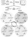
图1为本发明实施例中采用混合-读取法检测hOGG1的机理图。FIG. 1 is a schematic diagram of the mechanism of detecting hOGG1 by the hybrid-reading method in the embodiment of the present invention.
图2为本发明实施例中采用混合-读取法检测hOGG1可行性实验的结果图,(A)聚丙烯酰氨凝胶(PAGE)电泳对hOGG1酶切反应分析,(B)分别在hOGG1+APE1+UDG(实验)、hOGG1+APE1、hOGG1+UDG和APE1+UDG存在下实时监测反应产物的荧光曲线,(C)聚丙烯酰氨凝胶(PAGE)电泳对RCA反应产物分析,(D)分别在hOGG1+APE1+UDG(实验)、hOGG1+APE1、hOGG1+UDG和APE1+UDG存在下测定反应产物的荧光光谱。Figure 2 is the result diagram of the feasibility experiment of detecting hOGG1 by the mixed-reading method in the embodiment of the present invention, (A) polyacrylamide gel (PAGE) electrophoresis analysis of hOGG1 digestion reaction, (B) respectively in hOGG1+ Real-time monitoring of the fluorescence curves of reaction products in the presence of APE1+UDG (experimental), hOGG1+APE1, hOGG1+UDG and APE1+UDG, (C) Analysis of RCA reaction products by polyacrylamide gel (PAGE) electrophoresis, (D) The fluorescence spectra of the reaction products were measured in the presence of hOGG1+APE1+UDG (experimental), hOGG1+APE1, hOGG1+UDG and APE1+UDG, respectively.
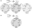
图3为本发明实施例中采用其他模板设计检测hOGG1的机理图,(A)模板-2,(B)模板-3。FIG. 3 is a schematic diagram of the mechanism of detecting hOGG1 using other template designs in the embodiment of the present invention, (A) template-2, (B) template-3.
图4为本发明实施例中采用其他模板设计检测hOGG1的荧光光谱,(A)模板-1,(B)模板-2,(C)模板-3,(D)三种模板(F-F0)/F0值的比较结果。4 is the fluorescence spectrum of hOGG1 detected by other template designs in the embodiment of the present invention, (A) template-1, (B) template-2, (C) template-3, (D) three templates (FF0 )/ Comparison result of F0 value.
图5为本发明实施例中检测hOGG1的优化实验结果图,(A)不同用量模板-1对应的荧光值变化,(B)不同缓冲液组合的荧光值变化,(C)不同用量APE1酶对应的荧光值变化,(D)不同用量UDG酶对应的荧光值变化,(E)不同用量KF酶对应的荧光值变化,(F)不同浓度dNTP/dUTP对应的荧光值变化,(G)不同反应时间对应的荧光值的变化。Figure 5 is a graph of the results of an optimization experiment for detecting hOGG1 in the embodiment of the present invention, (A) changes in fluorescence values corresponding to different dosages of template-1, (B) changes in fluorescence values with different buffer combinations, (C) corresponding to different dosages of APE1 enzyme (D) The fluorescence value changes corresponding to different dosages of UDG enzyme, (E) The fluorescence value changes corresponding to different dosages of KF enzyme, (F) The fluorescence value changes corresponding to different concentrations of dNTP/dUTP, (G) Different reactions Changes in fluorescence values corresponding to time.
图6为本发明实施例中检测hOGG1的灵敏度和特异性的检测结果图,(A)随hOGG1浓度变化的荧光光谱曲线,(B)荧光强度随hOGG1浓度变化的曲线,插图显示在1×10⁻⁷到5×10⁻⁵U/μL范围内荧光强度与hOGG1浓度的对数呈线性相关,(C)对0.02U/μL hOGG1、0.02g/L牛血清白蛋白、0.02g/L IgG、0.02U/μL AAG、0.02U/μL TDG和反应缓冲液(对照)的条件下进行荧光光谱测定。Figure 6 is a graph showing the detection results of the sensitivity and specificity of detecting hOGG1 in the embodiment of the present invention, (A) the fluorescence spectrum curve with the change of hOGG1 concentration, (B) the curve of the fluorescence intensity with the change of hOGG1 concentration, the inset is shown at 1×10 The fluorescence intensity was linearly correlated with the logarithm of hOGG1 concentration in the range of-7 to 5×10⁻⁵ U/μL, (C) for 0.02U/μL hOGG1, 0.02g/L bovine serum albumin, 0.02g/L IgG, Fluorescence spectrometry was performed under the conditions of 0.02U/μL AAG, 0.02U/μL TDG and reaction buffer (control).
图7为本发明实施例中不同浓度O8抑制剂对应的hOGG1相对活性结果图。FIG. 7 is a graph showing the relative activity results of hOGG1 corresponding to different concentrations of O8 inhibitors in the embodiment of the present invention.
图8为本发明实施例对实际样品检测的结果图,(A)测量A549、HeLa、HL-7702、HEK-293T细胞提取物和热失活A549细胞提取物的荧光强度,(B)荧光强度与A549细胞数呈线性关系。Figure 8 is a graph of the results of the actual sample detection in the embodiment of the present invention, (A) measuring the fluorescence intensity of A549, HeLa, HL-7702, HEK-293T cell extracts and heat-inactivated A549 cell extracts, (B) fluorescence intensity There is a linear relationship with the number of A549 cells.
具体实施方式Detailed ways
应该指出,以下详细说明都是示例性的,旨在对本发明提供进一步的说明。除非另有指明,本文使用的所有技术和科学术语具有与本发明所属技术领域的普通技术人员通常理解的相同含义。It should be noted that the following detailed description is exemplary and intended to provide further explanation of the invention. Unless otherwise defined, all technical and scientific terms used herein have the same meaning as commonly understood by one of ordinary skill in the art to which this invention belongs.
需要注意的是,这里所使用的术语仅是为了描述具体实施方式,而非意图限制根据本发明的示例性实施方式。如在这里所使用的,除非上下文另外明确指出,否则单数形式也意图包括复数形式,此外,还应当理解的是,当在本说明书中使用术语“包含”和/或“包括”时,其指明存在特征、步骤、操作、器件、组件和/或它们的组合。It should be noted that the terminology used herein is for the purpose of describing specific embodiments only, and is not intended to limit the exemplary embodiments according to the present invention. As used herein, unless the context clearly dictates otherwise, the singular is intended to include the plural as well, furthermore, it is to be understood that when the terms "comprising" and/or "including" are used in this specification, it indicates that There are features, steps, operations, devices, components, and/or combinations thereof.
鉴于切刻内切酶容易引起不必要的非特异性扩增而且影响EXPAR的反应速率,本发明提出了一步法荧光检测体系及DNA糖基化酶活性的检测方法和应用。Considering that the nickase easily causes unnecessary non-specific amplification and affects the reaction rate of EXPAR, the present invention proposes a one-step fluorescence detection system and a detection method and application of DNA glycosylase activity.
本发明的一种典型实施方式,提供了一种一步法荧光检测体系,包括,A typical embodiment of the present invention provides a one-step fluorescence detection system, including:
底物和模板:底物和模板均为单链DNA,底物含有8-氧鸟嘌呤,底物的3’端用NH2修饰;模板由至少一条DNA1和至少一条DNA2交错连接形成,DNA1与DNA2中均不含腺嘌呤,DNA1与DNA2的连接处含有腺嘌呤脱氧核苷酸;底物与DNA1、一部分DNA2互补,与8-氧鸟嘌呤互补的胞嘧啶位于DNA1;Substrate and template: Both the substrate and the template are single-stranded DNA, the substrate contains 8-oxoguanine, and the 3' end of the substrate is modified withNH2 ; the template is formed by at least one DNA1 and at least one DNA2. DNA2 does not contain adenine, and the junction between DNA1 and DNA2 contains adenine deoxynucleotide; the substrate is complementary to DNA1 and a part of DNA2, and the cytosine complementary to 8-oxoguanine is located in DNA1;
APE1酶:用于切割AP位点;APE1 enzyme: used to cut AP sites;
DNA聚合酶和dNTP/dUTP混合物:用于在切除AP位点后3’OH端进行延伸反应,dNTP中不含dTTP;DNA polymerase and dNTP/dUTP mixture: used for extension reaction at 3'OH end after excision of AP site, dNTP does not contain dTTP;
UDG酶:用于清除延伸反应获得延伸产物中的尿嘧啶;UDG enzyme: used to remove uracil in the extension product obtained by the extension reaction;
荧光染料:用于与扩增产物产生荧光。Fluorescent Dyes: Used to generate fluorescence with amplified products.
底物的3’端用NH2修饰用于防止非特异性扩增。底物含有8-氧鸟嘌呤,且与8-氧鸟嘌呤互补的胞嘧啶位于DNA1,能够使hOGG1在特定位置识别请切除8-氧鸟嘌呤产生AP位点,通过APE1酶切割产生3’OH端,然后通过DNA聚合酶和dNTP/dUTP混合物进行延伸反应,由于连接处的腺嘌呤脱氧核苷酸,使得尿嘧啶核苷酸掺入延伸产物中,再通过UDG酶识别请切除尿嘧啶,然后通过APE1酶切割产生3’OH端,从而进行扩增,通过荧光染料能够检测到扩增产物,从而实现对hOGG1活性的检测。The3 ' end of the substrate was modified with NH to prevent non-specific amplification. The substrate contains 8-oxoguanine, and the cytosine complementary to 8-oxoguanine is located in DNA1, which enables hOGG1 to recognize at a specific position. Please excise 8-oxoguanine to generate AP site, and generate 3'OH by APE1 enzyme cleavage Then, the extension reaction is carried out by DNA polymerase and dNTP/dUTP mixture. Due to the adenine deoxynucleotide at the junction, the uracil nucleotide is incorporated into the extension product, and then recognized by UDG enzyme. Please remove the uracil, and then The 3'OH end is generated by APE1 enzyme cleavage, so as to carry out amplification, and the amplification product can be detected by fluorescent dye, so as to realize the detection of hOGG1 activity.
模板中DNA1和DNA2的条数影响扩增重数,例如,当只有一条DNA1和一条DNA2时,只有一个连接处,通过该连接处的腺嘌呤碱基的作用只能进行单重扩增;当存在两条DNA1和一条DNA2时,有两个连接处,即含有2个腺嘌呤碱基,则可以实现三重扩增;而当存在两条DNA1和两条DNA2时,有三个连接处,即含有3个腺嘌呤碱基,则可以实现四重扩增;即DNA1和DNA2的条数影响腺嘌呤碱基的个数,从而影响扩增效率,扩增效率越大,检测效果越好。因而该实施方式的一些实施例中,模板由两条DNA1和两条DNA2交错连接形成。The number of DNA1 and DNA2 in the template affects the multiplicity of amplification. For example, when there is only one DNA1 and one DNA2, there is only one junction, and only single amplification can be performed through the action of adenine bases at this junction; when When there are two DNA1 and one DNA2, there are two junctions, that is, containing 2 adenine bases, and triple amplification can be achieved; and when there are two DNA1 and two DNA2, there are three junctions, that is, containing With 3 adenine bases, quadruple amplification can be achieved; that is, the number of DNA1 and DNA2 affects the number of adenine bases, thereby affecting the amplification efficiency. The greater the amplification efficiency, the better the detection effect. Thus, in some examples of this embodiment, the template is formed by staggered connection of two DNA1 and two DNA2.
该实施方式的一些实施例中,所述DNA聚合酶为Klenow大片段聚合酶。In some embodiments of this embodiment, the DNA polymerase is Klenow Large Fragment Polymerase.
使Make
该实施方式的一些实施例中,荧光染料为SYBR Green II。In some examples of this embodiment, the fluorescent dye is SYBR Green II.
该实施方式的一些实施例中,所述底物的核苷酸序列如SEQ ID NO.1所示,所述模板的核苷酸序列如SEQ ID NO.2所示。In some examples of this embodiment, the nucleotide sequence of the substrate is shown in SEQ ID NO.1, and the nucleotide sequence of the template is shown in SEQ ID NO.2.
经过优化实验表明,模板的量会影响荧光强度,从而影响检测结果,该实施方式的一些实施例中,底物的浓度为10nM时,模板的浓度为14~16nM。采用该浓度的模板时,荧光检测的荧光强度更好。The optimization experiment shows that the amount of the template will affect the fluorescence intensity, thereby affecting the detection result. In some examples of this embodiment, when the concentration of the substrate is 10 nM, the concentration of the template is 14-16 nM. When using this concentration of template, the fluorescence intensity of fluorescence detection is better.
APE1酶的用量也会影响荧光强度,该实施方式的一些实施例中,底物的量为2×10⁻⁴nmol时,APE1酶的用量为0.75~0.85U。采用该用量的APE1酶时,荧光检测的荧光强度更好。The amount of APE1 enzyme will also affect the fluorescence intensity. In some examples of this embodiment, when the amount of substrate is 2×10⁻⁴ nmol, the amount of APE1 enzyme is 0.75-0.85U. When the amount of APE1 enzyme is used, the fluorescence intensity of fluorescence detection is better.
UDG酶的用量也会影响荧光强度,该实施方式的一些实施例中,底物的量为2×10⁻⁴nmol时,UDG酶的用量不低于2U。当2U后不会有很大的增加,因而UDG酶的用量优选2.0~2.2U,在保证检测荧光强度的前提下,减少UDG酶的添加。The amount of UDG enzyme also affects the fluorescence intensity. In some examples of this embodiment, when the amountof substrate is 2×10⁻⁴ nmol, the amount of UDG enzyme is not less than 2U. When it is 2U, there will not be a great increase, so the dosage of UDG enzyme is preferably 2.0-2.2U. On the premise of ensuring the detection fluorescence intensity, the addition of UDG enzyme should be reduced.
Klenow大片段聚合酶的用量也会影响荧光强度,该实施方式的一些实施例中,底物的量为2×10⁻⁴nmol时,Klenow大片段聚合酶的用量为1.9~2.1U。实验表明,该条件下,荧光检测的荧光强度更好。The amount of Klenow large fragment polymerase also affects the fluorescence intensity. In some examples of this embodiment, when the amount of substrate is 2×10⁻⁴ nmol, the amount of Klenow large fragment polymerase is 1.9-2.1 U. Experiments show that under this condition, the fluorescence intensity of fluorescence detection is better.
dNTP/dUTP混合物的浓度也会影响荧光强度,该实施方式的一些实施例中,底物的浓度为10nM时,dNTP/dUTP混合物的浓度为58~62μM。实验表明,该条件下,荧光检测的荧光强度更好。The concentration of the dNTP/dUTP mixture also affects the fluorescence intensity. In some examples of this embodiment, when the concentration of the substrate is 10 nM, the concentration of the dNTP/dUTP mixture is 58-62 μM. Experiments show that under this condition, the fluorescence intensity of fluorescence detection is better.
本发明的另一种实施方式,提供了一种DNA糖基化酶活性的检测方法,将含有hOGG1的待测样品加入上述一步法荧光检测体系进行孵育,然后进行荧光检测。Another embodiment of the present invention provides a method for detecting DNA glycosylase activity. The sample to be tested containing hOGG1 is added to the above-mentioned one-step fluorescence detection system for incubation, and then fluorescence detection is performed.
该实施方式的一些实施例中,孵育温度为35~39℃。In some examples of this embodiment, the incubation temperature is 35-39°C.
经过实验表明,当孵育时间不低于40min时,检测效果更好。而当孵育时间为40~40.5min时,不仅能够保证检测效果,而且能够降低检测时间。Experiments show that the detection effect is better when the incubation time is not less than 40min. When the incubation time is 40-40.5 min, not only the detection effect can be ensured, but also the detection time can be reduced.
该实施方式的一些实施例中,缓冲液为缓冲液A与缓冲液B的组合,所述缓冲液A包括浓度为18~22mM、pH为7.9~8.1的Tris-HCl,0.9~1.1mM的EDTA,0.9~1.1mM的DTT,0.09~0.11mg/ml的BSA;缓冲液B包括45~55mM醋酸钾(potassium acetate),18~22mMTris-acetate、9~11mM醋酸镁(magnesium acetate)、0.9~1.1mM DTT。In some examples of this embodiment, the buffer is a combination of buffer A and buffer B, and the buffer A includes Tris-HCl at a concentration of 18-22 mM, pH 7.9-8.1, and EDTA at 0.9-1.1 mM , 0.9~1.1mM DTT, 0.09~0.11mg/ml BSA; Buffer B includes 45~55mM potassium acetate (potassium acetate), 18~22mM Tris-acetate, 9~11mM magnesium acetate (magnesium acetate), 0.9~1.1 mM DTT.
该实施方式的一些实施例中,荧光检测中,采用的激发波长为487~489纳米。In some examples of this embodiment, in the fluorescence detection, the excitation wavelength used is 487-489 nanometers.
本发明的第三种实施方式,提供了一种DNA糖基化酶活性的检测试剂盒,包括上述一步法荧光检测体系、缓冲溶液。The third embodiment of the present invention provides a detection kit for DNA glycosylase activity, comprising the above-mentioned one-step fluorescence detection system and a buffer solution.
本发明的第四种实施方式,提供了一种上述一步法荧光检测体系在筛选hOGG1抑制剂中的应用。The fourth embodiment of the present invention provides an application of the above-mentioned one-step fluorescence detection system in screening hOGG1 inhibitors.
本发明的第五种实施方式,提供了一种上述一步法荧光检测体系在制备检测癌细胞试剂中的应用。The fifth embodiment of the present invention provides an application of the above-mentioned one-step fluorescence detection system in the preparation of a reagent for detecting cancer cells.
具体的,所述癌细胞为A549细胞和/或HeLa细胞。Specifically, the cancer cells are A549 cells and/or HeLa cells.
为了使得本领域技术人员能够更加清楚地了解本发明的技术方案,以下将结合具体的实施例详细说明本发明的技术方案。In order to enable those skilled in the art to understand the technical solutions of the present invention more clearly, the technical solutions of the present invention will be described in detail below with reference to specific embodiments.
实施例Example
实施例中采用的核苷酸序列如表1所示。The nucleotide sequences used in the examples are shown in Table 1.
表1 核苷酸序列Table 1 Nucleotide sequences


底物中,带下划线的字母O表示8-oxoG。模板中,下划线处表示与底物互补的部分。In the substrate, the underlined letter O represents 8-oxoG. In the template, the underlined part indicates the part complementary to the substrate.
hOGG1的一步法检测:整个反应在20微升的溶液中进行,其中包括10nM底物、15nM模板-1、不同浓度的hOGG1、0.8U的APE1、2U的UDG、2U的Klenow大片段聚合酶、60μM的dNTP/dUTP混合物(dATP、dGTP、dCTP、dUTP,各物质的浓度均为60μM)、缓冲液A(20mMTris-HCl、1mMEDTA、1mM DTT、0.1mg/ml BSA、pH 8.0)、缓冲液B(50mM醋酸钾、20mMTris-Ac、10mM醋酸镁、1mM DTT、pH 7.9)。在37摄氏度条件下反应40分钟后,扩增产物与SYBR Green II混合,稀释至最终体积50微升,在激发波长为488纳米时,用荧光光谱仪在502-650纳米之间记录光谱。用525纳米处的最大荧光发射强度进行数据分析。One-step detection of hOGG1: The entire reaction was performed in 20 μl of solution including 10 nM substrate, 15 nM template-1, various concentrations of hOGG1, 0.8U of APE1, 2U of UDG, 2U of Klenow large fragment polymerase, 60 μM dNTP/dUTP mixture (dATP, dGTP, dCTP, dUTP, each at a concentration of 60 μM), buffer A (20 mM Tris-HCl, 1 mM EDTA, 1 mM DTT, 0.1 mg/ml BSA, pH 8.0), buffer B (50 mM potassium acetate, 20 mM Tris-Ac, 10 mM magnesium acetate, 1 mM DTT, pH 7.9). After 40 minutes of reaction at 37°C, the amplified product was mixed with SYBR Green II, diluted to a final volume of 50 μl, and the spectrum was recorded between 502-650 nm with a fluorescence spectrometer at an excitation wavelength of 488 nm. Data analysis was performed with the maximum fluorescence emission intensity at 525 nm.
凝胶电泳实验:扩增反应后,产物与荧光指示剂(SYBR Gold)混合加入10%的聚丙烯酰胺凝胶中,凝胶放入三羟甲基氨基甲烷-硼酸(TBE缓冲液:89mMTris-HCl、89mM boricacid、2mM EDTA,pH 8.3)中,在110V恒压室温条件下电泳40min。凝胶由ChemiDoc MP成像系统分析。为了研究hOGG1单独的切割修复过程,采用EPI-Green光源(520-545纳米激发)和577-613纳米滤光片对Cy3标记底物的反应产物进行了10%聚丙烯酰胺凝胶电泳分析。Gel electrophoresis experiment: After the amplification reaction, the product was mixed with a fluorescent indicator (SYBR Gold) and added to a 10% polyacrylamide gel, and the gel was placed in tris-boronic acid (TBE buffer: 89mM Tris- HCl, 89 mM boricacid, 2 mM EDTA, pH 8.3), electrophoresed for 40 min under constant pressure at 110 V at room temperature. Gels were analyzed by ChemiDoc MP imaging system. In order to study the cleavage repair process of hOGG1 alone, the reaction products of Cy3-labeled substrates were analyzed by 10% polyacrylamide gel electrophoresis using EPI-Green light source (520-545 nm excitation) and 577-613 nm filter.
抑制剂实验:将不同浓度的O8抑制剂与0.02U/μL的hOGG1及其他反应试剂在37℃进行反应,随后进行荧光测量。Inhibitor experiment: Different concentrations of O8 inhibitors were reacted with 0.02U/μL of hOGG1 and other reagents at 37°C, followed by fluorescence measurement.
细胞培养和细胞提取:在DMEM培养基中培养肺腺癌细胞系A549(或HeLa细胞、HL-7702细胞、HEK-293T细胞),培养基中加入10%胎牛血清和1%青霉素链霉素。培养基置于37℃,含有5%二氧化碳的培养箱中培养至细胞成熟。待细胞成熟,使用核提取试剂盒(ActiveMotif)根据说明书进行细胞提取物的提取。得到的提取物用于随后的检测。Cell culture and cell extraction: Lung adenocarcinoma cell line A549 (or HeLa cells, HL-7702 cells, HEK-293T cells) was cultured in DMEM medium supplemented with 10% fetal bovine serum and 1% penicillin-streptomycin . The medium was placed in an incubator containing 5% carbon dioxide at 37°C until the cells matured. After the cells were mature, the cell extracts were extracted using a nuclear extraction kit (ActiveMotif) according to the instructions. The resulting extract was used for subsequent assays.
四重扩增检测机理图如图1所示,首先设计了一种8-oxoG碱基修饰的单链DNA底物,其3’端用NH2修饰,从而可以防止非特异性扩增。底物与模板-1杂交,形成部分双链DNA,其中的8-oxoG/C碱基对可用于识别hOGG1。在hOGG1存在的情况下,hOGG1可以高效地与识别位点结合,从而从8-oxoG/C碱基对中切除受损的8-oxoG碱基,产生AP位点。AP位点可被APE1切割,导致切割底物3’OH末端的产生。当存在DNA聚合酶和dNTP/dUTP混合物(即dATP、dGTP、dCTP和dUTP)时,延伸反应在3’OH启动,并将尿嘧啶(U)核苷酸掺入延伸产物中,形成两个损伤位点。尿嘧啶损伤可通过UDG清除,产生AP位点。随后APE1切割这些AP位点,释放两种触发链(触发链-X,触发链-Y)。切割底物与模板-1的杂交能促进另一轮酶修复扩增,并产生大量的触发链(第一阶段)。生成的触发链-X可以继续与游离模板-1杂交,产生包含三个损伤位点的延伸产物,它在UDG和APE1的协同作用下导致新的循环酶修复扩增过程,释放了大量的触发链-X和触发链-Y(第二阶段);生成的触发链-Y可以继续与游离模板-1杂交,产生包含两个损伤位点的延伸产物,导致新的循环酶修复扩增过程,也释放了大量的触发链-X和触发链-Y(第三阶段)。同时,产生的触发链-X还可以模板-1结合产生不同于第二阶段的新的dsDNA(第四阶段)。hOGG1的存在可以诱导多轮酶修复扩增,包括切割底物、触发链-X和触发链-Y启动的扩增,最终导致指数级扩增和越来越多的触发链的产生。用SYBR Green II作为荧光染料可以简便地检测到扩增产物。与之相反,在没有hOGG1的情况下,不会发生8-oxoG碱基切除反应,也不会形成含3’OH末端的切割底物,从而无法导致酶修复扩增发生,因而不能观察到SYBR Green II信号。The schematic diagram of the quadruple amplification detection mechanism is shown in Fig. 1. First, an 8-oxoG base-modified single-stranded DNA substrate was designed, and its 3' end was modified withNH2 , which could prevent non-specific amplification. The substrate hybridizes to template-1 to form partially double-stranded DNA, in which the 8-oxoG/C base pair can be used to recognize hOGG1. In the presence of hOGG1, hOGG1 can efficiently bind to the recognition site, thereby excising the damaged 8-oxoG base from the 8-oxoG/C base pair, creating an AP site. The AP site can be cleaved by APE1, resulting in the creation of the 3'OH end of the cleavage substrate. In the presence of DNA polymerase and a dNTP/dUTP mixture (i.e., dATP, dGTP, dCTP, and dUTP), the extension reaction is initiated at the 3'OH and incorporates uracil (U) nucleotides into the extension product, forming two lesions site. Uracil damage can be cleared by UDG, resulting in AP sites. APE1 then cleaves these AP sites, releasing both trigger chains (trigger-X, trigger-Y). Hybridization of the cleavage substrate to template-1 promotes another round of enzymatic repair amplification and generates a large number of trigger strands (first stage). The resulting trigger-strand-X can continue to hybridize with free template-1 to generate an extension product containing three damage sites, which leads to a new cycle of enzymatic repair amplification under the synergistic action of UDG and APE1, releasing a large number of triggers Strand-X and trigger-strand-Y (second stage); the resulting trigger-strand-Y can continue to hybridize with the free template-1, resulting in an extension product containing both sites of damage, leading to a new cycle of enzymatic repair of the amplification process, Also released a lot of Trigger Chain-X and Trigger Chain-Y (Phase 3). At the same time, the generated trigger strand-X can also combine with template-1 to generate new dsDNA different from the second stage (fourth stage). The presence of hOGG1 induces multiple rounds of enzymatic repair amplification, including substrate cleavage, trigger-X and trigger-Y-initiated amplification, ultimately leading to exponential amplification and the production of increasingly more trigger strands. Amplification products can be easily detected using SYBR Green II as a fluorescent dye. In contrast, in the absence of hOGG1, no 8-oxoG base excision reaction occurs, and no cleavage substrate with a 3'OH terminus is formed, thereby causing no enzymatic repair amplification to occur, and SYBR cannot be observed Green II signal.
可行性实验Feasibility experiment
如图2A,为了验证该测定的可行性,使用凝胶电泳分析来验证实验的可行性。用一个8-oxoG碱基修饰且含Cy3标记的单链DNA底物(图2A,泳道5)来研究hOGG1的切割过程。反应产物用直接激发Cy3进行分析。在没有hOGG1的情况下,只观察到一条107nt带(它包含Cy3标记的底物和模板-1),表明没有发生切割反应(图2A,泳道1和2)。在hOGG1存在的情况下,观察到一条新的5nt条带,表明hOGG1可以识别和切割含有8-oxoG的底物(图2A,泳道3和4)。As shown in Figure 2A, to verify the feasibility of this assay, gel electrophoresis analysis was used to verify the feasibility of the experiment. The cleavage process of hOGG1 was investigated using an 8-oxoG base modified single-stranded DNA substrate containing Cy3 label (FIG. 2A, lane 5). The reaction products were analyzed with direct excitation Cy3. In the absence of hOGG1, only a 107nt band (which contains Cy3-labeled substrate and template-1) was observed, indicating that no cleavage reaction occurred (Fig. 2A,
为了探讨UDG酶在循环酶修复扩增反应中的必要性,在hOGG1、APE1和UDG三种酶参与下,进行了实时荧光检测。如图2B,在没有hOGG1或APE1的情况下,不能观察到荧光信号。在有hOGG1和APE1但没有UDG酶的情况下,由于单独发生聚合反应,观察到相对较弱的荧光信号。当存在hOGG1、APE1和UDG酶时,观察到较强的荧光信号且荧光信号随着反应时间的延长而增强,表明循环酶修复扩增反应以指数放大的方式进行。In order to explore the necessity of UDG enzyme in the cycle enzyme repair amplification reaction, real-time fluorescence detection was carried out with the participation of three enzymes hOGG1, APE1 and UDG. As shown in Figure 2B, in the absence of hOGG1 or APE1, no fluorescent signal could be observed. In the presence of hOGG1 and APE1 but without the UDG enzyme, a relatively weak fluorescence signal was observed due to the separate polymerization reaction. In the presence of hOGG1, APE1 and UDG enzymes, strong fluorescence signals were observed and the fluorescence signals increased with the prolongation of reaction time, indicating that the cyclic enzyme repair amplification reaction proceeded in an exponentially amplified manner.
如图2C,以SYBR Gold为指示剂,对hOGG1诱导的扩增产物进行PAGE分析。用20nt合成的触发链-X(图2C,泳道6)和21nt合成的触发链-Y(图2C,泳道5)来指示循环酶修复扩增的产物位置。只有在hOGG1、APE1和UDG酶存在的情况下,清晰的20-21nt条带(包含触发链-X和触发链-Y)才会出现(图2C,泳道3),表明循环酶修复扩增反应的发生。相反,在没有hOGG1(图2C,泳道2)或APE1(图2C,泳道1)的情况下,对照组没有出现20-21nt带;在有hOGG1和APE1存在但没有UDG酶的情况下产生迁移率降低的条带(图2C,泳道4)。As shown in Figure 2C, using SYBR Gold as an indicator, the amplification products induced by hOGG1 were subjected to PAGE analysis. The 20 nt synthesized trigger strand-X (Fig. 2C, lane 6) and the 21 nt synthesized trigger strand-Y (Fig. 2C, lane 5) were used to indicate the location of the amplified product for the cyclic enzyme repair. Only in the presence of hOGG1, APE1 and UDG enzymes, a clear 20-21nt band (comprising trigger-X and trigger-Y) appeared (Fig. 2C, lane 3), indicating that the cyclic enzymes repair the amplification reaction happened. In contrast, the 20-21nt band did not appear in the control group in the absence of hOGG1 (Fig. 2C, lane 2) or APE1 (Fig. 2C, lane 1); mobility was generated in the presence of hOGG1 and APE1 but without the UDG enzyme Decreased bands (Fig. 2C, lane 4).
扩增产物的荧光光谱测量进一步证实了上述结果(图2D)。在没有hOGG1或APE1的情况下,没有检测到明显的荧光强度。在有hOGG1和APE1但没有UDG酶的情况下,单独的聚合反应检测到相对较低的荧光强度。然而,在hOGG1、APE1和UDG酶存在的情况下,由于循环酶修复扩增,检测到了很强的荧光强度。有hOGG1、APE1和UDG酶的(F-F0)/F0值(F和F0分别为有hOGG1和无hOGG1时的荧光强度)远高于有hOGG1和APE1但无UDG酶时的(F-F0)/F0值,表明UDG酶的加入可以诱导酶的循环修复扩增。这些结果表明,与单独聚合反应相比,循环酶修复扩增能显著提高hOGG1检测的扩增效率。Fluorescence spectroscopic measurements of the amplified products further confirmed the above results (Fig. 2D). In the absence of hOGG1 or APE1, no significant fluorescence intensity was detected. In the presence of hOGG1 and APE1 but without UDGase, relatively low fluorescence intensities were detected for polymerization alone. However, in the presence of hOGG1, APE1 and UDG enzymes, strong fluorescence intensities were detected due to cycle enzyme repair amplification. With hOGG1, APE1 and UDG enzymes (FF0 )/F0 value (F and F0 are the fluorescence intensity with hOGG1 and without hOGG1, respectively) much higher than with hOGG1 and APE1 but without UDG enzyme (FF0 ) /F0 value, indicating that the addition of UDG enzyme can induce the cycle repair amplification of the enzyme. These results indicate that the cyclic enzyme repair amplification can significantly improve the amplification efficiency of hOGG1 detection compared with the polymerization reaction alone.
上述结果清晰表明该方法能用于检测DNA糖基化酶hOGG1。The above results clearly indicate that this method can be used to detect the DNA glycosylase hOGG1.
一步法检测体系模板的研究Research on the template of one-step detection system
为了研究多重循环酶修复扩增的过程,又设计了其他模板。与图1模板-1设计3个腺嘌呤(A)碱基可以达到四重扩增相比,如图3A,模板-2设计2个腺嘌呤(A)碱基,它可以达到三重扩增;如图3B,模板-3设计1个腺嘌呤(A)碱基,它仅可以实现单重扩增。这些模板可以使延申产物中分别产生3个、2个、1个尿嘧啶(U)损伤位点,其损伤位点处经过重复延伸、切割、释放小触发链的过程,从而达到循环的酶修复扩增,得到强荧光信号(图4A-C)。其中模板-1获得了高的扩增效率(图4D)。In order to study the process of multiple cycle enzyme repair amplification, other templates were designed. Compared with the design of 3 adenine (A) bases in template-1 in Figure 1, which can achieve quadruple amplification, as shown in Figure 3A, template-2 is designed with 2 adenine (A) bases, which can achieve triple amplification; As shown in Figure 3B, template-3 is designed with 1 adenine (A) base, which can only achieve single-plex amplification. These templates can generate 3, 2, and 1 uracil (U) damage sites in the extension product, respectively, and the damaged sites undergo the process of repeated extension, cleavage, and release of small trigger chains, so as to achieve a circulating enzyme Amplification was repaired and a strong fluorescent signal was obtained (Fig. 4A-C). Among them, template-1 obtained high amplification efficiency (Fig. 4D).
进一步测量了嵌入dNTP/dUTP的数目,从而计算出扩增效率。对应模板-1的四重循环扩增,扩增效率为6531倍,即在hOGG1存在条件下,每个单独8-oxoG碱基可以引起6531个dNTP/dUTP的嵌入。对于模板-2的三重扩增,扩增效率为5092倍;对于模板-3的单重扩增,扩增效率为3664倍。The number of intercalated dNTPs/dUTPs was further measured to calculate the amplification efficiency. Corresponding to the quadruple-cycle amplification of template-1, the amplification efficiency was 6531 times, that is, in the presence of hOGG1, each single 8-oxoG base could cause the intercalation of 6531 dNTPs/dUTPs. For triple amplification of template-2, the amplification efficiency was 5092-fold; for single-plex amplification of template-3, the amplification efficiency was 3664-fold.
一步法检测体系实验条件的优化Optimization of experimental conditions for one-step detection system
为了得到最佳的实验结果,优化了模板的浓度、不同缓冲液组合、APE1酶用量、UDG酶用量、KF酶用量、dNTP/dUTP的浓度、反应时间七个变量。In order to get the best experimental results, seven variables were optimized: template concentration, different buffer combinations, APE1 enzyme dosage, UDG enzyme dosage, KF enzyme dosage, dNTP/dUTP concentration, and reaction time.
模板-1的浓度在hOGG1的混合-读取检测中起着重要的作用。如图5A所示,(F-F0)/F0的值(F和F0分别是存在和不存在hOGG1时的荧光强度)随着模板-1的浓度从10nM增加到15nM而提高,超过15nM浓度后降低。因此,在后续研究中使用15nM的模板-1。The concentration of template-1 plays an important role in the mixed-read detection of hOGG1. As shown in Figure 5A, the value of (FF0 )/F0 (F and F0 are the fluorescence intensities in the presence and absence of hOGG1, respectively) increased as the concentration of template-1 increased from 10 nM to 15 nM, beyond the 15 nM concentration lower later. Therefore, template-1 at 15 nM was used in subsequent studies.
为了得到理想的缓冲液,对四种缓冲液的组合进行优化研究,包括缓冲液A(20mMTris-HCl(pH 8.0),1mM EDTA,1mM DTT,0.1mg/ml BSA),缓冲液B(50mM potassiumacetate,20mM Tris-acetate,10mM magnesium acetate,1mM DTT,pH 7.9),缓冲液C(50mMNaCl,10mM Tris-HCl,10mM MgCl2,1mM DTT,pH 7.9),and缓冲液D(20mM Tris-HCl,1mMDTT,1mM EDTA,pH 8.0)。如图5B,缓冲液A+B的组合(图5B,a)的(F-F0)/F0的值高于缓冲液A+B+C的组合(图5B,b)、缓冲液A+B+C+D的组合(图5B,c),因此,在后续研究中使用缓冲液A+B的组合。In order to obtain the ideal buffer, an optimization study was performed on the combination of four buffers, including buffer A (20 mM Tris-HCl (pH 8.0), 1 mM EDTA, 1 mM DTT, 0.1 mg/ml BSA), buffer B (50 mM potassiumacetate) , 20 mM Tris-acetate, 10 mM magnesium acetate, 1 mM DTT, pH 7.9), Buffer C (50 mM NaCl, 10 mM Tris-HCl, 10 mM MgCl2 , 1 mM DTT, pH 7.9), and Buffer D (20 mM Tris-HCl, 1 mM DTT , 1mM EDTA, pH 8.0). As shown in Fig. 5B, the value of (FF0 )/F0 for the combination of buffer A+B (Fig. 5B, a) was higher than that of the combination of buffer A+B+C (Fig. 5B, b), buffer A+B The combination of +C+D (Fig. 5B, c), therefore, the combination of buffers A+B was used in subsequent studies.
由于APE1可以刺激hOGG1催化的8-oxoG碱基切除修复反应,并且在循环酶修复扩增反应中起关键作用,因此研究了APE1的作用。在图5C中,(F-F0)/F0的值随着APE1的量从0.2增加到0.8U而提高,然后到0.8U以上减小,因此,在后续的实验中使用0.8U的APE1。Since APE1 can stimulate the 8-oxoG base excision repair reaction catalyzed by hOGG1 and plays a key role in the cyclic enzymatic repair amplification reaction, the role of APE1 was investigated. In Figure 5C, the value of (FF0 )/F0 increases as the amount of APE1 increases from 0.2 to 0.8U, and then decreases above 0.8U, so 0.8U of APE1 was used in subsequent experiments.
随后考察了UDG用量对循环扩增效率的影响。(F-F0)/F0的值首先随着UDG量的增加而增加,2U后不会有很大的增加(如图5D所示),因此选择2U进行后续的分析。Then, the effect of UDG dosage on cycle amplification efficiency was investigated. The value of (FF0 )/F0 first increased with the increase of the amount of UDG, and there would not be a great increase after 2U (as shown in Figure 5D), so 2U was selected for subsequent analysis.
如图5E所示,(F-F0)/F0值随Klenow大片段聚合酶加入量从0.5增加到2U而增大,超过2U后逐渐减小,因此在后续研究中使用2U的Klenow大片段聚合酶。As shown in Figure 5E, the (FF0 )/F0 value increased with the addition of Klenow large fragment polymerase from 0.5 to 2U, and gradually decreased after exceeding 2U. Therefore, 2U of Klenow large fragment polymerase was used in the follow-up study. enzymes.
如图5F所示,dNTP/dUTP混合物的(F-F0)/F0值随dNTP/dUTP混合物浓度从10μM增加到60μM而提高,超过60μM后下降。因此,选择60μM作为最佳dNTP/dUTP浓度。As shown in Figure 5F, the (FF0 )/F0 value of the dNTP/dUTP mixture increased as the concentration of the dNTP/dUTP mixture increased from 10 μM to 60 μM, and decreased beyond 60 μM. Therefore, 60 μM was chosen as the optimal dNTP/dUTP concentration.
此外,(F-F0)/F0的值随着循环酶修复扩增反应时间的增加而逐渐增大,40min后达到平台值(图5G),因此选择40min为最佳扩增时间。In addition, the value of (FF0 )/F0 gradually increased with the increase of the cycle enzyme repair amplification reaction time, and reached a plateau value after 40 min (Fig. 5G). Therefore, 40 min was selected as the optimal amplification time.
检测的灵敏度detection sensitivity
在最佳实验条件下,对灵敏度检测进行了评价。图6A显示了荧光发射光谱随不同浓度的hOGG1的变化。荧光强度随hOGG1在0-2×10⁻²U/μL浓度的增加而增强。为了进行定量分析和获得检测限,进一步得到了拟合方程。在对数标度(图6B插图)中,荧光强度与hOGG1浓度在1×10-7-5×10-5U/μL范围内呈线性关系,回归方程为F=3720.1+459.8log10C(R2=0.996),其中F和C分别为荧光强度和hOGG1浓度(U/μL)。根据空白信号的三倍标准差原理,计算出该方法的检出限为2.97×10-8U/μL。该检测限比基于滚环扩增的荧光法(1×10-3U/μL)和比色法(1.6×10-3U/μL)高约4个数量级。Sensitivity assays were evaluated under optimal experimental conditions. Figure 6A shows the fluorescence emission spectra as a function of different concentrations of hOGG1. The fluorescence intensity was enhanced with the increase of hOGG1 at the concentration of 0-2×10⁻² U/μL. In order to perform quantitative analysis and obtain detection limits, a fitting equation was further obtained. In the logarithmic scale (inset of Fig. 6B), the fluorescence intensity was linearly related to hOGG1 concentration in the range of 1 × 10⁻⁷ -5 × 10⁻⁵ U/μL, and the regression equation was F = 3720.1 + 459.8 log10 C ( R2 =0.996), where F and C are fluorescence intensity and hOGG1 concentration (U/μL), respectively. According to the principle of three times standard deviation of blank signal, the detection limit of this method was calculated to be 2.97×10⁻⁸ U/μL. This detection limit is about 4 orders of magnitude higher than that of the rolling circle amplification-based fluorescence method (1×10⁻³ U/μL) and the colorimetric method (1.6×10⁻³ U/μL).
检测的特异性specificity of detection
为了考察该方法检测hOGG1的选择性,以牛血清白蛋白(BSA)、免疫球蛋白G(IgG)、烷基腺嘌呤DNA糖基化酶(AAG)和胸腺嘧啶DNA糖基化酶(TDG)作为对照。如图6C所示,只有当加入特异性DNA糖基化酶hOGG1时,才能观察到显著的荧光信号。该结果表明本方法可以将hOGG1与无关蛋白和其他DNA糖基化酶成员区分开来,且高特异性。To investigate the selectivity of this method for the detection of hOGG1, bovine serum albumin (BSA), immunoglobulin G (IgG), alkyladenine DNA glycosylase (AAG) and thymidine DNA glycosylase (TDG) were used as comparison. As shown in Figure 6C, only when the specific DNA glycosylase hOGG1 was added, a significant fluorescent signal was observed. This result indicates that this method can distinguish hOGG1 from unrelated proteins and other DNA glycosylase members with high specificity.
抑制剂分析Inhibitor Analysis
使用O8抑制剂验证了该方法用于抑制试验的可行性。如图7所示,hOGG1的相对活性随着O8浓度的增加而下降。其半数抑制浓度(IC50)值为0.54μM,与凝胶测定法的IC50值0.22μM和荧光测定法的IC50值0.35μM相近。这些结果证实了O8是一种有效的hOGG1活性抑制剂,表明该方法可用于hOGG1抑制剂的筛选。The feasibility of this method for inhibition assays was validated using O8 inhibitors. As shown in Figure 7, the relative activity of hOGG1 decreased with increasing O8 concentration. Its median inhibitory concentration (IC50 ) value of 0.54 μM is similar to the IC50 value of 0.22 μM for the gel assay and0.35 μM for the fluorometric assay. These results confirm that O8 is a potent inhibitor of hOGG1 activity, indicating that this method can be used for the screening of hOGG1 inhibitors.
实际样品分析actual sample analysis
为了验证该方法在实际样品方面的应用,A549、HeLa、HL-7702、HEK-293T细胞提取物被用于检测。图8A,A549和HeLa细胞样品得到荧光强度远高于HL-7702、HEK-293T细胞样品和热失活A549细胞提取物。如图8B,随着A549细胞数量的增加,荧光强度增加。在1-10000细胞范围内,荧光强度与细胞数的对数呈线性相关。回归方程为F=595.5log10N+712.1(R2=0.999),其中F是荧光强度,N是A549细胞数,检测限为1个细胞。这些结果表明该方法可以用于检测细胞中的hOGG1酶。To verify the application of this method in real samples, A549, HeLa, HL-7702, HEK-293T cell extracts were used for detection. Figure 8A, A549 and HeLa cell samples obtained much higher fluorescence intensity than HL-7702, HEK-293T cell samples and heat-inactivated A549 cell extracts. As shown in Figure 8B, as the number of A549 cells increased, the fluorescence intensity increased. Fluorescence intensity is linearly related to the logarithm of cell number in the range of 1-10,000 cells. The regression equation is F = 595.5 log10 N + 712.1 (R2 =0.999), where F is the fluorescence intensity, N is the number of A549 cells, and the limit of detection is 1 cell. These results suggest that this method can be used to detect hOGG1 enzyme in cells.
以上所述仅为本发明的优选实施例而已,并不用于限制本发明,对于本领域的技术人员来说,本发明可以有各种更改和变化。凡在本发明的精神和原则之内,所作的任何修改、等同替换、改进等,均应包含在本发明的保护范围之内。The above descriptions are only preferred embodiments of the present invention, and are not intended to limit the present invention. For those skilled in the art, the present invention may have various modifications and changes. Any modification, equivalent replacement, improvement, etc. made within the spirit and principle of the present invention shall be included within the protection scope of the present invention.
SEQUENCE LISTINGSEQUENCE LISTING
<110> 山东师范大学<110> Shandong Normal University
<120> 一步法荧光检测体系及DNA糖基化酶活性的检测方法和应用<120> One-step fluorescence detection system and detection method and application of DNA glycosylase activity
<130><130>
<160> 7<160> 7
<170> PatentIn version 3.3<170> PatentIn version 3.3
<210> 1<210> 1
<211> 26<211> 26
<212> DNA<212> DNA
<213> 人工序列<213> Artificial sequences
<400> 1<400> 1
tacacagaca ggaaaagaag gtacac 26tacacagaca ggaaaagaag gtacac 26
<210> 2<210> 2
<211> 81<211> 81
<212> DNA<212> DNA
<213> 人工序列<213> Artificial sequences
<400> 2<400> 2
ccttcttttc ctgtctgtgt attctctctt tctgtcgtgt accttctttt cctgtctgtg 60ccttcttttc ctgtctgtgt attctctctt tctgtcgtgt accttctttt cctgtctgtg 60
tattctctct ttctgtcgtg t 81tattctctct ttctgtcgtg t 81
<210> 3<210> 3
<211> 62<211> 62
<212> DNA<212> DNA
<213> 人工序列<213> Artificial sequences
<400> 3<400> 3
ccttcttttc ctgtctgtgt attctctctt tctgtcgtgt accttctttt cctgtctgtg 60ccttcttttc ctgtctgtgt attctctctt tctgtcgtgt accttctttt cctgtctgtg 60
ta 62ta 62
<210> 4<210> 4
<211> 41<211> 41
<212> DNA<212> DNA
<213> 人工序列<213> Artificial sequences
<400> 4<400> 4
ttctctcttt ctgtcgtgta ccttcttttc ctgtctgtgt a 41ttctctcttt ctgtcgtgta ccttcttttc ctgtctgtgt a 41
<210> 5<210> 5
<211> 20<211> 20
<212> DNA<212> DNA
<213> 人工序列<213> Artificial sequences
<400> 5<400> 5
uacacgacag aaagagagaa 20
<210> 6<210> 6
<211> 21<211> 21
<212> DNA<212> DNA
<213> 人工序列<213> Artificial sequences
<400> 6<400> 6
uacacagaca ggaaaagaag g 21uacacagaca ggaaaagaag g 21
<210> 7<210> 7
<211> 26<211> 26
<212> DNA<212> DNA
<213> 人工序列<213> Artificial sequences
<400> 7<400> 7
tacacagaca ggaaaagaag gtacac 26tacacagaca ggaaaagaag gtacac 26
Claims (8)
Priority Applications (1)
| Application Number | Priority Date | Filing Date | Title |
|---|---|---|---|
| CN202110361586.4ACN113151420B (en) | 2021-04-02 | 2021-04-02 | One-step fluorescence detection system, and detection method and application of DNA glycosylase activity |
Applications Claiming Priority (1)
| Application Number | Priority Date | Filing Date | Title |
|---|---|---|---|
| CN202110361586.4ACN113151420B (en) | 2021-04-02 | 2021-04-02 | One-step fluorescence detection system, and detection method and application of DNA glycosylase activity |
Publications (2)
| Publication Number | Publication Date |
|---|---|
| CN113151420A CN113151420A (en) | 2021-07-23 |
| CN113151420Btrue CN113151420B (en) | 2022-06-21 |
Family
ID=76886387
Family Applications (1)
| Application Number | Title | Priority Date | Filing Date |
|---|---|---|---|
| CN202110361586.4AActiveCN113151420B (en) | 2021-04-02 | 2021-04-02 | One-step fluorescence detection system, and detection method and application of DNA glycosylase activity |
Country Status (1)
| Country | Link |
|---|---|
| CN (1) | CN113151420B (en) |
Families Citing this family (1)
| Publication number | Priority date | Publication date | Assignee | Title |
|---|---|---|---|---|
| CN113789363B (en)* | 2021-09-15 | 2023-07-28 | 聊城大学 | DTNS-mediated method for detecting 8-OG DNA glycosylase activity |
Citations (1)
| Publication number | Priority date | Publication date | Assignee | Title |
|---|---|---|---|---|
| CN105755101A (en)* | 2016-03-16 | 2016-07-13 | 山东师范大学 | Method for detecting DNA (deoxyribonucleic acid) glycosylase activity on basis of single quantum dot level |
- 2021
- 2021-04-02CNCN202110361586.4Apatent/CN113151420B/enactiveActive
Patent Citations (1)
| Publication number | Priority date | Publication date | Assignee | Title |
|---|---|---|---|---|
| CN105755101A (en)* | 2016-03-16 | 2016-07-13 | 山东师范大学 | Method for detecting DNA (deoxyribonucleic acid) glycosylase activity on basis of single quantum dot level |
Non-Patent Citations (1)
| Title |
|---|
| Construction of a sensitive protease sensor with DNA-peptide conjugates for single-molecule detection of multiple matrix metalloproteinases;Yueying Li等;《Biosens Bioelectron》;20200922;第169卷;第1-8页* |
Also Published As
| Publication number | Publication date |
|---|---|
| CN113151420A (en) | 2021-07-23 |
Similar Documents
| Publication | Publication Date | Title |
|---|---|---|
| CN111394430B (en) | Detection system based on CRISPR-Cas12a coupling enhanced strand displacement amplification and application thereof | |
| CN110144384B (en) | Fluorescent chemical sensor for detecting oxidative damage in telomere as well as detection method and application thereof | |
| CN107557455A (en) | A kind of detection method of the nucleic acid specific fragment based on CRISPR Cas13a | |
| CN103757106B (en) | Based on people's mthfr gene polymorphic detection test kit and the method for Taqman-MGB probe | |
| CN109161579B (en) | Ligase-based isothermal amplification method and its application in the detection of polynucleotide kinases | |
| CN103589777A (en) | DNA methylation detection probe, and detection method and detection kit thereof | |
| Wang et al. | Target-mediated hyperbranched amplification for sensitive detection of human alkyladenine DNA glycosylase from HeLa cells | |
| CN114540503B (en) | Tumor inhibition factor kit Let-7a detection kit based on strand displacement and enzyme-assisted circulation signal amplification and application method thereof | |
| CN113308519B (en) | Primer and probe for detecting single base mutation site and detection method | |
| CN107083437B (en) | A method for the ultrasensitive simultaneous detection of multiple DNA glycosylases using inherently fluorescent nucleotides | |
| Fan et al. | A highly sensitive method for simultaneous detection of hAAG and UDG activity based on multifunctional dsDNA probes mediated exponential rolling circle amplification | |
| Zhang et al. | Detection of DNA 3'-phosphatase activity based on exonuclease III-assisted cascade recycling amplification reaction | |
| CN113151420B (en) | One-step fluorescence detection system, and detection method and application of DNA glycosylase activity | |
| CN113088557A (en) | Fluorescent chemical sensor for simultaneously detecting multiple DNA glycosylases and detection method and application thereof | |
| Wang et al. | Ligase detection reaction amplification-activated CRISPR-Cas12a for single-molecule counting of FEN1 in breast cancer tissues | |
| CN114410793B (en) | Method for detecting FEN1 activity by label-free fluorescence | |
| Wu et al. | GlaI cleavage assistant isothermal exponential amplification coupling with CRISPR/Cas12a for ultrasensitive detection of CLDN11 methylation: A potential marker for lung adenocarcinoma | |
| CN115305278A (en) | A DNA methylation method combined with restriction endonuclease and nanopore detection | |
| CN113308518B (en) | Ultrasensitive detection method of DNA methylation and its application | |
| CN106222251A (en) | The cascade signal activated based on colocalization identification amplifies the method that strategy detects transcription factor | |
| CN118853835A (en) | An analytical method that can simultaneously quantify 5-methylcytosine and 5-hydroxymethylcytosine in the whole genome | |
| CN112522375A (en) | Detection kit and detection method for gene mutation of folate metabolism related molecular marker | |
| CN112011597A (en) | A cadmium ion sensing method based on inducible allosteric probe combined with rolling circle amplification | |
| Sun et al. | Label-free fluorescence detection of human 8-oxoguanine DNA glycosylase activity amplified by target-induced rolling circle amplification | |
| CN114480613B (en) | A detection method for MazF-mediated FTO enzyme and an inhibitor screening method |
Legal Events
| Date | Code | Title | Description |
|---|---|---|---|
| PB01 | Publication | ||
| PB01 | Publication | ||
| SE01 | Entry into force of request for substantive examination | ||
| SE01 | Entry into force of request for substantive examination | ||
| GR01 | Patent grant | ||
| GR01 | Patent grant | ||
| TR01 | Transfer of patent right | ||
| TR01 | Transfer of patent right | Effective date of registration:20250605 Address after:101300 No. 7, Huanghai Zhong Road, Lin Kong Economic Core Area, Shunyi District, Beijing Patentee after:Beijing Hongxinwang Biotechnology Co.,Ltd. Country or region after:China Address before:250014 Ji'nan Province, Lixia District, Shandong Province Cultural Road, No. 88 Patentee before:SHANDONG NORMAL University Country or region before:China |