CN112862785B - CTA image data identification method, device and storage medium - Google Patents
CTA image data identification method, device and storage mediumDownload PDFInfo
- Publication number
- CN112862785B CN112862785BCN202110183220.2ACN202110183220ACN112862785BCN 112862785 BCN112862785 BCN 112862785BCN 202110183220 ACN202110183220 ACN 202110183220ACN 112862785 BCN112862785 BCN 112862785B
- Authority
- CN
- China
- Prior art keywords
- aneurysm
- image
- encoder
- training
- graph
- Prior art date
- Legal status (The legal status is an assumption and is not a legal conclusion. Google has not performed a legal analysis and makes no representation as to the accuracy of the status listed.)
- Active
Links
Images


Classifications
- G—PHYSICS
- G06—COMPUTING OR CALCULATING; COUNTING
- G06T—IMAGE DATA PROCESSING OR GENERATION, IN GENERAL
- G06T7/00—Image analysis
- G06T7/0002—Inspection of images, e.g. flaw detection
- G06T7/0012—Biomedical image inspection
- G—PHYSICS
- G06—COMPUTING OR CALCULATING; COUNTING
- G06F—ELECTRIC DIGITAL DATA PROCESSING
- G06F18/00—Pattern recognition
- G06F18/20—Analysing
- G06F18/24—Classification techniques
- G06F18/241—Classification techniques relating to the classification model, e.g. parametric or non-parametric approaches
- G06F18/2411—Classification techniques relating to the classification model, e.g. parametric or non-parametric approaches based on the proximity to a decision surface, e.g. support vector machines
- G—PHYSICS
- G06—COMPUTING OR CALCULATING; COUNTING
- G06N—COMPUTING ARRANGEMENTS BASED ON SPECIFIC COMPUTATIONAL MODELS
- G06N3/00—Computing arrangements based on biological models
- G06N3/02—Neural networks
- G06N3/04—Architecture, e.g. interconnection topology
- G—PHYSICS
- G06—COMPUTING OR CALCULATING; COUNTING
- G06N—COMPUTING ARRANGEMENTS BASED ON SPECIFIC COMPUTATIONAL MODELS
- G06N3/00—Computing arrangements based on biological models
- G06N3/02—Neural networks
- G06N3/08—Learning methods
- G—PHYSICS
- G06—COMPUTING OR CALCULATING; COUNTING
- G06T—IMAGE DATA PROCESSING OR GENERATION, IN GENERAL
- G06T7/00—Image analysis
- G06T7/10—Segmentation; Edge detection
- G06T7/11—Region-based segmentation
- G—PHYSICS
- G06—COMPUTING OR CALCULATING; COUNTING
- G06T—IMAGE DATA PROCESSING OR GENERATION, IN GENERAL
- G06T7/00—Image analysis
- G06T7/10—Segmentation; Edge detection
- G06T7/136—Segmentation; Edge detection involving thresholding
- G—PHYSICS
- G06—COMPUTING OR CALCULATING; COUNTING
- G06V—IMAGE OR VIDEO RECOGNITION OR UNDERSTANDING
- G06V10/00—Arrangements for image or video recognition or understanding
- G06V10/40—Extraction of image or video features
- G06V10/44—Local feature extraction by analysis of parts of the pattern, e.g. by detecting edges, contours, loops, corners, strokes or intersections; Connectivity analysis, e.g. of connected components
- G—PHYSICS
- G06—COMPUTING OR CALCULATING; COUNTING
- G06V—IMAGE OR VIDEO RECOGNITION OR UNDERSTANDING
- G06V10/00—Arrangements for image or video recognition or understanding
- G06V10/40—Extraction of image or video features
- G06V10/46—Descriptors for shape, contour or point-related descriptors, e.g. scale invariant feature transform [SIFT] or bags of words [BoW]; Salient regional features
- G06V10/462—Salient features, e.g. scale invariant feature transforms [SIFT]
- G—PHYSICS
- G06—COMPUTING OR CALCULATING; COUNTING
- G06V—IMAGE OR VIDEO RECOGNITION OR UNDERSTANDING
- G06V10/00—Arrangements for image or video recognition or understanding
- G06V10/40—Extraction of image or video features
- G06V10/50—Extraction of image or video features by performing operations within image blocks; by using histograms, e.g. histogram of oriented gradients [HoG]; by summing image-intensity values; Projection analysis
- G—PHYSICS
- G16—INFORMATION AND COMMUNICATION TECHNOLOGY [ICT] SPECIALLY ADAPTED FOR SPECIFIC APPLICATION FIELDS
- G16H—HEALTHCARE INFORMATICS, i.e. INFORMATION AND COMMUNICATION TECHNOLOGY [ICT] SPECIALLY ADAPTED FOR THE HANDLING OR PROCESSING OF MEDICAL OR HEALTHCARE DATA
- G16H30/00—ICT specially adapted for the handling or processing of medical images
- G16H30/20—ICT specially adapted for the handling or processing of medical images for handling medical images, e.g. DICOM, HL7 or PACS
- G—PHYSICS
- G06—COMPUTING OR CALCULATING; COUNTING
- G06T—IMAGE DATA PROCESSING OR GENERATION, IN GENERAL
- G06T2207/00—Indexing scheme for image analysis or image enhancement
- G06T2207/20—Special algorithmic details
- G06T2207/20016—Hierarchical, coarse-to-fine, multiscale or multiresolution image processing; Pyramid transform
- G—PHYSICS
- G06—COMPUTING OR CALCULATING; COUNTING
- G06T—IMAGE DATA PROCESSING OR GENERATION, IN GENERAL
- G06T2207/00—Indexing scheme for image analysis or image enhancement
- G06T2207/30—Subject of image; Context of image processing
- G06T2207/30004—Biomedical image processing
- G06T2207/30096—Tumor; Lesion
- G—PHYSICS
- G06—COMPUTING OR CALCULATING; COUNTING
- G06T—IMAGE DATA PROCESSING OR GENERATION, IN GENERAL
- G06T2207/00—Indexing scheme for image analysis or image enhancement
- G06T2207/30—Subject of image; Context of image processing
- G06T2207/30004—Biomedical image processing
- G06T2207/30101—Blood vessel; Artery; Vein; Vascular
- G—PHYSICS
- G06—COMPUTING OR CALCULATING; COUNTING
- G06T—IMAGE DATA PROCESSING OR GENERATION, IN GENERAL
- G06T2211/00—Image generation
- G06T2211/40—Computed tomography
- G06T2211/404—Angiography
- G—PHYSICS
- G06—COMPUTING OR CALCULATING; COUNTING
- G06T—IMAGE DATA PROCESSING OR GENERATION, IN GENERAL
- G06T7/00—Image analysis
- G06T7/10—Segmentation; Edge detection
- G06T7/187—Segmentation; Edge detection involving region growing; involving region merging; involving connected component labelling
Landscapes
- Engineering & Computer Science (AREA)
- Theoretical Computer Science (AREA)
- Physics & Mathematics (AREA)
- General Physics & Mathematics (AREA)
- Computer Vision & Pattern Recognition (AREA)
- Health & Medical Sciences (AREA)
- General Health & Medical Sciences (AREA)
- Data Mining & Analysis (AREA)
- Multimedia (AREA)
- Evolutionary Computation (AREA)
- Artificial Intelligence (AREA)
- General Engineering & Computer Science (AREA)
- Life Sciences & Earth Sciences (AREA)
- Radiology & Medical Imaging (AREA)
- Biomedical Technology (AREA)
- Nuclear Medicine, Radiotherapy & Molecular Imaging (AREA)
- Software Systems (AREA)
- Mathematical Physics (AREA)
- Computing Systems (AREA)
- Medical Informatics (AREA)
- Molecular Biology (AREA)
- Computational Linguistics (AREA)
- Biophysics (AREA)
- Bioinformatics & Cheminformatics (AREA)
- Quality & Reliability (AREA)
- Public Health (AREA)
- Primary Health Care (AREA)
- Evolutionary Biology (AREA)
- Epidemiology (AREA)
- Bioinformatics & Computational Biology (AREA)
- Image Analysis (AREA)
- Apparatus For Radiation Diagnosis (AREA)
Abstract
Description
Technical Field
The present invention relates to CTA image processing technologies, and in particular, to a CTA image data identification method, device and storage medium.
Background
Intracranial aneurysms are relatively common life-threatening diseases with a prevalence of 3.2% in the general population and 85% in patients with spontaneous subarachnoid hemorrhage. Intracranial aneurysms are increasingly detected due to the widespread use of advanced imaging techniques. Aneurysms account for 5-10% of all strokes, but they can lead to high mortality rates, and survivors can suffer long-term neuropsychological effects and reduce quality of life. Early diagnosis by the primary care facility may affect both clinical management and guiding the prognostic management of patients with cerebral hemorrhage. For patients with spontaneous subarachnoid hemorrhage, timely and accurate identification of intracranial aneurysms is critical for immediate intervention or surgical treatment, while for patients without intracranial aneurysms, reliable exclusion of intracranial aneurysms is also important for professional treatment, and diagnosis of unbroken aneurysms is a critical clinical task.
Computed Tomography Angiography (CTA) is a non-invasive, convenient and reliable way to detect intracranial aneurysms. The american heart association and stroke association guidelines have suggested CTA as a useful tool for detection and follow-up of non-ruptured/ruptured intracranial aneurysms, as well as for preoperative planning. However, diagnosis of CTA is time consuming and requires the operation of a specially trained neuroradiologist, and the results of diagnosis often do not agree with one another, posing additional difficulties in reliable diagnosis. The accuracy of the diagnosis depends on a number of factors, including the size of the aneurysm, the diversity of Computed Tomography Imaging equipment (CT, 16 or 64 lines), the standardization of Digital Imaging and Communications in Medicine (DICOM), the quality of the images acquired by the equipment, the post-processing algorithms for the images, and the differences in the level of experience of the radiologist. These factors result in an effective diagnostic rate of CTA between 28% and 97.8%. Acute ischemic stroke guidelines also strongly recommend the use of CTA to follow patients with mechanical thrombectomy after the onset of disease. Thus, the effort of radiologists to detect or exclude intracranial aneurysms is rapidly increasing, and excluding them on CTAs remains a difficult task. This requires the radiologist to detect aneurysms from the images, particularly those of small size. This may lead to insufficient detection of aneurysms in clinical practice. At present, clinically, CTA screening and aneurysm diagnosis are still performed manually mainly by doctors in hospitals. Manual diagnosis presents significant challenges, both in terms of speed and effectiveness of diagnosis.
In view of the above challenges, it is highly desirable to have a high-performance computer-aided diagnosis (CAD) tool to assist in aneurysm diagnosis, improve efficiency and reduce the divergence between radiologists. Traditional styles of CAD systems are based on pre-provided features or imaging functions such as container curvature, thresholds, or a region growing algorithm. In the current aneurysm identification process, the identification rate of small aneurysms is low.
Disclosure of Invention
Embodiments of the present invention provide a CTA image data recognition method, device and storage medium, which can detect a target object having a variable rectangular box in an image or video, and further can detect aneurysms of various sizes in an artery, thereby improving accuracy of aneurysm recognition.
In a first aspect of the embodiments of the present invention, a CTA image data identification method is provided, including:
receiving a preprocessed image, wherein the preprocessed image only contains an artery graph;
predicting each voxel in the artery graph to obtain the probability that each voxel in the artery graph is an aneurysm;
and marking the aneurysm at the mark position with the probability of the voxel being the aneurysm larger than a preset value.
Optionally, in a possible implementation manner of the first aspect, the above three steps are performed by an HCNN model, which is a CNN with an encoder-decoder structure;
the encoder is configured to map a volume to an abstract low resolution encoding;
the decoder is configured to extend the encoding into a full resolution split volume.
Optionally, in a possible implementation manner of the first aspect, wherein the encoder adapts the decoder from a 50-layer SE-resenext network to a 3 × 3 convolutional transposed sequence;
the encoder is a pre-trained dynamics data set, and after pre-training the encoder, the last 3 rolling blocks and output layers of the encoder are deleted, and a spatial pyramid pool layer and a decoder are added;
the CTA image data recognition method further includes a second processing step and a third processing step.
Optionally, in a possible implementation manner of the first aspect, the HCNN model is trained by the following steps, including:
presetting a training sample, wherein the training sample comprises a proximity projection image and all annotated aneurysm positions in the image;
training the model based on the training sample to obtain a group of rectangular frames, wherein the rectangular frames comprise aneurysms on the image and the probability that each aneurysm is true positive;
and after the projected image is horizontally overturned to expand the training data set, training is carried out again.
Optionally, in a possible implementation manner of the first aspect, the method further includes:
and summarizing the results of all the marked aneurysms, and outputting the final detection result of the aneurysm.
In a second aspect of the embodiments of the present invention, a CTA image data recognition apparatus is provided, including:
the receiving module is used for receiving the preprocessed image, and the preprocessed image only contains an artery graph;
the prediction module is used for predicting each voxel in the artery graph to obtain the probability that each voxel in the artery graph is an aneurysm;
and the marking module is used for marking the aneurysm, wherein the probability of the voxel being the aneurysm is larger than a preset value.
Optionally, in a possible implementation manner of the second aspect, the receiving module, the predicting module, and the predicting module constitute an HCNN model, which is a CNN having an encoder-decoder structure;
the encoder is configured to map a volume to an abstract low resolution encoding;
the decoder is configured to extend the encoding into a full resolution split volume.
Optionally, in one possible implementation of the second aspect, wherein the encoder adapts the decoder from a 50-layer SE-resenext network to a 3 x 3 convolutional transposed sequence;
the encoder is a pre-trained dynamics data set, after pre-training the encoder, the last 3 rolling blocks and output layers of the encoder are deleted, and a spatial pyramid pool layer and a decoder are added.
Optionally, in a possible implementation manner of the second aspect, the HCNN model is trained by the following units including:
a presetting unit for presetting a training sample comprising the neighboring projection images and all annotated aneurysm locations in the images;
the training unit is used for training a model based on the training sample to obtain a group of rectangular frames, and the rectangular frames comprise the aneurysms on the image and the probability that each aneurysm is true positive;
and the expansion unit is used for performing horizontal turning expansion on the projected image to expand the training data set and then performing training again.
A third aspect of the embodiments of the present invention provides a readable storage medium, in which a computer program is stored, and the computer program is used for implementing the method of the first aspect and various possible designs of the first aspect of the present invention when executed by a processor.
The CTA image data identification method, the CTA image data identification device and the storage medium provided by the invention can detect the target object with the variable rectangular box in the image and the image based on the HCNN model, and further can detect the aneurysms with various sizes in the artery, thereby improving the accuracy of aneurysm identification. In the data set of the present invention, cerebral aneurysms vary widely from patient to patient, varying from 1 mm to 24 mm.
Drawings
FIG. 1 is a schematic view of a first embodiment of example 1 of the present invention;
FIG. 2 is a schematic view of a second embodiment of example 1 of the present invention;
FIG. 3 is a schematic view of a third embodiment of example 1 of the present invention;
FIG. 4 is a schematic view of a first embodiment of example 2 of the present invention;
FIG. 5 is a schematic view of a second embodiment of example 2 of the present invention;
FIG. 6 is a schematic view of a first embodiment of example 3 of the present invention;
FIG. 7 is a schematic view of a second embodiment of example 3 of the present invention;
FIG. 8 is a schematic view of a third embodiment of example 3 of the present invention;
FIG. 9 is a schematic view of a first embodiment of example 4 of the present invention;
FIG. 10 is a schematic view of a second embodiment of example 4 of the present invention;
FIG. 11 is a schematic view of a third embodiment of example 4 of the present invention.
Detailed Description
In order to make the objects, technical solutions and advantages of the embodiments of the present invention clearer, the technical solutions in the embodiments of the present invention will be clearly and completely described below with reference to the drawings in the embodiments of the present invention, and it is obvious that the described embodiments are only a part of the embodiments of the present invention, and not all of the embodiments. All other embodiments, which can be derived by a person skilled in the art from the embodiments given herein without making any creative effort, shall fall within the protection scope of the present invention.
The terms "first," "second," "third," "fourth," and the like in the description and in the claims, as well as in the drawings, if any, are used for distinguishing between similar elements and not necessarily for describing a particular sequential or chronological order. It is to be understood that the data so used is interchangeable under appropriate circumstances such that the embodiments of the invention described herein are capable of operation in sequences other than those illustrated or described herein.
It should be understood that, in various embodiments of the present invention, the sequence numbers of the processes do not mean the execution sequence, and the execution sequence of the processes should be determined by the functions and the internal logic of the processes, and should not constitute any limitation on the implementation process of the embodiments of the present invention.
It should be understood that in the present application, "comprising" and "having" and any variations thereof, are intended to cover a non-exclusive inclusion, such that a process, method, system, article, or apparatus that comprises a list of steps or elements is not necessarily limited to those steps or elements explicitly listed, but may include other steps or elements not expressly listed or inherent to such process, method, article, or apparatus.
It should be understood that, in the present invention, "a plurality" means two or more. "and/or" is merely an association describing an associated object, meaning that three relationships may exist, for example, and/or B, may mean: a exists alone, A and B exist simultaneously, and B exists alone. The character "/" generally indicates that the former and latter associated objects are in an "or" relationship. "comprises A, B and C" and "comprises A, B, C" means that all three of A, B, C comprise, "comprises A, B or C" means that one of three of A, B, C is comprised, "comprises A, B and/or C" means that any 1 or any 2 or 3 of the three of A, B, C is comprised.
It should be understood that in the present invention, "B corresponding to a", "a corresponds to B", or "B corresponds to a" means that B is associated with a, and B can be determined from a. Determining B from a does not mean determining B from a alone, but may be determined from a and/or other information. And the matching of A and B means that the similarity of A and B is greater than or equal to a preset threshold value.
As used herein, "if" can be interpreted as "at … …" or "at … …" or "in response to a determination" or "in response to a detection", depending on context.
The technical means of the present invention will be described in detail with reference to specific examples. The following several specific embodiments may be combined with each other, and details of the same or similar concepts or processes may not be repeated in some embodiments.
Example 1
The present invention provides a CTA image data recognition method, as shown in fig. 1, including:
step S310, receiving a preprocessed image, wherein the preprocessed image only comprises an artery graph;
s320, predicting each voxel in the artery graph to obtain the probability that each voxel in the artery graph is an aneurysm;
and step S330, marking the aneurysm at the mark position with the probability that the voxel is the aneurysm larger than a preset value.
The above three steps S310 to S330 execute processing by an HCNN model, such as the HCNN model shown in fig. 2, where the HCNN model is a CNN having an encoder-decoder structure;
the encoder is configured to map a volume to an abstract low resolution encoding;
the decoder is configured to extend the encoding into a full resolution split volume.
The CTA image data recognition method further includes a second processing step and a third processing step.
The invention develops and applies a neural network segmentation model (HCNN model) that can generate accurate voxel-by-voxel predictions of intracranial aneurysms on cranial Computed Tomography Angiography (CTA) imaging, and performs intracranial aneurysm segmentation and identification after passing through the pre-processed image acquired in the second step. In the design, development and training process of the present invention, a large training set of head CTA data is used to develop a three-dimensional convolutional neural network architecture to generate aneurysm segmentation. The function of a neural network is to construct parameters as a series of layers to learn different levels of abstraction. Convolutional neural networks are a type of neural network designed to process image data, while three-dimensional convolutional neural networks are particularly well-suited to process image sequences or volumes.
Wherein the encoder adapts the decoder from a 50-layer SE-resenext network to a 3 x 3 convolutional transposed sequence;
the encoder is a pre-trained dynamics data set, and after pre-training the encoder, the last 3 volume blocks and output layers of the encoder are deleted, and a spatial pyramid pool layer and a decoder are added.
The HCNN model is a CNN with an encoder-decoder structure, where the encoder maps a volume to abstract low resolution encoding, and the decoder extends the encoding to a full resolution split volume. The segmented volume is the same size as the corresponding study and specifies the aneurysm probability for each voxel, which is an atomic unit of a three-dimensional volume, similar to a pixel in a two-dimensional image. The encoder adapts the decoder from a 50-layer SE-resenext network to a 3 x 3 convolutional transposed sequence.
The encoder is a pre-trained dynamics data set, after pre-training the encoder, the last 3 volume blocks and output layers are deleted. Instead, a spatial pyramid pool layer and decoder are added. By the image preprocessing performed in the second step, the aneurysm is clearly displayed on the adjacent projection image, and the aneurysm is detected from the adjacent projection image by using the HCNN model. An important feature of the HCNN model is the ability to detect target objects with variable rectangular boxes, which enables it to detect aneurysms of various sizes. This feature is an important factor in aneurysm detection because in the data set of the present invention, cerebral aneurysms vary widely from patient to patient, varying from 1 mm to 24 mm.
The HCNN model is trained through the following steps of:
presetting a training sample, wherein the training sample comprises a proximity projection image and all annotated aneurysm positions in the image;
training the model based on the training sample to obtain a group of rectangular frames, wherein the rectangular frames comprise aneurysms on the image and the probability that each aneurysm is true positive;
and (5) performing horizontal turning on the projected image to expand the training data set, and then performing training again.
HCNN model training in detecting aneurysms, the system is monitored comprehensively using selected neighboring projection images and all annotated aneurysm locations. The model derives a set of rectangular boxes that include the aneurysms on the image and the probability that each aneurysm is true positive (aneurysm). The training data set is augmented using horizontal flipping. The time slicing time required for the search was 0.08 seconds. The total test time per case is 8-24 seconds, 100-300 tablets per case.
The voxel-based feature "speckle filter" of the present invention can be used for cerebral aneurysm detection. Despite the introduction of a large number of image features, there is no "one-stop" feature solution that can solve all the problems. Computer aided designers often manually select existing features or newly designed specialized features, and the selection and design of image features often has a large impact on the overall performance of the algorithm. Since developing a new specialized image feature for each question is a difficult task, a simpler approach is to use more generic, more well-known image features and search for their best combination. In this sense, general usability is an ideal attribute of image features. Three-dimensional image features can be divided into voxel-based features and region-based features. The voxel-based features are computed voxel by voxel, with one feature value per voxel. In this sense, the voxel-based feature may be viewed as a linear or non-linear image filter. For example, nonlinear filters are commonly used in the detection of intracranial aneurysms. Patch-based feature computation using sliding window techniques may also be considered voxel-based features, and a deep convolutional neural network (HCNN) with sliding window techniques may be used as a voxel-based feature generator.
Voxel-based features are used to detect and segment intracranial aneurysms. First, a large number of candidate voxels are determined by the previously used voxel-based classifier. These candidates are then integrated and segmented (e.g., by labeling, thresholding, and region growing) so that each candidate has a particular region. Finally, each region is classified as either a true positive or a false positive by a region-based classifier. The method is a region-based process for detecting a protrusion, such as an aneurysm. In protrusion detection, features are typically extracted from a graph structure, which is extracted from a vessel shape mask using a three-dimensional thinning (skeletonization) technique. In this approach, each branch or end point is the target of the computed features, and an aneurysm may be detected as an abnormally short branch.
Further comprising: and summarizing the results of all the marked aneurysms, and outputting the final detection result of the aneurysm.
And outputting the detection result of the aneurysm. And outputting a final detection result of the aneurysm for clinic according to the result of the image segmentation and identification in the third step. The system has higher patient level sensitivity and lesion level sensitivity than radiologists and neurosurgeons, and can perform risk early warning for potential intracranial aneurysms and prompt radiologists.
the receiving module is used for receiving the preprocessed image, and the preprocessed image only contains an artery graph;
the prediction module is used for predicting each voxel in the artery graph to obtain the probability that each voxel in the artery graph is an aneurysm;
and the marking module is used for marking the aneurysm, wherein the probability of the voxel being the aneurysm is larger than a preset value.
In one embodiment, the receiving module, the predicting module, and the predicting module form an HCNN model, the HCNN model being a CNN having an encoder-decoder structure;
the encoder is configured to map a volume to an abstract low resolution encoding;
the decoder is configured to extend the encoding into a full resolution split volume.
In one embodiment, where the encoder is from a 50-layer SE-resenext network, the adaptive decoder is a 3 x 3 convolutional transposed sequence;
the encoder is a pre-trained dynamics data set, after pre-training the encoder, the last 3 rolling blocks and output layers of the encoder are deleted, and a spatial pyramid pool layer and a decoder are added.
In one embodiment, the HCNN model is trained by the following units, including:
a presetting unit for presetting a training sample comprising the neighboring projection images and all annotated aneurysm locations in the images;
the training unit is used for training a model based on the training sample to obtain a group of rectangular frames, and the rectangular frames comprise the aneurysms on the image and the probability that each aneurysm is true positive;
and the expansion unit is used for performing horizontal turning expansion on the projected image to expand the training data set and then performing training again.
Example 2
Deep Learning (DL) has shown great potential for accurate lesion detection in the medical imaging field, the most predictable features can be learned directly from a large number of labeled image data sets, the expert diagnosis level has been reached or exceeded, and significant results have been achieved in the target detection field. Convolutional Neural Networks (CNN) is a deep learning method that is suitable for object detection in images. The CNN is advantageous in that it can directly use the original image as input and learn the features of the object in the training process. Therefore, it can automatically design features to detect targets, resulting in better performance. Because CTA is the preferred imaging method for intracranial aneurysm diagnosis, the invention is directed to a method for automatically detecting intracranial aneurysm based on CTA image. The invention can automatically detect the aneurysm in clinical application, and the detection model can take CTA image as the input of the whole system. The algorithm model of the invention can well solve the bone and vein images in the CTA image, and can be suitable for aneurysms of different sizes because of the great difference of sizes of different patients. The deep learning model of the present invention to help clinicians reliably identify clinically significant aneurysms in CTAs makes it possible to provide radiologists, neurosurgeons, and other clinicians with an easily accessible and immediately applicable diagnostic support tool.
The whole work flow of the invention is shown in figure 4. First, the present invention obtains CTA image data of an aneurysm from a CT via a data interface conforming to the standard DICOM 3.0 communication protocol. Secondly, preprocessing is carried out on CTA image data, a skeleton image of the CTA image is removed through a self-developed ResUNet algorithm model, and the vein image is screened out. Then, the segmentation and identification result of the CTA image aneurysm, i.e. the steps inembodiment 1 of the present invention, is identified through an autonomously developed HCNN algorithm model. And finally, outputting the aneurysm identification result report by the system, and sending the report to a doctor for browsing. For the detection of the aneurysm, the image preprocessing and the whole aneurysm algorithm processing flow are shown in fig. 5. Through the input of the CTA image, the result of the segmentation of the aneurysm image is obtained.
Example 3
The present invention provides a CTA image data processing method, which is the second processing step inembodiment 1, and is shown in the flowchart of fig. 6, including:
step S110, receiving CTA image data, where the CTA image data includes one or more original images. The system of the invention obtains CTA image data which accords with DICOM standard protocol through a system interface or a manual uploading mode. Data for CTA was generated by imported or domestic CT, including 16, 32, 64, 128 and 256 CT different models of equipment. The data acquisition module acquires an original image sequence of CTA in DICOM format from the DICOM server.
And step S120, processing the original image based on a preset three-dimensional CNN network model, and removing a bone graph and a vein graph in the original image to obtain a corrected image only with an artery graph.
Aiming at the problem of target detection in an original image and a two-dimensional image, the invention discloses a three-dimensional CNN network model of ResUNet, which is shown in figure 7. The network model mainly realizes the functions of importing original CTA examination, removing bone images, screening out vein images, finally obtaining adjacent projection images, and being capable of being clearly displayed on 2D images (original images and CTA).
Bright pixels (bones) and veins are removed from the original image, leaving most of the pixels as arteries. In order to introduce a deep learning model into the application of aneurysm detection, it is necessary to make the aneurysm more clearly shown on the image. The present invention uses a Neighbor Projection (NP) method to obtain modified images for training and testing of models. Each image is reconstructed by overlapping adjacent images with a certain attenuation rate. The decay rate of the gray value depends on the thickness of the CT radiograph, and is calculated to be 0.1 times the thickness of the radiograph. For example, for a CT with a layer thickness of 0.5 millimeters, the decay rate is set to 0.05 per layer.
In one embodiment, step S20 includes:
step S201, processing the original image to obtain a plurality of graphs in the original image, wherein the graphs are any one or more of bone graphs, vein graphs and artery graphs.
And S202, acquiring the gray value of each pixel point in each graph to obtain an average gray value corresponding to the graph, and deleting the graph of which the average gray value is greater than a preset value as a bone graph and a vein graph to obtain a segmentation image.
In one embodiment, the three-dimensional CNN network model includes an encoder-decoder architecture, a base module, and a dual audit module, including:
the encoder-decoder architecture is used for smooth gradual transition from an original image to a segmented mask;
the base module is used for allowing stable training to increase the network depth of the model;
the dual review module is used to learn remote context information to obtain a more reliable feature representation.
The ResUNet model provided by the present invention is a CNN with an encoder-decoder architecture, which contains an encoding module (encoder) for extracting context information and a symmetric decoding module (decoder) for expanding the encoded features into a full resolution map, the same size and dimensions as the input volume.
The ResUNet model is handled by a structure that, first, uses a encoder-decoder architecture for a smooth gradual transition from the original image to the segmented mask. Second, a base module is employed to allow stable training to increase the depth of the network. Third, a dual review module is embedded to learn remote context information to obtain a more reliable characterization. The results show that the model has good tolerance to image quality, and different manufacturer-generated images have a slight influence on system performance. The present invention replaces the stacked convolutional layers with basic blocks, and thus, the performance of the deep conventional network is improved by the connection. The dual review module is used to force the network to focus on information areas and features, resulting in significantly higher performance of the model of the invention.
In one embodiment, the three-dimensional CNN network model is trained by the steps comprising:
randomly collecting a 3D image patch, wherein the size of the 3D image patch is 80 multiplied by 80;
processing the 3D image patch to obtain a training sample set, wherein fifty percent of patches in the 3D image patch contain the aneurysm, and the other fifty percent of patches do not contain the aneurysm;
and training the three-dimensional CNN network model based on the training sample set.
In one embodiment, before processing the 3D image patch, the method further includes:
processing CTA image data in any one or more modes of rotation, scaling and turning, and obtaining a 3D image patch after the CTA image data is rotated, scaled and turned;
before the 3D image patch reaches the network, it is cropped to [0, 900] Huo Ensi field units, which are then normalized to [ -1,1].
In one embodiment, the training process of the three-dimensional CNN network model comprises a plurality of periods, and the period is multiplied after each training period is completed Wherein, the initial learning rate is 0.0001, and the training period is 100.

The invention replaces the original convolution block of U-Net with a basic module to ensure stable training when the depth of the network is significantly increased. The extended convolution is used at the top of the encoder to extend the reception range of the network, with only 3 downsamplings.
In order to enhance the performance of the network by exploring remote context information, a dual-rechecking module is embedded between the encoder and the decoder. The input size for the ResUNet model is 80X 80, which is sufficient to accommodate most intracranial aneurysms. During training, a 3D image patch having the above size is randomly sampled from the entire CTA volume.
To balance the number of training samples containing and not containing aneurysms, the sampled patch contains a 50% probability of containing an aneurysm. Data enhancements (e.g., rotation, scaling and flipping) are applied to the CTA data prior to patch sampling. Before arriving at the network, the input is clipped to [0, 900] Huo Ensi Field units (Hu) and then normalized to [ -1,1]. The model trains the network to optimize the weighted sum of the binary cross-entropy loss and the die (Dice) loss.
The Adam optimizer was used by setting the momentum and weight attenuation coefficients to 0.9 and 0.0001, respectively. The present invention employs a multivariate learning rate policy in which after each iteration, the initial learning rate is multiplied by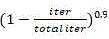 . The initial learning rate is 0.0001 and the training period is 100. At each cycle, the model first randomly selected images of 300 patients from the training set, and then randomly cropped 50 patches containing positive and negative examples from the images of each patient. A total of about 15,000 patches are used per epoch to train the modelAnd (4) molding. During model training, sub-volumes of 16 slices are randomly drawn from the volume. The dataset was preprocessed to find the contours of the skull, then each volume was cropped in the axial plane around the skull before resizing each slice to 208 x 208 pixels. The slice is then cropped to 192 × 192 pixels (random cropping is used during training, center cropping is used during testing), with the final input size for each example being 16 × 192 × 192; the same transformation is applied to the segment labels. The segmentation output is trained to optimize a weighted combination of voxel binary cross entropy and Dice loss.

The model crops the input into [ -300, 700] Huo Ensi field units, normalizes to [ -1,1], and starts from zero. The model was trained on 3 Graphics Processing Units (GPUs), with a minimum of 2 cases per GPU. The parameters of the model were optimized using a random gradient descent optimizer with a momentum of 0.9, a weight peak learning rate of 0.1 for random initialization, and a peak learning rate of 0.01 for pre-training weights. For normalization, the loss of all trainable parameters is increased by a weight decay of 0.001 and a random depth dip is used in the encoder block. To control class imbalance, 3 methods were used. First, an auxiliary penalty is added after the encoder, and a focus penalty is used to encourage larger parameter updates for misclassified positive samples. Second, the sampling frequency of the abnormal training example is higher than that of the normal example, so the abnormal case accounts for 30% of the training iteration number. The parameters of the decoder are not updated in the training iteration, where the segmentation labels consist of only background voxels.
To generate a segmented prediction of the entire volume, only the segmented outputs of the successive 16-slice subvolumes need to be connected together. If the number of slices is not evenly divisible by 16, the last input volume will be filled with 0 and the corresponding output volume will be truncated to the original size.
The present application also provides a CTA image data processing apparatus, as shown in fig. 8, including:
a receiving module to receive CTA image data, the CTA image data including one or more original images;
and the correction module is used for processing the original image based on a preset three-dimensional CNN network model, removing a bone graph and a vein graph in the original image and obtaining a corrected image only with an artery graph.
Example 4
The present invention provides a CTA image data processing method, which is the third processing step inembodiment 1, as shown in fig. 9, and includes:
step S210, receiving CTA image data, wherein the CTA image data comprises one or more original images;
step S220, processing the original image to obtain all nodes in the original image, wherein the nodes are points larger than a preset voxel value;
step S230, connecting every two adjacent nodes in the original image to generate a node frame graph;
step S240, a triangle path in the node frame graph is obtained to obtain a triangle path histogram, where the triangle path is a path where a plurality of nodes form a triangle.
Although the triangular path histogram is a set of voxel-based features, it is derived from the graph structure extracted from a given image. The graphical structure may simply be extracted from a binary image of the target structure (e.g. the vessel system).
The triangular path histogram feature set in the graph is defined at each node (i.e., each voxel) in a given graph based on a three-dimensional histogram of shortest path distances between the node and each of its neighboring node pairs. The feature vector effectively encodes the local graph network pattern around the node. Since the triangular path histogram in the graph does not use any three-dimensional thinning algorithm, the triangular path histogram does not have the problem of wrong node identification. Although the triangular path histogram features in the figures are particularly good at describing branch vessel structures, they may also describe a bump-like or node-like structure.
The triangle path histogram in the graph is powerful, and the triangle path histogram feature set in a single graph is enough to correctly detect the cerebral aneurysm by using a single support vector machine classifier. The triangular path histogram feature in the graph has robustness to non-rigid transformation, and the branched vessel structure and the protrusion-shaped structure can be effectively coded. A triangle path histogram characteristic set in a graph is defined on each node of any undirected graph.
In step S240, the method further includes:
an undirected graph G with a vertex set as V and two vertexes The shortest path length between is
 The shortest path is a path along the direction of the edge of the graph G;


presetting natural number The triplet of (2);

triple at node The node at which the triangular path histogram feature value is defined as the shortest path distance satisfies
 The conditions of (a) are:


triangle path histogram eigenvalue Is defined as follows:


wherein Is the number of elements of a given set

Triangle path histogram feature value By a series of triplets
 ) Is defined as


The sequence of triples is determined to satisfy And
And all distance combinations of conditions. For example, suppose
 ,
,


For example, suppose ,
,


And sequentially obtaining the triangular path histogram feature calculation model in the graph, as shown in fig. 10.
The invention also employs a multi-resolution strategy. A given binary volume will be rescaled by a factor of 0.5. Then, using the contracted volumes, the triangle path histogram feature in the graph is calculated. These features are then returned to the original voxels.
The present invention also uses two well-known Hessian-derived voxel-based features (shape index and point enhancement filters) to evaluate the effectiveness of the cooperative use of two different types of features, namely a grayscale-based and a graph-based feature set.
Processing the original image to obtain all nodes in the original image, wherein the nodes which are points larger than a preset voxel value comprise:
configuring the node enhancement filter to the following formula:

wherein Is the eigenvalue of the Hessian matrix;

the shape index is configured as:

wherein k1 and k2 are the principal curvatures;
based on the above steps, the features of the voxels are processed.
Each feature vector is mapped to a high-dimensional space by a kernel calculation before it is evaluated by a linear support vector machine classifier. The present invention solves this problem using an explicit feature mapping approach. In the explicit feature mapping method, the triangular path histogram feature vector in each graph adopts an index Approximate finite feature mapping.

Meanwhile, feature vectors based on the Hessian matrix are mapped by utilizing Gaussian kernels. The purpose of this explicit feature mapping is to reduce computation time significantly, while using two different kernels (i.e. exponentials) Nuclei and gaussian nuclei). Specifically, the triangle path histogram variables in all histogram-based maps are first multiplied by a factor such that the standard deviation of each feature becomes 1. Furthermore, all the grayscale-based features are linearly normalized such that their mean and standard deviation are 0 and 0, respectively
 。
。 The parameters control the weights between the triangle path histogram features and the grayscale-based features in the histogram-based map. It is further noted that the mean and standard deviation are calculated in advance from all training data sets.


The present invention uses a linear support vector machine as a voxel-based classifier. A base truth label roll is prepared prior to the training phase. Using these ground truth label volumes, foreground voxels are divided into positive and negative classes. To avoid an imbalance of the positive and negative sample size, the negative samples are randomly down-sampled so that only 0.5% of the negative samples remain in each case. And finally, collecting all positive and negative voxels from all training samples and training the support vector machine.
multiplying all triangle path histogram variables in the histogram-based graph by a factor to change the standard deviation of each feature to 1;
all voxels are linearly normalized for gray-based features such that the mean and standard deviation of the voxels are 0 and 0, respectively ;
;

Wherein, the parameters control the weights between the triangle path histogram features and the grayscale-based features in the histogram-based map.

The average value and/or the standard deviation are preset and/or obtained from all previous training data sets.
Processing the original image by criteria of window spacing [0, 450] and [ -50, 650], removing a CTA image of bone;
searching an initial region of the blood vessel by using a window with a preset threshold value, and reserving the maximum connectivity region as a final region of the blood vessel;
and obtaining corresponding cutting intervals to carry out normalization processing on the original image cutting based on the histogram of the brightness of the voxels in the final region.
The image characteristics of the triangular path histogram and the Hessian matrix in the graph have synergistic effect. The triangular path histogram feature in the graph in the algorithm model can cooperate with various existing image features. The invention simultaneously utilizes the triangular path histogram in the graph and the characteristic based on deep learning. Another important feature is that the triangle path histogram image features in the graph are invariant to translation and mirroring and robust to rotation and small local deformations. This is because the feature set comes from only one graph structure, which does not change significantly when small deformations occur locally.
Compared with the artificial data amplification technology frequently used in the deep learning-based method, the method based on the triangular path histogram in the graph does not need any data amplification (such as rotation and non-rigid deformation), so that the characteristic set of the triangular path histogram in the graph can successfully distinguish the vascular structure from the non-vascular structure in the detection task. Meanwhile, the triangular path histogram in the graph effectively captures the topological branch pattern of the human organ. In the inference stage, a segmented prediction of the entire image is generated by merging uniformly sampled predictions. Two adjacent patches may have a 1/8 overlap.
To detect intracranial aneurysms in some low contrast images cropped with a default window interval of [0, 900], two additional intervals [0, 450] and [ -50, 650] are used to normalize the source image. The setting is automatically selected according to the brightness distribution. Given a CTA image with bone removed, an initial region of the blood vessel is found using a threshold (e.g., 150 Hu), and then the maximum connectivity region is retained as the final region of the blood vessel. The histogram of the intensity of voxels in this region is analyzed to find the appropriate clipping interval. The present invention calculates the three distributions of [0, 200], [200, 300] and [300, 500] intervals, which correspond to [0, 450], [ -50, 650] and [0, 900]. Finally, a cropping interval corresponding to the dominant distribution interval is selected to normalize the source image.
The present invention also provides a CTA image data processing apparatus, as shown in fig. 11, including:
a receiving module to receive CTA image data, the CTA image data including one or more original images;
the processing module is used for processing the original image to obtain all nodes in the original image, wherein the nodes are points larger than a preset voxel value;
the generating module is used for connecting every two adjacent nodes in the original image to generate a node frame graph;
the acquisition module is used for acquiring a triangular path in the node frame graph to obtain a triangular path histogram, wherein the triangular path is a path of a triangle formed by a plurality of nodes.
In one embodiment, the obtaining module is further configured to perform the following steps, including:
an undirected graph G with V set of vertexes, and two vertexes The shortest path length between is
 The shortest path is a path along the direction of the edge of the graph G;


in advanceSetting natural number The triplet of (a);

triple unit At a node
 The node at which the triangular path histogram feature value is defined as the shortest path distance satisfies
 The conditions of (a) are:


triangle path histogram feature value Is defined as:


wherein Is the number of elements of a given set

Triangle path histogram feature value By a series of triplets
 ) Is defined as


Compared with the traditional aneurysm identification method, the method has the advantages that:
1. the invention can efficiently screen out the aneurysm through the CTA image, the detection speed, the diagnosis effective rate, the sensitivity and the specificity are all superior to those of common radiologists, the daily work efficiency of the radiologists can be improved, and misdiagnosis is avoided.
The bone and vein images of the CTA image can influence the diagnosis of the aneurysm, and the image preprocessing algorithm can effectively remove the bone and vein images, improve the effective diagnosis rate and avoid false positive.
3. The prevalence rates of aneurysms of different patients are very different, from common people (3% -7%) to subarachnoid hemorrhage patients (85%), the HCNN model can effectively deal with diagnosis of the aneurysms with large differences, and the detection result is stable.
The present invention allows radiologists to know the status of an aneurysm by giving them an assessment of sensitivity and specificity. Sensitivity indicates the number of negative results in total aneurysm positive cases, specificity indicates the number of positive results in total aneurysm negative cases, and accuracy indicates the number of positive and negative results in all tested cases.
To determine the robustness of the results and whether the results were due to the radiologist used, the present invention performed a sensitivity analysis and a T-test for differences in sensitivity, specificity, and accuracy. The quantitative variables are expressed as mean ± SD if the data is normally distributed, and median and quartile spacing are used when non-normally distributed data is used. The classification variables are expressed as frequency or percentage.
To evaluate the performance of the algorithmic tests, the models were evaluated in each cohort, respectively, for accuracy in correctly displaying the patient, patient level sensitivity, specificity, diagnostic efficacy, and variability was evaluated using 95% wilson score confidence intervals. In the test data set containing 357 aneurysms, 92.9% of the aneurysms were successfully detected by the present invention. Due to the flexible decision zone approach, the model performs well in detecting aneurysms of various sizes. The proposed bounding box can automatically fit the size of the aneurysm. The invention has 96.7 percent of total sensitivity for the aneurysm with the diameter of more than 3 millimeters.
The readable storage medium may be a computer storage medium or a communication medium. Communication media includes any medium that facilitates transfer of a computer program from one place to another. Computer storage media may be any available media that can be accessed by a general purpose or special purpose computer. For example, a readable storage medium is coupled to a processor such that the processor can read information from, and write information to, the readable storage medium. Of course, the readable storage medium may also be an integral part of the processor. The processor and the readable storage medium may reside in an Application Specific Integrated Circuits (ASIC). Additionally, the ASIC may reside in user equipment. Of course, the processor and the readable storage medium may also reside as discrete components in a communication device. The readable storage medium may be a read-only memory (ROM), a random-access memory (RAM), a CD-ROM, a magnetic tape, a floppy disk, an optical data storage device, and the like.
The present invention also provides a program product comprising execution instructions stored in a readable storage medium. The at least one processor of the device may read the execution instructions from the readable storage medium, and the execution of the execution instructions by the at least one processor causes the device to implement the methods provided by the various embodiments described above.
In the above embodiments of the terminal or the server, it should be understood that the Processor may be a Central Processing Unit (CPU), other general-purpose processors, a Digital Signal Processor (DSP), an Application Specific Integrated Circuit (ASIC), etc. A general purpose processor may be a microprocessor or the processor may be any conventional processor or the like. The steps of a method disclosed in connection with the present invention may be embodied directly in a hardware processor, or in a combination of the hardware and software modules within the processor.
Finally, it should be noted that: the above embodiments are only used to illustrate the technical solution of the present invention, and not to limit the same; while the invention has been described in detail and with reference to the foregoing embodiments, it will be understood by those skilled in the art that: the technical solutions described in the foregoing embodiments may still be modified, or some or all of the technical features may be equivalently replaced; and the modifications or the substitutions do not make the essence of the corresponding technical solutions depart from the scope of the technical solutions of the embodiments of the present invention.
Claims (4)
1. A CTA image data identification method, comprising:
step 1, receiving a preprocessed image, wherein the preprocessed image only contains an artery graph;
step 2, predicting each voxel in the artery graph to obtain the probability that each voxel in the artery graph is an aneurysm;
step 3, marking the aneurysm at the position where the probability of the voxel being the aneurysm is larger than a preset value;
the above three steps are processed by an HCNN model, which is a CNN having an encoder-decoder structure;
the encoder is configured to map a volume to an abstract low resolution encoding;
the decoder is used for expanding the code into a full-resolution partition volume;
wherein the encoder adapts the decoder from a 50-layer SE-resenext network to a 3 x 3 convolutional transposed sequence;
the encoder is a pre-trained dynamics data set, and after pre-training the encoder, the last 3 rolling blocks and output layers of the encoder are deleted, and a spatial pyramid pool layer and a decoder are added;
the HCNN model is trained by the following steps, including:
presetting a training sample, wherein the training sample comprises a proximity projection image and all annotated aneurysm positions in the image;
training the model based on the training sample to obtain a group of rectangular frames, wherein the rectangular frames comprise aneurysms on the image and the probability that each aneurysm is true positive;
and after the projected image is horizontally overturned to expand the training data set, training is carried out again.
2. The CTA image data recognition method according to claim 1, further comprising:
and summarizing the results of all the marked aneurysms, and outputting the final detection result of the aneurysm.
3. A CTA image data recognition apparatus, comprising:
the receiving module is used for receiving the preprocessed image, and the preprocessed image only contains an artery graph;
the prediction module is used for predicting each voxel in the artery graph to obtain the probability that each voxel in the artery graph is an aneurysm;
the marking module is used for marking the aneurysm, wherein the probability of the voxel being the aneurysm is larger than a preset value;
the receiving module, the predicting module and the predicting module form an HCNN model, and the HCNN model is a CNN with an encoder-decoder structure;
the encoder is configured to map a volume to an abstract low resolution encoding;
the decoder is used for expanding the code into a full-resolution partition volume;
wherein the encoder adapts the decoder from a 50-layer SE-resenext network to a 3 x 3 convolutional transposed sequence;
the encoder is a pre-trained dynamics data set, and after pre-training the encoder, the last 3 rolling blocks and output layers of the encoder are deleted, and a spatial pyramid pool layer and a decoder are added;
the HCNN model is trained by the following units, including:
a presetting unit for presetting a training sample comprising the neighboring projection images and all annotated aneurysm locations in the images;
the training unit is used for training a model based on the training sample to obtain a group of rectangular frames, and the rectangular frames comprise the aneurysms on the image and the probability that each aneurysm is true positive;
and the extension unit is used for carrying out horizontal turning on the projection image to extend the training data set and then carrying out training again.
4. A readable storage medium, characterized in that a computer program is stored in the readable storage medium, which computer program, when being executed by a processor, is adapted to carry out the method of one of the claims 1-2.
Priority Applications (1)
| Application Number | Priority Date | Filing Date | Title |
|---|---|---|---|
| CN202110183220.2ACN112862785B (en) | 2021-02-10 | 2021-02-10 | CTA image data identification method, device and storage medium |
Applications Claiming Priority (1)
| Application Number | Priority Date | Filing Date | Title |
|---|---|---|---|
| CN202110183220.2ACN112862785B (en) | 2021-02-10 | 2021-02-10 | CTA image data identification method, device and storage medium |
Publications (2)
| Publication Number | Publication Date |
|---|---|
| CN112862785A CN112862785A (en) | 2021-05-28 |
| CN112862785Btrue CN112862785B (en) | 2022-11-18 |
Family
ID=75989672
Family Applications (1)
| Application Number | Title | Priority Date | Filing Date |
|---|---|---|---|
| CN202110183220.2AActiveCN112862785B (en) | 2021-02-10 | 2021-02-10 | CTA image data identification method, device and storage medium |
Country Status (1)
| Country | Link |
|---|---|
| CN (1) | CN112862785B (en) |
Families Citing this family (1)
| Publication number | Priority date | Publication date | Assignee | Title |
|---|---|---|---|---|
| CN114155215B (en)* | 2021-11-24 | 2023-11-10 | 中山大学肿瘤防治中心(中山大学附属肿瘤医院、中山大学肿瘤研究所) | A method and system for nasopharyngeal cancer identification and tumor segmentation based on MR images |
Citations (3)
| Publication number | Priority date | Publication date | Assignee | Title |
|---|---|---|---|---|
| CN110929789A (en)* | 2019-11-22 | 2020-03-27 | 北京理工大学 | Automatic classification method and device for liver tumors based on multi-phase CT image analysis |
| CN112116605A (en)* | 2020-09-29 | 2020-12-22 | 西北工业大学深圳研究院 | A pancreas CT image segmentation method based on integrated deep convolutional neural network |
| CN112200773A (en)* | 2020-09-17 | 2021-01-08 | 苏州慧维智能医疗科技有限公司 | Large intestine polyp detection method based on encoder and decoder of cavity convolution |
- 2021
- 2021-02-10CNCN202110183220.2Apatent/CN112862785B/enactiveActive
Patent Citations (3)
| Publication number | Priority date | Publication date | Assignee | Title |
|---|---|---|---|---|
| CN110929789A (en)* | 2019-11-22 | 2020-03-27 | 北京理工大学 | Automatic classification method and device for liver tumors based on multi-phase CT image analysis |
| CN112200773A (en)* | 2020-09-17 | 2021-01-08 | 苏州慧维智能医疗科技有限公司 | Large intestine polyp detection method based on encoder and decoder of cavity convolution |
| CN112116605A (en)* | 2020-09-29 | 2020-12-22 | 西北工业大学深圳研究院 | A pancreas CT image segmentation method based on integrated deep convolutional neural network |
Non-Patent Citations (1)
| Title |
|---|
| Tielin Zhang 等.HCNN: A Neural Network Model for Combining Local and Global Features towards Human-like Classification.《International Journal of Pattern Recognition and Artificial Intelligence》.2015,全文.* |
Also Published As
| Publication number | Publication date |
|---|---|
| CN112862785A (en) | 2021-05-28 |
Similar Documents
| Publication | Publication Date | Title |
|---|---|---|
| Larrazabal et al. | Post-DAE: anatomically plausible segmentation via post-processing with denoising autoencoders | |
| CN111325739B (en) | Method and device for detecting lung focus and training method of image detection model | |
| EP3770850B1 (en) | Medical image identifying method, model training method, and computer device | |
| Chan et al. | Effective pneumothorax detection for chest X‐ray images using local binary pattern and support vector machine | |
| US20220198230A1 (en) | Auxiliary detection method and image recognition method for rib fractures based on deep learning | |
| RU2654199C1 (en) | Segmentation of human tissues in computer image | |
| Zhang et al. | Intelligent scanning: Automated standard plane selection and biometric measurement of early gestational sac in routine ultrasound examination | |
| EP3791316A1 (en) | Localization and classification of abnormalities in medical images | |
| WO2019200753A1 (en) | Lesion detection method, device, computer apparatus and storage medium | |
| US9269139B2 (en) | Rib suppression in radiographic images | |
| US9087370B2 (en) | Flow diverter detection in medical imaging | |
| JP2015528372A (en) | System and method for automatically detecting pulmonary nodules in medical images | |
| Osadebey et al. | Three-stage segmentation of lung region from CT images using deep neural networks | |
| Maiora et al. | Abdominal CTA image analisys through active learning and decision random forests: Aplication to AAA segmentation | |
| WO2019182520A1 (en) | Method and system of segmenting image of abdomen of human into image segments corresponding to fat compartments | |
| CN119487545A (en) | Analyzing medical images | |
| US20120321169A1 (en) | Shape Based Conditional Random Fields for Segmenting Intracranial Aneurysms | |
| WO2023207743A1 (en) | Image detection method and apparatus, and computer device, storage medium and program product | |
| CN105809175A (en) | Encephaledema segmentation method and system based on support vector machine algorithm | |
| US20240185577A1 (en) | Reinforced attention | |
| CN117809122B (en) | A method, system, electronic device and medium for processing intracranial large blood vessel images | |
| CN112381762A (en) | CT rib fracture auxiliary diagnosis system based on deep learning algorithm | |
| US20240386551A1 (en) | System and method for segmenting medical images | |
| Wang et al. | Automatic measurement of fetal head circumference using a novel GCN-assisted deep convolutional network | |
| US12182994B2 (en) | Method and system for generating a medical image with localized artifacts using machine learning |
Legal Events
| Date | Code | Title | Description |
|---|---|---|---|
| PB01 | Publication | ||
| PB01 | Publication | ||
| SE01 | Entry into force of request for substantive examination | ||
| SE01 | Entry into force of request for substantive examination | ||
| GR01 | Patent grant | ||
| GR01 | Patent grant |