CN112819076B - Training method and device for medical image classification model based on deep transfer learning - Google Patents
Training method and device for medical image classification model based on deep transfer learningDownload PDFInfo
- Publication number
- CN112819076B CN112819076BCN202110147726.8ACN202110147726ACN112819076BCN 112819076 BCN112819076 BCN 112819076BCN 202110147726 ACN202110147726 ACN 202110147726ACN 112819076 BCN112819076 BCN 112819076B
- Authority
- CN
- China
- Prior art keywords
- medical image
- model
- classification model
- training
- image
- Prior art date
- Legal status (The legal status is an assumption and is not a legal conclusion. Google has not performed a legal analysis and makes no representation as to the accuracy of the status listed.)
- Active
Links
Images


Classifications
- G—PHYSICS
- G06—COMPUTING OR CALCULATING; COUNTING
- G06F—ELECTRIC DIGITAL DATA PROCESSING
- G06F18/00—Pattern recognition
- G06F18/20—Analysing
- G06F18/24—Classification techniques
- G06F18/241—Classification techniques relating to the classification model, e.g. parametric or non-parametric approaches
- G06F18/2415—Classification techniques relating to the classification model, e.g. parametric or non-parametric approaches based on parametric or probabilistic models, e.g. based on likelihood ratio or false acceptance rate versus a false rejection rate
- G—PHYSICS
- G06—COMPUTING OR CALCULATING; COUNTING
- G06F—ELECTRIC DIGITAL DATA PROCESSING
- G06F18/00—Pattern recognition
- G06F18/20—Analysing
- G06F18/21—Design or setup of recognition systems or techniques; Extraction of features in feature space; Blind source separation
- G06F18/214—Generating training patterns; Bootstrap methods, e.g. bagging or boosting
- G—PHYSICS
- G06—COMPUTING OR CALCULATING; COUNTING
- G06N—COMPUTING ARRANGEMENTS BASED ON SPECIFIC COMPUTATIONAL MODELS
- G06N3/00—Computing arrangements based on biological models
- G06N3/02—Neural networks
- G06N3/04—Architecture, e.g. interconnection topology
- G06N3/045—Combinations of networks
- G—PHYSICS
- G06—COMPUTING OR CALCULATING; COUNTING
- G06N—COMPUTING ARRANGEMENTS BASED ON SPECIFIC COMPUTATIONAL MODELS
- G06N3/00—Computing arrangements based on biological models
- G06N3/02—Neural networks
- G06N3/04—Architecture, e.g. interconnection topology
- G06N3/047—Probabilistic or stochastic networks
- G—PHYSICS
- G06—COMPUTING OR CALCULATING; COUNTING
- G06N—COMPUTING ARRANGEMENTS BASED ON SPECIFIC COMPUTATIONAL MODELS
- G06N3/00—Computing arrangements based on biological models
- G06N3/02—Neural networks
- G06N3/08—Learning methods
- G—PHYSICS
- G06—COMPUTING OR CALCULATING; COUNTING
- G06T—IMAGE DATA PROCESSING OR GENERATION, IN GENERAL
- G06T7/00—Image analysis
- G06T7/0002—Inspection of images, e.g. flaw detection
- G06T7/0012—Biomedical image inspection
- G—PHYSICS
- G06—COMPUTING OR CALCULATING; COUNTING
- G06T—IMAGE DATA PROCESSING OR GENERATION, IN GENERAL
- G06T7/00—Image analysis
- G06T7/10—Segmentation; Edge detection
- G06T7/11—Region-based segmentation
- G—PHYSICS
- G06—COMPUTING OR CALCULATING; COUNTING
- G06T—IMAGE DATA PROCESSING OR GENERATION, IN GENERAL
- G06T7/00—Image analysis
- G06T7/30—Determination of transform parameters for the alignment of images, i.e. image registration
- G06T7/33—Determination of transform parameters for the alignment of images, i.e. image registration using feature-based methods
- G—PHYSICS
- G06—COMPUTING OR CALCULATING; COUNTING
- G06T—IMAGE DATA PROCESSING OR GENERATION, IN GENERAL
- G06T2207/00—Indexing scheme for image analysis or image enhancement
- G06T2207/10—Image acquisition modality
- G06T2207/10024—Color image
- G—PHYSICS
- G06—COMPUTING OR CALCULATING; COUNTING
- G06T—IMAGE DATA PROCESSING OR GENERATION, IN GENERAL
- G06T2207/00—Indexing scheme for image analysis or image enhancement
- G06T2207/20—Special algorithmic details
- G06T2207/20024—Filtering details
- G—PHYSICS
- G06—COMPUTING OR CALCULATING; COUNTING
- G06T—IMAGE DATA PROCESSING OR GENERATION, IN GENERAL
- G06T2207/00—Indexing scheme for image analysis or image enhancement
- G06T2207/20—Special algorithmic details
- G06T2207/20024—Filtering details
- G06T2207/20032—Median filtering
- G—PHYSICS
- G06—COMPUTING OR CALCULATING; COUNTING
- G06T—IMAGE DATA PROCESSING OR GENERATION, IN GENERAL
- G06T2207/00—Indexing scheme for image analysis or image enhancement
- G06T2207/20—Special algorithmic details
- G06T2207/20081—Training; Learning
- G—PHYSICS
- G06—COMPUTING OR CALCULATING; COUNTING
- G06T—IMAGE DATA PROCESSING OR GENERATION, IN GENERAL
- G06T2207/00—Indexing scheme for image analysis or image enhancement
- G06T2207/20—Special algorithmic details
- G06T2207/20084—Artificial neural networks [ANN]
- G—PHYSICS
- G06—COMPUTING OR CALCULATING; COUNTING
- G06T—IMAGE DATA PROCESSING OR GENERATION, IN GENERAL
- G06T2207/00—Indexing scheme for image analysis or image enhancement
- G06T2207/30—Subject of image; Context of image processing
- G06T2207/30004—Biomedical image processing
- G06T2207/30096—Tumor; Lesion
- G—PHYSICS
- G06—COMPUTING OR CALCULATING; COUNTING
- G06V—IMAGE OR VIDEO RECOGNITION OR UNDERSTANDING
- G06V2201/00—Indexing scheme relating to image or video recognition or understanding
- G06V2201/03—Recognition of patterns in medical or anatomical images
Landscapes
- Engineering & Computer Science (AREA)
- Physics & Mathematics (AREA)
- Theoretical Computer Science (AREA)
- General Physics & Mathematics (AREA)
- Data Mining & Analysis (AREA)
- Computer Vision & Pattern Recognition (AREA)
- General Engineering & Computer Science (AREA)
- Life Sciences & Earth Sciences (AREA)
- Artificial Intelligence (AREA)
- Evolutionary Computation (AREA)
- Health & Medical Sciences (AREA)
- General Health & Medical Sciences (AREA)
- Mathematical Physics (AREA)
- Computing Systems (AREA)
- Molecular Biology (AREA)
- Computational Linguistics (AREA)
- Biomedical Technology (AREA)
- Software Systems (AREA)
- Biophysics (AREA)
- Probability & Statistics with Applications (AREA)
- Bioinformatics & Computational Biology (AREA)
- Bioinformatics & Cheminformatics (AREA)
- Evolutionary Biology (AREA)
- Medical Informatics (AREA)
- Nuclear Medicine, Radiotherapy & Molecular Imaging (AREA)
- Radiology & Medical Imaging (AREA)
- Quality & Reliability (AREA)
- Image Analysis (AREA)
- Medical Treatment And Welfare Office Work (AREA)
Abstract
Translated fromChinese
Description
Translated fromChinese技术领域technical field
本发明涉及医学技术领域,尤其涉及一种基于深度迁移学习的医学图像分类模型的训练方法及装置。The invention relates to the field of medical technology, in particular to a training method and device for a medical image classification model based on deep transfer learning.
背景技术Background technique
前列腺癌是一种常见的癌症,也是导致癌症死亡的常见原因。目前的前列腺癌检测方法包括使用前列腺特异性抗原(PSA)检测进行筛查,然后经直肠穿刺活检,但存在假阴性和分期不足的问题。近年来,多参数磁共振成像(mpMRI)被发现是一种有价值的诊断工具,用于前列腺癌的检测、定位和分期。计算机辅助检测(CAD)系统在辅助可以提供更多可重复的结果,同时消耗更少的时间。利用数据表征算法,可以从图像中提取大量定量特征,通过深度学习算法可以建立特征和诊断之间的关系。Prostate cancer is a common cancer and a common cause of cancer death. Current prostate cancer testing methods include screening with a prostate-specific antigen (PSA) test followed by a transrectal biopsy, but have problems with false negatives and understaging. In recent years, multiparametric magnetic resonance imaging (mpMRI) has been found to be a valuable diagnostic tool for the detection, localization and staging of prostate cancer. Computer-aided inspection (CAD) systems can provide more repeatable results while consuming less time. Using data characterization algorithms, a large number of quantitative features can be extracted from images, and deep learning algorithms can establish relationships between features and diagnoses.
传统的分类方法依赖于手工特征的选择,并要求对先验领域知识有清晰的认识。特征学习方法可以有效地检测不同模式的视觉特征,但由于医学图像缺乏标注数据,无法准确地训练高可用度的模型。由于前列腺医学图像数据量稀少、病理特征复杂和人工标注耗时费力,现有的PCa(前列腺癌)分类的传统方法泛化性较差,且基于深度学习的PCa分类网络可解释性弱。Traditional classification methods rely on hand-crafted feature selection and require a clear understanding of prior domain knowledge. Feature learning methods can effectively detect different modalities of visual features, but cannot accurately train models with high availability due to the lack of labeled data in medical images. Due to the sparse amount of prostate medical image data, complex pathological features, and labor-intensive manual annotation, the existing traditional methods for PCa (prostate cancer) classification have poor generalization, and the deep learning-based PCa classification network is weak in interpretability.
发明内容SUMMARY OF THE INVENTION
本发明提供了一种基于深度迁移学习的医学图像分类模型的训练方法及装置,以解决现有的医学图像分类模型准确性低的问题。The present invention provides a training method and device for a medical image classification model based on deep migration learning, so as to solve the problem of low accuracy of the existing medical image classification model.
第一方面,提供了一种基于深度迁移学习的医学图像分类模型的训练方法,包括:In a first aspect, a training method for a medical image classification model based on deep transfer learning is provided, including:
获取已标注的医学图像并进行预处理,得到医学图像样本集;Obtain the labeled medical images and preprocess them to obtain a medical image sample set;
利用医学图像样本集训练CNN网络模型得到第一分类模型;Use the medical image sample set to train the CNN network model to obtain the first classification model;
获取在ImageNet图像数据集上预先训练好的VGG-16网络模型和ResNet-50网络模型;Obtain the VGG-16 network model and the ResNet-50 network model pre-trained on the ImageNet image dataset;
基于迁移学习方法分别利用医学图像样本集再次训练VGG-16网络模型和ResNet-50网络模型,分别得到第二分类模型和第三分类模型;Based on the transfer learning method, the VGG-16 network model and the ResNet-50 network model are retrained using the medical image sample set to obtain the second classification model and the third classification model respectively;
将第一分类模型、第二分类模型、第三分类模型的输出连接到投票机制模块的输入构成医学图像分类模型,其中,投票机制模块基于预设投票机制输出最终医学图像分类结果为医学图像分类模型的分类结果。The outputs of the first classification model, the second classification model and the third classification model are connected to the input of the voting mechanism module to form a medical image classification model, wherein the voting mechanism module outputs the final medical image classification result based on the preset voting mechanism as medical image classification The classification result of the model.
上述方案提供的医学图像分类模型的训练方法,借助ImageNet图像数据集预先训练好VGG-16网络模型和ResNet-50网络模型,然后基于迁移学习方法利用医学图像样本集再次训练VGG-16网络模型和ResNet-50网络模型,进而得到第二分类模型和第三分类模型,解决了标注医学图像少导致训练出来的模型精度低、可用度低的问题,提高了医学图像分类的准确性。本方案采用了三个不同分类模型,具备不同的功能,并将三个分类模型的预测结果进行综合结合投票机制得到最终的分类结果,提高了医学图像分类模型的准确性,具有更好的泛化性和鲁棒性。The training method of the medical image classification model provided by the above scheme, the VGG-16 network model and the ResNet-50 network model are pre-trained with the ImageNet image data set, and then the VGG-16 network model and the ResNet-50 network model are trained again based on the transfer learning method using the medical image sample set. The ResNet-50 network model, and then the second classification model and the third classification model are obtained, which solves the problem of low accuracy and low availability of the trained model due to less labeled medical images, and improves the accuracy of medical image classification. This solution uses three different classification models with different functions, and combines the prediction results of the three classification models with the voting mechanism to obtain the final classification result, which improves the accuracy of the medical image classification model and has better generalization capabilities. flexibility and robustness.
进一步地,对已标注的医学图像的预处理过程包括:Further, the preprocessing process of the labeled medical images includes:
对已标注的医学图像进行双立方插值处理,统一医学图像的分辨率;Perform bicubic interpolation processing on labeled medical images to unify the resolution of medical images;
对插值处理后的医学图像进行图像配准,将所有插值处理后的医学图像变换到同一坐标系下;Perform image registration on the interpolated medical images, and transform all the interpolated medical images into the same coordinate system;
对图像配准后的医学图像进行归一化处理,提取医学图像中的病灶区域,并使用Z-score标准化方法将其标准化。Normalize the medical image after image registration, extract the lesion area in the medical image, and use the Z-score normalization method to normalize it.
进一步地,所述归一化处理之后还包括:Further, after the normalization process, it also includes:
对归一化处理之后的医学图像进行数据增强,扩大医学图像样本集。Data enhancement is performed on the normalized medical images to expand the medical image sample set.
进一步地,所述数据增强包括对医学图像进行水平翻转50%、向上翻转、向下翻转、高斯滤波、均值滤波和中值滤波中的一种或多种,然后加入噪声。Further, the data enhancement includes performing one or more of horizontal flipping, up flipping, down flipping, Gaussian filtering, mean filtering and median filtering on the medical image, and then adding noise.
在已标注的医学图像数量有限的情况下,通过数据增强方式增加训练样本的多样性,可提高最后训练得到的模型的鲁棒性,避免过拟合。When the number of labeled medical images is limited, increasing the diversity of training samples through data augmentation can improve the robustness of the final trained model and avoid overfitting.
进一步地,所述对已标注的医学图像进行双立方插值处理,包括:Further, performing bicubic interpolation processing on the marked medical images includes:
采用基于BiCubic基函数的双立方插值法对已标注的医学图像进行插值处理;其中,BiCubic基函数的构造形式如下所示:The labeled medical images are interpolated by the bicubic interpolation method based on the BiCubic basis function; the construction form of the BiCubic basis function is as follows:

其中,a取-0.5,x表示源图像中像素点的横坐标或纵坐标,W(x)表示源图像中对应像素点的横坐标或纵坐标的权重;则源图像中像素点P对应在目标图像上的像素点B的像素值B(X,Y)可通过下式得到:Among them, a takes -0.5, x represents the abscissa or ordinate of the pixel in the source image, W(x) represents the weight of the abscissa or ordinate of the corresponding pixel in the source image; then the pixel P in the source image corresponds to The pixel value B(X, Y) of the pixel point B on the target image can be obtained by the following formula:

其中,am,n表示a(m,n)的像素值,W(m)表示a(m,n)横坐标上的权重,W(n)表示a(m,n)纵坐标上的权重,a(m,n)(m,n=0,1,2,3)表示源图像中距离像素点P最近的16个像素点;Among them, am,n represents the pixel value of a(m,n), W(m) represents the weight on the abscissa of a(m,n), and W(n) represents the weight on the ordinate of a(m,n) , a(m,n)(m,n=0,1,2,3) represents the 16 pixels closest to the pixel P in the source image;
所述对图像配准后的医学图像进行归一化处理,包括:The normalizing processing of the medical images after image registration includes:
根据已标注的医学图像去除非病灶区域,提取病灶区域;Remove the non-lesion area according to the labeled medical image, and extract the lesion area;
使用Z-score标准化方法将每个病灶区域的所有像素值转换为均值为0、标准差为1的公共尺度;其中,标准化过程中处理公式如下所示:The Z-score normalization method is used to convert all pixel values of each lesion area to a common scale with a mean of 0 and a standard deviation of 1; the processing formula in the normalization process is as follows:


其中,μ是病灶区域图像的均值,X表示病灶区域图像矩阵,σ表示标准差,N表示病灶区域图像的像素数量。Among them, μ is the mean of the image of the lesion area, X is the image matrix of the lesion area, σ is the standard deviation, and N is the number of pixels of the image of the lesion area.
对医学图像进行归一化操作,目的是将像素值变换到某个区间实现数据中心化,数据中心化符合数据分布规律,能增加模型的泛化能力。The purpose of normalizing medical images is to transform the pixel values into a certain interval to achieve data centralization. The data centralization conforms to the data distribution law and can increase the generalization ability of the model.
进一步地,所述第一分类模型通过如下过程训练得到:Further, the first classification model is obtained through the following process training:
基于CNN-6构建网络模型,其前两个卷积层包含大小为3×3的32个核,后两个卷积层包含大小为3×3的64个核;第2和第4个卷积层与2×2维的最大池化层交错,差值为0.25;倒数第二层是512个神经元的全连接层和0.5个缺失层,最后一层是30个神经元的全连接层;ReLU激活函数应用于所有四个卷积层和倒数第二层的全连接层,最后一层通过softmax函数输出预测概率,确定医学图像的分类类别;The network model is constructed based on CNN-6, the first two convolutional layers contain 32 kernels of size 3×3, and the last two convolutional layers contain 64 kernels of size 3×3; the 2nd and 4th volumes The accumulation layer is interleaved with a 2×2-dimensional max-pooling layer with a difference of 0.25; the penultimate layer is a fully connected layer of 512 neurons and 0.5 missing layers, and the last layer is a fully connected layer of 30 neurons. ; The ReLU activation function is applied to all four convolutional layers and the fully connected layer of the penultimate layer, and the last layer outputs the predicted probability through the softmax function to determine the classification category of the medical image;
使用gloot统一来初始权重,然后利用医学图像样本集训练上述基于CNN-6构建的网络模型,得到第一分类模型。Use gloot to unify the initial weights, and then use the medical image sample set to train the above network model based on CNN-6 to obtain the first classification model.
进一步地,所述第二分类模型通过如下过程训练得到:Further, the second classification model is obtained through the following process training:
获取ImageNet图像数据集;Get the ImageNet image dataset;
对ImageNet图像数据集中的ImageNet图像进行预处理,对RGB三个通道分别减去ImageNet图像数据集的均值,并进行正则化操作,得到ImageNet图像训练集;Preprocess the ImageNet images in the ImageNet image dataset, subtract the mean of the ImageNet image dataset from the three RGB channels, and perform regularization operations to obtain the ImageNet image training set;
利用ImageNet图像训练集对基于VGG-16网络结构的模型进行训练,得到预先训练好的VGG-16网络模型;Use the ImageNet image training set to train the model based on the VGG-16 network structure to obtain a pre-trained VGG-16 network model;
冻结预先训练好的VGG-16网络模型的预设数量的早期层,用30个神经元替换VGG-16网络模型最后一个全连接层,然后使用gloot统一重新初始化VGG-16网络模型最后的三个全连接层的权重;最后利用医学图像样本集对上述改进后的VGG-16网络模型进行训练,得到第二分类模型。Freeze a preset number of early layers of the pre-trained VGG-16 network model, replace the last fully connected layer of the VGG-16 network model with 30 neurons, and use gloot to uniformly re-initialize the last three VGG-16 network model. The weight of the fully connected layer; finally, the above-mentioned improved VGG-16 network model is trained by using the medical image sample set to obtain the second classification model.
进一步地,所述第三分类模型通过如下过程训练得到:Further, the third classification model is obtained through the following process training:
获取ImageNet图像数据集;Get the ImageNet image dataset;
对ImageNet图像数据集中的ImageNet图像进行预处理,对RGB三个通道分别减去ImageNet图像数据集的均值,并进行正则化操作,得到ImageNet图像训练集;Preprocess the ImageNet images in the ImageNet image dataset, subtract the mean of the ImageNet image dataset from the three RGB channels, and perform regularization operations to obtain the ImageNet image training set;
利用ImageNet图像训练集对基于ResNet-50网络结构的模型进行训练,得到预先训练好的ResNet-50网络模型;Use the ImageNet image training set to train the model based on the ResNet-50 network structure to obtain a pre-trained ResNet-50 network model;
冻结预先训练好的ResNet-50网络模型的预设数量的早期层,用30个神经元替换ResNet-50网络模型最后一个全连接层,然后使用gloot统一重新初始化ResNet-50网络模型最后一个全连接层的权重;最后利用医学图像样本集对上述改进后的ResNet-50网络模型进行训练,得到第三分类模型。Freeze a preset number of early layers of the pre-trained ResNet-50 network model, replace the last fully connected layer of the ResNet-50 network model with 30 neurons, and use gloot to uniformly re-initialize the last fully connected layer of the ResNet-50 network model The weight of the layer; finally, the above-mentioned improved ResNet-50 network model is trained by using the medical image sample set to obtain the third classification model.
本发明在训练第二分类模型和第三分类模型的过程中,使用Adma优化器和分类交叉熵损失函数在ImageNet图像数据集上对32个小批量的改进后的网络模型进行训练。In the process of training the second classification model and the third classification model, the present invention uses the Adma optimizer and the classification cross-entropy loss function to train the improved network models in 32 small batches on the ImageNet image data set.
设Z为n幅医学图像的医学图像训练数据集。从无到有训练最后一层(top layer(s))是一个迭代的过程,找到使CNN(VGG-16、ResNet-50均属于CNN网络结构)的经验损失最小的权重w。分类交叉熵损失函数如下式所示:Let Z be a medical image training dataset of n medical images. Training the last layer (top layer(s)) from scratch is an iterative process to find the weight w that minimizes the empirical loss of the CNN (VGG-16, ResNet-50 both belong to the CNN network structure). The categorical cross-entropy loss function is as follows:

其中,xi是Z的第i幅图像,f(xi,w)是xi的类别yi的预测概率,y′i是xi的真实类别,l(yi,y′i)是用于yi预测的惩罚函数,其如下式所示:where xi is the ith image of Z, f(xi ,w) is the predicted probability of classyi of xi , y′i is the true class of xi , and l(yi ,y′i ) is Penalty function for yi prediction, which is given by:

其中,yi∈{1,...,C},C为常数,其取值与类别总数相等,

当使用当前权重应用到小批量时,根据损失函数L的梯度计算更新的权重,使用Adma来计算个体的自适应学习率来控制权重更新的大小。When applied to a mini-batch using the current weights, the updated weights are calculated according to the gradient of the loss function L, and Adma is used to calculate the individual adaptive learning rate to control the size of the weight update.
进一步地,所述投票机制模块采用的投票机制通过如下公式表示:Further, the voting mechanism adopted by the voting mechanism module is expressed by the following formula:

其中,x表示输入的医学图像,y表示预测分类标号,
第二方面,提供了一种基于深度迁移学习的医学图像分类模型的训练装置,包括:In a second aspect, a training device for a medical image classification model based on deep transfer learning is provided, including:
图像获取模块,用于获取已标注的医学图像并进行预处理,得到医学图像样本集;The image acquisition module is used to acquire the labeled medical images and preprocess them to obtain a medical image sample set;
第一模型生成模块,用于利用医学图像样本集训练CNN网络模型得到第一分类模型;The first model generation module is used to train the CNN network model by using the medical image sample set to obtain the first classification model;
第二模型生成模块,用于获取在ImageNet图像数据集上预先训练好的VGG-16网络模型和ResNet-50网络模型;并基于迁移学习方法分别利用医学图像样本集再次训练VGG-16网络模型和ResNet-50网络模型,分别得到第二分类模型和第三分类模型;The second model generation module is used to obtain the pre-trained VGG-16 network model and ResNet-50 network model on the ImageNet image data set; and based on the transfer learning method, the VGG-16 network model and the medical image sample set are used to retrain the VGG-16 network model and ResNet-50 network model, respectively obtain the second classification model and the third classification model;
医学图像分类模型生成模块,用于将第一分类模型、第二分类模型、第三分类模型的输出连接到投票机制模块的输入构成医学图像分类模型,其中,投票机制模块基于预设投票机制输出最终医学图像分类结果为医学图像分类模型的分类结果。The medical image classification model generation module is used to connect the outputs of the first classification model, the second classification model, and the third classification model to the input of the voting mechanism module to form a medical image classification model, wherein the voting mechanism module outputs based on a preset voting mechanism The final medical image classification result is the classification result of the medical image classification model.
第三方面,提供了一种前列腺医学图像分类模型的训练方法,其训练方法采用如上所述的一种基于深度迁移学习的医学图像分类模型的训练方法,其中医学图像为前列腺医学图像。In a third aspect, a training method for a prostate medical image classification model is provided, and the training method adopts the above-mentioned training method for a medical image classification model based on deep transfer learning, wherein the medical image is a prostate medical image.
有益效果beneficial effect
本发明提出了一种基于深度迁移学习的医学图像分类模型的训练方法及装置,借助ImageNet图像数据集预先训练好VGG-16网络模型和ResNet-50网络模型,然后基于迁移学习方法利用医学图像样本集再次训练VGG-16网络模型和ResNet-50网络模型,进而得到第二分类模型和第三分类模型,解决了标注医学图像少导致训练出来的模型精度低、可用度低的问题,提高了医学图像分类的准确性。本方案采用了三个不同分类模型,具备不同的功能,并将三个分类模型的预测结果进行综合结合投票机制得到最终的分类结果,提高了医学图像分类模型的准确性,具有更好的泛化性和鲁棒性。The invention proposes a training method and device for a medical image classification model based on deep migration learning. The VGG-16 network model and the ResNet-50 network model are pre-trained with the help of the ImageNet image data set, and then the medical image samples are used based on the migration learning method. The VGG-16 network model and the ResNet-50 network model are trained again, and the second classification model and the third classification model are obtained, which solves the problem of low accuracy and low availability of the trained model due to less labeled medical images. Image classification accuracy. This solution uses three different classification models with different functions, and combines the prediction results of the three classification models with the voting mechanism to obtain the final classification result, which improves the accuracy of the medical image classification model and has better generalization capabilities. flexibility and robustness.
附图说明Description of drawings
为了更清楚地说明本发明实施例或现有技术中的技术方案,下面将对实施例或现有技术描述中所需要使用的附图作简单地介绍,显而易见地,下面描述中的附图仅仅是本发明的一些实施例,对于本领域普通技术人员来讲,在不付出创造性劳动的前提下,还可以根据这些附图获得其他的附图。In order to explain the embodiments of the present invention or the technical solutions in the prior art more clearly, the following briefly introduces the accompanying drawings that need to be used in the description of the embodiments or the prior art. Obviously, the accompanying drawings in the following description are only These are some embodiments of the present invention. For those of ordinary skill in the art, other drawings can also be obtained according to these drawings without creative efforts.
图1是本发明实施例提供的一种基于深度迁移学习的医学图像分类模型的训练方法的流程图;1 is a flowchart of a training method for a deep transfer learning-based medical image classification model provided by an embodiment of the present invention;
图2是本发明实施例提供的基于深度迁移学习的医学图像分类模型的分类架构图;2 is a classification architecture diagram of a medical image classification model based on deep transfer learning provided by an embodiment of the present invention;
图3是本发明实施例提供的图像配准过程图;3 is a diagram of an image registration process provided by an embodiment of the present invention;
图4是本发明实施例提供的一种对医学图像进行数据增强的示例图。FIG. 4 is an example diagram of performing data enhancement on a medical image according to an embodiment of the present invention.
具体实施方式Detailed ways
为使本发明的目的、技术方案和优点更加清楚,下面将对本发明的技术方案进行详细的描述。显然,所描述的实施例仅仅是本发明一部分实施例,而不是全部的实施例。基于本发明中的实施例,本领域普通技术人员在没有做出创造性劳动的前提下所得到的所有其它实施方式,都属于本发明所保护的范围。In order to make the objectives, technical solutions and advantages of the present invention clearer, the technical solutions of the present invention will be described in detail below. Obviously, the described embodiments are only some, but not all, embodiments of the present invention. Based on the embodiments of the present invention, all other implementations obtained by those of ordinary skill in the art without creative work fall within the protection scope of the present invention.
需要说明的是,在本发明的描述中,术语“第一”、“第二”等仅用于描述目的,而不能理解为指示或暗示相对重要性或顺序。此外,在本发明的描述中,除非另有说明,“多个”的含义是指至少两个。It should be noted that, in the description of the present invention, the terms "first", "second", etc. are only used for description purposes, and should not be construed as indicating or implying relative importance or order. Furthermore, in the description of the present invention, unless otherwise specified, the meaning of "plurality" means at least two.
实施例1Example 1
如图1、图2所示,本实施例提供了一种基于深度迁移学习的医学图像分类模型的训练方法,包括:As shown in FIG. 1 and FIG. 2 , this embodiment provides a training method for a medical image classification model based on deep transfer learning, including:
S1:获取已标注的医学图像并进行预处理,得到医学图像样本集;S1: Obtain the labeled medical images and perform preprocessing to obtain a medical image sample set;
S2:利用医学图像样本集训练CNN网络模型得到第一分类模型;S2: Use the medical image sample set to train the CNN network model to obtain the first classification model;
S3:获取在ImageNet图像数据集上预先训练好的VGG-16网络模型和ResNet-50网络模型;S3: Obtain the VGG-16 network model and ResNet-50 network model pre-trained on the ImageNet image dataset;
S4:基于迁移学习方法分别利用医学图像样本集再次训练VGG-16网络模型和ResNet-50网络模型,分别得到第二分类模型和第三分类模型;S4: Based on the transfer learning method, the VGG-16 network model and the ResNet-50 network model are retrained using the medical image sample set, respectively, to obtain the second classification model and the third classification model;
S5:将第一分类模型、第二分类模型、第三分类模型的输出连接到投票机制模块的输入构成医学图像分类模型,其中,投票机制模块基于预设投票机制输出最终医学图像分类结果为医学图像分类模型的分类结果。S5: Connect the outputs of the first classification model, the second classification model, and the third classification model to the input of the voting mechanism module to form a medical image classification model, wherein the voting mechanism module outputs the final medical image classification result based on the preset voting mechanism as a medical image classification model. The classification result of the image classification model.
上述方案提供的医学图像分类模型的训练方法,借助ImageNet图像数据集预先训练好VGG-16网络模型和ResNet-50网络模型,然后基于迁移学习方法利用医学图像样本集再次训练VGG-16网络模型和ResNet-50网络模型,进而得到第二分类模型和第三分类模型,解决了标注医学图像少导致训练出来的模型精度低、可用度低的问题,提高了医学图像分类的准确性。本方案采用了三个不同分类模型,具备不同的功能,并将三个分类模型的预测结果进行综合结合投票机制得到最终的分类结果,提高了医学图像分类模型的准确性,具有更好的泛化性和鲁棒性。The training method of the medical image classification model provided by the above scheme, the VGG-16 network model and the ResNet-50 network model are pre-trained with the ImageNet image data set, and then the VGG-16 network model and the ResNet-50 network model are trained again based on the transfer learning method using the medical image sample set. The ResNet-50 network model, and then the second classification model and the third classification model are obtained, which solves the problem of low accuracy and low availability of the trained model due to less labeled medical images, and improves the accuracy of medical image classification. This solution uses three different classification models with different functions, and combines the prediction results of the three classification models with the voting mechanism to obtain the final classification result, which improves the accuracy of the medical image classification model and has better generalization capabilities. flexibility and robustness.
本实施例中,对已标注的医学图像的预处理过程包括:In this embodiment, the preprocessing process for the labeled medical images includes:
S11:对已标注的医学图像进行双立方插值处理,统一医学图像的分辨率。考虑到医学图像是在不同的条件下采集的(例如,不同的扫描仪和采集配置),为了保证可靠的数据属性,需要进行图像插值步骤,可用图像的特征是在3D空间中具有相同的分辨率(即各向同性体素)。双立方插值的目的就是通过找到影响因子来获得目标图像对应点的像素值,达到图像缩放的目的,本实施例中使用基于BiCubic基函数的双立方插值法,以获得1.0mm3的分辨率。具体包括:S11: Perform bicubic interpolation processing on the labeled medical images to unify the resolution of the medical images. Considering that medical images are acquired under different conditions (e.g., different scanners and acquisition configurations), an image interpolation step is required to guarantee reliable data properties, and the available images are characterized by the same resolution in 3D space. rate (i.e. isotropic voxels). The purpose of bicubic interpolation is to obtain the pixel value of the corresponding point of the target image by finding the influence factor, so as to achieve the purpose of image scaling. In this embodiment, the bicubic interpolation method based on the BiCubic basis function is used to obtain a resolution of 1.0mm3 . Specifically include:
采用基于BiCubic基函数的双立方插值法对已标注的医学图像进行插值处理;其中,BiCubic基函数的构造形式如下所示:The labeled medical images are interpolated by the bicubic interpolation method based on the BiCubic basis function; the construction form of the BiCubic basis function is as follows:

其中,a取-0.5,x表示源图像中像素点的横坐标或纵坐标,W(x)表示源图像中对应像素点的横坐标或纵坐标的权重;则源图像中像素点P对应在目标图像上的像素点B的像素值B(X,Y)可通过下式得到:Among them, a takes -0.5, x represents the abscissa or ordinate of the pixel in the source image, W(x) represents the weight of the abscissa or ordinate of the corresponding pixel in the source image; then the pixel P in the source image corresponds to The pixel value B(X, Y) of the pixel point B on the target image can be obtained by the following formula:

其中,am,n表示a(m,n)的像素值,W(m)表示a(m,n)横坐标上的权重,W(n)表示a(m,n)纵坐标上的权重,a(m,n)(m,n=0,1,2,3)表示源图像中距离像素点P最近的16个像素点。Among them, am,n represents the pixel value of a(m,n), W(m) represents the weight on the abscissa of a(m,n), and W(n) represents the weight on the ordinate of a(m,n) , a(m,n)(m,n=0,1,2,3) represents the 16 pixel points closest to the pixel point P in the source image.
S12:对插值处理后的医学图像进行图像配准,将所有插值处理后的医学图像变换到同一坐标系下。医疗图像采集过程中,不同时刻或视角以及采集协议不一的情况下,需要将不同坐标系的图像变换到同一坐标系。图像配准则是通过寻找合适的空间变换,对图像进行刚性/非刚性变换,空间定位一致实现图像融合。以不同参数的核磁共振图为例,图像配准选择T2w为固定图像,PDw、ADC和Ktrans为运动图像,最后对运动图像进行变换使得四种图像在空间上互相配准,以上操作都是使用Dipy包完成的,Dipy包为现有技术,其配准过程可参见图3,在此不再做赘述。S12: Perform image registration on the interpolated medical images, and transform all the interpolated medical images into the same coordinate system. In the process of medical image acquisition, images of different coordinate systems need to be transformed into the same coordinate system at different times or viewing angles and under different acquisition protocols. The image matching criterion is to perform rigid/non-rigid transformation on the image by finding a suitable spatial transformation, and achieve image fusion with consistent spatial positioning. Taking the MRI images with different parameters as an example, the image registration selects T2w as the fixed image, PDw, ADC and Ktrans as the moving image, and finally transforms the moving image so that the four images are spatially registered with each other. The above operations are all using The Dipy package is completed, and the Dipy package is in the prior art, and its registration process can be seen in FIG. 3 , which will not be repeated here.
S13:对图像配准后的医学图像进行归一化处理,提取医学图像中的病灶区域,并使用Z-score标准化方法将其标准化。具体包括:S13: Normalize the medical image after image registration, extract the lesion area in the medical image, and use the Z-score normalization method to normalize it. Specifically include:
根据已标注的医学图像去除非病灶区域,提取病灶区域;以前列腺医学图像为例,则根据已标注的前列腺医学图像去除不包含前列腺区域的切片,减少负样本所占比例,对病变信息提取病灶区域;According to the labeled medical image, remove the non-lesion area, and extract the lesion area; take the prostate medical image as an example, remove the slices that do not contain the prostate area according to the labeled medical image of the prostate, reduce the proportion of negative samples, and extract the lesion information from the lesion information. area;
使用Z-score标准化方法将每个病灶区域的所有像素值转换为均值为0、标准差为1的公共尺度;其中,标准化过程中处理公式如下所示:The Z-score normalization method is used to convert all pixel values of each lesion area to a common scale with a mean of 0 and a standard deviation of 1; the processing formula in the normalization process is as follows:


其中,μ是病灶区域图像的均值,X表示病灶区域图像矩阵,σ表示标准差,N表示病灶区域图像的像素数量。对医学图像进行归一化操作,目的是将像素值变换到某个区间实现数据中心化,数据中心化符合数据分布规律,能增加模型的泛化能力。Among them, μ is the mean of the image of the lesion area, X is the image matrix of the lesion area, σ is the standard deviation, and N is the number of pixels of the image of the lesion area. The purpose of normalizing medical images is to transform the pixel values into a certain interval to achieve data centralization. The data centralization conforms to the data distribution law and can increase the generalization ability of the model.
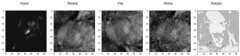
S14:对归一化处理之后的医学图像进行数据增强,扩大医学图像样本集。其中,所述数据增强包括对医学图像进行水平翻转50%、向上翻转、向下翻转、高斯滤波、均值滤波和中值滤波中的一种或多种,然后加入噪声。如图4所示,即为一种对医学图像进行数据增强的示例图。S14: Perform data enhancement on the normalized medical image to expand the medical image sample set. Wherein, the data enhancement includes performing one or more of horizontal flipping, upward flipping, downward flipping, Gaussian filtering, mean filtering and median filtering on the medical image, and then adding noise. As shown in FIG. 4 , it is an example diagram of data enhancement for medical images.
在已标注的医学图像数量有限的情况下,通过数据增强方式增加训练样本的多样性,可提高最后训练得到的模型的鲁棒性,避免过拟合。When the number of labeled medical images is limited, increasing the diversity of training samples through data augmentation can improve the robustness of the final trained model and avoid overfitting.
本实施例中,所述第一分类模型通过如下过程训练得到:In this embodiment, the first classification model is obtained through the following process training:
基于CNN-6构建网络模型,其前两个卷积层包含大小为3×3的32个核,后两个卷积层包含大小为3×3的64个核;第2和第4个卷积层与2×2维的最大池化层交错,差值为0.25;倒数第二层是512个神经元的全连接层和0.5个缺失层,最后一层是30个神经元的全连接层;ReLU激活函数应用于所有四个卷积层和倒数第二层的全连接层,最后一层通过softmax函数输出预测概率,确定医学图像的分类类别;The network model is constructed based on CNN-6, the first two convolutional layers contain 32 kernels of size 3×3, and the last two convolutional layers contain 64 kernels of size 3×3; the 2nd and 4th volumes The accumulation layer is interleaved with a 2×2-dimensional max-pooling layer with a difference of 0.25; the penultimate layer is a fully connected layer of 512 neurons and 0.5 missing layers, and the last layer is a fully connected layer of 30 neurons. ; The ReLU activation function is applied to all four convolutional layers and the fully connected layer of the penultimate layer, and the last layer outputs the predicted probability through the softmax function to determine the classification category of the medical image;
使用gloot统一来初始权重,然后利用医学图像样本集训练上述基于CNN-6构建的网络模型,得到第一分类模型。Use gloot to unify the initial weights, and then use the medical image sample set to train the above network model based on CNN-6 to obtain the first classification model.
本实施例中,所述第二分类模型通过如下过程训练得到:In this embodiment, the second classification model is obtained through the following process training:
获取ImageNet图像数据集;Get the ImageNet image dataset;
对ImageNet图像数据集中的ImageNet图像进行预处理,对RGB三个通道分别减去ImageNet图像数据集的均值,并进行正则化操作,得到ImageNet图像训练集;Preprocess the ImageNet images in the ImageNet image dataset, subtract the mean of the ImageNet image dataset from the three RGB channels, and perform regularization operations to obtain the ImageNet image training set;
利用ImageNet图像训练集对基于VGG-16网络结构的模型进行训练,得到预先训练好的VGG-16网络模型;Use the ImageNet image training set to train the model based on the VGG-16 network structure to obtain a pre-trained VGG-16 network model;
冻结预先训练好的VGG-16网络模型的预设数量的早期层,用30个神经元替换VGG-16网络模型最后一个全连接层,本实施例中,优选冻结除最后三层全连接层的其它层,然后使用gloot统一重新初始化VGG-16网络模型最后的三个全连接层的权重;最后利用医学图像样本集对上述改进后的VGG-16网络模型进行训练,得到第二分类模型。Freeze the preset number of early layers of the pre-trained VGG-16 network model, and replace the last fully connected layer of the VGG-16 network model with 30 neurons. In this embodiment, it is preferable to freeze all the fully connected layers except the last three layers Other layers, and then use gloot to uniformly re-initialize the weights of the last three fully connected layers of the VGG-16 network model; finally, use the medical image sample set to train the above-mentioned improved VGG-16 network model to obtain the second classification model.
本实施例中,所述第三分类模型通过如下过程训练得到:In this embodiment, the third classification model is obtained through the following process training:
获取ImageNet图像数据集;Get the ImageNet image dataset;
对ImageNet图像数据集中的ImageNet图像进行预处理,对RGB三个通道分别减去ImageNet图像数据集的均值,并进行正则化操作,得到ImageNet图像训练集;Preprocess the ImageNet images in the ImageNet image dataset, subtract the mean of the ImageNet image dataset from the three RGB channels, and perform regularization operations to obtain the ImageNet image training set;
利用ImageNet图像训练集对基于ResNet-50网络结构的模型进行训练,得到预先训练好的ResNet-50网络模型;Use the ImageNet image training set to train the model based on the ResNet-50 network structure to obtain a pre-trained ResNet-50 network model;
冻结预先训练好的ResNet-50网络模型的预设数量的早期层,本实施例中,优选冻结除最后一层全连接层的其它层,用30个神经元替换ResNet-50网络模型最后一个全连接层,然后使用gloot统一重新初始化ResNet-50网络模型最后一个全连接层的权重;最后利用医学图像样本集对上述改进后的ResNet-50网络模型进行训练,得到第三分类模型。Freeze the preset number of early layers of the pre-trained ResNet-50 network model. In this embodiment, it is preferable to freeze other layers except the last fully connected layer, and replace the last fully connected layer of the ResNet-50 network model with 30 neurons. connection layer, and then use gloot to uniformly re-initialize the weight of the last fully connected layer of the ResNet-50 network model; finally, use the medical image sample set to train the above-mentioned improved ResNet-50 network model to obtain the third classification model.
本实施例中,在训练第二分类模型和第三分类模型的过程中,使用Adma优化器和分类交叉熵损失函数在ImageNet图像数据集上对32个小批量的改进后的网络模型进行训练。In this embodiment, in the process of training the second classification model and the third classification model, the Adma optimizer and the classification cross-entropy loss function are used to train the improved network models in 32 mini-batches on the ImageNet image data set.
设Z为n幅医学图像的医学图像训练数据集。从无到有训练最后一层(top layer(s))是一个迭代的过程,找到使CNN(VGG-16、ResNet-50均属于CNN网络结构)的经验损失最小的权重w。分类交叉熵损失函数如下式所示:Let Z be a medical image training dataset of n medical images. Training the last layer (top layer(s)) from scratch is an iterative process to find the weight w that minimizes the empirical loss of the CNN (VGG-16, ResNet-50 both belong to the CNN network structure). The categorical cross-entropy loss function is as follows:

其中,xi是Z的第i幅图像,f(xi,w)是xi的类别yi的预测概率,y′i是xi的真实类别,k(yi,y′i)是用于yi预测的惩罚函数,其如下式所示:where xi is the ith image of Z, f(xi ,w) is the predicted probability of classyi of xi , y′i is the true class of xi , and k(yi ,y′i ) is Penalty function for yi prediction, which is given by:

其中,yi∈{1,...,C},C为常数,其取值与类别总数相等,

当使用当前权重应用到小批量时,根据损失函数L的梯度计算更新的权重,使用Adma来计算个体的自适应学习率来控制权重更新的大小。When applied to a mini-batch using the current weights, the updated weights are calculated according to the gradient of the loss function L, and Adma is used to calculate the individual adaptive learning rate to control the size of the weight update.
本实施例中,投票机制模块接收三个CNN计算的每种模式的强度,使用了不同的CNN,它们各自有不同的功能,来探索网络深度的核心重要性。CNN的输出组合负责产生每个模态的最终强度。投票机制是在Kuncheva等人的加权多数投票基础上的改进,本实施例使用了一个称为平均投票的组合规则,并给每个CNN产生的强度赋予不同的权重。In this embodiment, the voting mechanism module receives the strength of each mode calculated by three CNNs, and uses different CNNs, each with different functions, to explore the core importance of network depth. The output combination of the CNN is responsible for producing the final intensity of each modality. The voting mechanism is an improvement on the weighted majority voting of Kuncheva et al. This embodiment uses a combination rule called average voting and assigns different weights to the strengths produced by each CNN.
具体地,所述投票机制模块采用的投票机制通过如下公式表示:Specifically, the voting mechanism adopted by the voting mechanism module is expressed by the following formula:

其中,x表示输入的医学图像,y表示预测分类标号,
具体实施时,可利用Grad-CAM算法使用感兴趣或者制定类别的梯度解释在CNN模型的分类结果,可视化图像分类。In specific implementation, the Grad-CAM algorithm can be used to explain the classification results in the CNN model using the gradients of the interested or specified categories, and visualize the image classification.
实施例2Example 2
本实施例提供了一种基于深度迁移学习的医学图像分类模型的训练装置,包括:This embodiment provides a training device for a medical image classification model based on deep transfer learning, including:
图像获取模块,用于获取已标注的医学图像并进行预处理,得到医学图像样本集;The image acquisition module is used to acquire the labeled medical images and preprocess them to obtain a medical image sample set;
第一模型生成模块,用于利用医学图像样本集训练CNN网络模型得到第一分类模型;The first model generation module is used to train the CNN network model by using the medical image sample set to obtain the first classification model;
第二模型生成模块,用于获取在ImageNet图像数据集上预先训练好的VGG-16网络模型和ResNet-50网络模型;并基于迁移学习方法分别利用医学图像样本集再次训练VGG-16网络模型和ResNet-50网络模型,分别得到第二分类模型和第三分类模型;The second model generation module is used to obtain the pre-trained VGG-16 network model and ResNet-50 network model on the ImageNet image data set; and based on the transfer learning method, the VGG-16 network model and the medical image sample set are used to retrain the VGG-16 network model and ResNet-50 network model, respectively obtain the second classification model and the third classification model;
医学图像分类模型生成模块,用于将第一分类模型、第二分类模型、第三分类模型的输出连接到投票机制模块的输入构成医学图像分类模型,其中,投票机制模块基于预设投票机制输出最终医学图像分类结果为医学图像分类模型的分类结果。The medical image classification model generation module is used to connect the outputs of the first classification model, the second classification model, and the third classification model to the input of the voting mechanism module to form a medical image classification model, wherein the voting mechanism module outputs based on a preset voting mechanism The final medical image classification result is the classification result of the medical image classification model.
实施例3Example 3
本实施例提供了一种前列腺医学图像分类模型的训练方法,其训练方法采用如实施例1所述的一种基于深度迁移学习的医学图像分类模型的训练方法,其中医学图像为前列腺医学图像。This embodiment provides a training method for a prostate medical image classification model, and the training method adopts the training method for a medical image classification model based on deep transfer learning as described in Embodiment 1, wherein the medical image is a prostate medical image.
实施例4Example 4
本实施例提供了一种计算机可读存储介质,其存储有计算机程序,该计算机程序被处理器加载时执行如实施例1或实施例3所述的方法。This embodiment provides a computer-readable storage medium, which stores a computer program, and when the computer program is loaded by a processor, executes the method described in Embodiment 1 or Embodiment 3.
本领域内的技术人员应明白,本申请的实施例可提供为方法、系统、或计算机程序产品。因此,本申请可采用完全硬件实施例、完全软件实施例、或结合软件和硬件方面的实施例的形式。而且,本申请可采用在一个或多个其中包含有计算机可用程序代码的计算机可用存储介质(包括但不限于磁盘存储器、CD-ROM、光学存储器等)上实施的计算机程序产品的形式。As will be appreciated by those skilled in the art, the embodiments of the present application may be provided as a method, a system, or a computer program product. Accordingly, the present application may take the form of an entirely hardware embodiment, an entirely software embodiment, or an embodiment combining software and hardware aspects. Furthermore, the present application may take the form of a computer program product embodied on one or more computer-usable storage media (including, but not limited to, disk storage, CD-ROM, optical storage, etc.) having computer-usable program code embodied therein.
本申请是参照根据本申请实施例的方法、设备(系统)、和计算机程序产品的流程图和/或方框图来描述的。应理解可由计算机程序指令实现流程图和/或方框图中的每一流程和/或方框、以及流程图和/或方框图中的流程和/或方框的结合。可提供这些计算机程序指令到通用计算机、专用计算机、嵌入式处理机或其他可编程数据处理设备的处理器以产生一个机器,使得通过计算机或其他可编程数据处理设备的处理器执行的指令产生用于实现在流程图一个流程或多个流程和/或方框图一个方框或多个方框中指定的功能的装置。The present application is described with reference to flowchart illustrations and/or block diagrams of methods, apparatus (systems), and computer program products according to embodiments of the present application. It will be understood that each flow and/or block in the flowchart illustrations and/or block diagrams, and combinations of flows and/or blocks in the flowchart illustrations and/or block diagrams, can be implemented by computer program instructions. These computer program instructions may be provided to the processor of a general purpose computer, special purpose computer, embedded processor or other programmable data processing device to produce a machine such that the instructions executed by the processor of the computer or other programmable data processing device produce Means for implementing the functions specified in a flow or flow of a flowchart and/or a block or blocks of a block diagram.
这些计算机程序指令也可存储在能引导计算机或其他可编程数据处理设备以特定方式工作的计算机可读存储器中,使得存储在该计算机可读存储器中的指令产生包括指令装置的制造品,该指令装置实现在流程图一个流程或多个流程和/或方框图一个方框或多个方框中指定的功能。These computer program instructions may also be stored in a computer-readable memory capable of directing a computer or other programmable data processing apparatus to function in a particular manner, such that the instructions stored in the computer-readable memory result in an article of manufacture comprising instruction means, the instructions The apparatus implements the functions specified in the flow or flow of the flowcharts and/or the block or blocks of the block diagrams.
这些计算机程序指令也可装载到计算机或其他可编程数据处理设备上,使得在计算机或其他可编程设备上执行一系列操作步骤以产生计算机实现的处理,从而在计算机或其他可编程设备上执行的指令提供用于实现在流程图一个流程或多个流程和/或方框图一个方框或多个方框中指定的功能的步骤。These computer program instructions can also be loaded on a computer or other programmable data processing device to cause a series of operational steps to be performed on the computer or other programmable device to produce a computer-implemented process such that The instructions provide steps for implementing the functions specified in the flow or blocks of the flowcharts and/or the block or blocks of the block diagrams.
可以理解的是,上述各实施例中相同或相似部分可以相互参考,在一些实施例中未详细说明的内容可以参见其他实施例中相同或相似的内容。It can be understood that, the same or similar parts in the above embodiments may refer to each other, and the content not described in detail in some embodiments may refer to the same or similar content in other embodiments.
流程图中或在此以其他方式描述的任何过程或方法描述可以被理解为,表示包括一个或更多个用于实现特定逻辑功能或过程的步骤的可执行指令的代码的模块、片段或部分,并且本发明的优选实施方式的范围包括另外的实现,其中可以不按所示出或讨论的顺序,包括根据所涉及的功能按基本同时的方式或按相反的顺序,来执行功能,这应被本发明的实施例所属技术领域的技术人员所理解。Any description of a process or method in the flowcharts or otherwise described herein may be understood to represent a module, segment or portion of code comprising one or more executable instructions for implementing a specified logical function or step of the process , and the scope of the preferred embodiments of the invention includes alternative implementations in which the functions may be performed out of the order shown or discussed, including performing the functions substantially concurrently or in the reverse order depending upon the functions involved, which should It is understood by those skilled in the art to which the embodiments of the present invention belong.
为了进一步理解本发明的方案,在此以前列腺癌医学图像为例提供一个分类实验对本方案进行说明。In order to further understand the solution of the present invention, a classification experiment is provided to illustrate the solution by taking a medical image of prostate cancer as an example.
实验环境为Python,使用Keras库来实现深度cnn。在实验中加载Keras提供的预先训练好的cnn的权重。The experimental environment is Python, and the Keras library is used to implement deep cnn. Load the weights of the pre-trained CNN provided by Keras in the experiment.
数据集包括204名患者的训练数据集(330例可疑病变)和140名患者的测试数据集(208例)。由于T2横切面、ADC值、DWI、KTrans均为前列腺的横切面,故将它们结合起来用于前列腺病变的分类。T2矢状位序列是前列腺的侧视面,则没有使用。对于每一个发现,前列腺解剖区域的分配是可行的。前列腺可细分为4个解剖区:外周区(PZ),占腺组织的70-80%,约占前列腺癌的70%;过渡区(TZ),占腺组织的5%,约占PCa(前列腺癌)的25%;中央区域(仅作说明),占腺组织的20%,约占PCa的5%;和非腺体的前纤维肌间质(AS)。PROSTATEx挑战中的训练和测试样本来自PZ、TZ、AS和精囊(SV),如表1所示。The dataset consists of a training dataset of 204 patients (330 suspicious lesions) and a test dataset of 140 patients (208). Since T2 cross-section, ADC value, DWI, and KTrans are all cross-sections of the prostate, they are combined for the classification of prostate lesions. The T2 sagittal sequence is the lateral view of the prostate and is not used. For each finding, the assignment of prostate anatomical regions was feasible. The prostate can be subdivided into 4 anatomical zones: the peripheral zone (PZ), which accounts for 70-80% of the glandular tissue and accounts for about 70% of prostate cancers; the transition zone (TZ), which accounts for 5% of the glandular tissue and accounts for about 70% of the PCa ( 25% of prostate cancer); the central region (illustration only), which is 20% of glandular tissue and approximately 5% of PCa; and non-glandular prefibromyal stroma (AS). The training and testing samples in the PROSTATEx challenge are from PZ, TZ, AS, and seminal vesicle (SV), as shown in Table 1.
表1 ProstateX数据集划分Table 1 ProstateX dataset division

在对少量数据进行清理后,本发明选择了201名受试者,其中321项发现用于培训和验证目的。为了扩大和平衡训练数据集,本发明使用了翻转和转换原始数据。作为数据扩充的结果,本发明生成了5个交叉验证数据集,每个交叉验证数据集有10000个训练和2000个验证样本。在每一折叠中进行训练验证的分离,使得研究结果在前列腺区域的分布得以保留。图像强度归一化到[0,1]范围内。T2为40×40×40mm,DWI为32×32×12,DCE-MRI为32×32×12,以寻找位置为中心的3D patch作为CNN的输入。After cleaning a small amount of data, the present invention selected 201 subjects, of which 321 were found for training and validation purposes. To expand and balance the training dataset, the present invention uses flipping and transforming the original data. As a result of data augmentation, the present invention generates 5 cross-validation datasets, each with 10,000 training and 2,000 validation samples. A training-validation separation was performed within each fold, so that the distribution of findings across the prostate region was preserved. Image intensities are normalized to the range [0,1]. T2 is 40 × 40 × 40 mm, DWI is 32 × 32 × 12, and DCE-MRI is 32 × 32 × 12. The 3D patch centered on the search position is used as the input of CNN.
为了训练网络,本发明使用了随机梯度下降算法与Adam更新规则,10次一个小批量64,和一个二元交叉熵损失函数。本发明使用He方法从高斯分布随机初始化CNN的权值。本发明还对所有层的中间响应进行批处理正常化,以加速收敛。为了防止过拟合,除了批量归一化之外,本发明还使用了概率为0.25的dropout和对神经元权重有惩罚的L2正则化。通过监控验证性能,本发明使用了一种早期停止策略,并在验证集上选择了精确度最高的最佳模型。交叉验证用于为卷积层找到输入通道和过滤器数量的最佳组合。To train the network, the present invention uses stochastic gradient descent algorithm with Adam update rule, 10 times a mini-batch 64, and a binary cross-entropy loss function. The present invention uses the He method to randomly initialize the weights of the CNN from a Gaussian distribution. The present invention also batch normalizes the intermediate responses of all layers to speed up the convergence. To prevent overfitting, in addition to batch normalization, the present invention uses dropout with probability 0.25 and L2 regularization with penalty on neuron weights. By monitoring the validation performance, the present invention uses an early stopping strategy and selects the best model with the highest accuracy on the validation set. Cross-validation is used to find the best combination of input channels and number of filters for the convolutional layers.
表2不同模型下区域精度对比Table 2 Comparison of regional accuracy under different models

基于迁移学习和投票机制的分类模型在ProstateX(前列腺)数据集上进行端到端的训练,其Top-1 Accuary在PZ上高达93.08%,TZ达到81.34,AS达到83.45,SV达到82.93。在训练过程中,RseNet50网络分类结果,相对于VGG网络具有更少的尺寸和参数量,却有更高的精度;根据不同模型的加权平均组合,能够有限对前列腺区域分类,且精度达到最好的性能。The classification model based on transfer learning and voting mechanism is trained end-to-end on the ProstateX (prostate) dataset, and its Top-1 Accuary reaches 93.08% on PZ, 81.34 on TZ, 83.45 on AS, and 82.93 on SV. In the training process, the classification results of the RseNet50 network have less size and parameter quantity than the VGG network, but have higher accuracy; according to the weighted average combination of different models, the prostate region can be classified limitedly, and the accuracy is the best performance.
本发明提出了一种基于深度迁移学习的医学图像分类模型的训练方法及装置,该医学图像分类模型模型由三个不同深度的cnn组成,通过对预测概率加权平均进行组合。为了避免过拟合和获取特征信息,通过翻转和噪声等进行数据增强缓解类别不平衡,网络深度对于当前任务至关重要,迁移学习方法可以受益于在ImageNet数据集上预先训练的CNN捕捉到的一般特征,以及在医学图像上从头训练的极深CNN和另一个“浅层”CNN的顶层捕捉到的领域特定特征。实验结果表明引入深度迁移学习改善前列腺图像分类,具有泛化性和鲁棒性。The invention provides a training method and device for a medical image classification model based on deep migration learning. The medical image classification model model is composed of three CNNs with different depths, which are combined by weighted average of prediction probabilities. In order to avoid overfitting and obtain feature information, data augmentation through flipping and noise, etc. to alleviate class imbalance, network depth is crucial for current tasks, and transfer learning methods can benefit from pre-trained CNNs on ImageNet datasets. General features, as well as domain-specific features captured by the top layers of an extremely deep CNN trained from scratch on medical images and another "shallow" CNN. The experimental results show that the introduction of deep transfer learning improves prostate image classification with generalization and robustness.
尽管上面已经示出和描述了本发明的实施例,可以理解的是,上述实施例是示例性的,不能理解为对本发明的限制,本领域的普通技术人员在本发明的范围内可以对上述实施例进行变化、修改、替换和变型。Although the embodiments of the present invention have been shown and described above, it should be understood that the above embodiments are exemplary and should not be construed as limiting the present invention. Embodiments are subject to variations, modifications, substitutions and variations.
Claims (8)


Priority Applications (1)
| Application Number | Priority Date | Filing Date | Title |
|---|---|---|---|
| CN202110147726.8ACN112819076B (en) | 2021-02-03 | 2021-02-03 | Training method and device for medical image classification model based on deep transfer learning |
Applications Claiming Priority (1)
| Application Number | Priority Date | Filing Date | Title |
|---|---|---|---|
| CN202110147726.8ACN112819076B (en) | 2021-02-03 | 2021-02-03 | Training method and device for medical image classification model based on deep transfer learning |
Publications (2)
| Publication Number | Publication Date |
|---|---|
| CN112819076A CN112819076A (en) | 2021-05-18 |
| CN112819076Btrue CN112819076B (en) | 2022-06-17 |
Family
ID=75860794
Family Applications (1)
| Application Number | Title | Priority Date | Filing Date |
|---|---|---|---|
| CN202110147726.8AActiveCN112819076B (en) | 2021-02-03 | 2021-02-03 | Training method and device for medical image classification model based on deep transfer learning |
Country Status (1)
| Country | Link |
|---|---|
| CN (1) | CN112819076B (en) |
Families Citing this family (25)
| Publication number | Priority date | Publication date | Assignee | Title |
|---|---|---|---|---|
| CN112466461A (en)* | 2020-10-26 | 2021-03-09 | 淮阴工学院 | Medical image intelligent diagnosis method based on multi-network integration |
| CN113378984B (en)* | 2021-07-05 | 2023-05-02 | 国药(武汉)医学实验室有限公司 | Medical image classification method, system, terminal and storage medium |
| CN113592027B (en)* | 2021-08-16 | 2024-07-23 | 南京工程学院 | Medical image classification method based on transfer learning |
| CN114022521B (en)* | 2021-10-13 | 2024-09-13 | 华中科技大学 | A non-rigid multimodal medical image registration method and system |
| CN114092740A (en)* | 2021-11-11 | 2022-02-25 | 成都云芯医联科技有限公司 | AI-assisted analysis method for immune lateral flow sensing |
| CN114202524A (en)* | 2021-12-10 | 2022-03-18 | 中国人民解放军陆军特色医学中心 | Performance evaluation method and system of multi-modal medical image |
| CN114723985B (en)* | 2022-03-14 | 2025-08-01 | 山西三友和智慧信息技术股份有限公司 | Ultrasonic image classification method based on small training data set |
| CN114881155B (en)* | 2022-05-13 | 2024-11-29 | 重庆邮电大学 | Fruit image classification method based on deep migration learning |
| CN115035405B (en)* | 2022-05-26 | 2024-09-10 | 华南农业大学 | Citrus leaf disease identification method, system and equipment based on double transfer learning |
| CN115082742B (en)* | 2022-08-02 | 2025-04-29 | 浙江医准智能科技有限公司 | Image classification model training method, device, electronic device and storage medium |
| CN115331053A (en)* | 2022-08-11 | 2022-11-11 | 厦门大学 | An image classification model generation method and device based on L2NU activation function |
| CN115147668B (en)* | 2022-09-06 | 2022-12-27 | 北京鹰瞳科技发展股份有限公司 | Training method of disease classification model, disease classification method and related products |
| CN115546109B (en)* | 2022-09-09 | 2023-10-27 | 武汉中数医疗科技有限公司 | A method and device for identifying thyroid sampling data based on machine learning |
| US20240095907A1 (en)* | 2022-09-20 | 2024-03-21 | United Imaging Intelligence (Beijing) Co., Ltd. | Deep learning based systems and methods for detecting breast diseases |
| CN115797266A (en)* | 2022-11-09 | 2023-03-14 | 北京百度网讯科技有限公司 | Training method of medical image processing model, image processing method and device |
| CN116012649B (en)* | 2022-12-30 | 2023-09-19 | 东莞理工学院 | Integrated learning voting classification method, system and terminal for medical images |
| CN115797750B (en)* | 2023-02-02 | 2023-04-25 | 天津滨海迅腾科技集团有限公司 | Large-size image rapid transmission method based on deep learning algorithm |
| CN116486177A (en)* | 2023-05-15 | 2023-07-25 | 青岛中科研海海洋科技有限公司 | A method for underwater target recognition and classification based on deep learning |
| CN116704305A (en)* | 2023-06-20 | 2023-09-05 | 华中科技大学同济医学院附属协和医院 | Multi-modal and multi-section classification method for echocardiography based on deep learning algorithm |
| CN116824298A (en)* | 2023-06-27 | 2023-09-29 | 上海交通大学 | Automatic analysis method and system for medical images based on domain feature alignment transfer learning |
| CN116630867B (en)* | 2023-07-25 | 2023-11-17 | 深圳市美侨医疗科技有限公司 | Mask R-CNN-based leucorrhea trichomonas detection and tracking method |
| CN117058442A (en)* | 2023-07-27 | 2023-11-14 | 中国医科大学附属盛京医院 | High-risk fetal gallbladder classification method for biliary tract occlusion |
| CN117351293B (en)* | 2023-12-04 | 2024-02-06 | 天津医科大学口腔医院 | Combined learning periodontal disease image classification method and device |
| CN117454940B (en)* | 2023-12-21 | 2024-04-09 | 天津市肿瘤医院(天津医科大学肿瘤医院) | Training method and image processing method for predicting thyroid nodule metastasis |
| CN119649116A (en)* | 2024-11-27 | 2025-03-18 | 昆明医科大学第一附属医院(云南省皮肤病医院) | An AI-assisted screening method based on mammography |
Citations (10)
| Publication number | Priority date | Publication date | Assignee | Title |
|---|---|---|---|---|
| EP2073500A1 (en)* | 2007-12-21 | 2009-06-24 | SES Astra S.A. | Method for controlling the transfer of data entities from a server unit on a communication channel |
| CN102521656A (en)* | 2011-12-29 | 2012-06-27 | 北京工商大学 | Integrated transfer learning method for classification of unbalance samples |
| CN106780482A (en)* | 2017-01-08 | 2017-05-31 | 广东工业大学 | A kind of classification method of medical image |
| CN108647741A (en)* | 2018-05-18 | 2018-10-12 | 湖北工业大学 | A kind of image classification method and system based on transfer learning |
| CN108960073A (en)* | 2018-06-05 | 2018-12-07 | 大连理工大学 | Cross-module state image steganalysis method towards Biomedical literature |
| CN108985173A (en)* | 2018-06-19 | 2018-12-11 | 奕通信息科技(上海)股份有限公司 | Towards the depth network migration learning method for having the label apparent age data library of noise |
| EP3617943A1 (en)* | 2018-08-30 | 2020-03-04 | Topcon Corporation | Multivariate and multi-resolution retinal image anomaly detection system |
| CN111079620A (en)* | 2019-12-10 | 2020-04-28 | 北京小蝇科技有限责任公司 | Leukocyte image detection and identification model construction method based on transfer learning and application |
| CN111783841A (en)* | 2020-06-09 | 2020-10-16 | 中科院成都信息技术股份有限公司 | Garbage classification method, system and medium based on transfer learning and model fusion |
| CN112270379A (en)* | 2020-11-13 | 2021-01-26 | 北京百度网讯科技有限公司 | Training method of classification model, sample classification method, apparatus and equipment |
Family Cites Families (2)
| Publication number | Priority date | Publication date | Assignee | Title |
|---|---|---|---|---|
| US8331655B2 (en)* | 2008-06-30 | 2012-12-11 | Canon Kabushiki Kaisha | Learning apparatus for pattern detector, learning method and computer-readable storage medium |
| US10007863B1 (en)* | 2015-06-05 | 2018-06-26 | Gracenote, Inc. | Logo recognition in images and videos |
- 2021
- 2021-02-03CNCN202110147726.8Apatent/CN112819076B/enactiveActive
Patent Citations (10)
| Publication number | Priority date | Publication date | Assignee | Title |
|---|---|---|---|---|
| EP2073500A1 (en)* | 2007-12-21 | 2009-06-24 | SES Astra S.A. | Method for controlling the transfer of data entities from a server unit on a communication channel |
| CN102521656A (en)* | 2011-12-29 | 2012-06-27 | 北京工商大学 | Integrated transfer learning method for classification of unbalance samples |
| CN106780482A (en)* | 2017-01-08 | 2017-05-31 | 广东工业大学 | A kind of classification method of medical image |
| CN108647741A (en)* | 2018-05-18 | 2018-10-12 | 湖北工业大学 | A kind of image classification method and system based on transfer learning |
| CN108960073A (en)* | 2018-06-05 | 2018-12-07 | 大连理工大学 | Cross-module state image steganalysis method towards Biomedical literature |
| CN108985173A (en)* | 2018-06-19 | 2018-12-11 | 奕通信息科技(上海)股份有限公司 | Towards the depth network migration learning method for having the label apparent age data library of noise |
| EP3617943A1 (en)* | 2018-08-30 | 2020-03-04 | Topcon Corporation | Multivariate and multi-resolution retinal image anomaly detection system |
| CN111079620A (en)* | 2019-12-10 | 2020-04-28 | 北京小蝇科技有限责任公司 | Leukocyte image detection and identification model construction method based on transfer learning and application |
| CN111783841A (en)* | 2020-06-09 | 2020-10-16 | 中科院成都信息技术股份有限公司 | Garbage classification method, system and medium based on transfer learning and model fusion |
| CN112270379A (en)* | 2020-11-13 | 2021-01-26 | 北京百度网讯科技有限公司 | Training method of classification model, sample classification method, apparatus and equipment |
Non-Patent Citations (3)
| Title |
|---|
| Vote-based Image classification Using Linear Manifolds;Minegishi H et al;《Journal of the Institute of Image Electronics Engineers of Japan》;20111231;全文* |
| 基于图像目标特征空间自学习分类算法;贾广象等;《计算机工程与应用》;20151231;第51卷(第20期);全文* |
| 深度神经网络内部迁移的信息几何度量分析;陈力等;《湖南大学学报(自然科学版)》;20191231;第46卷(第02期);全文* |
Also Published As
| Publication number | Publication date |
|---|---|
| CN112819076A (en) | 2021-05-18 |
Similar Documents
| Publication | Publication Date | Title |
|---|---|---|
| CN112819076B (en) | Training method and device for medical image classification model based on deep transfer learning | |
| Agnes et al. | Classification of mammogram images using multiscale all convolutional neural network (MA-CNN) | |
| Singh et al. | Shallow 3D CNN for detecting acute brain hemorrhage from medical imaging sensors | |
| Yamuna et al. | Integrating AI for Improved Brain Tumor Detection and Classification | |
| Biffi et al. | Explainable anatomical shape analysis through deep hierarchical generative models | |
| Naeem et al. | DVFNet: A deep feature fusion-based model for the multiclassification of skin cancer utilizing dermoscopy images | |
| Ashwin et al. | Efficient and reliable lung nodule detection using a neural network based computer aided diagnosis system | |
| Shen et al. | Decision supporting model for one-year conversion probability from MCI to AD using CNN and SVM | |
| WO2022127500A1 (en) | Multiple neural networks-based mri image segmentation method and apparatus, and device | |
| Vinta et al. | Segmentation and classification of interstitial lung diseases based on hybrid deep learning network model | |
| Al Jannat et al. | Detection of multiple sclerosis using deep learning | |
| Karpakam et al. | A multi-modality framework for precise brain tumor detection and multi-class classification using hybrid GAN approach | |
| JP7519821B2 (en) | Medical system and medical information processing method | |
| Mankar et al. | A Review on Multiclass Brain Tumor Detection using Convolutional Neural Network and Support Vector Machines | |
| Kassim et al. | Sensitivity of cross-trained deep cnns for retinal vessel extraction | |
| Yu et al. | 3D convolutional networks based automatic diagnosis of Alzheimer's disease using structural MRI | |
| Adityasai et al. | Advancing Alzheimer's Diagnosis Through Transfer Learning with Deep MRI Analysis | |
| Radhika et al. | Effective Deep Learning Network Model for Multi-Class Skin Cancer Classification. | |
| Anitha et al. | Segmentation and classification of gastric cancer from endoscopic image dataset with the aid of artificial intelligence | |
| Gupta et al. | Lungs Disease Classification using VGG-16 architecture with PCA | |
| Nair et al. | A Review on DeepLungNet: CNN-Based Lung Cancer Detection Techniques Using CT Images | |
| Appavu | An Innovative Method for Detecting Brain Tumors that Combines Artificial Intelligence (AI), Machine Learning (ML), and Deep Learning with Artificial Data Augmentation | |
| Thota et al. | INCS: A Novel Brain Tumor Detection Methodology Design Based on Deep Learning Assisted Intelligent Neural Classification Strategy | |
| Vidyarthi et al. | Novel Architecture and Comparative Study for Deep Learning-Based Segmentation of 3D Brain MRI Images Using SegNet, V-Net, and U-Net | |
| Aiswarya et al. | Brain Tumor Classification Using ResNet-50 |
Legal Events
| Date | Code | Title | Description |
|---|---|---|---|
| PB01 | Publication | ||
| PB01 | Publication | ||
| SE01 | Entry into force of request for substantive examination | ||
| SE01 | Entry into force of request for substantive examination | ||
| GR01 | Patent grant | ||
| GR01 | Patent grant |