CN112603524B - Interatrial septum tissue stoma device and interatrial septum group weaving mouth system - Google Patents
Interatrial septum tissue stoma device and interatrial septum group weaving mouth systemDownload PDFInfo
- Publication number
- CN112603524B CN112603524BCN202011384593.8ACN202011384593ACN112603524BCN 112603524 BCN112603524 BCN 112603524BCN 202011384593 ACN202011384593 ACN 202011384593ACN 112603524 BCN112603524 BCN 112603524B
- Authority
- CN
- China
- Prior art keywords
- electrode
- ostomy
- stoma
- tissue
- interatrial septum
- Prior art date
- Legal status (The legal status is an assumption and is not a legal conclusion. Google has not performed a legal analysis and makes no representation as to the accuracy of the status listed.)
- Active
Links
Images


Classifications
- A—HUMAN NECESSITIES
- A61—MEDICAL OR VETERINARY SCIENCE; HYGIENE
- A61B—DIAGNOSIS; SURGERY; IDENTIFICATION
- A61B18/00—Surgical instruments, devices or methods for transferring non-mechanical forms of energy to or from the body
- A61B18/04—Surgical instruments, devices or methods for transferring non-mechanical forms of energy to or from the body by heating
- A61B18/12—Surgical instruments, devices or methods for transferring non-mechanical forms of energy to or from the body by heating by passing a current through the tissue to be heated, e.g. high-frequency current
- A61B18/14—Probes or electrodes therefor
- A—HUMAN NECESSITIES
- A61—MEDICAL OR VETERINARY SCIENCE; HYGIENE
- A61B—DIAGNOSIS; SURGERY; IDENTIFICATION
- A61B18/00—Surgical instruments, devices or methods for transferring non-mechanical forms of energy to or from the body
- A61B2018/00315—Surgical instruments, devices or methods for transferring non-mechanical forms of energy to or from the body for treatment of particular body parts
- A61B2018/00345—Vascular system
- A61B2018/00351—Heart
- A—HUMAN NECESSITIES
- A61—MEDICAL OR VETERINARY SCIENCE; HYGIENE
- A61B—DIAGNOSIS; SURGERY; IDENTIFICATION
- A61B18/00—Surgical instruments, devices or methods for transferring non-mechanical forms of energy to or from the body
- A61B2018/00571—Surgical instruments, devices or methods for transferring non-mechanical forms of energy to or from the body for achieving a particular surgical effect
- A61B2018/00595—Cauterization
- A—HUMAN NECESSITIES
- A61—MEDICAL OR VETERINARY SCIENCE; HYGIENE
- A61B—DIAGNOSIS; SURGERY; IDENTIFICATION
- A61B18/00—Surgical instruments, devices or methods for transferring non-mechanical forms of energy to or from the body
- A61B2018/00636—Sensing and controlling the application of energy
- A61B2018/00696—Controlled or regulated parameters
- A61B2018/00702—Power or energy
- A—HUMAN NECESSITIES
- A61—MEDICAL OR VETERINARY SCIENCE; HYGIENE
- A61B—DIAGNOSIS; SURGERY; IDENTIFICATION
- A61B18/00—Surgical instruments, devices or methods for transferring non-mechanical forms of energy to or from the body
- A61B2018/00636—Sensing and controlling the application of energy
- A61B2018/00696—Controlled or regulated parameters
- A61B2018/00714—Temperature
- A—HUMAN NECESSITIES
- A61—MEDICAL OR VETERINARY SCIENCE; HYGIENE
- A61B—DIAGNOSIS; SURGERY; IDENTIFICATION
- A61B18/00—Surgical instruments, devices or methods for transferring non-mechanical forms of energy to or from the body
- A61B18/04—Surgical instruments, devices or methods for transferring non-mechanical forms of energy to or from the body by heating
- A61B18/12—Surgical instruments, devices or methods for transferring non-mechanical forms of energy to or from the body by heating by passing a current through the tissue to be heated, e.g. high-frequency current
- A61B18/14—Probes or electrodes therefor
- A61B2018/1405—Electrodes having a specific shape
- A61B2018/1407—Loop
- A—HUMAN NECESSITIES
- A61—MEDICAL OR VETERINARY SCIENCE; HYGIENE
- A61B—DIAGNOSIS; SURGERY; IDENTIFICATION
- A61B18/00—Surgical instruments, devices or methods for transferring non-mechanical forms of energy to or from the body
- A61B18/04—Surgical instruments, devices or methods for transferring non-mechanical forms of energy to or from the body by heating
- A61B18/12—Surgical instruments, devices or methods for transferring non-mechanical forms of energy to or from the body by heating by passing a current through the tissue to be heated, e.g. high-frequency current
- A61B18/14—Probes or electrodes therefor
- A61B2018/1467—Probes or electrodes therefor using more than two electrodes on a single probe
Landscapes
- Health & Medical Sciences (AREA)
- Surgery (AREA)
- Engineering & Computer Science (AREA)
- Life Sciences & Earth Sciences (AREA)
- Biomedical Technology (AREA)
- Otolaryngology (AREA)
- Nuclear Medicine, Radiotherapy & Molecular Imaging (AREA)
- Plasma & Fusion (AREA)
- Physics & Mathematics (AREA)
- Heart & Thoracic Surgery (AREA)
- Medical Informatics (AREA)
- Molecular Biology (AREA)
- Animal Behavior & Ethology (AREA)
- General Health & Medical Sciences (AREA)
- Public Health (AREA)
- Veterinary Medicine (AREA)
- Surgical Instruments (AREA)
Abstract
Description
Translated fromChinese技术领域technical field
本发明涉及医疗器械技术领域,尤其涉及一种房间隔组织造口装置及房间隔组织造口系统。The invention relates to the technical field of medical devices, in particular to an atrial septal tissue stoma device and an atrial septal tissue stoma system.
背景技术Background technique
房间隔组织造口术通过在患者的左心房和右心房之间的房间隔组织上形成一个造口,以形成左右心房间的分流,而实现改善心衰患者或者肺动脉高血压患者的症状。Atrial septostomy is to improve the symptoms of patients with heart failure or pulmonary hypertension by forming an opening in the atrial septal tissue between the left atrium and right atrium to form a shunt between the left and right atrium.
传统的房间隔组织造口方法,通过在房间隔组织造口处植入分流器械。具体的,在经皮房间隔组织穿刺术后,在房间隔组织穿刺处植入分流器械,以保持分流开口处通畅。然而,分流器械容易导致血栓形成,且内皮容易爬附在分流器械上容易导致分流开口被封堵,从而失去左右心房间的分流作用。The traditional atrial septotomy method is to implant a shunt device at the atrial septal stoma. Specifically, after the percutaneous atrial septal tissue puncture, a shunt device is implanted at the puncture site of the atrial septal tissue to keep the opening of the shunt unobstructed. However, the shunt device easily leads to thrombus formation, and the endothelium is easy to climb and adhere to the shunt device, which may easily cause the shunt opening to be blocked, thereby losing the shunt effect of the left and right atrium.
现有的房间隔组织造口方法,通过造口器械对房间隔组织进行造口并在手术后撤离造口器械。造口器械包括切割装置和抓取装置。在造口过程中,抓取装置先对待切割的组织进行定位并抓取,然后由切割装置的切割部对抓取装置所抓取的部分组织进行切割,切割下来的部分组织被抓取装置带出体外,从而形成造口。由于在手术过程中通过使用机械或高频电刀对房间隔组织进行切割,因此存在较高的风险。此外,抓取装置在手术中容易发生松动而导致其它心肌组织受损,且抓取装置在回收时,可能导致所切割的组织脱落而导致栓塞形成。In the existing atrial septal tissue stoma method, a stoma instrument is used to stoma the atrial septal tissue and the ostomy instrument is withdrawn after the operation. An ostomy appliance includes a cutting device and a grasping device. During the stoma process, the grasping device first positions and grasps the tissue to be cut, and then the cutting part of the cutting device cuts part of the tissue grasped by the grasping device, and the cut part of the tissue is taken by the grasping device. out of the body, forming a stoma. There is a higher risk due to the cutting of atrial septal tissue during the procedure by using mechanical or high-frequency electrocautery. In addition, the grasping device is prone to loosening during the operation, causing damage to other myocardial tissues, and when the grasping device is recovered, the cut tissue may fall off, resulting in embolism.
现有的房间隔组织造口方法,在造口前需要对房间隔组织进行机械穿刺,不仅穿刺困难,且容易造成房间隔组织撕裂。In the existing atrial septal tissue stoma method, the atrial septal tissue needs to be mechanically punctured before the stoma, which is not only difficult to puncture, but also easily causes tearing of the atrial septal tissue.
发明内容Contents of the invention
有鉴于此,本发明实施例有必要提供一种房间隔组织造口装置及房间隔组织造口系统,以解决上述技术问题。In view of this, the embodiment of the present invention needs to provide an atrial septal tissue stoma device and an atrial septal tissue stoma system to solve the above technical problems.
第一方面,本发明实施例提供一种房间隔组织造口装置,包括造口支架及穿设于所述造口支架中的鞘芯,所述造口支架包括造口部,所述鞘芯的远端设置有与消融电源电连接的第一电极,所述第一电极用于对房间隔组织进行消融穿刺,以供所述造口部穿过所述房间隔组织的穿刺处并膨胀将所述房间隔组织撑开。In the first aspect, an embodiment of the present invention provides an atrial septal tissue ostomy device, which includes a stoma support and a sheath core pierced in the stoma support, the stoma support includes a stoma part, and the sheath core The distal end is provided with a first electrode electrically connected to the ablation power supply, and the first electrode is used to ablate and puncture the atrial septal tissue, so that the stoma can pass through the puncture site of the atrial septal tissue and expand. The interatrial septal tissue is stretched.
第二方面,本发明实施例提供一种房间隔组织造口系统,包括如上所述的房间隔组织造口装置、控制手柄及鞘管,所述控制手柄外接所述消融能源,所述控制手柄用于控制所述造口支架和所述鞘芯可活动地收容于所述鞘管内或伸出所述鞘管外。In the second aspect, an embodiment of the present invention provides an atrial septal tissue stoma system, including the above-mentioned atrial septal tissue stoma device, a control handle and a sheath tube, the control handle is externally connected to the ablation energy source, and the control handle It is used to control the ostomy support and the sheath core to be movably accommodated in the sheath or protruded out of the sheath.
本发明实施例提供了一种房间隔组织造口装置及房间隔组织造口系统,由于鞘芯的远端设置有与消融电源电连接的第一电极,因此可以通过使用第一电极接收到的射频能源对房间隔组织进行消融穿刺,无需切割房间隔组织,从而避免了机械穿刺造成的损伤问题、方便穿刺操作、且确保了房间隔组织造口不易回缩或闭合。此外,由于造口支架和鞘芯可撤离房间隔组织,因此避免留下器械而导致血栓形成或器械脱落造成栓塞的问题,从而提高了房间隔组织造口装置及其系统的使用安全性。Embodiments of the present invention provide an atrial septal tissue stoma device and an atrial septal tissue stoma system. Since the distal end of the sheath core is provided with a first electrode electrically connected to the ablation power supply, the The radiofrequency energy performs ablation and puncture of the atrial septal tissue without cutting the atrial septal tissue, thereby avoiding the damage caused by mechanical puncture, facilitating the puncture operation, and ensuring that the stoma of the atrial septal tissue is not easy to retract or close. In addition, since the stoma stent and the sheath core can be withdrawn from the atrial septal tissue, the problem of thrombus formation caused by leaving the device or embolism caused by the fall of the device is avoided, thereby improving the safety of the atrial septal tissue stoma device and its system.
附图说明Description of drawings
为了更清楚地说明本发明实施例或现有技术中的技术方案,下面将对实施例或现有技术描述中所需要使用的附图作简单地介绍,显而易见地,下面描述中的附图仅仅是本发明的一些实施例,对于本领域普通技术人员来讲,在不付出创造性劳动性的前提下,还可以根据这些附图获得其他的附图。In order to more clearly illustrate the embodiments of the present invention or the technical solutions in the prior art, the following will briefly introduce the drawings that need to be used in the description of the embodiments or the prior art. Obviously, the accompanying drawings in the following description are only These are some embodiments of the present invention. For those skilled in the art, other drawings can also be obtained according to these drawings on the premise of not paying creative efforts.
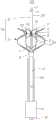
图1是本发明第一实施例提供的房间隔组织造口系统的结构示意图。Fig. 1 is a schematic structural view of an atrial septal tissue stoma system provided by the first embodiment of the present invention.
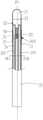
图2是图1中的房间隔组织造口系统的鞘芯的部分结构的剖面图。Fig. 2 is a cross-sectional view of the partial structure of the sheath core of the atrial septal tissue stoma system in Fig. 1 .
图3是图2中的鞘芯沿III-III线的剖面图。Fig. 3 is a sectional view of the sheath core along line III-III in Fig. 2 .
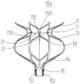
图4是图1中的房间隔组织造口系统的造口支架的结构示意图。Fig. 4 is a schematic structural view of the stoma bracket of the atrial septal tissue stoma system in Fig. 1 .
图5是本发明第二实施例提供的房间隔组织造口系统的结构示意图。Fig. 5 is a schematic structural view of the atrial septal tissue stoma system provided by the second embodiment of the present invention.
图6是图5中的房间隔组织造口系统的鞘芯的部分结构的剖面图。Fig. 6 is a cross-sectional view of a partial structure of the sheath core of the atrial septal tissue stoma system in Fig. 5 .
图7是图6中的鞘芯沿VII-VII线的剖面图。Fig. 7 is a cross-sectional view of the sheath core along line VII-VII in Fig. 6 .
图8是图5中的房间隔组织造口系统的造口支架的结构示意图。Fig. 8 is a schematic structural view of the stoma support of the atrial septal tissue stoma system in Fig. 5 .
主要元件符号说明Description of main component symbols
房间隔造口系统 1000,1000aAtrial Septostomy
房间隔造口装置 100,100aAtrial Septostomy
造口支架 10,10a
造口部 11
导电部 111Conductive part 111
控制孔 1101
显影定位件 113
第一定位件 12
第二定位件 13
回收部 14
回收口 15
螺母 151
内螺纹 152
鞘芯 20Sheath
收容槽 2010
可调弯段 2012
第一导线通道 201
第二导线通道 202
灌注通道 203
拉丝通道 204Brushed
第一电极 21
圆滑过渡结构 210
第一导线 22
灌注口 23
第二电极 25Second electrode 25
显影定位件 27
第二导线 24
可调弯结构 26
控制机构 30
控制线 31
连接环 32Connecting
导管 40
温度传感器 50
鞘管 200
控制手柄 300
连接口 301
具体实施方式Detailed ways
下面将结合本发明实施例中的附图,对本发明实施例中的技术方案进行清楚、完整地描述,显然,所描述的实施例仅是本发明一部分实施例,而不是全部的实施例。基于本发明中的实施例,本领域普通技术人员在没有做出创造性劳动前提下所获得的所有其它实施例,都属于本发明保护的范围。The following will clearly and completely describe the technical solutions in the embodiments of the present invention with reference to the accompanying drawings in the embodiments of the present invention. Obviously, the described embodiments are only some of the embodiments of the present invention, not all of them. Based on the embodiments of the present invention, all other embodiments obtained by persons of ordinary skill in the art without making creative efforts belong to the protection scope of the present invention.
首先需要说明的是,在介入医疗领域,通常将器械靠近操作者的一端称作近端(也即操作端),将器械远离操作者的一端称作远端(也即插入端)。具体的,远端是指器械可自由插入到动物或人体体内的一端。近端是指供用户或机器操作的一端或是用于连接其他器件的一端。此外,术语第一、第二等的使用不是指任何顺序或重要性,而是使用术语第一、第二等来将一个元件与另一元件区别开来。First of all, it needs to be explained that in the field of interventional medicine, the end of the instrument close to the operator is usually called the proximal end (ie, the operating end), and the end of the instrument away from the operator is called the distal end (ie, the insertion end). Specifically, the distal end refers to the end of the instrument that can be freely inserted into the animal or human body. The near end refers to the end for user or machine operation or the end for connecting other devices. Furthermore, the use of the terms first, second, etc. does not imply any order or importance, but rather the terms first, second, etc. are used to distinguish one element from another.
可以理解的,本发明的说明书和权利要求书及上述附图中的术语仅是为了描述特定实施例,并非要限制本发明。本发明的说明书和权利要求书及上述附图中的术语“第一”、“第二”等是用于区别不同对象,而非用于描述特定顺序。除非上下文另有明确表述,否则单数形式“一”和“所述”也旨在包括复数形式。术语“包括”以及它们任何变形,意图在于覆盖不排他的包含。此外,本发明可以以多种不同的形式来实现,并不限于本实施例所描述的实施例。提供以下具体实施例的目的是便于对本发明公开内容更清楚透彻的理解,其中上、下、左、右等指示方位的字词仅是针对所示结构在对应附图中位置而言。It can be understood that the terms in the specification, claims and the above-mentioned drawings of the present invention are only for describing specific embodiments, and are not intended to limit the present invention. The terms "first", "second" and the like in the specification and claims of the present invention and the above drawings are used to distinguish different objects, rather than to describe a specific order. The singular forms "a", "an" and "the" are intended to include the plural forms as well, unless the context clearly dictates otherwise. The term "comprise", as well as any variations thereof, is intended to cover non-exclusive inclusion. In addition, the present invention can be implemented in many different forms, and is not limited to the embodiments described in this embodiment. The purpose of providing the following specific embodiments is to facilitate a clearer and more thorough understanding of the disclosure of the present invention, wherein words indicating orientation such as up, down, left, and right are only for the positions of the structures shown in the corresponding drawings.
说明书后续描述为实施本发明的较佳实施例,然而上述描述乃以说明本发明的一般原则为目的,并非用以限定本发明的范围。本发明的保护范围当视所附权利要求所界定者为准。The following descriptions in the specification are preferred embodiments for implementing the present invention. However, the above descriptions are for the purpose of illustrating the general principles of the present invention, and are not intended to limit the scope of the present invention. The scope of protection of the present invention should be defined by the appended claims.
请参阅图1,图1所示为本发明第一实施例提供的房间隔造口系统1000的结构示意图。房间隔造口系统1000包括房间隔造口装置100、鞘管200及控制手柄300。控制手柄300外接有消融能源。控制手柄300用于控制造口支架10和鞘芯20可活动地收容于鞘管200内或伸出鞘管200外。Please refer to FIG. 1 . FIG. 1 is a schematic structural diagram of an
房间隔造口装置100包括造口支架10及穿设于造口支架10中的鞘芯20,造口支架10包括造口部11。鞘芯20的远端设置有与消融电源电连接的第一电极21。第一电极21用于对房间隔组织进行消融穿刺,以供造口部11穿过房间隔组织的穿刺处并膨胀将房间隔组织撑开。The
本领技术人员应当理解的是,所述图1仅是房间隔造口系统1000的示例,并不构成对房间隔造口系统1000的限定,且房间隔造口装置100及房间隔造口系统1000可以包括比图1所示更多或更少的部件,或者组合某些部件,或者不同的部件,例如房间隔造口装置100还可以包括用于测量造口直径的测量装置等,房间隔造口系统1000还可以包括推送器等。Those skilled in the art should understand that FIG. 1 is only an example of an
如此,基于鞘芯的远端设置有与消融电源电连接的第一电极,因此可以通过使用第一电极接收到的射频能源对房间隔组织进行消融穿刺,无需切割房间隔组织,从而避免了机械穿刺造成的损伤问题、方便穿刺操作、且确保了房间隔组织造口不易回缩或闭合。此外,由于造口支架和鞘芯可撤离房间隔组织,因此避免留下器械而导致血栓形成或器械脱落造成栓塞的问题,从而提高了房间隔组织造口装置及其系统的使用安全性。In this way, the distal end of the sheath core is provided with the first electrode electrically connected to the ablation power supply, so the atrial septal tissue can be ablated and punctured by using the radio frequency energy received by the first electrode without cutting the atrial septal tissue, thus avoiding mechanical The problem of injury caused by puncture is convenient for puncture operation, and it ensures that the atrial septal stoma is not easy to retract or close. In addition, since the stoma stent and the sheath core can be withdrawn from the atrial septal tissue, the problem of thrombus formation caused by leaving the device or embolism caused by the fall of the device is avoided, thereby improving the use safety of the atrial septal tissue stoma device and its system.
房间隔造口装置100还包括套设于鞘芯20外的导管40。造口支架10固定设置在导管40的远端。鞘芯20可活动地收容于导管40的内腔,以使鞘芯20可以伸出导管40并外露于造口支架10的远端。为了确保第一电极21的穿刺更加精准,鞘芯20外露出导管40的远端的最大轴向周度为50-60mm。导管40可活动地收容于鞘管200的内腔,以使造口支架10可以伸出鞘管200的远端。The
可以理解的,通过鞘管200输送造口支架10和鞘芯20至待穿刺组织,不仅方便运输,且避免造口支架10和鞘芯20对其他组织造成损伤的问题。在输送过程中,收缩造口支架10,以使造口支架10的径向外径小于鞘管200的内径而收纳于鞘管200内。在对房间隔组织进行造口时,将造口支架10从鞘管200内释放,此时造口支架10可以自动膨胀至预设的形状尺寸,并能对与其接触的组织产生一定的径向支撑作用。It can be understood that transporting the
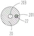
请一并参看图1、图2和图3,图2所示为房间隔造口装置100的鞘芯20的部分结构的剖面图,图3所示为鞘芯沿径向的剖面图。第一电极21的远端形成封闭的柔性尖端,且所述柔性尖端构造为圆滑过渡结构210,从而可以避免第一电极21在穿刺手术中划伤操作者或患者的非穿刺部位。第一电极21由金属材料制成。所述金属材料包括,但不局限于金、铂、铂铱合金中的至少一者。第一电极21通过第一导线22电连接于消融电源,以实现对房间隔组织进行消融穿刺。鞘芯20沿轴向开设收纳第一导线22的第一导线通道201。第一导线22的远端连接于第一电极21,第一导线22的近端连接于消融电源。Please refer to FIG. 1 , FIG. 2 and FIG. 3 together. FIG. 2 shows a cross-sectional view of a partial structure of the
在一些实施例中,第一电极21上设置有至少一个灌注口23。鞘芯20开设与至少一个灌注口23连通的灌注通道203。灌注通道203用于输送灌注液。灌注液可以是,但不局限于生理盐水、造影剂或药剂等流体。当灌注液为造影剂时,可以快速且精准地定位出第一电极21的位置,进而提高了穿刺手术的安全性。当灌注液为药剂,例如肝素时,可以避免穿刺手术中因血管堵塞造成的血管损伤或喷涌的问题。此外,灌注液还具有降温功能,在穿刺过程中,当温度传感器50检测到的温度过高,可以通过灌注液对第一电极21进行降温,从而避免患者在穿刺手术中产生的不适感。In some embodiments, at least one
至少一个灌注口23均匀间隔地设置在第一电极21上,以实现对第一电极21的快速降温或定位作用。灌注通道203与第一导线通道201隔离设置,以免灌注通道203的液体损坏或影响第一导线22的性能。在本实施例中,灌注通道203设置于鞘芯20的中心轴线P1的位置,以实现灌注液均匀地从至少一个灌注口23排出,提高第一电极21与鞘芯20的连接的稳定性。第一导线通道201靠近鞘芯20的外周面设置,即第一导线通道201偏离鞘芯20的中心轴线P1,简化第一电极21及鞘芯20的加工工艺,方便第一电极21与第一导线22的连接。At least one
需要说明的是,灌注通道203和第一导线通道201还可以设置于鞘芯20的其它位置,本发明不作具体限定。It should be noted that the
在一些实施例中,房间隔造口装置100还包括至少一个温度传感器50。至少一个温度传感器50电连接第一电极21。至少一个温度传感器50用于检测房间隔组织在消融穿刺过程中的温度,以防止温度过低或过高。若温度传感器50检测到的温度过低,控制调高消融电源的输出功率,若温度传感器50检测到的温度过高,则可以控制停止消融电源输出能量而停止对第一电极21进行加热;或者,可以通过灌注液对第一电极21进行降温,从而避免心脏内血栓形成。至少一个温度传感器50可以设置于第一电极21上;或者,还可以设置在第一电极21的附近位置上。In some embodiments, the
在本实施例中,至少一个温度传感器50可以设置在第一电极21上;或者,可以设置在第一电极21的附近位置。至少一个温度传感器50设置在第一电极21与鞘芯20的交界处,例如,温度传感器50设置在鞘芯20邻近第一电极21的外周壁,以在穿刺时接触房间隔组织,从而提高了温度传感器50的检测结果的精准度。优选地,至少一个温度传感器50设置于第一电极21上。至少一个温度传感器50与灌注通道203隔离设置,从而可以进一步提高温度传感器50的检测结果的精准度。In this embodiment, at least one
在本实施例中,鞘芯20可活动地穿设于造口支架10中。如此,在使用第一电极21接收到的射频能源对房间隔组织进行消融穿刺时,鞘芯20可以伸出造口支架10外,以使第一电极21外露于造口支架10外,从而使得第一电极21对房间隔组织穿刺更加精准、快速,且避免造口支架10在使用第一电极21对房间隔组织进行消融穿刺时而造成的损伤房间隔组织的周围组织的问题,进而提高了穿刺手术的安全性。此外,在第一电极21完成对房间隔组织造口后,鞘芯20可以回收至造口支架10内,以使第一电极21包埋于造口支架10内,从而避免第一电极21影响造口支架10对房间隔组织扩张造口而达到预期的造口直径。In this embodiment, the
造口支架10的形状大致呈球形或球台形。造口支架10为可径向收缩及膨胀的弹性支架。本实施例中,造口支架10为镍合金支架。造口支架10可以采用镍合金管切割而成,也可以采用镍合金丝编织而成。造口支架10的网状结构的疏密程度根据需要设定。本实施例中采用菱形结构单元连续周向排布一圈形成。造口支架10整体形状可以是灯笼状、直筒形、盘状、锥形等多种适用形状,在此不作限定。当造口支架10通过鞘管200输送时,直径可收缩至较小状态以便在鞘管200中输送;当在心脏中释放时,可自动膨胀至所需形状尺寸,并能对与其接触的组织产生一定的径向支撑作用。The
造口部11主要功能是径向膨胀将房间隔组织撑开。造口部11设置在造口支架10上。具体的,造口部11可以设置在造口支架10的远端;或者,可以设置在造口支架10的中部。造口部11沿造口支架10的周向设置,以实现均匀撑开房间隔组织。具体地,造口部11可以为,但不局限于波形支架、网状支架、杆状支架或它们组合形成的管状结构、筒状结构或环状结构。The main function of the
在本实施例中,在造口支架10处于完全释放的状态下,造口部11为杆状支架形成的圆筒状结构。造口部11由多根支杆交叉连接形成,其形状为圆筒形结构或椭圆筒形结构。同造口支架10一致,造口部11同样需要径向收缩,以实现在运输时收入到鞘管200内。In this embodiment, when the
造口部11所形成的形状可以有多种,例如造口部11可以为侧壁内凹或/和外凸的曲面形、圆筒形、椭圆筒形或者是它们的组合。曲面形是在周向形成一个封闭的曲面结构,外凸和内凹的位置可以根据需要设定,可以单独形成外凸结构或内凹结构,也可以将外凸或内凹结构相结合设置在同一个造口部11上。外凸结构如:盘状、球台形等。内凹结构如:腰鼓形。在本实施例中,采用圆筒形结构,与造口支架10平滑过渡形成一个整体圆筒结构。造口部11的轴向长度根据实际需要设定,一般与房间隔组织的厚度匹配即可。The
造口部11和第一电极21绝缘隔离设置,以避免在消融穿刺过程中损伤其他组织的问题,从而提高了房间隔造口装置100的使用安全性。The
在本实施例中,造口部11至少在外表面沿周向设置有与消融电源电连接的导电部111。导电部111用于在造口部11穿过所述房间隔组织的穿刺处后,对房间隔组织进行消融。In this embodiment, the
在本实施例中,导电部111可以为贴接于造口部11外表面的金属电极。导电部111与造口部11之间设有避免二者之间通电导通的绝缘体,或者与导电部111贴合处的造口部11至少为表面绝缘。这两种方式都可以采用,绝缘体可以采用绝缘垫片、绝缘涂层、绝缘套管等多种方式,本实施例中房间隔造口装置100的镍钛合金支架表面全部镀有PI的绝缘涂层,形成绝缘体,与导电部111绝缘。In this embodiment, the conductive part 111 may be a metal electrode attached to the outer surface of the
在一些实施例中,导电部111还可以为裸露的导电金属件。导电金属件可以单独设置在造口部11,也可以是造口部11的一部分或二者一体成型。单独设置是金属制成的导电部111镶嵌或粘贴在造口部11。采用造口部11的一部分是直接利用造口部11的金属材质导电的特性,导电部111为造口部11上外表面裸露的金属,直接用作导电部111。导电部111采用裸露的导电金属件是指直接采用金属制成导电部111,导电部111的形状可以是根据造口部11形状配合的各自独立的片状、网络状、杆状等,围绕造口部11一周间隔设置多个。导电部111也可以是围绕造口部11一周设置一圈连续或者间断的环状结构的导电部111。一圈的环状结构是能向中心收缩的结构或软性能弯折的结构,方便收入鞘管200。In some embodiments, the conductive portion 111 may also be a bare conductive metal piece. The conductive metal piece can be separately arranged on the
由于导电部111电连接于消融电源,因此导电部111可以用于消融与造口部11接触的部分组织。造口部11的导电部111导电只能在对应造口组织通电导通,而不能对心脏其他部分产生影响。因此,要求导电部111与造口部11之间设有避免二者之间通电导通的绝缘件,或者除导电部111以外的其余造口部11和造口支架10至少在与血液接触的外表面绝缘。本实施例中,是直接采用造口部11位于圆筒状结构的中部结构作为导电部111。在造口部11表面,除去导电部111面向房间隔组织的外表面(即导电部111背离鞘芯20一侧的表面)为裸露的金属,其他的造口部11外表面全部绝缘即采用派瑞林的绝缘涂层。外表面绝缘是指在表面涂覆有绝缘涂层。Since the conductive part 111 is electrically connected to the ablation power source, the conductive part 111 can be used to ablate part of the tissue in contact with the
导电部111设置有至少一个显影定位件113。具体的,导电部111开设至少一个控制孔1101。显影材料填充于控制孔1101内为形成显影定位件113。本实施例中,显影材料例如是,但不局限于金、铂或钽等贵金属材料。在本实施例中,显影定位件113为黄金显影定位件。显影材料的填充方式可以是机械变形的镶嵌、焊接、粘接等。显影定位件113用于在手术中显示导电部111的位置,用于准确将导电部111置于房间隔组织造口处。The conductive part 111 is provided with at least one developing
在一些实施例中,导电部111电连接于温度传感器。温度传感器邻接导电部111,且与房间隔组织接触,以检测导电部111的温度。温度传感器还电连接于消融电源。具体地,温度传感器可以设置在造口部11的导电部111上;或者可以设置于导电部111的附近位置。In some embodiments, the conductive part 111 is electrically connected to the temperature sensor. The temperature sensor is adjacent to the conductive part 111 and is in contact with the interatrial septal tissue to detect the temperature of the conductive part 111 . The temperature sensor is also electrically connected to the ablation power source. Specifically, the temperature sensor may be disposed on the conductive part 111 of the
本实施例中,采用顺应造口支架10和造口部11本身的结构延伸形成两个定位件,即造口部11的远端和近端分别连接有第一定位件12和第二定位件13。第一定位件12紧靠房间隔组织的左心房组织表面和第二定位件13紧靠房间隔组织的右心房组织表面,以使造口部11能够精准定位于房间隔组织的穿刺处。在本实施例中,第一定位件12和第二定位件13可以均构造为一锥面结构。第一定位件12和第二定位件13呈锥面法兰状。在另一实施例中,第一定位件和第二定位件还可以均构造为一平面结构。第一定位件和第二定位件呈平面法兰状。第一定位件和第二定位件的远端面分别形成定位面。在其他一实施例中,第一定位件构造为一锥面结构,第二定位件构造为一平面结构。第一定位件呈锥面法兰状,第二定位件呈平面法兰状。第一定位件和第二定位件的远端面分别形成定位面。In this embodiment, two positioning parts are formed by extending the structure conforming to the
造口支架10的近端连接有回收部14。回收部14大致呈圆锥状。回收部14设置有用于连接造口支架10和导管40的连接件。造口支架10与导管40的连接方式包括,但不局限于螺纹连接、粘接、焊接、压握或卡扣连接等等。造口支架10通过连接件与导管40间接连接在一起,不仅方便加工成型,且提高了造口支架10与导管40连接的可靠性。回收部14近端收缩形成回收口15,回收口15内设置有连接导管40的连接件。连接件根据回收方式可以是多种。例如,在本实施例中,连接件采用在回收口15设置有内螺纹152的螺母151,通过导管的外螺纹与内螺纹152的螺母151螺接。A
造口部11设有调节造口部11径向尺寸的调节机构30。调节机构30可以有多种实施方式,只要实现径向约束的结构都适用本发明。由于造口支架10需要置入鞘管200内进行传输,因此通过增设调节机构30可实现造口支架10径向收缩。调节机构30可以采用软性结构或伸缩结构。软性结构可以是控制线。The
在本实施例中,调节机构30包括至少两根控制线31,控制线31两端分别穿过造口部11周向上不同位置并向造口部11中心汇聚成束。本实施例中,调节机构30包括4根等长的控制线31,每根控制线31的两端从造口部11由外向内穿过两相邻的控制孔1101,每个控制孔1101均有两个线头穿过。所有的线头在造口部11的中心轴线处汇合并通过打结的方式形成汇合并形成连接环32。In this embodiment, the
在另一实施例中,调节机构包括一根控制线;控制线同时穿过造口部周向上不同位置且两端固定以限制造口部径向尺寸。In another embodiment, the adjustment mechanism includes a control line; the control line passes through different positions on the circumference of the stoma at the same time, and both ends are fixed to limit the radial dimension of the stoma.
在另一实施例中,调节机构包括至少一根控制线;控制线穿过造口部周向上不同位置,每条控制线的一端固定于造口部或与房间隔组织造口装置连接的输送系统远端,控制线另一端连接用于控制房间隔组织造口装置植入的控制机构,以控制造口部径向尺寸。In another embodiment, the adjustment mechanism includes at least one control wire; the control wire passes through different positions in the circumferential direction of the stoma, and one end of each control wire is fixed to the stoma or connected to the atrial septal tissue stoma device. At the far end of the system, the other end of the control line is connected to a control mechanism for controlling the implantation of an atrial septal tissue stoma device, so as to control the radial size of the stoma.
在另一实施例中,调节机构包括至少一根控制线;控制线穿过造口部周向上不同位置,每条控制线的两端中的至少一端通过输送系统穿出,通过手动操作,以控制造口部径向尺寸。In another embodiment, the adjustment mechanism includes at least one control line; the control line passes through different positions on the circumference of the stoma, at least one of the two ends of each control line passes through the delivery system, and is manually operated to Control the radial size of the stoma.
调节机构如果采用伸缩结构,可以是弹性圈、螺旋弹簧等,通过调节弹性圈、螺旋弹簧的长度或直径,实现造口部11的径向调节。If the adjustment mechanism adopts a telescopic structure, it can be an elastic ring, a helical spring, etc., and the radial adjustment of the
如图1和图4所示,房间隔造口装置100在完全释放的状态下,有一母线内凹的回转曲面的造口部11。在造口部11上设置有导电部111。造口部11最小直径处的圆周上,均布有4个控制孔1101。As shown in FIG. 1 and FIG. 4 , when the
本实施例中,请再次参阅图1至图4,造口支架10、鞘芯20、鞘管200和控制手柄300为一套完整的系统,本实施例的房间隔造口系统1000操作方法为:In this embodiment, please refer to Fig. 1 to Fig. 4 again, the
1.将房间隔造口装置100通过鞘管200送至右心房,将第一电极21从鞘管200中露出。将第一电极21的第一导线22连接至射频电源(消融电源),开启射频电源并设定参数(如功率30W,持续时间120S),然后使用第一电极21对房间隔组织进行射频穿刺。在穿刺过程中,通过第一电极21处的温度传感器50对房间隔组织进行检测,当温度过低或过高时应调节射频电源功率或停止加热。1. Send the
2.第一电极21对房间隔组织穿刺后,将鞘管200继续向前输送直至鞘管200的前端位于左心房内,控制孔1101处的显影定位件位于房间隔组织处。2. After the
3.缓慢后撤鞘管200,以使房间隔造口装置100的左房定位件完全出鞘,使用造口支架10远端的左房定位部对造口支架10进行定位。造口支架10的远端左房定位部紧靠房间隔组织左房表面,以确保造口部11能够精确定位于房间隔组织处。然后,将鞘管200继续后撤,使造口支架10完全释放。此时,造口部11位于房间隔组织处,可以进行后续的扩张与消融。3. Slowly withdraw the
4.当造口支架10完全释放时,造口支架10的造口部11处于最小直径。由于一次性扩张到位可能使房间隔组织撕裂,因此需要通过使用控制手柄300对造口支架10的造口部11处的直径大小进行控制而进行多次扩张,直到达到预设的造口直径(例如预设的造口直径范围为2mm-14mm)。此时,造口部11将位于房间隔组织的造口处并能够将房间隔组织撑开。4. When the
5.通过超声或DSC判断造口部11的造口直径是否已经达到预设的造口直径。当造口部11的造口直径达到预设的造口直径后,开启射频电源,并设置加热参数(如功率50W,持续时间30S),然后启动加热。此时,通过温度传感器对房间隔组织温度进行检测,当检测到的温度过高时,应当停止加热并用灌注液冲洗使造口支架10降温。5. Determine whether the stoma diameter of the
6.加热停止后,可将器械回收至鞘管200并撤除体外,并测量造口直径是否达到预期。6. After the heating is stopped, the instrument can be recovered to the
本发明实施例提供的房间隔组织造口系统,由于鞘芯的远端设置有与消融电源电连接的第一电极,因此可以通过使用第一电极接收到的射频能源对房间隔组织进行消融穿刺,无需切割房间隔组织,从而避免了机械穿刺造成的损伤问题、方便穿刺操作、且确保了房间隔组织造口不易回缩或闭合。此外,由于造口支架和鞘芯可撤离房间隔组织,因此避免留下器械而导致血栓形成或器械脱落造成栓塞的问题,从而提高了房间隔组织造口装置及其系统的使用安全性。In the atrial septal tissue stoma system provided by the embodiment of the present invention, since the distal end of the sheath core is provided with a first electrode electrically connected to the ablation power supply, the atrial septal tissue can be ablated and punctured by using the radio frequency energy received by the first electrode , without cutting the atrial septal tissue, thereby avoiding the damage caused by mechanical puncture, facilitating the puncture operation, and ensuring that the stoma of the atrial septal tissue is not easy to retract or close. In addition, since the stoma stent and the sheath core can be withdrawn from the atrial septal tissue, the problem of thrombus formation caused by leaving the device or embolism caused by the fall of the device is avoided, thereby improving the use safety of the atrial septal tissue stoma device and its system.
请一并参阅图1、图5至图8,图5所示为本发明第二实施例提供的房间隔造口系统1000a的结构示意图。在第二实施例中,房间隔造口装置100a不同于第一实施例中的房间隔造口装置100。Please refer to FIG. 1 , and FIG. 5 to FIG. 8 together. FIG. 5 is a schematic structural diagram of an
第二实施例中的房间隔造口装置100a与第一实施例中的房间隔造口装置100相似,不同的是,第二实施例中的房间隔造口装置100a还包括固定于鞘芯20a上的至少一个第二电极25。至少一个第二电极25设置于鞘芯20a的外周面,且位于第一电极21的近端。The
至少一个第二电极25及显影定位件27与鞘芯20的固定连接方式包括,但不局限于焊接、粘接、卡接等等。至少一个第二电极25用于在造口支架10a对房间隔组织造口后,再次对房间隔组织进行消融,从而可以进一步保证房间隔组织的造口不易回缩或闭合。The fixed connection methods of the at least one second electrode 25 and the developing
为了确保第一电极21与至少一个第二电极25相互独立的工作,至少一个第二电极25与第一电极21绝缘隔离设置。在本实施例中,第一电极21与至少一个第二电极25错开工作,即第一电极21执行对房间隔组织消融时,至少一个第二电极25停止执行对房间隔组织消融;或者至少一个第二电极25执行对房间隔组织消融时,第一电极21停止执行对房间隔组织消融,如此,可以避免至少一个第二电极25或第一电极21对房间隔造口处之外的其他组织进行消融穿刺所造成的损伤问题。In order to ensure that the
为了确保至少一个第二电极25正常工作,且防止第一电极21损伤房间隔造口处之外的其他组织,至少一个第二电极25与第一电极21之间的距离为10-15mm。至少一个第二电极25由金属材料制成,所述金属材料包括金、铂、铂铱合金中的至少一者。In order to ensure that the at least one second electrode 25 works normally and prevent the
每一第二电极25上设置显影定位件27,从而实现将第二电极25定位至房间隔造口处,从而更加精准对房间隔组织进行消融。在本实施例中,至少一个第二电极25为环状电极,所述显影定位件为显影环,所述显影环设置于所述环状电极的近端和远端,如此,在环状电极的周向提供更大的视角以快速并精准定位环状电极的位置,进而提高了穿刺手术的安全性,还可以使得显影环快速安装于环状电极,且提高了环状电极与显影环的连接的稳定性。在一些实施例中,所述显影环还可以设置于环状电极的近端;或者,环状电极的远端;或者,环状电极的其它位置,本发明不作具体限定。Each second electrode 25 is provided with a developing
在本实施例中,至少一个第二电极25的数量为一个。在一些实施例中,至少一个第二电极的数量为多个,且多个第二电极相互隔离设置。多个第二电极可以同时工作,也可以根据实际需求选择预设数量的第二电极同时工作。至少一个第二电极25的数量根据造口组织的结构来设定,本发明不作具体限定。In this embodiment, the number of at least one second electrode 25 is one. In some embodiments, there is a plurality of at least one second electrode, and the plurality of second electrodes are separated from each other. Multiple second electrodes can work at the same time, and a preset number of second electrodes can also be selected to work at the same time according to actual needs. The quantity of at least one second electrode 25 is set according to the structure of stoma tissue, which is not specifically limited in the present invention.
第二电极25与显影定位件27嵌设在鞘芯20上。具体的,至少一个第二电极25及显影定位件27套接于鞘芯20上,从而方便组装,且确保了至少一个第二电极25及显影定位件27与鞘芯20的连接稳定性及可靠性。鞘芯20的外周壁沿周向开设收容第二电极25与显影定位件27的收容槽2010。第二电极25及显影定位件27的外周壁与鞘芯20的外周壁相连接而形成连续光滑的外表面,从而提高了鞘芯20组装及手术输送过程中的顺畅性,且避免因不规则外壁对组织造成的损伤问题。The second electrode 25 and the developing
在本实施例中,第一电极21通过第一导线22电连接于消融电源。至少一个第二电极25通过第二导线24电连接于消融电源,第一导线22与第二导线24绝缘隔离设置。如此,由于第一导线22与第二导线24绝缘隔离设置,因此可防止第一导线22或第二导线24通电时发生串扰。In this embodiment, the
鞘芯20沿轴向开设间隔设置的第一导线通道201和第二导线通道202,第一导线22收纳于第一导线通道201内,第二导线24收容于第二导线通道202内。在一些实施例中,第一导线与第二导线还可以收纳于同一导线通道内,以简化鞘芯的加工工艺。第一导线通道201和第二导线通道202相对于鞘芯20的中心轴线P1呈对称方式排布设置,从而简化加工工艺。The
收容槽2010与第一导线通道201及第二导线通道202隔离设置。鞘芯20对应第二电极25的位置处开设与第二导线通道202及收容槽2010相贯通的通孔,第二导线24穿过通孔而电连接于第二电极25。如此,避免第二电极25与第一导线22电连接而影响第一电极21的工作性能。The receiving
房间隔造口装置100a还包括至少一个温度传感器50。至少一个温度传感器50设置于造口部11、第一电极21、至少一个第二电极25中的至少一者的所在位置或附近位置。如此,在穿刺过程中,当温度传感器50检测到的温度过高,可以通过灌注液对第一电极21进行降温,从而避免患者在穿刺手术中产生的不适感;当温度传感器50检测到的温度过低时,控制调高消融电源的输出功率。The
在一些实施例中,鞘芯20内设置有可调弯结构26。可调弯结构26用于调节鞘芯20的弯曲度,从而可以更精准地实现穿刺消融,提高了手术的安全性。可调弯结构26可以为拉丝。鞘芯20沿轴向开设收容所述拉丝的拉丝通道204。In some embodiments, an
鞘芯20的远端设置有可调弯段2012。第一电极21的远端外露于鞘芯20外。第一电极21的近端固定于可调弯段2012上。可调弯结构26的远端固定于第一电极21或可调弯段2012上,可调弯结构26的近端固定于控制手柄300上。可调弯结构26的近端通过控制手柄300调整可调弯结构26的弯曲状态或恢复平直状态,由于鞘芯20的结构灵活,因此更精准地实现第一电极21及至少一个第二电极25的消融穿刺,从而保证手术的顺利完成。The distal end of the
拉丝用于牵引可调弯段2012进行弯曲或者恢复平直,具有一定的强度。本实施例中,拉丝为单根结构,也可以采用多股结构。拉丝的截面形状可以为圆形等各种形状,在此不对其进行具体地限定。拉丝在具有一定强度以实现牵引功能的基础上,径向截面应尽量细小。拉丝为金属丝,即拉丝由金属材料制成。所述金属材料例如是,但不局限于不锈钢、钨合金、钴铬合金或者镍钛合金等,也可以由具有一定强度的高分子制成,在此不对其材料进行具体地限定。本实施例中,可调弯结构26优选为不锈钢丝。The wire drawing is used to pull the
拉丝通道204与第一导线通道201、第二导线通道202及灌注通道203均隔离设置,避免拉丝干扰第一导线22及第二导线24的正常工作,并且确保了第一导线22与第一电极21及第二导线24与第二电极25之间的连接稳定及可靠性。The wire-drawing
请再次参看图5和图8所示,在第二实施例中的造口支架10a与第一实施例中的造口支架10(参看图4)相似。不同的是,造口支架10a的造口部11上可以不设置导电部。在第一电极21对房间隔组织进行穿刺后,通过造口支架10a对房间隔组织进行造口,再采用第二电极25对房间隔造口处的组织进行消融,从而进一步确保房间隔组织造口不回缩或闭合。在一些实施例中,第一实施例中的造口支架10适用于第二实施例中的房间隔造口装置100a。Please refer to FIG. 5 and FIG. 8 again, the
本实施例中,请再次参阅图4至图8,造口支架10a、鞘芯20a、鞘管200和控制手柄300为一套完整的系统,本实施例的房间隔造口系统1000a操作方法为:In this embodiment, please refer to Fig. 4 to Fig. 8 again, the
1.将房间隔造口装置100a通过鞘管200送至右心房,将第一电极21从鞘管200中露出。将第一电极21的第一导线22连接至射频电源(消融电源),开启射频电源并设定参数(如功率30W,持续时间120S),然后使用第一电极21对房间隔组织进行射频穿刺。在穿刺过程中,通过第一电极21处的温度传感器50对房间隔组织进行检测,当温度过低或过高时应调节射频电源功率或停止加热。1. Send the
2.第一电极21对房间隔组织穿刺后,将鞘管200继续向前输送直至鞘管200的前端位于左心房内,控制孔1101处的显影定位件位于房间隔组织处。2. After the
3.缓慢后撤鞘管200,以使房间隔造口装置100的左房定位件完全出鞘,使用造口支架10远端的左房定位部对造口支架10进行定位。造口支架10的远端左房定位部紧靠房间隔组织左房表面,以确保造口部11能够精确定位于房间隔组织处。然后,将鞘管200继续后撤,使造口支架10完全释放。此时,造口部11位于房间隔组织处,可以进行后续的扩张。3. Slowly withdraw the
4.当造口支架10完全释放时,造口支架10的造口部11处于最小直径。由于一次性扩张到位可能使房间隔组织撕裂,因此需要通过使用控制手柄300对造口支架10的造口部11处的直径大小进行控制而进行多次扩张,直到达到预设的造口直径(例如预设的造口直径范围为2mm-14mm)。此时,造口部11将位于房间隔组织的造口处并能够将房间隔组织撑开。4. When the
5.通过超声或DSC判断造口部11的造口直径是否已经达到预设的造口直径。当造口部11的造口直径达到预设的造口直径后,将造口支架10a调整至最小直径并前推鞘管200(即控制鞘管200自造口支架10a近端朝远端推送),随着鞘管200的前推,造口支架10a被回收至鞘管200内。此时,将鞘管200稍微后撤预设位置,并通过位于第二电极25上的显影定位件27,将第二电极25定位于房间隔造口处。将第二电极25的第二导线24连接至射频电源(消融电源),开启射频电源并设定参数(如功率30W,持续时间120S),然后使用第二电极25对房间隔造口处的组织进行射频消融。此外,还可以通过操作控制手柄操纵可调弯结构26,以使第二电极25更灵活地对房间隔组织进行消融。最后,通过温度传感器对房间隔组织温度进行检测,当检测到的温度过高时,应当停止加热并用灌注液冲洗使造口支架10降温。5. Determine whether the stoma diameter of the
6.加热停止后,可将器械回收至鞘管200并撤除体外,并测量造口直径是否达到预期。6. After the heating is stopped, the instrument can be recovered to the
本发明实施例提供的房间隔组织造口系统,由于鞘芯的远端设置有与消融电源电连接的第一电极,因此可以通过使用第一电极接收到的射频能源对房间隔组织进行消融穿刺,无需切割房间隔组织,从而避免了机械穿刺造成的损伤问题、方便穿刺操作、且确保了房间隔组织造口不易回缩或闭合。此外,由于造口支架和鞘芯可撤离房间隔组织,因此避免留下器械而导致血栓形成或器械脱落造成栓塞的问题,从而提高了房间隔组织造口装置及其系统的使用安全性。此外,鞘芯的远端还设置有至少一个第二电极,从而在所述造口支架对所述房间隔组织造口后,至少一个第二电极可以再次对所述房间隔组织进行消融,从而进一步避免房间隔造口处的组装的回收或关闭。In the atrial septal tissue stoma system provided by the embodiment of the present invention, since the distal end of the sheath core is provided with a first electrode electrically connected to the ablation power supply, the atrial septal tissue can be ablated and punctured by using the radio frequency energy received by the first electrode , without cutting the atrial septal tissue, thereby avoiding the damage caused by mechanical puncture, facilitating the puncture operation, and ensuring that the stoma of the atrial septal tissue is not easy to retract or close. In addition, since the stoma stent and the sheath core can be withdrawn from the atrial septal tissue, the problem of thrombus formation caused by leaving the device or embolism caused by the fall of the device is avoided, thereby improving the use safety of the atrial septal tissue stoma device and its system. In addition, the distal end of the sheath core is also provided with at least one second electrode, so that after the stoma stent stomas the atrial septal tissue, the at least one second electrode can ablate the atrial septal tissue again, thereby Retrieval or closure of the assembly at the atrial septostomy is further avoided.
以上对本发明实施例进行了详细介绍,本文中应用了具体个例对本发明的原理及实施方式进行了阐述,以上实施例的说明只是用于帮助理解本发明的方法及其核心思想;同时,对于本领域的一般技术人员,依据本发明的思想,在具体实施方式及应用范围上均会有改变之处,综上上述,本说明书内容不应理解为对本发明的限制。The embodiments of the present invention have been described in detail above, and specific examples have been used in this paper to illustrate the principles and implementation methods of the present invention. The descriptions of the above embodiments are only used to help understand the method and core idea of the present invention; at the same time, for Those skilled in the art will have changes in the specific implementation and scope of application based on the idea of the present invention. In summary, the contents of this specification should not be construed as limiting the present invention.
Claims (18)
Priority Applications (2)
| Application Number | Priority Date | Filing Date | Title |
|---|---|---|---|
| CN202011384593.8ACN112603524B (en) | 2020-11-30 | 2020-11-30 | Interatrial septum tissue stoma device and interatrial septum group weaving mouth system |
| PCT/IB2021/061139WO2022113054A1 (en) | 2020-11-30 | 2021-11-30 | Stoma-creating device and stoma-creating system |
Applications Claiming Priority (1)
| Application Number | Priority Date | Filing Date | Title |
|---|---|---|---|
| CN202011384593.8ACN112603524B (en) | 2020-11-30 | 2020-11-30 | Interatrial septum tissue stoma device and interatrial septum group weaving mouth system |
Publications (2)
| Publication Number | Publication Date |
|---|---|
| CN112603524A CN112603524A (en) | 2021-04-06 |
| CN112603524Btrue CN112603524B (en) | 2023-02-24 |
Family
ID=75228348
Family Applications (1)
| Application Number | Title | Priority Date | Filing Date |
|---|---|---|---|
| CN202011384593.8AActiveCN112603524B (en) | 2020-11-30 | 2020-11-30 | Interatrial septum tissue stoma device and interatrial septum group weaving mouth system |
Country Status (2)
| Country | Link |
|---|---|
| CN (1) | CN112603524B (en) |
| WO (1) | WO2022113054A1 (en) |
Families Citing this family (7)
| Publication number | Priority date | Publication date | Assignee | Title |
|---|---|---|---|---|
| WO2022022528A1 (en)* | 2020-07-30 | 2022-02-03 | 杭州诺生医疗科技有限公司 | Atrial septostomy device |
| CN112603524B (en)* | 2020-11-30 | 2023-02-24 | 杭州诺生医疗科技有限公司 | Interatrial septum tissue stoma device and interatrial septum group weaving mouth system |
| CN114176720B (en)* | 2021-12-21 | 2024-07-02 | 上海申淇医疗科技有限公司 | Room separates ostomy device |
| US12376879B2 (en) | 2023-03-03 | 2025-08-05 | Theraheart Inc. | Expandable elements for shunting catheters |
| US12290310B2 (en) | 2023-04-06 | 2025-05-06 | Theraheart Inc. | Slicing elements for shunting catheters |
| US12262943B2 (en) | 2023-06-15 | 2025-04-01 | Theraheart Inc. | Expandable ablation mechanisms for shunting catheters |
| US12201354B1 (en) | 2024-04-01 | 2025-01-21 | Theraheart Inc. | Expandable ablation mechanisms for shunting catheters |
Citations (9)
| Publication number | Priority date | Publication date | Assignee | Title |
|---|---|---|---|---|
| US6200315B1 (en)* | 1997-12-18 | 2001-03-13 | Medtronic, Inc. | Left atrium ablation catheter |
| CN104042328A (en)* | 2013-03-07 | 2014-09-17 | 韦伯斯特生物官能(以色列)有限公司 | Irrigated ablation catheter having irrigation ports with reduced hydraulic resistance |
| CN104812297A (en)* | 2012-08-31 | 2015-07-29 | 阿库图森医疗有限公司 | Catheter system and methods of medical uses of same, including diagnostic and treatment uses for heart |
| CN106994207A (en)* | 2017-03-15 | 2017-08-01 | 刘志忠 | A kind of bipolar electrode sub-assembly and stimulating system |
| CN107693089A (en)* | 2016-08-09 | 2018-02-16 | 上海交通大学医学院附属新华医院 | Three-dimensional atrial septal puncture system |
| CN108784896A (en)* | 2017-10-31 | 2018-11-13 | 杭州诺生医疗科技有限公司 | Atrial septum ostomy appliance, atrial septum stoma system and its operating method |
| CN209377738U (en)* | 2018-11-20 | 2019-09-13 | 成都美创医疗科技股份有限公司 | A kind of plasma procedures electrode for mass puncture |
| CN110809435A (en)* | 2017-07-04 | 2020-02-18 | 泰尔茂株式会社 | Medical instrument and treatment method |
| CN211750039U (en)* | 2019-12-31 | 2020-10-27 | 杭州堃博生物科技有限公司 | Ablation catheter with mark |
Family Cites Families (7)
| Publication number | Priority date | Publication date | Assignee | Title |
|---|---|---|---|---|
| US6771996B2 (en)* | 2001-05-24 | 2004-08-03 | Cardiac Pacemakers, Inc. | Ablation and high-resolution mapping catheter system for pulmonary vein foci elimination |
| US8337518B2 (en)* | 2006-12-20 | 2012-12-25 | Onset Medical Corporation | Expandable trans-septal sheath |
| CN105193476A (en)* | 2014-06-19 | 2015-12-30 | 吕斐 | Interatrial septum puncturing assembly and interatrial septum puncturing method |
| IL274110B2 (en)* | 2017-10-31 | 2024-10-01 | Hangzhou Noya Medtech Co Ltd | Devices, systems and methods for interatrial leaks |
| CN111166462A (en)* | 2018-11-09 | 2020-05-19 | 杭州诺生医疗科技有限公司 | Interatrial septum stoma device and interatrial septum stoma system with improved ablation effect |
| CN210158675U (en)* | 2019-01-04 | 2020-03-20 | 科塞尔医疗科技(苏州)有限公司 | Interatrial septum puncture needle assembly |
| CN112603524B (en)* | 2020-11-30 | 2023-02-24 | 杭州诺生医疗科技有限公司 | Interatrial septum tissue stoma device and interatrial septum group weaving mouth system |
- 2020
- 2020-11-30CNCN202011384593.8Apatent/CN112603524B/enactiveActive
- 2021
- 2021-11-30WOPCT/IB2021/061139patent/WO2022113054A1/ennot_activeCeased
Patent Citations (9)
| Publication number | Priority date | Publication date | Assignee | Title |
|---|---|---|---|---|
| US6200315B1 (en)* | 1997-12-18 | 2001-03-13 | Medtronic, Inc. | Left atrium ablation catheter |
| CN104812297A (en)* | 2012-08-31 | 2015-07-29 | 阿库图森医疗有限公司 | Catheter system and methods of medical uses of same, including diagnostic and treatment uses for heart |
| CN104042328A (en)* | 2013-03-07 | 2014-09-17 | 韦伯斯特生物官能(以色列)有限公司 | Irrigated ablation catheter having irrigation ports with reduced hydraulic resistance |
| CN107693089A (en)* | 2016-08-09 | 2018-02-16 | 上海交通大学医学院附属新华医院 | Three-dimensional atrial septal puncture system |
| CN106994207A (en)* | 2017-03-15 | 2017-08-01 | 刘志忠 | A kind of bipolar electrode sub-assembly and stimulating system |
| CN110809435A (en)* | 2017-07-04 | 2020-02-18 | 泰尔茂株式会社 | Medical instrument and treatment method |
| CN108784896A (en)* | 2017-10-31 | 2018-11-13 | 杭州诺生医疗科技有限公司 | Atrial septum ostomy appliance, atrial septum stoma system and its operating method |
| CN209377738U (en)* | 2018-11-20 | 2019-09-13 | 成都美创医疗科技股份有限公司 | A kind of plasma procedures electrode for mass puncture |
| CN211750039U (en)* | 2019-12-31 | 2020-10-27 | 杭州堃博生物科技有限公司 | Ablation catheter with mark |
Also Published As
| Publication number | Publication date |
|---|---|
| CN112603524A (en) | 2021-04-06 |
| WO2022113054A1 (en) | 2022-06-02 |
Similar Documents
| Publication | Publication Date | Title |
|---|---|---|
| CN112603524B (en) | Interatrial septum tissue stoma device and interatrial septum group weaving mouth system | |
| JP6797173B2 (en) | Medical device for fluid communication | |
| CN108784896B (en) | Interatrial ostomy device, interatrial ostomy system and method of operating the same | |
| JP6835809B2 (en) | Electrosurgical device with lumens | |
| WO2022171142A1 (en) | Ablation catheter, ablation device and ablation system | |
| CN109965974A (en) | transcatheter septostomy device | |
| US9750564B2 (en) | Flexible catheter for high-frequency therapy of biological tissue and method of using same | |
| WO2014121664A1 (en) | Radio frequency ablation method, system and radio frequency ablation device thereof | |
| CN115363740B (en) | Ablation needle, ablation device and ablation system for myocardial ablation | |
| CN211834660U (en) | Lead extraction expansion head and extraction device | |
| CN119014968A (en) | Ablation needle, ablation device and ablation system | |
| CN109965972A (en) | ostomy device | |
| CN208892867U (en) | Atrial septum ostomy appliance and its atrial septum stoma system | |
| CN113813041A (en) | radiofrequency ablation device | |
| CN114831723A (en) | Medical catheter | |
| CN114246666A (en) | Ablation plugging device and ablation plugging system | |
| EP4302715A1 (en) | Medical device | |
| CN118806416A (en) | Adjustable curved catheter, ablation device and ablation system | |
| CN215651494U (en) | Medical catheter | |
| CN116172690A (en) | Ostomy device and ostomy system | |
| US20200268441A1 (en) | Energy delivery device for endovascular occlusion | |
| US10155125B2 (en) | Reverse loop ablation device | |
| US20200268442A1 (en) | Energy delivery device for endovascular occlusion | |
| CN114343827A (en) | Ablation catheter | |
| CN221831440U (en) | A balloon-type pulse ablation catheter for treating chronic obstructive pulmonary disease |
Legal Events
| Date | Code | Title | Description |
|---|---|---|---|
| PB01 | Publication | ||
| PB01 | Publication | ||
| SE01 | Entry into force of request for substantive examination | ||
| SE01 | Entry into force of request for substantive examination | ||
| GR01 | Patent grant | ||
| GR01 | Patent grant | ||
| PP01 | Preservation of patent right | Effective date of registration:20250620 Granted publication date:20230224 |