CN112493997B - Photoacoustic endoscopic imaging device and photoacoustic endoscopic imaging method based on same - Google Patents
Photoacoustic endoscopic imaging device and photoacoustic endoscopic imaging method based on sameDownload PDFInfo
- Publication number
- CN112493997B CN112493997BCN202011373366.5ACN202011373366ACN112493997BCN 112493997 BCN112493997 BCN 112493997BCN 202011373366 ACN202011373366 ACN 202011373366ACN 112493997 BCN112493997 BCN 112493997B
- Authority
- CN
- China
- Prior art keywords
- photoacoustic
- housing
- optical fiber
- endoscopic imaging
- imaging
- Prior art date
- Legal status (The legal status is an assumption and is not a legal conclusion. Google has not performed a legal analysis and makes no representation as to the accuracy of the status listed.)
- Active
Links
- 238000003384imaging methodMethods0.000titleclaimsabstractdescription83
- 239000013307optical fiberSubstances0.000claimsabstractdescription34
- 238000000034methodMethods0.000claimsabstractdescription12
- 238000009434installationMethods0.000claimsdescription16
- 230000008569processEffects0.000claimsdescription7
- 239000000945fillerSubstances0.000claimsdescription6
- 239000000463materialSubstances0.000claimsdescription3
- 239000003292glueSubstances0.000claimsdescription2
- 239000000523sampleSubstances0.000abstractdescription8
- 230000004075alterationEffects0.000abstractdescription7
- 230000008901benefitEffects0.000abstractdescription6
- 238000005516engineering processMethods0.000description15
- 230000003287optical effectEffects0.000description10
- 230000031700light absorptionEffects0.000description4
- 206010028980NeoplasmDiseases0.000description3
- 238000004364calculation methodMethods0.000description3
- 230000008859changeEffects0.000description3
- 230000000694effectsEffects0.000description3
- 230000007274generation of a signal involved in cell-cell signalingEffects0.000description3
- 230000007246mechanismEffects0.000description3
- 238000012634optical imagingMethods0.000description3
- 238000003848UV Light-CuringMethods0.000description2
- 239000000853adhesiveSubstances0.000description2
- 230000001070adhesive effectEffects0.000description2
- 238000006243chemical reactionMethods0.000description2
- 238000001723curingMethods0.000description2
- 230000007547defectEffects0.000description2
- 230000008021depositionEffects0.000description2
- 238000003745diagnosisMethods0.000description2
- 238000001839endoscopyMethods0.000description2
- 230000005284excitationEffects0.000description2
- 238000007689inspectionMethods0.000description2
- 230000001678irradiating effectEffects0.000description2
- 238000010521absorption reactionMethods0.000description1
- 239000013466adhesive and sealantSubstances0.000description1
- 230000002411adverseEffects0.000description1
- 230000009286beneficial effectEffects0.000description1
- 201000011510cancerDiseases0.000description1
- 238000010276constructionMethods0.000description1
- 238000001514detection methodMethods0.000description1
- 230000001079digestive effectEffects0.000description1
- 238000013399early diagnosisMethods0.000description1
- 230000004048modificationEffects0.000description1
- 238000012986modificationMethods0.000description1
- 230000035515penetrationEffects0.000description1
- 230000000737periodic effectEffects0.000description1
- 238000010895photoacoustic effectMethods0.000description1
- 239000002861polymer materialSubstances0.000description1
- 239000011347resinSubstances0.000description1
- 229920005989resinPolymers0.000description1
- 230000004083survival effectEffects0.000description1
- 238000002604ultrasonographyMethods0.000description1
- 238000012285ultrasound imagingMethods0.000description1
Images


Classifications
- A—HUMAN NECESSITIES
- A61—MEDICAL OR VETERINARY SCIENCE; HYGIENE
- A61B—DIAGNOSIS; SURGERY; IDENTIFICATION
- A61B5/00—Measuring for diagnostic purposes; Identification of persons
- A61B5/0093—Detecting, measuring or recording by applying one single type of energy and measuring its conversion into another type of energy
- A61B5/0095—Detecting, measuring or recording by applying one single type of energy and measuring its conversion into another type of energy by applying light and detecting acoustic waves, i.e. photoacoustic measurements
- A—HUMAN NECESSITIES
- A61—MEDICAL OR VETERINARY SCIENCE; HYGIENE
- A61B—DIAGNOSIS; SURGERY; IDENTIFICATION
- A61B5/00—Measuring for diagnostic purposes; Identification of persons
- A61B5/68—Arrangements of detecting, measuring or recording means, e.g. sensors, in relation to patient
- A61B5/6846—Arrangements of detecting, measuring or recording means, e.g. sensors, in relation to patient specially adapted to be brought in contact with an internal body part, i.e. invasive
- A61B5/6847—Arrangements of detecting, measuring or recording means, e.g. sensors, in relation to patient specially adapted to be brought in contact with an internal body part, i.e. invasive mounted on an invasive device
- A61B5/6852—Catheters
Landscapes
- Health & Medical Sciences (AREA)
- Life Sciences & Earth Sciences (AREA)
- Physics & Mathematics (AREA)
- Medical Informatics (AREA)
- Surgery (AREA)
- Engineering & Computer Science (AREA)
- Biomedical Technology (AREA)
- Heart & Thoracic Surgery (AREA)
- Biophysics (AREA)
- Molecular Biology (AREA)
- Pathology (AREA)
- Animal Behavior & Ethology (AREA)
- General Health & Medical Sciences (AREA)
- Public Health (AREA)
- Veterinary Medicine (AREA)
- Acoustics & Sound (AREA)
- Ultra Sonic Daignosis Equipment (AREA)
Abstract
Description
Translated fromChinese技术领域technical field
本发明专利涉及医疗器械的技术领域,具体而言,涉及一种光声内窥成像装置及基于该装置的光声内窥成像方法。The patent of the present invention relates to the technical field of medical devices, in particular, to a photoacoustic endoscopic imaging device and a photoacoustic endoscopic imaging method based on the device.
背景技术Background technique
我国肿瘤发病率近年来持续增高,早期发现与治疗能够显著程度提高患者的生存率。内窥镜以其检查操作无创的特点,是实现肿瘤诊断的常见光学仪器。光声成像(Photoacoustic Imaging,PAI)是近年来发展起来的一种非入侵式和非电离式的新型生物医学成像方法。The incidence of cancer in my country has continued to increase in recent years, and early detection and treatment can significantly improve the survival rate of patients. Endoscope is a common optical instrument for tumor diagnosis because of its non-invasive inspection operation. Photoacoustic Imaging (PAI) is a new non-invasive and non-ionizing biomedical imaging method developed in recent years.
光声成像的基本原理:当脉冲激光照射到生物组织中时,生物组织的光吸收域将产生超声信号,这种由光激发产生的超声信号即为光声信号。生物组织产生的光声信号携带了组织的光吸收特征信息,通过探测器接收光声信号就可重建出生物组织的光吸收分布图像。The basic principle of photoacoustic imaging: when the pulsed laser is irradiated into the biological tissue, the light absorption domain of the biological tissue will generate an ultrasonic signal, and the ultrasonic signal generated by the light excitation is the photoacoustic signal. The photoacoustic signal generated by the biological tissue carries the light absorption characteristic information of the tissue, and the light absorption distribution image of the biological tissue can be reconstructed by receiving the photoacoustic signal through the detector.
光声成像过程可以分为三个部分:信号的产生、信号的接收和信号处理及图像重建。The photoacoustic imaging process can be divided into three parts: signal generation, signal reception, signal processing and image reconstruction.
(1)信号的产生:光声信号的产生过程就是“光能”——“热能”——“机械能”的转化过程,由于脉冲激光器具有光声转换效率高的优点,因此通常会选择合适波长的激光作为激发源,并使吸收的光子的能量转化为热能的效率达到90%以上。脉冲激光器发出的激光束照射在待检测生物组织上,由于生物组织的吸收效应,在生物组织内部形成了与组织光学参数相关的能量沉积分布。由于激光脉宽很窄(ns)吸收的能量不能在短时间内释放,导致瞬间温度变化,从而通过热弹机制转化为热膨胀。周期性热流使周围的介质热胀冷缩而激发超声波,由于这种超声波信号的特殊产生机理,为了区别于其它的超声信号,通常称为光声信号。(1) Signal generation: The photoacoustic signal generation process is the conversion process of "light energy"-"thermal energy"-"mechanical energy". Since pulsed lasers have the advantages of high photoacoustic conversion efficiency, they usually choose the appropriate wavelength The laser is used as the excitation source, and the efficiency of converting the absorbed photon energy into heat energy reaches more than 90%. The laser beam emitted by the pulsed laser is irradiated on the biological tissue to be detected. Due to the absorption effect of the biological tissue, an energy deposition distribution related to the optical parameters of the tissue is formed inside the biological tissue. Because the laser pulse width is very narrow (ns), the absorbed energy cannot be released in a short time, resulting in an instantaneous temperature change, which is converted into thermal expansion through the thermoelastic mechanism. The periodic heat flow causes the surrounding medium to expand with heat and contract with cold to excite ultrasonic waves. Due to the special generation mechanism of this ultrasonic signal, in order to distinguish it from other ultrasonic signals, it is usually called photoacoustic signal.
(2)信号的接收和处理:利用超声探测器接收光声信号并对采集到的信号进行适当地处理。(2) Signal reception and processing: the photoacoustic signal is received by the ultrasonic detector and the collected signal is properly processed.
(3)图像重建:采用相应的图像重建算法,就能够得到生物组织内部光能量沉积的分布。当保证入射光的均匀性的前提下,光声重建图像与生物组织内部光吸收分布具有一一对应的关系。(3) Image reconstruction: Using the corresponding image reconstruction algorithm, the distribution of light energy deposition inside the biological tissue can be obtained. Under the premise of ensuring the uniformity of the incident light, there is a one-to-one correspondence between the photoacoustic reconstruction image and the light absorption distribution inside the biological tissue.
光声成像结合了纯光学组织成像中高选择特性和纯超声组织成像中深穿透特性的优点,可得到高分辨率和高对比度的生物组织图像,不仅弥补传统超声成像技术分辨率及对比度不足的缺点,而且改善了纯光学成像中散射效应对成像深度造成的不利影响(突破了高分辨率光学成像深度“软极限”(~1mm),可实现50mm的深层活体内组织成像),因此可以在较深的范围内实现高分辨、高对比度成像。该技术与消化内镜结合为实现肿瘤高灵敏早期诊断提供了新的诊疗模式,展现其巨大应用潜力。Photoacoustic imaging combines the advantages of high selectivity in pure optical tissue imaging and deep penetration in pure ultrasound tissue imaging to obtain high-resolution and high-contrast biological tissue images, which not only make up for the lack of resolution and contrast of traditional ultrasound imaging techniques In addition, it improves the adverse effect of scattering effect on imaging depth in pure optical imaging (breaks through the "soft limit" of high-resolution optical imaging depth (~1mm), and can achieve 50mm deep living tissue imaging), so it can be used in High-resolution, high-contrast imaging is achieved in a deeper range. The combination of this technology and digestive endoscopy provides a new diagnosis and treatment model for high-sensitivity early diagnosis of tumors, showing its great application potential.
根据光声信号的类型,可以将光声成像技术划分为经典光声成像技术与非线性光声成像技术,经典光声成像技术是以单个纳秒激光脉冲照射生物组织产生的光声信号实现成像,非线性光声成像技术是以两个纳秒激光脉冲先后照射生物组织相同区域产生的光声信号实现成像。According to the type of photoacoustic signal, photoacoustic imaging technology can be divided into classical photoacoustic imaging technology and nonlinear photoacoustic imaging technology. Classical photoacoustic imaging technology uses photoacoustic signals generated by a single nanosecond laser pulse to irradiate biological tissues to achieve imaging. , the nonlinear photoacoustic imaging technology realizes imaging by the photoacoustic signal generated by two nanosecond laser pulses successively irradiating the same area of biological tissue.
非线性光声成像技术在近年来获得了非常广泛的应用,原因在于其分辨率相比经典光声成像技术获得了显著的提升,因此可以获得更加清晰的图像。当选择多个波长激发样品产生光声效应实现功能成像时,不同波长的光束在空间中聚焦位置的一致性,是影响分辨率的主要因素。并且,通常情况下,实现多波长功能成像首先需要将每个波长的光束照射在生物组织中相同的区域,然后对原始数据进行解算才能够获取功能参数信息。因此,不同波长的光束在空间中聚焦位置的一致性,成为影响功能参数解算准确性的主要因素。目前,在光声内窥成像系统中实现光束聚焦的元件为透镜,光学成像的物理机制表明,透镜元件的色差会导致焦点位置随波长发生变化,导致成像的分辨率下降,进而降低了功能信息的解析精度。由此可知,透镜聚焦方案存在的固有缺陷,对非线性光声成像技术在生物医学的应用构成了极大阻碍。Nonlinear photoacoustic imaging technology has been widely used in recent years because its resolution has been significantly improved compared with classical photoacoustic imaging technology, so clearer images can be obtained. When multiple wavelengths are selected to excite the sample to generate photoacoustic effect to realize functional imaging, the consistency of the focusing positions of beams of different wavelengths in space is the main factor affecting the resolution. Moreover, under normal circumstances, to achieve multi-wavelength functional imaging, it is first necessary to irradiate the beam of each wavelength on the same area in the biological tissue, and then solve the raw data to obtain functional parameter information. Therefore, the consistency of the focusing positions of beams of different wavelengths in space becomes the main factor affecting the accuracy of functional parameter calculation. At present, in the photoacoustic endoscopic imaging system, the component that realizes beam focusing is the lens. The physical mechanism of optical imaging shows that the chromatic aberration of the lens element will cause the focus position to change with the wavelength, resulting in a decrease in the resolution of the imaging, which in turn reduces the functional information. resolution accuracy. It can be seen that the inherent defects of the lens focusing scheme constitute a great obstacle to the application of nonlinear photoacoustic imaging technology in biomedicine.
除此以外,为满足光声内窥的侧视成像应用需求,需要在光学系统中使用反射元件,将光束的主要传播方向由轴向转变为径向。现有成像系统的光路形式是以平面反射镜作为光束的反射元件,与透镜安装在探头前端的不同位置,透镜与平面反射镜的分离式安装使得每个元件分别占据一定空间长度,导致探头前端尺寸难以进一步缩短,限制了成像探头前端的自由运动范围,对光声内窥技术的在生物医学中的应用构成了阻碍。In addition, in order to meet the application requirements of side-view imaging for photoacoustic endoscopy, it is necessary to use reflective elements in the optical system to change the main propagation direction of the beam from axial to radial. The optical path form of the existing imaging system uses a plane reflector as the reflective element of the beam, and the lens is installed at a different position at the front end of the probe. The separate installation of the lens and the plane reflector makes each element occupy a certain space length, resulting in the front end of the probe The size is difficult to be further shortened, which limits the free movement range of the front end of the imaging probe, and hinders the application of photoacoustic endoscopic technology in biomedicine.
发明内容Contents of the invention
本发明的目的在于提供一种光声内窥成像装置及基于该装置的光声内窥成像方法,旨在解决现有技术中,采用透镜聚焦光束的焦点位置随波长变化的固有缺陷的问题。The purpose of the present invention is to provide a photoacoustic endoscopic imaging device and a photoacoustic endoscopic imaging method based on the device, aiming to solve the inherent defect of using a lens to focus the beam's focus position with wavelength changes in the prior art.
本发明是这样实现的,一种光声内窥成像装置,包括第一壳体、离轴反射镜、超声换能器、支撑件和光纤;The present invention is achieved in this way, a photoacoustic endoscopic imaging device, including a first housing, an off-axis mirror, an ultrasonic transducer, a support and an optical fiber;
所述支撑件设于所述第一壳体的第一端,所述离轴反射镜设于所述第一壳体的第二端,所述第一壳体的壁面上开设有信号窗;The support member is arranged at the first end of the first housing, the off-axis reflector is arranged at the second end of the first housing, and a signal window is opened on the wall of the first housing;
所述光纤安装于所述支撑件;所述支撑件设有换能器安装槽,所述超声换能器设于所述换能器安装槽。The optical fiber is installed on the support member; the support member is provided with a transducer installation groove, and the ultrasonic transducer is arranged in the transducer installation groove.
进一步的,所述第一壳体的第一端连接有扭矩弹簧。Further, a torsion spring is connected to the first end of the first housing.
进一步的,还包括第二壳体,所述第一壳体设于所述第二壳体内部。Further, a second casing is also included, and the first casing is arranged inside the second casing.
进一步的,所述光纤的前端安装于所述支撑件中心区域的贯通孔中,所述光纤的后端通过填充物固定于所述支撑件。Further, the front end of the optical fiber is installed in the through hole in the central area of the support, and the rear end of the optical fiber is fixed to the support by a filler.
进一步的,所述支撑件设有定位凸起,所述第一壳体内具有限位凸起,所述定位凸起与所述限位凸起的配合用于实现所述支撑件的安装定位。Further, the support is provided with a positioning protrusion, and the first housing has a position-limiting protrusion, and the cooperation between the positioning protrusion and the position-limiting protrusion is used to realize the installation and positioning of the support.
进一步的,所述第一壳体的第二端设有离轴反射镜安装槽,所述离轴反射镜上设有定位柱,所述定位柱安装于所述离轴反射镜安装槽。Further, the second end of the first housing is provided with an off-axis reflector installation groove, and the off-axis reflector is provided with a positioning post, and the positioning post is installed in the off-axis reflector installation groove.
进一步的,所述离轴反射镜的反射面为空间曲面。Further, the reflective surface of the off-axis reflector is a spatial curved surface.
进一步的,所述空间曲面为球面或非球面。Further, the space curved surface is spherical or aspheric.
基于光声内窥成像装置的光声内窥成像方法,包括以下步骤:A photoacoustic endoscopic imaging method based on a photoacoustic endoscopic imaging device, comprising the following steps:
(1)光声内窥成像装置设于一根导管内,插入待检测腔体内;(1) The photoacoustic endoscopic imaging device is set in a catheter and inserted into the cavity to be detected;
(2)激光光束沿所述光纤传输,在所述光纤的端部产生入射光束,所述入射光束经所述离轴反射镜的反射,穿过所述信号窗,汇聚至待检测部位,激发待检测部位产生光声信号;(2) The laser beam is transmitted along the optical fiber, and an incident beam is generated at the end of the optical fiber. The incident beam is reflected by the off-axis mirror, passes through the signal window, converges to the position to be detected, and excites A photoacoustic signal is generated at the part to be detected;
(3)所述超声换能器接收所述光声信号,经信号放大器放大后采集,采集的数据传输至数据采集卡;(3) The ultrasonic transducer receives the photoacoustic signal, collects it after being amplified by a signal amplifier, and transmits the collected data to a data acquisition card;
(4)对采集的数据进行处理,获得待检测部位的光声图像。(4) Process the collected data to obtain a photoacoustic image of the part to be detected.
进一步地,通过转动或移动所述光声内窥成像装置中的扭矩弹簧带动所述第一壳体及其内部的元件运动,实现对待检测部位所在区域的扫描成像。Further, by rotating or moving the torsion spring in the photoacoustic endoscopic imaging device to drive the first casing and its internal components to move, the scanning imaging of the area where the site to be detected is located is realized.
与现有技术相比,本发明提供的一种光声内窥成像装置,利用反射式聚焦成像系统无色差的天然优势,解决了不同波长下光束焦点发生偏移的问题,提高了光声成像的分辨率,进而能够获得更加准确的功能信息解算结果;同时,该装置将光束的反射及聚焦功能集成在一个元件中实现,能够显著缩小探头前端的尺寸,提升成像导管的运动姿态灵活性,有利于拓展光声内窥成像技术的应用领域。Compared with the prior art, the photoacoustic endoscopic imaging device provided by the present invention utilizes the natural advantage of no chromatic aberration of the reflective focusing imaging system, solves the problem of shifting the focal point of light beams at different wavelengths, and improves photoacoustic imaging. resolution, so that more accurate functional information calculation results can be obtained; at the same time, the device integrates the reflection and focusing functions of the beam into one component, which can significantly reduce the size of the front end of the probe and improve the flexibility of the imaging catheter's movement posture , which is conducive to expanding the application field of photoacoustic endoscopic imaging technology.
附图说明Description of drawings
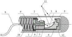
图1是本发明提供的一种光声内窥成像导管的剖面示意图;Fig. 1 is a schematic cross-sectional view of a photoacoustic endoscopic imaging catheter provided by the present invention;
图2是本发明提供的一种光声内窥成像导管的支撑件的立体示意图;Fig. 2 is a schematic perspective view of a support member of a photoacoustic endoscopic imaging catheter provided by the present invention;
图3是本发明提供的一种光声内窥成像导管的支撑件的剖面示意图;3 is a schematic cross-sectional view of a support for a photoacoustic endoscopic imaging catheter provided by the present invention;
图4是本发明提供的一种光声内窥成像导管的第一壳体的俯视图;Fig. 4 is a top view of the first casing of a photoacoustic endoscopic imaging catheter provided by the present invention;
图5是本发明提供的一种光声内窥成像导管的第一壳体的剖面示意图。Fig. 5 is a schematic cross-sectional view of a first casing of a photoacoustic endoscopic imaging catheter provided by the present invention.
附图标记说明:Explanation of reference signs:
1-填充物,2-超声换能器,3-反射光束,4-离轴反射镜,5-第一壳体,501-限位凸起,502-离轴反射镜安装槽,503-信号窗,6-入射光束,7-支撑件,701-换能器安装槽,702-定位凸起,8-光纤,9-扭矩弹簧,10-第二壳体,11-焦点,12-定位柱。1-filler, 2-ultrasonic transducer, 3-reflected beam, 4-off-axis reflector, 5-first housing, 501-limiting protrusion, 502-off-axis reflector mounting groove, 503-signal Window, 6-incident light beam, 7-support, 701-transducer installation groove, 702-positioning protrusion, 8-optical fiber, 9-torque spring, 10-second housing, 11-focus point, 12-positioning post .
具体实施方式Detailed ways
为了使本发明的目的、技术方案及优点更加清楚明白,以下结合附图及实施例,对本发明进行进一步详细说明。应当理解,此处所描述的具体实施例仅仅用以解释本发明,并不用于限定本发明。In order to make the object, technical solution and advantages of the present invention clearer, the present invention will be further described in detail below in conjunction with the accompanying drawings and embodiments. It should be understood that the specific embodiments described here are only used to explain the present invention, not to limit the present invention.
以下结合具体实施例对本发明的实现进行详细的描述。The implementation of the present invention will be described in detail below in conjunction with specific embodiments.
本实施例的附图中相同或相似的标号对应相同或相似的部件;在本发明的描述中,需要理解的是,若有术语“上”、“下”、“左”、“右”等指示的方位或位置关系为基于附图所示的方位或位置关系,仅是为了便于描述本发明和简化描述,而不是指示或暗示所指的装置或元件必须具有特定的方位、以特定的方位构造和操作,因此附图中描述位置关系的用语仅用于示例性说明,不能理解为对本专利的限制,对于本领域的普通技术人员而言,可以根据具体情况理解上述术语的具体含义。In the drawings of this embodiment, the same or similar symbols correspond to the same or similar components; The indicated orientation or positional relationship is based on the orientation or positional relationship shown in the drawings, and is only for the convenience of describing the present invention and simplifying the description, rather than indicating or implying that the referred device or element must have a specific orientation, or in a specific orientation. Construction and operation, so the words describing the positional relationship in the drawings are only for illustrative purposes, and should not be construed as limitations on this patent. Those of ordinary skill in the art can understand the specific meanings of the above terms according to specific situations.
参照图1-5所示,为本发明提供的较佳实施例。Referring to Fig. 1-5, it is a preferred embodiment provided by the present invention.
一种光声内窥成像装置,包括第一壳体5、离轴反射镜4、超声换能器2、支撑件7和光纤8;A photoacoustic endoscopic imaging device, comprising a
支撑件7设于第一壳体5的第一端,用于承载超声换能器2和光纤8;The support member 7 is arranged at the first end of the
离轴反射镜4设于第一壳体5的第二端,第一壳体5的壁面上开设有信号窗503,离轴反射镜4将光束反射后,穿过信号窗503,汇聚形成焦点11;The off-
光纤8安装于所述支撑件7,光纤8用于传导光束能量;支撑件7设有换能器安装槽701,超声换能器2设于所述换能器安装槽701,超声换能器2用于接收光声信号。The
第一壳体5用于封装离轴反射镜4、超声换能器2、支撑件7和光纤8。The
本实施例提供的一种光声内窥成像装置,利用反射式聚焦成像系统无色差的天然优势,解决了不同波长下光束焦点发生偏移的问题,提高了光声成像的分辨率,进而能够获得更加准确的功能信息解算结果;同时,该装置将光束的反射及聚焦功能集成在一个元件中实现,能够显著缩小探头前端的尺寸,提升成像导管的运动姿态灵活性,有利于拓展光声内窥成像技术的应用领域。The photoacoustic endoscopic imaging device provided in this embodiment solves the problem of shifting the focus of light beams at different wavelengths by utilizing the natural advantage of no chromatic aberration of the reflective focusing imaging system, improves the resolution of photoacoustic imaging, and can further Obtain more accurate functional information calculation results; at the same time, the device integrates the reflection and focusing functions of the beam into one component, which can significantly reduce the size of the front end of the probe, improve the flexibility of the imaging catheter's movement posture, and help expand the photoacoustic Fields of application of endoscopic imaging technology.
如附图1所示,一种光声内窥成像装置,包括第一壳体5、离轴反射镜4、超声换能器2、支撑件7、光纤8和第二壳体10。As shown in FIG. 1 , a photoacoustic endoscopic imaging device includes a
第一壳体5设于第二壳体10内,第二壳体10对第一壳体5及其内部元件起到保护作用。第二壳体10整体上可以是圆筒状结构,第二壳体10的前端表面可以是半球面或者其他无棱角的圆润表面,目的是防止有棱角的端部对生物组织等待检测部位造成损伤。第二壳体10由透明高分子材料制成,例如树脂材料,减少第二壳体10对光声信号的衰减。The
第一壳体5的第一端连接有扭矩弹簧9,扭矩弹簧9用于提供扭矩,实现第一壳体5的旋转运动及前后移动,扭矩弹簧9的层数为2层,增加了扭矩弹簧的强度,使其不易损坏。The first end of the
如附图4所示,第一壳体5的壁面上开设有信号窗503。As shown in FIG. 4 , a
如附图5所示,第一壳体5用于封装离轴反射镜4、超声换能器2、支撑件7和光纤8,形状为圆柱形,包括第一端、第二端和侧壁,第一端呈开放式,支撑件7及光纤8设于第一端;第二端呈封闭式,离轴反射镜4设于第二端;相距第一端的端部位置一定距离处,在第一壳体5内具有限位凸起501,用于支撑件7的装配定位;在第一壳体5内,第一壳体5的第二端设有离轴反射镜安装槽502,离轴反射镜4的底部设有定位柱12,离轴反射镜4通过定位柱12安装于离轴反射镜安装槽502。As shown in Figure 5, the
如附图2所示,支撑件7呈圆柱体状,其中心区域具有用于安装光纤8的贯通孔,侧面具有平面状的装配面,支撑件7设于第一壳体5的第一端,用于承载超声换能器2和光纤8。As shown in Figure 2, the support member 7 is in the shape of a cylinder, with a through hole for installing the
如附图3所示,支撑件7设有换能器安装槽701,超声换能器2设于所述换能器安装槽701,支撑件7与第一壳体5抵接的是定位凸起702;支撑件7的定位凸起702与第一壳体5内的限位凸起501相互配合,用于实现支撑件7的装配定位。As shown in Figure 3, the support member 7 is provided with a
光纤8的形式为多模光纤或单模光纤,用于传导光束能量,安装于支撑件7中心区域的贯通孔中,光纤8的后端通过填充物1固定于支撑件7,填充物1的材料为紫外固化胶。紫外固化胶,又称UV光固化胶,是一种单组份,不含溶剂,UV和可见光固化的粘接胶和密封胶,它可以用各种广泛的光源固化。The
离轴反射镜4设于第一壳体5的第二端,离轴反射镜4将光束反射后,穿过信号窗503,汇聚形成焦点11。The off-
离轴反射镜4的反射面为空间曲面,在空间上可以将入射到反射面上光束反射后汇聚到偏离轴向的某一径向位置。离轴反射镜4的反射面通常为球面或者非球面,球面的反射面较为容易加工,非球面的设计及加工较为复杂。对于成像质量具有严格要求的应用场合,可将离轴反射镜4的反射面加工成非球面,以减小像差。The reflective surface of the off-
离轴反射镜4的反射面较佳的实现方案为环形面,根据所需的反射光束焦点尺寸及位置来设计反射面的局部曲率半径、偏离光轴的距离及角度。The preferred implementation of the reflection surface of the off-
本实施例提供的光声内窥成像装置,其工作方式为:光束沿着光纤8进行传输,光纤8在支撑件7内的端部产生入射光束6,入射光束6经过离轴反射镜4的表面实现反射与汇聚,非线性光声信号焦点11区域产生;通过转动并回撤扭矩弹簧9带动第一壳体5及其内部的元件运动,实现光声内窥扫描成像。The photoacoustic endoscopic imaging device provided in this embodiment works in the following way: the light beam is transmitted along the
在本实施例中,采用以上光声内窥成像装置的光声内窥成像方法,包括以下步骤:In this embodiment, the photoacoustic endoscopic imaging method using the above photoacoustic endoscopic imaging device includes the following steps:
(1)光声内窥成像装置设于一根导管内,插入待检测腔体内;(1) The photoacoustic endoscopic imaging device is set in a catheter and inserted into the cavity to be detected;
(2)激光光束沿所述光纤8传输,在所述光纤8的端部产生入射光束6,所述入射光束6经所述离轴反射镜4的反射,穿过所述信号窗503,汇聚至待检测部位,激发待检测部位产生光声信号;(2) The laser beam is transmitted along the
(3)所述超声换能器2接收所述光声信号,经信号放大器放大后采集,采集的数据传输至数据采集卡;(3) The
(4)对采集的数据进行处理,获得待检测部位的光声图像。(4) Process the collected data to obtain a photoacoustic image of the part to be detected.
以上光声内窥成像方法可用于生物医学领域,通常可适用于非线性光声成像。非线性光声成像技术是以两个纳秒激光脉冲先后照射生物组织相同区域产生的光声信号实现成像。非线性光声成像技术相比经典的采用单个纳秒激光脉冲的光声成像技术,图像分辨率获得了显著的提升,因此可以获得更加清晰的图像。The above photoacoustic endoscopic imaging methods can be used in the field of biomedicine, and are generally applicable to nonlinear photoacoustic imaging. Nonlinear photoacoustic imaging technology is based on the photoacoustic signal generated by two nanosecond laser pulses sequentially irradiating the same area of biological tissue to achieve imaging. Compared with the classic photoacoustic imaging technology using a single nanosecond laser pulse, the nonlinear photoacoustic imaging technology has significantly improved image resolution, so a clearer image can be obtained.
在本实施例中,通过转动或前后移动所述光声内窥成像装置中的扭矩弹簧9带动所述第一壳体5及其内部的元件运动,实现对待检测部位所在区域的扫描成像,可以获得待检测部位所在区域的清晰图像,便于检测人员、医生等做出准确的判断。In this embodiment, by rotating or moving the
本发明提供的一种光声内窥成像装置及基于该装置的光声内窥成像方法,采用离轴反射镜对光束进行汇聚及折转,相比现有的光学透镜汇聚,平面反射镜折转的实现方式,本发明所述内容的有益效果为:1.离轴反射镜对光束进行汇聚时,所有波长的焦点位于空间相同的位置,光学专业术语称为无色差,因此相比存在色差的光学透镜聚焦方式,离轴反射镜聚焦的方式提高了分辨率。2.离轴反射镜能够集成了汇聚与折转两种功能,相比分立安装的光学透镜与平面反射镜,减小了光学系统占据的空间长度,从而缩小了探头的前端尺寸。A photoacoustic endoscopic imaging device and a photoacoustic endoscopic imaging method based on the device provided by the present invention use off-axis mirrors to converge and deflect light beams. The realization mode of the rotation, the beneficial effect of the content of the present invention is: 1. When the off-axis reflector converges the light beam, the focal points of all wavelengths are located at the same position in space, and the optical term is called achromatic aberration, so there is chromatic aberration compared with The focusing method of the optical lens and the focusing method of the off-axis mirror improve the resolution. 2. The off-axis mirror can integrate the two functions of convergence and deflection. Compared with the optical lens and plane mirror installed separately, the space length occupied by the optical system is reduced, thereby reducing the front-end size of the probe.
以上所述仅为本发明的较佳实施例而已,并不用以限制本发明,凡在本发明的精神和原则之内所作的任何修改、等同替换和改进等,均应包含在本发明的保护范围之内。The above descriptions are only preferred embodiments of the present invention, and are not intended to limit the present invention. Any modifications, equivalent replacements and improvements made within the spirit and principles of the present invention should be included in the protection of the present invention. within range.
Claims (4)
Translated fromChinesePriority Applications (1)
| Application Number | Priority Date | Filing Date | Title |
|---|---|---|---|
| CN202011373366.5ACN112493997B (en) | 2020-11-30 | 2020-11-30 | Photoacoustic endoscopic imaging device and photoacoustic endoscopic imaging method based on same |
Applications Claiming Priority (1)
| Application Number | Priority Date | Filing Date | Title |
|---|---|---|---|
| CN202011373366.5ACN112493997B (en) | 2020-11-30 | 2020-11-30 | Photoacoustic endoscopic imaging device and photoacoustic endoscopic imaging method based on same |
Publications (2)
| Publication Number | Publication Date |
|---|---|
| CN112493997A CN112493997A (en) | 2021-03-16 |
| CN112493997Btrue CN112493997B (en) | 2023-03-31 |
Family
ID=74968041
Family Applications (1)
| Application Number | Title | Priority Date | Filing Date |
|---|---|---|---|
| CN202011373366.5AActiveCN112493997B (en) | 2020-11-30 | 2020-11-30 | Photoacoustic endoscopic imaging device and photoacoustic endoscopic imaging method based on same |
Country Status (1)
| Country | Link |
|---|---|
| CN (1) | CN112493997B (en) |
Families Citing this family (2)
| Publication number | Priority date | Publication date | Assignee | Title |
|---|---|---|---|---|
| CN116262027A (en)* | 2021-12-15 | 2023-06-16 | 中国科学院深圳先进技术研究院 | Multi-point focusing photoacoustic endoscopic imaging catheter |
| CN116299837B (en)* | 2023-01-04 | 2024-03-15 | 华中科技大学 | Preparation method of full-light type lateral photoinduced ultrasonic self-collecting optical fiber endoscope |
Family Cites Families (8)
| Publication number | Priority date | Publication date | Assignee | Title |
|---|---|---|---|---|
| CN101262822B (en)* | 2005-07-18 | 2013-11-20 | 安德烈亚斯·曼德利斯 | Device for diagnosis of dental defects using infrared photothermal radiometry (PTR) and modulated laser luminescence (LUM) |
| US7935060B2 (en)* | 2006-11-08 | 2011-05-03 | Lightlab Imaging, Inc. | Opto-acoustic imaging devices and methods |
| WO2010080991A2 (en)* | 2009-01-09 | 2010-07-15 | Washington University In St. Louis | Miniaturized photoacoustic imaging apparatus including a rotatable reflector |
| CN104545811B (en)* | 2014-12-26 | 2017-06-27 | 深圳先进技术研究院 | An intravascular imaging system and method |
| CN106264604B (en)* | 2016-08-01 | 2019-12-31 | 苏州卓特医疗科技有限公司 | Full-scan photoacoustic dual-mode endoscopic probe |
| CN206700138U (en)* | 2016-12-30 | 2017-12-05 | 苏州阿格斯医疗技术有限公司 | A kind of endoscopic imaging probe for OCT systems |
| CN107411720B (en)* | 2017-09-19 | 2021-03-30 | 华南师范大学 | Intravascular photoacoustic/ultrasonic imaging endoscopic probe excited by high-efficiency collimated light |
| CN110251093B (en)* | 2019-07-24 | 2021-02-19 | 中南大学 | Acoustic focusing endoscopic photoacoustic/ultrasonic probe and scanning imaging method |
- 2020
- 2020-11-30CNCN202011373366.5Apatent/CN112493997B/enactiveActive
Also Published As
| Publication number | Publication date |
|---|---|
| CN112493997A (en) | 2021-03-16 |
Similar Documents
| Publication | Publication Date | Title |
|---|---|---|
| JP6538956B2 (en) | Imaging probe having imaging means combining ultrasound and optics | |
| US20140303476A1 (en) | Photoacoustic Imaging Using A Versatile Acoustic Lens | |
| CN103637819B (en) | Sound, light are total to the rectum optoacoustic endoscopy lens device of rotation sweep | |
| US8184367B2 (en) | Dynamically focused optical instrument | |
| US9351705B2 (en) | Miniaturized photoacoustic imaging apparatus including a rotatable reflector | |
| CN110859601B (en) | A photoacoustic imaging probe and photoacoustic imaging system | |
| CN101909512B (en) | An optical probe | |
| US20130190594A1 (en) | Scanning Optoacoustic Imaging System with High Resolution and Improved Signal Collection Efficiency | |
| US8401610B2 (en) | Rotating catheter probe using a light-drive apparatus | |
| US8184365B2 (en) | Optical instruments having dynamic focus | |
| CN108670177B (en) | Imaging probe of breast duct endoscope | |
| CN113229854B (en) | Probe integrating optical coherence tomography imaging and intravascular ultrasound | |
| CN112493997B (en) | Photoacoustic endoscopic imaging device and photoacoustic endoscopic imaging method based on same | |
| Li et al. | Miniature probe for forward-view wide-field optical-resolution photoacoustic endoscopy | |
| US20140160482A1 (en) | Optical system for endoscopic internally-referenced interferometric imaging, and method for employing the same | |
| CN111134591B (en) | Photoacoustic microscopic imaging pen and imaging method | |
| CN118986260B (en) | Arthroscopic device integrating optical-photoacoustic multimodal imaging and imaging method thereof | |
| CN108362646A (en) | A kind of system of miniature opto-acoustic microscopic imaging head, production method and its composition | |
| CN115251811B (en) | Large-depth photoacoustic multi-mode flexible endoscopic imaging probe based on micro stepping motor | |
| CN119344640A (en) | A photoacoustic-OCT dual-modality endoscopic imaging system based on multi-element focusing | |
| CN113080869A (en) | Ultrasonic imaging probe | |
| CN209899367U (en) | Dual-modality endoscope device based on liquid lens self-focusing | |
| CN115989989A (en) | Omnibearing rotary scanning endoscopic imaging device based on optical fiber sensor | |
| CN212698827U (en) | Optical coherence tomography endoscopic probe and imaging system | |
| JP7598475B2 (en) | Photoacoustic detection system combining transparent ultrasonic sensor |
Legal Events
| Date | Code | Title | Description |
|---|---|---|---|
| PB01 | Publication | ||
| PB01 | Publication | ||
| SE01 | Entry into force of request for substantive examination | ||
| SE01 | Entry into force of request for substantive examination | ||
| GR01 | Patent grant | ||
| GR01 | Patent grant |