CN112275335B - Self-priming valve compartmentalized chip, preparation method and detection method of Listeria monocytogenes - Google Patents
Self-priming valve compartmentalized chip, preparation method and detection method of Listeria monocytogenesDownload PDFInfo
- Publication number
- CN112275335B CN112275335BCN202011107828.9ACN202011107828ACN112275335BCN 112275335 BCN112275335 BCN 112275335BCN 202011107828 ACN202011107828 ACN 202011107828ACN 112275335 BCN112275335 BCN 112275335B
- Authority
- CN
- China
- Prior art keywords
- chip
- self
- priming valve
- mold
- listeria monocytogenes
- Prior art date
- Legal status (The legal status is an assumption and is not a legal conclusion. Google has not performed a legal analysis and makes no representation as to the accuracy of the status listed.)
- Active
Links
- 238000001514detection methodMethods0.000titleclaimsabstractdescription57
- 241000186779Listeria monocytogenesSpecies0.000titleclaimsabstractdescription46
- 238000002360preparation methodMethods0.000titleclaimsabstractdescription21
- 238000006243chemical reactionMethods0.000claimsabstractdescription29
- 239000012530fluidSubstances0.000claimsabstractdescription4
- 238000000034methodMethods0.000claimsdescription27
- OKTJSMMVPCPJKN-UHFFFAOYSA-NCarbonChemical compound[C]OKTJSMMVPCPJKN-UHFFFAOYSA-N0.000claimsdescription26
- 239000004205dimethyl polysiloxaneSubstances0.000claimsdescription24
- 229920000435poly(dimethylsiloxane)Polymers0.000claimsdescription24
- 239000000203mixtureSubstances0.000claimsdescription22
- -1polydimethylsiloxanePolymers0.000claimsdescription22
- XUIMIQQOPSSXEZ-UHFFFAOYSA-NSiliconChemical compound[Si]XUIMIQQOPSSXEZ-UHFFFAOYSA-N0.000claimsdescription21
- 229910021389grapheneInorganic materials0.000claimsdescription21
- 229910052710siliconInorganic materials0.000claimsdescription21
- 239000010703siliconSubstances0.000claimsdescription21
- 238000003756stirringMethods0.000claimsdescription14
- 108091023037AptamerProteins0.000claimsdescription13
- 239000000758substrateSubstances0.000claimsdescription13
- HEMHJVSKTPXQMS-UHFFFAOYSA-MSodium hydroxideChemical compound[OH-].[Na+]HEMHJVSKTPXQMS-UHFFFAOYSA-M0.000claimsdescription12
- IJOOHPMOJXWVHK-UHFFFAOYSA-NchlorotrimethylsilaneChemical compoundC[Si](C)(C)ClIJOOHPMOJXWVHK-UHFFFAOYSA-N0.000claimsdescription12
- 238000010438heat treatmentMethods0.000claimsdescription12
- 238000002347injectionMethods0.000claimsdescription12
- 239000007924injectionSubstances0.000claimsdescription12
- VWDWKYIASSYTQR-UHFFFAOYSA-Nsodium nitrateChemical compound[Na+].[O-][N+]([O-])=OVWDWKYIASSYTQR-UHFFFAOYSA-N0.000claimsdescription12
- XLYOFNOQVPJJNP-UHFFFAOYSA-NwaterChemical compoundOXLYOFNOQVPJJNP-UHFFFAOYSA-N0.000claimsdescription12
- 239000005662Paraffin oilSubstances0.000claimsdescription10
- 241001465754MetazoaSpecies0.000claimsdescription6
- 239000003795chemical substances by applicationSubstances0.000claimsdescription6
- 239000003431cross linking reagentSubstances0.000claimsdescription6
- 229920002120photoresistant polymerPolymers0.000claimsdescription6
- 235000010344sodium nitrateNutrition0.000claimsdescription6
- 239000004317sodium nitrateSubstances0.000claimsdescription6
- 239000005051trimethylchlorosilaneSubstances0.000claimsdescription6
- 238000007872degassingMethods0.000claimsdescription5
- 238000002073fluorescence micrographMethods0.000claimsdescription5
- 239000011521glassSubstances0.000claimsdescription5
- 238000004519manufacturing processMethods0.000claimsdescription5
- 239000012528membraneSubstances0.000claimsdescription5
- 238000002156mixingMethods0.000claimsdescription5
- 239000013642negative controlSubstances0.000claimsdescription5
- 239000008367deionised waterSubstances0.000claimsdescription4
- 229910021641deionized waterInorganic materials0.000claimsdescription4
- 238000000502dialysisMethods0.000claimsdescription4
- 238000001914filtrationMethods0.000claimsdescription4
- 150000002500ionsChemical class0.000claimsdescription4
- 238000001459lithographyMethods0.000claimsdescription4
- 239000002245particleSubstances0.000claimsdescription4
- 239000012286potassium permanganateSubstances0.000claimsdescription4
- 150000003839saltsChemical class0.000claimsdescription4
- 239000000243solutionSubstances0.000claimsdescription4
- 108091028043Nucleic acid sequenceProteins0.000claimsdescription3
- 150000007523nucleic acidsChemical class0.000claimsdescription3
- KPUWHANPEXNPJT-UHFFFAOYSA-NdisiloxaneChemical class[SiH3]O[SiH3]KPUWHANPEXNPJT-UHFFFAOYSA-N0.000claimsdescription2
- 238000009489vacuum treatmentMethods0.000claimsdescription2
- 238000011084recoveryMethods0.000abstractdescription4
- 230000035945sensitivityEffects0.000abstractdescription4
- 235000013870dimethyl polysiloxaneNutrition0.000description19
- 235000012431wafersNutrition0.000description19
- 241000894006BacteriaSpecies0.000description9
- BZTDTCNHAFUJOG-UHFFFAOYSA-N6-carboxyfluoresceinChemical compoundC12=CC=C(O)C=C2OC2=CC(O)=CC=C2C11OC(=O)C2=CC=C(C(=O)O)C=C21BZTDTCNHAFUJOG-UHFFFAOYSA-N0.000description5
- 235000015277porkNutrition0.000description5
- 230000008569processEffects0.000description5
- 238000012545processingMethods0.000description5
- 244000052616bacterial pathogenSpecies0.000description4
- 241000607142SalmonellaSpecies0.000description3
- 241000607272Vibrio parahaemolyticusSpecies0.000description3
- 238000013461designMethods0.000description3
- 238000001035dryingMethods0.000description3
- 230000005284excitationEffects0.000description3
- 235000013305foodNutrition0.000description3
- 238000011896sensitive detectionMethods0.000description3
- 238000002965ELISAMethods0.000description2
- 238000010586diagramMethods0.000description2
- 230000000694effectsEffects0.000description2
- 238000005516engineering processMethods0.000description2
- 230000001900immune effectEffects0.000description2
- 238000002955isolationMethods0.000description2
- 238000011068loading methodMethods0.000description2
- 239000000463materialSubstances0.000description2
- CXQXSVUQTKDNFP-UHFFFAOYSA-NoctamethyltrisiloxaneChemical groupC[Si](C)(C)O[Si](C)(C)O[Si](C)(C)CCXQXSVUQTKDNFP-UHFFFAOYSA-N0.000description2
- 244000052769pathogenSpecies0.000description2
- 238000004987plasma desorption mass spectroscopyMethods0.000description2
- 238000003752polymerase chain reactionMethods0.000description2
- 238000007789sealingMethods0.000description2
- 241001646719Escherichia coli O157:H7Species0.000description1
- 208000019331Foodborne diseaseDiseases0.000description1
- 206010024641ListeriosisDiseases0.000description1
- 201000009906MeningitisDiseases0.000description1
- 206010040047SepsisDiseases0.000description1
- FAPWRFPIFSIZLT-UHFFFAOYSA-MSodium chlorideChemical compound[Na+].[Cl-]FAPWRFPIFSIZLT-UHFFFAOYSA-M0.000description1
- 241000191967Staphylococcus aureusSpecies0.000description1
- QAOWNCQODCNURD-UHFFFAOYSA-NSulfuric acidChemical compoundOS(O)(=O)=OQAOWNCQODCNURD-UHFFFAOYSA-N0.000description1
- 230000009471actionEffects0.000description1
- 239000002390adhesive tapeSubstances0.000description1
- 150000001335aliphatic alkanesChemical class0.000description1
- 230000003321amplificationEffects0.000description1
- 239000000427antigenSubstances0.000description1
- 108091007433antigensProteins0.000description1
- 102000036639antigensHuman genes0.000description1
- 230000001580bacterial effectEffects0.000description1
- 230000009286beneficial effectEffects0.000description1
- 230000008901benefitEffects0.000description1
- 238000009739bindingMethods0.000description1
- 230000037029cross reactionEffects0.000description1
- 238000011161developmentMethods0.000description1
- 230000009977dual effectEffects0.000description1
- 235000013601eggsNutrition0.000description1
- 239000000706filtrateSubstances0.000description1
- 238000002866fluorescence resonance energy transferMethods0.000description1
- 244000078673foodborn pathogenSpecies0.000description1
- 235000012055fruits and vegetablesNutrition0.000description1
- 238000001027hydrothermal synthesisMethods0.000description1
- 238000011534incubationMethods0.000description1
- 208000015181infectious diseaseDiseases0.000description1
- 201000006747infectious mononucleosisDiseases0.000description1
- 230000003834intracellular effectEffects0.000description1
- 235000013372meatNutrition0.000description1
- 238000002493microarrayMethods0.000description1
- 235000013336milkNutrition0.000description1
- 239000008267milkSubstances0.000description1
- 210000004080milkAnatomy0.000description1
- 230000003472neutralizing effectEffects0.000description1
- 238000003199nucleic acid amplification methodMethods0.000description1
- 230000035699permeabilityEffects0.000description1
- 238000009832plasma treatmentMethods0.000description1
- 230000037452primingEffects0.000description1
- LLHKCFNBLRBOGN-UHFFFAOYSA-Npropylene glycol methyl ether acetateChemical compoundCOCC(C)OC(C)=OLLHKCFNBLRBOGN-UHFFFAOYSA-N0.000description1
- 108090000623proteins and genesProteins0.000description1
- 230000005180public healthEffects0.000description1
- 239000002994raw materialSubstances0.000description1
- 230000009870specific bindingEffects0.000description1
- 238000011895specific detectionMethods0.000description1
- 230000003068static effectEffects0.000description1
- 238000003860storageMethods0.000description1
- 239000000126substanceSubstances0.000description1
- 235000011149sulphuric acidNutrition0.000description1
- 238000012360testing methodMethods0.000description1
Images


Classifications
- B—PERFORMING OPERATIONS; TRANSPORTING
- B01—PHYSICAL OR CHEMICAL PROCESSES OR APPARATUS IN GENERAL
- B01L—CHEMICAL OR PHYSICAL LABORATORY APPARATUS FOR GENERAL USE
- B01L3/00—Containers or dishes for laboratory use, e.g. laboratory glassware; Droppers
- B01L3/50—Containers for the purpose of retaining a material to be analysed, e.g. test tubes
- B01L3/502—Containers for the purpose of retaining a material to be analysed, e.g. test tubes with fluid transport, e.g. in multi-compartment structures
- B01L3/5027—Containers for the purpose of retaining a material to be analysed, e.g. test tubes with fluid transport, e.g. in multi-compartment structures by integrated microfluidic structures, i.e. dimensions of channels and chambers are such that surface tension forces are important, e.g. lab-on-a-chip
- B01L3/502707—Containers for the purpose of retaining a material to be analysed, e.g. test tubes with fluid transport, e.g. in multi-compartment structures by integrated microfluidic structures, i.e. dimensions of channels and chambers are such that surface tension forces are important, e.g. lab-on-a-chip characterised by the manufacture of the container or its components
- G—PHYSICS
- G01—MEASURING; TESTING
- G01N—INVESTIGATING OR ANALYSING MATERIALS BY DETERMINING THEIR CHEMICAL OR PHYSICAL PROPERTIES
- G01N33/00—Investigating or analysing materials by specific methods not covered by groups G01N1/00 - G01N31/00
- G01N33/48—Biological material, e.g. blood, urine; Haemocytometers
- G01N33/50—Chemical analysis of biological material, e.g. blood, urine; Testing involving biospecific ligand binding methods; Immunological testing
- G01N33/53—Immunoassay; Biospecific binding assay; Materials therefor
- G01N33/5302—Apparatus specially adapted for immunological test procedures
- G01N33/5304—Reaction vessels, e.g. agglutination plates
- G—PHYSICS
- G01—MEASURING; TESTING
- G01N—INVESTIGATING OR ANALYSING MATERIALS BY DETERMINING THEIR CHEMICAL OR PHYSICAL PROPERTIES
- G01N33/00—Investigating or analysing materials by specific methods not covered by groups G01N1/00 - G01N31/00
- G01N33/48—Biological material, e.g. blood, urine; Haemocytometers
- G01N33/50—Chemical analysis of biological material, e.g. blood, urine; Testing involving biospecific ligand binding methods; Immunological testing
- G01N33/53—Immunoassay; Biospecific binding assay; Materials therefor
- G01N33/569—Immunoassay; Biospecific binding assay; Materials therefor for microorganisms, e.g. protozoa, bacteria, viruses
- G01N33/56911—Bacteria
Landscapes
- Health & Medical Sciences (AREA)
- Life Sciences & Earth Sciences (AREA)
- Chemical & Material Sciences (AREA)
- Immunology (AREA)
- Engineering & Computer Science (AREA)
- Hematology (AREA)
- Urology & Nephrology (AREA)
- Molecular Biology (AREA)
- Biomedical Technology (AREA)
- General Health & Medical Sciences (AREA)
- Analytical Chemistry (AREA)
- Chemical Kinetics & Catalysis (AREA)
- Biochemistry (AREA)
- Biotechnology (AREA)
- Food Science & Technology (AREA)
- Medicinal Chemistry (AREA)
- Physics & Mathematics (AREA)
- Microbiology (AREA)
- Pathology (AREA)
- Cell Biology (AREA)
- General Physics & Mathematics (AREA)
- Virology (AREA)
- Tropical Medicine & Parasitology (AREA)
- Dispersion Chemistry (AREA)
- Clinical Laboratory Science (AREA)
- Investigating Or Analysing Materials By The Use Of Chemical Reactions (AREA)
- Measuring Or Testing Involving Enzymes Or Micro-Organisms (AREA)
Abstract
Translated fromChinese
Description
Translated fromChinese技术领域technical field
本发明属于芯片制备领域,具体涉及一种自吸阀区隔式芯片、制备方法及单增李斯特菌的检测方法。The invention belongs to the field of chip preparation, and in particular relates to a self-priming valve compartmentalized chip, a preparation method and a detection method of Listeria monocytogenes.
背景技术Background technique
单核细胞增生性李斯特菌(Listeria monocytogenes,L.monocytogenes)为革兰氏阳性小杆菌,无芽孢,属兼性细胞内细菌,是导致食源性疾病爆发的最常见病原体之一。对理化因素抵抗力较强,常通过受污染的食物传播,如鸡蛋、果蔬、肉类和牛奶等。感染后能引起人、畜的李氏特菌病,主要表现为败血症、脑膜炎和单核细胞增多症。是最致命的食源性病原体之一,可造成二至三成的感染者死亡,其致死率甚至高过沙门氏菌及肉毒杆菌。此外,单核细胞增生李斯特菌不仅可以生存,而且还可以在冰箱中生长,这使食物储存和冷链运输变得困难。考虑到单核细胞增生李斯特氏菌对公共卫生的巨大威胁,迫切需要一种高度灵敏,特异的方法在食品中进行检测。Listeria monocytogenes (L.monocytogenes) is a small, Gram-positive, spore-free, facultative intracellular bacterium, which is one of the most common pathogens causing foodborne disease outbreaks. Strong resistance to physical and chemical factors, often transmitted through contaminated food, such as eggs, fruits and vegetables, meat and milk. After infection, it can cause listeriosis in humans and animals, mainly manifested as sepsis, meningitis and mononucleosis. It is one of the deadliest food-borne pathogens, causing 20-30% of infected people to die, and its fatality rate is even higher than that of Salmonella and botulinum. In addition, L. monocytogenes can not only survive, but also grow in refrigerators, making food storage and cold chain transportation difficult. Considering the enormous public health threat of Listeria monocytogenes, there is an urgent need for a highly sensitive, specific method for detection in food.
目前对单增李斯特菌的常见检测方法包括国家标准GB 4789.30-2016规定的传统分离培养鉴定法,分子生物学检测方法和免疫学检测方法等。(1)传统分离培养鉴定法是单增李斯特菌检测的“金标准”,该方法计数可靠,可以准确的鉴定单增李斯特菌并确定其浓度,具有成本较低的优势,便于普及应用,但检测周期过长,一般需要一周左右,无法满足副溶血性弧菌快速、灵敏的检测要求;(2)分子生物学检测方法中,常规聚合酶链式反应(Polymerase Chain Reaction,PCR)方法应用最为广泛,该方法通过扩增目标菌的基因序列实现对致病菌的检测,具有高灵敏度和特异性,在一定程度上能够满足灵敏检测的要求,但该方法同样需要较长时间的前增菌过程,且所需仪器设备昂贵,检测成本高,易出现假阳性结果;(3)免疫学检测方法中,酶联免疫吸附试验(Enzyme Linked Immuno SorbentAssay,ELISA)方法最为常见,该方法通过抗原与抗体的特异性结合反应,再辅以免疫放大技术实现对致病菌的检测,该方法与传统培养鉴定法相比提高了检测灵敏度和检测效率,但仍需要增菌和反复的孵育过程,检测步骤复杂,易出现交叉反应导致假阳性结果。因此,如何在短时间内实现对病原菌的灵敏检测,又不过分依赖设备就成为新一代检测技术发展的重点。At present, common detection methods for Listeria monocytogenes include traditional isolation and culture identification methods specified in the national standard GB 4789.30-2016, molecular biological detection methods and immunological detection methods. (1) The traditional isolation and culture identification method is the "gold standard" for the detection of Listeria monocytogenes. This method is reliable in counting, can accurately identify Listeria monocytogenes and determine its concentration. It has the advantage of low cost and is easy to popularize and apply. , but the detection period is too long, usually about a week, which cannot meet the rapid and sensitive detection requirements of Vibrio parahaemolyticus; (2) Among the molecular biology detection methods, the conventional polymerase chain reaction (PCR) method The most widely used method is to detect pathogenic bacteria by amplifying the gene sequence of the target bacteria. It has high sensitivity and specificity, and can meet the requirements of sensitive detection to a certain extent. However, this method also requires a long time before The enrichment process requires expensive equipment, high detection costs, and false positive results are prone to occur; (3) Among the immunological detection methods, the Enzyme Linked Immuno Sorbent Assay (ELISA) method is the most common, and this method passes The specific binding reaction of antigen and antibody, supplemented by immune amplification technology to achieve the detection of pathogenic bacteria, this method improves the detection sensitivity and detection efficiency compared with the traditional culture identification method, but still requires enrichment and repeated incubation process, The detection steps are complex, and cross-reactions are prone to lead to false positive results. Therefore, how to achieve sensitive detection of pathogens in a short period of time without relying too much on equipment has become the focus of the development of a new generation of detection technology.
发明内容SUMMARY OF THE INVENTION
本发明目的是提供一种自吸阀区隔式芯片、制备方法及单增李斯特菌的检测方法,该芯片基于整体的负压环境及氧化石墨烯和FAM标记特异性适配体和HNB染料,能够快速、灵敏、简便地检测单增李斯特菌。The purpose of the present invention is to provide a self-priming valve compartment chip, preparation method and detection method of Listeria monocytogenes, the chip is based on the overall negative pressure environment and graphene oxide and FAM label specific aptamer and HNB dye , which can detect Listeria monocytogenes quickly, sensitively and simply.
本发明首先提供一种自吸阀区隔式芯片,该芯片包括支撑层和设置在支撑层上的反应层,所述的反应层上设有若干流体微通道和反应微腔室,反应层的中间还设有进样口。The present invention first provides a self-priming valve compartmentalized chip, the chip includes a support layer and a reaction layer arranged on the support layer, the reaction layer is provided with a number of fluid microchannels and reaction microchambers, and the reaction layer is There is also an injection port in the middle.
本发明首先提供一种自吸阀区隔式芯片的制备方法,包括:The present invention first provides a method for preparing a self-priming valve segmented chip, comprising:
步骤一:模具的制备Step 1: Preparation of the mold
使用CorelDRAW X8设计自吸阀区隔式芯片的通道和腔室图案,然后将其印刷到一块透明膜上作为掩模,使用旋涂机,将负性光刻胶涂布于硅晶片上,共计涂布两层,随后将硅晶片放在掩模下方的单面光刻机上,调整掩模的位置,以使图案位于晶片的中间,曝光后对硅晶片进行烘烤,最后,用显影剂冲洗处理过的硅晶片,直至模具图形完全出现为止,再次对模具进行烘烤,得到模具;The channel and chamber patterns of the self-priming valve compartment chip were designed using CorelDRAW X8, then printed on a transparent film as a mask, and a spin coater was used to coat the negative photoresist on the silicon wafer, totaling Two coats are applied, the silicon wafer is then placed on a single-sided lithography machine under the mask, the mask is positioned so that the pattern is in the middle of the wafer, the silicon wafer is baked after exposure, and finally, rinsed with developer The processed silicon wafer is baked until the mold pattern completely appears, and the mold is baked again to obtain a mold;
步骤二:自吸阀区隔式芯片的制作Step 2: Fabrication of self-priming valve segmented chip
使用三甲基氯硅烷处理模具,将聚二甲基硅氧烷组分预固剂A和交联剂B混合去除气泡,倒入模具中,将螺栓和螺母拧到一端齐平,然后将齐平端向下放置在模具的阀门位置,经加热板烘烤固化后,将聚二甲基硅氧烷从模具表面揭除,使用打孔器对加样孔进行打孔,最后,对聚二甲基硅氧烷的通道面及玻璃片的表面同时使用等离子体处理,将被处理的平面相互贴合后烘烤固化形成自吸阀区隔式芯片。Treat the mold with trimethylchlorosilane, mix the polydimethylsiloxane component pre-curing agent A and crosslinking agent B to remove air bubbles, pour it into the mold, screw the bolt and nut flush to one end, and then place the The flat end is placed down on the valve position of the mold. After the heating plate is baked and cured, the polydimethylsiloxane is removed from the mold surface, and the sample hole is punched with a hole punch. Finally, the polydimethylsiloxane is drilled. The channel surface of the base siloxane and the surface of the glass sheet are treated with plasma at the same time.
优选的是,所述步骤一中硅晶片烘烤条件是60~70℃烘烤1~3min,随后是85~95℃下烘烤10min。Preferably, in the first step, the silicon wafer baking conditions are 60-70° C. for 1-3 minutes, followed by 85-95° C. for 10 minutes.
优选的是,所述步骤二中三甲基氯硅烷处理时间是10~20min。Preferably, the treatment time of trimethylchlorosilane in the second step is 10-20 min.
优选的是,所述步骤二中聚二甲基硅氧烷组分预固剂A和交联剂B的质量比10:1。Preferably, in the second step, the mass ratio of the polydimethylsiloxane component pre-fixing agent A and the cross-linking agent B is 10:1.
优选的是,所述步骤二中混合聚二甲基硅氧烷组分去除气泡的条件是1500~2000rpm旋转1~2min。Preferably, in the second step, the conditions for mixing the polydimethylsiloxane components to remove air bubbles are rotating at 1500-2000 rpm for 1-2 minutes.
优选的是,所述步骤二中烘烤固化的温度是70~90℃,时间是30~40min。Preferably, in the second step, the temperature for baking and curing is 70-90° C., and the time is 30-40 minutes.
优选的是,所述步骤二中芯片加热固化的温度是70~90℃,时间是30~40min。Preferably, in the second step, the temperature for heating and curing the chip is 70-90° C., and the time is 30-40 minutes.
本发明还提供一种基于自吸阀区隔式芯片的单增李斯特菌的检测方法,包括:The present invention also provides a method for detecting Listeria monocytogenes based on a self-priming valve segmented chip, comprising:
步骤一:氧化石墨烯的制备Step 1: Preparation of graphene oxide
将石墨粉加入H2SO4和硝酸钠的混合物中,于冰浴中搅拌,剧烈搅拌下,将高锰酸钾缓慢加入上述混合物,继续在30~40℃搅拌,然后加热至140~150℃搅拌,加入去离子水终止反应体系,在冰浴中用氢氧化钠中和H2SO4,将PH调至5.5~6,得到的浅黄色溶液经过离子过滤膜过滤去除大颗粒,然后在透析袋中透析2~3天去除盐分,得到氧化石墨烯;Add graphite powder to the mixture of H2 SO4 and sodium nitrate, stir in an ice bath, slowly add potassium permanganate to the above mixture under vigorous stirring, continue to stir at 30-40 ℃, and then heat to 140-150 ℃ Stir, add deionized water to terminate the reaction system, neutralize H2 SO4 with sodium hydroxide in an ice bath, adjust the pH to 5.5-6, and the resulting pale yellow solution is filtered through an ion filtration membrane to remove large particles, and then dialyzed Dialysis in the bag for 2 to 3 days to remove salt to obtain graphene oxide;
步骤二:自吸阀区隔式芯片的前处理Step 2: Pretreatment of self-priming valve compartment chip
使用透明胶带覆盖芯片顶部,置于真空泵中进行脱气处理;Cover the top of the chip with scotch tape and place it in a vacuum pump for degassing;
步骤三:检测Step 3: Detection
将步骤一得到氧化石墨烯、FAM修饰的单增李斯特菌特异性适配体和HNB混合,作为阴性对照,通过底物进样孔在负压条件下使混合物均匀分布到样品孔中,随后将透明胶带揭除,置于加热板上加热,待底物完全干燥后,重新在芯片表面贴附胶带并置于真空泵中抽真空处理,将待测样品以及石蜡油注入总进样孔中,利用石蜡油将每个检测小室分开,反应后,将芯片置于小动物成像仪中拍摄荧光图像。Mix the graphene oxide, FAM-modified Listeria monocytogenes specific aptamer and HNB obtained in
优选的是,所述步骤三的氧化石墨烯浓度为0.4~0.5mg·mL-1,FAM修饰的单增李斯特菌特异性适配体浓度为1~1.2μM,HNB的浓度为12.5μM。Preferably, the concentration of graphene oxide in the third step is 0.4-0.5 mg·mL-1 , the concentration of the FAM-modified Listeria monocytogenes-specific aptamer is 1-1.2 μM, and the concentration of HNB is 12.5 μM.
本发明的有益效果The beneficial effects of the present invention
本发明提供一种自吸阀区隔式芯片、制备方法及单增李斯特菌的检测方法,该方法首先是制作芯片的模具,然后利用聚二甲基硅氧烷的可塑性通过加热固化,打孔,等离子体处理等过程,依托模具制作自吸阀区隔式芯片,在芯片表面贴附透明胶带,置于真空泵中抽真空使得芯片内部形成负压环境。而后将HNB、FAM染料标记的单增李斯特菌特异性适配体和氧化石墨烯按一定比例混合,通过底物进样孔注入芯片中,由于大气压的作用使得混合物将均匀分布到各检测小室中。由于氧化石墨烯的荧光共振能量转移效应使得FAM的绿色荧光被淬灭,此时混合物只剩HNB的红色荧光。揭去表面的胶带,将含有底物的芯片置于加热板上干燥,待小室内底物完全干燥后,重新于芯片表面贴附胶带,置于真空泵中抽真空使得芯片内部形成负压环境。将含有病原菌的待检测样品通过进样孔注入芯片中,当存在目标细菌时,6-FAM标记的适体会捕获目标细菌,远离氧化石墨烯表面使得绿色荧光恢复,混合物显示绿色,没有目标细菌存在时,6-FAM的荧光仍处于淬灭状态,混合物显示红色,因此通过红绿色可以区分有无目标菌存在,而根据绿色荧光的强度可以进一步对病原菌进行定量分析。The invention provides a self-priming valve segmented chip, a preparation method and a method for detecting Listeria monocytogenes. The method firstly prepares a mold for the chip, and then utilizes the plasticity of polydimethylsiloxane to cure by heating to mold Hole, plasma treatment and other processes, rely on the mold to make a self-priming valve compartment chip, attach transparent tape to the surface of the chip, and place it in a vacuum pump to evacuate to form a negative pressure environment inside the chip. Then, HNB, FAM dye-labeled Listeria monocytogenes-specific aptamer and graphene oxide are mixed in a certain proportion, and injected into the chip through the substrate injection hole. Due to the action of atmospheric pressure, the mixture will be evenly distributed to each detection chamber. middle. Due to the fluorescence resonance energy transfer effect of graphene oxide, the green fluorescence of FAM was quenched, and only the red fluorescence of HNB remained in the mixture. Remove the tape on the surface, put the chip containing the substrate on a heating plate to dry, and after the substrate in the chamber is completely dry, re-attach the tape on the surface of the chip, and place it in a vacuum pump to vacuum to form a negative pressure environment inside the chip. The sample to be detected containing pathogenic bacteria is injected into the chip through the injection hole. When the target bacteria is present, the 6-FAM-labeled aptamer will capture the target bacteria and keep away from the graphene oxide surface to restore the green fluorescence, the mixture shows green, and there is no target bacteria. At , the fluorescence of 6-FAM is still in a quenched state, and the mixture shows red, so the presence or absence of target bacteria can be distinguished by red and green, and the pathogenic bacteria can be further quantitatively analyzed according to the intensity of green fluorescence.
本发明设计制作了自吸阀区隔式芯片,并以该芯片为载体,将HNB染料引入到FAM染料标记的单增李斯特菌特异性适配体和氧化石墨烯的混合物中,建立了双荧光淬灭-恢复体系用于检测单增李斯特菌,操作简单,极大的缩短了检测时间,定量检测时变异系数小,最低检测浓度为46.8CFU/mL,加标回收率达到98.08%,灵敏度高,稳定性好。The invention designs and manufactures a self-priming valve compartment chip, and uses the chip as a carrier to introduce HNB dye into the mixture of FAM dye-labeled Listeria monocytogenes specific aptamer and graphene oxide to establish a dual The fluorescence quenching-recovery system is used to detect Listeria monocytogenes. The operation is simple, the detection time is greatly shortened, the coefficient of variation during quantitative detection is small, the minimum detection concentration is 46.8CFU/mL, and the recovery rate of standard addition reaches 98.08%. High sensitivity and good stability.
附图说明Description of drawings
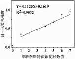
图1为实施例3中单增李斯特菌检测标准曲线图。Fig. 1 is the standard curve diagram of the detection of Listeria monocytogenes in Example 3.
图2为实施例3中单增李斯特菌与副溶血性弧菌和沙门氏菌检测的特异性结果图。2 is a graph showing the specificity results of the detection of Listeria monocytogenes, Vibrio parahaemolyticus and Salmonella in Example 3.
图3本发明实施例4中单增李斯特菌检测标准曲线图。3 is a graph of the standard curve for the detection of Listeria monocytogenes in Example 4 of the present invention.
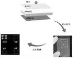
图4为本发明自吸阀区隔式芯片的制备及基于自吸阀区隔式芯片的单增李斯特菌的检测方法流程图。4 is a flow chart of the preparation of the self-priming valve compartmentalized chip and the detection method of Listeria monocytogenes based on the self-priming valve compartmentalized chip of the present invention.
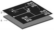
图5为本发明自吸阀区隔式芯片的结构示意图。FIG. 5 is a schematic structural diagram of the self-priming valve compartmentalized chip of the present invention.
图中,1、反应微腔室,2、进样口,3、反应层,4、支撑层。In the figure, 1, the reaction microchamber, 2, the injection port, 3, the reaction layer, 4, the support layer.
具体实施方式Detailed ways
本发明首先提供一种自吸阀区隔式芯片,如图5所示,该芯片包括支撑层4和设置在支撑层4上的反应层3,所述的反应层3上设有若干流体微通道和反应微腔室1,反应层3的中间还设有进样口2。为了在芯片脱气处理时起到密闭作用,优选在反应层上设有密封层,所述的密封层优选为胶带。The present invention first provides a self-priming valve compartmentalized chip, as shown in FIG. 5 , the chip includes a
所述的反应层的材料优选为PDMS材料,支撑层的下方设有玻璃基底,尺寸优选为40mm*60mm;所述的进样口是将一组螺栓和螺母作为阀门开关。The material of the reaction layer is preferably PDMS material, and a glass substrate is provided below the support layer, and the size is preferably 40mm*60mm; the injection port uses a set of bolts and nuts as valve switches.
本发明还提供一种自吸阀区隔式芯片的制备,包括:The present invention also provides a preparation of a self-priming valve segmented chip, comprising:
步骤一:模具的制备Step 1: Preparation of the mold
使用CorelDRAW X8设计自吸阀区隔式芯片的通道和腔室图案,然后将其印刷到一块透明膜上作为掩模,考虑到腔室的高度,优选于硅晶片上涂覆两层负性光刻胶,使用Spincoat G3P-8旋涂机,将负性光刻胶涂布于干净的干燥硅晶片上,旋涂机的使用条件优选为2000~3000rpm,时间为30s-1min,然后再次重复此步骤以形成第二层,随后将硅晶片放在掩模下方的“h49-25c 4型”单面光刻机上,在曝光之前,调整掩模的位置,以使图案位于晶片的中间,以便后续的芯片制作,所述的曝光时间优选为25s~40s,将硅晶片优选在60~70℃烘烤1~3min,随后在85~95℃下烘烤10min,最后,用显影剂冲洗处理过的硅晶片,直至模具图形完全出现为止;所述的显影剂优选为SU-8,为了使模具具有更佳的机械性能,优选在300℃下烘烤20-40min,至此模具制备完成;Use CorelDRAW X8 to design the channel and chamber pattern of the self-priming valve compartment chip, and then print it on a transparent film as a mask. Considering the height of the chamber, it is preferable to coat the silicon wafer with two layers of negative light For photoresist, use Spincoat G3P-8 spin coater to apply negative photoresist on clean and dry silicon wafers. The operating conditions of the spin coater are preferably 2000-3000rpm, and the time is 30s-1min, and then repeat this process again. step to form the second layer, then place the silicon wafer on a "h49-
步骤二:自吸阀区隔式芯片的制作Step 2: Fabrication of self-priming valve segmented chip
使用三甲基氯硅烷处理模具,优选处理时间为10-20min,以防聚二甲基硅氧烷粘附,然后将聚二甲基硅氧烷组分预固剂A和交联剂B混合,优选混合质量比为10:1,优选1500~2000rpm旋转1~2min去除混合时产生的气泡并倒入模具中,待芯片静止约1min后,除去聚二甲基硅氧烷中的气泡,将螺栓和螺母拧到一端齐平,然后将齐平端向下放置在模具的阀门位置,注意避免将气泡引入聚二甲基硅氧烷中,以免影响芯片性能,随后优选于70~90℃加热板上烘烤30~40min,待聚二甲基硅氧烷层完全固化后小心从模具上剥离,优选1.0/1.2mm打孔器对加样孔进行打孔;最后,对聚二甲基硅氧烷的通道面及玻璃片的表面同时使用等离子预处理,所述的等离子预处理的条件优选为200V处理1min,将被处理的平面相互贴合后优选90℃烘烤30min形成牢固稳定的芯片;随后使用透明胶带覆盖芯片顶部,获得自吸阀区隔式芯片。Treat the mold with trimethylchlorosilane, preferably for 10-20min, to prevent the polydimethylsiloxane from sticking, and then mix the polydimethylsiloxane component pre-curing agent A and crosslinking agent B , preferably the mixing mass ratio is 10:1, preferably 1500~2000rpm for 1~2min to remove the bubbles generated during mixing and pour it into the mold, after the chip is static for about 1min, remove the polydimethylsiloxane. Screw the bolt and nut until one end is flush, and then place the flush end down on the valve position of the mold, taking care to avoid introducing air bubbles into the polydimethylsiloxane, so as not to affect the performance of the chip, and then preferably heating the plate at 70-90 °C Bake for 30 to 40 minutes, and carefully peel off the polydimethylsiloxane layer from the mold after the polydimethylsiloxane layer is completely cured, preferably with a 1.0/1.2mm hole puncher to punch the sample hole; The channel surface of the alkane and the surface of the glass sheet are pretreated with plasma at the same time. The conditions of the plasma pretreatment are preferably 200V for 1min, and after the planes to be treated are bonded to each other, it is preferably baked at 90°C for 30min to form a firm and stable chip; The top of the chip was then covered with scotch tape to obtain a self-priming valve compartment chip.
本发明还提供一种基于自吸阀区隔式芯片的单增李斯特菌的检测方法,包括:The present invention also provides a method for detecting Listeria monocytogenes based on a self-priming valve segmented chip, comprising:
步骤一:氧化石墨烯的制备Step 1: Preparation of graphene oxide
将石墨粉加入H2SO4和硝酸钠的混合物中,于冰浴中搅拌25~30min,剧烈搅拌下,将3.0~4.0g高锰酸钾缓慢加入上述混合物(将温度保持0℃,但是在10min内完成),搅拌1.5~2h,继续在30~40℃搅拌1.5~2h,然后加热至140~150℃搅拌2~3h,加入去离子水终止反应体系,然后在冰浴中用氢氧化钠中和H2SO4,将PH调至5.5~6,得到的浅黄色溶液经过0.22μm的离子过滤膜过滤去除大颗粒,然后在透析袋(MWCO 1000da)中透析2~3天去除盐分,获得氧化石墨烯;所述石墨粉和硝酸钠质量比优选为1.0~1.5:40~46,H2SO4体积mL:硝酸钠的质量g优选为80~100:40~46;Add graphite powder to the mixture of H2 SO4 and sodium nitrate, stir in an ice bath for 25-30 min, under vigorous stirring, slowly add 3.0-4.0 g of potassium permanganate to the above mixture (keep the temperature at 0°C, but at Complete within 10min), stir for 1.5~2h, continue to stir at 30~40℃ for 1.5~2h, then heat to 140~150℃, stir for 2~3h, add deionized water to terminate the reaction system, then use sodium hydroxide in an ice bath Neutralize H2 SO4 , adjust the pH to 5.5-6, the obtained pale yellow solution is filtered through a 0.22 μm ion filtration membrane to remove large particles, and then dialyzed in a dialysis bag (MWCO 1000da) for 2 to 3 days to remove salt, and the obtained Graphene oxide; the mass ratio of the graphite powder and sodium nitrate is preferably 1.0-1.5: 40-46, and the volume mL of H2 SO4 : the mass g of sodium nitrate is preferably 80-100: 40-46;
步骤二:自吸阀区隔式芯片的前处理Step 2: Pretreatment of self-priming valve compartment chip
使用透明胶带覆盖芯片顶部,置于真空泵中进行脱气处理,优选处理时间为40~50min;Cover the top of the chip with scotch tape, place it in a vacuum pump for degassing, and the preferred treatment time is 40 to 50 minutes;
步骤三:检测Step 3: Detection
将步骤一的氧化石墨烯,6-FAM标记的单增李斯特菌特异性适配体和HNB混合,作为阴性对照,通过底物进样孔在负压条件下使混合物均匀分布到样品孔中;所述还原氧化石墨烯浓度优选为0.4~0.5mg·mL-1,FAM修饰的单增李斯特菌特异性适配体浓度优选为1~1.2μM,HNB的浓度优选为12.5μM;随后将透明胶带揭除,以增加透气性方便干燥,置于45℃加热板上加热2~2.5h,待底物完全干燥后,重新在芯片表面贴附胶带并置于真空泵中抽真空,处理时长优选为40~50min,将待测样品以及随后的石蜡油注入总进样孔中,利用石蜡油将每个检测小室分开,反应10min后,将芯片置于小动物成像仪中,在455nm蓝光激发条件下拍摄荧光图像,通过红色与绿色荧光的区别来鉴定阴/阳性,通过绿色荧光的荧光强度对目标菌进行定量。Mix the graphene oxide in
下面结合具体实施例对本发明做进一步详细的描述,实施例中涉及到的原料均为商购获得The present invention will be described in further detail below with reference to specific examples, and the raw materials involved in the examples are all commercially available
实施例1自吸阀分隔式芯片的制备Example 1 Preparation of self-priming valve partitioned chip
首先制备模具,使用CorelDRAW X8设计自吸阀区隔式芯片的通道和腔室图案,然后将其印刷到一块透明膜上作为掩模,考虑到腔室的高度,于硅晶片上涂覆两层负性光刻胶。使用Spincoat G3P-8旋涂机,将负性光刻胶以2700rpm的速度作为第一层涂布于干净的干燥硅晶片上,时长为30s,然后再次重复此步骤以形成第二层。随后将硅晶片放在掩模下方的“h49-25c 4型”单面光刻机上。在曝光之前,调整掩模的位置,以使图案大致位于晶片的中间,以便后续的芯片制作。曝光25s后,将硅晶片在65℃烘烤1min,然后在95℃下烘烤10min。最后,用SU-8显影剂冲洗处理过的硅晶片,直至模具图形完全出现为止。为了使模具具有更佳的机械性能,在300℃下烘烤20min,至此模具制备完成。First prepare the mold, use CorelDRAW X8 to design the channel and chamber pattern of the self-priming valve compartment chip, then print it on a piece of transparent film as a mask, and apply two layers on the silicon wafer considering the height of the chamber Negative photoresist. Using a Spincoat G3P-8 spin coater, a negative photoresist was applied as a first layer on a clean, dry silicon wafer at 2700 rpm for 30 s, and the procedure was repeated again to form a second layer. The silicon wafer was then placed on a "h49-
使用三甲基氯硅烷处理模具10min,以防聚二甲基硅氧烷粘附。然后将聚二甲基硅氧烷组分预固剂A和交联剂B以10:1的质量比混合,2000rpm旋转1min去除混合时产生的气泡并倒入模具中。待芯片静止约1min后,除去PDMS中的气泡。将螺栓和螺母拧到一端齐平,然后将齐平端向下放置在模具的阀门位置,注意避免将气泡引入聚二甲基硅氧烷中,以免影响芯片性能。随后置于90℃加热板上烘烤40min,待聚二甲基硅氧烷层完全固化后小心从模具上剥离,使用1.2mm打孔器对加样孔进行打孔。最后,对聚二甲基硅氧烷的通道面及玻璃片的表面同时使用等离子体预处理,将被处理的平面相互贴合后于90℃烘烤30min形成牢固稳定的芯片。随后使用透明胶带覆盖芯片顶部,使之可以更好地脱气以便后续使用。The molds were treated with trimethylchlorosilane for 10 min to prevent polydimethylsiloxane from sticking. Then, the polydimethylsiloxane component pre-curing agent A and cross-linking agent B were mixed in a mass ratio of 10:1, rotated at 2000 rpm for 1 min to remove air bubbles generated during mixing, and poured into a mold. After the chip was at rest for about 1 min, the air bubbles in the PDMS were removed. Thread the bolt and nut flush on one end, then place the flush end down at the valve position of the mold, taking care to avoid introducing air bubbles into the polydimethylsiloxane that could affect chip performance. Then place it on a 90° C. hot plate to bake for 40 min. After the polydimethylsiloxane layer is completely cured, it is carefully peeled off from the mold, and a 1.2 mm hole punch is used to punch the sample hole. Finally, the channel surface of polydimethylsiloxane and the surface of the glass sheet were pretreated with plasma at the same time, and the treated planes were bonded to each other and baked at 90° C. for 30 minutes to form a firm and stable chip. The top of the chip is then covered with scotch tape to allow better degassing for subsequent use.
实施例2水热法合成氧化石墨烯Example 2 Hydrothermal synthesis of graphene oxide
将1.0g石墨粉加入100mL H2SO4和46g硝酸钠的混合物中,于冰浴中搅拌30min。剧烈搅拌下,将3.0g高锰酸钾缓慢加入上述混合物(将温度保持0℃,但是在10min内完成),搅拌2h。继续在40℃搅拌2h,然后加热至150℃搅拌3h。加入200mL去离子水终止反应体系。然后在冰浴中用氢氧化钠中和H2SO4,将PH调至6。得到的浅黄色溶液经过0.22μm的离子过滤膜过滤去除大颗粒,然后在透析袋(MWCO 1000da)中透析3天去除盐分,获得氧化石墨烯。1.0 g of graphite powder was added to a mixture of 100 mL of H2 SO4 and 46 g of sodium nitrate, and stirred in an ice bath for 30 min. With vigorous stirring, 3.0 g of potassium permanganate was slowly added to the above mixture (the temperature was kept at 0°C, but completed within 10 min) and stirred for 2 h. Continue to stir at 40°C for 2h, then heat to 150°C and stir for 3h. The reaction system was terminated by adding 200 mL of deionized water. The pH was then adjusted to6 by neutralizing theH2SO4 with sodium hydroxide in an ice bath. The resulting pale yellow solution was filtered through a 0.22 μm ion filtration membrane to remove large particles, and then dialyzed in a dialysis bag (MWCO 1000da) for 3 days to remove salt to obtain graphene oxide.
实施例3基于自吸阀区隔式芯片的单增李斯特菌的检测方法
检测流程如图4,取一自吸阀分隔式芯片,表面贴附透明胶带并置于真空泵中抽真空,处理时长为40min,而后将0.4mg·mL-1的氧化石墨烯,1μM的6-FAM标记的单增李斯特菌特异性适配体和12.5μM HNB混合(0.4mg·mL-1的氧化石墨烯,1μM的6-FAM标记的另一段核酸序列和12.5μM HNB混合作为阴性对照),通过底物进样口进样,上样量为28μL。随后将透明胶带揭除,以增加透气性方便干燥。置于45℃加热板上加热,待底物完全干燥后,重新在芯片表面贴附胶带并置于真空泵中抽真空,处理时长为40min。将总共26μL的待测样品以及随后的石蜡油注入总进样孔中,利用石蜡油将每个孔室分开。反应10min后,将芯片置于小动物成像仪中,在455nm蓝光激发条件下拍摄荧光图像。通过红色与绿色的区别来鉴定阴/阳性,通过绿色的荧光强度对目标军进行定量。参考所作的标准曲线图2,确定样品中单增李斯特菌的数量。定量检测该菌的浓度范围在10-107CFU/mL。The detection process is shown in Figure 4. A self- priming valve-separated chip is taken, the surface is affixed with scotch tape and placed in a vacuum pump to be evacuated. The processing time is 40min. FAM-labeled Listeria monocytogenes-specific aptamer was mixed with 12.5 μM HNB (0.4 mg·mL-1 of graphene oxide, 1 μM of 6-FAM labeled another nucleic acid sequence and 12.5 μM HNB mixed as a negative control) , the sample was injected through the substrate inlet, and the loading volume was 28 μL. The scotch tape is then removed to increase breathability and facilitate drying. Put it on a 45°C heating plate to heat, and after the substrate is completely dry, re-attach tape on the chip surface and place it in a vacuum pump for vacuuming, and the processing time is 40 min. A total of 26 μL of the sample to be tested, followed by paraffin oil, was injected into the total injection well, with paraffin oil separating each well. After 10 min of reaction, the chip was placed in a small animal imager, and fluorescence images were taken under the excitation condition of 455 nm blue light. Negative/positive was identified by the difference between red and green, and the target army was quantified by the fluorescence intensity of green. Determine the amount of L. monocytogenes in the sample with reference to the standard curve made in Figure 2. Quantitative detection of the bacterial concentration ranged from 10-107 CFU/mL.
所述的使用的单增李斯特菌的适配体序列为:5’-6-FAM-TTTTTTTTTTATCCATGGGGCGGAGATGAGGGGGAGGAGGGCGGGTACCCGGTTGAT-3’,由上海生工生物公司合成。The aptamer sequence of the used Listeria monocytogenes was: 5'-6-FAM-TTTTTTTTTATCCATGGGGCGGAGATGAGGGGGAGGAGGGCGGGTACCCGGTTGAT-3', which was synthesized by Shanghai Sangon Biological Company.
图1为实施例3中单增李斯特菌检测标准曲线图,其中横坐标为单增李斯特菌的浓度对数值,纵坐标为荧光强度。检测浓度范围:10-107CFU/mL。1 is a graph showing the standard curve for the detection of Listeria monocytogenes in Example 3, wherein the abscissa is the logarithm of the concentration of Listeria monocytogenes, and the ordinate is the fluorescence intensity. Detection concentration range: 10-107 CFU/mL.
本发明方法检测稳定,检测限可低至46.8CFU/mL,检测时间短,操作简单,检测效果好。对单增李斯特菌、沙门氏菌、金黄色葡萄球菌、副溶血性弧菌、大肠杆菌O157:H7等进行检测,只有单增李斯特菌的检测结果呈阳性(绿色),其余均为阴性(红色),该方法特异性强,未见假阳性和假阴性结果,结果如图2和表1。The method of the invention has stable detection, the detection limit can be as low as 46.8 CFU/mL, the detection time is short, the operation is simple, and the detection effect is good. For Listeria monocytogenes, Salmonella, Staphylococcus aureus, Vibrio parahaemolyticus, Escherichia coli O157:H7, etc., only the test results of Listeria monocytogenes were positive (green), and the rest were negative (red). ), the method has strong specificity, and no false positive and false negative results were found. The results are shown in Figure 2 and Table 1.
表1对单增李斯特菌的特异性检测结果Table 1. The specific detection results of Listeria monocytogenes

注:<+>表示单核细胞增生性李斯特菌阳性,<—>表示单核细胞增生性李斯特菌阴性。Note: <+> means positive for Listeria monocytogenes, <—> means negative for Listeria monocytogenes.
模拟样品的检测Detection of simulated samples
制备猪肉模拟样品:取5g新鲜猪肉切成碎末,然后在15mL无菌生理盐水中于4℃浸泡过夜。第二天用0.22μm滤膜对滤出液进行抽滤;取5*102、5*106CFU/mL的单增李斯特菌接种于猪肉浸出液中,用本发明检测方法对其进行检测,具体为:Preparation of pork mock samples: 5 g of fresh pork was minced, and then soaked in 15 mL of sterile normal saline at 4°C overnight. The filtrate was suction filtered with a 0.22 μm filter membrane on the second day; 5*102 , 5*106 CFU/mL of Listeria monocytogenes was inoculated into the pork extract, and the detection method of the present invention was used to detect it. ,Specifically:
取一自吸阀分隔式芯片,表面贴附透明胶带并置于真空泵中抽真空,处理时长为40min,而后将0.4mg·mL-1的氧化石墨烯,1μM的6-FAM标记的单增李斯特菌特异性适配体和12.5μM HNB混合(0.4mg·mL-1的氧化石墨烯,1μM的6-FAM标记的另一段核酸序列和12.5μMHNB混合作为阴性对照),通过底物进样口进样,上样量为28μL。随后将透明胶带揭除,以增加透气性方便干燥。置于45℃加热板上加热,待底物完全干燥后,重新在芯片表面贴附胶带并置于真空泵中抽真空,处理时长为40min。将待测猪肉模拟样品26μL及石蜡油注入总进样孔中,利用石蜡油将每个孔室分开。反应10min后,将芯片置于小动物成像仪中,在455nm蓝光激发条件下拍摄荧光图像。通过红色与绿色的区别来鉴定阴/阳性,通过绿色的荧光强度对目标军进行定量。参考所做的标准曲线图2,确定样品中单增李斯特菌的数量,定量检测该菌的浓度范围在10-107CFU/mL。本发明方法检测稳定,加标回收率达98.08%。Take a self-priming valve- separated chip, attach transparent tape on the surface and place it in a vacuum pump for evacuating, and the processing time is 40 min. The special bacteria-specific aptamer was mixed with 12.5μM HNB (0.4mg·mL-1 of graphene oxide, 1μM of 6-FAM labeled another nucleic acid sequence and 12.5μM HNB mixed as a negative control), through the substrate inlet The sample was injected, and the loading volume was 28 μL. The scotch tape is then removed to increase breathability and facilitate drying. Put it on a 45°C heating plate to heat, and after the substrate is completely dry, re-attach tape on the chip surface and place it in a vacuum pump for vacuuming, and the processing time is 40 min. Inject 26 μL of the pork mock sample to be tested and paraffin oil into the total injection hole, and use paraffin oil to separate each well. After 10 min of reaction, the chip was placed in a small animal imager, and fluorescence images were taken under the excitation condition of 455 nm blue light. Negative/positive was identified by the difference between red and green, and the target army was quantified by the fluorescence intensity of green. Referring to Figure 2 of the standard curve made, the quantity of Listeria monocytogenes in the sample was determined, and the concentration of the bacteria was quantitatively detected in the range of 10-107 CFU/mL. The method of the invention is stable in detection, and the recovery rate of standard addition reaches 98.08%.
图3本发明实施例4中单增李斯特菌检测标准曲线图。横坐标为猪肉模拟样品中单增李斯特菌的浓度对数值,纵坐标为归一化荧光强度。检测浓度范围:10-107CFU/mL。3 is a graph of the standard curve for the detection of Listeria monocytogenes in Example 4 of the present invention. The abscissa is the logarithm of the concentration of Listeria monocytogenes in the simulated pork samples, and the ordinate is the normalized fluorescence intensity. Detection concentration range: 10-107 CFU/mL.
Claims (8)
Translated fromChinesePriority Applications (1)
| Application Number | Priority Date | Filing Date | Title |
|---|---|---|---|
| CN202011107828.9ACN112275335B (en) | 2020-10-16 | 2020-10-16 | Self-priming valve compartmentalized chip, preparation method and detection method of Listeria monocytogenes |
Applications Claiming Priority (1)
| Application Number | Priority Date | Filing Date | Title |
|---|---|---|---|
| CN202011107828.9ACN112275335B (en) | 2020-10-16 | 2020-10-16 | Self-priming valve compartmentalized chip, preparation method and detection method of Listeria monocytogenes |
Publications (2)
| Publication Number | Publication Date |
|---|---|
| CN112275335A CN112275335A (en) | 2021-01-29 |
| CN112275335Btrue CN112275335B (en) | 2022-06-28 |
Family
ID=74497381
Family Applications (1)
| Application Number | Title | Priority Date | Filing Date |
|---|---|---|---|
| CN202011107828.9AActiveCN112275335B (en) | 2020-10-16 | 2020-10-16 | Self-priming valve compartmentalized chip, preparation method and detection method of Listeria monocytogenes |
Country Status (1)
| Country | Link |
|---|---|
| CN (1) | CN112275335B (en) |
Citations (2)
| Publication number | Priority date | Publication date | Assignee | Title |
|---|---|---|---|---|
| CN105803074A (en)* | 2016-04-12 | 2016-07-27 | 浙江大学 | Primer-type nucleic acid fluorescent probe subjected to two-way strand displacement |
| CN107988046A (en)* | 2018-01-23 | 2018-05-04 | 吉林大学 | Self-absorption multichannel detection of pathogens micro-fluidic chip based on LAMP |
Family Cites Families (15)
| Publication number | Priority date | Publication date | Assignee | Title |
|---|---|---|---|---|
| WO2002063049A2 (en)* | 2001-02-02 | 2002-08-15 | Genome Therapeutics Corporation | Methods for determining a nucleotide at a specific location within a nucleic acid molecule |
| US8334101B2 (en)* | 2008-09-26 | 2012-12-18 | University Of Massachusetts | Intracellular DNA receptor |
| US20110312078A1 (en)* | 2010-06-17 | 2011-12-22 | Geneasys Pty Ltd | Microfluidic device for detecting target nucleic acid sequences in mitochondrial dna |
| CN104697968B (en)* | 2013-12-06 | 2018-11-09 | 中国科学院深圳先进技术研究院 | Construction method based on near-infrared fluorescent energy transfer biosensor |
| CN104807987B (en)* | 2014-01-27 | 2017-04-19 | 广州阳普医疗科技股份有限公司 | Paper chip, making method thereof, and bio-molecule detection method |
| CN105295899B (en)* | 2015-09-21 | 2017-05-10 | 山东大学 | A ratiometric fluorescent probe for detecting hydrogen sulfide and its application |
| CN105861295A (en)* | 2016-05-24 | 2016-08-17 | 长沙医学院 | Biosensor for detecting salmonella typhimurium and preparation and detection methods |
| CN205741036U (en)* | 2016-05-24 | 2016-11-30 | 长沙医学院 | A kind of detection DNA rat salmonella typhi nano biological sensor Han SSeC |
| CN106086173B (en)* | 2016-06-14 | 2019-12-24 | 西安交通大学 | A Rapid Bacterial Detection Method Based on Upconversion Fluorescence Resonance Energy Transfer |
| US11371938B2 (en)* | 2017-03-28 | 2022-06-28 | Agency For Science, Technology And Research | Nanomaterial-based bacterial sensors |
| CN107064091A (en)* | 2017-04-19 | 2017-08-18 | 中国科学院电子学研究所 | A kind of micro-fluidic chip, single cell protein quantitative testing device and method |
| CN107262170B (en)* | 2017-07-03 | 2019-04-09 | 重庆大学 | A kind of multiplex digital PCR chip and using method thereof |
| CN109406470B (en)* | 2018-10-26 | 2021-03-26 | 云南大学 | Construction method and application of fluorescent sensor based on competitive identification |
| CN110286107B (en)* | 2019-06-26 | 2022-04-01 | 湖北工业大学 | Detection method of heavy metal lead ions |
| CN111744566A (en)* | 2020-06-30 | 2020-10-09 | 吉林大学 | A kind of biological chip, its preparation method, its application and test kit |
- 2020
- 2020-10-16CNCN202011107828.9Apatent/CN112275335B/enactiveActive
Patent Citations (2)
| Publication number | Priority date | Publication date | Assignee | Title |
|---|---|---|---|---|
| CN105803074A (en)* | 2016-04-12 | 2016-07-27 | 浙江大学 | Primer-type nucleic acid fluorescent probe subjected to two-way strand displacement |
| CN107988046A (en)* | 2018-01-23 | 2018-05-04 | 吉林大学 | Self-absorption multichannel detection of pathogens micro-fluidic chip based on LAMP |
Non-Patent Citations (1)
| Title |
|---|
| A Graphene Oxide-Based Fluorescent;Jie Tan;《Nanoscale Research Letters》;20180401;第1-8页* |
Also Published As
| Publication number | Publication date |
|---|---|
| CN112275335A (en) | 2021-01-29 |
Similar Documents
| Publication | Publication Date | Title |
|---|---|---|
| Yin et al. | A “sample-in-multiplex-digital-answer-out” chip for fast detection of pathogens | |
| Wang et al. | A lab-on-chip device for the sample-in-result-out detection of viable Salmonella using loop-mediated isothermal amplification and real-time turbidity monitoring | |
| CN103071548A (en) | Power source-free and valve-free type single molecule detection chip and applications thereof | |
| CN111662804A (en) | Digital loop-mediated isothermal amplification micro-fluidic chip for food microorganism detection | |
| CN113984728B (en) | Construction method of fluorescent biosensor for rapid detection of listeria monocytogenes | |
| CN106916906B (en) | Primer composition for detecting infectious diarrhea pathogens and kit thereof | |
| Wang et al. | Automatic and multi-channel detection of bacteria on a slidable centrifugal disc based on FTA card nucleic acid extraction and recombinase aided amplification | |
| CN102943113B (en) | Loop-mediated isothermal amplification detection primer set, detection method and kit for Escherichia coli O157 | |
| CN110575852B (en) | Multi-digital RPA micro-fluidic chip integrating sample pretreatment | |
| CN107988046A (en) | Self-absorption multichannel detection of pathogens micro-fluidic chip based on LAMP | |
| CN112275335B (en) | Self-priming valve compartmentalized chip, preparation method and detection method of Listeria monocytogenes | |
| Jin et al. | Multiplexed bead-based mesofluidic system for detection of food-borne pathogenic bacteria | |
| CN116047067B (en) | Double-rod circulation micro-fluidic chip based on aptamer and complementary strand modification and preparation method and application thereof | |
| CN102586157A (en) | Method for enriching and capturing vibrio patahaemolyticus with high throughput | |
| CN108531556A (en) | The kit and its application method of multiple pathogens are detected based on micro-fluidic chip | |
| Chen et al. | Development of a photothermal bead-based nucleic acid amplification test (pbbNAAT) technique for a high-performance loop-mediated isothermal amplification (LAMP)–based point-of-care test (POCT) | |
| CN111323596A (en) | Staphylococcus aureus detection kit and preparation method thereof | |
| WO2019140618A1 (en) | Method for quantitatively detecting bacteria in vbnc state | |
| CN112255397B (en) | Kit for detecting Listeria monocytogenes, Vibrio parahaemolyticus and Salmonella typhimurium and preparation method thereof | |
| Duarte-Guevara et al. | On-chip PMA labeling of foodborne pathogenic bacteria for viable qPCR and qLAMP detection | |
| CN112903386A (en) | Microneedle patch capable of rapidly extracting phosphoprotein and preparation method and application thereof | |
| CN104805010B (en) | A kind of pathogenic bacterium fast enriching chip and preparation method thereof | |
| CN106987616A (en) | A kind of salmonella quick determination method | |
| CN113774162A (en) | Novel coronavirus isothermal amplification primer, detection method and application | |
| CN104388526A (en) | Staphylococcus aureus selective chromogenic culture medium and test paper thereof |
Legal Events
| Date | Code | Title | Description |
|---|---|---|---|
| PB01 | Publication | ||
| PB01 | Publication | ||
| SE01 | Entry into force of request for substantive examination | ||
| SE01 | Entry into force of request for substantive examination | ||
| GR01 | Patent grant | ||
| GR01 | Patent grant |