CN112245006B - Liver tumor surgery method and system based on triangular model - Google Patents
Liver tumor surgery method and system based on triangular modelDownload PDFInfo
- Publication number
- CN112245006B CN112245006BCN202011270325.3ACN202011270325ACN112245006BCN 112245006 BCN112245006 BCN 112245006BCN 202011270325 ACN202011270325 ACN 202011270325ACN 112245006 BCN112245006 BCN 112245006B
- Authority
- CN
- China
- Prior art keywords
- liver
- point
- incision
- tumor
- angle
- Prior art date
- Legal status (The legal status is an assumption and is not a legal conclusion. Google has not performed a legal analysis and makes no representation as to the accuracy of the status listed.)
- Expired - Fee Related
Links
Images


Classifications
- A—HUMAN NECESSITIES
- A61—MEDICAL OR VETERINARY SCIENCE; HYGIENE
- A61B—DIAGNOSIS; SURGERY; IDENTIFICATION
- A61B34/00—Computer-aided surgery; Manipulators or robots specially adapted for use in surgery
- A61B34/10—Computer-aided planning, simulation or modelling of surgical operations
- G—PHYSICS
- G06—COMPUTING OR CALCULATING; COUNTING
- G06T—IMAGE DATA PROCESSING OR GENERATION, IN GENERAL
- G06T17/00—Three dimensional [3D] modelling, e.g. data description of 3D objects
- G—PHYSICS
- G06—COMPUTING OR CALCULATING; COUNTING
- G06T—IMAGE DATA PROCESSING OR GENERATION, IN GENERAL
- G06T7/00—Image analysis
- G06T7/0002—Inspection of images, e.g. flaw detection
- G06T7/0012—Biomedical image inspection
- G—PHYSICS
- G06—COMPUTING OR CALCULATING; COUNTING
- G06T—IMAGE DATA PROCESSING OR GENERATION, IN GENERAL
- G06T7/00—Image analysis
- G06T7/10—Segmentation; Edge detection
- G06T7/13—Edge detection
- A—HUMAN NECESSITIES
- A61—MEDICAL OR VETERINARY SCIENCE; HYGIENE
- A61B—DIAGNOSIS; SURGERY; IDENTIFICATION
- A61B34/00—Computer-aided surgery; Manipulators or robots specially adapted for use in surgery
- A61B34/10—Computer-aided planning, simulation or modelling of surgical operations
- A61B2034/101—Computer-aided simulation of surgical operations
- A61B2034/105—Modelling of the patient, e.g. for ligaments or bones
- A—HUMAN NECESSITIES
- A61—MEDICAL OR VETERINARY SCIENCE; HYGIENE
- A61B—DIAGNOSIS; SURGERY; IDENTIFICATION
- A61B34/00—Computer-aided surgery; Manipulators or robots specially adapted for use in surgery
- A61B34/10—Computer-aided planning, simulation or modelling of surgical operations
- A61B2034/108—Computer aided selection or customisation of medical implants or cutting guides
- G—PHYSICS
- G06—COMPUTING OR CALCULATING; COUNTING
- G06T—IMAGE DATA PROCESSING OR GENERATION, IN GENERAL
- G06T2207/00—Indexing scheme for image analysis or image enhancement
- G06T2207/10—Image acquisition modality
- G06T2207/10072—Tomographic images
- G06T2207/10081—Computed x-ray tomography [CT]
- G—PHYSICS
- G06—COMPUTING OR CALCULATING; COUNTING
- G06T—IMAGE DATA PROCESSING OR GENERATION, IN GENERAL
- G06T2207/00—Indexing scheme for image analysis or image enhancement
- G06T2207/10—Image acquisition modality
- G06T2207/10132—Ultrasound image
- G—PHYSICS
- G06—COMPUTING OR CALCULATING; COUNTING
- G06T—IMAGE DATA PROCESSING OR GENERATION, IN GENERAL
- G06T2207/00—Indexing scheme for image analysis or image enhancement
- G06T2207/30—Subject of image; Context of image processing
- G06T2207/30004—Biomedical image processing
- G06T2207/30056—Liver; Hepatic
- G—PHYSICS
- G06—COMPUTING OR CALCULATING; COUNTING
- G06T—IMAGE DATA PROCESSING OR GENERATION, IN GENERAL
- G06T2207/00—Indexing scheme for image analysis or image enhancement
- G06T2207/30—Subject of image; Context of image processing
- G06T2207/30004—Biomedical image processing
- G06T2207/30096—Tumor; Lesion
Landscapes
- Engineering & Computer Science (AREA)
- Health & Medical Sciences (AREA)
- Physics & Mathematics (AREA)
- General Physics & Mathematics (AREA)
- Theoretical Computer Science (AREA)
- Medical Informatics (AREA)
- Computer Vision & Pattern Recognition (AREA)
- General Health & Medical Sciences (AREA)
- Surgery (AREA)
- Life Sciences & Earth Sciences (AREA)
- Nuclear Medicine, Radiotherapy & Molecular Imaging (AREA)
- Animal Behavior & Ethology (AREA)
- Heart & Thoracic Surgery (AREA)
- Molecular Biology (AREA)
- Biomedical Technology (AREA)
- Robotics (AREA)
- Public Health (AREA)
- Veterinary Medicine (AREA)
- Radiology & Medical Imaging (AREA)
- Quality & Reliability (AREA)
- Computer Graphics (AREA)
- Geometry (AREA)
- Software Systems (AREA)
- Apparatus For Radiation Diagnosis (AREA)
Abstract
Translated fromChinese
Description
Technical Field
The invention relates to the field of surgery, in particular to a liver tumor surgery method and system based on a triangular model.
Background
Liver surgery has already been brought to the era of preoperative three-dimensional visualization planning and intraoperative fluorescence real-time navigation, and these technologies have indeed improved the precision of our operations and radical cure thoroughness in oncology meaning. However, due to the complexity of liver anatomy, the two techniques have limitations in resection of tumors on the upper segment of the middle liver lobe, three-dimensional visualization does not conform to the actual situation in clinical practice, the operation complexity of fluorescent staining is high, and meanwhile, expensive fluorescent surgical equipment and three-dimensional visualization techniques are difficult to popularize in common hospitals.
The concept of the upper part of the middle lobe of the liver, which spans the left lobe and the right lobe of the liver, has unique anatomical characteristics, is positioned in the central area of the liver close to the head, is different from the marginal area of the liver, if the upper part of the middle lobe of the liver is cut off in sections, the cut surfaces are mostly hemispherical surfaces with the tumor as the center and are 360 degrees, and the cut surfaces are more adjacent to the trunk of three hepatic veins of the second hepatic portal, so that the thick and large hepatic vein reflux branches are easily damaged in the operation, and the major hemorrhage is caused. How to plan the resection range of the upper liver lobe tumor is one of the difficulties of the current endoscopic liver resection.
If selective blocking of tumor-bearing regions is selected in conjunction with fluorescent counterstaining, the targeted tumor-bearing regions, in which the upper segment is completely exposed, are negatively stained by dissecting a portion of the hepatic parenchyma by separating it along the Glissonian sheath of the first hepatic portal and severing the branches of the inferior segment of the middle hepatic lobe. Finding a fluorescent stain of tumor-bearing areas, while accurate, free liver pedicles increase the risk of bleeding.
If the portal vein branch puncture dyeing of the target liver segment positioned by ultrasonic in the operation is used for determining the surgical excision range, a plurality of liver pedicles need to be punctured in a combined mode, the puncture dyeing technical requirement is high, and the situations of false dyeing and missed dyeing exist. If the liver parenchyma is split from the second hepatic portal on the head side to the foot side, and after finding the hepatic vena medialis or the interfissure veins, the target hepatic pedicle is found, so that the side injury of the operation is increased.
The determination of the extent of resection of a superior lobe tumor also lacks a uniform, programmable approach. The key problem is how to determine the incision point of the liver fracture plane and the incision depth from the fracture plane to ensure sufficient incisional margin.
Disclosure of Invention
In order to solve the problems in the prior art, the invention provides a liver tumor surgery method based on a triangular model, wherein the triangular model is constructed according to a projection point of the maximum liver tumor section on the surface of the liver and the deepest tumor section in a graph, the incision point, the incision angle and the incision length on the surface of the liver are determined through calculation, and the liver section which is wide enough is unfolded from the incision point to the left and the right to perform surgery.
A liver tumor surgery method based on a trigonometric model, the method comprising:
obtaining a maximum cross-sectional view of the liver tumor;
and constructing a triangular model according to the projection point of the maximum section of the liver tumor on the surface of the liver and the deepest tumor in the image, and determining the incision point, the incision angle and the incision length of the surface of the liver through calculation.
Preferably, the constructing a triangular model according to the projection point of the maximum section of the liver tumor on the surface of the liver and the deepest tumor in the map comprises:
selecting a projection point of the maximum section of the liver tumor on the surface of the liver, and marking the projection point as a point A;
selecting the deepest tumor in the maximum cross-sectional view of the liver tumor, and marking the deepest tumor as a point D;
connecting a point A and a point D, extending AD to a point C, defining AC as a first right-angle side and DC as a cutting edge, and measuring to obtain an AC distance value;
based on a predefined incision angle alpha, constructing a hypotenuse BC by taking the point C as a vertex, so that the point B is positioned on the surface of the liver, and the angle ABC is alpha, the angle BAC is a right triangle, the angle BAC is 90 degrees, the point B is an operative incision point on the surface of the liver, and the BC is an operative incision length.
Preferably, the obtaining mode of the maximum cross-sectional view of the liver comprises: CT scan imaging and magnetic resonance imaging.
Preferably, the preset range of the incision angle α is defined according to a laparoscopic working angle and a surgical operation angle.
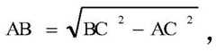
Further, the calculation formula of the cut-in length BC is:

in the formula, B is a surgical incision point, AC represents a distance value measured before the surgery, BC represents a distance between the incision point and the tumor length-a distance from the incision point to the deepest incisal margin, and α represents an incision angle selected within a preset range.
Further, the length AB of the liver surface entry point B from the point a is calculated by:

and determining the specific position of the liver surface incision point B based on the predefined incision angle alpha and the AB length acquired after calculation.
A liver tumor surgery system based on a trigonometric model, the system comprising:
the acquisition module is used for acquiring a maximum cross-sectional view of the liver tumor;
the construction module is used for constructing a triangular model according to the projection point of the maximum section of the liver tumor on the surface of the liver and the extension point of the deepest tumor in the image;
and the calculation module is used for determining the liver surface incision point, the incision angle and the incision length through calculation.
Preferably, the building block comprises:
the first selecting unit is used for selecting a projection point of the maximum section of the liver tumor on the surface of the liver and marking the projection point as a point A;
the second selecting unit is used for selecting the deepest part of the tumor in the maximum cross-sectional view of the liver tumor and marking the deepest part of the tumor as a point D;
the measuring unit is used for connecting the point A and the point D, extending AD to a point C, defining AC as a first right-angle side and DC as a cutting edge, and measuring to obtain an AC distance value;
and the defining unit is used for constructing a hypotenuse BC by taking the point C as a vertex based on a predefined incision angle alpha, so that the point B is positioned on the surface of the liver, and the angle ABC is alpha, the delta BAC is a right triangle, the angle BAC is 90 degrees, the point B is a surgical incision point on the surface of the liver, and the BC is a surgical incision length.
Preferably, the calculation module comprises:
a first determination unit configured to calculate the plunge length by:

in the formula, B is a surgical incision point, AC represents a distance value measured before the surgery, BC represents a distance between the incision point and the tumor length-a distance from the incision point to the deepest incisal margin, and α represents an incision angle selected within a preset range.
Preferably, the calculation module further comprises:
a second determining unit, configured to calculate a length AB of the liver surface entry point B from the point a by:

and determining the specific position of the liver surface incision point B based on the predefined incision angle alpha and the AB length obtained after calculation.
The invention has the beneficial effects that:
1. the invention determines a uniform and programmable approach for the resection range of the upper segment tumor of the middle liver lobe.
2. The invention can determine and calculate the incision point of the liver fracture plane and the incision depth from the fracture plane, thereby ensuring enough incisional margin.
3. The surgical resection of the upper liver lobe tumor is planned by the method, complex equipment is not needed, the operability in the operation is strong, and the conditions that the three-dimensional visualization is inconsistent with the actual condition in the clinical practice in the preoperative three-dimensional visualization planning, the operation complexity and the success rate of fluorescent staining, mixed interference caused by traffic branches and the like are effectively overcome.
Drawings
In order to more clearly illustrate the detailed description of the invention or the technical solutions in the prior art, the drawings that are needed in the detailed description of the invention or the prior art will be briefly described below. Throughout the drawings, like elements or portions are generally identified by like reference numerals. In the drawings, elements or portions are not necessarily drawn to scale.
FIG. 1 is a flow chart of a liver tumor surgery method based on a triangular model according to the present invention;
FIG. 2 is a schematic diagram of the system of the present invention;
FIG. 3 is a schematic view of a trigonometric model for surgery of upper liver tumor according to the present invention;
FIG. 4 is a schematic diagram of a triangle model of the present invention for the surgical incision point, incision angle and incision length of the upper liver segment tumor;
FIG. 5 is a partial view of the deepest point of entry C for the operation of the upper liver segment tumor according to the present invention;
in the drawing, 1, a liver tumor maximum cross-sectional view, 2, an acquisition module, 3, a construction module, 301, a first selection unit, 302, a second selection unit, 303, a measurement unit, 304, a definition unit, 4, a calculation module, 401, a first selection unit, 402, a second selection unit, 5, an input device, 6 and a display device.
Detailed Description
Embodiments of the present invention will be described in detail below with reference to the accompanying drawings. The following examples are only for illustrating the technical solutions of the present invention more clearly, and therefore are only examples, and the protection scope of the present invention is not limited thereby.
It is to be noted that, unless otherwise specified, technical or scientific terms used herein shall have the ordinary meaning as understood by those skilled in the art to which the invention pertains.
The specific embodiment of the present invention provides a liver tumor surgery method based on a triangular model, as shown in fig. 1, the method includes:
s1, acquiring a maximum cross-sectional view of the liver tumor;
s2, constructing a triangular model according to the projection point of the maximum section of the liver tumor on the surface of the liver and the deepest part of the tumor in the image, and determining the incision point, the incision angle and the incision length of the surface of the liver through calculation.
Based on the same inventive concept, the above scheme is applied to the following specific embodiments:
example 1
With reference to fig. 2 and fig. 3, before an operation is started, a maximumcross-sectional view 1 of a liver tumor is obtained through CT scan imaging or magnetic resonance imaging, the maximumcross-sectional view 1 of the liver tumor is obtained by an obtainingmodule 2, and is uploaded to aconstructing module 3, and theconstructing module 3 is configured to construct a triangular model according to a projection point of the maximumcross-sectional view 1 of the liver tumor on a liver surface and a deepest tumor position in the diagram, as shown in fig. 3, specifically as follows:
the first selectingunit 301 included in theconstruction module 3 selects a projection point of the maximum liver tumor section on the liver surface, and the projection point is marked as a point A;
a second selectingunit 302 included in theconstruction module 3 selects the deepest part of the tumor in the maximum cross-sectional view of the liver tumor, and marks the deepest part of the tumor as a point D;
themeasurement unit 303 included in theconstruction module 3 connects the point a and the point D, extends the AD to a point C, defines AC as a first right-angle side of the triangular model, defines DC as a cutting edge, and obtains an AC distance value by measuring in themaximum section 1 of the liver tumor obtained by CT scan imaging or nuclear magnetic resonance imaging;
according to different requirements of a laparoscope working angle and a surgery operation angle in different surgeries, a pre-defined incision angle alpha is input into thebuilding module 3 before surgery execution through theinput device 5, thebuilding module 3 comprises a definingunit 304, the definingunit 304 constructs a right-angled triangle hypotenuse BC by taking the point C as a vertex, so that a point B is positioned on the surface of the liver, and the angle ABC is equal to alpha, the Delta BAC is equal to the right-angled triangle, the angle BAC is equal to 90 degrees, the point B is a surgery incision point on the surface of the liver, and the BC is a surgery incision length.
Theconstruction module 3 transmits the construction information to the calculation module 4, and the calculation module 4 is used for determining the liver surface incision point, the incision angle and the incision length in a specific operation through calculation, and specifically calculates as follows:
the calculation module 4 comprises:
a first determiningunit 401, configured to calculate the cutting length by:

in the formula, B is a surgical incision point, AC represents a distance value measured before the surgery, BC represents a distance between the incision point and the tumor length-a distance from the incision point to the deepest incisal margin, and α represents an incision angle selected within a preset range.
The calculation module 4 further comprises:
a second determiningunit 402, configured to calculate a length AB of the liver surface entry point B from the point a by:

and determining the specific position of the liver surface incision point B based on the predefined incision angle alpha and the AB length obtained after calculation, and displaying the incision point B, the incision angle alpha and the incision length BC required by the operation through adisplay device 6.
Example 2
Taking metastatic liver cancer of a certain patient as an example, a tumor is located at the ventral segment of S8 and is 2-3cm away from the liver surface, a preoperative nuclear magnetic resonance imaging is used for obtaining a maximum cross section image of the liver tumor and is closely attached to P8, the tumor is 2-3cm deep in the liver parenchyma, a preoperative nuclear magnetic resonance imaging is found in the operation for obtaining the maximum cross section 1 of the liver tumor, an obtaining unit 2 obtains the maximum cross section image and transmits the maximum cross section image to a constructing module 3, a first selecting unit 301 is used for determining a point A of the liver surface, a second selecting unit 302 is used for obtaining a deepest part D of the tumor, a measuring unit 303 is used for connecting the point A with the point D and extending AD to a point C, an AC distance obtained by an AC distance value is measured to be 5.6cm, wherein an incisional edge DC is 1cm, an operation preset incisional angle alpha is input through an input device 6 and is 45 degrees, a first determining unit 401 calculates BC to be 7.9cm, a second circling unit 402 obtains AB to be 5.6cm, thereby determining an incisional point B, and develops the liver section from the incisional point B to the left and the left, when the incision depth reaches about 7cm, the deeper resection is stopped when the hepatic median vein trunk is encountered, the preoperative planned BC length is 7.9cm, the error is 0.9cm in order to retain the hepatic median vein trunk in the actual resection, the incisal margin is 0.6cm and 0.4cm in the section view of the tumor, and for the tumor deep in the parenchyma of the liver, the incisal margin treatment range of 0.6cm is larger than the radio frequency.
As shown in fig. 4, during a specific surgical operation, the maximum cross section of the liver tumor corresponding to the preoperative operation is found through ultrasound during the operation, and a tumor a point is marked on the liver surface, that is, a projection of the tumor at a point on the cross section closest to the liver surface. Determining a liver surface entry point B according to a length value B' of the AC measured before the operation; according to a set cut-in angle alpha, from shallow to deep, a liver section with enough width is unfolded from a point B to the left and right by taking a point C of a constructed virtual right triangle as a center, the liver section is cut into the liver from the point B according to the length of BC, a Glissonian branch of a section S4a or S8 can be encountered generally when the cut-out process is less than 2cm, and branches of hepatic veins S4a and S8 can be encountered when the cut-out process is about 3cm, and the branches are cut off one by one.
As shown in fig. 5, in the preoperative planning of the present invention, a triangle is constructed representing a cross section of the liver surface incision point B and the incision deepest point C. In the process of surgical excision, the section is pushed and unfolded left and right by taking the tumor as the center, so that the tumor area can be completely covered, and the margin is ensured. According to
 The incision angle value alpha can represent the visual field range seen by the laparoscope from the foot side, the larger the angle is, the deeper the incision is, the harder the important pipeline on the liver incision surface is observed from the foot side under the laparoscope, otherwise, the smaller the angle is, the better the visual field is, the convenience is brought to the exposure, cutting, hemostasis and other treatments of the important pipeline on the liver incision surface, but the farther the incision point B is away from the tumor, the more normal liver tissues are excised. In the resection operation of the upper segment tumor of the middle liver lobe, the angle of the incision angle value alpha can be set between 30 and 45 degrees because the lens has a visual angle of 30 degrees, and the deep pipeline structure of the liver section can be clearly displayed through the lens. According to the formula, sin alpha is the largest when the cutting angle value alpha is 45 degrees, BC can be ensured under the condition that the AC length value b' is determined, the shortest, namely the minimum amount of the cut normal liver is achieved, and the important pipeline on the liver section can be fully exposed. But require the surgical team to work through tacit coordination; on the contrary, if the operation team starts, the experience is not mature, the incision angle value alpha can be reduced as much as possible, the length of the hepatectomy line BC is prolonged, and a farther incision point B is selected.


Finally, it should be noted that: the above embodiments are only used to illustrate the technical solution of the present invention, and not to limit the same; while the invention has been described in detail and with reference to the foregoing embodiments, it will be understood by those skilled in the art that: the technical solutions described in the foregoing embodiments may still be modified, or some or all of the technical features may be equivalently replaced; such modifications and substitutions do not depart from the spirit and scope of the present invention, and they should be construed as being included in the following claims and description.
Claims (3)
1. A liver tumor surgery system based on a trigonometric model, the system comprising:
the acquisition module is used for acquiring a maximum cross-sectional view of the liver tumor;
the construction module is used for constructing a triangular model according to the projection point of the maximum section of the liver tumor on the surface of the liver and the extension point of the deepest tumor in the image;
the calculation module is used for determining the liver surface incision point, the incision angle and the incision length through calculation; the building module comprises:
the first selecting unit is used for selecting a projection point of the maximum section of the liver tumor on the surface of the liver and marking the projection point as a point A;
the second selecting unit is used for selecting the deepest part of the tumor in the maximum cross-sectional view of the liver tumor and marking the deepest part of the tumor as a point D;
the measuring unit is used for connecting the point A and the point D, extending AD to a point C, defining AC as a first right-angle side and DC as a cutting edge, and measuring to obtain an AC distance value;
and the defining unit is used for constructing a hypotenuse BC by taking the point C as a vertex based on a predefined incision angle alpha, so that the point B is positioned on the surface of the liver, and the angle ABC is alpha, the delta BAC is a right triangle, the angle BAC is 90 degrees, the point B is a surgical incision point on the surface of the liver, and the BC is a surgical incision length.
2. The liver tumor surgery system based on triangular model according to claim 1, wherein the calculation module comprises:
a first determination unit configured to calculate the plunge length by:

in the formula, B is a surgical incision point, AC represents a preoperative measured distance value, BC represents an incision length, and α represents an incision angle selected within a preset range.
3. The liver tumor surgery system based on triangular model according to claim 1, wherein the calculation module further comprises:
a second determining unit, configured to calculate a length AB of the liver surface entry point B from the point a by:

and determining the specific position of the liver surface incision point B based on the predefined incision angle alpha and the calculated length AB.
Priority Applications (1)
| Application Number | Priority Date | Filing Date | Title |
|---|---|---|---|
| CN202011270325.3ACN112245006B (en) | 2020-11-13 | 2020-11-13 | Liver tumor surgery method and system based on triangular model |
Applications Claiming Priority (1)
| Application Number | Priority Date | Filing Date | Title |
|---|---|---|---|
| CN202011270325.3ACN112245006B (en) | 2020-11-13 | 2020-11-13 | Liver tumor surgery method and system based on triangular model |
Publications (2)
| Publication Number | Publication Date |
|---|---|
| CN112245006A CN112245006A (en) | 2021-01-22 |
| CN112245006Btrue CN112245006B (en) | 2022-03-04 |
Family
ID=74265730
Family Applications (1)
| Application Number | Title | Priority Date | Filing Date |
|---|---|---|---|
| CN202011270325.3AExpired - Fee RelatedCN112245006B (en) | 2020-11-13 | 2020-11-13 | Liver tumor surgery method and system based on triangular model |
Country Status (1)
| Country | Link |
|---|---|
| CN (1) | CN112245006B (en) |
Families Citing this family (2)
| Publication number | Priority date | Publication date | Assignee | Title |
|---|---|---|---|---|
| CN114681067A (en)* | 2022-03-22 | 2022-07-01 | 范宁 | Laser-assisted tumor positioning and cutting device |
| CN116262072A (en)* | 2023-02-14 | 2023-06-16 | 广东省中医院(广州中医药大学第二附属医院、广州中医药大学第二临床医学院、广东省中医药科学院) | Real-time surgical navigation method and system for electromagnetic tracking assisted triangulation |
Citations (4)
| Publication number | Priority date | Publication date | Assignee | Title |
|---|---|---|---|---|
| CN101141924A (en)* | 2005-01-10 | 2008-03-12 | 霍尔特医学公司 | Gynecological ablation procedure and system |
| CN104408398A (en)* | 2014-10-21 | 2015-03-11 | 无锡海斯凯尔医学技术有限公司 | Liver boundary identification method and system |
| CN105147362A (en)* | 2015-07-17 | 2015-12-16 | 哈尔滨工程大学 | Brain tumor surgery incision locating and approach planning method |
| CN108305255A (en)* | 2017-01-12 | 2018-07-20 | 浙江京新术派医疗科技有限公司 | The generation method and generating means of operation on liver cut surface |
Family Cites Families (5)
| Publication number | Priority date | Publication date | Assignee | Title |
|---|---|---|---|---|
| US20070118100A1 (en)* | 2005-11-22 | 2007-05-24 | General Electric Company | System and method for improved ablation of tumors |
| US8315812B2 (en)* | 2010-08-12 | 2012-11-20 | Heartflow, Inc. | Method and system for patient-specific modeling of blood flow |
| CN203647434U (en)* | 2014-01-03 | 2014-06-18 | 张丽红 | Surgical knife used for cutting tumour |
| US10307209B1 (en)* | 2018-08-07 | 2019-06-04 | Sony Corporation | Boundary localization of an internal organ of a subject for providing assistance during surgery |
| CN110013306B (en)* | 2019-03-22 | 2021-04-02 | 北京工业大学 | Puncture path planning method for CT-guided hepatic tumor thermal ablation treatment |
- 2020
- 2020-11-13CNCN202011270325.3Apatent/CN112245006B/ennot_activeExpired - Fee Related
Patent Citations (4)
| Publication number | Priority date | Publication date | Assignee | Title |
|---|---|---|---|---|
| CN101141924A (en)* | 2005-01-10 | 2008-03-12 | 霍尔特医学公司 | Gynecological ablation procedure and system |
| CN104408398A (en)* | 2014-10-21 | 2015-03-11 | 无锡海斯凯尔医学技术有限公司 | Liver boundary identification method and system |
| CN105147362A (en)* | 2015-07-17 | 2015-12-16 | 哈尔滨工程大学 | Brain tumor surgery incision locating and approach planning method |
| CN108305255A (en)* | 2017-01-12 | 2018-07-20 | 浙江京新术派医疗科技有限公司 | The generation method and generating means of operation on liver cut surface |
Also Published As
| Publication number | Publication date |
|---|---|
| CN112245006A (en) | 2021-01-22 |
Similar Documents
| Publication | Publication Date | Title |
|---|---|---|
| Araki et al. | Intraoperative ultrasonography of laparoscopic hepatectomy: key technique for safe liver transection | |
| JP6858127B2 (en) | Equipment and methods to support tissue ablation | |
| CN111093516B (en) | Ultrasound system and method for planning ablation | |
| US11064954B2 (en) | Flattened organ display | |
| JP5685546B2 (en) | A feedback system that integrates interventional planning and navigation | |
| CN112245006B (en) | Liver tumor surgery method and system based on triangular model | |
| US20180235576A1 (en) | Ultrasound doppler and elastography for ablation prediction and monitoring | |
| US20120029387A1 (en) | Methods and systems for real-time surgical procedure assistance using an electronic organ map | |
| US20250312057A1 (en) | Methods and devices for delivering cancer therapy to a target tissue site via a cored tissue cavity | |
| US20210393332A1 (en) | Methods and devices for navigating a tissue resection device | |
| CN110023883B (en) | Method and system for interactive mesh placement and measurement for lesion removal | |
| JP2014111083A (en) | Puncture assist device | |
| JP2013540517A (en) | System and method for temperature feedback for adaptive radio frequency ablation | |
| US20210322092A1 (en) | A system and method for the ablation of uterine fibroids | |
| US20210378731A1 (en) | Tissue resection control systems and methods | |
| Monden et al. | Intrahepatic glissonean approach for laparoscopic bisegmentectomy 7 and 8 with root-side hepatic vein exposure | |
| CN112043377A (en) | Method and system for ablation path planning assisted by ultrasound field simulation in any CT slice | |
| CN102609620A (en) | Ablation therapy image guide device with image segmenting device | |
| Kim et al. | Video-assisted thoracic surgery segmentectomy | |
| CN113271881A (en) | System for monitoring ablation progress using a remote temperature probe | |
| US20220047314A1 (en) | Tissue dilation and resection systems and methods | |
| KR20230117694A (en) | Tissue ablation control system and method | |
| Shi et al. | Monitoring of radiofrequency ablation with shear wave delay mapping | |
| Hagendoorn | Robotic Systems, Equipment, Instrumentation, and Troubleshooting | |
| CN116262072A (en) | Real-time surgical navigation method and system for electromagnetic tracking assisted triangulation |
Legal Events
| Date | Code | Title | Description |
|---|---|---|---|
| PB01 | Publication | ||
| PB01 | Publication | ||
| SE01 | Entry into force of request for substantive examination | ||
| SE01 | Entry into force of request for substantive examination | ||
| GR01 | Patent grant | ||
| GR01 | Patent grant | ||
| TR01 | Transfer of patent right | ||
| TR01 | Transfer of patent right | Effective date of registration:20220622 Address after:266000 No. 16, Jiangsu Road, Qingdao, Shandong Patentee after:THE AFFILIATED HOSPITAL OF QINGDAO University Address before:No.10, 3rd floor, building 31, liupukang 2nd District, Xicheng District, Beijing Patentee before:Fan Ning | |
| CF01 | Termination of patent right due to non-payment of annual fee | ||
| CF01 | Termination of patent right due to non-payment of annual fee | Granted publication date:20220304 |