CN112138208A - Scaffold material for promoting vascularization tissue regeneration and preparation method thereof - Google Patents
Scaffold material for promoting vascularization tissue regeneration and preparation method thereofDownload PDFInfo
- Publication number
- CN112138208A CN112138208ACN202011038579.2ACN202011038579ACN112138208ACN 112138208 ACN112138208 ACN 112138208ACN 202011038579 ACN202011038579 ACN 202011038579ACN 112138208 ACN112138208 ACN 112138208A
- Authority
- CN
- China
- Prior art keywords
- promoting
- tissue regeneration
- polypeptide
- vascularized tissue
- polymer material
- Prior art date
- Legal status (The legal status is an assumption and is not a legal conclusion. Google has not performed a legal analysis and makes no representation as to the accuracy of the status listed.)
- Pending
Links
- 239000000463materialSubstances0.000titleclaimsabstractdescription67
- 230000001737promoting effectEffects0.000titleclaimsabstractdescription35
- 230000017423tissue regenerationEffects0.000titleclaimsabstractdescription34
- 238000002360preparation methodMethods0.000titleclaimsabstractdescription6
- 229920001184polypeptidePolymers0.000claimsabstractdescription45
- 102000004196processed proteins & peptidesHuman genes0.000claimsabstractdescription45
- 108090000765processed proteins & peptidesProteins0.000claimsabstractdescription45
- 239000002861polymer materialSubstances0.000claimsabstractdescription39
- 239000002121nanofiberSubstances0.000claimsabstractdescription32
- 230000008929regenerationEffects0.000claimsabstractdescription17
- 238000011069regeneration methodMethods0.000claimsabstractdescription17
- 230000002165photosensitisationEffects0.000claimsabstractdescription9
- 239000003504photosensitizing agentSubstances0.000claimsabstractdescription9
- 150000004676glycansChemical class0.000claimsabstractdescription4
- 229920001282polysaccharidePolymers0.000claimsabstractdescription4
- 239000005017polysaccharideSubstances0.000claimsabstractdescription4
- 102000004169proteins and genesHuman genes0.000claimsabstractdescription4
- 108090000623proteins and genesProteins0.000claimsabstractdescription4
- 239000000843powderSubstances0.000claimsdescription32
- 206010034972Photosensitivity reactionDiseases0.000claimsdescription22
- 239000000017hydrogelSubstances0.000claimsdescription17
- 108010010803GelatinProteins0.000claimsdescription15
- 229920000159gelatinPolymers0.000claimsdescription15
- 239000008273gelatinSubstances0.000claimsdescription15
- 235000019322gelatineNutrition0.000claimsdescription15
- 235000011852gelatine dessertsNutrition0.000claimsdescription15
- CERQOIWHTDAKMF-UHFFFAOYSA-NMethacrylic acidChemical compoundCC(=C)C(O)=OCERQOIWHTDAKMF-UHFFFAOYSA-N0.000claimsdescription12
- 239000002131composite materialSubstances0.000claimsdescription12
- 229920001661ChitosanPolymers0.000claimsdescription11
- 102000008186CollagenHuman genes0.000claimsdescription11
- 108010035532CollagenProteins0.000claimsdescription11
- 229920001436collagenPolymers0.000claimsdescription11
- 239000008367deionised waterSubstances0.000claimsdescription11
- 229910021641deionized waterInorganic materials0.000claimsdescription11
- 239000002002slurrySubstances0.000claimsdescription11
- XLYOFNOQVPJJNP-UHFFFAOYSA-NwaterChemical compoundOXLYOFNOQVPJJNP-UHFFFAOYSA-N0.000claimsdescription11
- 238000006243chemical reactionMethods0.000claimsdescription10
- 229920005615natural polymerPolymers0.000claimsdescription10
- 238000000034methodMethods0.000claimsdescription9
- 229920000642polymerPolymers0.000claimsdescription8
- 238000002156mixingMethods0.000claimsdescription5
- 239000002904solventSubstances0.000claimsdescription5
- KIUKXJAPPMFGSW-DNGZLQJQSA-N(2S,3S,4S,5R,6R)-6-[(2S,3R,4R,5S,6R)-3-Acetamido-2-[(2S,3S,4R,5R,6R)-6-[(2R,3R,4R,5S,6R)-3-acetamido-2,5-dihydroxy-6-(hydroxymethyl)oxan-4-yl]oxy-2-carboxy-4,5-dihydroxyoxan-3-yl]oxy-5-hydroxy-6-(hydroxymethyl)oxan-4-yl]oxy-3,4,5-trihydroxyoxane-2-carboxylic acidChemical compoundCC(=O)N[C@H]1[C@H](O)O[C@H](CO)[C@@H](O)[C@@H]1O[C@H]1[C@H](O)[C@@H](O)[C@H](O[C@H]2[C@@H]([C@@H](O[C@H]3[C@@H]([C@@H](O)[C@H](O)[C@H](O3)C(O)=O)O)[C@H](O)[C@@H](CO)O2)NC(C)=O)[C@@H](C(O)=O)O1KIUKXJAPPMFGSW-DNGZLQJQSA-N0.000claimsdescription4
- IXPNQXFRVYWDDI-UHFFFAOYSA-N1-methyl-2,4-dioxo-1,3-diazinane-5-carboximidamideChemical compoundCN1CC(C(N)=N)C(=O)NC1=OIXPNQXFRVYWDDI-UHFFFAOYSA-N0.000claimsdescription4
- 229920000936AgarosePolymers0.000claimsdescription4
- 229920002674hyaluronanPolymers0.000claimsdescription4
- 229960003160hyaluronic acidDrugs0.000claimsdescription4
- 235000010413sodium alginateNutrition0.000claimsdescription4
- 229940005550sodium alginateDrugs0.000claimsdescription4
- 239000000661sodium alginateSubstances0.000claimsdescription4
- 238000012545processingMethods0.000claimsdescription3
- 239000008363phosphate bufferSubstances0.000claimsdescription2
- 125000003275alpha amino acid groupChemical group0.000claims2
- 239000003292glueSubstances0.000claims1
- QTBSBXVTEAMEQO-UHFFFAOYSA-NAcetic acidChemical compoundCC(O)=OQTBSBXVTEAMEQO-UHFFFAOYSA-N0.000description9
- 150000001413amino acidsChemical group0.000description6
- 238000000502dialysisMethods0.000description6
- 230000035876healingEffects0.000description5
- 230000008439repair processEffects0.000description5
- 230000015572biosynthetic processEffects0.000description4
- 230000007547defectEffects0.000description4
- 230000002500effect on skinEffects0.000description4
- 238000004108freeze dryingMethods0.000description4
- DCUFMVPCXCSVNP-UHFFFAOYSA-Nmethacrylic anhydrideChemical compoundCC(=C)C(=O)OC(=O)C(C)=CDCUFMVPCXCSVNP-UHFFFAOYSA-N0.000description4
- 206010052428WoundDiseases0.000description3
- 208000027418Wounds and injuryDiseases0.000description3
- 210000004204blood vesselAnatomy0.000description3
- 230000000052comparative effectEffects0.000description3
- 238000011532immunohistochemical stainingMethods0.000description3
- 238000007639printingMethods0.000description3
- 239000004471GlycineSubstances0.000description2
- 206010021143HypoxiaDiseases0.000description2
- 230000033115angiogenesisEffects0.000description2
- 230000000694effectsEffects0.000description2
- 230000010595endothelial cell migrationEffects0.000description2
- 238000001125extrusionMethods0.000description2
- 230000006870functionEffects0.000description2
- 230000007954hypoxiaEffects0.000description2
- 238000003786synthesis reactionMethods0.000description2
- 238000005406washingMethods0.000description2
- 102100024616Platelet endothelial cell adhesion moleculeHuman genes0.000description1
- 230000006907apoptotic processEffects0.000description1
- 210000004027cellAnatomy0.000description1
- 230000003915cell functionEffects0.000description1
- 230000001684chronic effectEffects0.000description1
- 238000010276constructionMethods0.000description1
- 238000002242deionisation methodMethods0.000description1
- 238000010586diagramMethods0.000description1
- 201000010099diseaseDiseases0.000description1
- 208000037265diseases, disorders, signs and symptomsDiseases0.000description1
- 238000005516engineering processMethods0.000description1
- 239000004220glutamic acidSubstances0.000description1
- 238000010438heat treatmentMethods0.000description1
- 208000015181infectious diseaseDiseases0.000description1
- 208000028867ischemiaDiseases0.000description1
- 210000004088microvesselAnatomy0.000description1
- 238000012986modificationMethods0.000description1
- 230000004048modificationEffects0.000description1
- 230000017074necrotic cell deathEffects0.000description1
- 235000015097nutrientsNutrition0.000description1
- 230000001172regenerating effectEffects0.000description1
- 238000001338self-assemblyMethods0.000description1
- 238000010532solid phase synthesis reactionMethods0.000description1
- 239000000126substanceSubstances0.000description1
- 230000004083survival effectEffects0.000description1
- 238000012360testing methodMethods0.000description1
- 230000002792vascularEffects0.000description1
Images


Classifications
- A—HUMAN NECESSITIES
- A61—MEDICAL OR VETERINARY SCIENCE; HYGIENE
- A61L—METHODS OR APPARATUS FOR STERILISING MATERIALS OR OBJECTS IN GENERAL; DISINFECTION, STERILISATION OR DEODORISATION OF AIR; CHEMICAL ASPECTS OF BANDAGES, DRESSINGS, ABSORBENT PADS OR SURGICAL ARTICLES; MATERIALS FOR BANDAGES, DRESSINGS, ABSORBENT PADS OR SURGICAL ARTICLES
- A61L27/00—Materials for grafts or prostheses or for coating grafts or prostheses
- A61L27/14—Macromolecular materials
- A61L27/16—Macromolecular materials obtained by reactions only involving carbon-to-carbon unsaturated bonds
- A—HUMAN NECESSITIES
- A61—MEDICAL OR VETERINARY SCIENCE; HYGIENE
- A61L—METHODS OR APPARATUS FOR STERILISING MATERIALS OR OBJECTS IN GENERAL; DISINFECTION, STERILISATION OR DEODORISATION OF AIR; CHEMICAL ASPECTS OF BANDAGES, DRESSINGS, ABSORBENT PADS OR SURGICAL ARTICLES; MATERIALS FOR BANDAGES, DRESSINGS, ABSORBENT PADS OR SURGICAL ARTICLES
- A61L27/00—Materials for grafts or prostheses or for coating grafts or prostheses
- A61L27/14—Macromolecular materials
- A61L27/20—Polysaccharides
- A—HUMAN NECESSITIES
- A61—MEDICAL OR VETERINARY SCIENCE; HYGIENE
- A61L—METHODS OR APPARATUS FOR STERILISING MATERIALS OR OBJECTS IN GENERAL; DISINFECTION, STERILISATION OR DEODORISATION OF AIR; CHEMICAL ASPECTS OF BANDAGES, DRESSINGS, ABSORBENT PADS OR SURGICAL ARTICLES; MATERIALS FOR BANDAGES, DRESSINGS, ABSORBENT PADS OR SURGICAL ARTICLES
- A61L27/00—Materials for grafts or prostheses or for coating grafts or prostheses
- A61L27/14—Macromolecular materials
- A61L27/22—Polypeptides or derivatives thereof, e.g. degradation products
- A61L27/227—Other specific proteins or polypeptides not covered by A61L27/222, A61L27/225 or A61L27/24
- A—HUMAN NECESSITIES
- A61—MEDICAL OR VETERINARY SCIENCE; HYGIENE
- A61L—METHODS OR APPARATUS FOR STERILISING MATERIALS OR OBJECTS IN GENERAL; DISINFECTION, STERILISATION OR DEODORISATION OF AIR; CHEMICAL ASPECTS OF BANDAGES, DRESSINGS, ABSORBENT PADS OR SURGICAL ARTICLES; MATERIALS FOR BANDAGES, DRESSINGS, ABSORBENT PADS OR SURGICAL ARTICLES
- A61L27/00—Materials for grafts or prostheses or for coating grafts or prostheses
- A61L27/14—Macromolecular materials
- A61L27/22—Polypeptides or derivatives thereof, e.g. degradation products
- A61L27/24—Collagen
- A—HUMAN NECESSITIES
- A61—MEDICAL OR VETERINARY SCIENCE; HYGIENE
- A61L—METHODS OR APPARATUS FOR STERILISING MATERIALS OR OBJECTS IN GENERAL; DISINFECTION, STERILISATION OR DEODORISATION OF AIR; CHEMICAL ASPECTS OF BANDAGES, DRESSINGS, ABSORBENT PADS OR SURGICAL ARTICLES; MATERIALS FOR BANDAGES, DRESSINGS, ABSORBENT PADS OR SURGICAL ARTICLES
- A61L27/00—Materials for grafts or prostheses or for coating grafts or prostheses
- A61L27/50—Materials characterised by their function or physical properties, e.g. injectable or lubricating compositions, shape-memory materials, surface modified materials
- A61L27/52—Hydrogels or hydrocolloids
- A—HUMAN NECESSITIES
- A61—MEDICAL OR VETERINARY SCIENCE; HYGIENE
- A61L—METHODS OR APPARATUS FOR STERILISING MATERIALS OR OBJECTS IN GENERAL; DISINFECTION, STERILISATION OR DEODORISATION OF AIR; CHEMICAL ASPECTS OF BANDAGES, DRESSINGS, ABSORBENT PADS OR SURGICAL ARTICLES; MATERIALS FOR BANDAGES, DRESSINGS, ABSORBENT PADS OR SURGICAL ARTICLES
- A61L27/00—Materials for grafts or prostheses or for coating grafts or prostheses
- A61L27/50—Materials characterised by their function or physical properties, e.g. injectable or lubricating compositions, shape-memory materials, surface modified materials
- A61L27/54—Biologically active materials, e.g. therapeutic substances
- A—HUMAN NECESSITIES
- A61—MEDICAL OR VETERINARY SCIENCE; HYGIENE
- A61L—METHODS OR APPARATUS FOR STERILISING MATERIALS OR OBJECTS IN GENERAL; DISINFECTION, STERILISATION OR DEODORISATION OF AIR; CHEMICAL ASPECTS OF BANDAGES, DRESSINGS, ABSORBENT PADS OR SURGICAL ARTICLES; MATERIALS FOR BANDAGES, DRESSINGS, ABSORBENT PADS OR SURGICAL ARTICLES
- A61L2300/00—Biologically active materials used in bandages, wound dressings, absorbent pads or medical devices
- A61L2300/20—Biologically active materials used in bandages, wound dressings, absorbent pads or medical devices containing or releasing organic materials
- A61L2300/252—Polypeptides, proteins, e.g. glycoproteins, lipoproteins, cytokines
- A—HUMAN NECESSITIES
- A61—MEDICAL OR VETERINARY SCIENCE; HYGIENE
- A61L—METHODS OR APPARATUS FOR STERILISING MATERIALS OR OBJECTS IN GENERAL; DISINFECTION, STERILISATION OR DEODORISATION OF AIR; CHEMICAL ASPECTS OF BANDAGES, DRESSINGS, ABSORBENT PADS OR SURGICAL ARTICLES; MATERIALS FOR BANDAGES, DRESSINGS, ABSORBENT PADS OR SURGICAL ARTICLES
- A61L2300/00—Biologically active materials used in bandages, wound dressings, absorbent pads or medical devices
- A61L2300/40—Biologically active materials used in bandages, wound dressings, absorbent pads or medical devices characterised by a specific therapeutic activity or mode of action
- A61L2300/412—Tissue-regenerating or healing or proliferative agents
- A—HUMAN NECESSITIES
- A61—MEDICAL OR VETERINARY SCIENCE; HYGIENE
- A61L—METHODS OR APPARATUS FOR STERILISING MATERIALS OR OBJECTS IN GENERAL; DISINFECTION, STERILISATION OR DEODORISATION OF AIR; CHEMICAL ASPECTS OF BANDAGES, DRESSINGS, ABSORBENT PADS OR SURGICAL ARTICLES; MATERIALS FOR BANDAGES, DRESSINGS, ABSORBENT PADS OR SURGICAL ARTICLES
- A61L2400/00—Materials characterised by their function or physical properties
- A61L2400/12—Nanosized materials, e.g. nanofibres, nanoparticles, nanowires, nanotubes; Nanostructured surfaces
Landscapes
- Health & Medical Sciences (AREA)
- Chemical & Material Sciences (AREA)
- Life Sciences & Earth Sciences (AREA)
- Medicinal Chemistry (AREA)
- General Health & Medical Sciences (AREA)
- Dermatology (AREA)
- Transplantation (AREA)
- Epidemiology (AREA)
- Veterinary Medicine (AREA)
- Animal Behavior & Ethology (AREA)
- Oral & Maxillofacial Surgery (AREA)
- Public Health (AREA)
- Chemical Kinetics & Catalysis (AREA)
- Engineering & Computer Science (AREA)
- Biomedical Technology (AREA)
- Molecular Biology (AREA)
- Biophysics (AREA)
- Dispersion Chemistry (AREA)
- Materials For Medical Uses (AREA)
Abstract
Translated fromChinese
Description
Translated fromChinese技术领域technical field
本发明涉及生物医用材料领域,尤其涉及一种促血管化组织再生的支架材料及其制备方法。The invention relates to the field of biomedical materials, in particular to a scaffold material for promoting vascularized tissue regeneration and a preparation method thereof.
背景技术Background technique
机体组织的损伤修复和再生急需新生血管提供营养物质,支持相关细胞的生存并维持细胞正常功能,组织的再生与重建往往基于丰富的毛细血管网络系统。血管网的生成对预防缺氧、防止细胞凋亡和组织坏死至关重要。受损组织的再生修复速度、成败和质量通常取决于早期血管化程度。特别是对一些愈合时间长、愈合过程难的疾病如慢性创面,微血管网的快速构建可以逆转创面缺血、缺氧和降低感染,引导创面往愈合的方向发展。The damage repair and regeneration of body tissues urgently need new blood vessels to provide nutrients to support the survival of related cells and maintain normal cell function. The regeneration and reconstruction of tissues are often based on the rich capillary network system. The formation of the vascular network is essential for preventing hypoxia, apoptosis and tissue necrosis. The speed, success, and quality of regenerative repair of damaged tissue often depends on the degree of early vascularization. Especially for some diseases with long healing time and difficult healing process, such as chronic wounds, the rapid construction of microvascular network can reverse wound ischemia, hypoxia and reduce infection, and guide the wound to develop in the direction of healing.
然而,目前市场上的组织修复产品(如真皮支架)内部血管化时间较长,增加了病人的治疗周期和临床周转压力,从而增加了治疗成本。However, the tissue repair products currently on the market (such as dermal stents) have a long internal vascularization time, which increases the patient's treatment cycle and clinical turnaround pressure, thereby increasing treatment costs.
发明内容SUMMARY OF THE INVENTION
有鉴于此,有必要提供一种能够促进血管化以及组织再生的支架材料。In view of this, it is necessary to provide a scaffold material that can promote vascularization and tissue regeneration.
另,还有必要提供一种所述支架材料的制备方法。In addition, it is also necessary to provide a preparation method of the scaffold material.
本发明提供一种促血管化组织再生的支架材料,包括光敏化高分子材料以及与所述光敏化高分子材料复合的多肽纳米纤维材料,所述光敏化高分子材料包括蛋白质和多糖中的至少一种,所述多肽纳米纤维材料用于促进血管化再生。The present invention provides a scaffold material for promoting the regeneration of vascularized tissue, comprising a photosensitizing polymer material and a polypeptide nanofiber material compounded with the photosensitizing polymer material, wherein the photosensitizing polymer material includes at least one of proteins and polysaccharides. One, the polypeptide nanofiber material is used to promote vascularization and regeneration.
本发明还提供一种所述促血管化组织再生的支架材料的制备方法,包括以下步骤:The present invention also provides a method for preparing the scaffold material for promoting vascularized tissue regeneration, comprising the following steps:
将天然高分子材料以及甲基丙烯酸或甲基丙烯酸衍生物溶于溶剂中反应,处理后得到光敏化高分子材料粉体;Dissolving the natural polymer material and methacrylic acid or methacrylic acid derivative in a solvent to react, and after processing, the photosensitive polymer material powder is obtained;
提供多肽材料粉体,将所述多肽材料粉体溶于去离子水中,调节pH后形成多肽纳米纤维水凝胶;providing a polypeptide material powder, dissolving the polypeptide material powder in deionized water, and adjusting the pH to form a polypeptide nanofiber hydrogel;
将所述光敏化高分子材料粉体与所述多肽纳米纤维水凝胶混合,得到复合浆料;以及Mixing the light-sensitized polymer material powder with the polypeptide nanofiber hydrogel to obtain a composite slurry; and
打印并光照所述复合浆料,从而得到所述促血管化组织再生的支架材料。The composite slurry is printed and illuminated to obtain the scaffold material for promoting vascularized tissue regeneration.
本发明中的所述多肽纳米纤维材料具有促血管化再生的功能,从而使得所述述促血管化组织再生的支架材料能够有效促进血管再生和组织再生,有效促进软、硬组织缺损的再生修复,实现快速愈合的目标。The polypeptide nanofiber material in the present invention has the function of promoting vascularization and regeneration, so that the scaffold material for promoting vascularization tissue regeneration can effectively promote blood vessel regeneration and tissue regeneration, and effectively promote the regeneration and repair of soft and hard tissue defects , to achieve the goal of rapid healing.
附图说明Description of drawings
图1是本发明较佳实施例提供的促血管化组织再生的支架材料的制备方法的流程图。FIG. 1 is a flow chart of a method for preparing a scaffold material for promoting vascularized tissue regeneration provided by a preferred embodiment of the present invention.
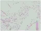
图2是本发明实施例1制备的促血管化组织再生的支架材料对真皮组织缺损修复后的免疫组化染色图。FIG. 2 is a picture of immunohistochemical staining after repair of dermal tissue defect by the scaffold material for promoting vascularized tissue regeneration prepared in Example 1 of the present invention.
图3是本发明对比例制备的促血管化组织再生的支架材料对真皮组织缺损修复后的免疫组化染色图。Fig. 3 is an immunohistochemical staining diagram of dermal tissue defect repaired by the scaffold material for promoting vascularized tissue regeneration prepared by the comparative example of the present invention.
具体实施方式Detailed ways
下面将结合本发明实施例中的附图,对本发明实施例中的技术方案进行清楚、完整地描述,显然,所描述的实施例仅仅是本发明一部分实施例,而不是全部的实施例。基于本发明中的实施例,本领域普通技术人员在没有做出创造性劳动前提下所获得的所有其他实施例,都属于本发明保护的范围。The technical solutions in the embodiments of the present invention will be clearly and completely described below with reference to the accompanying drawings in the embodiments of the present invention. Obviously, the described embodiments are only a part of the embodiments of the present invention, but not all of the embodiments. Based on the embodiments of the present invention, all other embodiments obtained by those of ordinary skill in the art without creative efforts shall fall within the protection scope of the present invention.
除非另有定义,本文所使用的所有的技术和科学术语与属于本发明的技术领域的技术人员通常理解的含义相同。本文中在本发明的说明书中所使用的术语只是为了描述具体的实施例的目的,不是旨在于限制本发明。Unless otherwise defined, all technical and scientific terms used herein have the same meaning as commonly understood by one of ordinary skill in the art to which this invention belongs. The terms used herein in the description of the present invention are for the purpose of describing specific embodiments only, and are not intended to limit the present invention.
本发明较佳实施例提供一种促血管化组织再生的支架材料,包括光敏化高分子材料以及与所述光敏化高分子材料复合的多肽纳米纤维材料。所述支架材料具有多个通孔,所述通孔之间相互连通。其中,所述通孔的孔径为100-1000微米。A preferred embodiment of the present invention provides a scaffold material for promoting the regeneration of vascularized tissue, including a photosensitized polymer material and a polypeptide nanofiber material compounded with the photosensitized polymer material. The support material has a plurality of through holes, and the through holes communicate with each other. Wherein, the diameter of the through hole is 100-1000 microns.
所述光敏化高分子材料包括蛋白质和多糖中的至少一种。具体地,所述光敏化高分子材料包括甲基丙烯酸或甲基丙烯酸衍生物修饰改性的天然高分子材料。其中,所述天然高分子材料选自胶原、明胶、壳聚糖、琼脂糖、透明质酸以及海藻酸钠中的至少一种。The photosensitive polymer material includes at least one of protein and polysaccharide. Specifically, the photosensitive polymer material includes a natural polymer material modified by methacrylic acid or a methacrylic acid derivative. Wherein, the natural polymer material is selected from at least one of collagen, gelatin, chitosan, agarose, hyaluronic acid and sodium alginate.
所述多肽纳米纤维材料用于促进血管化再生。所述多肽纳米纤维材料的氨基酸序列包括SLSLSLSLSLSLKGEETEVTVEGLEPG。即丝氨酸-亮氨酸-丝氨酸-亮氨酸-丝氨酸-亮氨酸-丝氨酸-亮氨酸-丝氨酸-亮氨酸-丝氨酸-亮氨酸-赖氨酸-甘氨酸-谷氨酸-谷氨酸-苏氨酸-谷氨酸-缬氨酸-苏氨酸-缬氨酸-谷氨酸-甘氨酸-亮氨酸-谷氨酸-脯氨酸-甘氨酸。其中,所述氨基酸序列具有促进内皮细胞迁移及加速毛细血管网络再生的作用。The polypeptide nanofiber material is used to promote vascularization and regeneration. The amino acid sequence of the polypeptide nanofiber material includes SLSLSLSLSLSLKGEETEVTVEGLEPG. i.e. serine-leucine-serine-leucine-serine-leucine-serine-leucine-serine-leucine-serine-leucine-lysine-glycine-glutamic acid-glutamic acid -Threonine-Glutamic acid-valine-threonine-valine-glutamic acid-glycine-leucine-glutamic acid-proline-glycine. Wherein, the amino acid sequence has the effect of promoting endothelial cell migration and accelerating capillary network regeneration.
请参阅图1,本发明较佳实施例还提供一种所述促血管化组织再生的支架材料的制备方法,包括以下步骤:Referring to FIG. 1, a preferred embodiment of the present invention also provides a method for preparing the scaffold material for promoting vascularized tissue regeneration, comprising the following steps:
步骤S11,将天然高分子材料以及甲基丙烯酸或甲基丙烯酸衍生物溶于溶剂中反应,处理后得到光敏化高分子材料粉体。In step S11, the natural polymer material and the methacrylic acid or methacrylic acid derivative are dissolved in a solvent for reaction, and the photosensitive polymer material powder is obtained after treatment.
具体地,将所述天然高分子材料溶于所述溶剂中至完全溶解,加热后缓慢滴加甲基丙烯酸或甲基丙烯酸衍生物,反应一定时间后加磷酸缓冲液终止反应,透析后冷冻干燥。Specifically, the natural polymer material is dissolved in the solvent until it is completely dissolved, methacrylic acid or a methacrylic acid derivative is slowly added dropwise after heating, phosphate buffer is added after the reaction for a certain period of time to terminate the reaction, and lyophilized after dialysis .
其中,所述天然高分子材料包括胶原、明胶、壳聚糖、琼脂糖、透明质酸以及海藻酸钠中的至少一种。所述溶剂可为去离子水以及醋酸等。Wherein, the natural polymer material includes at least one of collagen, gelatin, chitosan, agarose, hyaluronic acid and sodium alginate. The solvent can be deionized water, acetic acid and the like.
本发明通过化学接枝的方式修饰所述天然高分子材料以得到所述光敏化高分子材料粉体,使得所述光敏化高分子材料粉体具有双键,从而使得所述光敏化高分子材料粉体能够在紫外光照射下迅速交联固化。In the present invention, the natural polymer material is modified by chemical grafting to obtain the light-sensitized polymer material powder, so that the light-sensitized polymer material powder has a double bond, so that the light-sensitized polymer material powder has a double bond. The powder can be quickly cross-linked and cured under UV light irradiation.
步骤S12,提供多肽材料粉体,将所述多肽材料粉体溶于去离子水中,调节pH后形成多肽纳米纤维水凝胶。Step S12, providing a polypeptide material powder, dissolving the polypeptide material powder in deionized water, and adjusting the pH to form a polypeptide nanofiber hydrogel.
具体地,调节pH至5.5,静置过夜后自组装形成所述多肽纳米纤维水凝胶。Specifically, the pH was adjusted to 5.5, and the polypeptide nanofiber hydrogel was formed by self-assembly after standing overnight.
在本实施例中,可采用固相合成技术合成所述多肽材料粉体。具体地,按氨基酸顺序依次添加合成:丝氨酸-亮氨酸-丝氨酸-亮氨酸-丝氨酸-亮氨酸-丝氨酸-亮氨酸-丝氨酸-亮氨酸-丝氨酸-亮氨酸-赖氨酸-甘氨酸-谷氨酸-谷氨酸-苏氨酸-谷氨酸-缬氨酸-苏氨酸-缬氨酸-谷氨酸-甘氨酸-亮氨酸-谷氨酸-脯氨酸-甘氨酸,清洗、透析以及冻干,得到所述多肽材料粉体。In this embodiment, the polypeptide material powder can be synthesized by solid-phase synthesis technology. Specifically, the synthesis was sequentially added in amino acid order: serine-leucine-serine-leucine-serine-leucine-serine-leucine-serine-leucine-serine-leucine-lysine- Glycine-glutamic acid-glutamic acid-threonine-glutamic acid-valine-threonine-valine-glutamic acid-glycine-leucine-glutamic acid-proline-glycine, Washing, dialysis and freeze-drying to obtain the polypeptide material powder.
其中,所述多肽材料粉体的氨基酸序列包括SLSLSLSLSLSLKGEETEVTVEGLEPG。其中,所述氨基酸序列具有促进内皮细胞迁移及加速毛细血管网络再生的作用。Wherein, the amino acid sequence of the polypeptide material powder includes SLSLSLSLSLSLKGEETEVTVEGLEPG. Wherein, the amino acid sequence has the effect of promoting endothelial cell migration and accelerating capillary network regeneration.
步骤S13,将所述光敏化高分子材料粉体与所述多肽纳米纤维水凝胶混合,得到复合浆料。Step S13, mixing the light-sensitized polymer material powder with the polypeptide nanofiber hydrogel to obtain a composite slurry.
具体地,可将所述光敏化高分子材料粉体溶于去离子水中,得到光敏化高分子溶液,并将所述光敏化高分子溶液与所述多肽纳米纤维水凝胶以一定比例混合,从而得到所述复合浆料。Specifically, the photosensitized polymer material powder can be dissolved in deionized water to obtain a photosensitized polymer solution, and the photosensitized polymer solution can be mixed with the polypeptide nanofiber hydrogel in a certain proportion, Thereby, the composite slurry is obtained.
其中,所述光敏化高分子溶液的浓度为1%-30%,所述多肽纳米纤维水凝胶的浓度为0.5%-5%。所述光敏化高分子溶液与所述多肽纳米纤维水凝胶体积比为99:1-50:50。Wherein, the concentration of the photosensitized polymer solution is 1%-30%, and the concentration of the polypeptide nanofiber hydrogel is 0.5%-5%. The volume ratio of the photosensitized polymer solution to the polypeptide nanofiber hydrogel is 99:1-50:50.
步骤S14,打印并光照所述复合浆料,从而得到所述促血管化组织再生的支架材料。Step S14, printing and illuminating the composite slurry, thereby obtaining the scaffold material for promoting vascularized tissue regeneration.
具体地,可通过3D打印机打印所述复合浆料。其中,所述光照可为紫外光照射。所述紫外光照射可使所述光敏化高分子材料粉体迅速交联固化。Specifically, the composite paste may be printed by a 3D printer. Wherein, the light irradiation may be ultraviolet light irradiation. The ultraviolet light irradiation can rapidly cross-link and solidify the photosensitive polymer material powder.
其中,所述促血管化组织再生的支架材料包括光敏化高分子材料以及与所述光敏化高分子材料复合的多肽纳米纤维材料。Wherein, the scaffold material for promoting vascularized tissue regeneration includes a photosensitized polymer material and a polypeptide nanofiber material compounded with the photosensitized polymer material.
下面通过实施例对本发明进行具体说明。The present invention will be specifically described below through examples.
实施例1Example 1
第一步,将质量为1g的明胶溶解在10mL的去离子水中,40℃加热,得到明胶溶液,向所述明胶溶液中缓慢滴加0.3mL的甲基丙烯酸酐,反应4小时后加40mL的PBS溶液终止反应,透析3天后冷冻干燥得到光敏化明胶粉体。In the first step, 1 g of gelatin was dissolved in 10 mL of deionized water, heated at 40° C. to obtain a gelatin solution, 0.3 mL of methacrylic anhydride was slowly added dropwise to the gelatin solution, and 40 mL of methacrylic anhydride was added after 4 hours of reaction. The reaction was terminated by the PBS solution, and the photosensitized gelatin powder was obtained by freeze-drying after dialysis for 3 days.
第二步,将所述光敏化明胶粉体溶于去离子中,配制成浓度为20%的光敏化明胶溶液。In the second step, the light-sensitized gelatin powder is dissolved in deionized water to prepare a light-sensitized gelatin solution with a concentration of 20%.
第三步,按氨基酸顺序依次添加合成:丝氨酸-亮氨酸-丝氨酸-亮氨酸-丝氨酸-亮氨酸-丝氨酸-亮氨酸-丝氨酸-亮氨酸-丝氨酸-亮氨酸-赖氨酸-甘氨酸-谷氨酸-谷氨酸-苏氨酸-谷氨酸-缬氨酸-苏氨酸-缬氨酸-谷氨酸-甘氨酸-亮氨酸-谷氨酸-脯氨酸-甘氨酸,清洗、透析以及冻干后得到多肽材料粉体。The third step is to add synthesis in order of amino acids: serine-leucine-serine-leucine-serine-leucine-serine-leucine-serine-leucine-serine-leucine-lysine -Glycine-Glutamic acid-Glutamic acid-threonine-glutamic acid-valine-threonine-valine-glutamic acid-glycine-leucine-glutamic acid-proline-glycine , after washing, dialysis and freeze-drying, the polypeptide material powder is obtained.
第四步,将所述多肽材料粉体溶解于去离子水中,配制成浓度为10mg/mL的溶液,调节pH至5.5后静置过夜,得到多肽纳米纤维水凝胶。In the fourth step, the polypeptide material powder is dissolved in deionized water to prepare a solution with a concentration of 10 mg/mL, and the pH is adjusted to 5.5 and left to stand overnight to obtain a polypeptide nanofiber hydrogel.
第五步,将所述光敏化明胶溶液与所述多肽纳米纤维水凝胶按体积比9:1混合,得到复合浆料。In the fifth step, the photosensitized gelatin solution is mixed with the polypeptide nanofiber hydrogel in a volume ratio of 9:1 to obtain a composite slurry.
第六步,将所述复合浆料转移至3D打印机打印针筒中,通过气压挤出的方式打印,从而得到促血管化组织再生的支架材料。In the sixth step, the composite slurry is transferred to a 3D printer printing syringe and printed by air pressure extrusion, thereby obtaining a scaffold material for promoting vascularized tissue regeneration.
实施例2Example 2
第一步,将质量为2g的壳聚糖溶解在50mL的醋酸中,40℃加热,得到壳聚糖溶液,向所述壳聚糖溶液中缓慢滴加1mL的甲基丙烯酸酐,反应4小时后加200mL的PBS溶液终止反应,透析3天后冷冻干燥得到光敏化壳聚糖粉体。In the first step, 2 g of chitosan was dissolved in 50 mL of acetic acid, heated at 40° C. to obtain a chitosan solution, and 1 mL of methacrylic anhydride was slowly added dropwise to the chitosan solution, and the reaction was carried out for 4 hours. Then, 200 mL of PBS solution was added to terminate the reaction, and after 3 days of dialysis, the photosensitive chitosan powder was obtained by freeze-drying.
第二步,将所述光敏化壳聚糖粉体溶于去离子中,配制成浓度为10%的光敏化壳聚糖溶液。In the second step, the light-sensitized chitosan powder is dissolved in deionization to prepare a light-sensitized chitosan solution with a concentration of 10%.
第三步,该步骤与实施例1中的第三步相同,具体请参考实施例1。The third step is the same as the third step in Embodiment 1. For details, please refer to Embodiment 1.
第四步,将所述多肽材料粉体溶解于去离子水中,配制成浓度为20mg/mL的溶液,调节pH至5.5后静置过夜,得到多肽纳米纤维水凝胶。In the fourth step, the polypeptide material powder is dissolved in deionized water to prepare a solution with a concentration of 20 mg/mL, the pH is adjusted to 5.5, and then allowed to stand overnight to obtain a polypeptide nanofiber hydrogel.
第五步,将所述光敏化壳聚糖溶液与所述多肽纳米纤维水凝胶按体积比8:2混合,得到复合浆料。In the fifth step, the photosensitized chitosan solution is mixed with the polypeptide nanofiber hydrogel in a volume ratio of 8:2 to obtain a composite slurry.
第六步,该步骤与实施例1中的第六步相同,具体请参考实施例1。The sixth step, this step is the same as the sixth step in Embodiment 1, please refer to Embodiment 1 for details.
实施例3Example 3
第一步,将质量为1g的胶原溶解在80mL的醋酸中,40℃加热,得到胶原溶液,向所述胶原溶液中缓慢滴加0.3mL的甲基丙烯酸酐,反应4小时后加320mL的PBS溶液终止反应,透析3天后冷冻干燥得到光敏化胶原粉体。In the first step, 1 g of collagen was dissolved in 80 mL of acetic acid, heated at 40°C to obtain a collagen solution, 0.3 mL of methacrylic anhydride was slowly added dropwise to the collagen solution, and 320 mL of PBS was added after the reaction for 4 hours. The solution terminated the reaction, and lyophilized after dialysis for 3 days to obtain the photosensitized collagen powder.
第二步,将所述光敏化胶原粉体溶于去离子中,配制成浓度为5%的光敏化胶原溶液。In the second step, the photosensitized collagen powder is dissolved in deionized water to prepare a photosensitized collagen solution with a concentration of 5%.
第三步,该步骤与实施例1中的第三步相同,具体请参考实施例1。The third step is the same as the third step in Embodiment 1. For details, please refer to Embodiment 1.
第四步,该步骤与实施例1中的第四步相同,具体请参考实施例1。The fourth step is the same as the fourth step in Embodiment 1. For details, please refer to Embodiment 1.
第五步,将所述光敏化胶原溶液与所述多肽纳米纤维水凝胶按体积比19:1混合,得到复合浆料。In the fifth step, the photosensitized collagen solution is mixed with the polypeptide nanofiber hydrogel in a volume ratio of 19:1 to obtain a composite slurry.
第六步,该步骤与实施例1中的第六步相同,具体请参考实施例1。The sixth step, this step is the same as the sixth step in Embodiment 1, please refer to Embodiment 1 for details.
对比例Comparative ratio
将实施例1制备的光敏化明胶溶液转移至3D打印机打印针筒中,通过气压挤出的方式打印,从而得到明胶支架材料。The light-sensitized gelatin solution prepared in Example 1 was transferred to a 3D printer printing syringe, and printed by air pressure extrusion, thereby obtaining a gelatin scaffold material.
将实施例1-3获得的促血管化组织再生的支架材料进行测试,具体测试结果请参见表1。实施例1-3制备的所述支架材料均具有多个通孔,所述通孔之间相互连通。其中,实施例1-3制备的所述支架材料的所述通孔的孔径分别为500μm、300μm以及400μm。The scaffold materials for promoting vascularized tissue regeneration obtained in Examples 1-3 were tested, and the specific test results are shown in Table 1. The scaffold materials prepared in Examples 1-3 all have a plurality of through holes, and the through holes communicate with each other. Wherein, the diameters of the through holes of the scaffold materials prepared in Examples 1-3 are 500 μm, 300 μm and 400 μm, respectively.
将实施例1制备的所述促血管化组织再生的支架材料以及对比例制备的所述明胶支架材料分别进行CD31真皮组织修复,7天后进行免疫组化染色测试。请参阅图2和图3,由图可知,所述促血管化组织再生的支架材料的通孔中具有大量的毛细血管生成,所述明胶支架材料中则无毛细血管生成。由此可知,所述多肽纳米纤维材料可促进毛细血管的生成。The vascularized tissue regeneration-promoting scaffold prepared in Example 1 and the gelatin scaffold prepared in Comparative Example were respectively subjected to CD31 dermal tissue repair, and immunohistochemical staining was performed 7 days later. Referring to FIG. 2 and FIG. 3 , it can be seen from the figures that there is a large amount of capillary angiogenesis in the through-holes of the scaffold material for promoting vascularized tissue regeneration, but there is no capillary angiogenesis in the gelatin scaffold material. It can be seen that the polypeptide nanofiber material can promote the formation of capillaries.
本发明中的所述多肽纳米纤维材料具有促血管化再生的功能,从而使得所述述促血管化组织再生的支架材料能够有效促进血管再生和组织再生,有效促进软、硬组织缺损的再生修复,实现快速愈合的目标。The polypeptide nanofiber material in the present invention has the function of promoting vascularization and regeneration, so that the scaffold material for promoting vascularization tissue regeneration can effectively promote blood vessel regeneration and tissue regeneration, and effectively promote the regeneration and repair of soft and hard tissue defects , to achieve the goal of rapid healing.
以上实施方式仅用以说明本发明实施例的技术方案而非限制,尽管参照以上较佳实施方式对本发明实施例进行了详细说明,本领域的普通技术人员应当理解,可以对本发明实施例的技术方案进行修改或等同替换都不应脱离本发明实施例的技术方案的精神和范围。The above embodiments are only used to illustrate the technical solutions of the embodiments of the present invention and not limit them. Although the embodiments of the present invention have been described in detail with reference to the above preferred embodiments, those of ordinary skill in the art should understand that the technical solutions of the embodiments of the present invention can be Modifications or equivalent replacements of the solutions should not depart from the spirit and scope of the technical solutions of the embodiments of the present invention.
Claims (10)
Translated fromChinesePriority Applications (1)
| Application Number | Priority Date | Filing Date | Title |
|---|---|---|---|
| CN202011038579.2ACN112138208A (en) | 2020-09-28 | 2020-09-28 | Scaffold material for promoting vascularization tissue regeneration and preparation method thereof |
Applications Claiming Priority (1)
| Application Number | Priority Date | Filing Date | Title |
|---|---|---|---|
| CN202011038579.2ACN112138208A (en) | 2020-09-28 | 2020-09-28 | Scaffold material for promoting vascularization tissue regeneration and preparation method thereof |
Publications (1)
| Publication Number | Publication Date |
|---|---|
| CN112138208Atrue CN112138208A (en) | 2020-12-29 |
Family
ID=73895571
Family Applications (1)
| Application Number | Title | Priority Date | Filing Date |
|---|---|---|---|
| CN202011038579.2APendingCN112138208A (en) | 2020-09-28 | 2020-09-28 | Scaffold material for promoting vascularization tissue regeneration and preparation method thereof |
Country Status (1)
| Country | Link |
|---|---|
| CN (1) | CN112138208A (en) |
Citations (8)
| Publication number | Priority date | Publication date | Assignee | Title |
|---|---|---|---|---|
| US20080145934A1 (en)* | 2004-09-28 | 2008-06-19 | Ian Ross Harris | Tissue-engineering scaffolds containing self-assembled-peptide hydrogels |
| US20110110922A1 (en)* | 2008-01-08 | 2011-05-12 | Tufts University | Wound Healing Peptides and Methods of Use Thereof |
| US20140349933A1 (en)* | 2011-11-04 | 2014-11-27 | Agency For Science, Technology And Research | Self-assembled composite ultrasmall peptide-polymer hydrogels |
| US9526762B1 (en)* | 2013-08-09 | 2016-12-27 | William Marsh Rice University | Multidomain peptides for promoting angiogenesis |
| CN106983912A (en)* | 2017-04-17 | 2017-07-28 | 广东省生物工程研究所(广州甘蔗糖业研究所) | A kind of anti-bacterial hydrogel recovery support of 3D printing and preparation method thereof |
| CN107663377A (en)* | 2017-08-24 | 2018-02-06 | 浙江大学 | It is a kind of that there is temperature sensitive and the mixing hydrogel and its 3D printing method of light sensitive characteristic |
| CN107899075A (en)* | 2017-10-20 | 2018-04-13 | 浙江大学 | A kind of Self-Assembled based on specificity interaction and preparation method thereof |
| US20200000875A1 (en)* | 2018-06-15 | 2020-01-02 | New Jersey Institute Of Technology | Injectable Self-assembling Antibacterial Peptide Hydrogels |
- 2020
- 2020-09-28CNCN202011038579.2Apatent/CN112138208A/enactivePending
Patent Citations (8)
| Publication number | Priority date | Publication date | Assignee | Title |
|---|---|---|---|---|
| US20080145934A1 (en)* | 2004-09-28 | 2008-06-19 | Ian Ross Harris | Tissue-engineering scaffolds containing self-assembled-peptide hydrogels |
| US20110110922A1 (en)* | 2008-01-08 | 2011-05-12 | Tufts University | Wound Healing Peptides and Methods of Use Thereof |
| US20140349933A1 (en)* | 2011-11-04 | 2014-11-27 | Agency For Science, Technology And Research | Self-assembled composite ultrasmall peptide-polymer hydrogels |
| US9526762B1 (en)* | 2013-08-09 | 2016-12-27 | William Marsh Rice University | Multidomain peptides for promoting angiogenesis |
| CN106983912A (en)* | 2017-04-17 | 2017-07-28 | 广东省生物工程研究所(广州甘蔗糖业研究所) | A kind of anti-bacterial hydrogel recovery support of 3D printing and preparation method thereof |
| CN107663377A (en)* | 2017-08-24 | 2018-02-06 | 浙江大学 | It is a kind of that there is temperature sensitive and the mixing hydrogel and its 3D printing method of light sensitive characteristic |
| CN107899075A (en)* | 2017-10-20 | 2018-04-13 | 浙江大学 | A kind of Self-Assembled based on specificity interaction and preparation method thereof |
| US20200000875A1 (en)* | 2018-06-15 | 2020-01-02 | New Jersey Institute Of Technology | Injectable Self-assembling Antibacterial Peptide Hydrogels |
Non-Patent Citations (1)
| Title |
|---|
| 严梦玲: "光交联水凝胶可打印性与生物活性研究", 《中国优秀博硕士学位论文全文数据库(硕士) 工程科技Ⅰ辑》* |
Similar Documents
| Publication | Publication Date | Title |
|---|---|---|
| CN106822183B (en) | Photosensitive platelet-rich plasma gel and preparation method and application thereof | |
| CN110330797A (en) | A kind of double modified gelatin-compounded bio-inks of fibroin albumen-and its preparation method and application of photo-crosslinking | |
| CN114524953A (en) | Silk fibroin/hyaluronic acid composite hydrogel, preparation method and application | |
| CN107998450A (en) | The preparation method and application of artificial skin and artificial skin | |
| CN114874455B (en) | A method for constructing modified collagen and gel that are neutrally soluble, have self-assembly ability and photo-cross-linking ability | |
| CN110522947A (en) | A kind of preparation method of 4D-chitosan thermosensitive gel | |
| CN104857569A (en) | Preparation method of fibroin and graphene oxide composite bracket material | |
| CN113214476B (en) | Bionic glycomacropeptide hydrogel and preparation method and application thereof | |
| CN107149700A (en) | A kind of three component biological glues and its preparation and application | |
| CN112126080A (en) | Photocuring hydrogel based on sulfydryl-alkene click reaction, and preparation method and application thereof | |
| CN114507364B (en) | Preparation of light-cured casein hydrogel and its application in hemostasis and skin repair | |
| CN107854729A (en) | A kind of fibroin albumen base self-healing hydrogel and preparation method thereof | |
| CN114773629A (en) | Preparation method of injectable photocurable hemostatic hydrogel for traumatic brain injury | |
| CN108642115A (en) | A kind of high quality can scale-up version Isin glue collagen method for extraction and purification | |
| CN111388758A (en) | Composite biological ink based on methacrylated hydrogel/hydroxyethyl cellulose/acellular matrix and preparation method thereof | |
| JP2025531968A (en) | Use of silk fibroin fusion proteins in the preparation of topical medications for skin wounds | |
| CN115636960B (en) | Photo-crosslinking silk fibroin hemostatic adhesive and preparation method thereof | |
| CN108912284A (en) | A kind of acrylic acid-grafted natural collagen protein and preparation method thereof with fibrosis energy | |
| CN111607047A (en) | A high-throughput biomaterial screening bidirectional gradient hydrogel and its preparation method | |
| CN108295029A (en) | A kind of injection Multifunctional composite water gel and preparation method thereof | |
| CN110478529A (en) | A kind of 3D printing konjac glucomannan hydrogel scaffold and preparation method and application | |
| CN110755685A (en) | 3D printing guar gum gel bracket and preparation method thereof | |
| CN112138208A (en) | Scaffold material for promoting vascularization tissue regeneration and preparation method thereof | |
| CN117919511A (en) | Preparation method of collagen matrix biological film, collagen matrix biological film and application thereof | |
| Palai et al. | Functionalization of viscoelastic gels with decellularized extracellular matrix microparticles enhances tissue adhesion, cell spreading, and tissue regeneration |
Legal Events
| Date | Code | Title | Description |
|---|---|---|---|
| PB01 | Publication | ||
| PB01 | Publication | ||
| SE01 | Entry into force of request for substantive examination | ||
| SE01 | Entry into force of request for substantive examination | ||
| RJ01 | Rejection of invention patent application after publication | Application publication date:20201229 | |
| RJ01 | Rejection of invention patent application after publication |