CN112102321A - Focal image segmentation method and system based on deep convolutional neural network - Google Patents
Focal image segmentation method and system based on deep convolutional neural networkDownload PDFInfo
- Publication number
- CN112102321A CN112102321ACN202010788632.4ACN202010788632ACN112102321ACN 112102321 ACN112102321 ACN 112102321ACN 202010788632 ACN202010788632 ACN 202010788632ACN 112102321 ACN112102321 ACN 112102321A
- Authority
- CN
- China
- Prior art keywords
- feature
- image segmentation
- lesion image
- neural network
- lesion
- Prior art date
- Legal status (The legal status is an assumption and is not a legal conclusion. Google has not performed a legal analysis and makes no representation as to the accuracy of the status listed.)
- Granted
Links
Images


Classifications
- G—PHYSICS
- G06—COMPUTING OR CALCULATING; COUNTING
- G06T—IMAGE DATA PROCESSING OR GENERATION, IN GENERAL
- G06T7/00—Image analysis
- G06T7/10—Segmentation; Edge detection
- G—PHYSICS
- G06—COMPUTING OR CALCULATING; COUNTING
- G06N—COMPUTING ARRANGEMENTS BASED ON SPECIFIC COMPUTATIONAL MODELS
- G06N3/00—Computing arrangements based on biological models
- G06N3/02—Neural networks
- G06N3/04—Architecture, e.g. interconnection topology
- G06N3/045—Combinations of networks
- G—PHYSICS
- G06—COMPUTING OR CALCULATING; COUNTING
- G06N—COMPUTING ARRANGEMENTS BASED ON SPECIFIC COMPUTATIONAL MODELS
- G06N3/00—Computing arrangements based on biological models
- G06N3/02—Neural networks
- G06N3/04—Architecture, e.g. interconnection topology
- G06N3/047—Probabilistic or stochastic networks
- G—PHYSICS
- G06—COMPUTING OR CALCULATING; COUNTING
- G06N—COMPUTING ARRANGEMENTS BASED ON SPECIFIC COMPUTATIONAL MODELS
- G06N3/00—Computing arrangements based on biological models
- G06N3/02—Neural networks
- G06N3/08—Learning methods
- G—PHYSICS
- G06—COMPUTING OR CALCULATING; COUNTING
- G06T—IMAGE DATA PROCESSING OR GENERATION, IN GENERAL
- G06T2207/00—Indexing scheme for image analysis or image enhancement
- G06T2207/10—Image acquisition modality
- G06T2207/10072—Tomographic images
- G06T2207/10081—Computed x-ray tomography [CT]
- G—PHYSICS
- G06—COMPUTING OR CALCULATING; COUNTING
- G06T—IMAGE DATA PROCESSING OR GENERATION, IN GENERAL
- G06T2207/00—Indexing scheme for image analysis or image enhancement
- G06T2207/20—Special algorithmic details
- G06T2207/20081—Training; Learning
- G—PHYSICS
- G06—COMPUTING OR CALCULATING; COUNTING
- G06T—IMAGE DATA PROCESSING OR GENERATION, IN GENERAL
- G06T2207/00—Indexing scheme for image analysis or image enhancement
- G06T2207/20—Special algorithmic details
- G06T2207/20084—Artificial neural networks [ANN]
- G—PHYSICS
- G06—COMPUTING OR CALCULATING; COUNTING
- G06T—IMAGE DATA PROCESSING OR GENERATION, IN GENERAL
- G06T2207/00—Indexing scheme for image analysis or image enhancement
- G06T2207/20—Special algorithmic details
- G06T2207/20212—Image combination
- G06T2207/20221—Image fusion; Image merging
- G—PHYSICS
- G06—COMPUTING OR CALCULATING; COUNTING
- G06T—IMAGE DATA PROCESSING OR GENERATION, IN GENERAL
- G06T2207/00—Indexing scheme for image analysis or image enhancement
- G06T2207/30—Subject of image; Context of image processing
- G06T2207/30004—Biomedical image processing
- G06T2207/30092—Stomach; Gastric
- G—PHYSICS
- G06—COMPUTING OR CALCULATING; COUNTING
- G06T—IMAGE DATA PROCESSING OR GENERATION, IN GENERAL
- G06T2207/00—Indexing scheme for image analysis or image enhancement
- G06T2207/30—Subject of image; Context of image processing
- G06T2207/30004—Biomedical image processing
- G06T2207/30096—Tumor; Lesion
Landscapes
- Engineering & Computer Science (AREA)
- Physics & Mathematics (AREA)
- Theoretical Computer Science (AREA)
- General Physics & Mathematics (AREA)
- Computational Linguistics (AREA)
- General Health & Medical Sciences (AREA)
- Life Sciences & Earth Sciences (AREA)
- Artificial Intelligence (AREA)
- Biomedical Technology (AREA)
- Biophysics (AREA)
- Software Systems (AREA)
- Data Mining & Analysis (AREA)
- Evolutionary Computation (AREA)
- Health & Medical Sciences (AREA)
- Molecular Biology (AREA)
- Computing Systems (AREA)
- General Engineering & Computer Science (AREA)
- Mathematical Physics (AREA)
- Computer Vision & Pattern Recognition (AREA)
- Probability & Statistics with Applications (AREA)
- Image Analysis (AREA)
- Image Processing (AREA)
Abstract
Translated fromChinese
Description
Translated fromChinese技术领域technical field
本发明涉及临床影像技术领域,特别涉及一种基于深度卷积神经网络的病灶图像分割方法及系统。The invention relates to the technical field of clinical imaging, in particular to a method and system for segmenting a lesion image based on a deep convolutional neural network.
背景技术Background technique
胃癌(gastric cancer)是起源于胃黏膜上皮的恶性肿瘤,也是消化道系统种非常普遍的癌症,通常采用CT作为其常规的胃癌影像检查方式,而从CT影像中自动分割病灶图像对于术前具有指导且重要亟需意义。目前,自动分割方法有多种,尤其是运用深度学习在医学影像分割性格完成巨大成功。通常采用的自动分割方法有特征金字塔网络、多视点特征金字塔网络以及基于深层注意特征的特征金字塔网络等,但上述自动分割方法往往忽略多尺度特征之间的不一致性或没有考虑到单层特征和多层特征之间的低级信息和高级信息之间的互补作用,使得影像分割精度低且浪费资源。Gastric cancer is a malignant tumor originating from the gastric mucosa epithelium, and it is also a very common cancer in the digestive tract. CT is usually used as its conventional imaging method for gastric cancer. Guiding and important urgently need meaning. At present, there are many automatic segmentation methods, especially the use of deep learning to achieve great success in medical image segmentation. The commonly used automatic segmentation methods include feature pyramid network, multi-view feature pyramid network, and feature pyramid network based on deep attention features, etc., but the above automatic segmentation methods often ignore the inconsistency between multi-scale features or do not consider single-layer features and The complementary effect between low-level information and high-level information between multi-layer features makes image segmentation low precision and wastes resources.
因此,现有技术还有待改进和提高。Therefore, the existing technology still needs to be improved and improved.
发明内容SUMMARY OF THE INVENTION
基于此,有必要针对现有技术中影像分割精度低且浪费资源的技术问题,提供一种基于深度卷积神经网络的病灶图像分割方法及系统。Based on this, it is necessary to provide a lesion image segmentation method and system based on a deep convolutional neural network in order to solve the technical problems of low image segmentation accuracy and waste of resources in the prior art.
为了解决上述技术问题,本发明所采用的技术方案如下:In order to solve the above-mentioned technical problems, the technical scheme adopted in the present invention is as follows:
第一方面,本申请提供一种基于深度卷积神经网络的病灶图像分割方法,所述基于深度卷积神经网络的病灶图像分割方法包括以下步骤:In a first aspect, the present application provides a method for segmenting a lesion image based on a deep convolutional neural network. The method for segmenting a lesion image based on a deep convolutional neural network includes the following steps:
采集待测部位的原始图像;Collect the original image of the part to be tested;
将所述原始图像输入经过训练的病灶图像分割模型,通过所述病灶图像分割模型确定所述待测部位的病灶图像;其中,所述病灶图像从所述原始图像分割出。The original image is input into a trained lesion image segmentation model, and the lesion image of the to-be-measured site is determined by the lesion image segmentation model; wherein, the lesion image is segmented from the original image.
可选地,所述病灶图像分割模型包括三层深度神经网络、三维空间特征融合模块、单级特征细化模块以及多级特征细化模块,所述将所述原始图像输入经过训练的病灶图像分割模型,通过所述病灶图像分割模型确定所述待测部位的病灶图像具体包括:Optionally, the lesion image segmentation model includes a three-layer deep neural network, a three-dimensional spatial feature fusion module, a single-level feature refinement module, and a multi-level feature refinement module, and the original image is input into the trained lesion image. Segmentation model, the lesion image of the to-be-measured site determined by the lesion image segmentation model specifically includes:
所述原始图像输入至所述三层深度神经网络,通过所述三层深度神经网络确定所述原始图像的三个初始多尺度特征图;The original image is input to the three-layer deep neural network, and three initial multi-scale feature maps of the original image are determined by the three-layer deep neural network;
三个初始多尺度特征图输入至所述三维空间特征融合模块,通过所述三维空间特征融合模块确定每个初始多尺度特征图对应的修正多尺度特征图;Three initial multi-scale feature maps are input to the three-dimensional spatial feature fusion module, and the modified multi-scale feature map corresponding to each initial multi-scale feature map is determined by the three-dimensional spatial feature fusion module;
将单个初始多尺度特征图以及对应的修正多尺度特征图组合输入至所述单级特征细化模块,通过所述单级特征细化模块确定所述初始多尺度特征图对应的多层深度注意特征图;其中,所述多层深度注意特征图的深度高于所述初始多尺度特征图;A single initial multi-scale feature map and a corresponding modified multi-scale feature map are combined and input to the single-level feature refinement module, and the single-level feature refinement module determines the multi-layer depth attention corresponding to the initial multi-scale feature map. feature map; wherein, the depth of the multi-layer depth attention feature map is higher than the initial multi-scale feature map;
将三个多层深度注意特征图输入至所述多级特征细化模块,通过所述多级特征细化模块确定所述原始图像的病灶图像。The three multi-layer depth attention feature maps are input to the multi-level feature refinement module, and the lesion image of the original image is determined by the multi-level feature refinement module.
可选地,所述三层深度神经网络包括一个下采样层、级联的三个编码层以及级联的三个解码层,所述编码层与所述解码层一一对应;Optionally, the three-layer deep neural network includes a downsampling layer, three concatenated encoding layers, and three concatenated decoding layers, and the encoding layers are in one-to-one correspondence with the decoding layers;
所述三维空间特征融合模块包括三个三维空间特征融合网络,一个三维空间特征融合网络与一个所述解码层单层级联,每个三维空间特征融合网络均包括特征修正单元、特征提取单元以及特征融合单元;The three-dimensional space feature fusion module includes three three-dimensional space feature fusion networks, one three-dimensional space feature fusion network and a single layer of the decoding layer concatenated, and each three-dimensional space feature fusion network includes a feature correction unit, a feature extraction unit, and a feature extraction unit. Feature fusion unit;
所述单级特征细化模块包括三个单级特征细化网络,一个单级特征细化网络与一个所述三维空间特征融合网络单层级联,每个单级特征细化网络均包括单级通道拼接单元以及级联的三个单级整流线性单元;The single-level feature refinement module includes three single-level feature refinement networks, a single-level feature refinement network and a single-level concatenation of the three-dimensional spatial feature fusion network, and each single-level feature refinement network includes a single-level feature refinement network. Stage channel splicing unit and cascaded three single-stage rectifier linear units;
所述多级特征细化模块包括多级通道拼接单元以及级联的三个多级整流线性单元。The multi-level feature refinement module includes a multi-level channel splicing unit and three cascaded multi-level rectification linear units.
可选地,所述病灶图像分割模型的训练具体包括:Optionally, the training of the lesion image segmentation model specifically includes:
构建预设网络模型以及构建混合损失函数;Build preset network models and build hybrid loss functions;
获取初始训练集;其中,所述初始训练集包括若干个训练CT图像;Obtain an initial training set; wherein, the initial training set includes several training CT images;
获取每个训练CT图像预设尺寸的CT图像块作为目标训练集,对所述目标训练集进行增强,以得到增强后的目标训练集;Obtain the CT image block of each training CT image preset size as the target training set, and enhance the target training set to obtain the enhanced target training set;
基于增强后的目标训练集对预设网络模型进行训练,通过所述混合损失函数修正所述预设网络模型的参数,以得到所述病灶图像分割模型。The preset network model is trained based on the enhanced target training set, and the parameters of the preset network model are modified through the mixed loss function to obtain the lesion image segmentation model.
可选地,所述构建混合损失函数具体包括:Optionally, the constructing the hybrid loss function specifically includes:
构建第一损失函数;其中,所述第一损失函数是用于修正所述预设网络模型中三维空间特征融合模块与单级特征细化模块之间的性能参数的Jaccard损失:Construct a first loss function; wherein, the first loss function is the Jaccard loss used to correct the performance parameters between the three-dimensional spatial feature fusion module and the single-stage feature refinement module in the preset network model:
构建第二损失函数;其中,所述第二损失函数是用于平衡所述预设网络模型中所述单级特征细化模块与多级特征细化模块之间的数的Focal损失;constructing a second loss function; wherein, the second loss function is a Focal loss used to balance the numbers between the single-level feature refinement module and the multi-level feature refinement module in the preset network model;
基于所述第一损失函数和所述第二损失函数的加权和确定所述混合损失函数。The hybrid loss function is determined based on a weighted sum of the first loss function and the second loss function.
可选地,所述第一损失函数的公式为:Optionally, the formula of the first loss function is:

其中,n为输入CT图像块的体素数;ε表示一个平滑因子;pi∈[0,1]表示第i个体素的预测概率,qi∈{0,1}表示对应的CT图像块的体素值;Among them, n is the number of voxels of the input CT image block; ε represents a smoothing factor; pi ∈ [0, 1] represents the prediction probability of the i-th voxel, and qi ∈ {0, 1} represents the corresponding CT image block. voxel value;
所述第二损失函数的公式为:The formula of the second loss function is:

其中,α表示Focal损失的一个平衡因子;γ表示一个聚焦参数平滑调整加权率;Among them, α represents a balance factor of Focal loss; γ represents a focus parameter smooth adjustment weighting rate;
单个混合损失函数的公式为:The formula for a single mixed loss function is:
Losssingal=λ·Lossjaccard+η·LossfocalLosssingal =λ·Loss jaccard+ η·Lossfocal
其中,其中,λ和η分别表示Jaccard损失和Focal损失的权重因子;Among them, λ and η represent the weight factors of Jaccard loss and Focal loss, respectively;
所有单个混合损失函数的损失之和作为所述混合损失函数,所述混合损失函数的公式为:The sum of the losses of all individual mixed loss functions is used as the mixed loss function, and the formula of the mixed loss function is:

其中,ws和

可选地,所述增强包括数据增强和图像增强,所述数据增强包括翻转、旋转、平移中一种或多种,所述图像增强包括归一化、体素空间重采样中一种或多种。Optionally, the enhancement includes data enhancement and image enhancement, the data enhancement includes one or more of flipping, rotation, and translation, and the image enhancement includes one or more of normalization and voxel space resampling. kind.
可选地,所述体素空间重采样用于执行三阶样条插值方法,所述归一化用于执行将前景体素的0.5%到99.5%强度值的剪切操作。Optionally, the voxel space resampling is used to perform a third-order spline interpolation method, and the normalization is used to perform a clipping operation of 0.5% to 99.5% intensity values of foreground voxels.
可选地,所述病灶图像分割模型训练时使用Adam算法作为优化器,并使用了学习率衰减策略。Optionally, Adam algorithm is used as an optimizer during training of the lesion image segmentation model, and a learning rate decay strategy is used.
第二方面,本申请还提供一种基于深度卷积神经网络的病灶图像分割系统,所述基于深度卷积神经网络的病灶图像分割系统包括:In a second aspect, the present application further provides a lesion image segmentation system based on a deep convolutional neural network, and the lesion image segmentation system based on a deep convolutional neural network includes:
图像采集装置用于采集待测部位的原始图像;The image acquisition device is used to acquire the original image of the part to be tested;
病灶图像分割装置用于确定所述待测部位的原始图像对应的病灶图像;其中,所述病灶图像分割装置配置有经过训练的病灶图像分割模型。The lesion image segmentation device is used for determining the lesion image corresponding to the original image of the part to be tested; wherein, the lesion image segmentation device is configured with a trained lesion image segmentation model.
有益效果:Beneficial effects:
本发明所提供的一种基于深度卷积神经网络的病灶图像分割方法及系统,利用经过训练的病灶图像分割模型中的三维特征融合模块通过映射关系转化待测部位的原始图像所提取的初始多尺度特征图以匹配不同的特征分辨率,进而提高模型精度,利用病灶图像分割模型中的单级特征细化模块来细化和融合三维特征融合模块以及三层深度神经网络中同层级的特征,以解决不同尺度特征间的不一致性,以得到具有高一致性的多层深度注意特征图;利用病灶图像分割模型中的多级特征细化模块缓解替代消失问题,利用构建的混合损失函数增强SDS(Stage-wise Deep Supervision,阶段间深监督)来得到病灶图像,以避免多层深度注意特征图直接平均用于预测病灶区域,提高模型性能和准确性。A method and system for lesion image segmentation based on a deep convolutional neural network provided by the present invention utilizes the three-dimensional feature fusion module in the trained lesion image segmentation model to transform the original image of the part to be tested through the mapping relationship. The scale feature map is used to match different feature resolutions, thereby improving the accuracy of the model. The single-level feature refinement module in the lesion image segmentation model is used to refine and fuse the three-dimensional feature fusion module and the features of the same level in the three-layer deep neural network. In order to solve the inconsistency between features at different scales, to obtain a multi-layer depth attention feature map with high consistency; use the multi-level feature refinement module in the lesion image segmentation model to alleviate the problem of substitution disappearance, and use the constructed hybrid loss function to enhance SDS (Stage-wise Deep Supervision, deep supervision between stages) to obtain lesion images, to avoid the direct averaging of multi-layer deep attention feature maps for predicting lesion areas, and improve model performance and accuracy.
附图说明Description of drawings
图1为本发明提供一种基于深度卷积神经网络的病灶图像分割方法的流程图。FIG. 1 is a flowchart of a lesion image segmentation method based on a deep convolutional neural network provided by the present invention.
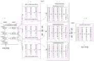
图2为本发明的病灶图像分割模型的结构框图。FIG. 2 is a structural block diagram of the lesion image segmentation model of the present invention.
图3为本发明提供的基于深度卷积神经网络的病灶图像分割系统的结构框图。FIG. 3 is a structural block diagram of a lesion image segmentation system based on a deep convolutional neural network provided by the present invention.
具体实施方式Detailed ways
本发明提供一种基于深度卷积神经网络的病灶图像分割方法及系统,为使本发明的目的、技术方案及效果更加清楚、明确,以下参照附图并举实施例对本发明进一步详细说明。应当理解,此处所描述的具体实施例仅用以解释本发明,并不用于限定本发明。The present invention provides a lesion image segmentation method and system based on deep convolutional neural network. It should be understood that the specific embodiments described herein are only used to explain the present invention, but not to limit the present invention.
本技术领域技术人员可以理解,除非特意声明,这里使用的单数形式“一”、“一个”、“所述”和“该”也可包括复数形式。应该进一步理解的是,本发明的说明书中使用的措辞“包括”是指存在所述特征、整数、步骤、操作、元件和/或组件,但是并不排除存在或添加一个或多个其他特征、整数、步骤、操作、元件、组件和/或它们的组。应该理解,当我们称元件被“连接”或“耦接”到另一元件时,它可以直接连接或耦接到其他元件,或者也可以存在中间元件。此外,这里使用的“连接”或“耦接”可以包括无线连接或无线耦接。这里使用的措辞“和/或”包括一个或更多个相关联的列出项的全部或任一单元和全部组合。It will be understood by those skilled in the art that the singular forms "a", "an", "the" and "the" as used herein can include the plural forms as well, unless expressly stated otherwise. It should be further understood that the word "comprising" used in the description of the present invention refers to the presence of stated features, integers, steps, operations, elements and/or components, but does not exclude the presence or addition of one or more other features, Integers, steps, operations, elements, components and/or groups thereof. It will be understood that when we refer to an element as being "connected" or "coupled" to another element, it can be directly connected or coupled to the other element or intervening elements may also be present. Furthermore, "connected" or "coupled" as used herein may include wirelessly connected or wirelessly coupled. As used herein, the term "and/or" includes all or any element and all combination of one or more of the associated listed items.
本技术领域技术人员可以理解,除非另外定义,这里使用的所有术语(包括技术术语和科学术语),具有与本发明所属领域中的普通技术人员的一般理解相同的意义。还应该理解的是,诸如通用字典中定义的那些术语,应该被理解为具有与现有技术的上下文中的意义一致的意义,并且除非像这里一样被特定定义,否则不会用理想化或过于正式的含义来解释。It will be understood by those skilled in the art that, unless otherwise defined, all terms (including technical and scientific terms) used herein have the same meaning as commonly understood by one of ordinary skill in the art to which this invention belongs. It should also be understood that terms, such as those defined in a general dictionary, should be understood to have meanings consistent with their meanings in the context of the prior art and, unless specifically defined as herein, should not be interpreted in idealistic or overly formal meaning to explain.
经发明人研究发现,特征金字塔网络(FPN)是一个典型的深度学习网络,其拥有学习多级特征表达的特征金字塔结构,在医学影像目标检测和语义分割中均有出色的性能表现。现有的自动分割方法如一种具有位置感知注意的多视点特征金字塔网络,用于深度普遍的病灶检测,又如基于深层注意特征的特征金字塔网络,用于三维经直肠超声前列腺分割,再如一种具有特征细化的3DESPNet,用于脑肿瘤的分割,但这些方法存在以下问题:要么忽略多尺度特征之间的不一致性,从而导致病灶部位对应的图像精度不高,所预测的病灶部位准确性低;要么没有考虑到单层特征和多层特征之间的低级信息和高级信息之间的互补作用,从而导致资源浪费,占用空间,增加成本。The inventor's research found that the Feature Pyramid Network (FPN) is a typical deep learning network, which has a feature pyramid structure for learning multi-level feature expressions, and has excellent performance in medical image target detection and semantic segmentation. Existing automatic segmentation methods such as a multi-view feature pyramid network with position-aware attention for deep and universal lesion detection, and a feature pyramid network based on deep attention features for 3D transrectal ultrasound prostate segmentation, and another example. 3DESPNet with feature refinement is used for brain tumor segmentation, but these methods have the following problems: either ignore the inconsistency between multi-scale features, resulting in low accuracy of the image corresponding to the lesion and the accuracy of the predicted lesion. low; or the complementary effect between low-level information and high-level information between single-layer features and multi-layer features is not considered, resulting in waste of resources, occupying space, and increasing costs.
因此,基于上述问题,本申请提出一种基于深度卷积神经网络的病灶图像分割方法和系统,利用经过训练的病灶图像分割模型中的三维特征融合模块通过映射关系转化待测部位的原始图像所提取的初始多尺度特征图以匹配不同的特征分辨率,进而提高模型精度,利用病灶图像分割模型中的单级特征细化模块来细化和融合三维特征融合模块以及三层深度神经网络中同层级的特征,以解决不同尺度特征间的不一致性,以得到具有高一致性的多层深度注意特征图;利用病灶图像分割模型中的多级特征细化模块缓解替代消失问题,利用构建的混合损失函数增强SDS(Stage-wise Deep Supervision,阶段间深监督)来得到病灶图像,以避免多层深度注意特征图直接平均用于预测病灶区域,提高模型性能和准确性。Therefore, based on the above problems, the present application proposes a method and system for lesion image segmentation based on a deep convolutional neural network, using the three-dimensional feature fusion module in the trained lesion image segmentation model to transform the original image of the part to be tested through the mapping relationship. The extracted initial multi-scale feature maps are used to match different feature resolutions, thereby improving the model accuracy. The single-level feature refinement module in the lesion image segmentation model is used to refine and fuse the 3D feature fusion module and the same in the three-layer deep neural network. Hierarchical features to solve the inconsistency between features at different scales to obtain multi-layer deep attention feature maps with high consistency; use the multi-level feature refinement module in the lesion image segmentation model to alleviate the problem of substitution disappearance, and use the constructed hybrid The loss function enhances SDS (Stage-wise Deep Supervision, deep supervision between stages) to obtain lesion images, so as to avoid the direct averaging of multi-layer deep attention feature maps for predicting lesion areas, and improve model performance and accuracy.
下面结合附图,详细说明本发明的技术方案,具体如下:Below in conjunction with the accompanying drawings, the technical solutions of the present invention are described in detail, specifically as follows:
请参阅图1,图1是本发明提供的一种基于深度卷积神经网络的病灶图像分割方法的流程图,应该说明的是,本发明实施方式的基于深度卷积神经网络的病灶图像分割方法并不限于图1所示的流程图中的步骤及顺序,根据不同的需求,流程图中的步骤可以增加、移除或者改变顺序。Please refer to FIG. 1. FIG. 1 is a flowchart of a method for segmenting a lesion image based on a deep convolutional neural network provided by the present invention. It should be noted that the method for segmenting a lesion image based on a deep convolutional neural network according to an embodiment of the present invention It is not limited to the steps and sequence in the flowchart shown in FIG. 1 . According to different requirements, the steps in the flowchart can be added, removed or changed in sequence.
如图1所示,所述基于深度卷积神经网络的病灶图像分割方法包括以下步骤:As shown in Figure 1, the method for segmenting a lesion image based on a deep convolutional neural network includes the following steps:
S10、采集待测部位的原始图像。S10, collect the original image of the part to be tested.
在本申请实施例中,所述待测部位指的是患者机体中待检查区域,机体局部发生病变的部位成为病灶,病灶可以发生在机体的任何组织或者器官,如牙周病、阑尾炎等疾病。本实施例中,所述待测部位指的是胃部区域。其采用医学影像设备采集胃部区域的原始图像。该原始图像即为胃部区域的CT影像。待测部位中病灶对应的图像成为病灶图像,该病灶图像用于辅助专业医生对患者情况判断以指导专业医生进行有效手术治疗。In the embodiments of the present application, the site to be tested refers to the region to be examined in the patient's body, and the site where the local disease occurs in the body becomes the lesion, and the lesion can occur in any tissue or organ of the body, such as periodontal disease, appendicitis and other diseases . In this embodiment, the site to be tested refers to the stomach region. It uses medical imaging equipment to capture raw images of the stomach area. The original image is the CT image of the stomach region. The image corresponding to the lesion in the part to be measured becomes the lesion image, and the lesion image is used to assist the professional doctor in judging the patient's condition to guide the professional doctor to perform effective surgical treatment.
S20、将所述原始图像输入经过训练的病灶图像分割模型,通过所述病灶图像分割模型确定所述待测部位的病灶图像;其中,所述病灶图像从所述原始图像分割出。S20. Input the original image into a trained lesion image segmentation model, and determine the lesion image of the to-be-measured site by using the lesion image segmentation model; wherein, the lesion image is segmented from the original image.
在本申请实施例中,所述病灶图像分割模型应用于胃肿瘤分割网络。如图2所示,所述病灶图像分割模型包括三层深度神经网络1、三维空间特征融合模块2、单级特征细化模块3以及多级特征细化模块4,所述原始图像经过该病灶图像分割模型可以输出该待测部位中可能属于病灶区域对应的病灶图像。In the embodiments of the present application, the lesion image segmentation model is applied to a gastric tumor segmentation network. As shown in FIG. 2 , the lesion image segmentation model includes a three-layer deep neural network 1, a three-dimensional spatial
所述三层深度神经网络1使用3D FPN架构,用于提取待测部位的原始图像中不同尺度的特征,得到初始多尺度特征图,这样,通过自顶向下的路径和横向连接获得不同尺度的粗略特征图。所述初始多尺度特征图包括小尺寸特征图和大尺寸特征图,所述小尺寸特征图具有分辨率低,语义信息层次高的特点,所述大尺寸特征图具有分辨率高,细节信息丰富的特点。The three-layer deep neural network 1 uses a 3D FPN architecture to extract features of different scales in the original image of the part to be tested, and obtain an initial multi-scale feature map. In this way, different scales are obtained through top-down paths and lateral connections. The rough feature map of . The initial multi-scale feature map includes a small-size feature map and a large-size feature map. The small-size feature map has the characteristics of low resolution and high level of semantic information, and the large-size feature map has high resolution and rich detailed information. specialty.
本申请的三层深度神经网络结构简单,占用计算机内存小,同时能用于不同尺度的特征提取。继续参阅图2,所述三层深度神经网络1包括一个下采样层、级联的三个编码层以及级联的三个解码层,所述编码层与所述解码层一一对应。所述下采样层作为第0层(layer0),三个编码层以及对应的解码层分别对应第1层(layer1)、第2层(layer2)以及第3层(layer3)。具体实施时,设置卷积核大小为(1,2,2),通过该卷积核对第0层、第1层和第2层进行下采样操作,第1层、第2层以及第3层的解码层分别输出一个初始多尺度特征图。随着网络层次的加深,初始多尺度特征图的尺度不一致性也越来越明显。因此,在进行下采样操作时,在第2层和第3层之间利用空洞卷积来聚合多尺度语义信息。The three-layer deep neural network of the present application has a simple structure, occupies little computer memory, and can be used for feature extraction of different scales. Continuing to refer to FIG. 2 , the three-layer deep neural network 1 includes a downsampling layer, three concatenated encoding layers, and three concatenated decoding layers, and the encoding layers are in one-to-one correspondence with the decoding layers. The down-sampling layer is the 0th layer (layer0), and the three coding layers and the corresponding decoding layers correspond to the first layer (layer1), the second layer (layer2), and the third layer (layer3), respectively. In specific implementation, the size of the convolution kernel is set to (1, 2, 2), and the convolution kernel is used to perform downsampling operations on the 0th layer, the first layer and the second layer, and the first layer, the second layer and the third layer. The decoding layers of each output an initial multi-scale feature map. As the network layer deepens, the scale inconsistency of the initial multi-scale feature maps becomes more and more obvious. Therefore, atrous convolution is utilized between
图2中,stage0阶段是一个编码层,用于提取粗略的初始多尺度特征图,其stage1阶段是一个解码层,其和编码层组成基础骨架,同样得到的是粗略的初始多尺度特征图。In Figure 2, the stage0 stage is an encoding layer used to extract a rough initial multi-scale feature map, and the stage1 stage is a decoding layer, which forms the basic skeleton with the encoding layer, and also obtains a rough initial multi-scale feature map.
请继续参阅图2,所述三维空间特征融合模块2(又称为3D ASFF)包括三个三维空间特征融合网络,一个三维空间特征融合网络与一个所述解码层单层级联,每个三维空间特征融合网络均包括特征修正单元、特征提取单元以及特征融合单元。所述三维空间特征融合模块2分为特征细化和特征融合两个阶段,其特征细化阶段是将三维空间分辨率转化为一个简单的映射问题,即yn→l=f(xn),其中xn是三维空间特征融合模块2提取的第n层特征;f为上采样或下采样操作;yn→l表示调整尺寸后的特征;n∈{1,2,3},l∈{1,2,3},并且n≠l。Please continue to refer to FIG. 2, the 3D spatial feature fusion module 2 (also known as 3D ASFF) includes three 3D spatial feature fusion networks, one 3D spatial feature fusion network and a single layer of the decoding layer. The spatial feature fusion network includes a feature correction unit, a feature extraction unit and a feature fusion unit. The three-dimensional spatial
其特征融合阶段是将三个解码层提取出的初始多尺度特征融合成修正多尺度特征图。具体实施时,对yl进行卷积、组归一化和参数化整流线性单元(PRelu)运算,得到特征融合权值

其中


请继续参阅图2,所述单级特征细化模块3(SLFR)用于提取更深层次的空间信息和语义信息,所述单级特征细化模块3包括三个单级特征细化网络,一个单级特征细化网络与一个所述三维空间特征融合网络单层级联,每个单级特征细化网络均包括单级通道拼接单元、级联的三个单级整流线性单元以及卷积注意力模块。Please continue to refer to FIG. 2 , the single-level feature refinement module 3 (SLFR) is used to extract deeper spatial information and semantic information. The single-level
其中,所述单级通道拼接单元指的是layer1、layer2和layer3上横向单层级特征通道间拼接,如layer3该层的解码层输出的初始多尺度特征图与同层的所述三维空间特征融合模块2中特征融合单元输出的修正多尺度特征图拼接。The single-level channel splicing unit refers to the splicing between horizontal single-level feature channels on layer1, layer2 and layer3, such as the initial multi-scale feature map output by the decoding layer of layer3 and the three-dimensional spatial feature of the same layer. The modified multi-scale feature map stitching output by the feature fusion unit in the
每个单级整流线性单元为卷积层,均包括一个卷积、一个组归一化和一个PRelu。第一个卷积层使用1×1×1的内核进行参数化整流线性激活,最后两个卷积层使用3×3×3的内核进一步提取有用信息。然后,利用卷积注意力模块CBAM(包括通道注意力和空间注意力操作)得到单层级的细化特征,即三个成绩的细化特征为多层深度注意特征图。需要说明的是,所述多层深度注意特征图的深度高于所述初始多尺度特征图。并且,所述所述多层深度注意特征图为高一致性的多尺度特征图。Each single-stage rectified linear unit is a convolutional layer, including a convolution, a group normalization, and a PRelu. The first convolutional layer uses 1×1×1 kernels for parametric rectified linear activation, and the last two convolutional layers use 3×3×3 kernels to further extract useful information. Then, the convolutional attention module CBAM (including channel attention and spatial attention operations) is used to obtain single-level refined features, that is, the refined features of the three grades are multi-layer deep attention feature maps. It should be noted that the depth of the multi-layer deep attention feature map is higher than that of the initial multi-scale feature map. Moreover, the multi-layer depth attention feature map is a highly consistent multi-scale feature map.
由于初始多尺度特征图是经过编码器而获得,具有不同的分辨率和低特征一致性,低特征一致性会导致特征融合时不能进行有效的特征表达。因此,在单层级间进行特征复用,以提高网络中间层的特征表达能力,如stage2的输入来自三维特征融合模块以及解码层的特征图,从解码层到stage2是进行跳跃连接操作,即stage1的特征通过跳跃连接(图2中虚线)操作实现特征复用,正因重复利用stage1的特征,再联合stage2的特征才能实现单层级上特征的细化。Since the initial multi-scale feature maps are obtained through the encoder, they have different resolutions and low feature consistency. Low feature consistency will lead to ineffective feature expression during feature fusion. Therefore, feature multiplexing is performed between single layers to improve the feature expression capability of the middle layer of the network. For example, the input of stage2 comes from the three-dimensional feature fusion module and the feature map of the decoding layer. From the decoding layer to stage2, a skip connection operation is performed, that is The features of stage1 realize feature multiplexing through the operation of skip connection (dotted line in Figure 2). It is precisely because the features of stage1 are reused that the features of stage2 can be combined to achieve the refinement of features on a single level.
请继续参阅图2,所述多级特征细化模块4(MLFR)用于更好预测肿瘤区域,以避免将得到的多层深度注意特征图直接平均用于预测肿瘤区域,因此,所述多级特征细化模块4是将特征通过不同采样率的空洞卷积进行重采样,这样,多级特征细化模块4输出的病灶图像比直接平均多层深度注意力特征图更高精度,其方法的复杂度更低。如图2所示,多级特征细化模块4包括多级通道拼接单元以及级联的三个多级整流线性单元。具体实施时,将三个多层深度注意特征图输入至所述多级通道拼接单元进行通道拼接后,输出至所述级联的三个多级整流线性单元进行卷积、归一化处理后经过PRelu激活函数,然后执行上采样操作后输出所述原始图像的病灶图像。Please continue to refer to Fig. 2, the multi-level feature refinement module 4 (MLFR) is used to better predict tumor regions, so as to avoid directly averaging the obtained multi-layer deep attention feature maps for predicting tumor regions. The level feature refinement module 4 resamples the features through atrous convolution with different sampling rates. In this way, the lesion image output by the multilevel feature refinement module 4 is more accurate than the direct average multi-layer depth attention feature map. lower complexity. As shown in FIG. 2 , the multi-level feature refinement module 4 includes a multi-level channel splicing unit and three cascaded multi-level rectifying linear units. During specific implementation, the three multi-layer depth attention feature maps are input to the multi-level channel splicing unit for channel splicing, and then output to the cascaded three multi-level rectified linear units for convolution and normalization processing. After the PRelu activation function, the upsampling operation is performed and the lesion image of the original image is output.
也就是说,在本申请的一个实施方式中,所述将所述原始图像输入经过训练的病灶图像分割模型,通过所述病灶图像分割模型确定所述待测部位的病灶图像具体包括:That is to say, in an embodiment of the present application, inputting the original image into a trained lesion image segmentation model, and determining the lesion image of the to-be-measured site by the lesion image segmentation model specifically includes:
所述原始图像输入至所述三层深度神经网络,通过所述三层深度神经网络确定所述原始图像的三个初始多尺度特征图;The original image is input to the three-layer deep neural network, and three initial multi-scale feature maps of the original image are determined by the three-layer deep neural network;
三个初始多尺度特征图输入至所述三维空间特征融合模块,通过所述三维空间特征融合模块确定每个初始多尺度特征图对应的修正多尺度特征图;Three initial multi-scale feature maps are input to the three-dimensional spatial feature fusion module, and the modified multi-scale feature map corresponding to each initial multi-scale feature map is determined by the three-dimensional spatial feature fusion module;
将单个初始多尺度特征图以及对应的修正多尺度特征图组合输入至所述单级特征细化模块,通过所述单级特征细化模块确定所述初始多尺度特征图对应的多层深度注意特征图;其中,所述多层深度注意特征图的深度高于所述初始多尺度特征图;A single initial multi-scale feature map and a corresponding modified multi-scale feature map are combined and input to the single-level feature refinement module, and the single-level feature refinement module determines the multi-layer depth attention corresponding to the initial multi-scale feature map. feature map; wherein, the depth of the multi-layer depth attention feature map is higher than the initial multi-scale feature map;
将三个多层深度注意特征图输入至所述多级特征细化模块,通过所述多级特征细化模块确定所述原始图像的病灶图像。The three multi-layer depth attention feature maps are input to the multi-level feature refinement module, and the lesion image of the original image is determined by the multi-level feature refinement module.
进一步地,上述病灶图像分割模型的训练过程为:Further, the training process of the above-mentioned lesion image segmentation model is:
1、获取初始训练集;其中,所述初始训练集包括若干个训练CT图像;1. Obtain an initial training set; wherein, the initial training set includes several training CT images;
2、获取每个训练CT图像预设尺寸的CT图像块作为目标训练集,对所述目标训练集进行数据增强和图像增强,所述数据增强包括翻转、旋转、平移中一种或多种,所述图像增强包括归一化、体素空间重采样中一种或多种,然后得到增强后的目标训练集。2. Obtain the CT image block of each training CT image preset size as the target training set, and carry out data enhancement and image enhancement to the target training set, and the data enhancement includes one or more of flipping, rotating, and translating, The image enhancement includes one or more of normalization and voxel space resampling, and then an enhanced target training set is obtained.
其中,所述体素空间重采样用于执行三阶样条插值方法,所述归一化用于执行将前景体素的0.5%到99.5%强度值的剪切操作。The voxel space resampling is used to perform a third-order spline interpolation method, and the normalization is used to perform a clipping operation of 0.5% to 99.5% intensity values of foreground voxels.
3、构建预设网络模型以及构建混合损失函数。3. Build a preset network model and build a mixed loss function.
所述预设网络模型如图2所示,stage2的特征是来自3DASFF操作模块的输出,用于缓解多尺度特征之间的不一致性;stage3的特征是来自SLFR操作模块的输出,SLFR输入的是stage2特征和stage1特征(共称单层级特征),得到更深的且有效的多尺度特征。最后,stage3的多尺度特征输入到MLFR模块,再经历上采样操作生成和标签尺寸相同的最终预测病灶图像。The preset network model is shown in Figure 2. The feature of stage2 is the output from the 3DASFF operation module, which is used to alleviate the inconsistency between multi-scale features; the feature of stage3 is the output from the SLFR operation module, and the input of SLFR is stage2 features and stage1 features (together called single-level features), to obtain deeper and effective multi-scale features. Finally, the multi-scale features of stage3 are input to the MLFR module, and then undergo an upsampling operation to generate the final predicted lesion image with the same size as the label.
在训练过程中,使用多级特征的深度监督网络可以细化每个阶段的多级特征,该阶段间深监督机制(Stage-wise Deep Supervision,SDS)不仅更适合多级特征预测,而且更有利于训练权重参数的设置,也就是说,通过减小最终预测的权重个数和重新设计的混合损失函数来促进深监督机制。这样,SDS机制可以有效地利用网络后两个阶段的多级特征融合模块来缓解梯度消失问题。During the training process, a deep supervised network using multi-level features can refine the multi-level features of each stage, and the inter-stage deep supervision mechanism (Stage-wise Deep Supervision, SDS) is not only more suitable for multi-level feature prediction, but more Facilitates the setting of training weight parameters, that is, facilitates deep supervision by reducing the number of weights for final prediction and redesigning the hybrid loss function. In this way, the SDS mechanism can effectively use the multi-level feature fusion modules in the latter two stages of the network to alleviate the gradient vanishing problem.
构建一混合损失函数来增强SDS,通过引入Focalloss是为了解决类别不均衡导致难以最佳收敛的问题。所述混合损失函数由第一损失函数和第二损失函数的加权和组成,不是使用二进制类的交叉熵损失或Dice损失进行肿瘤分割。A hybrid loss function is constructed to enhance SDS, and Focalloss is introduced to solve the problem of difficulty in optimal convergence caused by class imbalance. The hybrid loss function consists of a weighted sum of the first loss function and the second loss function, instead of using binary-class cross-entropy loss or Dice loss for tumor segmentation.
所述第一损失函数是用于修正所述预设网络模型中三维空间特征融合模块与单级特征细化模块之间的性能参数的Jaccard损失;所述第一损失函数的公式为:The first loss function is the Jaccard loss used to correct the performance parameters between the three-dimensional spatial feature fusion module and the single-stage feature refinement module in the preset network model; the formula of the first loss function is:

其中,n为输入CT图像块的体素数;ε表示一个平滑因子,设置为1.0;pi∈[0,1]表示第i个体素的预测概率,qi∈{0,1}表示对应的CT图像块的体素值;Among them, n is the number of voxels of the input CT image block; ε represents a smoothing factor, which is set to 1.0; pi ∈ [0, 1] represents the predicted probability of the i-th voxel, and qi ∈ {0, 1} represents the corresponding The voxel value of the CT image block;
所述第二损失函数是用于平衡所述预设网络模型中所述单级特征细化模块与多级特征细化模块之间的数的Focal损失,以解决正负样本不平滑问题,其用于指导小目标肿瘤区域的模型分割;所述第二损失函数的公式为:The second loss function is a Focal loss used to balance the number between the single-level feature refinement module and the multi-level feature refinement module in the preset network model, so as to solve the problem of unsmooth positive and negative samples. It is used to guide the model segmentation of small target tumor regions; the formula of the second loss function is:

其中,其中,α表示Focal损失的一个平衡因子,设置为0.2;γ表示一个聚焦参数平滑调整加权率,设置为1。Among them, α represents a balance factor of Focal loss, set to 0.2; γ represents a focus parameter smooth adjustment weighting rate, set to 1.
因此,每个监督信号损失,即单个混合损失函数的公式为:Therefore, the formula for each supervised signal loss, i.e. a single mixed loss function, is:
Losssingal=λ·Lossjaccard+η·Lossfocal, (4)Losssingal =λ·Loss jaccard+ η·Lossfocal , (4)
其中,λ和η分别表示Jaccard损失和Focal损失的权重因子,λ是设置为1,η是设置为0.1。Among them, λ and η represent the weight factors of Jaccard loss and Focal loss, respectively, λ is set to 1, η is set to 0.1.
最后,SDS损失定义为所有监督信号的损失之和,所有单个混合损失函数的损失之和作为所述混合损失函数,所述混合损失函数的公式为:Finally, the SDS loss is defined as the sum of the losses of all supervised signals, and the sum of the losses of all individual mixed loss functions is used as the mixed loss function, and the formula of the mixed loss function is:

其中,ws和

4、基于增强后的目标训练集对预设网络模型进行训练,通过所述混合损失函数修正所述预设网络模型的参数,以得到所述病灶图像分割模型。4. The preset network model is trained based on the enhanced target training set, and the parameters of the preset network model are modified through the hybrid loss function to obtain the lesion image segmentation model.
实验数据:Experimental data:
使用三种医疗器械(东芝320层CT、西门子64层CT和飞利浦128层CT)采集,采集到的数据集包含160个CT图像样本(160个普通CT数据块和63个增强CT数据块),并作了分割标签。Using three medical instruments (Toshiba 320-slice CT, Siemens 64-slice CT and Philips 128-slice CT) to acquire, the acquired dataset contains 160 CT image samples (160 normal CT data blocks and 63 enhanced CT data blocks), And made a split label.
该病灶图像分割模型在PyTorch平台上实现,使用1个NVIDIA GeForce GTX2080Ti GPU(11GB)进行训练,采用五折组交叉验证策略。由于肿瘤区域比背景区域小,为了应对3D数据对计算机内存消耗的限制,对数据集做预处理,首先将每个体积裁剪为大小为24×256×256的块(patch),然后进行数据增强(例如,翻转、旋转、平移)操作,而且还执行CT图像归一化(从所有前景体素的0.5%到99.5%强度值剪切操作)和体素空间重采样(使用三阶样条插值),得到目标训练集。The lesion image segmentation model was implemented on the PyTorch platform and trained using an NVIDIA GeForce GTX2080Ti GPU (11GB), using a five-fold group cross-validation strategy. Since the tumor area is smaller than the background area, in order to cope with the limitation of 3D data on computer memory consumption, the dataset is preprocessed by first cropping each volume into a patch of size 24×256×256, and then performing data augmentation (eg flip, rotate, translate) operations, but also perform CT image normalization (a clipping operation from 0.5% to 99.5% intensity values of all foreground voxels) and voxel space resampling (using third-order spline interpolation ) to get the target training set.
该模型使用Adam算法作为优化器,初始学习率设置为0.003,并使用了学习率衰减策略。与此同时,将批量大小设置为2,总学习epoch设置为500。定量评价分割性能的指标包括:Dice相似系数(Dice)、Jaccard指数(JI)、精确度(Pre)、召回率(Recall)、平均表面距离(Average surface distance,ASD,In voxel)和95%豪斯多夫距离(Hausdorff distance,95HD,Invoxel),直至训练的病灶图像分割模型具有泛化能力,使得训练后输出的预测病灶图像精度高、准确性高。The model uses the Adam algorithm as the optimizer, the initial learning rate is set to 0.003, and a learning rate decay strategy is used. At the same time, set the batch size to 2 and the total learning epoch to 500. Metrics to quantitatively evaluate segmentation performance include: Dice similarity coefficient (Dice), Jaccard index (JI), precision (Pre), recall (Recall), average surface distance (ASD, In voxel) and 95% Hao Hausdorff distance (95HD, Invoxel), until the trained lesion image segmentation model has the generalization ability, so that the predicted lesion image output after training has high precision and high accuracy.
基于上述基于深度卷积神经网络的病灶图像分割方法,本申请还提供一种基于深度卷积神经网络的病灶图像分割系统,如图3所示,所述基于深度卷积神经网络的病灶图像分割系统100包括:Based on the above-mentioned method for segmenting a lesion image based on a deep convolutional neural network, the present application also provides a system for segmenting a lesion image based on a deep convolutional neural network, as shown in FIG.
图像采集装置11用于采集待测部位的原始图像;其中,所述图像采集装置11可以为医学影像CT设备等。The
病灶图像分割装置22用于确定所述待测部位的原始图像对应的病灶图像;其中,所述病灶图像分割装置配置有经过训练的病灶图像分割模型,其结构如图2所示,所述基于深度卷积神经网络的病灶图像分割系统100用于实现基于深度卷积神经网络的病灶图像分割方法中的步骤,具体如上述所示。The lesion
需要说明的是,图3仅示出了系统100的部分组件,但是应理解的是,并不要求实施所有示出的组件,可以替代的实施更多或者更少的组件。It should be noted that FIG. 3 only shows some components of the
例如,其还包括处理器以及与所述处理器连接的存储器,所述存储器在一些实施例中可以是所述基于深度卷积神经网络的病灶图像分割系统200的内部存储单元,例如基于深度卷积神经网络的病灶图像分割系统200的内存。For example, it further includes a processor and a memory connected to the processor, the memory may be an internal storage unit of the deep convolutional neural network-based lesion image segmentation system 200 in some embodiments, for example, based on a depth volume The memory of the lesion image segmentation system 200 of the integrated neural network.
所述存储器在另一些实施例中也可以是所述基于深度卷积神经网络的病灶图像分割系统200的外部存储设备,例如所述基于深度卷积神经网络的病灶图像分割系统200上配备的插接式U盘,智能存储卡(Smart Media Card,SMC),安全数字(Secure Digital,SD)卡,闪存卡(FlashCard)等。进一步地,所述存储器还可以既包括所述基于深度卷积神经网络的病灶图像分割系统200的内部存储单元也包括外部存储设备。所述存储器用于存储安装于所述基于深度卷积神经网络的病灶图像分割系统200的应用软件及各类数据,例如所述基于深度卷积神经网络的病灶图像分割程序代码等。所述存储器还可以用于暂时地存储已经输出或者将要输出的数据。在一实施例中,存储器上存储有基于深度卷积神经网络的病灶图像分割程序,该基于深度卷积神经网络的病灶图像分割程序可被处理器所执行,从而实现本申请中基于深度卷积神经网络的病灶图像分割方法,具体如上述方法所述。In other embodiments, the memory may also be an external storage device of the deep convolutional neural network-based lesion image segmentation system 200 , for example, an inserter equipped on the deep convolutional neural network-based lesion image segmentation system 200 . Connected U disk, Smart Media Card (SMC), Secure Digital (SD) card, Flash Card (FlashCard), etc. Further, the memory may also include both an internal storage unit of the system 200 for lesion image segmentation based on a deep convolutional neural network and an external storage device. The memory is used to store application software and various data installed in the deep convolutional neural network-based lesion image segmentation system 200, such as the deep convolutional neural network-based lesion image segmentation program code and the like. The memory may also be used to temporarily store data that has been output or is to be output. In one embodiment, a lesion image segmentation program based on a deep convolutional neural network is stored in the memory, and the lesion image segmentation program based on a deep convolutional neural network can be executed by a processor, so as to realize the depth convolution based on the present application. The lesion image segmentation method of the neural network is specifically described in the above method.
所述处理器在一些实施例中可以是一中央处理器(Central Processing Unit,CPU),微处理器,基带处理器或其他数据处理芯片,用于运行所述存储器中存储的程序代码或处理数据,例如执行所述基于深度卷积神经网络的病灶图像分割方法等,具体如上述方法所述。In some embodiments, the processor may be a central processing unit (Central Processing Unit, CPU), a microprocessor, a baseband processor or other data processing chips for running program codes or processing data stored in the memory , for example, executing the deep convolutional neural network-based lesion image segmentation method, etc., specifically as described in the above method.
综上所述,本发明提供了一种基于深度卷积神经网络的病灶图像分割方法及系统,所述基于深度卷积神经网络的病灶图像分割方法包括以下步骤:采集待测部位的原始图像;将所述原始图像输入经过训练的病灶图像分割模型,通过所述病灶图像分割模型确定所述待测部位的病灶图像;其中,所述病灶图像从所述原始图像分割出。本申请利用经过训练的病灶图像分割模型中的三维特征融合模块通过映射关系转化待测部位的原始图像所提取的初始多尺度特征图以匹配不同的特征分辨率,进而提高模型精度,利用病灶图像分割模型中的单级特征细化模块来细化和融合三维特征融合模块以及三层深度神经网络中同层级的特征,以解决不同尺度特征间的不一致性,以得到具有高一致性的多层深度注意特征图;利用病灶图像分割模型中的多级特征细化模块缓解替代消失问题,利用构建的混合损失函数增强SDS以得到病灶图像,以避免多层深度注意特征图直接平均用于预测病灶区域,提高模型性能和准确性。To sum up, the present invention provides a method and system for segmenting a lesion image based on a deep convolutional neural network, and the method for segmenting a lesion image based on a deep convolutional neural network includes the following steps: collecting the original image of the part to be tested; The original image is input into a trained lesion image segmentation model, and the lesion image of the to-be-measured site is determined by the lesion image segmentation model; wherein, the lesion image is segmented from the original image. The present application uses the three-dimensional feature fusion module in the trained lesion image segmentation model to transform the initial multi-scale feature map extracted from the original image of the part to be tested through the mapping relationship to match different feature resolutions, thereby improving the model accuracy. The single-level feature refinement module in the segmentation model refines and fuses the 3D feature fusion module and the features of the same level in the three-layer deep neural network to solve the inconsistency between features at different scales to obtain a multi-layer with high consistency Deep attention feature map; use the multi-level feature refinement module in the lesion image segmentation model to alleviate the problem of substitution disappearance, and use the constructed hybrid loss function to enhance the SDS to obtain the lesion image, so as to avoid the direct averaging of the multi-layer deep attention feature maps for predicting lesions regions to improve model performance and accuracy.
最后应说明的是:以上实施例仅用以说明本发明的技术方案,而非对其限制;尽管参照前述实施例对本发明进行了详细的说明,本领域的普通技术人员应当理解:其依然可以对前述各实施例所记载的技术方案进行修改,或者对其中部分技术特征进行等同替换;而这些修改或者替换,并不使相应技术方案的本质脱离本发明各实施例技术方案的精神和范围。Finally, it should be noted that: the above embodiments are only used to illustrate the technical solutions of the present invention, but not to limit them; although the present invention has been described in detail with reference to the foregoing embodiments, those of ordinary skill in the art should understand: it can still be Modifications are made to the technical solutions described in the foregoing embodiments, or some technical features thereof are equivalently replaced; and these modifications or replacements do not make the essence of the corresponding technical solutions depart from the spirit and scope of the technical solutions of the embodiments of the present invention.
Claims (10)
Translated fromChinese


Priority Applications (1)
| Application Number | Priority Date | Filing Date | Title |
|---|---|---|---|
| CN202010788632.4ACN112102321B (en) | 2020-08-07 | 2020-08-07 | A lesion image segmentation method and system based on deep convolutional neural network |
Applications Claiming Priority (1)
| Application Number | Priority Date | Filing Date | Title |
|---|---|---|---|
| CN202010788632.4ACN112102321B (en) | 2020-08-07 | 2020-08-07 | A lesion image segmentation method and system based on deep convolutional neural network |
Publications (2)
| Publication Number | Publication Date |
|---|---|
| CN112102321Atrue CN112102321A (en) | 2020-12-18 |
| CN112102321B CN112102321B (en) | 2023-09-01 |
Family
ID=73752876
Family Applications (1)
| Application Number | Title | Priority Date | Filing Date |
|---|---|---|---|
| CN202010788632.4AActiveCN112102321B (en) | 2020-08-07 | 2020-08-07 | A lesion image segmentation method and system based on deep convolutional neural network |
Country Status (1)
| Country | Link |
|---|---|
| CN (1) | CN112102321B (en) |
Cited By (22)
| Publication number | Priority date | Publication date | Assignee | Title |
|---|---|---|---|---|
| CN112614145A (en)* | 2020-12-31 | 2021-04-06 | 湘潭大学 | Deep learning-based intracranial hemorrhage CT image segmentation method |
| CN112712528A (en)* | 2020-12-24 | 2021-04-27 | 浙江工业大学 | Multi-scale U-shaped residual encoder and integral reverse attention mechanism combined intestinal tract lesion segmentation method |
| CN112801168A (en)* | 2021-01-25 | 2021-05-14 | 江苏大学 | Tumor image focal region prediction analysis method and system and terminal equipment |
| CN112837280A (en)* | 2021-01-26 | 2021-05-25 | 南京英沃夫科技有限公司 | Medical image image processing method, device, electronic device and medium |
| CN112991263A (en)* | 2021-02-06 | 2021-06-18 | 杭州迪英加科技有限公司 | Method and equipment for improving calculation accuracy of TPS (acute respiratory syndrome) of PD-L1 immunohistochemical pathological section |
| CN113192633A (en)* | 2021-05-24 | 2021-07-30 | 山西大学 | Stomach cancer fine-grained classification method based on attention mechanism |
| CN113256641A (en)* | 2021-07-08 | 2021-08-13 | 湖南大学 | Skin lesion image segmentation method based on deep learning |
| CN113450381A (en)* | 2021-06-16 | 2021-09-28 | 上海深至信息科技有限公司 | System and method for evaluating accuracy of image segmentation model |
| CN113658332A (en)* | 2021-08-24 | 2021-11-16 | 电子科技大学 | Ultrasonic image-based intelligent abdominal rectus muscle segmentation and reconstruction method and device |
| CN113674253A (en)* | 2021-08-25 | 2021-11-19 | 浙江财经大学 | Rectal cancer CT image automatic segmentation method based on U-transducer |
| CN113870289A (en)* | 2021-09-22 | 2021-12-31 | 浙江大学 | A method and device for decoupling, dividing and conquering facial nerve segmentation |
| CN114022462A (en)* | 2021-11-10 | 2022-02-08 | 华东理工大学 | Method, system, device, processor and computer-readable storage medium for realizing lesion segmentation of multi-parameter nuclear magnetic resonance images |
| CN114119627A (en)* | 2021-10-19 | 2022-03-01 | 北京科技大学 | Method and device for image segmentation of superalloy microstructure based on deep learning |
| CN114322793A (en)* | 2022-03-16 | 2022-04-12 | 科大天工智能装备技术(天津)有限公司 | Workpiece size measurement method, device and storage medium based on global segmentation network |
| CN114913183A (en)* | 2021-02-07 | 2022-08-16 | 上海交通大学 | Image segmentation method, system, apparatus and medium based on constraint |
| CN115222946A (en)* | 2022-09-19 | 2022-10-21 | 南京信息工程大学 | A single-stage instance image segmentation method, apparatus and computer equipment |
| CN115713535A (en)* | 2022-11-07 | 2023-02-24 | 阿里巴巴(中国)有限公司 | Image segmentation model determination method and image segmentation method |
| CN116503607A (en)* | 2023-06-28 | 2023-07-28 | 天津市中西医结合医院(天津市南开医院) | CT image segmentation method and system based on deep learning |
| CN116563285A (en)* | 2023-07-10 | 2023-08-08 | 邦世科技(南京)有限公司 | Focus characteristic identifying and dividing method and system based on full neural network |
| WO2024016575A1 (en)* | 2022-07-22 | 2024-01-25 | 重庆文理学院 | Cbam mechanism-based residual network medical image auxiliary detection method |
| CN117635639A (en)* | 2023-11-29 | 2024-03-01 | 陕西中科通大生命科学技术有限公司 | Neural network-based system and method for segmenting abnormal focus of female reproductive system |
| WO2024183000A1 (en)* | 2023-03-07 | 2024-09-12 | 中国科学院深圳先进技术研究院 | Image segmentation method based on complementary information-based loss function |
Citations (4)
| Publication number | Priority date | Publication date | Assignee | Title |
|---|---|---|---|---|
| US20190228529A1 (en)* | 2016-08-26 | 2019-07-25 | Hangzhou Hikvision Digital Technology Co., Ltd. | Image Segmentation Method, Apparatus, and Fully Convolutional Network System |
| US10482603B1 (en)* | 2019-06-25 | 2019-11-19 | Artificial Intelligence, Ltd. | Medical image segmentation using an integrated edge guidance module and object segmentation network |
| CN110675419A (en)* | 2019-10-11 | 2020-01-10 | 上海海事大学 | A multimodal glioma image segmentation method with adaptive attention gate |
| CN111127493A (en)* | 2019-11-12 | 2020-05-08 | 中国矿业大学 | Remote sensing image semantic segmentation method based on attention multi-scale feature fusion |
- 2020
- 2020-08-07CNCN202010788632.4Apatent/CN112102321B/enactiveActive
Patent Citations (4)
| Publication number | Priority date | Publication date | Assignee | Title |
|---|---|---|---|---|
| US20190228529A1 (en)* | 2016-08-26 | 2019-07-25 | Hangzhou Hikvision Digital Technology Co., Ltd. | Image Segmentation Method, Apparatus, and Fully Convolutional Network System |
| US10482603B1 (en)* | 2019-06-25 | 2019-11-19 | Artificial Intelligence, Ltd. | Medical image segmentation using an integrated edge guidance module and object segmentation network |
| CN110675419A (en)* | 2019-10-11 | 2020-01-10 | 上海海事大学 | A multimodal glioma image segmentation method with adaptive attention gate |
| CN111127493A (en)* | 2019-11-12 | 2020-05-08 | 中国矿业大学 | Remote sensing image semantic segmentation method based on attention multi-scale feature fusion |
Non-Patent Citations (2)
| Title |
|---|
| 徐胜军;欧阳朴衍;郭学源;KHAN TAHA MUTHAR;: "基于多尺度特征融合模型的遥感图像建筑物分割", 计算机测量与控制, no. 07, pages 220 - 225* |
| 石陆魁;杜伟?;马红祺;张军;: "基于多尺度和特征融合的肺癌识别方法", 计算机工程与设计, no. 05, pages 235 - 241* |
Cited By (38)
| Publication number | Priority date | Publication date | Assignee | Title |
|---|---|---|---|---|
| CN112712528B (en)* | 2020-12-24 | 2024-03-26 | 浙江工业大学 | Intestinal tract focus segmentation method combining multi-scale U-shaped residual error encoder and integral reverse attention mechanism |
| CN112712528A (en)* | 2020-12-24 | 2021-04-27 | 浙江工业大学 | Multi-scale U-shaped residual encoder and integral reverse attention mechanism combined intestinal tract lesion segmentation method |
| CN112614145A (en)* | 2020-12-31 | 2021-04-06 | 湘潭大学 | Deep learning-based intracranial hemorrhage CT image segmentation method |
| CN112801168A (en)* | 2021-01-25 | 2021-05-14 | 江苏大学 | Tumor image focal region prediction analysis method and system and terminal equipment |
| CN112801168B (en)* | 2021-01-25 | 2024-10-08 | 江苏大学 | Lesion area prediction analysis method, system and terminal equipment of tumor image |
| CN112837280A (en)* | 2021-01-26 | 2021-05-25 | 南京英沃夫科技有限公司 | Medical image image processing method, device, electronic device and medium |
| CN112991263A (en)* | 2021-02-06 | 2021-06-18 | 杭州迪英加科技有限公司 | Method and equipment for improving calculation accuracy of TPS (acute respiratory syndrome) of PD-L1 immunohistochemical pathological section |
| CN112991263B (en)* | 2021-02-06 | 2022-07-22 | 杭州迪英加科技有限公司 | Method and equipment for improving TPS (tissue specific differentiation) calculation accuracy of PD-L1 immunohistochemical pathological section |
| CN114913183A (en)* | 2021-02-07 | 2022-08-16 | 上海交通大学 | Image segmentation method, system, apparatus and medium based on constraint |
| CN114913183B (en)* | 2021-02-07 | 2025-03-18 | 上海交通大学 | Constraint-based image segmentation method, system, device and medium |
| CN113192633A (en)* | 2021-05-24 | 2021-07-30 | 山西大学 | Stomach cancer fine-grained classification method based on attention mechanism |
| CN113192633B (en)* | 2021-05-24 | 2022-05-31 | 山西大学 | Attention-based fine-grained classification of gastric cancer |
| CN113450381A (en)* | 2021-06-16 | 2021-09-28 | 上海深至信息科技有限公司 | System and method for evaluating accuracy of image segmentation model |
| CN113256641A (en)* | 2021-07-08 | 2021-08-13 | 湖南大学 | Skin lesion image segmentation method based on deep learning |
| CN113256641B (en)* | 2021-07-08 | 2021-10-01 | 湖南大学 | A deep learning-based image segmentation method for skin lesions |
| CN113658332A (en)* | 2021-08-24 | 2021-11-16 | 电子科技大学 | Ultrasonic image-based intelligent abdominal rectus muscle segmentation and reconstruction method and device |
| CN113658332B (en)* | 2021-08-24 | 2023-04-11 | 电子科技大学 | Ultrasonic image-based intelligent abdominal rectus muscle segmentation and reconstruction method and device |
| CN113674253A (en)* | 2021-08-25 | 2021-11-19 | 浙江财经大学 | Rectal cancer CT image automatic segmentation method based on U-transducer |
| CN113870289B (en)* | 2021-09-22 | 2022-03-15 | 浙江大学 | A method and device for decoupling, dividing and conquering facial nerve segmentation |
| CN113870289A (en)* | 2021-09-22 | 2021-12-31 | 浙江大学 | A method and device for decoupling, dividing and conquering facial nerve segmentation |
| US12230019B2 (en) | 2021-09-22 | 2025-02-18 | Zhejiang University | Decoupling divide-and-conquer facial nerve segmentation method and device |
| WO2023045231A1 (en)* | 2021-09-22 | 2023-03-30 | 浙江大学 | Method and apparatus for facial nerve segmentation by decoupling and divide-and-conquer |
| CN114119627A (en)* | 2021-10-19 | 2022-03-01 | 北京科技大学 | Method and device for image segmentation of superalloy microstructure based on deep learning |
| CN114119627B (en)* | 2021-10-19 | 2022-05-17 | 北京科技大学 | Method and device for image segmentation of superalloy microstructure based on deep learning |
| CN114022462A (en)* | 2021-11-10 | 2022-02-08 | 华东理工大学 | Method, system, device, processor and computer-readable storage medium for realizing lesion segmentation of multi-parameter nuclear magnetic resonance images |
| CN114322793B (en)* | 2022-03-16 | 2022-07-15 | 科大天工智能装备技术(天津)有限公司 | Workpiece size measuring method and device based on global segmentation network and storage medium |
| CN114322793A (en)* | 2022-03-16 | 2022-04-12 | 科大天工智能装备技术(天津)有限公司 | Workpiece size measurement method, device and storage medium based on global segmentation network |
| WO2024016575A1 (en)* | 2022-07-22 | 2024-01-25 | 重庆文理学院 | Cbam mechanism-based residual network medical image auxiliary detection method |
| CN115222946B (en)* | 2022-09-19 | 2022-11-25 | 南京信息工程大学 | A single-stage instance image segmentation method, device and computer equipment |
| CN115222946A (en)* | 2022-09-19 | 2022-10-21 | 南京信息工程大学 | A single-stage instance image segmentation method, apparatus and computer equipment |
| CN115713535A (en)* | 2022-11-07 | 2023-02-24 | 阿里巴巴(中国)有限公司 | Image segmentation model determination method and image segmentation method |
| CN115713535B (en)* | 2022-11-07 | 2024-05-14 | 阿里巴巴(中国)有限公司 | Image segmentation model determination method and image segmentation method |
| WO2024183000A1 (en)* | 2023-03-07 | 2024-09-12 | 中国科学院深圳先进技术研究院 | Image segmentation method based on complementary information-based loss function |
| CN116503607A (en)* | 2023-06-28 | 2023-07-28 | 天津市中西医结合医院(天津市南开医院) | CT image segmentation method and system based on deep learning |
| CN116503607B (en)* | 2023-06-28 | 2023-09-19 | 天津市中西医结合医院(天津市南开医院) | CT image segmentation method and system based on deep learning |
| CN116563285A (en)* | 2023-07-10 | 2023-08-08 | 邦世科技(南京)有限公司 | Focus characteristic identifying and dividing method and system based on full neural network |
| CN116563285B (en)* | 2023-07-10 | 2023-09-19 | 邦世科技(南京)有限公司 | Focus characteristic identifying and dividing method and system based on full neural network |
| CN117635639A (en)* | 2023-11-29 | 2024-03-01 | 陕西中科通大生命科学技术有限公司 | Neural network-based system and method for segmenting abnormal focus of female reproductive system |
Also Published As
| Publication number | Publication date |
|---|---|
| CN112102321B (en) | 2023-09-01 |
Similar Documents
| Publication | Publication Date | Title |
|---|---|---|
| CN112102321B (en) | A lesion image segmentation method and system based on deep convolutional neural network | |
| CN112241766B (en) | Liver CT image multi-lesion classification method based on sample generation and transfer learning | |
| US11704808B1 (en) | Segmentation method for tumor regions in pathological images of clear cell renal cell carcinoma based on deep learning | |
| WO2023098524A1 (en) | Multi-modal medical data fusion evaluation method and apparatus, device, and storage medium | |
| CN118314350A (en) | MRI brain tumor segmentation method based on attention bottleneck fusion | |
| CN108898175A (en) | Area of computer aided model building method based on deep learning gastric cancer pathological section | |
| CN117746119A (en) | Ultrasound image breast tumor classification method based on feature fusion and attention mechanism | |
| CN112381846A (en) | Ultrasonic thyroid nodule segmentation method based on asymmetric network | |
| CN118196121A (en) | Breast ultrasound image segmentation method based on denoising diffusion probability model | |
| CN116051589A (en) | Method and device for segmenting lung parenchyma and pulmonary blood vessels in CT image | |
| CN112884788A (en) | Cup optic disk segmentation method and imaging method based on rich context network | |
| CN112634265A (en) | Method and system for constructing and segmenting fully-automatic pancreas segmentation model based on DNN (deep neural network) | |
| CN116596890A (en) | Dynamic image thyroid cancer risk layering prediction method based on graph convolution network | |
| CN114581474A (en) | A method for automatic delineation of clinical target volume based on CT images of cervical cancer | |
| CN116309615A (en) | Multi-mode MRI brain tumor image segmentation method | |
| Nguyen et al. | PolyPooling: An accurate polyp segmentation from colonoscopy images | |
| CN114897870B (en) | Colon polyp segmentation method based on cascade structure attention mechanism network | |
| CN116739988A (en) | Deep learning cerebral hemorrhage classification method based on multi-difficulty course learning | |
| CN116229071A (en) | Integrated MP-Unet segmentation method based on multi-mode MRI | |
| CN115294023A (en) | Liver tumor automatic segmentation method and device | |
| Zhang et al. | Bgra-net: Boundary-guided and region-aware convolutional neural network for the segmentation of breast ultrasound images | |
| CN119107322A (en) | A lightweight image segmentation method, device, computer equipment and storage medium | |
| CN119131383A (en) | Medical image segmentation method, device, equipment and computer-readable storage medium | |
| CN118735945A (en) | An anchor-based cue learning approach for pulmonary nodule segmentation | |
| CN118762177A (en) | An automatic segmentation method for pancreatic tumors based on CT images |
Legal Events
| Date | Code | Title | Description |
|---|---|---|---|
| PB01 | Publication | ||
| PB01 | Publication | ||
| SE01 | Entry into force of request for substantive examination | ||
| SE01 | Entry into force of request for substantive examination | ||
| CB03 | Change of inventor or designer information | Inventor after:Wang Tianfu Inventor after:Gao Wenwen Inventor after:Chen Yue Inventor after:Liu Xiuxiu Inventor after:Wang Yige Inventor after:Zhang Yongtao Inventor after:Ma Guolin Inventor after:Lei Baiying Inventor after:Li Haimei Inventor after:Liu Bing Inventor after:Pay over Inventor after:Han Xiaowei Inventor after:Du Lei Inventor before:Wang Tianfu Inventor before:Lei Baiying Inventor before:Zhou Guangqian Inventor before:Yue Guanghui Inventor before:Wang Yongjun Inventor before:Liao Jinqi | |
| CB03 | Change of inventor or designer information | ||
| GR01 | Patent grant | ||
| GR01 | Patent grant |