CN112043287A - Cerebral blood oxygen non-invasive monitoring method and monitoring device - Google Patents
Cerebral blood oxygen non-invasive monitoring method and monitoring deviceDownload PDFInfo
- Publication number
- CN112043287A CN112043287ACN202011065976.9ACN202011065976ACN112043287ACN 112043287 ACN112043287 ACN 112043287ACN 202011065976 ACN202011065976 ACN 202011065976ACN 112043287 ACN112043287 ACN 112043287A
- Authority
- CN
- China
- Prior art keywords
- blood oxygen
- infrared light
- cerebral blood
- brain
- light source
- Prior art date
- Legal status (The legal status is an assumption and is not a legal conclusion. Google has not performed a legal analysis and makes no representation as to the accuracy of the status listed.)
- Granted
Links
- QVGXLLKOCUKJST-UHFFFAOYSA-Natomic oxygenChemical compound[O]QVGXLLKOCUKJST-UHFFFAOYSA-N0.000titleclaimsabstractdescription384
- 229910052760oxygenInorganic materials0.000titleclaimsabstractdescription384
- 239000001301oxygenSubstances0.000titleclaimsabstractdescription384
- 239000008280bloodSubstances0.000titleclaimsabstractdescription376
- 210000004369bloodAnatomy0.000titleclaimsabstractdescription376
- 230000002490cerebral effectEffects0.000titleclaimsabstractdescription308
- 238000012544monitoring processMethods0.000titleclaimsabstractdescription223
- 238000000034methodMethods0.000titleclaimsabstractdescription58
- 238000012806monitoring deviceMethods0.000titleclaimsabstractdescription41
- 210000004556brainAnatomy0.000claimsabstractdescription154
- 230000002360prefrontal effectEffects0.000claimsabstractdescription107
- 238000012937correctionMethods0.000claimsabstractdescription54
- 108010064719OxyhemoglobinsProteins0.000claimsabstractdescription43
- 238000012545processingMethods0.000claimsabstractdescription32
- 210000003128headAnatomy0.000claimsdescription77
- 108010054147HemoglobinsProteins0.000claimsdescription68
- 102000001554HemoglobinsHuman genes0.000claimsdescription68
- 238000010521absorption reactionMethods0.000claimsdescription43
- 239000000523sampleSubstances0.000claimsdescription37
- 230000008033biological extinctionEffects0.000claimsdescription31
- 230000008569processEffects0.000claimsdescription21
- 238000006243chemical reactionMethods0.000claimsdescription20
- 230000003321amplificationEffects0.000claimsdescription16
- 238000003199nucleic acid amplification methodMethods0.000claimsdescription16
- 238000004364calculation methodMethods0.000claimsdescription15
- 238000001914filtrationMethods0.000claimsdescription15
- 238000007781pre-processingMethods0.000claimsdescription14
- 238000001514detection methodMethods0.000claimsdescription11
- 238000012512characterization methodMethods0.000claimsdescription7
- 230000009467reductionEffects0.000claimsdescription7
- 230000001603reducing effectEffects0.000claimsdescription5
- 230000031700light absorptionEffects0.000claimsdescription4
- 239000000463materialSubstances0.000claimsdescription2
- 230000002452interceptive effectEffects0.000claims7
- 210000001061foreheadAnatomy0.000claims6
- 230000003760hair shineEffects0.000claims2
- 238000006213oxygenation reactionMethods0.000claims1
- XUMBMVFBXHLACL-UHFFFAOYSA-NMelaninChemical compoundO=C1C(=O)C(C2=CNC3=C(C(C(=O)C4=C32)=O)C)=C2C4=CNC2=C1CXUMBMVFBXHLACL-UHFFFAOYSA-N0.000abstractdescription42
- INGWEZCOABYORO-UHFFFAOYSA-N2-(furan-2-yl)-7-methyl-1h-1,8-naphthyridin-4-oneChemical compoundN=1C2=NC(C)=CC=C2C(O)=CC=1C1=CC=CO1INGWEZCOABYORO-UHFFFAOYSA-N0.000abstractdescription7
- 238000002835absorbanceMethods0.000abstractdescription6
- 108010002255deoxyhemoglobinProteins0.000abstractdescription6
- 230000006378damageEffects0.000abstractdescription5
- 230000001737promoting effectEffects0.000abstractdescription2
- 230000002349favourable effectEffects0.000abstract1
- 210000001519tissueAnatomy0.000description61
- 230000008859changeEffects0.000description23
- 230000006854communicationEffects0.000description21
- 238000004891communicationMethods0.000description19
- 230000001678irradiating effectEffects0.000description17
- 238000013461designMethods0.000description15
- 210000005013brain tissueAnatomy0.000description13
- 238000005516engineering processMethods0.000description10
- 230000003993interactionEffects0.000description9
- 238000005259measurementMethods0.000description9
- 101150002826inf2 geneProteins0.000description8
- 230000003287optical effectEffects0.000description8
- 238000011160researchMethods0.000description7
- 230000000694effectsEffects0.000description5
- 206010021143HypoxiaDiseases0.000description4
- 230000002159abnormal effectEffects0.000description4
- 230000005540biological transmissionEffects0.000description4
- 230000014509gene expressionEffects0.000description4
- 230000007954hypoxiaEffects0.000description4
- 101100452236Caenorhabditis elegans inf-1 geneProteins0.000description3
- 208000010496Heart ArrestDiseases0.000description3
- 238000004497NIR spectroscopyMethods0.000description3
- 238000010586diagramMethods0.000description3
- 238000000691measurement methodMethods0.000description3
- 230000000541pulsatile effectEffects0.000description3
- 238000005070samplingMethods0.000description3
- 239000007779soft materialSubstances0.000description3
- 230000003595spectral effectEffects0.000description3
- 238000004611spectroscopical analysisMethods0.000description3
- 238000001356surgical procedureMethods0.000description3
- 206010002091AnaesthesiaDiseases0.000description2
- 238000005481NMR spectroscopyMethods0.000description2
- 230000037005anaesthesiaEffects0.000description2
- 210000001367arteryAnatomy0.000description2
- 230000001276controlling effectEffects0.000description2
- 238000011161developmentMethods0.000description2
- 208000037265diseases, disorders, signs and symptomsDiseases0.000description2
- 210000001652frontal lobeAnatomy0.000description2
- 238000004868gas analysisMethods0.000description2
- 230000004060metabolic processEffects0.000description2
- 238000012821model calculationMethods0.000description2
- 230000036284oxygen consumptionEffects0.000description2
- 238000012360testing methodMethods0.000description2
- 210000003462veinAnatomy0.000description2
- 239000011345viscous materialSubstances0.000description2
- 208000028399Critical IllnessDiseases0.000description1
- 208000001953HypotensionDiseases0.000description1
- 238000004566IR spectroscopyMethods0.000description1
- 208000008589ObesityDiseases0.000description1
- 238000000862absorption spectrumMethods0.000description1
- 238000000149argon plasma sinteringMethods0.000description1
- 235000013405beerNutrition0.000description1
- 230000009286beneficial effectEffects0.000description1
- 230000017531blood circulationEffects0.000description1
- 210000000988bone and boneAnatomy0.000description1
- 238000013130cardiovascular surgeryMethods0.000description1
- 210000001627cerebral arteryAnatomy0.000description1
- 238000004590computer programMethods0.000description1
- 238000010276constructionMethods0.000description1
- 230000007812deficiencyEffects0.000description1
- 238000003745diagnosisMethods0.000description1
- 201000010099diseaseDiseases0.000description1
- 208000035475disorderDiseases0.000description1
- 239000003814drugSubstances0.000description1
- 229940079593drugDrugs0.000description1
- 230000009977dual effectEffects0.000description1
- 230000007613environmental effectEffects0.000description1
- 230000000763evoking effectEffects0.000description1
- 238000002474experimental methodMethods0.000description1
- 230000002631hypothermal effectEffects0.000description1
- 230000010354integrationEffects0.000description1
- 238000011835investigationMethods0.000description1
- 230000002427irreversible effectEffects0.000description1
- 208000028867ischemiaDiseases0.000description1
- 208000012866low blood pressureDiseases0.000description1
- 230000037323metabolic rateEffects0.000description1
- 210000003205muscleAnatomy0.000description1
- 238000001320near-infrared absorption spectroscopyMethods0.000description1
- 235000020824obesityNutrition0.000description1
- 210000000056organAnatomy0.000description1
- 230000000149penetrating effectEffects0.000description1
- 230000035515penetrationEffects0.000description1
- 230000002093peripheral effectEffects0.000description1
- 230000005622photoelectricityEffects0.000description1
- 239000000049pigmentSubstances0.000description1
- 238000002600positron emission tomographyMethods0.000description1
- 230000001902propagating effectEffects0.000description1
- 230000010349pulsationEffects0.000description1
- 238000002106pulse oximetryMethods0.000description1
- 230000001105regulatory effectEffects0.000description1
- 210000004761scalpAnatomy0.000description1
- 210000003625skullAnatomy0.000description1
- 230000003238somatosensory effectEffects0.000description1
- 241000894007speciesSpecies0.000description1
- 239000000126substanceSubstances0.000description1
- 210000000115thoracic cavityAnatomy0.000description1
- 230000002792vascularEffects0.000description1
Images


Classifications
- A—HUMAN NECESSITIES
- A61—MEDICAL OR VETERINARY SCIENCE; HYGIENE
- A61B—DIAGNOSIS; SURGERY; IDENTIFICATION
- A61B5/00—Measuring for diagnostic purposes; Identification of persons
- A61B5/145—Measuring characteristics of blood in vivo, e.g. gas concentration or pH-value ; Measuring characteristics of body fluids or tissues, e.g. interstitial fluid or cerebral tissue
- A61B5/1455—Measuring characteristics of blood in vivo, e.g. gas concentration or pH-value ; Measuring characteristics of body fluids or tissues, e.g. interstitial fluid or cerebral tissue using optical sensors, e.g. spectral photometrical oximeters
- A61B5/14551—Measuring characteristics of blood in vivo, e.g. gas concentration or pH-value ; Measuring characteristics of body fluids or tissues, e.g. interstitial fluid or cerebral tissue using optical sensors, e.g. spectral photometrical oximeters for measuring blood gases
- A61B5/14553—Measuring characteristics of blood in vivo, e.g. gas concentration or pH-value ; Measuring characteristics of body fluids or tissues, e.g. interstitial fluid or cerebral tissue using optical sensors, e.g. spectral photometrical oximeters for measuring blood gases specially adapted for cerebral tissue
- A—HUMAN NECESSITIES
- A61—MEDICAL OR VETERINARY SCIENCE; HYGIENE
- A61B—DIAGNOSIS; SURGERY; IDENTIFICATION
- A61B5/00—Measuring for diagnostic purposes; Identification of persons
- A61B5/72—Signal processing specially adapted for physiological signals or for diagnostic purposes
- A61B5/7235—Details of waveform analysis
- A61B5/725—Details of waveform analysis using specific filters therefor, e.g. Kalman or adaptive filters
Landscapes
- Health & Medical Sciences (AREA)
- Life Sciences & Earth Sciences (AREA)
- Physics & Mathematics (AREA)
- Engineering & Computer Science (AREA)
- Public Health (AREA)
- Medical Informatics (AREA)
- Veterinary Medicine (AREA)
- General Health & Medical Sciences (AREA)
- Animal Behavior & Ethology (AREA)
- Biophysics (AREA)
- Pathology (AREA)
- Biomedical Technology (AREA)
- Heart & Thoracic Surgery (AREA)
- Surgery (AREA)
- Molecular Biology (AREA)
- Psychiatry (AREA)
- Computer Vision & Pattern Recognition (AREA)
- Physiology (AREA)
- Artificial Intelligence (AREA)
- Signal Processing (AREA)
- Neurology (AREA)
- Spectroscopy & Molecular Physics (AREA)
- Optics & Photonics (AREA)
- Measurement Of The Respiration, Hearing Ability, Form, And Blood Characteristics Of Living Organisms (AREA)
Abstract
Description
Translated fromChinese技术领域technical field
本发明涉及生物医学信号采集和处理技术领域,具体涉及一种脑血氧无创监测方法及监测装置。The invention relates to the technical field of biomedical signal acquisition and processing, in particular to a non-invasive monitoring method and monitoring device for cerebral blood oxygen.
背景技术Background technique
氧是维持人体新陈代谢的重要物质。人体组织缺氧是导致某些疾病的重要原因,甚至可能产生严重后果,直接危及生命。人体组织的血氧饱和度是反映组织氧供应的重要参数,有着极重要的临床价值。Oxygen is an important substance to maintain human metabolism. Hypoxia in human tissue is an important cause of certain diseases, and may even have serious consequences, directly endangering life. The blood oxygen saturation of human tissue is an important parameter reflecting tissue oxygen supply, and has extremely important clinical value.
脑组织新陈代谢率高,耗氧量占全身耗氧量的20%,而且对缺氧特别敏感,短时间缺氧就有可能造成中枢系统不可恢复的损伤。在深低温停循环的心血管手术中、神经外科的血管内手术中、脑意外的急救中、危重病人抢救时、心脏骤停后大脑复苏的治疗等情况下,一个重要问题就是脑保护。为避免缺氧或缺血导致病人出现严重紊乱,降低手术并发症的发生,需连续监测脑血氧含量,密切关注脑供氧和脑代谢的状况,及时优化传输到脑的氧量,以防对大脑的损伤。Brain tissue has a high metabolic rate, oxygen consumption accounts for 20% of the whole body oxygen consumption, and is particularly sensitive to hypoxia. Short-term hypoxia may cause irreversible damage to the central system. An important issue is brain protection in cardiovascular surgery with deep hypothermic circulatory arrest, endovascular surgery in neurosurgery, emergency cerebral accident, rescue of critically ill patients, and cerebral resuscitation after cardiac arrest. In order to avoid serious disorders of patients caused by hypoxia or ischemia and reduce the occurrence of surgical complications, it is necessary to continuously monitor cerebral blood oxygen content, pay close attention to the status of cerebral oxygen supply and cerebral metabolism, and optimize the amount of oxygen transmitted to the brain in time to prevent damage to the brain.
常规临床方法获得脑氧供应情况的方法主要有脑电图测量、体感诱发电位测量、颈静脉血氧饱和度测量、经颅多普勒测量脑中小动脉血流速度。但是这些方法都存在一些不可克服的问题。它们有的是有创的或是操作特别复杂,并且所得结果解释困难,最重要的是由于存在过多的假阴性和假阳性结果而使这些方法显得不可靠。核磁共振(NMR)和正电子断层扫描(PET)能可靠的反映脑氧供应状况,但它们不能实现手术中实时监测且设备昂贵。Routine clinical methods to obtain cerebral oxygen supply mainly include EEG measurement, somatosensory evoked potential measurement, jugular venous oxygen saturation measurement, and transcranial Doppler measurement of the blood flow velocity of small and medium cerebral arteries. But these methods have some insurmountable problems. Some of them are invasive or extremely complex to perform, and the results obtained are difficult to interpret, most importantly, because of the excessive number of false-negative and false-positive results that make these methods unreliable. Nuclear magnetic resonance (NMR) and positron emission tomography (PET) can reliably reflect cerebral oxygen supply, but they cannot achieve real-time intraoperative monitoring and are expensive.
近红外光谱法监测脑氧供应情况为近年来发展起来的一种极有前途的技术,它为临床提供了一种便携、实时、连续、操作简单、相对廉价的无创伤测量方法,可广泛用于脑氧监测的各种场合,获得易于临床解释的脑血氧饱和度值。The monitoring of cerebral oxygen supply by near-infrared spectroscopy is an extremely promising technology developed in recent years. It provides a portable, real-time, continuous, simple and relatively inexpensive non-invasive measurement method for clinical use, which can be widely used. In various occasions of cerebral oxygen monitoring, the cerebral blood oxygen saturation value which is easy to be clinically interpreted can be obtained.
近红外光谱法测量血氧饱和度以朗伯—比尔定律(The Lambert-Beer Law)和光散射理论为基础,利用还原血红蛋白和氧合血红蛋白的光吸收系数的差别来进行。朗伯—比尔定律是:The measurement of blood oxygen saturation by near-infrared spectroscopy is based on the Lambert-Beer Law and light scattering theory, using the difference in the light absorption coefficients of reduced hemoglobin and oxyhemoglobin. The Lambert-Beer law is:

其中,A为吸光度,I为入射光强,Io为出射光强,μa为介质的吸收系数,d为光穿过介质的路径,ε为分子消光系数,c为介质的浓度。Among them, A is the absorbance, I is the incident light intensity, Io is the outgoing light intensity, μa is the absorption coefficient of the medium, d is the path of light passing through the medium, ε is the molecular extinction coefficient, and c is the concentration of the medium.
在生物组织光谱学中,常用光密度(Optical Density,OD)来描述光在生物组织中传播时的能量损失,通常把光密度的变化量当作研究对象。吸光度的定义为:In biological tissue spectroscopy, Optical Density (OD) is often used to describe the energy loss when light propagates in biological tissue, and the change in optical density is usually regarded as the research object. Absorbance is defined as:

如果路径d为常数,则光密度OD与物质浓度c成正比。在红光谱区(622nm~760nm),HbO2与HbR的吸收系数差距较大,波长越短,HbR对光的吸收能力越强。而当光波长逐渐增加,进入红外光谱区(780nm~1mm)后,这两者的吸收系数会出现交替领先的情况,其中805nm左右(通常为800nm~820nm)的区间为血红蛋白(氧合血红蛋白和还原血红蛋白)的等吸收点。由于在红光和红外光区里,氧合血红蛋白和还原血红蛋白有自己独特的吸收光谱,因此能决定每一种成分的相对百分含量,即血氧饱和度。If the path d is constant, the optical density OD is proportional to the species concentration c. In the red spectral region (622nm-760nm), the difference between the absorption coefficients of HbO2 and HbR is large, and the shorter the wavelength, the stronger the absorption ability of HbR to light. When the wavelength of light gradually increases and enters the infrared spectral region (780nm~1mm), the absorption coefficients of the two will alternately lead, among which the interval around 805nm (usually 800nm~820nm) is hemoglobin (oxyhemoglobin and isosbestic point of reduced hemoglobin). Oxygenated hemoglobin and reduced hemoglobin have their own unique absorption spectra in the red and infrared light regions, so the relative percentage content of each component, that is, blood oxygen saturation, can be determined.
上述频谱范围内的光对人体有很强的穿透能力,它能透过头皮、头骨和脑组织数厘米的深度。人脑中每100克组织含血红蛋白600~1000mg,因而人脑是极适合红外光谱测量血红蛋白和氧合血红蛋白的器官。大脑组织中动、静脉交错,静脉占75%,动脉占20%,毛细血管占5%,脑血氧饱和度实质是局部大脑血红蛋白混合氧饱和度,主要代表静脉部分。由于脑血氧饱和度主要测量的是静脉信号,因而能在低血压、脉搏搏动减弱甚至心脏停止跳动的情况下使用不受限制,可应用于脑氧供需情况监测的各种场合,而在这些场合下,目前临床广泛使用的脉搏血氧计作用受到限制。Light in the above-mentioned spectral range is highly penetrating to the human body and can penetrate the scalp, skull and brain tissue to a depth of several centimeters. The human brain contains 600-1000 mg of hemoglobin per 100 grams of tissue, so the human brain is an organ that is very suitable for measuring hemoglobin and oxyhemoglobin by infrared spectroscopy. In the brain tissue, arteries and veins are interlaced, with veins accounting for 75%, arteries accounting for 20%, and capillaries accounting for 5%. Since the cerebral blood oxygen saturation mainly measures the venous signal, it can be used without restriction in the case of low blood pressure, weakened pulse or even cardiac arrest. It can be used in various occasions for monitoring the supply and demand of cerebral oxygen. On occasions, the role of the pulse oximeter widely used in clinical practice is limited.
在脉搏血氧饱和度的测量方法中,一个重要概念是:当光通过血管组织时,透射光分为两部分:一部分是稳定的或称直流成分(DC),主要反映各种组织属于非脉动部分(如肌肉、骨骼、色素、脂肪、静脉血等)的吸收情况,另一部分是脉动的或称交流部分(AC),主要反映动脉血的吸收情况。由于探测的脉动波完全是由动脉血产生的,所以可以通过红光和红外光的传输变化推断出动脉血氧饱和度。脑血氧测量设备与脉博血氧计有着不同的测量目的和测量手段,测量条件也不相同。脉搏血氧计原理显示,只有在有动脉搏动的情况下,脉搏血氧计才能工作,因而脑血氧计有着其特殊的临床应用领域,是脉搏血氧计不能替代的。In the measurement method of pulse oximetry, an important concept is: when light passes through vascular tissue, the transmitted light is divided into two parts: one part is stable or direct current component (DC), which mainly reflects that various tissues are non-pulsatile The absorption of some parts (such as muscle, bone, pigment, fat, venous blood, etc.), and the other part is the pulsatile or alternating part (AC), which mainly reflects the absorption of arterial blood. Since the detected pulsatile waves are entirely generated by arterial blood, the arterial blood oxygen saturation can be inferred from the transmission changes of red and infrared light. Cerebral blood oxygen measurement equipment and pulse oximeter have different measurement purposes and measurement methods, and measurement conditions are also different. The principle of the pulse oximeter shows that the pulse oximeter can work only when there is arterial pulsation, so the cerebral oximeter has its special clinical application field and cannot be replaced by the pulse oximeter.
国外对于使用近红外光谱监测脑血氧的技术已有较多研究,相关技术较为成熟,并且已经有相应的产品在临床使用。现有技术中,主要是依据朗伯-比尔定律,利用脱氧血红蛋白与氧合血红蛋白对600-900nm不同波长光的吸收率差异性,从而获取氧合血红蛋白与脱氧血红蛋白的含量,以此得到脑部区域的血氧数据。国外的脑血氧设备以日立公司的ETG4000-ETG7000系列系统、岛津公司的FOIRE3000系统、美国TechEn公司的CW5-CW6系列系统以及美国CAS系统等为代表,已在医院的麻醉科、神经外科、胸外科、监护室等科室获得了应用。国外的脑血氧设备,大多体积庞大、使用复杂,无论是对于仪器的操作,还是病患电极的佩戴,都有着相当高的要求,对于使用者有着相当高的专业知识的要求。同时仪器高昂的价格对于医院的采购也有着一定的限制,病患的诊疗费用也是水涨船高,大大限制了脑血氧监测设备的普及性。近年来,国外逐渐研发出小型便携式的脑血氧监测设备,但依旧存在价格昂贵,引入费用高等普遍问题。There have been many studies on the technology of monitoring cerebral blood oxygen using near-infrared spectroscopy abroad, and the related technology is relatively mature, and there are already corresponding products in clinical use. In the prior art, mainly based on Lambert-Beer's law, the difference in the absorption rate of deoxyhemoglobin and oxyhemoglobin to 600-900nm light with different wavelengths is used to obtain the content of oxyhemoglobin and deoxyhemoglobin, thereby obtaining the brain. Regional blood oxygen data. Foreign cerebral blood oxygen equipment is represented by Hitachi's ETG4000-ETG7000 series system, Shimadzu's FOIRE3000 system, American TechEn's CW5-CW6 series system and American CAS system, etc. Departments such as thoracic surgery and intensive care units have been applied. Most of the foreign cerebral blood oxygen equipment are bulky and complicated to use. No matter it is the operation of the instrument or the wearing of the patient's electrodes, there are very high requirements, and the users have very high professional knowledge requirements. At the same time, the high price of instruments also has certain restrictions on hospital purchases, and the cost of diagnosis and treatment for patients has also risen, which greatly limits the popularity of cerebral blood oxygen monitoring equipment. In recent years, small and portable cerebral blood oxygen monitoring equipment has been gradually developed abroad, but there are still common problems of high price and high introduction cost.
国内的脑血氧监测技术发展一直处于落后地位,但这几年也在奋起直追。清华大学、华中科技大学、南京航天航空大学等都有基于近红外光的脑局部血氧检测装置的研究论文发表。国内的医疗器械生产企业也开始关注脑血氧监测技术的研究。武汉一海数字工程有限公司于2009年推出了ES-5002,ES-5006双波长脑血氧监测仪;重庆名希医疗器械有限公司于2015年推出了MNIR-P100脑血氧无创监测仪;2019年中科搏锐基于NIRS原理,并结合中科院自动化所脑网络组研究中心在脑部结构与光学特性方面积累的技术,研发了无创、多通道、实时监测的便携式无创脑血氧监护仪和穿戴式无线脑血氧头带。但从国家药监局的网站上查询可知,总体上国内目前取得医疗器械产品注册证的脑血氧无创监测产品还非常少,只有重庆名希医疗器械有限公司和河北金康安医疗器械有限公司的脑血氧无创监测仪,而武汉一海数字工程有限公司的脑血氧产品注册证到期后尚未看到延续注册的信息。临床应用效果调查也表明目前国产脑血氧无创监测设备尚不完全符合临床应用需求。The development of cerebral blood oxygen monitoring technology in China has always been in a backward position, but it has also caught up in recent years. Tsinghua University, Huazhong University of Science and Technology, Nanjing University of Aeronautics and Astronautics, etc. have published research papers on near-infrared light-based brain local blood oxygen detection devices. Domestic medical device manufacturers have also begun to pay attention to the research on cerebral blood oxygen monitoring technology. Wuhan Yihai Digital Engineering Co., Ltd. launched ES-5002, ES-5006 dual-wavelength cerebral blood oxygen monitor in 2009; Chongqing Mingxi Medical Instrument Co., Ltd. launched MNIR-P100 non-invasive cerebral blood oxygen monitor in 2015; 2019 Based on the NIRS principle and combined with the technology accumulated by the Brain Network Group Research Center of the Institute of Automation, Chinese Academy of Sciences in terms of brain structure and optical characteristics, Cobray has developed a non-invasive, multi-channel, real-time monitoring portable non-invasive cerebral blood oxygen monitor and wearable wireless cerebral blood oxygen headband. However, it can be seen from the website of the State Food and Drug Administration that in general, there are very few non-invasive monitoring products for cerebral blood oxygen that have obtained the registration certificate of medical device products in China. Blood oxygen non-invasive monitor, and Wuhan Yihai Digital Engineering Co., Ltd. has not seen the information of renewal registration after the cerebral blood oxygen product registration certificate expired. The clinical application effect investigation also shows that the current domestic non-invasive monitoring equipment for cerebral blood oxygen does not fully meet the needs of clinical application.
目前,国内在这一领域的研究中常见的脑血氧预测模型大多基于修正后的朗伯-比尔定律来构建,近年来也逐渐有研究人员利用稳态空间分辨光谱技术(SRS)来构建脑血氧预测模型。然而,大量文献表明,国内研究人员大多使用双波长光源用于检测脑血氧信号进而构建脑血氧预测模型,该类模型原理相对简单,但稳定性和预测精度上存在一定的不足,容易受人体头部组织环境因素的影响和干扰。At present, most of the cerebral blood oxygen prediction models commonly used in domestic research in this field are based on the revised Lambert-Beer law. In recent years, researchers have gradually used steady-state spatially resolved spectroscopy (SRS) to construct Blood oxygen prediction model. However, a large number of literatures show that most domestic researchers use dual-wavelength light sources to detect cerebral blood oxygen signals and then build cerebral blood oxygen prediction models. Influence and interference of environmental factors in human head tissue.
发明内容SUMMARY OF THE INVENTION
针对现有技术存在的上述不足,本发明要解决的技术问题是如何提供一种脑血氧无创监测装置及监测方法的解决新方案,以提高脑血氧无创监测的稳定性和准确性。In view of the above deficiencies in the prior art, the technical problem to be solved by the present invention is how to provide a new solution for a non-invasive monitoring device and monitoring method for cerebral blood oxygen, so as to improve the stability and accuracy of non-invasive monitoring of cerebral blood oxygen.
为解决上述技术问题,本发明采用了如下的技术方案:In order to solve the above-mentioned technical problems, the present invention adopts the following technical solutions:
一种脑血氧无创监测方法,以人体头部对应脑前额叶的区域作为脑血氧无创监测区,通过采集脑血氧无创监测区对红光的吸收情况作为对人体头部组织干扰信号的表征值,通过采集脑血氧无创监测区对两种不同波长的红外光的吸收情况分别作为脑前额叶区局部氧合血红蛋白浓度、脑前额叶区局部还原血红蛋白浓度的表征值,进而分别求取去除人体头部组织干扰信号的脑前额叶区局部氧合血红蛋白浓度值和脑前额叶区局部还原血红蛋白浓度值,从而得到去除人体头部组织干扰信号的脑前额叶区局部血氧饱和度监测值,实现脑血氧无创监测。A non-invasive monitoring method for cerebral blood oxygen, the area of the human head corresponding to the prefrontal lobe of the brain is used as the non-invasive monitoring area for cerebral blood oxygen, and the absorption of red light in the non-invasive monitoring area for cerebral blood oxygen is collected as the interference signal to the human head tissue. The characteristic value is obtained by collecting the absorption of two different wavelengths of infrared light in the non-invasive monitoring area of cerebral blood oxygen as the characteristic value of the local oxyhemoglobin concentration in the prefrontal lobe area and the local reduced hemoglobin concentration in the prefrontal lobe area of the brain, respectively. The local oxyhemoglobin concentration value in the prefrontal lobe region of the brain and the local reduced hemoglobin concentration value in the prefrontal lobe region of the brain are removed from the interference signal of the human head tissue, so as to obtain the local blood oxygen saturation monitoring value in the prefrontal lobe region of the brain that removes the interference signal of the human head tissue , to achieve non-invasive monitoring of cerebral blood oxygen.
上述的脑血氧无创监测方法中,作为优选方案,采集脑血氧无创监测区对红光吸收情况所用的红光源的发光波长为680nm~700nm,优选为700nm;In the above non-invasive monitoring method for cerebral blood oxygen, as a preferred solution, the emission wavelength of the red light source used to collect the absorption of red light in the non-invasive monitoring area of cerebral blood oxygen is 680nm-700nm, preferably 700nm;
采集脑血氧无创监测区对两种不同波长的红外光的吸收情况所用的第一红外光源的发光波长为760nm~790nm、第二红外光源的发光波长为840nm~900nm;其中,760nm~790nm红外光的吸收情况用于表征脑前额叶区局部还原血红蛋白浓度,优选采用760nm红外光;840nm~900nm红外光的吸收情况用于表征脑前额叶区局部氧合血红蛋白浓度,优选采用850nm红外光;The emission wavelength of the first infrared light source used to collect the absorption of infrared light of two different wavelengths in the non-invasive monitoring area of cerebral blood oxygen is 760nm-790nm, and the emission wavelength of the second infrared light source is 840nm-900nm; The light absorption is used to characterize the local reduced hemoglobin concentration in the prefrontal lobe region of the brain, preferably 760nm infrared light; the absorption of 840nm-900nm infrared light is used to characterize the local oxyhemoglobin concentration in the prefrontal lobe region of the brain, preferably 850nm infrared light;
每个脑血氧无创监测区均通过相间隔的两个光电探测器进行出射光强度检测,以所述两个光电探测器检测红光源照射脑血氧无创监测区反射出的红光的出射光强度差值作为对人体头部组织干扰信号的表征值,以所述两个光电探测器检测所述两种不同波长的红外光源照射脑血氧无创监测区反射出的红外光的出射光强度差值分别作为脑前额叶区局部氧合血红蛋白浓度、脑前额叶区局部还原血红蛋白浓度的表征值。Each cerebral blood oxygen non-invasive monitoring area detects the intensity of the outgoing light by two photodetectors at intervals, and the two photodetectors detect the outgoing light of the red light reflected by the red light source irradiating the cerebral blood oxygen non-invasive monitoring area The intensity difference is used as the characterization value of the interference signal to the human head tissue, and the two photodetectors are used to detect the difference in the outgoing light intensity of the infrared light reflected by the two different wavelengths of infrared light sources irradiating the cerebral blood oxygen non-invasive monitoring area. The values were used as the representative values of the local oxyhemoglobin concentration in the prefrontal lobe region of the brain and the local reduced hemoglobin concentration in the prefrontal lobe region of the brain.
上述的脑血氧无创监测方法中,作为优选方案,脑前额叶区局部血氧饱和度监测值rSO2(P)按如下模型求得:In the above-mentioned non-invasive monitoring method for cerebral blood oxygen, as a preferred solution, the local blood oxygen saturation monitoring value rSO2 (P) in the prefrontal lobe region of the brain is obtained according to the following model:

其中,K1为第一修正系数;CHbO2为去除人体头部组织干扰信号的脑前额叶区局部还原血红蛋白浓度值,CHbR为去除人体头部组织干扰信号的脑前额叶区局部氧合血红蛋白浓度值,且有:Among them, K1 is the first correction coefficient;CHbO2 is the local reduced hemoglobin concentration value in the prefrontal lobe area of the brain after removing the interference signal of the human head tissue, andCHbR is the local oxyhemoglobin in the prefrontal lobe area of the brain after removing the interference signal from the human head tissue concentration value, and has:


其中,ΔODred表示两个光电探测器检测红光源照射脑血氧无创监测区反射出的红光的出射光强度差值;ΔODinf1表示两个光电探测器检测第一红外光源的照射脑血氧无创监测区反射出的红外光的出射光强度差值;ΔODinf2表示两个光电探测器检测第二红外光源的照射脑血氧无创监测区反射出的红外光的出射光强度差值;

上述的脑血氧无创监测方法中,作为优选方案,还通过采集脑血氧无创监测区对血红蛋白等吸收点波长的红外光的吸收情况作为个体差异修正因子的表征值,用于在得到去除人体头部组织干扰信号的脑前额叶区局部血氧饱和度监测值的基础上,进一步进行个体差异修正,实现脑血氧无创监测;采集脑血氧无创监测区对血红蛋白等吸收点波长的红外光的吸收情况所用的第三红外光源的发光波长为800nm~820nm,优选为805nm;In the above-mentioned non-invasive monitoring method of cerebral blood oxygen, as a preferred solution, the absorption of infrared light of the absorption point wavelength such as hemoglobin in the non-invasive monitoring area of cerebral blood oxygen is also collected as the characteristic value of the individual difference correction factor, which is used to obtain the removal of human body. On the basis of the local blood oxygen saturation monitoring value of the brain prefrontal lobe area of the head tissue interference signal, further individual differences are corrected to realize non-invasive monitoring of cerebral blood oxygen. The emission wavelength of the third infrared light source used in the absorption situation is 800nm~820nm, preferably 805nm;
个体差异修正的脑前额叶区局部血氧饱和度监测值rSO2(P)按如下模型求得:The regional blood oxygen saturation monitoring value rSO2 (P) in the prefrontal region of the brain corrected for individual differences is obtained according to the following model:

其中,K1、K2分别为第一修正系数和第二修正系数;CHbO2为去除人体头部组织干扰信号的脑前额叶区局部还原血红蛋白浓度值,CHbR为去除人体头部组织干扰信号的脑前额叶区局部氧合血红蛋白浓度值,CID为个体差异修正因子,且有:Among them, K1 and K2 are the first correction coefficient and the second correction coefficient, respectively;CHbO2 is the local reduced hemoglobin concentration value in the prefrontal lobe region of the brain after removing the interference signal of the human head tissue, andCHbR is the removal of the interference signal of the human head tissue. The local oxyhemoglobin concentration value in the prefrontal lobe region of the brain,CID is the individual difference correction factor, and has:


其中,ΔODred表示两个光电探测器检测红光源照射脑血氧无创监测区反射出的红光的出射光强度差值;ΔODinf1表示两个光电探测器检测第一红外光源的照射脑血氧无创监测区反射出的红外光的出射光强度差值;ΔODinf2表示两个光电探测器检测第二红外光源的照射脑血氧无创监测区反射出的红外光的出射光强度差值;

本发明提供的脑血氧无创监测装置的解决方案如下:The solution of the non-invasive monitoring device for cerebral blood oxygen provided by the present invention is as follows:
一种脑血氧无创监测装置,包括脑血氧信息采集子系统、采集控制子系统和监测计算处理子系统;A cerebral blood oxygen non-invasive monitoring device, comprising a cerebral blood oxygen information acquisition subsystem, a acquisition control subsystem and a monitoring computing processing subsystem;
所述脑血氧信息采集子系统包括一组或两组用于采集脑血氧信息的脑血氧信息采集探头,以及用于对采集的脑血氧信息进行信号转换以及滤波放大预处理的信号预处理电路;每组脑血氧信息采集探头具有用于贴合在人体头部对应脑前额叶区域的贴合部,以及布置在所述贴合部上的发光波长为680nm~700nm的红光源、发光波长为760nm~790nm的第一红外光源、发光波长为840nm~900nm的第二红外光源、以及相间隔的两个光电探测器;所述脑血氧信息采集探头用于通过其贴合部贴合在作为脑血氧无创监测区的人体头部对应脑前额叶的区域,并用于通过两个光电探测器检测红光源照射脑血氧无创监测区反射出的红光的出射光强度差值作为对人体头部组织干扰信号的表征值,通过两个光电探测器检测所述第一红外光源照射脑血氧无创监测区反射出的红外光的出射光强度差值作为脑前额叶区局部还原血红蛋白浓度的表征值,通过两个光电探测器检测所述第一红外光源照射脑血氧无创监测区反射出的红外光的出射光强度差值作为脑前额叶区局部氧合血红蛋白浓度的表征值;The cerebral blood oxygen information acquisition subsystem includes one or two sets of cerebral blood oxygen information acquisition probes for acquiring cerebral blood oxygen information, and signals for signal conversion and filtering, amplification and preprocessing for the acquired cerebral blood oxygen information. Preprocessing circuit; each group of cerebral blood oxygen information acquisition probes has a fitting part for fitting on the human head corresponding to the prefrontal lobe region of the brain, and a red light source with a light-emitting wavelength of 680nm-700nm arranged on the fitting part , a first infrared light source with an emission wavelength of 760nm-790nm, a second infrared light source with an emission wavelength of 840nm-900nm, and two photodetectors spaced apart; the cerebral blood oxygen information collection probe is used for It is attached to the area of the human head corresponding to the prefrontal lobe of the brain, which is the non-invasive monitoring area for cerebral blood oxygen, and is used to detect the difference in the outgoing light intensity of the red light reflected by the red light source irradiating the non-invasive monitoring area for cerebral blood oxygen through two photodetectors. As the characterization value of the interference signal to the human head tissue, two photodetectors are used to detect the difference in the outgoing light intensity of the infrared light reflected by the first infrared light source irradiating the cerebral blood oxygen non-invasive monitoring area as the local restoration of the prefrontal lobe area of the brain The characteristic value of the hemoglobin concentration, the difference in the outgoing light intensity of the infrared light reflected by the first infrared light source irradiating the non-invasive monitoring area for cerebral blood oxygen is detected by two photodetectors as the characteristic value of the local oxyhemoglobin concentration in the prefrontal lobe area of the brain ;
所述采集控制子系统用于对脑血氧信息采集子系统的脑血氧信息采集过程进行驱动控制;The acquisition control subsystem is used to drive and control the cerebral blood oxygen information acquisition process of the cerebral blood oxygen information acquisition subsystem;
所述监测计算处理子系统用于接收脑血氧信息采集子系统所采集的脑血氧信息,分别求取去除人体头部组织干扰信号的脑前额叶区局部氧合血红蛋白浓度值和脑前额叶区局部还原血红蛋白浓度值,从而计算得到去除人体头部组织干扰信号的脑前额叶区局部血氧饱和度监测值,实现脑血氧无创监测。The monitoring and computing processing subsystem is used to receive the cerebral blood oxygen information collected by the cerebral blood oxygen information acquisition subsystem, and respectively obtain the local oxyhemoglobin concentration value in the prefrontal lobe region of the brain and the prefrontal lobe brain after removing the interference signal of the human head tissue. The local reduction of hemoglobin concentration in the area can be calculated to obtain the local blood oxygen saturation monitoring value in the prefrontal lobe area of the brain that removes the interference signal of the human head tissue, so as to realize the non-invasive monitoring of cerebral blood oxygen.
上述的脑血氧无创监测装置中,作为优选方案,所述脑血氧信息采集探头的贴合部为柔软粘性材质,用于粘附贴合在人体头部对应脑前额叶的区域;In the above-mentioned non-invasive monitoring device for cerebral blood oxygen, as a preferred solution, the bonding portion of the cerebral blood oxygen information collection probe is made of a soft and viscous material, which is used to adhere and fit on the area of the human head corresponding to the prefrontal lobe of the brain;
所述脑血氧信息采集探头还具有用于对所述贴合部进行遮光的软质遮光外壳,用于减少环境光对贴合部的光干扰。The cerebral blood oxygen information collection probe also has a soft light-shielding casing for shielding the abutting part from light, so as to reduce the light interference of ambient light on the abutting part.
上述的脑血氧无创监测装置中,作为优选方案,所述信号预处理电路包括信号转换升压电路单元、信号滤波电路单元和信号放大电路单元;In the above non-invasive monitoring device for cerebral blood oxygen, as a preferred solution, the signal preprocessing circuit includes a signal conversion booster circuit unit, a signal filter circuit unit and a signal amplification circuit unit;
所述信号转换升压电路单元包括信号转换电路和升压电路,所述信号转换电路用于将脑血氧信息采集探头中光电探测器的电流信号转换为电压信号,所述升压电路用于对电压信号进行升压;The signal conversion booster circuit unit includes a signal conversion circuit and a booster circuit, the signal conversion circuit is used to convert the current signal of the photodetector in the cerebral blood oxygen information acquisition probe into a voltage signal, and the booster circuit is used for Boost the voltage signal;
所述信号滤波电路单元包括10Hz低通滤波电路,用于滤除10Hz以上的干扰信号;The signal filter circuit unit includes a 10Hz low-pass filter circuit for filtering out interference signals above 10Hz;
所述信号放大电路单元包括前置放大电路和二级放大电路,用于对信号进行二级放大。The signal amplifying circuit unit includes a pre-amplifying circuit and a second-level amplifying circuit, which are used for second-level amplifying the signal.
上述的脑血氧无创监测装置中,作为优选方案,所述采集控制子系统通过分时间隔驱动控制脑血氧信息采集探头中的各不同光源交替发光,使得脑血氧信息采集探头中的光电探测器能够在不同时段检测不同光源照射脑血氧无创监测区反射出的出射光强度。In the above-mentioned non-invasive monitoring device for cerebral blood oxygen, as a preferred solution, the acquisition control subsystem drives and controls the different light sources in the cerebral blood oxygen information acquisition probe to emit light alternately through time-division intervals, so that the photoelectricity in the cerebral blood oxygen information acquisition probe is illuminated alternately. The detector can detect the intensity of outgoing light reflected from the non-invasive monitoring area of cerebral blood oxygen irradiated by different light sources at different time periods.
上述的脑血氧无创监测装置中,作为优选方案,所述监测计算处理子系统采用如下模型计算得到脑前额叶区局部血氧饱和度监测值rSO2(P):In the above non-invasive monitoring device for cerebral blood oxygen, as a preferred solution, the monitoring and computing processing subsystem adopts the following model to calculate and obtain the local blood oxygen saturation monitoring value rSO2 (P) in the prefrontal lobe region of the brain:

其中,K1为第一修正系数;CHbO2为去除人体头部组织干扰信号的脑前额叶区局部还原血红蛋白浓度值,CHbR为去除人体头部组织干扰信号的脑前额叶区局部氧合血红蛋白浓度值,且有:Among them, K1 is the first correction coefficient;CHbO2 is the local reduced hemoglobin concentration value in the prefrontal lobe area of the brain after removing the interference signal of the human head tissue, andCHbR is the local oxyhemoglobin in the prefrontal lobe area of the brain after removing the interference signal from the human head tissue concentration value, and has:


其中,ΔODred表示两个光电探测器检测红光源照射脑血氧无创监测区反射出的红光的出射光强度差值;ΔODinf1表示两个光电探测器检测第一红外光源的照射脑血氧无创监测区反射出的红外光的出射光强度差值;ΔODinf2表示两个光电探测器检测第二红外光源的照射脑血氧无创监测区反射出的红外光的出射光强度差值;

上述的脑血氧无创监测装置中,作为优选方案,所述脑血氧信息采集探头的贴合部上还布置有发光波长为800nm~820nm的第三红外光源,用于通过两个光电探测器检测所述第三红外光源照射脑血氧无创监测区反射出的红外光的出射光强度差值作为个体差异修正因子的表征值;In the above non-invasive monitoring device for cerebral blood oxygen, as a preferred solution, a third infrared light source with an emission wavelength of 800 nm to 820 nm is also arranged on the bonding part of the cerebral blood oxygen information collection probe, which is used to pass through two photodetectors. Detecting the difference in the outgoing light intensity of the infrared light reflected by the third infrared light source irradiating the cerebral blood oxygen non-invasive monitoring area as the characteristic value of the individual difference correction factor;
所述监测计算处理子系统还用于在得到去除人体头部组织干扰信号的脑前额叶区局部血氧饱和度监测值的基础上,进一步利用所述个体差异修正因子对于脑前额叶区局部血氧饱和度监测值进行个体差异修正;采用如下模型计算得到个体差异修正的脑前额叶区局部血氧饱和度监测值rSO2(P):The monitoring and computing processing subsystem is also used to further utilize the individual difference correction factor to determine the local blood oxygen saturation in the prefrontal lobe region of the brain on the basis of obtaining the monitoring value of the local blood oxygen saturation in the prefrontal lobe region of the brain from which the interference signal of the human head tissue is removed. The oxygen saturation monitoring value is corrected for individual differences; the following model is used to calculate the individual difference-corrected local blood oxygen saturation monitoring value rSO2 (P) in the prefrontal area of the brain:

其中,K1、K2分别为第一修正系数和第二修正系数;


其中,ΔODred表示两个光电探测器检测红光源照射脑血氧无创监测区反射出的红光的出射光强度差值;ΔODinf1表示两个光电探测器检测第一红外光源的照射脑血氧无创监测区反射出的红外光的出射光强度差值;ΔODinf2表示两个光电探测器检测第二红外光源的照射脑血氧无创监测区反射出的红外光的出射光强度差值;

于现有技术,本发明的有益效果在于:In the prior art, the beneficial effects of the present invention are:
1、本发明利用氧合血红蛋白与脱氧血红蛋白对近红外光的吸收度的不同,开发了一种对人体脑部局部组织血氧饱和度的无创监测方法,该方法不会对人体造成伤害,且通过连续脑血氧值预测模型可以实现脑血氧值的连续实时监测,考虑了黑色素的影响以及加入了修正因子,相比现有常见的双波长预测模型更加稳定,预测精度更高。1. The present invention utilizes the difference in the absorbance of oxyhemoglobin and deoxyhemoglobin to near-infrared light to develop a non-invasive monitoring method for the blood oxygen saturation of local tissue in the human brain, which will not cause harm to the human body, and The continuous real-time monitoring of cerebral blood oxygen value can be realized through the continuous cerebral blood oxygen value prediction model, considering the influence of melanin and adding a correction factor, which is more stable and more accurate than the existing common dual-wavelength prediction model.
2、本发明的脑血氧无创监测方法,其监测区选择人体头部对应脑前额叶的区域无密集毛发覆盖,减小了干扰,近红外光可以更好地穿透外部结构进入脑组织,从而获得的信号包含更多有用信息,并且考虑了人体组织中黑色素的影响,利用红光的吸收情况作为对人体头部组织黑色素干扰信号的表征,分别检测到表层干扰信号与深层有用信号,采集到的信号内容更加丰富,方便处理得到信噪比高的脑血氧信号,进而求取去除人体头部组织干扰信号的脑前额叶区局部血氧饱和度监测值,使得脑血氧连续监测稳定性更好、监测精度更高。2. In the non-invasive monitoring method of cerebral blood oxygen of the present invention, the monitoring area selects the area of the human head corresponding to the prefrontal lobe of the brain without dense hair coverage, reducing interference, and the near-infrared light can better penetrate the external structure and enter the brain tissue, The obtained signal contains more useful information, and the influence of melanin in human tissue is considered. The absorption of red light is used as the characterization of the melanin interference signal of human head tissue, and the surface interference signal and the deep useful signal are detected respectively. The obtained signal content is richer, which is convenient to process and obtain the cerebral blood oxygen signal with high signal-to-noise ratio, and then obtain the local blood oxygen saturation monitoring value in the prefrontal lobe area of the brain without the interference signal of the human head tissue, so that the continuous monitoring of cerebral blood oxygen is stable. Better performance and higher monitoring accuracy.
3、本发明脑血氧无创监测装置优选采用下位机端、上位机端的分布式集成化设计,这样可以使得脑血氧无创监测装置可以基于嵌入式技术加以设计,可以形成一个独立的小型化装置,具有便携、灵活性好、成本低、方便推广、适应性强的优点。3. The cerebral blood oxygen non-invasive monitoring device of the present invention preferably adopts the distributed integrated design of the lower computer end and the upper computer end, so that the cerebral blood oxygen non-invasive monitoring device can be designed based on embedded technology, and an independent miniaturized device can be formed. , has the advantages of portability, good flexibility, low cost, convenient promotion and strong adaptability.
4、本发明的脑血氧无创监测装置,还可以更进一步的优化设计良好的人机交互功能,以简化操作、增强显示效果,在显示脑血氧波形、脑血氧值的同时,还可以显示对应监测区域的参考值和相对变化量,以便多参数观察比较,在异常情况发生时及时做出反应;并且还可以通过人机交互界面的系统设置的设计,实现对信号采集模式、显示模式的调整控制,以便适用于更多的模式来应对更多的应用场景。4. The cerebral blood oxygen non-invasive monitoring device of the present invention can further optimize the well-designed human-computer interaction function to simplify the operation and enhance the display effect. While displaying the cerebral blood oxygen waveform and cerebral blood oxygen value, it can also Display the reference value and relative change of the corresponding monitoring area, so as to observe and compare multiple parameters, and respond in time when an abnormal situation occurs; and can also realize the signal acquisition mode and display mode through the design of the system settings of the human-computer interaction interface. The adjustment control is suitable for more modes to deal with more application scenarios.
附图说明Description of drawings
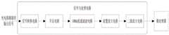
图1是本发明一种具体实施方式的脑血氧无创监测装置的系统框图。FIG. 1 is a system block diagram of a non-invasive monitoring device for cerebral blood oxygen according to a specific embodiment of the present invention.
图2是本发明一种具体实施方式的脑血氧无创监测装置中下位机端与上位机端选择连接通信的程序示例流程图。FIG. 2 is a flow chart of an example of a program for selecting connection and communication between the lower computer end and the upper computer end in the non-invasive monitoring device for cerebral blood oxygen according to a specific embodiment of the present invention.
图3是本发明一种具体实施方式的脑血氧无创监测装置中上位机端人机交互以及通信过程流程图。FIG. 3 is a flow chart of the human-computer interaction and communication process of the host computer in the non-invasive monitoring device for cerebral blood oxygen according to a specific embodiment of the present invention.
图4是本发明一种具体实施方式的脑血氧无创监测装置中信号预处理电路的电路结构框图。4 is a block diagram of a circuit structure of a signal preprocessing circuit in a non-invasive monitoring device for cerebral blood oxygen according to a specific embodiment of the present invention.
具体实施方式Detailed ways
近年来,国内外研究开发脑血氧无创监测设备的科研人员越来越多,但就目前国内情况而言,成熟的设备依旧稀缺,而国外的无创脑血氧监护设备购置价格昂贵,诊断费用高,国内的普及率不高。因此本发明的提出,能有效填补该领域的部分空缺,为国内的脑血氧监护设备的发展提供一定的支持。针对脑血氧信号监测这一问题,由于其应用环境的特殊性,往往是在临床手术环境中使用该类设备装置,因此需要充分考虑监测部位与监测参数的种类。首先,本发明面向的对象是临床麻醉后进行手术的病人,其往往是平躺于手术台上,为了监测到的脑血氧信号受到尽可能小的干扰,因此选择人体头部对应脑前额叶的区域作为脑血氧无创监测区。之所以研究无创监测,也是出于对病人的手术体验的考虑,目前国内大部分手术中对脑血氧值的检测是通过对动、静脉血的血气分析得到的,这类方法无法连续监测且有创,有一定的危险性。在手术中,医生最需要的两个脑血氧的相关信息,一是脑血氧信号波形的变化情况,是否出现剧烈突变;二是脑血氧值,当人处于正常状态时,人体脑血氧值是在一个稳定范围内的,若手术时出现数值异常或者波动较大,则需要采取一定的手段弥补。In recent years, more and more researchers at home and abroad have researched and developed non-invasive monitoring equipment for cerebral blood oxygen. However, as far as the current domestic situation is concerned, mature equipment is still scarce, and foreign non-invasive monitoring equipment for cerebral blood oxygen is expensive to purchase and diagnose. High, the domestic penetration rate is not high. Therefore, the present invention can effectively fill some vacancies in this field, and provide certain support for the development of domestic cerebral blood oxygen monitoring equipment. For the monitoring of cerebral blood oxygen signal, due to the particularity of its application environment, this type of equipment is often used in a clinical operation environment, so it is necessary to fully consider the types of monitoring sites and monitoring parameters. First of all, the object of the present invention is the patient who undergoes surgery after clinical anesthesia, who is usually lying flat on the operating table. In order to monitor the cerebral blood oxygen signal with as little interference as possible, the head of the human body is selected to correspond to the prefrontal lobe of the brain. The area is used as a non-invasive monitoring area for cerebral blood oxygen. The reason for the study of non-invasive monitoring is also due to the consideration of the patient's surgical experience. At present, most of the detection of cerebral blood oxygen value in domestic operations is obtained by blood gas analysis of arterial and venous blood. Invasive, there is a certain degree of risk. During the operation, the doctor needs two information about cerebral blood oxygen most, one is the change of the cerebral blood oxygen signal waveform, whether there is a sudden change; the other is the cerebral blood oxygen value. The oxygen value is within a stable range. If the value is abnormal or fluctuates greatly during the operation, certain measures need to be taken to make up for it.
综合以上背景原因,经过更深入的研究发现,人体头部组织中对脑血氧监测产生干扰的信号,主要来自于人体皮肤组织中黑色素成分对于脑血氧信号连续监测造成的影响,因为人体组织中黑色素成分也会吸收近红外光,从而导致常规检测中,由黑色素成分吸收的近红外光部分也被误计算为血红蛋白对近红外光的吸收,造成血红蛋白对近红外光的吸收计算量虚高,进而对最终监测的脑血氧值产生干扰,引起较大的脑血氧监测误差。而进一步研究发现,人体中黑色素成分对红光的吸收系数远大于血红蛋白的吸收系数,因此,在构建脑血氧无创预测模型时,可以近似认为红光照射监测区域后的出射光光密度的变化量主要是人体黑色素成分吸收造成的;而且,在近红外波段内,随着波长的增加,人体黑色素对近红外光的吸收系数的变化不大。因此,可以考虑将脑血氧无创监测区对红光的吸收情况作为对人体头部组织干扰信号的表征,进而借此去除该干扰信号值,以提高脑血氧无创监测的稳定性和准确性。Based on the above background reasons, after more in-depth research, it was found that the signals that interfere with the monitoring of cerebral blood oxygen in the human head tissue mainly come from the influence of the melanin component in the human skin tissue on the continuous monitoring of the cerebral blood oxygen signal. The melanin component also absorbs near-infrared light, which leads to the miscalculation of the near-infrared light absorbed by the melanin component in the routine detection as the absorption of near-infrared light by hemoglobin, resulting in a falsely high calculation of the absorption of near-infrared light by hemoglobin. , which in turn interferes with the final monitored cerebral blood oxygen value, causing a larger cerebral blood oxygen monitoring error. Further research found that the absorption coefficient of red light by melanin in the human body is much larger than that of hemoglobin. Therefore, when constructing a non-invasive prediction model of cerebral blood oxygen, it can be approximated that the change in the optical density of the outgoing light after the red light irradiates the monitoring area The amount is mainly caused by the absorption of human melanin components; moreover, in the near-infrared band, with the increase of wavelength, the absorption coefficient of human melanin to near-infrared light does not change much. Therefore, the absorption of red light in the non-invasive monitoring area of cerebral blood oxygen can be considered as a representation of the interference signal of human head tissue, and then the value of the interference signal can be removed to improve the stability and accuracy of non-invasive monitoring of cerebral blood oxygen. .
基于前述研究,本发明提出了一种脑血氧无创监测方法,旨在通过人体脑部两种血红蛋白对于红光和近红外光吸收程度的不同,利用连续脑血氧预测模型实现患者连续实时的脑血氧监测,为脑血氧的无创监测提供一种解决新方案。Based on the aforementioned research, the present invention proposes a non-invasive monitoring method for cerebral blood oxygen, which aims to realize continuous real-time monitoring of patients by using a continuous cerebral blood oxygen prediction model through the difference in the absorption of red light and near-infrared light by two hemoglobins in the human brain. Cerebral blood oxygen monitoring provides a new solution for non-invasive monitoring of cerebral blood oxygen.
本发明的脑血氧无创监测方法,以人体头部对应脑前额叶的区域作为脑血氧无创监测区,通过采集脑血氧无创监测区对红光的吸收情况作为对人体头部组织干扰信号的表征值,通过采集脑血氧无创监测区对两种不同波长的红外光的吸收情况分别作为脑前额叶区局部氧合血红蛋白浓度、脑前额叶区局部还原血红蛋白浓度的表征值,分别检测到表层干扰信号与深层有用信号,采集到的信号内容更加丰富,方便处理得到信噪比高的脑血氧信号,进而分别求取去除人体头部组织干扰信号的脑前额叶区局部氧合血红蛋白浓度值和脑前额叶区局部还原血红蛋白浓度值,从而得到去除人体头部组织干扰信号的脑前额叶区局部血氧饱和度监测值,实现脑血氧无创监测。In the non-invasive monitoring method for cerebral blood oxygen of the present invention, the area of the human head corresponding to the prefrontal lobe of the brain is used as the non-invasive monitoring area for cerebral blood oxygen, and the absorption of red light in the non-invasive monitoring area for cerebral blood oxygen is collected as the interference signal to the head tissue of the human body By collecting the absorption of two different wavelengths of infrared light in the non-invasive monitoring area of cerebral blood oxygen as the representative value of the local oxyhemoglobin concentration in the prefrontal area of the brain and the local reduced hemoglobin concentration in the prefrontal area of the brain, respectively, The surface interference signal and the deep useful signal are collected with richer signal content, which is convenient to process and obtain the cerebral blood oxygen signal with a high signal-to-noise ratio, and then obtain the local oxyhemoglobin concentration in the prefrontal lobe region of the brain that removes the interference signal of the human head tissue. The value and the local reduced hemoglobin concentration value in the prefrontal lobe area of the brain can be obtained to obtain the local blood oxygen saturation monitoring value in the prefrontal lobe area of the brain that removes the interference signal of the human head tissue, and realizes non-invasive monitoring of cerebral blood oxygen.
本发明的脑血氧无创监测方法,其监测区选择人体头部对应脑前额叶的区域无密集毛发覆盖,减小了干扰,近红外光可以更好地穿透外部结构进入脑组织,从而获得的信号包含更多有用信息,并且考虑了人体组织中黑色素的影响,利用红光的吸收情况作为对人体头部组织黑色素干扰信号的表征,进而求取去除人体头部组织干扰信号的脑前额叶区局部血氧饱和度监测值,使得脑血氧连续监测稳定性更好、监测精度更高。In the non-invasive monitoring method for cerebral blood oxygen of the present invention, the monitoring area selects the area of the human head corresponding to the prefrontal lobe of the brain without dense hair coverage, reducing interference, and the near-infrared light can better penetrate the external structure and enter the brain tissue, thereby obtaining The signal contains more useful information, and considering the influence of melanin in human tissue, the absorption of red light is used as a representation of the melanin interference signal in human head tissue, and then the prefrontal lobe of the brain that removes the interference signal in human head tissue is obtained. The monitoring value of local blood oxygen saturation in the region makes the continuous monitoring of cerebral blood oxygen more stable and more accurate.
为了更好的体现本发明脑血氧无创监测方法的技术可实施性和技术优势,下面通过基于本发明脑血氧无创监测方法设计思路的脑血氧无创监测装置实施例,来做进一步的说明。In order to better reflect the technical practicability and technical advantages of the non-invasive monitoring method for cerebral blood oxygen of the present invention, the following is a further description of the embodiment of the non-invasive monitoring device for cerebral blood oxygen based on the design idea of the non-invasive monitoring method for cerebral blood oxygen of the present invention. .
本发明提出的脑血氧无创监测装置,包括脑血氧信息采集子系统、采集控制子系统和监测计算处理子系统;其中,脑血氧信息采集子系统和采集控制子系统可集成作为下位机端,监测计算处理子系统可独立集成作为上位机端,下位机端与上位机端可以通过数据传输串口等有线通信方式,或者WIFI、蓝牙等无线通信方式,建立相互之间的数据传输连接。其装置构架原理框图如图1所示。The non-invasive monitoring device for cerebral blood oxygen proposed by the present invention includes a cerebral blood oxygen information acquisition subsystem, a acquisition control subsystem and a monitoring calculation processing subsystem; wherein, the cerebral blood oxygen information acquisition subsystem and the acquisition control subsystem can be integrated as a lower computer The monitoring and computing processing subsystem can be independently integrated as the host computer side. The lower computer side and the host computer side can establish a data transmission connection between each other through wired communication methods such as data transmission serial ports, or wireless communication methods such as WIFI and Bluetooth. The principle block diagram of its device architecture is shown in Figure 1.
本发明的脑血氧无创监测装置中,脑血氧信息采集子系统可以设计一组或两组用于采集脑血氧信息的脑血氧信息采集探头,以及用于对采集的脑血氧信息进行信号转换以及滤波放大预处理的信号预处理电路。每组脑血氧信息采集探头具有用于贴合在人体头部对应脑前额叶区域的贴合部,贴合部上至少需要布置有发光波长为680nm~700nm的红光源、发光波长为760nm~790nm的第一红外光源、发光波长为840nm~900nm的第二红外光源、以及相间隔的两个光电探测器。脑血氧信息采集探头用于通过其贴合部贴合在作为脑血氧无创监测区的人体头部对应脑前额叶的区域,并用于通过两个光电探测器检测红光源照射脑血氧无创监测区反射出的红光的出射光强度差值作为对人体头部组织干扰信号的表征值,通过两个光电探测器检测所述第一红外光源照射脑血氧无创监测区反射出的红外光的出射光强度差值作为脑前额叶区局部还原血红蛋白浓度的表征值,通过两个光电探测器检测所述第一红外光源照射脑血氧无创监测区反射出的红外光的出射光强度差值作为脑前额叶区局部氧合血红蛋白浓度的表征值。此外,脑血氧信息采集探头的贴合部上还可以设计布置发光波长为800nm~820nm的第三红外光源,用于通过两个光电探测器检测所述第三红外光源照射脑血氧无创监测区反射出的红外光的出射光强度差值作为个体差异修正因子的表征值,以用于进行个体差异修正。In the cerebral blood oxygen non-invasive monitoring device of the present invention, the cerebral blood oxygen information acquisition subsystem can design one or two sets of cerebral blood oxygen information acquisition probes for collecting cerebral blood oxygen A signal preprocessing circuit that performs signal conversion and filter amplification preprocessing. Each set of cerebral blood oxygen information acquisition probes has a fitting portion for fitting on the human head corresponding to the prefrontal lobe region of the brain. At least a red light source with a light-emitting wavelength of 680nm-700nm and a light-emitting wavelength of 760nm~ A first infrared light source with a wavelength of 790 nm, a second infrared light source with an emission wavelength of 840 nm to 900 nm, and two photodetectors spaced apart. The cerebral blood oxygen information acquisition probe is used to fit the area of the human head corresponding to the prefrontal lobe of the brain, which is the non-invasive monitoring area of cerebral blood oxygen, through its fitting part, and is used to detect the red light source through two photodetectors to illuminate the cerebral blood oxygen non-invasively. The difference in intensity of outgoing light of the red light reflected from the monitoring area is used as a characterization value of the interference signal to the human head tissue, and two photodetectors are used to detect the infrared light reflected from the first infrared light source irradiating the non-invasive monitoring area for cerebral blood oxygen. The difference in intensity of the outgoing light is used as a representative value of the local reduced hemoglobin concentration in the prefrontal lobe area of the brain, and the difference in intensity of outgoing light reflected by the first infrared light source irradiating the non-invasive monitoring area for cerebral blood oxygen is detected by two photodetectors. As a representative value of local oxyhemoglobin concentration in the prefrontal area of the brain. In addition, a third infrared light source with an emission wavelength of 800 nm to 820 nm can also be designed and arranged on the bonding part of the cerebral blood oxygen information acquisition probe, which is used for non-invasive monitoring of cerebral blood oxygen by detecting the third infrared light source through two photodetectors. The difference of the outgoing light intensity of the infrared light reflected from the area is used as the characteristic value of the individual difference correction factor for performing the individual difference correction.
采集控制子系统用于对脑血氧信息采集子系统的脑血氧信息采集过程进行驱动控制。具体应用时,采集控制子系统通过分时间隔驱动控制脑血氧信息采集探头中的各不同光源交替发光,使得脑血氧信息采集探头中的光电探测器能够在不同时段检测不同光源照射脑血氧无创监测区反射出的出射光强度。The acquisition control subsystem is used to drive and control the cerebral blood oxygen information acquisition process of the cerebral blood oxygen information acquisition subsystem. In specific applications, the acquisition control subsystem drives and controls the different light sources in the cerebral blood oxygen information acquisition probe to emit light alternately through time-division intervals, so that the photodetector in the cerebral blood oxygen information acquisition probe can detect different light sources irradiating cerebral blood in different periods of time. The intensity of the outgoing light reflected from the oxygen non-invasive monitoring area.
监测计算处理子系统用于接收脑血氧信息采集子系统所采集的脑血氧信息,分别求取去除人体头部组织干扰信号的脑前额叶区局部氧合血红蛋白浓度值和脑前额叶区局部还原血红蛋白浓度值,从而计算得到去除人体头部组织干扰信号的脑前额叶区局部血氧饱和度监测值,实现脑血氧无创监测;在脑血氧信息采集探头的贴合部上还布置有第三红外光源的条件下,监测计算处理子系统还可以用于在得到去除人体头部组织干扰信号的脑前额叶区局部血氧饱和度监测值的基础上,进一步利用所述个体差异修正因子对于脑前额叶区局部血氧饱和度监测值进行个体差异修正。The monitoring and computing processing subsystem is used to receive the cerebral blood oxygen information collected by the cerebral blood oxygen information acquisition subsystem, and respectively obtain the local oxyhemoglobin concentration value in the prefrontal lobe area and the local prefrontal lobe area after removing the interference signal of the human head tissue. The hemoglobin concentration value is restored to calculate the local blood oxygen saturation monitoring value in the prefrontal lobe area of the brain that removes the interference signal of the human head tissue, so as to realize the non-invasive monitoring of cerebral blood oxygen. Under the condition of the third infrared light source, the monitoring and computing processing subsystem can also be used to further utilize the individual difference correction factor on the basis of obtaining the monitoring value of the local blood oxygen saturation in the prefrontal lobe region of the brain from which the interference signal of the human head tissue is removed. Individual differences were corrected for the monitoring values of local blood oxygen saturation in the prefrontal lobe region of the brain.
监测计算处理子系统在执行脑前额叶区局部血氧饱和度监测值的分析计算过程中,需要使用到本发明提出的脑血氧无创预测模型;该脑血氧无创预测模型基于修正的朗伯-比尔定律,利用前述的红光和不同波长的红外光通过脑局部组织前后光密度的变化信息,以及相间隔的两个光电探测器分别检测浅层和深层的组织信息,构建出初步的模型,之后将模型进行优化,根据脑血氧饱和度的定义构建出最终的无创脑血氧预测模型,从而实时计算得到脑前额叶区局部血氧饱和度监测值。并且作为上位机端的软件交互设计,还可以进一步的增加人机交互功能,例如对接收到的脑血氧信息进行显示,方便观察,还可以根据近红外脑血氧检测时波形的变化特性,定位识别出变化的特征点,并在上位机端的人机交互界面上进行显示和分析,等等。In the process of analyzing and calculating the monitoring value of local blood oxygen saturation in the prefrontal lobe region of the brain, the monitoring and computing processing subsystem needs to use the non-invasive prediction model of cerebral blood oxygen proposed by the present invention; the non-invasive prediction model of cerebral blood oxygen is based on the modified Lambertian - Beer's Law, using the aforementioned red light and infrared light of different wavelengths to pass through the brain tissue before and after the change in optical density information, and two spaced photodetectors to detect the information of the superficial and deep tissue respectively, to build a preliminary model Then, the model is optimized, and the final non-invasive cerebral blood oxygen prediction model is constructed according to the definition of cerebral blood oxygen saturation, so as to obtain the monitoring value of local blood oxygen saturation in the prefrontal lobe region of the brain through real-time calculation. And as the software interaction design of the host computer, it can further increase the human-computer interaction function, such as displaying the received cerebral blood oxygen information, which is convenient for observation, and can also locate according to the changing characteristics of the waveform during the near-infrared cerebral blood oxygen detection. Identify the changing feature points, and display and analyze them on the human-computer interface of the host computer, and so on.
具体到本实施例中而言,脑血氧无创监测装置中具体的技术设计要点主要分为如下的几个部分:Specifically in this embodiment, the specific technical design points in the non-invasive monitoring device for cerebral blood oxygen are mainly divided into the following parts:
1)脑血氧信息采集探头的具体结构设计;1) The specific structure design of the cerebral blood oxygen information acquisition probe;
2)采集控制子系统对脑血氧信息采集过程的驱动和控制实现方式;2) The driving and control implementation method of the acquisition control subsystem for the acquisition process of cerebral blood oxygen information;
3)信号预处理电路对于脑血氧信号的预处理过程;3) The preprocessing process of the cerebral blood oxygen signal by the signal preprocessing circuit;
4)监测计算处理子系统对接收到的脑血氧信息数据的二次处理以及波形绘制、显示;4) The secondary processing of the received cerebral blood oxygen information data by the monitoring and calculation processing subsystem, as well as the waveform drawing and display;
5)监测计算处理子系统中脑血氧无创预测模型的构建以及基于计算的软件实现、计算和显示。5) The construction of the non-invasive prediction model of cerebral blood oxygen in the monitoring and computing processing subsystem and the software realization, calculation and display based on calculation.
下面分别对各个部分展开进行详细说明。The following is a detailed description of each part.
作为一种具体的优选设计方式,在本实施例中,脑血氧无创监测装置的各个子系统采用统一供电,再根据各模块所需要的工作电压,采用对应的稳压电源模块,为各个模块提供额定的工作电压。As a specific preferred design method, in this embodiment, each subsystem of the cerebral blood oxygen non-invasive monitoring device adopts a unified power supply, and then according to the working voltage required by each module, a corresponding regulated power supply module is used to provide the power supply for each module. Provides rated working voltage.
在本实施例中,如图1所示,上述第1)部分的技术内容中,脑血氧信息采集子系统中设计了两组脑血氧信息采集探头,分别用于贴合检测人体头部两侧的脑前额叶区域位置;每组脑血氧信息采集探头的贴合部上布置了发光波长为700nm的红光源、发光波长为760nm的第一红外光源、发光波长为850nm的第二红外光源、发光波长为805nm的第三红外光源、以及相间隔的两个光电探测器。其中,700nm波长的红光用于监测皮肤组织中黑色素成分对红光的吸收情况;760nm波长的红光是用于监测脱氧血红蛋白的浓度变化情况;805nm波长的近红外光是两种血红蛋白的等吸收点,用于模型的修正;850nm波长的近红外光是用于监测氧合血红蛋白的浓度变化情况。同时,在结构设计上,该脑血氧信息采集探头采用了柔软粘性材质制作贴合部,例如可将贴合部制作成采用柔性贴片等形式,用于粘附贴合在人体头部对应脑前额叶的区域,以更好地贴合皮肤,避免漏光,减少能量损耗和外部干扰;同时,脑血氧信息采集探头还设计有用于对所述贴合部进行遮光的软质遮光外壳,且外壳颜色最好采用黑色等深色以尽量吸收环境光,软质遮光外壳一方面用于对贴合部上布置的各个光源和两个光电探测器的结构加以保护,另一方面可用于减少环境光对贴合部的光干扰,并且外壳采用柔软材质可以产生一定的形变来更好地贴合人脑前额叶,尽量减少被监测者的不舒适感。In this embodiment, as shown in FIG. 1 , in the technical content of the above-mentioned part 1), two sets of cerebral blood oxygen information acquisition probes are designed in the cerebral blood oxygen information acquisition subsystem, which are respectively used to fit and detect the human head. The position of the prefrontal lobe area on both sides of the brain; on the bonding part of each group of cerebral blood oxygen information collection probes, a red light source with a luminous wavelength of 700 nm, a first infrared light source with a luminous wavelength of 760 nm, and a second infrared light source with a luminous wavelength of 850 nm are arranged A light source, a third infrared light source with an emission wavelength of 805 nm, and two photodetectors spaced apart. Among them, the red light with a wavelength of 700nm is used to monitor the absorption of red light by melanin components in skin tissue; the red light with a wavelength of 760nm is used to monitor the concentration change of deoxyhemoglobin; the near-infrared light with a wavelength of 805nm is used for two kinds of hemoglobin, etc. The absorption point is used for model correction; near-infrared light with a wavelength of 850 nm is used to monitor changes in the concentration of oxyhemoglobin. At the same time, in terms of structural design, the cerebral blood oxygen information acquisition probe is made of soft and viscous material to make the bonding part. The area of the prefrontal lobe of the brain can better fit the skin, avoid light leakage, reduce energy loss and external interference; at the same time, the cerebral blood oxygen information acquisition probe is also designed with a soft shading shell for shading the fitting part. And the color of the shell is preferably dark, such as black, to absorb the ambient light as much as possible. The ambient light interferes with the light of the fitting part, and the shell is made of soft material, which can produce a certain deformation to better fit the prefrontal lobe of the human brain and minimize the discomfort of the monitored person.
本实施例中,脑血氧无创监测装置优选采用下位机端、上位机端的分布式集成化设计,脑血氧信息采集子系统和采集控制子系统集成作为下位机端,监测计算处理子系统独立集成作为上位机端,下位机端与上位机端之间通过有线或无线通信方式建立数据传输连接。这样可以使得脑血氧无创监测装置可以基于嵌入式技术加以设计,使得脑血氧无创监测装置产品将下位机系统与上位机系统结合起来,可以形成一个独立的小型化装置,具有便携、灵活性好、成本低、方便推广、适应性强的优点。In this embodiment, the cerebral blood oxygen non-invasive monitoring device preferably adopts the distributed integrated design of the lower computer and the upper computer, the cerebral blood oxygen information acquisition subsystem and the acquisition control subsystem are integrated as the lower computer, and the monitoring and computing processing subsystem is independent The integration is used as the upper computer end, and the data transmission connection is established between the lower computer end and the upper computer end through wired or wireless communication. In this way, the cerebral blood oxygen non-invasive monitoring device can be designed based on embedded technology, so that the cerebral blood oxygen non-invasive monitoring device product can combine the lower computer system and the upper computer system to form an independent miniaturized device, which is portable and flexible. It has the advantages of good, low cost, convenient promotion and strong adaptability.
其次,在软件的系统设置中连接相应的通信方式,选择合适的监测模式并完成其他设置后,即可开始接收来自下位机采集的脑血氧信号数据。例如,图2给出了下位机端与上位机端选择连接通信的一种程序示例流程图,以该图示示例为例,可以设计下位机端与上位机端选择连接通信的方式包括串口通信、WIFI通信、蓝牙通信等不同形式,使用者可通过软件的系统设置选择所需的连接通信方式后,系统则根据相应通信方式执行下位机端与上位机之间的通信连接处理,直至完成通信连接。Secondly, connect the corresponding communication mode in the system settings of the software, select the appropriate monitoring mode and complete other settings, then you can start to receive the cerebral blood oxygen signal data collected from the lower computer. For example, Fig. 2 shows a flow chart of a program example for selecting connection and communication between the lower computer and the upper computer. Taking this illustrated example as an example, it is possible to design a method for selecting connection and communication between the lower computer and the upper computer, including serial communication. , WIFI communication, Bluetooth communication, etc. After the user can select the required connection communication method through the system settings of the software, the system will execute the communication connection processing between the lower computer and the upper computer according to the corresponding communication method until the communication is completed. connect.
此外,在采集过程中若出现探头掉落,连接断开等情况,上位机端会自动终止通信,并提示警告信息。上述描述为上位机和下位机的在脑血氧信号采集中的人机交互以及通信过程,具体流程示例如图3所示。下位机利用上述自主设计的两组脑血氧信息采集探头,分别对人体头部对应左、右侧前额叶脑局部位置进行信号的采集,两侧通道同时工作进行信号的采集。In addition, if the probe is dropped or the connection is disconnected during the acquisition process, the host computer will automatically terminate the communication and prompt a warning message. The above description is the human-computer interaction and communication process of the upper computer and the lower computer in the acquisition of the cerebral blood oxygen signal, and a specific process example is shown in FIG. 3 . The lower computer uses the above two sets of cerebral blood oxygen information acquisition probes independently designed to collect signals from the local positions of the human head corresponding to the left and right prefrontal lobes respectively, and the two channels work simultaneously to collect signals.
在本实施例中,上述第2)部分的技术内容中,采集控制子系统对脑血氧信息采集过程的驱动和控制实现方式又具体包括如下技术内容:In this embodiment, in the technical content of the above-mentioned part 2), the driving and control implementation manner of the acquisition control subsystem for the acquisition process of cerebral blood oxygen information specifically includes the following technical content:
2a)在采集人体头部两侧前额叶脑血氧信号时,两侧脑血氧信息采集探头的通道是同时工作的,其中的各个红光、红外光源均采用LED光源,其工作先后顺序可设计为700nm(红光源)、760nm(第一红外光源)、805nm(第三红外光源)、850nm(第二红外光源),它们交替发光(当然也可以设计为其它顺序),以确保四种LED光源的工作时段中间包含一定长度的间隔期,用来防止不同波长LED光源之间的光干扰。采集控制子系统包括电源模块、光源驱动模块、微控制器。其中,LED光源是由采集控制子系统中的光源驱动模块电路控制工作的,通过微控制器的I/O管脚输出PWM信号,控制光源驱动模块电路输出驱动信号让相应的LED光源发光;电源模块则为其它各电子器件供电。微控制器还用于对采集得到的脑血氧信号进行收集,并与监测计算处理子系统进行数据通信,将采集到的脑血氧信号上传给监测计算处理子系统。2a) When collecting the cerebral blood oxygen signal of the prefrontal lobe on both sides of the human head, the channels of the cerebral blood oxygen information acquisition probes on both sides work simultaneously, and each red light and infrared light source are LED light sources, and the working order can be changed. It is designed to be 700nm (red light source), 760nm (first infrared light source), 805nm (third infrared light source), 850nm (second infrared light source), and they emit light alternately (of course, they can also be designed in other orders) to ensure that four kinds of LEDs The working period of the light source includes a certain length of interval, which is used to prevent light interference between LED light sources of different wavelengths. The acquisition control subsystem includes a power supply module, a light source drive module, and a microcontroller. Among them, the LED light source is controlled by the light source driving module circuit in the acquisition control subsystem, and the PWM signal is output through the I/O pin of the microcontroller, and the light source driving module circuit is controlled to output the driving signal to make the corresponding LED light source emit light; the power supply The module supplies power to various other electronic devices. The microcontroller is also used for collecting the collected cerebral blood oxygen signal, and performing data communication with the monitoring computing processing subsystem, and uploading the collected cerebral blood oxygen signal to the monitoring computing processing subsystem.
2b)驱动信号的产生,是通过微控制器的I/O管脚输出PWM信号来控制光源驱动模块电路实现的。红光与近红外光LED的工作电压往往在1.1V~1.6V之间,通过电路分压来限制进入LED的电压信号,并利用三极管组合来满足其工作需要的额定电流,进而实现驱动发光的目的。2b) The generation of the driving signal is realized by controlling the light source driving module circuit by outputting the PWM signal from the I/O pin of the microcontroller. The working voltage of red light and near-infrared light LEDs is often between 1.1V and 1.6V. The voltage signal entering the LED is limited by the circuit voltage divider, and the combination of triodes is used to meet the rated current required for its work, thereby realizing the driving light-emitting Purpose.
2c)各个LED在驱动发光后,会依次有序发光,入射光穿透人体脑部前额叶,经“香蕉型”路径传播后会产生反射,由两个光电探测器接收到出射光信号,转变成微弱的电流信号。2c) After each LED is driven to emit light, it will emit light in sequence. The incident light penetrates the prefrontal lobe of the human brain, and will be reflected after propagating through the "banana-shaped" path. The outgoing light signal is received by two photodetectors and converted into into a weak current signal.
在本实施例中,上述第3)部分的技术内容中,作为优选方案,如图4所示,信号预处理电路包括信号转换升压电路单元、信号滤波电路单元和信号放大电路单元;信号转换升压电路单元包括信号转换电路和升压电路,信号转换电路用于将脑血氧信息采集探头中光电探测器的电流信号转换为电压信号,升压电路用于对电压信号进行升压;信号滤波电路单元包括10Hz低通滤波电路,用于滤除10Hz以上的干扰信号;信号放大电路单元包括前置放大电路和二级放大电路,用于对信号进行二级放大。其中,具体包括如下技术内容:In this embodiment, in the technical content of the above-mentioned part 3), as a preferred solution, as shown in FIG. 4 , the signal preprocessing circuit includes a signal conversion booster circuit unit, a signal filter circuit unit and a signal amplification circuit unit; The boost circuit unit includes a signal conversion circuit and a boost circuit, the signal conversion circuit is used to convert the current signal of the photodetector in the cerebral blood oxygen information acquisition probe into a voltage signal, and the boost circuit is used to boost the voltage signal; The filter circuit unit includes a 10Hz low-pass filter circuit for filtering out interference signals above 10Hz; the signal amplifier circuit unit includes a preamplifier circuit and a secondary amplifier circuit for secondary amplification of the signal. Among them, the specific technical content includes the following:
3a)本装置中光电探测器接收到透过人体头部两侧前额叶的出射光之后,会随之产生一个微弱的电流信号。为了信号可以进行A/D转换,需要将其转换成电压信号,即需要一个信号转换电路,将电流信号转换为电压信号。本专利中使用了OPA380芯片作为信号转换电路,来实现电流转电压的目的,该放大器具有极低的偏置电流,同时通过选择电路中合适阻抗值的电阻,作为升压电路,实现一个107倍的升压放大效果,即信号放大部分的前置放大。3a) After the photodetector in the device receives the outgoing light passing through the frontal lobes on both sides of the human head, a weak current signal will be generated accordingly. In order to perform A/D conversion on the signal, it needs to be converted into a voltage signal, that is, a signal conversion circuit is required to convert the current signal into a voltage signal. In this patent, the OPA380 chip is used as a signal conversion circuit to achieve the purpose of converting current to voltage. The amplifier has a very low bias current, and at the same time, by selecting a resistor with a suitable impedance value in the circuit, it is used as a boost circuit to achieve a 107 The boost amplification effect is doubled, that is, the pre-amplification of the signal amplification part.
3b)在电流转电压的同时进行一个107倍数的放大,是为了方便之后的滤波处理。根据资料显示,人体脑血氧信号的频率一般在0.01Hz以下,而常见的几种噪声,如50Hz工频干扰,各种高频白噪声等,都能轻易地将脑血氧信号掩盖。因此,要先进行一定程度的放大,才能保证滤波之后的信号里仍然能提取到所需要的脑血氧信号。本实施例所使用的低通滤波器截止频率为10Hz,作为10Hz低通滤波电路,主要目的是为了滤除10Hz以上的干扰信号,例如50Hz工频干扰以及一些白噪声等,同时保留信号的一些细节信息,考虑到该装置往往是应用于临床手术情况下,病人基本都处于麻醉状态,因此硬件设施上不考虑滤除低频噪声的部分。3b) Amplification by a factor of 107 is performed at the same time as the current is converted into the voltage, in order to facilitate subsequent filtering processing. According to the data, the frequency of human cerebral blood oxygen signal is generally below 0.01Hz, and several common noises, such as 50Hz power frequency interference, various high-frequency white noise, etc., can easily cover the cerebral blood oxygen signal. Therefore, a certain degree of amplification must be performed first to ensure that the required cerebral blood oxygen signal can still be extracted from the filtered signal. The cutoff frequency of the low-pass filter used in this embodiment is 10Hz. As a 10Hz low-pass filter circuit, the main purpose is to filter out interference signals above 10Hz, such as 50Hz power frequency interference and some white noise, while retaining some of the signal. For detailed information, considering that the device is often used in clinical operations, the patients are basically under anesthesia, so the part of filtering out low-frequency noise is not considered in the hardware facilities.
3c)在经过10Hz低通滤波之后,信噪比会有一个比较显著的提升。然后,对该信号再次进行放大处理,从毫伏级达到伏级。在前置放大电路对信号进行放大之后,信号的变化幅度仍然比较微小,不便于后续的处理计算,因此要再经过二级放大电路进行二级放大。3c) After 10Hz low-pass filtering, the signal-to-noise ratio will be significantly improved. Then, the signal is amplified again from the millivolt level to the volt level. After the pre-amplifier circuit amplifies the signal, the change range of the signal is still relatively small, which is inconvenient for subsequent processing and calculation.
3d)本实施例中该装置所用的芯片多为双极性的芯片,供电也采用正负双电压供电,因此会允许出现负电压的情况。然而,微控制器自带的A/D采样模块无法采集负电压,因此需要对负电压部分进行一定的电压抬升,使整个脑血氧信号都位于正电压范围,才能进行后续处理。该电压抬升电路是一个简单的加法器电路,通过多种电阻的设计搭配,来实现电压抬升。最后,经过信号预处理电路处理后的脑血氧信号由微控制器进行收集,上传给监测计算处理子系统。3d) In this embodiment, most of the chips used in the device are bipolar chips, and the power supply also adopts positive and negative dual voltage power supply, so the situation of negative voltage is allowed. However, the A/D sampling module that comes with the microcontroller cannot collect negative voltages, so it is necessary to raise the negative voltage part to a certain extent so that the entire cerebral blood oxygen signal is in the positive voltage range before subsequent processing can be performed. The voltage boost circuit is a simple adder circuit, and the voltage boost is realized through the design and combination of various resistors. Finally, the cerebral blood oxygen signal processed by the signal preprocessing circuit is collected by the microcontroller and uploaded to the monitoring and computing processing subsystem.
在本实施例中,上述第4)部分的技术内容中,在具体应用实施中,上位机端可以采用PC(桌面级个人电脑)、嵌入式处理系统等计算机端来得以实现。作为优选方案,上位机端接收到来自下位机微控制器发送上来的脑血氧信号数据交由监测计算处理子系统进行计算处理之前,可以对这些已经进行初步预处理理的信号再次进行数字滤波,用数字滤波的方法去除其中剩余的一些明显噪声,使信号更加平滑稳定。接着,根据近红外脑血氧检测时波形的变化特性,定位识别出变化的特征点。该应用软件会默认实时显示处理之后的人体头部两侧前额叶的脑血氧信号波形,方便直观地观察人体脑血氧的变化趋势,及时作出应对措施。另外,可以通过应用软件中的功能选项,来选择需要的波形信息进行显示,如每个波长的光对应的变化曲线,脑局部组织血氧相对值的变化曲线等。In this embodiment, in the technical content of the above-mentioned part 4), in the specific application implementation, the host computer side can be realized by using a computer side such as a PC (desktop personal computer) and an embedded processing system. As a preferred solution, before the upper computer receives the cerebral blood oxygen signal data sent from the microcontroller of the lower computer and sends it to the monitoring and calculation processing subsystem for calculation and processing, the signals that have undergone preliminary preprocessing can be digitally filtered again. , use digital filtering to remove some of the remaining obvious noise to make the signal more smooth and stable. Next, according to the change characteristics of the waveform during the near-infrared cerebral blood oxygen detection, the characteristic points of the change are located and identified. The application software will display the processed cerebral blood oxygen signal waveform of the prefrontal lobes on both sides of the human head in real time by default, so that it is convenient and intuitive to observe the changing trend of human cerebral blood oxygen and take timely countermeasures. In addition, the required waveform information can be selected and displayed through the function options in the application software, such as the change curve corresponding to each wavelength of light, the change curve of the relative value of blood oxygen in the brain tissue, etc.
在本实施例中,上述第5)部分的技术内容中,监测计算处理子系统中脑血氧无创预测模型,需要基于修正的朗伯-比尔定律加以建立和优化,从而利用构建的无创脑血氧预测模型来实时计算被测者当前被测区域的脑前额叶区局部血氧饱和度监测值。下面对脑血氧无创预测模型的建立过程加以展开说明。In this embodiment, in the technical content of the above-mentioned part 5), the non-invasive prediction model of cerebral blood oxygen in the monitoring and computing processing subsystem needs to be established and optimized based on the modified Lambert-Beer law, so as to use the constructed non-invasive cerebral blood oxygen The oxygen prediction model is used to calculate the local blood oxygen saturation monitoring value of the prefrontal area of the brain in the current measured area of the subject in real time. The following describes the establishment process of the non-invasive prediction model of cerebral blood oxygen.
由修正的朗伯-比尔定律可得:From the modified Lambert-Beer law we get:

Ir为入射光光强度,Io为出射光光强度,差分路径因子DPF(大于1),r是光源到检测器的直线距离,ε为摩尔消光系数,C为实验生物组织浓度;G则是其他外围生物组织对光的散射吸收等种种消减作用。当探测波长λ以及探测距离r一定时,ελ,DPFλ,Gλ均为常数。其中,r×DPF=L表示轨迹的实际行程。Ir is the light intensity of the incident light, Io is the light intensity of the outgoing light, the differential path factor DPF (greater than 1), r is the straight-line distance from the light source to the detector, ε is the molar extinction coefficient, C is the concentration of the experimental biological tissue; G is the It is a variety of reducing effects such as scattering and absorption of light by other peripheral biological tissues. When the detection wavelength λ and the detection distance r are constant, ελ , DPFλ , and Gλ are all constants. Among them, r×DPF=L represents the actual stroke of the trajectory.
人体中黑色素成分对700nm红光的吸收系数远大于血红蛋白的吸收系数,因此,在构建脑血氧无创预测模型时,近似认为700nm的出射光光密度的变化量是黑色素成分吸收造成的。而且,在近红外波段内,随着波长的增加,黑色素对近红外光的吸收系数的变化不大。The absorption coefficient of melanin components to 700nm red light in the human body is much larger than that of hemoglobin. Therefore, when building a non-invasive prediction model of cerebral blood oxygen, it is approximated that the change in the optical density of the emitted light at 700nm is caused by the absorption of melanin components. Moreover, in the near-infrared band, as the wavelength increases, the absorption coefficient of melanin to near-infrared light does not change much.
结合修正的朗伯-比尔定律,则有:Combined with the modified Lambert-Beer law, we have:


ΔODλ为波长为λ出射光光密度的变化量,在本实施例中,波长λ的取值针对不同的光源分别为700nm、760nm、850nm、805nm;即,ΔOD700表示两个光电探测器检测红光源照射脑血氧无创监测区反射出的红光的出射光强度差值;ΔOD760表示两个光电探测器检测第一红外光源的照射脑血氧无创监测区反射出的红外光的出射光强度差值;ΔOD850表示两个光电探测器检测第二红外光源的照射脑血氧无创监测区反射出的红外光的出射光强度差值;


ΔL为两个光电探测器与光源距离的差值。ΔL is the difference between the distances between the two photodetectors and the light source.
求解上述方程组,分别得到氧合血红蛋白与还原血红蛋白的浓度:Solve the above equations to obtain the concentrations of oxyhemoglobin and reduced hemoglobin, respectively:


考虑到红光源、第一红外光源、第二红外光源、第三红外光源的发光波长不局限于选择为700nm、760nm、850nm、805nm(红光源的发光波长可在680nm~700nm范围中选取、第一红外光源的发光波长可在760nm~790nm范围中选取、第二红外光源的发光波长可在840nm~900nm范围中选取、第三红外光源的发光波长可在800nm~820nm范围中选取),那么,如果采用ΔODred表示两个光电探测器检测红光源照射脑血氧无创监测区反射出的红光的出射光强度差值,ΔODinf1表示两个光电探测器检测第一红外光源的照射脑血氧无创监测区反射出的红外光的出射光强度差值,ΔODinf2表示两个光电探测器检测第二红外光源的照射脑血氧无创监测区反射出的红外光的出射光强度差值,ΔODinf3表示两个光电探测器检测第三红外光源的照射脑血氧无创监测区反射出的红外光的出射光强度差值,


其中,ΔODred、ΔODinf1、ΔODinf2、ΔODinf3四个出射光强度差值均可通过光电探测器检测获得;脑部还原血红蛋白针对第一红外光源、第二红外光源、第三红外光源的摩尔消光系数


再结合组织氧饱和度的计算公式:Combined with the formula for calculating tissue oxygen saturation:

求解上述方程组,构建脑血氧无创预测模型的展开式可表达为:Solving the above equations, the expansion formula for constructing the non-invasive prediction model of cerebral blood oxygen can be expressed as:

考虑到个体差异性,每个人的BMI指数不同,肥胖程度不一样,头部结构不尽相同,以及存在年龄段的显著差异,因此上述脑血氧预测模型还需要加入合适的修正因子,来减少这些因素的影响。805nm的近红外光源是氧合血红蛋白与还原血红蛋白的等吸收点,通过其与左右两侧光源吸收情况的对比,本实施例还提出模型中的个体差异修正因子CID:Taking into account individual differences, each person's BMI index is different, the degree of obesity is different, the structure of the head is different, and there are significant differences in age groups, so the above cerebral blood oxygen prediction model also needs to add appropriate correction factors to reduce influence of these factors. The near-infrared light source of 805 nm is the isoabsorption point of oxyhemoglobin and reduced hemoglobin. By comparing it with the absorption of the left and right light sources, this embodiment also proposes the individual difference correction factor CID in the model:

同样,考虑到红光源、第一红外光源、第二红外光源、第三红外光源的发光波长不局限于选择为700nm、760nm、850nm、805nm(红光源的发光波长可在680nm~700nm范围中选取、第一红外光源的发光波长可在760nm~790nm范围中选取、第二红外光源的发光波长可在840nm~900nm范围中选取、第三红外光源的发光波长可在800nm~820nm范围中选取),那么,如果采用ΔODred表示两个光电探测器检测红光源照射脑血氧无创监测区反射出的红光的出射光强度差值,ΔODinf1表示两个光电探测器检测第一红外光源的照射脑血氧无创监测区反射出的红外光的出射光强度差值,ΔODinf2表示两个光电探测器检测第二红外光源的照射脑血氧无创监测区反射出的红外光的出射光强度差值,ΔODinf3表示两个光电探测器检测第三红外光源的照射脑血氧无创监测区反射出的红外光的出射光强度差值,


利用个体差异修正因子CID对脑前额叶区局部血氧饱和度监测值进行修正,即:Using the individual difference correction factor CID to correct the local blood oxygen saturation monitoring value in the prefrontal lobe area, namely:
CID·rSO2;CID ·rSO2 ;
本发明还引入了两个修正系数:第一修正系数K1,第二修正系数K2。The present invention also introduces two correction coefficients: a first correction coefficient K1 , and a second correction coefficient K2 .
考虑到在建模过程中会为了达到目的而提出一些背景条件,从而忽略部分实际情况,如脑局部组织中其他成分对光的吸收、反射、散射等,这些都会导致构建的模型预测出来的结果与实际结果有差距。为了减少这种差距,本模型提出了第一修正因子K1,用以对模型计算得到脑前额叶区局部血氧饱和度监测值进行修正,即第一修正系数K1修正后的脑前额叶区局部血氧饱和度监测值为:Considering that some background conditions will be proposed for the purpose during the modeling process, and some actual conditions will be ignored, such as the absorption, reflection, and scattering of light by other components in the local brain tissue, which will lead to the results predicted by the constructed model. There is a discrepancy with the actual result. In order to reduce this gap, this model proposes a first correction factor K1 to correct the local blood oxygen saturation monitoring value in the prefrontal lobe area calculated by the model, that is, the prefrontal lobe of the brain after the correction by the first correction factor K1 Regional local blood oxygen saturation monitoring value is:

此外,为了调节修正因子CID的效果,在上述基础上继续提出了第二修正系数K2,它是为了防止CID过度修正,导致结果与实际值差距过大。因此,结合个体差异修正因子CID以及第一修正系数K1和第二修正系数K2对脑前额叶区局部血氧饱和度监测值进行修正后,即:In addition, in order to adjust the effect of the correction factor CID , a second correction coefficient K2 is proposed on the basis of the above, which is to prevent excessive correction of CID , resulting in an excessive difference between the result and the actual value. Therefore, combined with the individual difference correction factor CID and the first correction coefficient K1 and the second correction coefficient K2 , the local blood oxygen saturation monitoring value in the prefrontal lobe region of the brain is corrected, namely:

其中,第一修正因子K1是针对脑血氧无创预测模型计算得到脑前额叶区局部血氧饱和度监测值进行修正的修正参数,在脑血氧无创预测模型得以确定后即为固定值;而第二修正因子K2的值针对每个被测者而言均为个性化的固定值;因此,K1和K2的值均可通过使用脑血氧无创监测装置监测前的信号测试来确定。Wherein, the first correction factor K1 is a correction parameter for the correction of the local blood oxygen saturation monitoring value in the prefrontal lobe region of the brain calculated by the non-invasive prediction model of cerebral blood oxygen, and it is a fixed value after the non-invasive prediction model of cerebral blood oxygen is determined; The value of the second correction factor K2 is an individualized fixed value for each subject; therefore, the values of K1 and K2 can be determined by using the signal test before monitoring with the cerebral blood oxygen non-invasive monitoring device Sure.
最终应用于上位机端脑血氧无创监测软件的脑血氧无创预测模型表达式为:The expression of the non-invasive prediction model of cerebral blood oxygen finally applied to the non-invasive monitoring software of cerebral blood oxygen on the host computer is:

这里的rSO2(P)为通过模型计算得到个体差异修正的脑前额叶区局部血氧饱和度监测值。Here, rSO2 (P) is the monitoring value of local blood oxygen saturation in the prefrontal lobe region of the brain obtained by model calculation and corrected for individual differences.
具体应用实施时,可基于计算机端的应用软件将上述模型编程实现,基于上述脑血氧无创预测模型,实时计算双通道的脑血氧值,并在人机交互界面中进行显示,方便观察被测者的实时情况,同时进行显示的还有脑局部组织血氧饱和度的参考值与相对变化量。During the specific application and implementation, the above model can be programmed and realized based on the application software on the computer side. Based on the above non-invasive prediction model of cerebral blood oxygen, the dual-channel cerebral blood oxygen value can be calculated in real time, and displayed in the human-computer interaction interface, which is convenient for observing the measured value. At the same time, the reference value and relative change of blood oxygen saturation in local brain tissue are also displayed.
由此,利用本发明的脑血氧无创监测装置对人体脑局部组织血氧饱和度监测的具体步骤包括:Thus, the specific steps for monitoring the blood oxygen saturation of the human brain local tissue by using the cerebral blood oxygen non-invasive monitoring device of the present invention include:
a)下位机系统与上位机系统的连接和数据通信:a) Connection and data communication between the lower computer system and the upper computer system:
在这个过程中,脑血氧无创监测装置供电工作后,下位机通过装置内部的串口线与上位机相连接,上位机计算机端主要通过脑血氧无创监测软件来实现与下位机的通信。在软件的系统设置中连接相应的串口,选择合适的监测模式并完成其他设置后,即可开始接收来自下位机采集的脑血氧信号数据。采集过程中若出现探头掉落,连接断开等情况,上位机端会自动终止通信,并提示警告信息。In this process, after the cerebral blood oxygen non-invasive monitoring device is powered on, the lower computer is connected to the upper computer through the serial line inside the device, and the computer end of the upper computer mainly realizes the communication with the lower computer through the cerebral blood oxygen non-invasive monitoring software. Connect the corresponding serial port in the system settings of the software, select the appropriate monitoring mode and complete other settings, you can start to receive the cerebral blood oxygen signal data collected from the lower computer. If the probe is dropped or the connection is disconnected during the acquisition process, the host computer will automatically terminate the communication and prompt a warning message.
b)下位机对人体头部前额叶脑血氧信号的采集:b) Acquisition of the blood oxygen signal of the prefrontal lobe of the human head by the lower computer:
在这个过程中,左右两侧前额叶上的通道是同时工作的,每一路的探头里包含680nm~700nm的红光源、760nm~790nm的第一红外光源、840nm~900nm的第二红外光源、800nm~820nm的第三红外光源(上述实施例中采用的700nm的红光源、760nm的第一红外光源、850nm的第二红外光源、805nm的第三红外光源)四种波长的入射光和覆盖对应检测范围的光电探测器。驱动脑血氧探头是通过微控制器I/O管脚输出的PWM信号控制驱动电路来实现的,考虑到LED对工作电压及工作电流有规定,因此驱动电路中需要设置一个分压电路,来控制输出电压,并通过三极管的搭配来满足额定电流的需求。在LED被驱动后,会依次有序发光,透过人体前额叶后产生一个反射信号,由光电探测器接收并形成对应的电流信号。由于两个光电探测器与光源的距离不同,接收到的信号自然有区别,该设计是为了同时监测浅层的背景干扰和深层的有用信号。In this process, the channels on the left and right prefrontal lobes work at the same time. Each probe contains a red light source of 680nm-700nm, a first infrared light source of 760nm to 790nm, a second infrared light source of 840nm to 900nm, and a 800nm light source. The third infrared light source of ~820 nm (the red light source of 700 nm, the first infrared light source of 760 nm, the second infrared light source of 850 nm, and the third infrared light source of 805 nm used in the above embodiment) The incident light of four wavelengths and the corresponding detection of coverage range of photodetectors. The driving of the cerebral blood oxygen probe is realized by controlling the driving circuit through the PWM signal output by the I/O pin of the microcontroller. Considering that the LED has regulations on the working voltage and working current, a voltage divider circuit needs to be set in the driving circuit to Control the output voltage and meet the rated current requirements through the combination of triodes. After the LED is driven, it will emit light in order, and a reflection signal will be generated after passing through the frontal lobe of the human body, which will be received by the photodetector and form a corresponding current signal. Due to the different distances of the two photodetectors from the light source, the received signals are naturally differentiated, and the design is to simultaneously monitor the background interference in the shallow layer and the useful signal in the deep layer.
c)下位机对于脑血氧信号的初步信号处理:c) Preliminary signal processing of the cerebral blood oxygen signal by the lower computer:
在这个过程中,由光电探测器产生的电流信号会经过信号转换单元、信号滤波单元、电压转换单元、信号放大单元。由于光电探测器产生的电流信号过于微弱,不方便处理,首先要进行电流转电压的转换,同时对它进行足够程度的放大。接着,通过低通滤波电路,滤除信号中带有的如50Hz工频干扰和高频白噪声等干扰信号。为了避免负电压可能对单片机A/D采样存在的影响,将信号通过加法器进行一次简单的电压抬升,使其完全处于正电压范围。为了提高信噪比,在电压抬升之后再次通过低通滤波电路。之后,通过一个放大电路对信号进行二次放大,达到A/D采样的电压范围要求。In this process, the current signal generated by the photodetector will pass through the signal conversion unit, the signal filtering unit, the voltage conversion unit, and the signal amplification unit. Since the current signal generated by the photodetector is too weak and inconvenient to handle, the current-to-voltage conversion must first be performed, and at the same time, it must be amplified to a sufficient degree. Then, through the low-pass filter circuit, the interference signals such as 50Hz power frequency interference and high-frequency white noise in the signal are filtered out. In order to avoid the influence that the negative voltage may have on the A/D sampling of the single-chip microcomputer, a simple voltage boost is carried out through the adder to make the signal completely in the positive voltage range. In order to improve the signal-to-noise ratio, the low-pass filter circuit is passed again after the voltage rise. After that, the signal is amplified twice by an amplifier circuit to meet the voltage range requirement of A/D sampling.
d)上位机对于数据的二次处理以及波形显示:d) Secondary processing of data by the host computer and waveform display:
在这个过程中,上位机计算机端的应用软件在打开对应串口之后,接收到来自下位机微控制器通过串口发送上来的脑血氧信号数据,对这些已经进行初步处理的信号再次进行数字滤波,用数字滤波的方法去除其中剩余的一些明显噪声,使信号更加平滑稳定。接着,根据近红外脑血氧检测时波形的变化特性,定位识别出变化的特征点。该应用软件会默认实时显示处理之后的人体头部两侧前额叶的脑血氧信号波形,方便直观地观察人体脑血氧的变化趋势,及时作出应对措施。另外,可以通过应用软件中的功能设置,来选择需要的波形信息进行显示,如每个波长的光对应的变化曲线,脑局部组织血氧相对值的变化曲线等。In this process, after opening the corresponding serial port, the application software on the computer side of the upper computer receives the cerebral blood oxygen signal data sent by the microcontroller of the lower computer through the serial port, and performs digital filtering on these preliminarily processed signals again. The method of digital filtering removes some of the remaining obvious noise and makes the signal more smooth and stable. Next, according to the change characteristics of the waveform during the near-infrared cerebral blood oxygen detection, the characteristic points of the change are located and identified. The application software will display the processed cerebral blood oxygen signal waveform of the prefrontal lobes on both sides of the human head in real time by default, so that it is convenient and intuitive to observe the changing trend of human cerebral blood oxygen and take timely countermeasures. In addition, the required waveform information can be selected and displayed through the function settings in the application software, such as the change curve corresponding to each wavelength of light, the change curve of the relative value of blood oxygen in the brain tissue, etc.
e)脑血氧无创预测模型基于PC的软件实现以及参数的计算和显示:e) The PC-based software implementation of the non-invasive prediction model of cerebral blood oxygen and the calculation and display of parameters:
在这个过程中,首先基于修正的朗伯-比尔定律构建脑血氧无创预测模型,将四种波长的入射光通过脑局部组织前后的吸光度信息作为输入参数,脑局部组织血氧监测值作为输出参数,构建出初步的模型,之后将模型进行优化,加入多个修正因子,提高模型的稳定性与预测精度。由此建立得到的脑血氧无创预测模型表达式为:In this process, a non-invasive prediction model of cerebral blood oxygen is first constructed based on the modified Lambert-Beer law, and the absorbance information of four wavelengths of incident light before and after passing through the local brain tissue is used as the input parameter, and the blood oxygen monitoring value of the local brain tissue is used as the output. parameters, build a preliminary model, and then optimize the model and add multiple correction factors to improve the stability and prediction accuracy of the model. The expression of the non-invasive prediction model of cerebral blood oxygen thus established is:

这里的rSO2(P)为通过模型计算得到个体差异修正的脑前额叶区局部血氧饱和度监测值;
接着,基于计算机端的应用软件将该模型编程实现,实时计算双通道的脑血氧值,并在人机交互界面中进行显示,方便观察被测者的实时情况,同时进行显示的还有脑局部组织血氧饱和度的参考值与相对变化量。Then, the application software based on the computer program realizes the model, calculates the dual-channel cerebral blood oxygen value in real time, and displays it in the human-computer interaction interface, which is convenient to observe the real-time situation of the subject, and also displays the brain part at the same time. Reference value and relative change of tissue oxygen saturation.
在具体应用中,本发明的脑血氧无创监测装置的上位机端还可以更进一步的优化设计良好的人机交互功能,以简化操作、增强显示效果,在显示脑血氧波形、脑血氧值的同时,还可以显示对应监测区域的参考值和相对变化量,以便多参数观察比较,在异常情况发生时及时做出反应;并且还可以通过人机交互界面的系统设置的设计,实现对信号采集模式、显示模式的调整控制,以便适用于更多的模式来应对更多的应用场景。In specific applications, the host computer of the non-invasive monitoring device for cerebral blood oxygen of the present invention can further optimize and design a well-designed human-computer interaction function to simplify the operation and enhance the display effect. At the same time, it can also display the reference value and relative change of the corresponding monitoring area, so as to observe and compare multi-parameters, and respond in time when an abnormal situation occurs. The adjustment control of signal acquisition mode and display mode is suitable for more modes to deal with more application scenarios.
通过上述流程可以看到,本发明的脑血氧无创监测装置及脑血氧无创监测方法,能够实现对人体脑部血氧饱和度的无创连续监测。当系统上电之后,完成上、下位机的连接,将信号采集前端放置于被测者脑部两侧前额叶,设置好相关信息后,即可开始连续无创地监测被测者的脑部血氧饱和度,与目前常见的通过在颈部安置针管定时采集血液进行血气分析来获得脑血氧值的方法相比,更加方便、安全,节约了人力、物力,成本更低。当出现异常情况时,医生以及相关医护人员可以第一时间观察到脑血氧信号波形的变化和脑血氧值的突变,及时实施救援。It can be seen from the above process that the non-invasive monitoring device for cerebral blood oxygen and the non-invasive monitoring method for cerebral blood oxygen of the present invention can realize the non-invasive continuous monitoring of the blood oxygen saturation of the human brain. After the system is powered on, the connection between the upper and lower computers is completed, and the signal acquisition front end is placed on the prefrontal lobes on both sides of the subject's brain. After setting the relevant information, the continuous non-invasive monitoring of the subject's brain blood can begin. Compared with the common method of obtaining cerebral blood oxygen value by placing a needle in the neck to periodically collect blood for blood gas analysis, oxygen saturation is more convenient and safe, saves manpower and material resources, and costs less. When an abnormal situation occurs, doctors and relevant medical staff can immediately observe the changes of the cerebral blood oxygen signal waveform and the sudden change of the cerebral blood oxygen value, and implement rescue in time.
此外,作为另一种实施方式,如果上述的脑血氧无创监测装置中,如果不考虑个体差异性,主要考虑人体头部组织干扰信号对脑血氧无创监测的影响,监测中则只需要使用到对红光源、第一红外光源和第二红外光源三种信号的计算,那么脑血氧无创预测模型可以简化为:In addition, as another embodiment, if in the above-mentioned non-invasive monitoring device for cerebral blood oxygen, if individual differences are not considered, and the influence of interference signals of human head tissue on the non-invasive monitoring of cerebral blood oxygen is mainly considered, in monitoring, only the use of To the calculation of the three signals of the red light source, the first infrared light source and the second infrared light source, the non-invasive prediction model of cerebral blood oxygen can be simplified as:

其中,in,


ΔODred表示两个光电探测器检测红光源照射脑血氧无创监测区反射出的红光的出射光强度差值;ΔODinf1表示两个光电探测器检测第一红外光源的照射脑血氧无创监测区反射出的红外光的出射光强度差值;ΔODinf2表示两个光电探测器检测第二红外光源的照射脑血氧无创监测区反射出的红外光的出射光强度差值;

综上所述,本发明利用氧合血红蛋白与脱氧血红蛋白对近红外光的吸收度的不同,开发了一种对人体脑部局部组织血氧饱和度的无创监测方法,该方法不会对人体造成伤害,且通过连续脑血氧值预测模型可以实现脑血氧值的连续实时监测,考虑了黑色素的影响以及加入了修正因子,相比现有常见的双波长预测模型更加稳定,预测精度更高。本发明的脑血氧无创监测方法,其监测区选择人体头部对应脑前额叶的区域无密集毛发覆盖,减小了干扰,近红外光可以更好地穿透外部结构进入脑组织,从而获得的信号包含更多有用信息,并且考虑了人体组织中黑色素的影响,利用红光的吸收情况作为对人体头部组织黑色素干扰信号的表征,分别检测到表层干扰信号与深层有用信号,采集到的信号内容更加丰富,方便处理得到信噪比高的脑血氧信号,进而求取去除人体头部组织干扰信号的脑前额叶区局部血氧饱和度监测值,使得脑血氧连续监测稳定性更好、监测精度更高。而本发明脑血氧无创监测装置可优选采用下位机端、上位机端的分布式集成化设计,这样可以使得脑血氧无创监测装置可以基于嵌入式技术加以设计,可以形成一个独立的小型化装置,具有便携、灵活性好、成本低、方便推广、适应性强的优点。由此可见,本发明对脑血氧无创监测提供了新的解决方案,提高了脑血氧无创监测的稳定性和准确性,更有利于推进脑血氧无创监测的临床应用。To sum up, the present invention utilizes the difference in the absorbance of oxyhemoglobin and deoxyhemoglobin to near-infrared light, and develops a non-invasive monitoring method for the blood oxygen saturation of the local tissue of the human brain, which will not cause any damage to the human body. In addition, the continuous real-time monitoring of cerebral blood oxygen value can be realized through the continuous cerebral blood oxygen value prediction model. The influence of melanin and the addition of correction factors are considered. Compared with the existing common dual-wavelength prediction model, it is more stable and has higher prediction accuracy. . In the non-invasive monitoring method for cerebral blood oxygen of the present invention, the monitoring area selects the area of the human head corresponding to the prefrontal lobe of the brain without dense hair coverage, reducing interference, and the near-infrared light can better penetrate the external structure and enter the brain tissue, thereby obtaining The signal contains more useful information, and considering the influence of melanin in human tissue, the absorption of red light is used as the characterization of the melanin interference signal of human head tissue, and the surface interference signal and the deep useful signal are detected respectively. The signal content is more abundant, and it is convenient to process and obtain the cerebral blood oxygen signal with high signal-to-noise ratio, and then obtain the local blood oxygen saturation monitoring value in the prefrontal lobe area of the brain without the interference signal of the human head tissue, which makes the continuous monitoring of cerebral blood oxygen more stable. Good, the monitoring accuracy is higher. The cerebral blood oxygen non-invasive monitoring device of the present invention can preferably adopt the distributed integrated design of the lower computer end and the upper computer end, so that the cerebral blood oxygen non-invasive monitoring device can be designed based on embedded technology, and an independent miniaturized device can be formed. , has the advantages of portability, good flexibility, low cost, convenient promotion and strong adaptability. It can be seen that the present invention provides a new solution for non-invasive monitoring of cerebral blood oxygen, improves the stability and accuracy of non-invasive monitoring of cerebral blood oxygen, and is more conducive to promoting the clinical application of non-invasive monitoring of cerebral blood oxygen.
最后说明的是,以上实施例仅用以说明本发明的技术方案而非限制,尽管通过参照本发明的优选实施例已经对本发明进行了描述,但本领域的普通技术人员应当理解,可以在形式上和细节上对其作出各种各样的改变,而不偏离所附权利要求书所限定的本发明的精神和范围。Finally, it should be noted that the above embodiments are only used to illustrate the technical solutions of the present invention and not to limit them. Although the present invention has been described with reference to the preferred embodiments of the present invention, those of ordinary skill in the art should Various changes in the above and in the details may be made therein without departing from the spirit and scope of the present invention as defined by the appended claims.
Claims (10)


Priority Applications (1)
| Application Number | Priority Date | Filing Date | Title |
|---|---|---|---|
| CN202011065976.9ACN112043287B (en) | 2020-09-30 | 2020-09-30 | Cerebral blood oxygen non-invasive monitoring method and monitoring device |
Applications Claiming Priority (1)
| Application Number | Priority Date | Filing Date | Title |
|---|---|---|---|
| CN202011065976.9ACN112043287B (en) | 2020-09-30 | 2020-09-30 | Cerebral blood oxygen non-invasive monitoring method and monitoring device |
Publications (2)
| Publication Number | Publication Date |
|---|---|
| CN112043287Atrue CN112043287A (en) | 2020-12-08 |
| CN112043287B CN112043287B (en) | 2021-07-20 |
Family
ID=73606218
Family Applications (1)
| Application Number | Title | Priority Date | Filing Date |
|---|---|---|---|
| CN202011065976.9AActiveCN112043287B (en) | 2020-09-30 | 2020-09-30 | Cerebral blood oxygen non-invasive monitoring method and monitoring device |
Country Status (1)
| Country | Link |
|---|---|
| CN (1) | CN112043287B (en) |
Cited By (15)
| Publication number | Priority date | Publication date | Assignee | Title |
|---|---|---|---|---|
| CN112494039A (en)* | 2021-02-07 | 2021-03-16 | 深圳市第二人民医院(深圳市转化医学研究院) | Visual intelligent monitoring system of brain blood oxygen |
| CN112617825A (en)* | 2020-12-17 | 2021-04-09 | 中国人民解放军西部战区总医院 | Application of lead electrode in rapid positioning of epileptic focus |
| CN112641444A (en)* | 2020-12-10 | 2021-04-13 | 中国科学院深圳先进技术研究院 | Cerebral blood oxygen detection system and method |
| CN113017623A (en)* | 2021-03-04 | 2021-06-25 | 江苏盖睿健康科技有限公司 | Measuring method, device and storage medium for hypoperfusion blood oxygen saturation |
| CN113057630A (en)* | 2021-03-09 | 2021-07-02 | 宝宝 | Finger-clamping oximeter detection circuit |
| CN113100760A (en)* | 2021-05-24 | 2021-07-13 | 中国科学院合肥物质科学研究院 | Cerebral blood oxygen saturation detection system and method not disturbed by scalp blood flow |
| CN113171087A (en)* | 2021-04-26 | 2021-07-27 | 重庆大学 | A non-invasive monitoring device for cerebral blood oxygen |
| CN113925465A (en)* | 2021-10-20 | 2022-01-14 | 佛山科学技术学院 | A rehabilitation training evaluation method and system based on muscle oxygen feedback information |
| CN115251915A (en)* | 2022-07-07 | 2022-11-01 | 深圳东海浪潮科技有限公司 | Noninvasive brain tissue oxygen concentration measuring method |
| CN116115187A (en)* | 2022-12-27 | 2023-05-16 | 浙江大学 | Multi-mode cooperative detection system based on near infrared photoelectric and electroencephalogram signals |
| CN116223439A (en)* | 2023-03-20 | 2023-06-06 | 中国科学院合肥物质科学研究院 | Method for establishing water-resistant interference model for hemoglobin based on near infrared spectrum technology |
| CN116269365A (en)* | 2023-01-04 | 2023-06-23 | 中科搏锐(北京)科技有限公司 | Cerebral blood oxygen monitoring probe, head-mounted equipment and cerebral oxygen monitoring system |
| CN116392116A (en)* | 2023-04-12 | 2023-07-07 | 上海大学 | Near-infrared cerebral blood oxygen monitoring equipment for personnel emergency behavior |
| CN117497150A (en)* | 2023-11-08 | 2024-02-02 | 首都医科大学附属北京天坛医院 | Symptom detection method and system |
| CN118177771A (en)* | 2024-03-15 | 2024-06-14 | 中国医学科学院北京协和医院 | Portable brain blood flow signal monitoring system |
Citations (16)
| Publication number | Priority date | Publication date | Assignee | Title |
|---|---|---|---|---|
| JP2000121555A (en)* | 1998-10-09 | 2000-04-28 | Japan Science & Technology Corp | Oxygen monitor by heterodyne detection |
| CN1542434A (en)* | 2003-11-07 | 2004-11-03 | 清华大学 | Tissue blood oxygen parameter detection method that can correct the influence of outer tissue |
| WO2007035934A2 (en)* | 2005-09-22 | 2007-03-29 | Skyline Biomedical, Inc. | Apparatus and method for non-invasive and minimally-invasive sensing of parameters relating to blood |
| WO2009100423A1 (en)* | 2008-02-08 | 2009-08-13 | Cas Medical Systems, Inc. | Improved method for spectrophotometric blood oxygenation monitoring |
| CN102156541A (en)* | 2010-05-13 | 2011-08-17 | 天津大学 | Prefrontal electroencephalogram information and blood oxygen information fused human-computer interaction method |
| CN202526183U (en)* | 2012-04-05 | 2012-11-14 | 王培勇 | Human tissue oxygen content monitor |
| EP2712544A1 (en)* | 2012-09-28 | 2014-04-02 | Cas Medical Systems, Inc. | Systems and methods for spectroscopic measurement of a characteristic of biological tissue |
| CN104382604A (en)* | 2014-12-02 | 2015-03-04 | 重庆名希医疗器械有限公司 | Noninvasive brain blood oxygen parameter measuring method |
| CN104997518A (en)* | 2015-06-25 | 2015-10-28 | 广州视源电子科技股份有限公司 | Method and system for counteracting motion interference of blood oxygen measuring equipment |
| CN106999113A (en)* | 2014-07-08 | 2017-08-01 | 德克萨斯大学系统董事会 | Systems and methods for measuring fetal brain oxygenation |
| CN107822592A (en)* | 2016-09-15 | 2018-03-23 | 超级医药成像有限公司 | system and method for measuring tissue oxygenation |
| CN109414226A (en)* | 2016-04-21 | 2019-03-01 | 维奥普蒂克斯公司 | It is corrected using melanin and determines tissue oxygenation saturation |
| CN109464150A (en)* | 2018-11-12 | 2019-03-15 | 中科搏锐(北京)科技有限公司 | A non-invasive intra-abdominal fetal cerebral blood oxygen detection instrument and method thereof |
| CN110179472A (en)* | 2019-06-05 | 2019-08-30 | 佛山科学技术学院 | A kind of method and system of brain function detection |
| CN110393514A (en)* | 2019-06-24 | 2019-11-01 | 华为技术有限公司 | Wearable Devices and Photoelectric Pulse Sensing Components |
| CN110575181A (en)* | 2019-09-10 | 2019-12-17 | 重庆大学 | Near-infrared spectroscopy non-invasive blood glucose detection network model training method |
- 2020
- 2020-09-30CNCN202011065976.9Apatent/CN112043287B/enactiveActive
Patent Citations (16)
| Publication number | Priority date | Publication date | Assignee | Title |
|---|---|---|---|---|
| JP2000121555A (en)* | 1998-10-09 | 2000-04-28 | Japan Science & Technology Corp | Oxygen monitor by heterodyne detection |
| CN1542434A (en)* | 2003-11-07 | 2004-11-03 | 清华大学 | Tissue blood oxygen parameter detection method that can correct the influence of outer tissue |
| WO2007035934A2 (en)* | 2005-09-22 | 2007-03-29 | Skyline Biomedical, Inc. | Apparatus and method for non-invasive and minimally-invasive sensing of parameters relating to blood |
| WO2009100423A1 (en)* | 2008-02-08 | 2009-08-13 | Cas Medical Systems, Inc. | Improved method for spectrophotometric blood oxygenation monitoring |
| CN102156541A (en)* | 2010-05-13 | 2011-08-17 | 天津大学 | Prefrontal electroencephalogram information and blood oxygen information fused human-computer interaction method |
| CN202526183U (en)* | 2012-04-05 | 2012-11-14 | 王培勇 | Human tissue oxygen content monitor |
| EP2712544A1 (en)* | 2012-09-28 | 2014-04-02 | Cas Medical Systems, Inc. | Systems and methods for spectroscopic measurement of a characteristic of biological tissue |
| CN106999113A (en)* | 2014-07-08 | 2017-08-01 | 德克萨斯大学系统董事会 | Systems and methods for measuring fetal brain oxygenation |
| CN104382604A (en)* | 2014-12-02 | 2015-03-04 | 重庆名希医疗器械有限公司 | Noninvasive brain blood oxygen parameter measuring method |
| CN104997518A (en)* | 2015-06-25 | 2015-10-28 | 广州视源电子科技股份有限公司 | Method and system for counteracting motion interference of blood oxygen measuring equipment |
| CN109414226A (en)* | 2016-04-21 | 2019-03-01 | 维奥普蒂克斯公司 | It is corrected using melanin and determines tissue oxygenation saturation |
| CN107822592A (en)* | 2016-09-15 | 2018-03-23 | 超级医药成像有限公司 | system and method for measuring tissue oxygenation |
| CN109464150A (en)* | 2018-11-12 | 2019-03-15 | 中科搏锐(北京)科技有限公司 | A non-invasive intra-abdominal fetal cerebral blood oxygen detection instrument and method thereof |
| CN110179472A (en)* | 2019-06-05 | 2019-08-30 | 佛山科学技术学院 | A kind of method and system of brain function detection |
| CN110393514A (en)* | 2019-06-24 | 2019-11-01 | 华为技术有限公司 | Wearable Devices and Photoelectric Pulse Sensing Components |
| CN110575181A (en)* | 2019-09-10 | 2019-12-17 | 重庆大学 | Near-infrared spectroscopy non-invasive blood glucose detection network model training method |
Non-Patent Citations (3)
| Title |
|---|
| CEM, ARITURK: "Efficacy of Near-Infrared Spectrometry for Monitoring the Cerebral Effects of Severe Dilutional Anemia", 《HEART SURGERY FORUM》* |
| 刘光达: "基于近红外光谱的抗扰动脑血氧分析仪", 《光学精密工程》* |
| 廖小华: "光谱法用于血氧饱和度无损测量", 《激光与光电子学进展》* |
Cited By (21)
| Publication number | Priority date | Publication date | Assignee | Title |
|---|---|---|---|---|
| CN112641444A (en)* | 2020-12-10 | 2021-04-13 | 中国科学院深圳先进技术研究院 | Cerebral blood oxygen detection system and method |
| CN112617825A (en)* | 2020-12-17 | 2021-04-09 | 中国人民解放军西部战区总医院 | Application of lead electrode in rapid positioning of epileptic focus |
| CN112494039B (en)* | 2021-02-07 | 2021-04-20 | 深圳市第二人民医院(深圳市转化医学研究院) | A visual intelligent monitoring system for cerebral blood oxygen |
| CN112494039A (en)* | 2021-02-07 | 2021-03-16 | 深圳市第二人民医院(深圳市转化医学研究院) | Visual intelligent monitoring system of brain blood oxygen |
| CN113017623A (en)* | 2021-03-04 | 2021-06-25 | 江苏盖睿健康科技有限公司 | Measuring method, device and storage medium for hypoperfusion blood oxygen saturation |
| CN113057630A (en)* | 2021-03-09 | 2021-07-02 | 宝宝 | Finger-clamping oximeter detection circuit |
| CN113171087A (en)* | 2021-04-26 | 2021-07-27 | 重庆大学 | A non-invasive monitoring device for cerebral blood oxygen |
| CN113100760A (en)* | 2021-05-24 | 2021-07-13 | 中国科学院合肥物质科学研究院 | Cerebral blood oxygen saturation detection system and method not disturbed by scalp blood flow |
| CN113100760B (en)* | 2021-05-24 | 2024-11-29 | 中国科学院合肥物质科学研究院 | Cerebral blood oxygen saturation detection system and method free from scalp blood flow interference |
| CN113925465B (en)* | 2021-10-20 | 2023-06-27 | 佛山科学技术学院 | Rehabilitation training evaluation method and system based on myooxygen feedback information |
| CN113925465A (en)* | 2021-10-20 | 2022-01-14 | 佛山科学技术学院 | A rehabilitation training evaluation method and system based on muscle oxygen feedback information |
| CN115251915A (en)* | 2022-07-07 | 2022-11-01 | 深圳东海浪潮科技有限公司 | Noninvasive brain tissue oxygen concentration measuring method |
| CN116115187A (en)* | 2022-12-27 | 2023-05-16 | 浙江大学 | Multi-mode cooperative detection system based on near infrared photoelectric and electroencephalogram signals |
| CN116269365A (en)* | 2023-01-04 | 2023-06-23 | 中科搏锐(北京)科技有限公司 | Cerebral blood oxygen monitoring probe, head-mounted equipment and cerebral oxygen monitoring system |
| CN116269365B (en)* | 2023-01-04 | 2024-03-12 | 中科搏锐(北京)科技有限公司 | Cerebral blood oxygen monitoring probe, head-mounted equipment and cerebral oxygen monitoring system |
| CN116223439A (en)* | 2023-03-20 | 2023-06-06 | 中国科学院合肥物质科学研究院 | Method for establishing water-resistant interference model for hemoglobin based on near infrared spectrum technology |
| CN116392116A (en)* | 2023-04-12 | 2023-07-07 | 上海大学 | Near-infrared cerebral blood oxygen monitoring equipment for personnel emergency behavior |
| CN117497150A (en)* | 2023-11-08 | 2024-02-02 | 首都医科大学附属北京天坛医院 | Symptom detection method and system |
| CN117497150B (en)* | 2023-11-08 | 2024-06-18 | 首都医科大学附属北京天坛医院 | A symptom detection method and system |
| CN118177771A (en)* | 2024-03-15 | 2024-06-14 | 中国医学科学院北京协和医院 | Portable brain blood flow signal monitoring system |
| CN118177771B (en)* | 2024-03-15 | 2024-11-15 | 中国医学科学院北京协和医院 | A portable brain blood flow signal monitoring system |
Also Published As
| Publication number | Publication date |
|---|---|
| CN112043287B (en) | 2021-07-20 |
Similar Documents
| Publication | Publication Date | Title |
|---|---|---|
| CN112043287B (en) | Cerebral blood oxygen non-invasive monitoring method and monitoring device | |
| Ray et al. | A review of wearable multi-wavelength photoplethysmography | |
| Haahr et al. | An electronic patch for wearable health monitoring by reflectance pulse oximetry | |
| JP3824615B2 (en) | Sleep apnea diagnostic apparatus and method | |
| CN101605495B (en) | Medical measuring device | |
| US20180177412A1 (en) | Medical Measuring Device | |
| US8428673B2 (en) | Medical measuring device | |
| Molavi et al. | Noninvasive optical monitoring of bladder filling to capacity using a wireless near infrared spectroscopy device | |
| Wan et al. | Reflective type blood oxygen saturation detection system based on MAX30100 | |
| CN113171087A (en) | A non-invasive monitoring device for cerebral blood oxygen | |
| CN209899402U (en) | Reflection type oximeter | |
| CN111481174A (en) | Anesthesia and consciousness depth monitoring system and method | |
| CN109009064A (en) | A kind of intracranial pressure noninvasive monitoring device and method | |
| TWM542444U (en) | Wearable multifunctional pulse wave diagnosis and analysis device | |
| JP2024511975A (en) | hemodilution detector | |
| Kuncoro et al. | Wireless photoplethysmography sensor for continuous blood pressure biosignal shape acquisition | |
| Kiruthiga et al. | Reflectance pulse oximetry for blood oxygen saturation measurement from diverse locations-a preliminary analysis | |
| CN116746921A (en) | A dual-modal real-time monitoring device for cerebral blood flow | |
| Laulkar et al. | Applications of finger photoplethysmography | |
| Buyanov et al. | Continuous cerebral and tissue oximetry by three-wave NIRS device | |
| Shriram et al. | Application of high & low brightness LEDs to human tissue to capture photoplethysmogram at a finger tip | |
| Khong et al. | The evolution of heart beat rate measurement techniques from contact based photoplethysmography to non-contact based photoplethysmography imaging | |
| Zhu et al. | Absolute measurement of tissue oxygen saturation with NIRS | |
| Kraitl et al. | Optical non-invasive methods for characterization of the human health status | |
| CN115969348B (en) | A portable non-invasive early screening device for thrombosis |
Legal Events
| Date | Code | Title | Description |
|---|---|---|---|
| PB01 | Publication | ||
| PB01 | Publication | ||
| SE01 | Entry into force of request for substantive examination | ||
| SE01 | Entry into force of request for substantive examination | ||
| GR01 | Patent grant | ||
| GR01 | Patent grant |