CN112011509A - Separation method and application of primary microglia of rat with spinal cord injury - Google Patents
Separation method and application of primary microglia of rat with spinal cord injuryDownload PDFInfo
- Publication number
- CN112011509A CN112011509ACN202011021176.7ACN202011021176ACN112011509ACN 112011509 ACN112011509 ACN 112011509ACN 202011021176 ACN202011021176 ACN 202011021176ACN 112011509 ACN112011509 ACN 112011509A
- Authority
- CN
- China
- Prior art keywords
- spinal cord
- microglia
- cord injury
- rat
- isolating
- Prior art date
- Legal status (The legal status is an assumption and is not a legal conclusion. Google has not performed a legal analysis and makes no representation as to the accuracy of the status listed.)
- Pending
Links
Images


Classifications
- C—CHEMISTRY; METALLURGY
- C12—BIOCHEMISTRY; BEER; SPIRITS; WINE; VINEGAR; MICROBIOLOGY; ENZYMOLOGY; MUTATION OR GENETIC ENGINEERING
- C12N—MICROORGANISMS OR ENZYMES; COMPOSITIONS THEREOF; PROPAGATING, PRESERVING, OR MAINTAINING MICROORGANISMS; MUTATION OR GENETIC ENGINEERING; CULTURE MEDIA
- C12N5/00—Undifferentiated human, animal or plant cells, e.g. cell lines; Tissues; Cultivation or maintenance thereof; Culture media therefor
- C12N5/06—Animal cells or tissues; Human cells or tissues
- C12N5/0602—Vertebrate cells
- C12N5/0618—Cells of the nervous system
- C12N5/0622—Glial cells, e.g. astrocytes, oligodendrocytes; Schwann cells
- C—CHEMISTRY; METALLURGY
- C12—BIOCHEMISTRY; BEER; SPIRITS; WINE; VINEGAR; MICROBIOLOGY; ENZYMOLOGY; MUTATION OR GENETIC ENGINEERING
- C12N—MICROORGANISMS OR ENZYMES; COMPOSITIONS THEREOF; PROPAGATING, PRESERVING, OR MAINTAINING MICROORGANISMS; MUTATION OR GENETIC ENGINEERING; CULTURE MEDIA
- C12N2509/00—Methods for the dissociation of cells, e.g. specific use of enzymes
- C12N2509/10—Mechanical dissociation
Landscapes
- Engineering & Computer Science (AREA)
- Health & Medical Sciences (AREA)
- Biomedical Technology (AREA)
- Life Sciences & Earth Sciences (AREA)
- Zoology (AREA)
- Wood Science & Technology (AREA)
- Biotechnology (AREA)
- Organic Chemistry (AREA)
- Bioinformatics & Cheminformatics (AREA)
- Genetics & Genomics (AREA)
- Chemical & Material Sciences (AREA)
- Neurosurgery (AREA)
- Neurology (AREA)
- Microbiology (AREA)
- Biochemistry (AREA)
- General Engineering & Computer Science (AREA)
- General Health & Medical Sciences (AREA)
- Cell Biology (AREA)
- Medicines Containing Material From Animals Or Micro-Organisms (AREA)
Abstract
Translated fromChinese
Description
Translated fromChinese技术领域technical field
本发明属于生物技术领域,涉及生物模型的制备、功能细胞的分离、鉴定及培养技术,具体为脊髓损伤大鼠原代小胶质细胞分离方法及应用。The invention belongs to the field of biotechnology, and relates to the preparation of biological models, the separation, identification and culture of functional cells, in particular to a method and application of the separation of primary microglia in rats with spinal cord injury.
背景技术Background technique
脊髓损伤可导致感觉和运动功能的丧失,是中枢神经系统严重受损的疾病之一,给社会、家庭带来沉重的经济负担。小胶质细胞是中枢神经系统的固有免疫细胞,在脊髓损伤早期被活化,不仅参与中枢神经系统的成熟,而且直接影响成年后的神经元新生。例如,在受损伤的视神经或脊髓中,植入新的小胶质细胞或者巨噬细胞可以显著改善神经纤维的创伤后再生,这为神经再生研究提供了新的方向。Spinal cord injury can lead to the loss of sensory and motor functions, and is one of the diseases that seriously damage the central nervous system, which brings a heavy economic burden to society and families. Microglia are the innate immune cells of the central nervous system. They are activated in the early stage of spinal cord injury and not only participate in the maturation of the central nervous system, but also directly affect neuronal regeneration in adulthood. For example, in the injured optic nerve or spinal cord, implantation of new microglia or macrophages can significantly improve the posttraumatic regeneration of nerve fibers, which provides a new direction for nerve regeneration research.
目前已有大量关于新生鼠皮层小胶质细胞培养方法的报道,主要通过培养混合胶质细胞,再采用“温和胰酶消化法”、“分层振荡法”、“密度梯度离心法”或“免疫磁珠分选技术”等提纯小胶质细胞,但有关脊髓小胶质细胞分离纯化方法的文献报道较少,多为参照以上培养方法,周期较长约为10天,而且产量及纯度不高。此外,关于脊髓损伤后的小胶质细胞提取方法较为少见,而且传统的小胶质细胞提取方法多为培养增殖一段时间后的细胞(状态已发生改变),不能在短时间内获得数量较多的高纯度小胶质细胞,这与在体细胞的差异较大,制约了脊髓损伤后小胶质细胞的机制研究。At present, there have been a large number of reports on the culture method of neonatal rat cortical microglia, mainly by culturing mixed glial cells, and then using "mild trypsin digestion method", "layered shaking method", "density gradient centrifugation method" or " However, there are few literature reports on the separation and purification methods of spinal cord microglia, and most of them refer to the above culture methods. The cycle is about 10 days, and the yield and purity are not high. high. In addition, the extraction method of microglia after spinal cord injury is relatively rare, and the traditional extraction methods of microglia are mostly cells that have been cultured and proliferated for a period of time (the state has changed), and a large number of cells cannot be obtained in a short time. The high purity of microglia, which is quite different from somatic cells, restricts the study of the mechanism of microglia after spinal cord injury.
发明内容SUMMARY OF THE INVENTION
解决的技术问题:为了克服现有技术的缺陷,通过简单的研磨过滤方法在脊髓损伤后的短时间内获取高纯度的小胶质细胞,鉴于此,本发明提供了脊髓损伤大鼠原代小胶质细胞分离方法及应用。Technical problem to be solved: In order to overcome the defects of the prior art, high-purity microglia can be obtained in a short time after spinal cord injury by a simple grinding and filtering method. Glial cell isolation method and application.
技术方案:脊髓损伤大鼠原代小胶质细胞分离方法,所述方法包括以下步骤:Technical solution: a method for isolating primary microglia in rats with spinal cord injury, the method includes the following steps:
第1步、脊髓损伤大鼠模型制备Step 1. Preparation of spinal cord injury rat model
a、使用SD大鼠,测量体重后进行腹膜内麻醉,剃除背部鼠毛,铺巾并用碘伏消毒,使用无菌器械切开皮肤、分离肌肉筋膜、咬除椎板并完整暴露脊髓;a. Using SD rats, measure the body weight, perform intraperitoneal anesthesia, shave the back of the rat, spread the towel and disinfect with iodophor, use sterile instruments to incise the skin, separate the muscle fascia, bite off the lamina and completely expose the spinal cord;
b、采用脊髓打击器打击脊髓,然后缝合皮肤,术后用碘伏消毒、定期排尿并注意保暖;b. Use a spinal cord striker to strike the spinal cord, then suture the skin, disinfect with iodophor after surgery, urinate regularly and keep warm;
第2步、小胶质细胞的分离培养Step 2. Isolation and culture of microglia
c、将步骤b饲养1-14天的大鼠模型处死,消毒后使用无菌器械取出受损区脊髓,漂洗后置于0℃的DMEM/F12(HAM)完全培养基中剥离、剔除脊膜和血管;c. The rat model that had been reared in step b for 1-14 days was put to death. After disinfection, the spinal cord in the damaged area was taken out using sterile instruments. After rinsing, it was placed in DMEM/F12 (HAM) complete medium at 0°C to strip and remove the meninges. and blood vessels;
d、将脊髓置于滤网上,研磨后采用DMEM/F12(HAM)完全培养基冲洗于离心管中,离心后弃上清并重悬;d. Put the spinal cord on a filter screen, rinse it in a centrifuge tube with DMEM/F12 (HAM) complete medium after grinding, discard the supernatant after centrifugation and resuspend;
e、接种d步骤的重悬细胞,37℃、5%CO2培养箱中孵育后换液培养;e. Inoculate the resuspended cells in step d, incubate in a 37°C, 5% CO2 incubator, and then change the medium for culture;
第3步、小胶质细胞的鉴定Step 3. Identification of Microglia
f、取步骤e培养的细胞,采用CD11b免疫荧光染色法鉴定为小胶质细胞。f. Take the cells cultured in step e and identify them as microglia by CD11b immunofluorescence staining.
优选的,步骤a中完整暴露T10脊髓节段。Preferably, in step a, the T10 spinal cord segment is completely exposed.
优选的,步骤b中采用脊髓打击器打击T10段脊髓,落物质量和直径分别为10g和2.5mm,下落高度和深度分别为10cm和2mm。Preferably, in step b, a spinal cord striker is used to strike the spinal cord of the T10 segment, and the mass and diameter of the dropped objects are 10 g and 2.5 mm, respectively, and the falling height and depth are 10 cm and 2 mm, respectively.
优选的,步骤c中将饲养3天后的脊髓损伤大鼠模型处死。Preferably, in step c, the spinal cord injury rat model after being reared for 3 days is sacrificed.
优选的,所述完全培养基的配方为89%的DMEM/F12(HAM),10%的胎牛血清,1%的100U/mL青链霉素。Preferably, the formula of the complete medium is 89% DMEM/F12 (HAM), 10% fetal bovine serum, and 1% 100 U/mL penicillin streptomycin.
优选的,步骤d中滤网的孔径为40μm,并采用注射器尾部研磨脊髓。Preferably, in step d, the pore size of the filter screen is 40 μm, and the spinal cord is ground with the tail of a syringe.
优选的,步骤e中细胞孵育4h后换液培养。Preferably, in step e, the cells are incubated for 4 hours and then cultured with a medium change.
优选的,步骤f中CD11b免疫荧光染色法鉴定小胶质细胞纯度为98%以上。Preferably, the purity of microglia identified by CD11b immunofluorescence staining in step f is more than 98%.
以上任一所述脊髓损伤大鼠原代小胶质细胞分离方法在获取脊髓损伤短时间内高纯度小胶质细胞中的应用。Application of any of the above-mentioned methods for isolating primary microglia in rats with spinal cord injury in obtaining high-purity microglia cells in a short period of time after spinal cord injury.
优选的,所述短时间为脊髓损伤后2-3天,所述高纯度为98%以上。Preferably, the short time is 2-3 days after spinal cord injury, and the high purity is above 98%.
本发明所述脊髓损伤大鼠原代小胶质细胞分离方法的设计思路/原理在于:脊髓损伤后2-3天,小胶质细胞聚集、浸润于脊髓损伤区域,本发明在该段时间内通过简单的研磨过滤步骤获取数量多且高纯度的小胶质细胞。采用本发明所述方法获取的小胶质细胞种板后依然保持在体细胞的形态,这为研究脊髓损伤后小胶质细胞的机制提供了基础。The design idea/principle of the method for isolating primary microglia in rats with spinal cord injury according to the present invention is as follows: 2-3 days after spinal cord injury, microglia aggregate and infiltrate the spinal cord injury area. Obtain a high number and high purity of microglia by a simple grinding and filtration step. The microglia obtained by the method of the present invention remain in the form of somatic cells after seeding, which provides a basis for studying the mechanism of microglia after spinal cord injury.
有益效果:(1)本发明所述方法将脊髓损伤短期内的大鼠作为试验原料,通过简单的研磨、过滤等步骤很快即可获得数量可观、高纯度的小胶质细胞;该方法大大缩短了传统方法提取小胶质细胞的时间,并为实时动态检测在体小胶质细胞的功能提供了基础;(2)所述方法克服了传统小胶质细胞纯化难工艺成本高的不足,能够高效、经济地获得高纯度的小胶质细胞,为脊髓损伤后小胶质细胞的研究提供基础。Beneficial effects: (1) The method of the present invention uses rats with spinal cord injury in a short period of time as the test material, and can quickly obtain a considerable number of high-purity microglia cells through simple grinding, filtering and other steps; The time for extracting microglia cells by traditional methods is shortened, and provides a basis for real-time dynamic detection of the functions of microglia cells in vivo; (2) the method overcomes the shortcomings of traditional microglial cell purification processes, which are difficult and costly, and The high-purity microglia can be obtained efficiently and economically, providing a basis for the research of microglia after spinal cord injury.
附图说明Description of drawings
图1是脊髓打击器打击大鼠T10段脊髓图;Fig. 1 is the spinal cord diagram of the T10 segment of the rat hitting the spinal cord striker;
图2是小胶质细胞的形态特征图,其中a为100倍镜下的形态特征图,b为200倍镜下的形态特征图;Figure 2 is a morphological feature map of microglia, where a is the morphological feature map under a 100x microscope, and b is a morphological feature map under a 200x microscope;
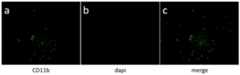
图3是小胶质细胞表面抗原CD11b免疫荧光鉴定结果图,其中a为表面抗原CD11b染色(CD11b绿色),b为核染色(dapi蓝色),c为混合(merge);Figure 3 is the result of immunofluorescence identification of microglia surface antigen CD11b, in which a is the surface antigen CD11b staining (CD11b green), b is nuclear staining (dapi blue), and c is merge (merge);
图4是脊髓损伤后不同时间段内提取的小胶质细胞形态特征图及细胞数量柱状图,其中a为正常组即脊髓未损伤,b为脊髓损伤1天后,c为脊髓损伤3天后,d为脊髓损伤7天后,e为脊髓损伤14天后;Figure 4 is the morphological characteristic map and cell number histogram of microglia extracted in different time periods after spinal cord injury, in which a is the normal group, that is, the spinal cord is not injured, b is 1 day after spinal cord injury, c is 3 days after spinal cord injury, d is 7 days after spinal cord injury, e is 14 days after spinal cord injury;
图5是脊髓损伤后不同时间段内提取的小胶质细胞数量柱状图,其中,3d与7d相比,*P<0.05;Figure 5 is a histogram of the number of microglia extracted in different time periods after spinal cord injury, in which, compared with 3d and 7d, *P<0.05;
图6是脊髓损伤3天后小胶质的形态特征图,其中a为种板后1天,b为种板后2天,c为种板后4天。Figure 6 is a graph showing the morphological characteristics of microglia 3 days after spinal cord injury, wherein a is 1 day after seeding, b is 2 days after seeding, and c is 4 days after seeding.
具体实施方式Detailed ways
以下实施例进一步说明本发明的内容,但不应理解为对本发明的限制。在不背离本发明精神和实质的情况下,对本发明方法、步骤或条件所作的修改和替换,均属于本发明的范围。若未特别指明,实施例中所用的技术手段为本领域技术人员所熟知的常规手段。The following examples further illustrate the content of the present invention, but should not be construed as limiting the present invention. Modifications and substitutions made to the methods, steps or conditions of the present invention without departing from the spirit and essence of the present invention all belong to the scope of the present invention. Unless otherwise specified, the technical means used in the examples are conventional means well known to those skilled in the art.
实施例1Example 1
1、脊髓损伤大鼠模型的制备1. Preparation of spinal cord injury rat model
测量180-200g的SPF级SD大鼠的体重,用1%戊巴比妥钠(50mg/kg)进行腹膜内麻醉;麻醉5-10min后剃除背部鼠毛,将大鼠置于已消毒的鼠板上,带上无菌手套后铺巾并用碘伏消毒背部皮肤。The body weight of SPF grade SD rats of 180-200 g was measured, and intraperitoneal anesthesia was performed with 1% sodium pentobarbital (50 mg/kg). On the mouse plate, put on sterile gloves, spread the towel and disinfect the back skin with iodophor.
以T10棘突为切口中心切开皮肤,切口大小为3cm,钝性分离筋膜、背斜方肌,以手术刀紧贴T9-11椎体棘突分离棘突旁肌群,暴露T9-11椎板后夹持T9椎体两侧,提起大鼠,使用弯头剪剪断T10-11间韧带,并沿椎板两侧剪下T10椎板,向上掀开T10椎板,暴露T10节段脊髓。The skin was incised with the spinous process of T10 as the center of the incision, the size of the incision was 3 cm, the fascia and dorsal trapezius were bluntly separated, and the paraspinous muscle group was separated with a scalpel close to the spinous process of the T9-11 vertebral body, and the T9-11 was exposed. Hold both sides of the T9 vertebral body behind the lamina, lift the rat, use elbow scissors to cut the ligament between T10-11, and cut the T10 lamina along both sides of the lamina, lift the T10 lamina upward to expose the spinal cord of the T10 segment .
采用脊髓打击器打击T10段脊髓,如图1所示,落物质量和直径分别为10g和2.5mm,下落高度和深度分别为10cm和2mm,大鼠双后肢的张力立即消除,表明该模型已成功建立,逐层缝合创口后创面消毒。术后用碘伏消毒创口及尿道口,定期按摩排尿并注意保暖。The spinal cord of the T10 segment was hit with a spinal cord striker. As shown in Figure 1, the weight and diameter of the dropped objects were 10g and 2.5mm, respectively, and the height and depth of the drop were 10cm and 2mm, respectively. The tension on both hind limbs of the rat was immediately eliminated, indicating that the model has been After successful establishment, the wound was sutured layer by layer and then the wound surface was disinfected. After the operation, the wound and urethral opening were disinfected with iodophor, and the urination was massaged regularly and kept warm.
2、小胶质细胞的分离2. Isolation of Microglia
将正常组、1天、3天、7天、14天后的上述脊髓损伤模型麻醉脱颈处死,置于75%乙醇中消毒30s,将大鼠放置在已消毒的超净台内,使用无菌手术刀沿旧伤口划开,以T10为中心用剪刀剪断上下两个椎体,取出受损区2-3cm的脊髓,PBS漂洗后暂存于6孔板内0℃的DMEM/F12(HAM)+10%胎牛血清+1%青链霉素培养基中。The above spinal cord injury models in the normal group, 1 day, 3 days, 7 days, and 14 days were anesthetized and sacrificed by de-cervical, placed in 75% ethanol for sterilization for 30s, and the rats were placed in a sterilized ultra-clean table, using sterile The scalpel cut along the old wound, and the upper and lower vertebral bodies were cut with scissors with T10 as the center. The spinal cord of 2-3 cm in the damaged area was taken out. After rinsing with PBS, it was temporarily stored in DMEM/F12 (HAM) at 0°C in a 6-well plate +10% fetal bovine serum + 1% penicillin-streptomycin medium.
用显微镊快速剥离、剔除脊膜和血管,更换上述培养基后用1ml枪头将脊髓置于40um滤网上,用20ml注射器尾部研磨组织约30s,吸取上述培养基冲洗于50ml离心管中,1000rpm离心5min后弃上清,以上述培养基重悬;Use microtweezers to quickly peel and remove meninges and blood vessels. After replacing the above medium, place the spinal cord on a 40um filter with a 1ml pipette tip, grind the tissue with the tail of a 20ml syringe for about 30s, absorb the above medium and rinse it in a 50ml centrifuge tube. After centrifugation at 1000rpm for 5min, the supernatant was discarded and resuspended with the above medium;
将重悬细胞接种于已包被的6孔板中(前一天用0.01%多聚赖氨酸包被6孔板4h,PBS洗两遍后晾干备用),37℃、5%CO2培养箱中孵育4h后换液观察细胞形态,镜下可见细胞形态呈圆形,且呈贴壁样生长,如图2所示。统计分析后发现大鼠脊髓损伤3天后,分离得到的小胶质细胞数量最多,如图4、5所示。The resuspended cells were seeded in the coated 6-well plate (the 6-well plate was coated with 0.01% poly-lysine for 4h the day before, washed twice with PBS and then air-dried for use), and cultured at 37°C, 5% CO2 After incubating in the box for 4 hours, the medium was changed to observe the cell morphology. Under the microscope, the cell morphology was round and showed adherent growth, as shown in Figure 2. After statistical analysis, it was found that the number of isolated microglia was the largest 3 days after the rat spinal cord injury, as shown in Figures 4 and 5.
3、小胶质细胞的鉴定及培养3. Identification and culture of microglia
取培养的细胞,采用CD11b免疫荧光染色法鉴定小胶质细胞。具体操作如下:The cultured cells were taken, and microglia were identified by CD11b immunofluorescence staining. The specific operations are as follows:
取出6孔板,吸弃培养基后用PBS洗涤3次,每次5min,以4%多聚甲醛固定10min,以0.2%Triton-100通透10min,并用PBS洗涤3次,每次5min。以5%山羊血清室温封闭60min,加入一抗CD11b(1:300),4℃过夜。用PBS洗涤3次,每次5min,加入二抗Alexa 488,室温避光孵育1h,PBS洗涤3次。Dapi复染后PBS洗涤,滴加抗荧光淬灭剂后荧光倒置显微镜下观察,鉴定其纯度为98%,如图3所示。The 6-well plate was taken out, the culture medium was aspirated, washed three times with PBS for 5 min each, fixed with 4% paraformaldehyde for 10 min, permeabilized with 0.2% Triton-100 for 10 min, and washed three times with PBS for 5 min each time. The cells were blocked with 5% goat serum at room temperature for 60 min, and the primary antibody CD11b (1:300) was added, and the cells were kept at 4°C overnight. Wash with PBS for 3 times, 5 min each time, add secondary antibody Alexa 488, incubate for 1 h at room temperature in the dark, and wash 3 times with PBS. After Dapi counterstaining, washing with PBS, dropwise addition of anti-fluorescence quencher, and observation under a fluorescence inverted microscope, the purity was identified as 98%, as shown in Figure 3.
取培养脊髓损伤3天后的小胶质细胞,继续培养1天、2天、4天,镜下观察发现小胶质细胞在种板1天时,细胞呈圆形且胞体较大,呈阿米巴样;种板2天、4天时,细胞的分支逐渐增多,胞体逐渐变小,如图6所示。Take the microglia cultured for 3 days after spinal cord injury, and continue to culture them for 1 day, 2 days, and 4 days. The microglia were observed under the microscope and found that the cells were round and the cell body was larger, showing the appearance of amoeba. After 2 days and 4 days of seeding, the branches of the cells gradually increased, and the cell bodies gradually became smaller, as shown in Figure 6.
综上所述,通过实施例1所述方法可高效、经济地获得数量可观、纯度高的小胶质细胞,为脊髓损伤后小胶质细胞的研究提供基础。In summary, the method described in Example 1 can efficiently and economically obtain microglia with a considerable number and high purity, which provides a basis for the study of microglia after spinal cord injury.
Claims (10)
Priority Applications (1)
| Application Number | Priority Date | Filing Date | Title |
|---|---|---|---|
| CN202011021176.7ACN112011509A (en) | 2020-09-25 | 2020-09-25 | Separation method and application of primary microglia of rat with spinal cord injury |
Applications Claiming Priority (1)
| Application Number | Priority Date | Filing Date | Title |
|---|---|---|---|
| CN202011021176.7ACN112011509A (en) | 2020-09-25 | 2020-09-25 | Separation method and application of primary microglia of rat with spinal cord injury |
Publications (1)
| Publication Number | Publication Date |
|---|---|
| CN112011509Atrue CN112011509A (en) | 2020-12-01 |
Family
ID=73527283
Family Applications (1)
| Application Number | Title | Priority Date | Filing Date |
|---|---|---|---|
| CN202011021176.7APendingCN112011509A (en) | 2020-09-25 | 2020-09-25 | Separation method and application of primary microglia of rat with spinal cord injury |
Country Status (1)
| Country | Link |
|---|---|
| CN (1) | CN112011509A (en) |
Cited By (2)
| Publication number | Priority date | Publication date | Assignee | Title |
|---|---|---|---|---|
| CN114561355A (en)* | 2022-01-23 | 2022-05-31 | 四川大学华西医院 | Acute and rapid separation method for spinal cord scar tissue cells |
| CN116240170A (en)* | 2021-12-08 | 2023-06-09 | 中国科学院苏州纳米技术与纳米仿生研究所 | A kind of culture method and application of spinal cord microglial cells |
Citations (3)
| Publication number | Priority date | Publication date | Assignee | Title |
|---|---|---|---|---|
| US20120225821A1 (en)* | 2009-08-17 | 2012-09-06 | University-Industry Cooperation Group Of Kyung Hee | Composition for preventing or treating a spinal cord injury |
| CN108251372A (en)* | 2018-01-16 | 2018-07-06 | 南京中医药大学 | Primary microglia/injured neuron co-culture system and its construction method and application |
| US20200239844A1 (en)* | 2017-02-28 | 2020-07-30 | The Regents Of The University Of California | Differentiation and use of human microglia-like cells from pluripotent stem cells and hematopoietic progenitors |
- 2020
- 2020-09-25CNCN202011021176.7Apatent/CN112011509A/enactivePending
Patent Citations (3)
| Publication number | Priority date | Publication date | Assignee | Title |
|---|---|---|---|---|
| US20120225821A1 (en)* | 2009-08-17 | 2012-09-06 | University-Industry Cooperation Group Of Kyung Hee | Composition for preventing or treating a spinal cord injury |
| US20200239844A1 (en)* | 2017-02-28 | 2020-07-30 | The Regents Of The University Of California | Differentiation and use of human microglia-like cells from pluripotent stem cells and hematopoietic progenitors |
| CN108251372A (en)* | 2018-01-16 | 2018-07-06 | 南京中医药大学 | Primary microglia/injured neuron co-culture system and its construction method and application |
Non-Patent Citations (18)
| Title |
|---|
| DASA CIZKOVA 等: "Alterations of protein composition along the rostro-caudal axis after spinal cord injury: proteomic, in vitro and in vivo analyses", 《FRONT CELL NEUROSCI》* |
| DASA CIZKOVA 等: "Alterations of protein composition along the rostro-caudal axis after spinal cord injury: proteomic, in vitro and in vivo analyses", 《FRONT CELL NEUROSCI》, no. 8, 17 April 2014 (2014-04-17), pages 105* |
| ZHAOYUN YANG 等: "Down-Regulation of miRNA-128 Contributes to Neuropathic Pain Following Spinal Cord Injury via Activation of P38", 《MED SCI MONIT》* |
| ZHAOYUN YANG 等: "Down-Regulation of miRNA-128 Contributes to Neuropathic Pain Following Spinal Cord Injury via Activation of P38", 《MED SCI MONIT》, no. 23, 23 January 2017 (2017-01-23), pages 405 - 411* |
| 吴斌等: "大鼠脊髓源性少突胶质前体细胞的生物学特性", 《华中科技大学学报(医学版)》* |
| 吴斌等: "大鼠脊髓源性少突胶质前体细胞的生物学特性", 《华中科技大学学报(医学版)》, no. 06, 15 December 2012 (2012-12-15), pages 723 - 726* |
| 李明: "大鼠实验性脊髓损伤后小胶质和少突胶质细胞的变化", 《安徽医药》* |
| 李明: "大鼠实验性脊髓损伤后小胶质和少突胶质细胞的变化", 《安徽医药》, no. 03, 30 March 2010 (2010-03-30), pages 1* |
| 潘娅岚等: "脊髓康对共培养体系中小胶质细胞吞噬及损伤神经元再生的影响", 《中国免疫学杂志》* |
| 潘娅岚等: "脊髓康对共培养体系中小胶质细胞吞噬及损伤神经元再生的影响", 《中国免疫学杂志》, no. 11, 20 November 2017 (2017-11-20), pages 1652 - 1657* |
| 王子田等: "脊髓挤压伤所致小胶质细胞的变化与血脊髓屏障的关系", 《神经解剖学杂志》* |
| 王子田等: "脊髓挤压伤所致小胶质细胞的变化与血脊髓屏障的关系", 《神经解剖学杂志》, no. 02, 31 March 2008 (2008-03-31), pages 113 - 118* |
| 钱凯: "新生大鼠原代小胶质细胞分离培养方法的改良", 临床神经外科杂志, vol. 16, no. 01, pages 1 - 5* |
| 陈雪,张婷,李彩红,王伟,骆翔,喻志源: "大鼠脊髓小胶质细胞体外纯化培养方法的建立", 神经损伤与功能重建, vol. 9, no. 06, pages 1 - 1* |
| 黄秀艳等: "人小胶质细胞的分离、纯化、特异分子表达与吞噬功能研究", 《中国病理生理杂志》* |
| 黄秀艳等: "人小胶质细胞的分离、纯化、特异分子表达与吞噬功能研究", 《中国病理生理杂志》, no. 05, 15 May 2008 (2008-05-15), pages 1 - 2* |
| 黎晓慧等: "树原代小胶质细胞的分离培养、纯化与鉴定", 《动物学杂志》* |
| 黎晓慧等: "树原代小胶质细胞的分离培养、纯化与鉴定", 《动物学杂志》, no. 03, 3 June 2019 (2019-06-03), pages 445 - 450* |
Cited By (3)
| Publication number | Priority date | Publication date | Assignee | Title |
|---|---|---|---|---|
| CN116240170A (en)* | 2021-12-08 | 2023-06-09 | 中国科学院苏州纳米技术与纳米仿生研究所 | A kind of culture method and application of spinal cord microglial cells |
| CN114561355A (en)* | 2022-01-23 | 2022-05-31 | 四川大学华西医院 | Acute and rapid separation method for spinal cord scar tissue cells |
| CN114561355B (en)* | 2022-01-23 | 2023-04-11 | 四川大学华西医院 | Acute and rapid separation method for spinal cord scar tissue cells |
Similar Documents
| Publication | Publication Date | Title |
|---|---|---|
| JP6687757B2 (en) | Methods for preparing 3D cartilage organoid blocks | |
| EP1604695A1 (en) | Amnion-origin medical material and method of preparing the same | |
| RU2323252C1 (en) | Method for culturing human mesenchymal stem cells ex vivo | |
| CN1962858A (en) | Creation of tissue engineered female reproductive organs | |
| CN106801032A (en) | The construction method of people's amnioic epithelium stem cell bank | |
| CN112011509A (en) | Separation method and application of primary microglia of rat with spinal cord injury | |
| CN105238738A (en) | Isolated culture method of piglet myocardial fibroblasts | |
| US20200392465A1 (en) | A process of preparing buccal epithelial cell suspension and its use | |
| CN109971704A (en) | A kind of preparation method of Trichophyton rubrum infection model | |
| CN104152408B (en) | The preparation method of Subaerial blue green algae | |
| WO2006003818A1 (en) | Corneal epithelial sheet and process for producing the same | |
| CN106377799A (en) | Preparation method of dental pulp stem cell and chitosan scaffold complex | |
| CN104877955A (en) | Method for separating and purifying rat dermal microvascular endothelium cells | |
| CN110295137B (en) | Channa argus kidney cell line and construction method and application thereof | |
| CN109294972A (en) | A method for in vitro culture of lamb small intestine epithelial cells | |
| JP2002534068A (en) | Transplant animal model for high induction of papilloma, propagation of papillomavirus, and evaluation of candidate therapeutics | |
| EP1568386A1 (en) | Method of organ regeneration | |
| CN110373381A (en) | A kind of preparation method by the efficient placenta mesenchyma stem cell of homogenizer | |
| CN116875534A (en) | Method for improving expression level of Sox2 in DP (DP) cells | |
| CN110464877A (en) | A kind of preparation method and its effect evaluation method of acellular nerve allografts | |
| CN117050937A (en) | Method for extracting primary cells of terminal bud base of salamander | |
| CN109777774A (en) | Preparation method of macrophage-derived extracellular matrix | |
| CN116836912A (en) | Lateolabrax japonicus kistrodon cell line and construction method and application thereof | |
| CN102168062B (en) | Human pulmonary artery smooth muscle cell separation and culturing method and application of same | |
| CN112501115B (en) | A method for extracting, separating and purifying rabbit muscle stem cells |
Legal Events
| Date | Code | Title | Description |
|---|---|---|---|
| PB01 | Publication | ||
| PB01 | Publication | ||
| SE01 | Entry into force of request for substantive examination | ||
| SE01 | Entry into force of request for substantive examination | ||
| RJ01 | Rejection of invention patent application after publication | Application publication date:20201201 | |
| RJ01 | Rejection of invention patent application after publication |