CN111986181A - Intravascular stent image segmentation method and system based on double-attention machine system - Google Patents
Intravascular stent image segmentation method and system based on double-attention machine systemDownload PDFInfo
- Publication number
- CN111986181A CN111986181ACN202010859488.9ACN202010859488ACN111986181ACN 111986181 ACN111986181 ACN 111986181ACN 202010859488 ACN202010859488 ACN 202010859488ACN 111986181 ACN111986181 ACN 111986181A
- Authority
- CN
- China
- Prior art keywords
- attention
- feature
- block
- dual
- decoding
- Prior art date
- Legal status (The legal status is an assumption and is not a legal conclusion. Google has not performed a legal analysis and makes no representation as to the accuracy of the status listed.)
- Granted
Links
Images


Classifications
- G—PHYSICS
- G06—COMPUTING OR CALCULATING; COUNTING
- G06T—IMAGE DATA PROCESSING OR GENERATION, IN GENERAL
- G06T7/00—Image analysis
- G06T7/0002—Inspection of images, e.g. flaw detection
- G06T7/0012—Biomedical image inspection
- G—PHYSICS
- G06—COMPUTING OR CALCULATING; COUNTING
- G06N—COMPUTING ARRANGEMENTS BASED ON SPECIFIC COMPUTATIONAL MODELS
- G06N3/00—Computing arrangements based on biological models
- G06N3/02—Neural networks
- G06N3/04—Architecture, e.g. interconnection topology
- G06N3/045—Combinations of networks
- G—PHYSICS
- G06—COMPUTING OR CALCULATING; COUNTING
- G06N—COMPUTING ARRANGEMENTS BASED ON SPECIFIC COMPUTATIONAL MODELS
- G06N3/00—Computing arrangements based on biological models
- G06N3/02—Neural networks
- G06N3/08—Learning methods
- G—PHYSICS
- G06—COMPUTING OR CALCULATING; COUNTING
- G06T—IMAGE DATA PROCESSING OR GENERATION, IN GENERAL
- G06T7/00—Image analysis
- G06T7/10—Segmentation; Edge detection
- G06T7/11—Region-based segmentation
- G—PHYSICS
- G06—COMPUTING OR CALCULATING; COUNTING
- G06T—IMAGE DATA PROCESSING OR GENERATION, IN GENERAL
- G06T2207/00—Indexing scheme for image analysis or image enhancement
- G06T2207/10—Image acquisition modality
- G06T2207/10072—Tomographic images
- G06T2207/10081—Computed x-ray tomography [CT]
- G—PHYSICS
- G06—COMPUTING OR CALCULATING; COUNTING
- G06T—IMAGE DATA PROCESSING OR GENERATION, IN GENERAL
- G06T2207/00—Indexing scheme for image analysis or image enhancement
- G06T2207/10—Image acquisition modality
- G06T2207/10116—X-ray image
- G—PHYSICS
- G06—COMPUTING OR CALCULATING; COUNTING
- G06T—IMAGE DATA PROCESSING OR GENERATION, IN GENERAL
- G06T2207/00—Indexing scheme for image analysis or image enhancement
- G06T2207/20—Special algorithmic details
- G06T2207/20081—Training; Learning
- G—PHYSICS
- G06—COMPUTING OR CALCULATING; COUNTING
- G06T—IMAGE DATA PROCESSING OR GENERATION, IN GENERAL
- G06T2207/00—Indexing scheme for image analysis or image enhancement
- G06T2207/20—Special algorithmic details
- G06T2207/20084—Artificial neural networks [ANN]
- G—PHYSICS
- G06—COMPUTING OR CALCULATING; COUNTING
- G06T—IMAGE DATA PROCESSING OR GENERATION, IN GENERAL
- G06T2207/00—Indexing scheme for image analysis or image enhancement
- G06T2207/30—Subject of image; Context of image processing
- G06T2207/30004—Biomedical image processing
- G06T2207/30101—Blood vessel; Artery; Vein; Vascular
Landscapes
- Engineering & Computer Science (AREA)
- Theoretical Computer Science (AREA)
- Physics & Mathematics (AREA)
- General Physics & Mathematics (AREA)
- General Health & Medical Sciences (AREA)
- Health & Medical Sciences (AREA)
- Computing Systems (AREA)
- Mathematical Physics (AREA)
- Data Mining & Analysis (AREA)
- Evolutionary Computation (AREA)
- Biophysics (AREA)
- Molecular Biology (AREA)
- Biomedical Technology (AREA)
- General Engineering & Computer Science (AREA)
- Artificial Intelligence (AREA)
- Computational Linguistics (AREA)
- Software Systems (AREA)
- Life Sciences & Earth Sciences (AREA)
- Computer Vision & Pattern Recognition (AREA)
- Medical Informatics (AREA)
- Nuclear Medicine, Radiotherapy & Molecular Imaging (AREA)
- Radiology & Medical Imaging (AREA)
- Quality & Reliability (AREA)
- Image Analysis (AREA)
Abstract
Description
Translated fromChinese技术领域technical field
本发明属于图像识别领域,具体涉及了一种基于双注意力机制的血管内支架图像分割方法和系统。The invention belongs to the field of image recognition, and in particular relates to a method and system for image segmentation of intravascular stents based on a dual attention mechanism.
背景技术Background technique
腹部主动脉瘤(Abdominal aortic aneurysm,AAA)是最常见的一种动脉瘤。通常腹部主动脉瘤在破裂之前不会出现典型症状,因此通常导致85%到90%的病死率。临床研究表明,比起开放式修复方法,血管内动脉瘤修复(Endovascular aneurysm repair,EVAR)手术可以有效降低患者在围手术期的发病率和死亡率,并维持相同程度的术后存活率。然而,由于EVAR手术的复杂性,在介入手术的过程中通常需要长时间的辐射和大剂量的造影剂注射,进而可能会导致病人出现如肾衰竭之类的常见并发症。因此,减少EVAR手术的时间是非常必要的。Abdominal aortic aneurysm (AAA) is the most common type of aneurysm. Abdominal aortic aneurysms typically do not present with typical symptoms until they rupture, and thus usually result in an 85% to 90% case fatality rate. Clinical studies have shown that endovascular aneurysm repair (EVAR) surgery can effectively reduce perioperative morbidity and mortality, and maintain the same degree of postoperative survival compared with open repair methods. However, due to the complexity of EVAR surgery, long-term radiation and high-dose contrast injection are usually required during interventional procedures, which may lead to common complications such as renal failure in patients. Therefore, it is very necessary to reduce the time of EVAR surgery.
在介入手术中,融合术前数据(CT图像)和术中X光造影能够降低造影剂和辐射的使用剂量。然而,这个融合可能会因为病人的移动和介入器械造成的血管形变而变得不准确。为了避免重复使用造影剂,在手术过程中,对比血管内支架的分割图像和术前数据可以评估检测当前的融合效果。然而,血管内支架分割目前有以下几部分难点:(1)不同介入手术中采用的支架具有不同的形态学特征,例如大小和形状;(2)由于支架的像素数量远小于背景的像素数量,前景和背景类别数量极度不平均;(3)造影剂和其他一些丝状结构(例如脊椎、导丝等)会干扰支架的边缘像素的分类准确性。In interventional procedures, the fusion of preoperative data (CT images) and intraoperative X-ray contrast can reduce the dose of contrast agents and radiation used. However, this fusion may become inaccurate due to patient movement and vessel deformation caused by interventional instruments. In order to avoid repeated use of contrast agents, during the operation, comparing the segmented images of the endovascular stent with the preoperative data can evaluate and detect the current fusion effect. However, endovascular stent segmentation currently has the following difficulties: (1) stents used in different interventional procedures have different morphological characteristics, such as size and shape; (2) because the number of pixels of the stent is much smaller than that of the background, The number of foreground and background categories is extremely uneven; (3) contrast agents and some other filamentary structures (eg, spine, guide wire, etc.) can interfere with the classification accuracy of the edge pixels of the stent.
目前针对血管内支架分割的研究相对较少。Demirci等人提出了一种基于模型的方法。该方法的数据预处理部分采用了一种基于海森矩阵的滤波器。尽管该方法能直接复原支架的三维形状,它需要提前定义支架的模型,因此在支架的选择上有一定的局限性。At present, there are relatively few studies on endovascular stent segmentation. A model-based approach was proposed by Demirci et al. The data preprocessing part of the method adopts a filter based on Hessian matrix. Although this method can directly restore the three-dimensional shape of the stent, it needs to define the stent model in advance, so there are certain limitations in the selection of stents.
近年来,深度学习在医学图像处理领域里大放异彩。Breininger等人提出了一种具有收缩和舒张模式的全卷积网络,用于动脉血管支架的分割。然而,由于该方法在主干部分对残差模块的应用,其实时性能不是很好。In recent years, deep learning has shined in the field of medical image processing. Breininger et al. proposed a fully convolutional network with systolic and diastolic modes for segmentation of arterial vascular stents. However, due to the application of the residual module in the backbone part of the method, its real-time performance is not very good.
发明内容SUMMARY OF THE INVENTION
为了解决现有技术中的上述问题,即现有技术无法实时并且精确地从手术中X光透射图像中将血管内支架分割出来的问题,本发明提供了一种基于双注意力机制的血管内支架图像分割方法,所述方法包括:In order to solve the above-mentioned problem in the prior art, that is, the prior art cannot segment the intravascular stent from the X-ray transmission image during the operation in real time and accurately, the present invention provides an intravascular stent based on a dual attention mechanism. A stent image segmentation method, the method comprising:
步骤S10,获取手术过程中包含支架的区域的X光透射视频序列作为待检测视频序列;Step S10, acquiring the X-ray transmission video sequence of the region including the stent during the operation as the video sequence to be detected;
步骤S20,基于所述待检测视频序列,通过训练好的基于深度学习的轻量化双注意力融合网络生成显示血管内支架的二值分割掩膜序列;Step S20, based on the video sequence to be detected, generate a binary segmentation mask sequence showing the intravascular stent through the trained lightweight dual-attention fusion network based on deep learning;
步骤S30,将所述二值分割掩膜序列覆盖在所述待检测视屏序列上获得血管内支架的视频序列;Step S30, overlaying the binary segmentation mask sequence on the to-be-detected video sequence to obtain a video sequence of the intravascular stent;
其中,所述基于深度学习的轻量化双注意力融合网络包括顺次连接的第一卷积层、多级嵌套的编码解码结构和第二卷积层;所述多级嵌套的编码解码结构在各级编码器与解码器之间嵌套插入下一级编码解码结构,在最下一级的编码解码结构的编码模块和解码模块之间插入一个特征注意力块;Wherein, the lightweight dual-attention fusion network based on deep learning includes a first convolution layer connected in sequence, a multi-level nested encoding and decoding structure and a second convolution layer; the multi-level nested encoding and decoding structure The structure inserts the next-level encoding-decoding structure between the encoder and the decoder at each level, and inserts a feature attention block between the encoding module and the decoding module of the encoding-decoding structure at the lowest level;
所述多级嵌套的编码解码结构,其编码器和解码器分别包括多个编码模块和多个解码模块;所述编码模块通过残差连接与对应的同级解码模块连接;In the multi-level nested encoding and decoding structure, the encoder and the decoder respectively include a plurality of encoding modules and a plurality of decoding modules; the encoding modules are connected with corresponding decoding modules of the same level through residual connection;
所述编码模块基于MobileNetV2网络构建,在MobileNetV2中将标准卷积层替换为深度可分卷积层;The encoding module is constructed based on the MobileNetV2 network, and the standard convolutional layer is replaced by a depthwise separable convolutional layer in MobileNetV2;
所述解码模块由解码块和关联注意力块组成。The decoding module consists of decoding blocks and associated attention blocks.
进一步地,所述训练好的基于深度学习的轻量化双注意力融合网络,其获得方法包括:Further, the method for obtaining the trained lightweight dual-attention fusion network based on deep learning includes:
步骤A100,获取训练视频序列,按时间顺序提取所述训练视频序列中的第t帧作为当前帧;Step A100, obtaining a training video sequence, and extracting the t-th frame in the training video sequence in chronological order as a current frame;
步骤A200,基于所述当前帧,通过所述第一卷积层生成第一特征图像;Step A200, based on the current frame, generate a first feature image through the first convolutional layer;
步骤A300,基于所述第一特征图像,通过所述基于深度学习的轻量化双注意力融合网络的编码器中的编码模块进行分级编码,获得特征压缩图像;Step A300, based on the first feature image, perform hierarchical coding through the coding module in the encoder of the deep learning-based lightweight dual-attention fusion network, to obtain a feature compressed image;
步骤A400,通过所述特征注意力模块获取所述特征压缩图像三个不同尺度的特征,结合所述不同尺度的特征生成编码特征向量;Step A400, obtaining features of three different scales of the feature compressed image through the feature attention module, and generating an encoded feature vector in combination with the features of the different scales;
步骤A500,基于所述编码特征向量,通过所述基于深度学习的轻量化双注意力融合网络中的解码器中的解码块,进行分级解码,每级解码后通过解码器中的所述关联注意力块凸显支架特征以及消除残差连接中的不相关响应和噪声响应,生成上采样特征图像;Step A500, based on the encoded feature vector, perform hierarchical decoding through the decoding block in the decoder in the light-weight dual-attention fusion network based on deep learning, and pass the associated attention in the decoder after each level of decoding. The force block highlights bracket features and eliminates irrelevant and noisy responses in residual connections to generate upsampled feature images;
步骤A600,基于所述上采样特征图像,通过所述基于深度学习的轻量化双注意力融合网络的第二卷积层生成所述当前帧对应的支架的二值分割掩膜;Step A600, based on the up-sampled feature image, generate a binary segmentation mask of the bracket corresponding to the current frame through the second convolution layer of the deep learning-based lightweight dual-attention fusion network;
步骤A700,若网络全局损失函数不低于设定阈值,则通过随机梯度下降法调节特征注意力块的注意力系数、关联注意力块的激活函数及权重和网络参数,令t=t+1并跳转步骤A100,直至全局损失函数小于预设的阈值,获得训练好的基于轻量化双注意力融合网络。Step A700, if the global loss function of the network is not lower than the set threshold, adjust the attention coefficient of the feature attention block, the activation function of the associated attention block, the weight and the network parameters through the stochastic gradient descent method, so that t=t+1 And jump to step A100 until the global loss function is less than the preset threshold, and the trained lightweight dual-attention fusion network is obtained.
进一步地,所述特征注意力块输入端连接特征注意力块第一支线、特征注意力块第二支线和特征注意力块第三支线;Further, the input end of the feature attention block is connected to the first branch line of the feature attention block, the second branch line of the feature attention block and the third branch line of the feature attention block;
所述特征注意力块第一支线,为与所述特征注意力块输入端顺次连接的自适应平均池化层、卷积核为1×1的卷积层和上采样层;The first branch of the feature attention block is an adaptive average pooling layer, a convolution layer with a convolution kernel of 1×1, and an upsampling layer sequentially connected to the input end of the feature attention block;
所述特征注意力块第二支线,为与所述特征注意力块输入端连接的卷积核为1×1的卷积层;The second branch of the feature attention block is a convolution layer with a 1×1 convolution kernel connected to the input end of the feature attention block;
所述特征注意力块第三支线,为与所述特征注意力块输入端顺次连接的呈U型结构的卷积核分别为7×7的卷积层、卷积核为5×5的卷积层和卷积核为3×3的卷积层;The third branch of the feature attention block is a convolution kernel with a U-shaped structure connected in sequence with the input end of the feature attention block, which is a 7×7 convolution layer and a 5×5 convolution kernel The convolution layer and the convolution kernel are 3×3 convolution layers;
特征注意力块第三支线与特征注意力块第二支线的输出做乘法运算连接后与特征注意力块第一支线的输出做加法运算。The output of the third branch of the feature attention block and the output of the second branch of the feature attention block are multiplied and connected, and then added with the output of the first branch of the feature attention block.
进一步地,所述关联注意力块输入端连关联注意力块第一支线和关联注意力块第二支线;Further, the input end of the associated attention block is connected to the first branch of the associated attention block and the second branch of the associated attention block;
所述关联注意力块第一支线,为与关联注意力块输入端顺次连接的卷积核为3×3的卷积层和卷积核为1×1的卷积层;The first branch of the associated attention block is a convolution layer with a convolution kernel of 3×3 and a convolution layer with a convolution kernel of 1×1 sequentially connected to the input end of the associated attention block;
所述关联注意力块第二支线,为与关联注意力块输入端连接的卷积核为1×1的卷积层;The second branch of the associated attention block is a convolution layer with a 1×1 convolution kernel connected to the input end of the associated attention block;
其中,关联注意力块的输入信息通过所述关联注意力块第一支线获取关键特征图,通过所述关联注意力块第二支线获取通用大小特征图;将所述关键特征图和通用大小特征图结合生成增强特征图;将所述增强特征图进行线性修正、线性变化、二分类和重采样后与关联注意力块的输入信息结合注意力系数作乘法运算生成上采样特征图像。Wherein, the input information of the associated attention block obtains the key feature map through the first branch of the associated attention block, and obtains the general size feature map through the second branch of the associated attention block; the key feature map and the general size feature are combined. The enhanced feature map is combined to generate an enhanced feature map; the enhanced feature map is subjected to linear correction, linear change, binary classification and resampling, and the input information of the associated attention block is combined with the attention coefficient to perform a multiplication operation to generate an up-sampled feature image.
进一步地,上采样特征图像

其中,


进一步地,关联注意力块通过门向量gi判断每个向量i是否处于焦点区域;所述门向量由环境信息删除底层特征响应组成。Further, the associated attention block judges whether each vector i is in the focus area through the gate vector gi ; the gate vector is composed of the environmental information deletion underlying feature response.
进一步地,所述关联注意力块的注意力系数

其中,σ1表示线性修正的激活函数,σ2表示二分类的激活函数,gi表示门向量,x为上采样特征图像,i为像素标号,

进一步地,最终注意力系数

进一步地,所述全局损失函数L为:Further, the global loss function L is:
L=LR-Focal+λLDiceL=LR-Focal +λLDice
其中,LDice是Dice系数损失函数,LR-Focal是聚焦损失函数,λ为用于调整聚焦损失和Dice系数损失之间平衡的超参数。where LDice is the Dice coefficient loss function, LR-Focal is the focus loss function, and λ is a hyperparameter used to adjust the balance between focus loss and Dice coefficient loss.
进一步地,所述聚焦损失函数LR-Focal为:Further, the focusing loss function LR-Focal is:

其中,yi是第i个像素的标签,1代表支架,0代表背景,pi是第i个像素的预测概率值,权重因子β和调制因子γ大于等于0。Among them,yi is the label of the ith pixel, 1 represents the bracket, 0 represents the background, pi is the predicted probability value of theith pixel, and the weight factor β and modulation factor γ are greater than or equal to 0.
本发明的另一方面,提出了一种基于双注意力机制的血管内支架图像分割系统,所述系统包括待测视频获取单元、掩膜生成单元和支架显现单元;In another aspect of the present invention, an intravascular stent image segmentation system based on a dual attention mechanism is proposed, the system includes a video acquisition unit to be tested, a mask generation unit and a stent display unit;
所述视频获取单元,用于获取手术过程中包含支架的区域的X光透射视频序列作为待检测视频序列;The video acquisition unit is used to acquire the X-ray transmission video sequence of the region including the stent during the operation as the video sequence to be detected;
所述掩膜生成单元,用于基于所述待检测视频序列,通过训练好的基于深度学习的轻量化双注意力融合网络生成显示血管内支架的二值分割掩膜序列;The mask generation unit is configured to generate a binary segmentation mask sequence showing the intravascular stent through the trained lightweight dual-attention fusion network based on deep learning based on the video sequence to be detected;
所述支架显现单元,用于将所述二值分割掩膜序列覆盖在所述待检测视屏序列上获得血管内支架的视频序列;the stent presentation unit, configured to overlay the binary segmentation mask sequence on the to-be-detected video sequence to obtain a video sequence of the intravascular stent;
其中,所述基于深度学习的轻量化双注意力融合网络包括顺次连接的第一卷积层、多级嵌套的编码解码结构和第二卷积层;所述多级嵌套的编码解码结构在各级编码器与解码器之间嵌套插入下一级编码解码结构,在最下一级的编码解码结构的编码模块和解码模块之间插入一个特征注意力块;Wherein, the lightweight dual-attention fusion network based on deep learning includes a first convolution layer connected in sequence, a multi-level nested encoding and decoding structure and a second convolution layer; the multi-level nested encoding and decoding structure The structure inserts the next-level encoding-decoding structure between the encoder and the decoder at each level, and inserts a feature attention block between the encoding module and the decoding module of the encoding-decoding structure at the lowest level;
所述多级嵌套的编码解码结构,其编码器和解码器分别包括多个编码模块和多个解码模块;所述编码模块通过残差连接与对应的同级解码模块连接;In the multi-level nested encoding and decoding structure, the encoder and the decoder respectively include a plurality of encoding modules and a plurality of decoding modules; the encoding modules are connected with corresponding decoding modules of the same level through residual connection;
所述编码模块基于MobileNetV2网络构建,在MobileNetV2中将标准卷积层替换为深度可分卷积层;The encoding module is constructed based on the MobileNetV2 network, and the standard convolutional layer is replaced by a depthwise separable convolutional layer in MobileNetV2;
所述解码模块由解码块和关联注意力块组成。The decoding module consists of decoding blocks and associated attention blocks.
本发明的第三方面,提出了一种存储装置,其中存储有多条程序,所述程序适于由处理器加载并执行以实现上述的基于双注意力机制的血管内支架图像分割方法。In a third aspect of the present invention, a storage device is provided, wherein a plurality of programs are stored, and the programs are adapted to be loaded and executed by a processor to realize the above-mentioned method for segmenting an intravascular stent image based on a dual-attention mechanism.
本发明的第四方面,提出了一种处理装置,包括处理器、存储装置;所述处理器,适于执行各条程序;所述存储装置,适于存储多条程序;所述程序适于由处理器加载并执行以实现上述的基于双注意力机制的血管内支架图像分割方法。In a fourth aspect of the present invention, a processing device is provided, including a processor and a storage device; the processor is suitable for executing various programs; the storage device is suitable for storing multiple programs; the program is suitable for It is loaded and executed by the processor to realize the above-mentioned method for segmentation of intravascular stent images based on the dual attention mechanism.
本发明的有益效果:Beneficial effects of the present invention:
(1)本发明基于双注意力机制的血管内支架图像分割方法,采用轻量化双注意力融合网络中的特征注意力块通过下采样和上采样逐步结合不同尺度的信息更准确地获取相关特征,还采用自适应平均化层进一步改善模型表现,提高了现有的图像分割方法用于识别血管内支架图像的精确度。(1) The image segmentation method for intravascular stents based on the dual attention mechanism of the present invention adopts the feature attention blocks in the lightweight dual attention fusion network to gradually combine information of different scales through downsampling and upsampling to obtain relevant features more accurately , and an adaptive averaging layer is used to further improve the model performance and improve the accuracy of existing image segmentation methods for identifying intravascular stent images.
(2)本发明基于双注意力机制的血管内支架图像分割方法,采用轻量化双注意力融合网络中的关联注意力块将每一级输入的信息分成关键特征图和通用大小特征图并将两部分结合起来提高非线性特征并滤去不相关信息以及残差连接中的噪音,提高了图像分割方法的精确度。(2) The present invention is based on the dual-attention mechanism-based image segmentation method for intravascular stents. The associated attention blocks in the lightweight dual-attention fusion network are used to divide the input information of each level into key feature maps and general-size feature maps, and then use the associated attention blocks in the lightweight dual-attention fusion network to divide the input information of each level into a key feature map and a general size feature map. The two parts are combined to improve nonlinear features and filter out irrelevant information and noise in residual connections, which improves the accuracy of image segmentation methods.
(3)本发明基于双注意力机制的血管内支架图像分割方法,采用Dice损失函数替换交叉熵损失函数解决了在血管内支架细长的结构导致的间类数据不平衡的问题,避免了由于造影剂和丝状结构的干扰导致的支架边缘像素被误分类的问题。(3) The image segmentation method of the intravascular stent based on the dual attention mechanism of the present invention adopts the Dice loss function to replace the cross-entropy loss function to solve the problem of unbalanced data between categories caused by the slender structure of the intravascular stent, and avoids the problem of The problem of misclassification of stent edge pixels due to interference from contrast agents and filamentous structures.
(4)本发明基于双注意力机制的血管内支架图像分割方法,通过聚焦损失中的调制因子可以自动减少简单数据的权重,并迅速聚焦于错分类的数据,提高了血管内支架图像识别的精确度。(4) The image segmentation method of intravascular stent based on the dual attention mechanism of the present invention can automatically reduce the weight of simple data through the modulation factor in the focus loss, and quickly focus on the misclassified data, which improves the recognition efficiency of intravascular stent images. Accuracy.
(5)本发明基于双注意力机制的血管内支架图像分割方法,采用轻量化的网络生成显示支架的二值分割掩膜,无需复杂的启发信息,提高了图像处理的速度,速度可达12.6fps,满足了血管内支架手术的实时性要求。(5) The image segmentation method of the intravascular stent based on the dual attention mechanism of the present invention adopts a lightweight network to generate a binary segmentation mask for displaying the stent, without complex heuristic information, and improves the speed of image processing, and the speed can reach 12.6 fps, which meets the real-time requirements of endovascular stent surgery.
附图说明Description of drawings
通过阅读参照以下附图所作的对非限制性实施例所作的详细描述,本申请的其它特征、目的和优点将会变得更明显:Other features, objects and advantages of the present application will become more apparent by reading the detailed description of non-limiting embodiments made with reference to the following drawings:
图1是本发明基于双注意力机制的血管内支架图像分割方法第一实施例的流程示意图;FIG. 1 is a schematic flowchart of a first embodiment of an intravascular stent image segmentation method based on a dual attention mechanism of the present invention;
图2是本发明第一实施例的基于深度学习的轻量化双注意力融合网络结构示意图;2 is a schematic structural diagram of a lightweight dual-attention fusion network based on deep learning according to the first embodiment of the present invention;
图3是本发明第一实施例的基于深度学习的轻量化双注意力融合网络中的特征注意力块结构示意图;3 is a schematic structural diagram of a feature attention block in a lightweight dual attention fusion network based on deep learning according to the first embodiment of the present invention;
图4是本发明第一实施例的基于深度学习的轻量化双注意力融合网络中的关联注意力块结构示意图;4 is a schematic structural diagram of an associated attention block in a lightweight dual-attention fusion network based on deep learning according to the first embodiment of the present invention;
图5是本发明第一实施例的分割不同支架的效果图;5 is an effect diagram of dividing different brackets according to the first embodiment of the present invention;
图6是移除了双注意力机制的分割支架效果图;Figure 6 is the effect diagram of the split bracket with the dual attention mechanism removed;
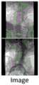
图7是本发明在数据集PUGSeg进行对比实验中的获取到的图像;Fig. 7 is the image that the present invention obtains in the data set PUGSeg to carry out the contrast experiment;
图8是本发明在数据集PUGSeg进行对比实验中的通过GT方式分割的导管示意图;8 is a schematic diagram of the catheter segmented by GT method in the comparative experiment of the data set PUGSeg of the present invention;
图9是本发明在数据集PUGSeg进行对比试验中通过U-Net方式分割的导管示意图;9 is a schematic diagram of a catheter segmented by U-Net mode in the data set PUGSeg performing a comparative test according to the present invention;
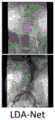
图10是本发明在数据集PUGSeg进行对比实验中通过本发明提出的LDA-Net分割的导管示意图;FIG. 10 is a schematic diagram of a catheter segmented by the LDA-Net proposed by the present invention in the comparative experiment of the data set PUGSeg of the present invention;
图11是本发明在NLM胸部X光数据集进行对比实验中获取到的图像示意图;11 is a schematic diagram of an image obtained by the present invention in a comparison experiment performed on an NLM chest X-ray dataset;
图12是本发明在NLM胸部X光数据集进行对比实验中通过GT方式获取的捕捉轮廓效果示意图;12 is a schematic diagram of the capture contour effect obtained by the GT method in the comparative experiment of the NLM chest X-ray dataset according to the present invention;
图13是本发明在NLM胸部X光数据集进行对比实验中通过Att.U-Net方式获取的捕捉轮廓效果示意图;13 is a schematic diagram of the capture contour effect obtained by the Att.U-Net method in the comparative experiment of the NLM chest X-ray data set of the present invention;
图14是本发明在NLM胸部X光数据集进行对比实验中通过本发明提出的LDA-Net方式获取的捕捉轮廓效果示意图。FIG. 14 is a schematic diagram of the capture contour effect obtained by the LDA-Net method proposed by the present invention in the comparative experiment of the NLM chest X-ray dataset of the present invention.
具体实施方式Detailed ways
下面结合附图和实施例对本申请作进一步的详细说明。可以理解的是,此处所描述的具体实施例仅用于解释相关发明,而非对该发明的限定。另外还需要说明的是,为了便于描述,附图中仅示出了与有关发明相关的部分。The present application will be further described in detail below with reference to the accompanying drawings and embodiments. It should be understood that the specific embodiments described herein are only used to explain the related invention, but not to limit the invention. In addition, it should be noted that, for the convenience of description, only the parts related to the related invention are shown in the drawings.
需要说明的是,在不冲突的情况下,本申请中的实施例及实施例中的特征可以相互组合。下面将参考附图并结合实施例来详细说明本申请。It should be noted that the embodiments in the present application and the features of the embodiments may be combined with each other in the case of no conflict. The present application will be described in detail below with reference to the accompanying drawings and in conjunction with the embodiments.
本发明提供一种基于双注意力机制的血管内支架图像分割方法,本方法包括:The present invention provides an intravascular stent image segmentation method based on a dual attention mechanism, the method comprising:
步骤S10,获取手术过程中包含支架的区域的X光透射视频序列作为待检测视频序列;Step S10, acquiring the X-ray transmission video sequence of the region including the stent during the operation as the video sequence to be detected;
步骤S20,基于所述待检测视频序列,通过训练好的基于深度学习的轻量化双注意力融合网络生成显示血管内支架的二值分割掩膜序列;Step S20, based on the video sequence to be detected, generate a binary segmentation mask sequence showing the intravascular stent through the trained lightweight dual-attention fusion network based on deep learning;
步骤S30,将所述二值分割掩膜序列覆盖在所述待检测视屏序列上获得血管内支架的视频序列;Step S30, overlaying the binary segmentation mask sequence on the to-be-detected video sequence to obtain a video sequence of the intravascular stent;
其中,所述基于深度学习的轻量化双注意力融合网络包括顺次连接的第一卷积层、多级嵌套的编码解码结构和第二卷积层;所述多级嵌套的编码解码结构在各级编码器与解码器之间嵌套插入下一级编码解码结构,在最下一级的编码解码结构的编码模块和解码模块之间插入一个特征注意力块;Wherein, the lightweight dual-attention fusion network based on deep learning includes a first convolution layer connected in sequence, a multi-level nested encoding and decoding structure and a second convolution layer; the multi-level nested encoding and decoding structure The structure inserts the next-level encoding-decoding structure between the encoder and the decoder at each level, and inserts a feature attention block between the encoding module and the decoding module of the encoding-decoding structure at the lowest level;
所述多级嵌套的编码解码结构,其编码器和解码器分别包括多个编码模块和多个解码模块;所述编码模块通过残差连接与对应的同级解码模块连接;In the multi-level nested encoding and decoding structure, the encoder and the decoder respectively include a plurality of encoding modules and a plurality of decoding modules; the encoding modules are connected with corresponding decoding modules of the same level through residual connection;
所述编码模块基于MobileNetV2网络构建,在MobileNetV2中将标准卷积层替换为深度可分卷积层;The encoding module is constructed based on the MobileNetV2 network, and the standard convolutional layer is replaced by a depthwise separable convolutional layer in MobileNetV2;
所述解码模块由解码块和关联注意力块组成。The decoding module consists of decoding blocks and associated attention blocks.
为了更清晰地对本发明基于双注意力机制的血管内支架图像分割方法进行说明,下面结合图1对本发明方法实施例中各步骤展开详述。In order to more clearly describe the image segmentation method of the intravascular stent based on the dual attention mechanism of the present invention, each step in the embodiment of the method of the present invention will be described in detail below with reference to FIG. 1 .
本发明一种实施例的基于双注意力机制的血管内支架图像分割方法,包括步骤S10-步骤S30,各步骤详细描述如下:A method for segmenting an intravascular stent image based on a dual attention mechanism according to an embodiment of the present invention includes steps S10 to S30, and each step is described in detail as follows:
步骤S10,获取手术过程中包含支架的区域的X光透射视频序列作为待检测视频序列;Step S10, acquiring the X-ray transmission video sequence of the region including the stent during the operation as the video sequence to be detected;
步骤S20,基于所述待检测视频序列,通过训练好的基于深度学习的轻量化双注意力融合网络生成显示血管内支架的二值分割掩膜序列;Step S20, based on the video sequence to be detected, generate a binary segmentation mask sequence showing the intravascular stent through the trained lightweight dual-attention fusion network based on deep learning;
在本实施例中,基于深度学习的轻量化双注意力融合网络结构如图2所示,在图2中,1为第一卷积层,2、3、4和5为编码模块,6为特征注意力块,7、9、11和13为解码块,8、10、12和14为关联注意力块,15为第二卷积层。In this embodiment, the structure of the lightweight dual-attention fusion network based on deep learning is shown in Figure 2. In Figure 2, 1 is the first convolutional layer, 2, 3, 4 and 5 are the encoding modules, and 6 is the Feature attention blocks, 7, 9, 11, and 13 are decoding blocks, 8, 10, 12, and 14 are associated attention blocks, and 15 is the second convolutional layer.
所述基于深度学习的轻量化双注意力融合网络,其训练方法为:The training method of the deep learning-based lightweight dual-attention fusion network is:
步骤A100,获取训练视频序列,按时间顺序提取所述训练视频序列中的第t帧作为当前帧;Step A100, obtaining a training video sequence, and extracting the t-th frame in the training video sequence in chronological order as a current frame;
步骤A200,基于所述当前帧,通过所述第一卷积层生成第一特征图像;Step A200, based on the current frame, generate a first feature image through the first convolutional layer;
步骤A300,基于所述第一特征图像,通过所述基于深度学习的轻量化双注意力融合网络的编码器中的编码模块进行分级编码,获得特征压缩图像;Step A300, based on the first feature image, perform hierarchical coding through the coding module in the encoder of the deep learning-based lightweight dual-attention fusion network, to obtain a feature compressed image;
步骤A400,通过所述特征注意力模块获取所述特征压缩图像三个不同尺度的特征,结合所述不同尺度的特征生成编码特征向量;Step A400, obtaining features of three different scales of the feature compressed image through the feature attention module, and generating an encoded feature vector in combination with the features of the different scales;
在本实施例中,如图4所示,所述特征注意力块输入端连接特征注意力块第一支线、特征注意力块第二支线和特征注意力块第三支线;In this embodiment, as shown in FIG. 4 , the input end of the feature attention block is connected to the first branch line of the feature attention block, the second branch line of the feature attention block, and the third branch line of the feature attention block;
所述特征注意力块第一支线,为与所述特征注意力块输入端顺次连接的自适应平均池化层、卷积核为1×1的卷积层和上采样层;The first branch of the feature attention block is an adaptive average pooling layer, a convolution layer with a convolution kernel of 1×1, and an upsampling layer sequentially connected to the input end of the feature attention block;
所述特征注意力块第二支线,为与所述特征注意力块输入端连接的卷积核为1×1的卷积层;The second branch of the feature attention block is a convolution layer with a 1×1 convolution kernel connected to the input end of the feature attention block;
所述特征注意力块第三支线,为与所述特征注意力块输入端顺次连接的呈U型结构的卷积核分别为7×7的卷积层、卷积核为5×5的卷积层和卷积核为3×3的卷积层;The third branch of the feature attention block is a convolution kernel with a U-shaped structure connected in sequence with the input end of the feature attention block, which is a 7×7 convolution layer and a 5×5 convolution kernel The convolution layer and the convolution kernel are 3×3 convolution layers;
特征注意力块第三支线与特征注意力块第二支线的输出做乘法运算连接后与特征注意力块第一支线的输出做加法运算。The output of the third branch of the feature attention block and the output of the second branch of the feature attention block are multiplied and connected, and then added with the output of the first branch of the feature attention block.
步骤A500,基于所述编码特征向量,通过所述基于深度学习的轻量化双注意力融合网络中的解码器中的解码块,进行分级解码,每级解码后通过解码器中的所述关联注意力块凸显支架特征以及消除残差连接中的不相关响应和噪声响应,生成上采样特征图像;Step A500, based on the encoded feature vector, perform hierarchical decoding through the decoding block in the decoder in the light-weight dual-attention fusion network based on deep learning, and pass the associated attention in the decoder after each level of decoding. The force block highlights bracket features and eliminates irrelevant and noisy responses in residual connections to generate upsampled feature images;
本实施例中,如图5所示,所述关联注意力块输入端连关联注意力块第一支线和关联注意力块第二支线;In this embodiment, as shown in FIG. 5 , the input end of the associated attention block is connected to the first branch of the associated attention block and the second branch of the associated attention block;
所述关联注意力块第一支线,为与关联注意力块输入端顺次连接的卷积核为3×3的卷积层和卷积核为1×1的卷积层;The first branch of the associated attention block is a convolution layer with a convolution kernel of 3×3 and a convolution layer with a convolution kernel of 1×1 sequentially connected to the input end of the associated attention block;
所述关联注意力块第二支线,为与关联注意力块输入端连接的卷积核为1×1的卷积层;The second branch of the associated attention block is a convolution layer with a 1×1 convolution kernel connected to the input end of the associated attention block;
其中,关联注意力块的输入信息通过所述关联注意力块第一支线获取关键特征图,通过所述关联注意力块第二支线获取通用大小特征图;将所述关键特征图和通用大小特征图结合生成增强特征图;将所述增强特征图进行线性修正、线性变化、二分类和重采样后与关联注意力块的输入信息结合注意力系数作乘法运算生成上采样特征图像。Wherein, the input information of the associated attention block obtains the key feature map through the first branch of the associated attention block, and obtains the general size feature map through the second branch of the associated attention block; the key feature map and the general size feature are combined. The enhanced feature map is combined to generate an enhanced feature map; the enhanced feature map is subjected to linear correction, linear change, binary classification and resampling, and the input information of the associated attention block is combined with the attention coefficient to perform a multiplication operation to generate an up-sampled feature image.
本实施例中,所述上采样特征图像

其中,


在本实施例中,所述关联注意力块通过门向量gi判断每个向量i是否处于焦点区域;所述门向量由环境信息删除底层特征响应组成。In this embodiment, the associative attention block judges whether each vector i is in the focus area through the gate vector gi ; the gate vector is composed of the response of the environment information deletion underlying feature.
在本实施例中,关联注意力块的注意力系数

其中,σ1表示线性修正的激活函数,σ2表示二分类的激活函数,gi表示门向量,x为上采样特征图像,i为像素标号,

最终注意力系数

步骤A600,基于所述上采样特征图像,通过所述基于深度学习的轻量化双注意力融合网络的第二卷积层生成所述当前帧对应的支架的二值分割掩膜;Step A600, based on the up-sampled feature image, generate a binary segmentation mask of the bracket corresponding to the current frame through the second convolution layer of the deep learning-based lightweight dual-attention fusion network;
步骤A700,若网络全局损失函数不低于设定阈值,则通过随机梯度下降法调节特征注意力块的注意力系数、关联注意力块的激活函数及权重和网络参数,令t=t+1并跳转步骤A100,直至全局损失函数小于预设的阈值,获得训练好的基于轻量化双注意力融合网络。Step A700, if the global loss function of the network is not lower than the set threshold, adjust the attention coefficient of the feature attention block, the activation function of the associated attention block, the weight and the network parameters through the stochastic gradient descent method, so that t=t+1 And jump to step A100 until the global loss function is less than the preset threshold, and the trained lightweight dual-attention fusion network is obtained.
在本实施例中,所述全局损失函数L如公式(3)所示:In this embodiment, the global loss function L is shown in formula (3):
L=LR-Focal+λLDiceL=LR-Focal +λLDice
(3)(3)
其中,LDice是Dice系数损失函数,LR-Focal是聚焦损失函数,λ为用于调整聚焦损失和Dice系数损失之间平衡的超参数。where LDice is the Dice coefficient loss function, LR-Focal is the focus loss function, and λ is a hyperparameter used to adjust the balance between focus loss and Dice coefficient loss.
在本实施例中,所述聚焦损失函数LR-Focal如公式(4)所示:In this embodiment, the focusing loss function LR-Focal is shown in formula (4):

其中,yi是第i个像素的标签,1代表支架,0代表背景,pi是第i个像素的预测概率值,权重因子β和调制因子γ大于等于0。Among them,yi is the label of the ith pixel, 1 represents the bracket, 0 represents the background, pi is the predicted probability value of theith pixel, and the weight factor β and modulation factor γ are greater than or equal to 0.
本发明提出的模型采用的优化器是随机梯度下降算法(Stochastic gradientdecent,SGD),初始学习率为0.001,权重衰减为0.0005,动量参数为0.9。为了找到最佳模型表现,本发明采用了多元学习率策略,当验证准确度饱和时,会给学习率乘以0.9。每次训练模型的batch size为8,epoch为180。The optimizer used in the model proposed by the present invention is Stochastic gradient descent (SGD), the initial learning rate is 0.001, the weight decay is 0.0005, and the momentum parameter is 0.9. In order to find the best model performance, the present invention adopts a multivariate learning rate strategy, when the verification accuracy is saturated, the learning rate is multiplied by 0.9. The batch size of each training model is 8, and the epoch is 180.
步骤S30,将所述二值分割掩膜序列覆盖在所述待检测视屏序列上获得血管内支架的视频序列;Step S30, overlaying the binary segmentation mask sequence on the to-be-detected video sequence to obtain a video sequence of the intravascular stent;
其中,所述基于深度学习的轻量化双注意力融合网络包括顺次连接的第一卷积层、多级嵌套的编码解码结构和第二卷积层;所述多级嵌套的编码解码结构在各级编码器与解码器之间嵌套插入下一级编码解码结构,在最下一级的编码解码结构的编码模块和解码模块之间插入一个特征注意力块;Wherein, the lightweight dual-attention fusion network based on deep learning includes a first convolution layer connected in sequence, a multi-level nested encoding and decoding structure and a second convolution layer; the multi-level nested encoding and decoding structure The structure inserts the next-level encoding-decoding structure between the encoder and the decoder at each level, and inserts a feature attention block between the encoding module and the decoding module of the encoding-decoding structure at the lowest level;
所述多级嵌套的编码解码结构,其编码器和解码器分别包括多个编码模块和多个解码模块;所述编码模块通过残差连接与对应的同级解码模块连接;In the multi-level nested encoding and decoding structure, the encoder and the decoder respectively include a plurality of encoding modules and a plurality of decoding modules; the encoding modules are connected with corresponding decoding modules of the same level through residual connection;
所述编码模块基于MobileNetV2网络构建,在MobileNetV2中将标准卷积层替换为深度可分卷积层;The encoding module is constructed based on the MobileNetV2 network, and the standard convolutional layer is replaced by a depthwise separable convolutional layer in MobileNetV2;
所述解码模块由解码块和关联注意力块组成。The decoding module consists of decoding blocks and associated attention blocks.
本实施例中将本发明提出的模型LDA-Net分别在三个不同的数据集上进行测试,分别为SeTaX,PUGSeg和NLM胸部X光数据集。SeTaX是一个基于X光造影的术中支架数据集,由北京协和医院提供。该数据集包括1269张训练集图像、254张验证集图像和381张测试集图像。PUGSeg是一个包含多种导丝的介入手术器械数据集,由上海华东医院和北京协和医院提供。该数据集包括1585张训练集图像、317张验证集图像和476张测试集图像。NLM胸部X光数据集是一个针对结核的标准数字图像数据集,包含336例结核数据和326例正常数据。In this embodiment, the model LDA-Net proposed by the present invention is tested on three different data sets, namely SeTaX, PUGSeg and NLM chest X-ray data sets. SeTaX is an angiography-based intraoperative stent dataset provided by Peking Union Medical College Hospital. The dataset consists of 1269 training set images, 254 validation set images and 381 test set images. PUGSeg is a dataset of interventional surgical instruments containing various guide wires, provided by Shanghai Huadong Hospital and Peking Union Medical College Hospital. The dataset consists of 1585 training set images, 317 validation set images and 476 test set images. The NLM chest X-ray dataset is a standard digital image dataset for tuberculosis, containing 336 tuberculosis cases and 326 normal cases.
为了评估不同模块对于本发明所述方法的贡献,本实施例对不同设置下的模型分别在数据集SeTaX上进行了测试,结果如表1所示:In order to evaluate the contribution of different modules to the method of the present invention, the models under different settings are tested on the dataset SeTaX respectively, and the results are shown in Table 1:
表1消融实验结果Table 1 Results of ablation experiments


其中,BaseNet就是常规的U-Net。BCE代表二值交叉熵损失函数(Binary CrossEntropy Loss)。DL代表Dice Loss,FL代表聚焦损失函数。DRF代表前文所述的混合损失函数。Among them, BaseNet is a conventional U-Net. BCE stands for Binary CrossEntropy Loss. DL stands for Dice Loss and FL stands for focus loss function. DRF stands for the hybrid loss function described earlier.
表1清楚地展现出了双注意力模块对模型表现的提升。具体地,加入FAM后模型的表现从0.898提升到了0.946,加入RAM后模型的表现能平均提升0.227。当把FAM和RAM都加入模型中后。模型的平均F1值达到了0.969,提升了约7.91%。同时从运行时间可以看出,加入双注意力模块后,模型的计算量也只是略有增加,而没有带来太多的计算负担。Table 1 clearly shows the improvement of the model performance by the dual attention module. Specifically, after adding FAM, the performance of the model is improved from 0.898 to 0.946, and the performance of the model after adding RAM can be improved by an average of 0.227. When both FAM and RAM are added to the model. The average F1 value of the model reached 0.969, an improvement of about 7.91%. At the same time, it can be seen from the running time that after adding the dual attention module, the calculation amount of the model is only slightly increased without bringing too much computational burden.
为了验证主干算法的表现,本实施例还将原始的主干网络分别替换为ResNet和VGGNet进行了测试。从表1中可清楚看出,采用预训练的MobileNetV2可以大幅减少程序运行时间。In order to verify the performance of the backbone algorithm, this embodiment also replaces the original backbone network with ResNet and VGGNet for testing. It is clear from Table 1 that using pre-trained MobileNetV2 can greatly reduce the program running time.
为了验证损失函数的表现,本实施例还对模型分别应用了另外三种损失函数,分别为二值交叉熵损失函数(BCE Loss)、Dice Loss和聚焦损失函数(Focal Loss)。从表1中可看出,本发明提出的混合损失函数的表现优于另外三种损失函数。In order to verify the performance of the loss function, this embodiment also applies three other loss functions to the model, which are binary cross-entropy loss function (BCE Loss), Dice Loss and focus loss function (Focal Loss). It can be seen from Table 1 that the hybrid loss function proposed in the present invention outperforms the other three loss functions.
为了展示本方法的优越性,本实施例将本方法与三种常用的网络(U-Net,LinkNet和TernausNet)、两种基于注意力的网络(Attention U-Net和CS-Net)、以及一种最近提出的方法(KBS)在数据集SeTaX上进行了测试。对其他方法本实施例中采用的都是最佳参数。测试结果如表2所示;In order to demonstrate the superiority of this method, this embodiment combines this method with three commonly used networks (U-Net, LinkNet and TernausNet), two attention-based networks (Attention U-Net and CS-Net), and a A recently proposed method (KBS) was tested on the dataset SeTaX. For other methods, the best parameters are used in this embodiment. The test results are shown in Table 2;
表2与尖端技术的量化对比(基于SeTax数据集)Table 2 Quantitative comparison with cutting-edge technologies (based on SeTax dataset)


表2中清晰地显示出,本发明提出的方法可以达到更高的准确度,同时用的处理时间也是最短的。此外,本方法的对每张图像的平均处理时间为79.6ms(12.6FPS),可以达到实时性的要求。本发明实施例的分割支架效果示意图如图5所示,通过对比没有双注意力的支架分割效果示意图如图6,本发明提出的方法具有很强的鲁棒性,对于各种不同介入手术的术中支架都能准确地分割出来,且无需任何附加处理步骤。It is clearly shown in Table 2 that the method proposed by the present invention can achieve higher accuracy and at the same time use the shortest processing time. In addition, the average processing time for each image of this method is 79.6ms (12.6FPS), which can meet the real-time requirement. The schematic diagram of the effect of splitting the stent according to the embodiment of the present invention is shown in FIG. 5 , and the schematic diagram of the stent segmentation effect without dual attention is shown in FIG. 6 . Intraoperative stents can be accurately segmented without any additional processing steps.
更进一步地,为了验证本发明提出的模型LDA-Net的有效性,本实施例又在另外两个数据集PUGSeg和NLM胸部X光数据集上进行了测试。测试结果如表3所示。Furthermore, in order to verify the effectiveness of the model LDA-Net proposed by the present invention, this embodiment is tested on two other datasets, PUGSeg and NLM chest X-ray dataset. The test results are shown in Table 3.

表3与尖端技术的量化对比(基于PUGSeg和NLM Chest X-ray Database数据集)Table 3 Quantitative comparison with cutting-edge technologies (based on PUGSeg and NLM Chest X-ray Database datasets)
从表3中可以清楚看到,本发明提出的方法在准确性和鲁棒性上都有很好的表现,且显著优于其他方法。图7、图8、图9、图10、图11、图12、图13和图14是本对比实验的可视化结果。相比于U-Net和AttentionU-Net,本方法能够更好地捕捉其他方法难以捕捉到的轮廓信息,从而获得更准确和平滑的分割淹没。It can be clearly seen from Table 3 that the method proposed by the present invention has good performance in both accuracy and robustness, and is significantly better than other methods. Figure 7, Figure 8, Figure 9, Figure 10, Figure 11, Figure 12, Figure 13 and Figure 14 are the visualization results of this comparative experiment. Compared with U-Net and AttentionU-Net, this method can better capture the contour information that is difficult to capture by other methods, so as to obtain more accurate and smooth segmentation submerged.
本发明提出的用于血管内支架分割的方法,是目前第一个实现术中X光造影时全自动实时支架分割的方法,速度可达12.6FPS。该方法中涉及的双注意力模型和混合损失函数都能提高模型对支架像素的敏感度,且无需复杂的启发信息。此外,该方法经在三个不同的数据集(SeTaX,PUGSeg,NLM Chest)上测试后,均实现了目前所有尖端方法中最好的分割结果。The method for intravascular stent segmentation proposed by the present invention is currently the first method for realizing fully automatic real-time stent segmentation during intraoperative X-ray angiography, and the speed can reach 12.6 FPS. Both the dual attention model and the hybrid loss function involved in this method can improve the sensitivity of the model to bracket pixels without complex heuristic information. Furthermore, the method achieves the best segmentation results among all state-of-the-art methods after being tested on three different datasets (SeTaX, PUGSeg, NLM Chest).
本发明第二实施例的基于双注意力机制的血管内支架图像分割系统,所述系统包括待测视频获取单元、掩膜生成单元和支架显现单元;An intravascular stent image segmentation system based on a dual attention mechanism according to a second embodiment of the present invention, the system includes a video acquisition unit to be tested, a mask generation unit, and a stent display unit;
所述视频获取单元,用于获取手术过程中包含支架的区域的X光透射视频序列作为待检测视频序列;The video acquisition unit is used to acquire the X-ray transmission video sequence of the region including the stent during the operation as the video sequence to be detected;
所述掩膜生成单元,用于基于所述待检测视频序列,通过训练好的基于深度学习的轻量化双注意力融合网络生成显示血管内支架的二值分割掩膜序列;The mask generation unit is configured to generate a binary segmentation mask sequence showing the intravascular stent through the trained lightweight dual-attention fusion network based on deep learning based on the video sequence to be detected;
所述支架显现单元,用于将所述二值分割掩膜序列覆盖在所述待检测视屏序列上获得血管内支架的视频序列;the stent presentation unit, configured to overlay the binary segmentation mask sequence on the to-be-detected video sequence to obtain a video sequence of the intravascular stent;
其中,所述基于深度学习的轻量化双注意力融合网络包括顺次连接的第一卷积层、多级嵌套的编码解码结构和第二卷积层;所述多级嵌套的编码解码结构在各级编码器与解码器之间嵌套插入下一级编码解码结构,在最下一级的编码解码结构的编码模块和解码模块之间插入一个特征注意力块;Wherein, the lightweight dual-attention fusion network based on deep learning includes a first convolution layer connected in sequence, a multi-level nested encoding and decoding structure and a second convolution layer; the multi-level nested encoding and decoding structure The structure inserts the next-level encoding-decoding structure between the encoder and the decoder at each level, and inserts a feature attention block between the encoding module and the decoding module of the encoding-decoding structure at the lowest level;
所述多级嵌套的编码解码结构,其编码器和解码器分别包括多个编码模块和多个解码模块;所述编码模块通过残差连接与对应的同级解码模块连接;In the multi-level nested encoding and decoding structure, the encoder and the decoder respectively include a plurality of encoding modules and a plurality of decoding modules; the encoding modules are connected with corresponding decoding modules of the same level through residual connection;
所述编码模块基于MobileNetV2网络构建,在MobileNetV2中将标准卷积层替换为深度可分卷积层;The encoding module is constructed based on the MobileNetV2 network, and the standard convolutional layer is replaced by a depthwise separable convolutional layer in MobileNetV2;
所述解码模块由解码块和关联注意力块组成。The decoding module consists of decoding blocks and associated attention blocks.
所属技术领域的技术人员可以清楚地了解到,为描述的方便和简洁,上述描述的系统的具体工作过程及有关说明,可以参考前述方法实施例中的对应过程,在此不再赘述。Those skilled in the art can clearly understand that, for the convenience and brevity of description, for the specific working process and related description of the system described above, reference may be made to the corresponding process in the foregoing method embodiments, which will not be repeated here.
需要说明的是,上述实施例提供的基于双注意力机制的血管内支架图像分割系统,仅以上述各功能模块的划分进行举例说明,在实际应用中,可以根据需要而将上述功能分配由不同的功能模块来完成,即将本发明实施例中的模块或者步骤再分解或者组合,例如,上述实施例的模块可以合并为一个模块,也可以进一步拆分成多个子模块,以完成以上描述的全部或者部分功能。对于本发明实施例中涉及的模块、步骤的名称,仅仅是为了区分各个模块或者步骤,不视为对本发明的不当限定。It should be noted that the intravascular stent image segmentation system based on the dual-attention mechanism provided by the above-mentioned embodiments is only illustrated by the division of the above-mentioned functional modules. That is, the modules or steps in the embodiments of the present invention are decomposed or combined. For example, the modules in the above-mentioned embodiments can be combined into one module, or can be further split into multiple sub-modules, so as to complete all the above descriptions. or some functions. The names of the modules and steps involved in the embodiments of the present invention are only for distinguishing each module or step, and should not be regarded as an improper limitation of the present invention.
本发明第三实施例的一种存储装置,其中存储有多条程序,所述程序适于由处理器加载并执行以实现上述的基于双注意力机制的血管内支架图像分割方法。A storage device according to a third embodiment of the present invention stores a plurality of programs, and the programs are adapted to be loaded and executed by a processor to implement the above-mentioned method for segmenting an intravascular stent image based on a dual-attention mechanism.
本发明第四实施例的一种处理装置,包括处理器、存储装置;处理器,适于执行各条程序;存储装置,适于存储多条程序;所述程序适于由处理器加载并执行以实现上述的基于双注意力机制的血管内支架图像分割方法。A processing device according to a fourth embodiment of the present invention includes a processor and a storage device; the processor is adapted to execute various programs; the storage device is adapted to store multiple programs; the programs are adapted to be loaded and executed by the processor In order to realize the above-mentioned method for segmentation of intravascular stent images based on the dual attention mechanism.
所属技术领域的技术人员可以清楚地了解到,为描述的方便和简洁,上述描述的存储装置、处理装置的具体工作过程及有关说明,可以参考前述方法实施例中的对应过程,在此不再赘述。Those skilled in the art can clearly understand that, for the convenience and brevity of description, the specific working process and relevant description of the storage device and processing device described above can refer to the corresponding process in the foregoing method embodiments, which is not repeated here. Repeat.
本领域技术人员应该能够意识到,结合本文中所公开的实施例描述的各示例的模块、方法步骤,能够以电子硬件、计算机软件或者二者的结合来实现,软件模块、方法步骤对应的程序可以置于随机存储器(RAM)、内存、只读存储器(ROM)、电可编程ROM、电可擦除可编程ROM、寄存器、硬盘、可移动磁盘、CD-ROM、或技术领域内所公知的任意其它形式的存储介质中。为了清楚地说明电子硬件和软件的可互换性,在上述说明中已经按照功能一般性地描述了各示例的组成及步骤。这些功能究竟以电子硬件还是软件方式来执行,取决于技术方案的特定应用和设计约束条件。本领域技术人员可以对每个特定的应用来使用不同方法来实现所描述的功能,但是这种实现不应认为超出本发明的范围。Those skilled in the art should be aware that the modules and method steps of each example described in conjunction with the embodiments disclosed herein can be implemented by electronic hardware, computer software or a combination of the two, and the programs corresponding to the software modules and method steps Can be placed in random access memory (RAM), internal memory, read only memory (ROM), electrically programmable ROM, electrically erasable programmable ROM, registers, hard disk, removable disk, CD-ROM, or as known in the art in any other form of storage medium. In order to clearly illustrate the interchangeability of electronic hardware and software, the components and steps of each example have been described generally in terms of functionality in the foregoing description. Whether these functions are performed in electronic hardware or software depends on the specific application and design constraints of the technical solution. Skilled artisans may use different methods of implementing the described functionality for each particular application, but such implementations should not be considered beyond the scope of the present invention.
术语“第一”、“第二”等是用于区别类似的对象,而不是用于描述或表示特定的顺序或先后次序。The terms "first," "second," etc. are used to distinguish between similar objects, and are not used to describe or indicate a particular order or sequence.
术语“包括”或者任何其它类似用语旨在涵盖非排他性的包含,从而使得包括一系列要素的过程、方法、物品或者设备/装置不仅包括那些要素,而且还包括没有明确列出的其它要素,或者还包括这些过程、方法、物品或者设备/装置所固有的要素。The term "comprising" or any other similar term is intended to encompass a non-exclusive inclusion such that a process, method, article or device/means comprising a list of elements includes not only those elements but also other elements not expressly listed, or Also included are elements inherent to these processes, methods, articles or devices/devices.
至此,已经结合附图所示的优选实施方式描述了本发明的技术方案,但是,本领域技术人员容易理解的是,本发明的保护范围显然不局限于这些具体实施方式。在不偏离本发明的原理的前提下,本领域技术人员可以对相关技术特征作出等同的更改或替换,这些更改或替换之后的技术方案都将落入本发明的保护范围之内。So far, the technical solutions of the present invention have been described with reference to the preferred embodiments shown in the accompanying drawings, however, those skilled in the art can easily understand that the protection scope of the present invention is obviously not limited to these specific embodiments. Without departing from the principle of the present invention, those skilled in the art can make equivalent changes or substitutions to the relevant technical features, and the technical solutions after these changes or substitutions will fall within the protection scope of the present invention.
Claims (13)
Translated fromChinese


Priority Applications (1)
| Application Number | Priority Date | Filing Date | Title |
|---|---|---|---|
| CN202010859488.9ACN111986181B (en) | 2020-08-24 | 2020-08-24 | Intravascular stent image segmentation method and system based on dual attention mechanism |
Applications Claiming Priority (1)
| Application Number | Priority Date | Filing Date | Title |
|---|---|---|---|
| CN202010859488.9ACN111986181B (en) | 2020-08-24 | 2020-08-24 | Intravascular stent image segmentation method and system based on dual attention mechanism |
Publications (2)
| Publication Number | Publication Date |
|---|---|
| CN111986181Atrue CN111986181A (en) | 2020-11-24 |
| CN111986181B CN111986181B (en) | 2021-07-30 |
Family
ID=73443739
Family Applications (1)
| Application Number | Title | Priority Date | Filing Date |
|---|---|---|---|
| CN202010859488.9AActiveCN111986181B (en) | 2020-08-24 | 2020-08-24 | Intravascular stent image segmentation method and system based on dual attention mechanism |
Country Status (1)
| Country | Link |
|---|---|
| CN (1) | CN111986181B (en) |
Cited By (14)
| Publication number | Priority date | Publication date | Assignee | Title |
|---|---|---|---|---|
| CN113012172A (en)* | 2021-04-09 | 2021-06-22 | 杭州师范大学 | AS-UNet-based medical image segmentation method and system |
| CN113192023A (en)* | 2021-04-28 | 2021-07-30 | 珠海横乐医学科技有限公司 | Guide wire segmentation method, device and medium based on pixel topological coupling |
| CN113223002A (en)* | 2021-05-07 | 2021-08-06 | 西安智诊智能科技有限公司 | Blood vessel image segmentation method |
| CN113223704A (en)* | 2021-05-20 | 2021-08-06 | 吉林大学 | Auxiliary diagnosis method for computed tomography aortic aneurysm based on deep learning |
| CN113378907A (en)* | 2021-06-04 | 2021-09-10 | 南京大学 | Automatic software traceability recovery method for enhancing data preprocessing process |
| CN113420770A (en)* | 2021-06-21 | 2021-09-21 | 梅卡曼德(北京)机器人科技有限公司 | Image data processing method, image data processing device, electronic equipment and storage medium |
| CN113469258A (en)* | 2021-07-08 | 2021-10-01 | 中国科学院自动化研究所 | X-ray angiography image matching method and system based on two-stage CNN |
| CN113538475A (en)* | 2021-07-19 | 2021-10-22 | 中国科学院自动化研究所 | Real-time multi-instrument segmentation method and system based on multi-task algorithm |
| CN114494482A (en)* | 2021-12-24 | 2022-05-13 | 中国人民解放军总医院第一医学中心 | Method for generating CT blood vessel imaging based on flat scanning CT |
| CN114565770A (en)* | 2022-03-23 | 2022-05-31 | 中南大学 | Image segmentation method and system based on edge-assisted computation and mask attention |
| CN114648634A (en)* | 2022-02-14 | 2022-06-21 | 北京百度网讯科技有限公司 | Method for segmenting optic cup of optic disk, method, device, equipment and medium for training model |
| CN114723760A (en)* | 2022-05-19 | 2022-07-08 | 北京世纪好未来教育科技有限公司 | Portrait segmentation model training method and device, and portrait segmentation method and device |
| CN116402780A (en)* | 2023-03-31 | 2023-07-07 | 北京长木谷医疗科技有限公司 | Thoracic spine image segmentation method and device based on dual self-attention and deep learning |
| CN116758087A (en)* | 2023-08-22 | 2023-09-15 | 邦世科技(南京)有限公司 | Lumbar vertebra CT bone window side recess gap detection method and device |
Citations (19)
| Publication number | Priority date | Publication date | Assignee | Title |
|---|---|---|---|---|
| CN104463965A (en)* | 2014-12-17 | 2015-03-25 | 中国科学院自动化研究所 | Training scene simulation system and method for minimally invasive cardiovascular interventional operation |
| US20180276813A1 (en)* | 2017-03-23 | 2018-09-27 | International Business Machines Corporation | Weakly supervised probabilistic atlas generation through multi-atlas label fusion |
| CN109389091A (en)* | 2018-10-22 | 2019-02-26 | 重庆邮电大学 | The character identification system and method combined based on neural network and attention mechanism |
| CN109448006A (en)* | 2018-11-01 | 2019-03-08 | 江西理工大学 | A kind of U-shaped intensive connection Segmentation Method of Retinal Blood Vessels of attention mechanism |
| CN109584246A (en)* | 2018-11-16 | 2019-04-05 | 成都信息工程大学 | Based on the pyramidal DCM cardiac muscle diagnosis and treatment irradiation image dividing method of Analysis On Multi-scale Features |
| CN109685813A (en)* | 2018-12-27 | 2019-04-26 | 江西理工大学 | A kind of U-shaped Segmentation Method of Retinal Blood Vessels of adaptive scale information |
| CN109886273A (en)* | 2019-02-26 | 2019-06-14 | 四川大学华西医院 | A CMR Image Segmentation and Classification System |
| CN110163878A (en)* | 2019-05-28 | 2019-08-23 | 四川智盈科技有限公司 | A kind of image, semantic dividing method based on dual multiple dimensioned attention mechanism |
| CN110675406A (en)* | 2019-09-16 | 2020-01-10 | 南京信息工程大学 | CT image kidney segmentation algorithm based on residual double-attention depth network |
| CN110727824A (en)* | 2019-10-11 | 2020-01-24 | 浙江大学 | Method for solving question-answering task of object relationship in video by using multiple interaction attention mechanism |
| US20200051238A1 (en)* | 2018-08-13 | 2020-02-13 | International Business Machines Corporation | Anatomical Segmentation Identifying Modes and Viewpoints with Deep Learning Across Modalities |
| CN111079649A (en)* | 2019-12-17 | 2020-04-28 | 西安电子科技大学 | Remote sensing image ground feature classification method based on lightweight semantic segmentation network |
| CN111127493A (en)* | 2019-11-12 | 2020-05-08 | 中国矿业大学 | Remote sensing image semantic segmentation method based on attention multi-scale feature fusion |
| CN111145170A (en)* | 2019-12-31 | 2020-05-12 | 电子科技大学 | Medical image segmentation method based on deep learning |
| US20200160515A1 (en)* | 2018-11-21 | 2020-05-21 | Siemens Healthcare Gmbh | Processing a Medical Image |
| CN111402310A (en)* | 2020-02-29 | 2020-07-10 | 同济大学 | A monocular image depth estimation method and system based on depth estimation network |
| CN111402259A (en)* | 2020-03-23 | 2020-07-10 | 杭州健培科技有限公司 | Brain tumor segmentation method based on multi-level structure relation learning network |
| CN111445474A (en)* | 2020-05-25 | 2020-07-24 | 南京信息工程大学 | Kidney CT image segmentation method based on bidirectional complex attention deep network |
| CN111526434A (en)* | 2020-04-24 | 2020-08-11 | 西北工业大学 | Converter-based video summarization method |
- 2020
- 2020-08-24CNCN202010859488.9Apatent/CN111986181B/enactiveActive
Patent Citations (19)
| Publication number | Priority date | Publication date | Assignee | Title |
|---|---|---|---|---|
| CN104463965A (en)* | 2014-12-17 | 2015-03-25 | 中国科学院自动化研究所 | Training scene simulation system and method for minimally invasive cardiovascular interventional operation |
| US20180276813A1 (en)* | 2017-03-23 | 2018-09-27 | International Business Machines Corporation | Weakly supervised probabilistic atlas generation through multi-atlas label fusion |
| US20200051238A1 (en)* | 2018-08-13 | 2020-02-13 | International Business Machines Corporation | Anatomical Segmentation Identifying Modes and Viewpoints with Deep Learning Across Modalities |
| CN109389091A (en)* | 2018-10-22 | 2019-02-26 | 重庆邮电大学 | The character identification system and method combined based on neural network and attention mechanism |
| CN109448006A (en)* | 2018-11-01 | 2019-03-08 | 江西理工大学 | A kind of U-shaped intensive connection Segmentation Method of Retinal Blood Vessels of attention mechanism |
| CN109584246A (en)* | 2018-11-16 | 2019-04-05 | 成都信息工程大学 | Based on the pyramidal DCM cardiac muscle diagnosis and treatment irradiation image dividing method of Analysis On Multi-scale Features |
| US20200160515A1 (en)* | 2018-11-21 | 2020-05-21 | Siemens Healthcare Gmbh | Processing a Medical Image |
| CN109685813A (en)* | 2018-12-27 | 2019-04-26 | 江西理工大学 | A kind of U-shaped Segmentation Method of Retinal Blood Vessels of adaptive scale information |
| CN109886273A (en)* | 2019-02-26 | 2019-06-14 | 四川大学华西医院 | A CMR Image Segmentation and Classification System |
| CN110163878A (en)* | 2019-05-28 | 2019-08-23 | 四川智盈科技有限公司 | A kind of image, semantic dividing method based on dual multiple dimensioned attention mechanism |
| CN110675406A (en)* | 2019-09-16 | 2020-01-10 | 南京信息工程大学 | CT image kidney segmentation algorithm based on residual double-attention depth network |
| CN110727824A (en)* | 2019-10-11 | 2020-01-24 | 浙江大学 | Method for solving question-answering task of object relationship in video by using multiple interaction attention mechanism |
| CN111127493A (en)* | 2019-11-12 | 2020-05-08 | 中国矿业大学 | Remote sensing image semantic segmentation method based on attention multi-scale feature fusion |
| CN111079649A (en)* | 2019-12-17 | 2020-04-28 | 西安电子科技大学 | Remote sensing image ground feature classification method based on lightweight semantic segmentation network |
| CN111145170A (en)* | 2019-12-31 | 2020-05-12 | 电子科技大学 | Medical image segmentation method based on deep learning |
| CN111402310A (en)* | 2020-02-29 | 2020-07-10 | 同济大学 | A monocular image depth estimation method and system based on depth estimation network |
| CN111402259A (en)* | 2020-03-23 | 2020-07-10 | 杭州健培科技有限公司 | Brain tumor segmentation method based on multi-level structure relation learning network |
| CN111526434A (en)* | 2020-04-24 | 2020-08-11 | 西北工业大学 | Converter-based video summarization method |
| CN111445474A (en)* | 2020-05-25 | 2020-07-24 | 南京信息工程大学 | Kidney CT image segmentation method based on bidirectional complex attention deep network |
Non-Patent Citations (8)
| Title |
|---|
| HANCHAO LI 等: "Pyramid Attention Network for Semantic Segmentation", 《ARXIV:1805.10180V3》* |
| JIWEI ZHANG 等: "Lightweight Attention Pyramid Network for Object Detection and Instance Segmentation", 《APPLIED SCIENCES》* |
| LEI MOU 等: "CS-Net: Channel and Spatial Attention Network for Curvilinear Structure Segmentation", 《MICCAI 2019》* |
| MARK SANDLER 等: "MobileNetV2: Inverted Residuals and Linear Bottlenecks", 《ARXIV:1801.04381V3》* |
| 李天培 等: "基于双注意力编码-解码器架构的视网膜血管分割", 《计算机科学》* |
| 梅旭璋 等: "基于密集注意力网络的视网膜血管图像分割", 《计算机工程》* |
| 沈烨 等: "嵌入注意力机制的多模型融合冠脉CTA分割算法", 《计算机科学与探索》* |
| 郝晓宇 等: "融合双注意力机制3D U-Net的肺肿瘤分割", 《中国图象图形学报》* |
Cited By (20)
| Publication number | Priority date | Publication date | Assignee | Title |
|---|---|---|---|---|
| CN113012172B (en)* | 2021-04-09 | 2023-10-03 | 杭州师范大学 | AS-UNet-based medical image segmentation method and system |
| CN113012172A (en)* | 2021-04-09 | 2021-06-22 | 杭州师范大学 | AS-UNet-based medical image segmentation method and system |
| CN113192023A (en)* | 2021-04-28 | 2021-07-30 | 珠海横乐医学科技有限公司 | Guide wire segmentation method, device and medium based on pixel topological coupling |
| CN113192023B (en)* | 2021-04-28 | 2025-03-04 | 珠海横乐医疗科技有限公司 | Guidewire segmentation method, device and medium based on pixel topological coupling |
| CN113223002A (en)* | 2021-05-07 | 2021-08-06 | 西安智诊智能科技有限公司 | Blood vessel image segmentation method |
| CN113223704A (en)* | 2021-05-20 | 2021-08-06 | 吉林大学 | Auxiliary diagnosis method for computed tomography aortic aneurysm based on deep learning |
| CN113378907A (en)* | 2021-06-04 | 2021-09-10 | 南京大学 | Automatic software traceability recovery method for enhancing data preprocessing process |
| CN113378907B (en)* | 2021-06-04 | 2024-01-09 | 南京大学 | Automated software traceability recovery method for enhancing data preprocessing process |
| CN113420770A (en)* | 2021-06-21 | 2021-09-21 | 梅卡曼德(北京)机器人科技有限公司 | Image data processing method, image data processing device, electronic equipment and storage medium |
| CN113469258A (en)* | 2021-07-08 | 2021-10-01 | 中国科学院自动化研究所 | X-ray angiography image matching method and system based on two-stage CNN |
| CN113538475A (en)* | 2021-07-19 | 2021-10-22 | 中国科学院自动化研究所 | Real-time multi-instrument segmentation method and system based on multi-task algorithm |
| CN114494482A (en)* | 2021-12-24 | 2022-05-13 | 中国人民解放军总医院第一医学中心 | Method for generating CT blood vessel imaging based on flat scanning CT |
| CN114648634A (en)* | 2022-02-14 | 2022-06-21 | 北京百度网讯科技有限公司 | Method for segmenting optic cup of optic disk, method, device, equipment and medium for training model |
| CN114565770A (en)* | 2022-03-23 | 2022-05-31 | 中南大学 | Image segmentation method and system based on edge-assisted computation and mask attention |
| CN114723760A (en)* | 2022-05-19 | 2022-07-08 | 北京世纪好未来教育科技有限公司 | Portrait segmentation model training method and device, and portrait segmentation method and device |
| CN114723760B (en)* | 2022-05-19 | 2022-08-23 | 北京世纪好未来教育科技有限公司 | Portrait segmentation model training method and device, and portrait segmentation method and device |
| CN116402780A (en)* | 2023-03-31 | 2023-07-07 | 北京长木谷医疗科技有限公司 | Thoracic spine image segmentation method and device based on dual self-attention and deep learning |
| CN116402780B (en)* | 2023-03-31 | 2024-04-02 | 北京长木谷医疗科技股份有限公司 | Thoracic vertebra image segmentation method and device based on double self-attention and deep learning |
| CN116758087A (en)* | 2023-08-22 | 2023-09-15 | 邦世科技(南京)有限公司 | Lumbar vertebra CT bone window side recess gap detection method and device |
| CN116758087B (en)* | 2023-08-22 | 2023-10-31 | 邦世科技(南京)有限公司 | Lumbar vertebra CT bone window side recess gap detection method and device |
Also Published As
| Publication number | Publication date |
|---|---|
| CN111986181B (en) | 2021-07-30 |
Similar Documents
| Publication | Publication Date | Title |
|---|---|---|
| CN111986181B (en) | Intravascular stent image segmentation method and system based on dual attention mechanism | |
| CN110197493B (en) | Fundus image blood vessel segmentation method | |
| CN115546570B (en) | A vascular image segmentation method and system based on three-dimensional deep network | |
| CN112348883B (en) | System, method and device for real-time positioning of endpoints of interventional instruments in vascular interventional surgery | |
| CN114066884B (en) | Retinal blood vessel segmentation method and device, electronic device and storage medium | |
| US20240273723A1 (en) | Method and System for Automated Analysis of Coronary Angiograms | |
| CN109447963B (en) | A method and device for brain image recognition | |
| CN111985485A (en) | Pyramid attention cycle network-based surgical interventional instrument tracking method | |
| CN114271939A (en) | Aneurysm operation planning method, device, electronic device and readable storage medium | |
| CN111724365B (en) | Interventional instrument detection method, system and device for endovascular aneurysm repair operation | |
| US20220409161A1 (en) | Medical image synthesis for motion correction using generative adversarial networks | |
| CN112862835A (en) | Coronary vessel segmentation method, device, equipment and computer readable storage medium | |
| CN116228781A (en) | Coronary vessel segmentation method, device, electronic equipment and storage medium | |
| KR20210093696A (en) | Cerebral aneurysm rupture prediction system | |
| CN117726633A (en) | Segmentation method and system of double-branch coronary artery image based on feature fusion | |
| CN118172369A (en) | Intravascular exception segmentation method based on double decoders and local feature enhancement network | |
| CN111507455B (en) | Neural network system generation method and device, image processing method and electronic equipment | |
| CN115760651A (en) | Improved RegGAN low-dose CT image denoising method and related device | |
| CN112419462A (en) | Rendering synthesis method, system and storage medium for three-dimensional blood vessel | |
| US20250061620A1 (en) | 3d dsa image reconstruction | |
| CN116630386A (en) | CTA scanning image processing method and system thereof | |
| CN113538475B (en) | Real-time multi-instrument segmentation method and system based on multi-task algorithm | |
| CN116993812A (en) | Coronary vessel centerline extraction method, device, equipment and storage medium | |
| CN115965780A (en) | Retina blood vessel refining segmentation method, module, equipment and storage medium based on multiple loss functions | |
| CN114820480A (en) | Aneurysm type identification method and device and computer equipment |
Legal Events
| Date | Code | Title | Description |
|---|---|---|---|
| PB01 | Publication | ||
| PB01 | Publication | ||
| SE01 | Entry into force of request for substantive examination | ||
| SE01 | Entry into force of request for substantive examination | ||
| GR01 | Patent grant | ||
| GR01 | Patent grant |