CN111973268A - Volumetric LAT map - Google Patents
Volumetric LAT mapDownload PDFInfo
- Publication number
- CN111973268A CN111973268ACN202010440937.6ACN202010440937ACN111973268ACN 111973268 ACN111973268 ACN 111973268ACN 202010440937 ACN202010440937 ACN 202010440937ACN 111973268 ACN111973268 ACN 111973268A
- Authority
- CN
- China
- Prior art keywords
- tissue
- value
- voxels
- processor
- voxel
- Prior art date
- Legal status (The legal status is an assumption and is not a legal conclusion. Google has not performed a legal analysis and makes no representation as to the accuracy of the status listed.)
- Granted
Links
Images


Classifications
- A—HUMAN NECESSITIES
- A61—MEDICAL OR VETERINARY SCIENCE; HYGIENE
- A61B—DIAGNOSIS; SURGERY; IDENTIFICATION
- A61B5/00—Measuring for diagnostic purposes; Identification of persons
- A61B5/24—Detecting, measuring or recording bioelectric or biomagnetic signals of the body or parts thereof
- A61B5/316—Modalities, i.e. specific diagnostic methods
- A—HUMAN NECESSITIES
- A61—MEDICAL OR VETERINARY SCIENCE; HYGIENE
- A61B—DIAGNOSIS; SURGERY; IDENTIFICATION
- A61B34/00—Computer-aided surgery; Manipulators or robots specially adapted for use in surgery
- A61B34/10—Computer-aided planning, simulation or modelling of surgical operations
- A—HUMAN NECESSITIES
- A61—MEDICAL OR VETERINARY SCIENCE; HYGIENE
- A61B—DIAGNOSIS; SURGERY; IDENTIFICATION
- A61B5/00—Measuring for diagnostic purposes; Identification of persons
- A61B5/24—Detecting, measuring or recording bioelectric or biomagnetic signals of the body or parts thereof
- A61B5/25—Bioelectric electrodes therefor
- A61B5/279—Bioelectric electrodes therefor specially adapted for particular uses
- A61B5/28—Bioelectric electrodes therefor specially adapted for particular uses for electrocardiography [ECG]
- A61B5/283—Invasive
- A61B5/287—Holders for multiple electrodes, e.g. electrode catheters for electrophysiological study [EPS]
- A—HUMAN NECESSITIES
- A61—MEDICAL OR VETERINARY SCIENCE; HYGIENE
- A61B—DIAGNOSIS; SURGERY; IDENTIFICATION
- A61B5/00—Measuring for diagnostic purposes; Identification of persons
- A61B5/24—Detecting, measuring or recording bioelectric or biomagnetic signals of the body or parts thereof
- A61B5/316—Modalities, i.e. specific diagnostic methods
- A61B5/318—Heart-related electrical modalities, e.g. electrocardiography [ECG]
- A61B5/339—Displays specially adapted therefor
- G—PHYSICS
- G06—COMPUTING OR CALCULATING; COUNTING
- G06T—IMAGE DATA PROCESSING OR GENERATION, IN GENERAL
- G06T17/00—Three dimensional [3D] modelling, e.g. data description of 3D objects
- G—PHYSICS
- G06—COMPUTING OR CALCULATING; COUNTING
- G06T—IMAGE DATA PROCESSING OR GENERATION, IN GENERAL
- G06T7/00—Image analysis
- G06T7/0002—Inspection of images, e.g. flaw detection
- G06T7/0012—Biomedical image inspection
- G—PHYSICS
- G06—COMPUTING OR CALCULATING; COUNTING
- G06T—IMAGE DATA PROCESSING OR GENERATION, IN GENERAL
- G06T7/00—Image analysis
- G06T7/30—Determination of transform parameters for the alignment of images, i.e. image registration
- G06T7/33—Determination of transform parameters for the alignment of images, i.e. image registration using feature-based methods
- G06T7/344—Determination of transform parameters for the alignment of images, i.e. image registration using feature-based methods involving models
- G—PHYSICS
- G16—INFORMATION AND COMMUNICATION TECHNOLOGY [ICT] SPECIALLY ADAPTED FOR SPECIFIC APPLICATION FIELDS
- G16H—HEALTHCARE INFORMATICS, i.e. INFORMATION AND COMMUNICATION TECHNOLOGY [ICT] SPECIALLY ADAPTED FOR THE HANDLING OR PROCESSING OF MEDICAL OR HEALTHCARE DATA
- G16H50/00—ICT specially adapted for medical diagnosis, medical simulation or medical data mining; ICT specially adapted for detecting, monitoring or modelling epidemics or pandemics
- G16H50/50—ICT specially adapted for medical diagnosis, medical simulation or medical data mining; ICT specially adapted for detecting, monitoring or modelling epidemics or pandemics for simulation or modelling of medical disorders
- A—HUMAN NECESSITIES
- A61—MEDICAL OR VETERINARY SCIENCE; HYGIENE
- A61B—DIAGNOSIS; SURGERY; IDENTIFICATION
- A61B34/00—Computer-aided surgery; Manipulators or robots specially adapted for use in surgery
- A61B34/10—Computer-aided planning, simulation or modelling of surgical operations
- A61B2034/101—Computer-aided simulation of surgical operations
- A61B2034/105—Modelling of the patient, e.g. for ligaments or bones
- A—HUMAN NECESSITIES
- A61—MEDICAL OR VETERINARY SCIENCE; HYGIENE
- A61B—DIAGNOSIS; SURGERY; IDENTIFICATION
- A61B34/00—Computer-aided surgery; Manipulators or robots specially adapted for use in surgery
- A61B34/10—Computer-aided planning, simulation or modelling of surgical operations
- A61B2034/108—Computer aided selection or customisation of medical implants or cutting guides
- A—HUMAN NECESSITIES
- A61—MEDICAL OR VETERINARY SCIENCE; HYGIENE
- A61B—DIAGNOSIS; SURGERY; IDENTIFICATION
- A61B5/00—Measuring for diagnostic purposes; Identification of persons
- A61B5/24—Detecting, measuring or recording bioelectric or biomagnetic signals of the body or parts thereof
- A61B5/25—Bioelectric electrodes therefor
- A61B5/279—Bioelectric electrodes therefor specially adapted for particular uses
- A61B5/28—Bioelectric electrodes therefor specially adapted for particular uses for electrocardiography [ECG]
- A61B5/283—Invasive
- A—HUMAN NECESSITIES
- A61—MEDICAL OR VETERINARY SCIENCE; HYGIENE
- A61B—DIAGNOSIS; SURGERY; IDENTIFICATION
- A61B5/00—Measuring for diagnostic purposes; Identification of persons
- A61B5/48—Other medical applications
- A61B5/4836—Diagnosis combined with treatment in closed-loop systems or methods
- G—PHYSICS
- G06—COMPUTING OR CALCULATING; COUNTING
- G06T—IMAGE DATA PROCESSING OR GENERATION, IN GENERAL
- G06T2207/00—Indexing scheme for image analysis or image enhancement
- G06T2207/30—Subject of image; Context of image processing
- G06T2207/30004—Biomedical image processing
- G06T2207/30048—Heart; Cardiac
Landscapes
- Health & Medical Sciences (AREA)
- Engineering & Computer Science (AREA)
- Life Sciences & Earth Sciences (AREA)
- Medical Informatics (AREA)
- Public Health (AREA)
- Physics & Mathematics (AREA)
- General Health & Medical Sciences (AREA)
- Biomedical Technology (AREA)
- Pathology (AREA)
- Surgery (AREA)
- Veterinary Medicine (AREA)
- Animal Behavior & Ethology (AREA)
- Molecular Biology (AREA)
- Heart & Thoracic Surgery (AREA)
- Biophysics (AREA)
- General Physics & Mathematics (AREA)
- Theoretical Computer Science (AREA)
- Cardiology (AREA)
- Computer Vision & Pattern Recognition (AREA)
- Epidemiology (AREA)
- Primary Health Care (AREA)
- Databases & Information Systems (AREA)
- Data Mining & Analysis (AREA)
- Nuclear Medicine, Radiotherapy & Molecular Imaging (AREA)
- Physiology (AREA)
- Computer Graphics (AREA)
- Geometry (AREA)
- Software Systems (AREA)
- Quality & Reliability (AREA)
- Radiology & Medical Imaging (AREA)
- Robotics (AREA)
- Measuring And Recording Apparatus For Diagnosis (AREA)
- Measurement And Recording Of Electrical Phenomena And Electrical Characteristics Of The Living Body (AREA)
- Apparatus For Radiation Diagnosis (AREA)
- Led Devices (AREA)
- Gyroscopes (AREA)
Abstract
Description
Translated fromChinese相关申请的交叉引用CROSS-REFERENCE TO RELATED APPLICATIONS
本申请要求于2019年5月23日提交的标题为“Volume metric LAT MAP”的美国临时申请号62/852,266的权益,该申请的公开内容以引用方式并入本文。This application claims the benefit of US Provisional Application No. 62/852,266, filed May 23, 2019, entitled "Volume metric LAT MAP," the disclosure of which is incorporated herein by reference.
技术领域technical field
本发明涉及解剖和电生理模型,尤其是心脏的解剖和电生理模型。The present invention relates to anatomical and electrophysiological models, especially anatomical and electrophysiological models of the heart.
背景技术Background technique
心脏的组织上的特定位置处的局部激活时间(LAT)为电传播的波前经过该位置时的时间。局部激活时间通常是由特定参考时间测得的,诸如,体表心电图(ECG)记录的QRS波群中的时间点。The local activation time (LAT) at a particular location on the tissue of the heart is the time when an electrically propagating wavefront passes that location. The local activation time is usually measured from a specific reference time, such as a time point in the QRS complex recorded by a surface electrocardiogram (ECG).
美国专利申请公布2006/0084970描述了采集和标测心室中生理数据的方法。该方法包括将具有电极的导管插入心室中。用电极采集心室中的生理数据。确定电极的位置,并且使用电极的位置确定所采集的生理数据的位置。将所采集的生理数据与所采集的生理数据的位置整合。接收与心室的至少一部分的三维几何形状相关的信息,并且创建生理数据的连续三维颜色编码的标测图并将其叠加在三维几何形状信息的几何表示上。然后利用标测图来递送消融治疗。US Patent Application Publication 2006/0084970 describes methods of acquiring and mapping physiological data in the ventricle. The method includes inserting a catheter with electrodes into the ventricle. Physiological data in the ventricle is collected with electrodes. The positions of the electrodes are determined, and the positions of the acquired physiological data are determined using the positions of the electrodes. The acquired physiological data is integrated with the location of the acquired physiological data. Information related to the three-dimensional geometry of at least a portion of the ventricle is received, and a continuous three-dimensional color-coded map of the physiological data is created and superimposed on the geometric representation of the three-dimensional geometry information. The ablation therapy is then delivered using the map.
美国专利申请公布2016/0100770描述了一种用于诊断心律失常并引导导管治疗的系统,该系统可允许对身体内的空间电生理(EP)模式进行测量、分类、分析和标测。该系统还可引导心律失常治疗并在治疗进行时更新标测图。该系统可以使用具有高密度传感器的医疗装置,该传感器具有用于收集EP数据和定位数据的已知空间配置。此外,该系统还可使用电子控制系统(ECU)来计算并向用户提供多种度量、导数度量、高清晰度(HD)标测图、HD合成标测图和用于与显示装置上所示的几何解剖模型相关联的一般视觉辅助工具。US Patent Application Publication 2016/0100770 describes a system for diagnosing cardiac arrhythmias and guiding catheter therapy that allows for the measurement, classification, analysis and mapping of spatial electrophysiological (EP) patterns within the body. The system can also guide arrhythmia treatment and update maps as treatment progresses. The system can use a medical device with a high density of sensors with a known spatial configuration for collecting EP data and positioning data. In addition, the system can use an electronic control system (ECU) to calculate and provide the user with a variety of metrics, derivative metrics, high definition (HD) maps, HD composite maps, and General visual aids associated with geometric anatomical models.
发明内容SUMMARY OF THE INVENTION
根据本发明的一些实施方案,提供了一种包括监视器和处理器的系统。处理器被配置为将心脏的腔室的组织上的相应位置处的参数的相应第一值分配给组织的模型中的第一体素,第一体素分别表示位置。位置中的一些位置在组织的心内膜表面上,并且位置中的其他位置在组织的心外膜表面上。处理器还被配置为通过内插第一值将相应第二值分配给模型中的第二体素,第二体素的子集表示心内膜表面和心外膜表面之间的组织的一部分。处理器还被配置为在监视器上显示模型。According to some embodiments of the present invention, there is provided a system including a monitor and a processor. The processor is configured to assign respective first values of the parameters at respective positions on the tissue of the chambers of the heart to first voxels in the model of the tissue, the first voxels respectively representing the positions. Some of the locations are on the endocardial surface of the tissue, and other of the locations are on the epicardial surface of the tissue. The processor is further configured to assign a corresponding second value to a second voxel in the model by interpolating the first value, a subset of the second voxel representing a portion of tissue between the endocardial surface and the epicardial surface . The processor is also configured to display the model on the monitor.
在一些实施方案中,参数包括组织的属性。In some embodiments, the parameters include properties of the tissue.
在一些实施方案中,属性包括局部激活时间(LAT)。In some embodiments, the attribute includes local activation time (LAT).
在一些实施方案中,该处理器被进一步配置为:In some embodiments, the processor is further configured to:
基于第一值和第二值识别使电传播减速的至少一个区域,并且at least one region that decelerates electrical propagation is identified based on the first value and the second value, and
生成指示该区域的输出。Generate output indicating the region.
在一些实施方案中,参数包括递送到组织的能量的量。In some embodiments, the parameter includes the amount of energy delivered to the tissue.
在一些实施方案中,处理器被配置为通过迭代地将体素的直接邻域的平均值分配给第二体素中的每个体素来内插第一值。In some embodiments, the processor is configured to interpolate the first value by iteratively assigning to each of the second voxels an average value of the voxel's immediate neighborhood.
在一些实施方案中,处理器被配置为通过分配加权平均值来分配平均值,其中直接邻域由相应权重加权,该相应权重从与第一值相关联的相应置信度水平导出。In some embodiments, the processor is configured to assign the mean by assigning a weighted mean, wherein the immediate neighborhood is weighted by a respective weight derived from a respective confidence level associated with the first value.
在一些实施方案中,处理器被配置为通过以下方式内插第一值:在迭代地将平均值分配给第二体素中的每个体素之前,使用任何类型的最近邻内插将相应初始值分配给第二体素中的每个体素。In some embodiments, the processor is configured to interpolate the first value by using any type of nearest neighbor interpolation to interpolate the corresponding initial value before iteratively assigning the mean value to each of the second voxels Values are assigned to each voxel in the second voxel.
在一些实施方案中,处理器被配置为能够显示模型,以便指示分配给子集的第二值中的那些。In some embodiments, the processor is configured to be able to display the model to indicate those of the second values assigned to the subset.
根据本发明的一些实施方案,还提供了一种方法,该方法包括将心脏的腔室的组织上的相应位置处的参数的相应第一值分配给组织的模型中的第一体素,该第一体素分别表示该位置。位置中的一些位置在组织的心内膜表面上,并且位置中的其他位置在组织的心外膜表面上。该方法还包括通过内插第一值将相应第二值分配给模型中的第二体素,第二体素的子集表示心内膜表面和心外膜表面之间的组织的一部分。According to some embodiments of the present invention there is also provided a method comprising assigning respective first values of parameters at respective locations on tissue of a chamber of the heart to a first voxel in a model of tissue, the The first voxel respectively represents the location. Some of the locations are on the endocardial surface of the tissue, and other of the locations are on the epicardial surface of the tissue. The method also includes assigning corresponding second values to second voxels in the model by interpolating the first values, the subset of second voxels representing a portion of tissue between the endocardial surface and the epicardial surface.
根据本发明的一些实施方案,还提供了一种包括存储有程序指令的有形非暂态计算机可读介质的计算机软件产品。当指令由处理器读取时,指令使得处理器将心脏的腔室的组织上的相应位置处的参数的相应第一值分配给组织的模型中的第一体素,第一体素分别表示位置。位置中的一些位置在组织的心内膜表面上,并且位置中的其他位置在组织的心外膜表面上。指令还使得处理器通过内插第一值来将相应第二值分配给模型中的第二体素,第二体素的子集表示心内膜表面和心外膜表面之间的组织的一部分。According to some embodiments of the present invention, there is also provided a computer software product comprising a tangible non-transitory computer readable medium having program instructions stored thereon. The instructions, when read by the processor, cause the processor to assign respective first values of the parameters at respective locations on the tissue of the chambers of the heart to first voxels in the model of the tissue, the first voxels respectively representing Location. Some of the locations are on the endocardial surface of the tissue, and other of the locations are on the epicardial surface of the tissue. The instructions also cause the processor to assign a corresponding second value to a second voxel in the model by interpolating the first value, a subset of the second voxels representing a portion of tissue between the endocardial surface and the epicardial surface .
结合附图,通过以下对本发明的实施方案的详细描述,将更全面地理解本公开,其中:The present disclosure will be more fully understood from the following detailed description of embodiments of the invention, taken in conjunction with the accompanying drawings, in which:
附图说明Description of drawings
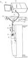
图1是根据本发明的一些实施方案的用于生成心脏组织的扩充模型的系统的示意图;1 is a schematic diagram of a system for generating an augmented model of cardiac tissue in accordance with some embodiments of the present invention;
图2是根据本发明的一些实施方案的用于扩充心脏组织的模型的技术的流程图;并且2 is a flow diagram of a technique for augmenting a model of cardiac tissue in accordance with some embodiments of the present invention; and
图3示出了根据本发明的一些实施方案的用于扩充心脏组织的模型的技术的各方面。3 illustrates aspects of a technique for augmenting a model of cardiac tissue in accordance with some embodiments of the invention.
具体实施方式Detailed ways
概述Overview
在一些实施方案中,构建了受检者心脏的一部分的电生理标测图。电生理标测图包括心脏的该部分的解剖结构的计算机化表示,以及叠加的电生理数据。此类标测图的示例是LAT标测图,其使用例如滑动比色刻度尺指示各种解剖位置处的相应LAT值。In some embodiments, an electrophysiological map of a portion of the subject's heart is constructed. The electrophysiological map includes a computerized representation of the anatomy of the portion of the heart, and superimposed electrophysiological data. An example of such a map is a LAT map, which indicates corresponding LAT values at various anatomical locations using, for example, a sliding colorimetric scale.
为了构建LAT标测图,导管的远侧端部处的一个或多个电极首先采集心脏组织上的各个位置的LAT值的“点云”。然后将该点云标测到组织的体素化解剖模型,使得所采集的LAT值被分别分配给模型中的体素的子集。随后,使用合适的内插技术,为剩余的体素分配内插LAT值。To construct the LAT map, one or more electrodes at the distal end of the catheter first acquire a "point cloud" of LAT values at various locations on the cardiac tissue. This point cloud is then mapped to a voxelized anatomical model of the tissue so that the acquired LAT values are assigned to subsets of voxels in the model, respectively. Then, using a suitable interpolation technique, the remaining voxels are assigned interpolated LAT values.
根据常规技术,分别针对心脏的相关部分的心内膜表面和心外膜表面构建两个完全独立的LAT标测图。然而,本发明人已认识到,由于电流通过内部或“壁内”心脏组织的传播,心脏的心内膜表面的电生理属性与心外膜表面的电生理属性相关。因此,由于考虑到隔离中的每个表面,上述常规技术可能提供不准确的内插LAT值。According to conventional techniques, two completely independent LAT maps are constructed for the endocardial and epicardial surfaces of the relevant part of the heart, respectively. However, the inventors have recognized that the electrophysiological properties of the endocardial surface of the heart correlate with the electrophysiological properties of the epicardial surface due to the propagation of current through the interior or "intramural" cardiac tissue. Thus, the conventional techniques described above may provide inaccurate interpolated LAT values due to consideration of each surface in the isolation.
因此,本发明的实施方案提供体积(即,三维)LAT标测图,其考虑了心内膜表面和心外膜表面两者以及壁内组织。首先,构建心脏组织(包括心内膜表面、心外膜表面和壁内组织)的三维解剖模型。接下来,针对心内膜表面和心外膜表面采集相应的LAT点云,并且将LAT点云标测到模型。随后,使用合适的内插技术,估计剩余表面和壁内体素的LAT值。例如,可使用迭代内插技术,由此在每次迭代期间,为每个体素分配其直接邻域的值的平均值。Accordingly, embodiments of the present invention provide volumetric (ie, three-dimensional) LAT maps that take into account both the endocardial and epicardial surfaces as well as intramural tissue. First, a three-dimensional anatomical model of cardiac tissue (including endocardial surface, epicardial surface, and intramural tissue) is constructed. Next, the corresponding LAT point clouds are acquired for the endocardial and epicardial surfaces, and the LAT point clouds are mapped to the model. Subsequently, using suitable interpolation techniques, the LAT values of the remaining surface and intramural voxels are estimated. For example, iterative interpolation techniques can be used whereby during each iteration, each voxel is assigned an average of the values of its immediate neighbor.
有利的是,如本文所述构建的体积标测图对于心内膜表面和心外膜表面两者通常是准确的。此外,体积标测图可允许医师可视化壁内组织的电生理属性。此外,体积标测图可有利于更准确地识别电传播减速的区域。Advantageously, volume maps constructed as described herein are generally accurate for both endocardial and epicardial surfaces. Additionally, volume maps may allow physicians to visualize the electrophysiological properties of intramural tissue. In addition, volumetric maps may facilitate more accurate identification of areas of electrical propagation deceleration.
除了局部激活时间之外,本文所述的技术可用于构建与组织相关联的其他参数的体积标测图。此类参数包括电压、循环长度、温度和递送到组织的能量的量。In addition to local activation time, the techniques described herein can be used to construct volumetric maps of other parameters associated with tissue. Such parameters include voltage, cycle length, temperature, and the amount of energy delivered to the tissue.
系统描述System specification
首先参考图1,该图是根据本发明的一些实施方案的用于生成心脏组织的扩充模型的系统20的示意图。Reference is first made to FIG. 1, which is a schematic diagram of a
在图1中,示出了医师30沿受检者22的心脏24的腔室的组织移动导管26的远侧端部28。具体地讲,医师30沿组织的心内膜表面和心外膜表面两者移动远侧端部28。在一些实施方案中,远侧端部28包括可用于处理(例如,消融)组织的一个或多个区域的一个或多个处理电极。In FIG. 1 , a
当导管的远侧端部沿组织移动时,属于系统20的处理器32跟踪远侧端部,即,探知远侧端部28设置在其处的组织上的多个位置。(为方便起见,这些位置中的每个位置在下文中简称为导管的位置。)如上所述,这些位置中的一些位置在组织的心内膜表面上,而这些位置中的其他位置在组织的心外膜表面上。As the distal end of the catheter is moved along the tissue, the
此外,当导管的远侧端部沿组织移动时,设置在远侧端部28处的电极和/或其他传感器(例如,温度或力传感器)采集与至少一个参数相关的数据。由处理器32经由电接口34(诸如端口或插口)接收这些数据。基于这些数据,处理器32在多个位置处探知参数的相应值。Additionally, electrodes and/or other sensors (eg, temperature or force sensors) disposed at the
通常,由远侧端部28采集的数据包括在远侧端部28穿过的组织的各个位置处的相应电压信号。另选地或除此之外,数据可包括位置处的相应温度值。另选地或除此之外,数据可包括导管压靠组织的力。Typically, the data acquired by the
在一些实施方案中,基于所探知的导管的位置,处理器32构建组织的解剖模型。然后利用前述参数的值来扩充该解剖模型,如下文参考图2至图3进一步所述。在其他实施方案中,处理器32利用参数值扩充预先存在的解剖模型。In some embodiments,
为了促进跟踪导管的远侧端部,导管的远侧端部可包括一个或多个电磁传感器,该一个或多个电磁传感器在存在所生成的磁场的情况下输出指示传感器的相应位置的信号。由处理器32经由电接口34接收这些信号。基于该信号,处理器32可探知导管的位置。To facilitate tracking of the distal end of the catheter, the distal end of the catheter may include one or more electromagnetic sensors that, in the presence of the generated magnetic field, output signals indicative of the respective positions of the sensors. These signals are received by
另选地,导管的远侧端部可包括导管电极,并且多个电极贴片可耦接到受检者22的身体。当电压施加在导管电极与电极贴片之间时,可测量导管电极与电极贴片之间的电流的相应量值。基于这些电流量值,处理器可探知导管的位置。Alternatively, the distal end of the catheter may include a catheter electrode, and a plurality of electrode patches may be coupled to the body of the subject 22 . When a voltage is applied between the catheter electrode and the electrode patch, the corresponding magnitude of the current between the catheter electrode and the electrode patch can be measured. Based on these current magnitudes, the processor can detect the location of the catheter.
作为又一种替代形式,上述跟踪技术中的两者可彼此组合使用,如例如美国专利8,456,182中所述,该专利的公开内容以引用方式并入本文。另选地或除此之外,可使用任何其他合适的跟踪技术,例如,如美国专利8,456,182中所述。As yet another alternative, both of the above tracking techniques may be used in combination with each other, as described, for example, in US Pat. No. 8,456,182, the disclosure of which is incorporated herein by reference. Alternatively or in addition, any other suitable tracking technique may be used, eg, as described in US Pat. No. 8,456,182.
通常,系统20还包括监视器36。在医师操作导管26时,处理器32可在监视器36上将表示导管的远侧端部的图标叠加在受检者心脏的图像上,使得医师可视觉地跟踪导管。另选地或除此之外,处理器可在监视器36上显示组织的扩充模型,可如下文参考图2至图3详细描述的那样构建该扩充模型。Typically, the
一般来讲,处理器32可被实施为单个处理器或一组协作式联网或集群处理器。在一些实施方案中,如本文所述,处理器32的功能可例如使用一个或多个专用集成电路(ASIC)或现场可编程门阵列(FPGA)仅以硬件来实现。在其他实施方案中,处理器32的功能至少部分地以软件实现。例如,在一些实施方案中,处理器32为编程化数字计算设备,该编程化数字计算设备包括中央处理单元(CPU)和/或图形处理单元(GPU)、随机存取存储器(RAM)、非易失性辅助存储装置(诸如硬盘驱动器或CD ROM驱动器)、网络接口和/或外围设备。如本领域所公知的,将包括软件程序的程序代码和/或数据加载到RAM中以用于由CPU和/或GPU执行和处理,并且生成结果以用于显示、输出、传输或存储。例如,程序代码和/或数据可以电子形式通过网络而被下载到计算机,或者替代地或除此之外,其可被提供和/或存储在非暂态有形介质(诸如磁性存储器、光学存储器、或电子存储器)上。此类程序代码和/或数据在被提供给处理器时,产生被配置为执行本文所述的任务的机器或专用计算机。In general,
扩充模型Augmented model
现在参考图2,其为根据本发明的一些实施方案的用于扩充心脏组织的模型的技术48的流程图的示意图。进一步参考图3,其示出了技术48的各方面。(应当注意,出于说明的目的,图3所示的量纯粹是假设的。)Reference is now made to FIG. 2 , which is a schematic illustration of a flowchart of a
如上文参考图1所述,在探知步骤50处,处理器32探知心脏24的腔室的心内膜表面和心外膜表面上的多个位置处的特定参数的相应值。例如,处理器可探知组织属性的相应值,诸如组织的电压、LAT、循环长度或温度。(可从从组织采集的电压信号导出LAT和循环长度值。)另选地或除此之外,处理器可探知由处理电极递送到组织的能量(诸如射频(RF)能量)的量的相应值。可基于诸如递送到处理电极的能量的量、组织的温度和导管压靠组织的压力的因素来计算能量的量。As described above with reference to FIG. 1 , at the probing
随后,处理器将所探知的值与组织的三维模型38相关联。模型38包括多个体素,每个体素表示组织的不同相应部分。具体地讲,限定模型的第一表面40的那些体素(在本文中称为“心外膜体素”)表示组织的心外膜表面,限定第二表面42的那些体素(在本文中称为“心内膜体素”)表示心内膜表面,并且位于第一表面40和第二表面42之间的那些体素(在本文中称为“壁内体素”)表示壁内组织。表示表现出所探知的参数值的组织的相应部分的那些体素在本文中称为第一体素44。The processor then associates the learned values with the three-
更具体地讲,在第一分配步骤52处,将在探知步骤50处探知的参数值分别分配给第一体素44,如图3的部分A所示。换句话讲,为每个第一体素44分配在由体素表示的组织部分处表现出的值。接下来,如图3的部分B-D所示,通过内插分配给第一体素44的值,处理器对在本文中被称为第二体素46的剩余体素赋值(即,为其分配相应值)。(为清楚起见,图3将分配给第二体素46的值用斜体表示。)More specifically, at a
通常,为了对第二体素赋值,处理器首先在初始化步骤54处初始化第二体素,即,处理器将相应初始值分配给每个第二体素相应46。为了执行该初始化,处理器可使用任何合适类型的最近邻内插。例如,如图3的部分B所示,处理器可使用标准最近邻内插技术,因为可以为每个第二体素分配最靠近它的第一体素的值。另选地,例如,加权最近邻内插技术可用于该初始化。Typically, to assign a value to a second voxel, the processor first initializes the second voxel at
通常,在初始化之后,处理器迭代地将第二体素的直接邻域的相应值的平均值分配给每个第二体素。(对于其中不执行上述初始化的实施方案,仅对已经分配了值的那些直接邻域执行取平均值。)该迭代平均可被称为“拉普拉斯内插”。Typically, after initialization, the processor iteratively assigns to each second voxel the average of the corresponding values of the immediate neighborhood of the second voxel. (For embodiments in which the above initialization is not performed, averaging is performed only on those immediate neighborhoods to which values have been assigned.) This iterative averaging may be referred to as "Laplace interpolation."
在一些实施方案中,迭代次数是预定义的。在其他实施方案中,处理器执行迭代平均,直到满足一个或多个预定义的停止标准。例如,可执行迭代平均,直到任何相邻体素对之间的最大差值小于预定义的阈值。In some embodiments, the number of iterations is predefined. In other embodiments, the processor performs iterative averaging until one or more predefined stopping criteria are met. For example, iterative averaging can be performed until the maximum difference between any pair of adjacent voxels is less than a predefined threshold.
因此,例如,如图2所示,迭代平均可包括第二分配步骤56和检查步骤58。在第二分配步骤56处,处理器将其直接邻域的平均值分配给每个第二体素。在检查步骤58处,处理器检查是否已执行预定义迭代次数,或者是否已满足停止标准。如果是,则迭代平均结束。否则,处理器返回到第二分配步骤56。Thus, for example, as shown in FIG. 2 , the iterative averaging may include a second assigning
在一些实施方案中,如果两个体素共享至少一个顶点,则一个体素被认为是另一个体素的直接邻域(或与另一个体素“相邻”)。因此,体素可具有最多26个直接邻域。(由于体素的二维表示,图3示出了最多八个而不是26个直接邻域。)在其他实施方案中,仅当两个体素共享至少一个面时,这两个体素才被认为是彼此的直接邻域;因此,体素可具有最多仅六个直接邻域。另选地,可使用其他标准来确定体素的直接邻域。In some embodiments, a voxel is considered to be a direct neighbor of (or "adjacent" to) another voxel if it shares at least one vertex. Therefore, a voxel can have up to 26 direct neighbors. (Due to the two-dimensional representation of voxels, Figure 3 shows a maximum of eight instead of 26 direct neighbors.) In other embodiments, two voxels are considered only if they share at least one facet are direct neighbors of each other; thus, a voxel can have at most only six direct neighbors. Alternatively, other criteria may be used to determine the direct neighborhood of a voxel.
通过举例说明的方式,假设共享至少一个公共顶点的一对体素被认为是彼此的直接邻域,图3的部分C和D示出了上述平均值的两次迭代。(应当注意,图3不考虑图中未完全示出的任何体素;因此,例如,通过对仅三个直接邻域取平均值来对角体素赋值。)By way of illustration, assuming that a pair of voxels sharing at least one common vertex are considered to be direct neighbors of each other, Parts C and D of Figure 3 show two iterations of the above average. (It should be noted that Figure 3 does not take into account any voxels that are not fully shown in the figure; thus, for example, corner voxels are assigned by averaging only three immediate neighbors.)
在一些实施方案中,在对每个第二体素赋值时,第二体素的直接邻域被相等地加权,如图3所假设的。在其他实施方案中,由相应权重加权平均值,该权重从与分配给第一体素44的值相关联的相应置信度水平导出。这些置信度水平通常是从导管的远侧端部接收相关数据的质量的函数。In some embodiments, when assigning each second voxel, the immediate neighbors of the second voxel are weighted equally, as assumed in FIG. 3 . In other embodiments, the average is weighted by respective weights derived from respective confidence levels associated with the values assigned to the
例如,假设心外膜表面(由第一表面40表示)的置信度水平大于心内膜表面的置信度水平,则处理器可向每个心外膜第一体素以及初始化为心外膜第一体素的值的每个“子”第二体素赋予更大的权重。因此,例如,假设每个心外膜第一体素及其子体素的权重为1.2,并且每个心内膜第一体素及其子体素的权重仅为1,则部分C中所示的特定第二体素46a将被分配103.3(=(1.2*500+330)/(1.2*5+3))的值,而不是103.8。For example, assuming that the confidence level of the epicardial surface (represented by the first surface 40 ) is greater than the confidence level of the endocardial surface, the processor may Each "child" second voxel of a voxel's value is given greater weight. So, for example, assuming that each epicardial first voxel and its child voxels have a weight of 1.2, and each endocardial first voxel and its child voxels have a weight of only 1, then the The particular
作为拉普拉斯内插的替代或补充,可以使用其他内插技术来对第二体素46赋值。此类技术包括例如捏合、距离倒数加权、样条内插、自然相邻内插、以及如上文已经描述的最近邻内插。一般来讲,响应于内插参数的属性来选择内插技术。例如,对于在整个组织上线性变化的局部激活时间,可使用线性内插技术,诸如拉普拉斯内插。另一方面,对于递送的能量,可使用非线性的基于热力学的内插技术。例如,处理器可假设递送的能量的量从处理电极接触组织的部位以指数方式衰减。Alternatively or in addition to Laplacian interpolation, other interpolation techniques may be used to assign values to the
在一些实施方案中,使用多个并行执行线程(例如,在图形处理单元(GPU)上运行)对第二体素46赋值。因此,例如,在拉普拉斯内插的每次迭代期间,可以并行处理所有第二体素。In some embodiments, the
在一些情况下,不将对应于疤痕组织的体素进行赋值,并且对其他体素的赋值没有贡献。疤痕组织可以由医师手动识别或由处理器32基于从组织采集的电压信号自动识别。In some cases, voxels corresponding to scar tissue are not assigned and do not contribute to the assignment of other voxels. Scar tissue may be manually identified by the physician or automatically by the
通常,在对第二体素赋值之后,处理器在显示步骤60处在监视器36(图1)上显示模型38,以便指示参数值。例如,处理器可以根据对应于由参数获得的值的范围的比色刻度尺对模型的体素进行着色。如上文概述中所述,在显示模型时,处理器通常指示分配给壁内体素的值。因此,有利的是,相对于是否仅示出心内膜表面和心外膜表面,医师可获得对组织的电属性的更好理解。Typically, after assigning the second voxel, the processor displays the
在一些实施方案中,基于分配给模型38的LAT值,处理器识别使电传播减速的任何区域。有利的是,模型38的三维性质有利于以更高的准确度识别这些区域。In some embodiments, based on the LAT value assigned to the
例如,在具有坐标(x0,y0,z0)的每个体素处,处理器可将电传播的归一化速度计算为V(x0,y0,z0)=((L(x0+1,y0,z0)-L(x0-1,y0,z0))-1,(L(x0,y0+1,z0)-L(x0,y0-1,z0))-1,(L(x0,y0,z0+1)-L(x0,y0,z0-1))-1),其中L(x,y,z)指示具有坐标(x,y,z)的体素处的LAT,(x0±1,y0,z0)是体素沿x轴的直接邻域,(x0,y0±1,z0)是体素沿y轴的直接邻域,并且(x0,y0,z0±1)是体素沿z轴的直接邻域。处理器然后可将速度的导数计算为dV=(V(x0+1,y0,z0)-V(x0-1,y0,z0)),V(x0,y0+1,z0)-V(x0,y0-1,z0),V(x0,y0,z0+1)-V(x0,y0,z0-1))。随后,处理器可计算点积V·dV。如果该点积为负,则假设体素表示使电传播减速的区域的一部分。For example, at each voxel with coordinates (x0,y0,z0), the processor may calculate the normalized velocity of electrical propagation as V(x0,y0,z0) =((L(x0+1,y0, z0) -L(x0-1,y0,z0) )-1 , (L(x0,y0+1,z0) -L(x0,y0-1,z0) )-1 ,(L(x0,y0, z0+1) -L(x0,y0,z0-1) )-1 ), where L(x,y,z) indicates the LAT at the voxel with coordinates (x,y,z), (x0±1 ,y0,z0) is the direct neighborhood of the voxel along the x-axis, (x0,y0±1,z0) is the direct neighborhood of the voxel along the y-axis, and (x0,y0,z0±1) is the voxel's direct neighborhood along the y-axis The immediate neighborhood of the z-axis. The processor may then calculate the derivative of velocity as dV=(V(x0+1,y0,z0) -V(x0-1,y0,z0) ),V(x0,y0+1,z0) -V(x0 ,y0-1,z0) ,V(x0,y0,z0+1) -V(x0,y0,z0-1) ). The processor may then calculate the dot product V·dV. If the dot product is negative, the voxel is assumed to represent a portion of the region that slows down electrical propagation.
响应于识别使电传播减速的至少一个区域,处理器可生成指示该区域的输出。例如,在显示模型时,处理器可以着色或以其他方式注释表示区域的体素。In response to identifying at least one region that decelerates electrical propagation, the processor may generate an output indicative of the region. For example, when displaying the model, the processor can color or otherwise annotate the voxels representing the region.
本领域技术人员应当理解,本发明不限于上文具体示出和描述的内容。相反,本发明的实施方案的范围包括上文所述的各种特征的组合与子组合两者,以及本领域的技术人员在阅读上述说明书时可能想到的未在现有技术范围内的变型和修改。以引用方式并入本专利申请的文献被视为本申请的整体部分,不同的是如果这些并入的文献中限定的任何术语与本说明书中明确或隐含地给出的定义相冲突,则应仅考虑本说明书中的定义。It should be understood by those skilled in the art that the present invention is not limited to what has been specifically shown and described above. Rather, the scope of embodiments of the invention includes both combinations and sub-combinations of the various features described above, as well as variations and modifications that may occur to those skilled in the art upon reading the foregoing description that are not within the scope of the prior art. Revise. Documents incorporated by reference into this patent application are considered an integral part of this application, except that if any term defined in such incorporated document conflicts with a definition expressly or implicitly given in this specification, then Only the definitions in this specification should be considered.
Claims (20)
Translated fromChineseApplications Claiming Priority (4)
| Application Number | Priority Date | Filing Date | Title |
|---|---|---|---|
| US201962852266P | 2019-05-23 | 2019-05-23 | |
| US62/852266 | 2019-05-23 | ||
| US16/817537 | 2020-03-12 | ||
| US16/817,537US11564610B2 (en) | 2019-05-23 | 2020-03-12 | Volumetric LAT map |
Publications (2)
| Publication Number | Publication Date |
|---|---|
| CN111973268Atrue CN111973268A (en) | 2020-11-24 |
| CN111973268B CN111973268B (en) | 2024-11-29 |
Family
ID=70804543
Family Applications (1)
| Application Number | Title | Priority Date | Filing Date |
|---|---|---|---|
| CN202010440937.6AActiveCN111973268B (en) | 2019-05-23 | 2020-05-22 | Volumetric LAT map |
Country Status (5)
| Country | Link |
|---|---|
| US (3) | US11564610B2 (en) |
| EP (1) | EP3744240B1 (en) |
| JP (1) | JP7471914B2 (en) |
| CN (1) | CN111973268B (en) |
| IL (1) | IL274726B2 (en) |
Families Citing this family (3)
| Publication number | Priority date | Publication date | Assignee | Title |
|---|---|---|---|---|
| US11564610B2 (en) | 2019-05-23 | 2023-01-31 | Biosense Webster (Israel) Ltd. | Volumetric LAT map |
| US12310737B2 (en) | 2020-09-30 | 2025-05-27 | Boston Scientific Scimed, Inc. | Interactive 2D scatter plot of EGM characteristic metrics |
| WO2024141350A1 (en) | 2022-12-26 | 2024-07-04 | Koninklijke Philips N.V. | Producing combined error values |
Citations (13)
| Publication number | Priority date | Publication date | Assignee | Title |
|---|---|---|---|---|
| US5687737A (en)* | 1992-10-09 | 1997-11-18 | Washington University | Computerized three-dimensional cardiac mapping with interactive visual displays |
| US20030176799A1 (en)* | 1992-09-23 | 2003-09-18 | Beatty Graydon Ernest | Software for mapping potential distribution of a heart chamber |
| US20080058657A1 (en)* | 2006-09-06 | 2008-03-06 | Yitzhack Schwartz | Correlation of cardiac electrical maps with body surface measurements |
| CN101204335A (en)* | 2007-12-14 | 2008-06-25 | 浙江工业大学 | Cardiac Motion Analysis Method Based on Statistical Model |
| CN103354730A (en)* | 2010-12-30 | 2013-10-16 | 圣犹达医疗用品电生理部门有限公司 | System and method for diagnosing arrhythmias and directing catheter therapies |
| CN104622459A (en)* | 2013-11-13 | 2015-05-20 | 韦伯斯特生物官能(以色列)有限公司 | Reverse ECG Mapping |
| US20150313480A1 (en)* | 2014-05-05 | 2015-11-05 | Pacesetter Inc. | Method and system for calculating strain from characterization data of a cardiac chamber |
| US20150317448A1 (en)* | 2014-05-05 | 2015-11-05 | Pacesetter Inc. | Method and system to automatically assign map points to anatomical segments and determine mechanical activation time |
| US20160283687A1 (en)* | 2015-03-27 | 2016-09-29 | Siemens Aktiengesellschaft | System and Method for Non-Invasively Estimating Electrophysiological Maps and Measurements from Cardio-Thoracic 3D Images and Electrocardiography Data |
| CN106691438A (en)* | 2016-12-07 | 2017-05-24 | 首都医科大学附属北京安贞医院 | Integralheart three-dimensional mapping system for complex arrhythmias |
| JP2017142788A (en)* | 2016-01-28 | 2017-08-17 | バイオセンス・ウエブスター・(イスラエル)・リミテッドBiosense Webster (Israel), Ltd. | High definition coloring of heart chambers |
| CN107260157A (en)* | 2016-03-31 | 2017-10-20 | 韦伯斯特生物官能(以色列)有限公司 | The mapping of atrial fibrillation |
| US20180256056A1 (en)* | 2017-03-07 | 2018-09-13 | Biosense Webster (Israel) Ltd. | 3-d electrophysiology heart simulation system and related methods |
Family Cites Families (4)
| Publication number | Priority date | Publication date | Assignee | Title |
|---|---|---|---|---|
| US8456182B2 (en) | 2008-09-30 | 2013-06-04 | Biosense Webster, Inc. | Current localization tracker |
| DE102011081987B4 (en)* | 2011-09-01 | 2014-05-28 | Tomtec Imaging Systems Gmbh | Method for producing a model of a surface of a cavity wall |
| US20190053728A1 (en) | 2017-08-16 | 2019-02-21 | Regents Of The University Of Minnesota | System and method for activation recovery interval imaging of cardiac disorders |
| US11564610B2 (en) | 2019-05-23 | 2023-01-31 | Biosense Webster (Israel) Ltd. | Volumetric LAT map |
- 2020
- 2020-03-12USUS16/817,537patent/US11564610B2/enactiveActive
- 2020-05-17ILIL274726Apatent/IL274726B2/enunknown
- 2020-05-22JPJP2020089400Apatent/JP7471914B2/enactiveActive
- 2020-05-22EPEP20176116.0Apatent/EP3744240B1/enactiveActive
- 2020-05-22CNCN202010440937.6Apatent/CN111973268B/enactiveActive
- 2023
- 2023-01-09USUS18/094,470patent/US11844619B2/enactiveActive
- 2023-11-14USUS18/389,240patent/US12408858B2/enactiveActive
Patent Citations (16)
| Publication number | Priority date | Publication date | Assignee | Title |
|---|---|---|---|---|
| US20030176799A1 (en)* | 1992-09-23 | 2003-09-18 | Beatty Graydon Ernest | Software for mapping potential distribution of a heart chamber |
| US20060084970A1 (en)* | 1992-09-23 | 2006-04-20 | Endocardial Solutions, Inc. | Mapping physiological data in a heart chamber |
| US5687737A (en)* | 1992-10-09 | 1997-11-18 | Washington University | Computerized three-dimensional cardiac mapping with interactive visual displays |
| US20080058657A1 (en)* | 2006-09-06 | 2008-03-06 | Yitzhack Schwartz | Correlation of cardiac electrical maps with body surface measurements |
| CN101204335A (en)* | 2007-12-14 | 2008-06-25 | 浙江工业大学 | Cardiac Motion Analysis Method Based on Statistical Model |
| CN103354730A (en)* | 2010-12-30 | 2013-10-16 | 圣犹达医疗用品电生理部门有限公司 | System and method for diagnosing arrhythmias and directing catheter therapies |
| US20160100770A1 (en)* | 2010-12-30 | 2016-04-14 | St. Jude Medical, Atrial Fibrillation Division, Inc. | System and method for diagnosing arrhythmias and directing catheter therapies |
| CN104622459A (en)* | 2013-11-13 | 2015-05-20 | 韦伯斯特生物官能(以色列)有限公司 | Reverse ECG Mapping |
| US20150317448A1 (en)* | 2014-05-05 | 2015-11-05 | Pacesetter Inc. | Method and system to automatically assign map points to anatomical segments and determine mechanical activation time |
| US20150313480A1 (en)* | 2014-05-05 | 2015-11-05 | Pacesetter Inc. | Method and system for calculating strain from characterization data of a cardiac chamber |
| US20160283687A1 (en)* | 2015-03-27 | 2016-09-29 | Siemens Aktiengesellschaft | System and Method for Non-Invasively Estimating Electrophysiological Maps and Measurements from Cardio-Thoracic 3D Images and Electrocardiography Data |
| JP2017142788A (en)* | 2016-01-28 | 2017-08-17 | バイオセンス・ウエブスター・(イスラエル)・リミテッドBiosense Webster (Israel), Ltd. | High definition coloring of heart chambers |
| CN107085861A (en)* | 2016-01-28 | 2017-08-22 | 韦伯斯特生物官能(以色列)有限公司 | High definition coloring of the ventricle |
| CN107260157A (en)* | 2016-03-31 | 2017-10-20 | 韦伯斯特生物官能(以色列)有限公司 | The mapping of atrial fibrillation |
| CN106691438A (en)* | 2016-12-07 | 2017-05-24 | 首都医科大学附属北京安贞医院 | Integralheart three-dimensional mapping system for complex arrhythmias |
| US20180256056A1 (en)* | 2017-03-07 | 2018-09-13 | Biosense Webster (Israel) Ltd. | 3-d electrophysiology heart simulation system and related methods |
Also Published As
| Publication number | Publication date |
|---|---|
| IL274726B2 (en) | 2023-07-01 |
| EP3744240B1 (en) | 2022-09-07 |
| CN111973268B (en) | 2024-11-29 |
| US11564610B2 (en) | 2023-01-31 |
| JP2020189094A (en) | 2020-11-26 |
| IL274726A (en) | 2020-11-30 |
| US20230157612A1 (en) | 2023-05-25 |
| US20200367778A1 (en) | 2020-11-26 |
| IL274726B1 (en) | 2023-03-01 |
| US12408858B2 (en) | 2025-09-09 |
| US20240074689A1 (en) | 2024-03-07 |
| EP3744240A1 (en) | 2020-12-02 |
| US11844619B2 (en) | 2023-12-19 |
| JP7471914B2 (en) | 2024-04-22 |
Similar Documents
| Publication | Publication Date | Title |
|---|---|---|
| CN107260157B (en) | Mapping of atrial fibrillation | |
| CN111150390B (en) | Cardiac Electrophysiology (EP) activated iterative coherent mapping including scar effects | |
| US9610023B2 (en) | System and methods for computing activation maps | |
| CN111150391B (en) | Iterative coherence mapping of cardiac electrophysiological (EP) activation including reentry | |
| US12408858B2 (en) | Volumetric LAT map | |
| US10342620B2 (en) | Efficient treatment of atrial fibrillation using three-dimensional electrical potential model | |
| US11013447B2 (en) | Cardiac mapping using a 3D grid | |
| CN109276315B (en) | Impedance-based location tracking performance using dispersive interpolation | |
| KR20200138017A (en) | Determining occurrence of focal and/or rotor arrhythmogenic activity in cardiac tissue regions | |
| CN114711779A (en) | Incorporating confidence levels into electrophysiological (EP) maps | |
| EP3821812A1 (en) | Historical ultrasound data for display of live location data | |
| US10694949B2 (en) | Computer implemented method for calculating values indicative for the local spatial structure of conducting properties of heart muscle tissue and computer programs thereof | |
| EP3821806B1 (en) | Visual route indication for activation clusters | |
| CN115211965A (en) | Improved electrophysiological (EP) map coloring by accounting for outliers | |
| JP2024527971A (en) | Tissue Treatment System | |
| CN111345810A (en) | Electrophysiological ripple mapping visualization method | |
| RU2772201C2 (en) | Iterative coherent mapping of electrophysiological activation of heart, including scar effects | |
| RU2771797C2 (en) | Iterative coherent mapping of electrophysiological activation of heart, including effect of re-entry | |
| EP4122395A1 (en) | Layered multi-activation local activation times (lat) mapping | |
| CN118251177A (en) | Tissue Therapy System | |
| CN119384250A (en) | Projection of activation wave velocity onto the mapped cardiac chambers |
Legal Events
| Date | Code | Title | Description |
|---|---|---|---|
| PB01 | Publication | ||
| PB01 | Publication | ||
| SE01 | Entry into force of request for substantive examination | ||
| SE01 | Entry into force of request for substantive examination | ||
| GR01 | Patent grant | ||
| GR01 | Patent grant |