CN111899213A - Cerebrovascular reconstruction method based on high-angle resolution diffusion imaging - Google Patents
Cerebrovascular reconstruction method based on high-angle resolution diffusion imagingDownload PDFInfo
- Publication number
- CN111899213A CN111899213ACN202010492235.2ACN202010492235ACN111899213ACN 111899213 ACN111899213 ACN 111899213ACN 202010492235 ACN202010492235 ACN 202010492235ACN 111899213 ACN111899213 ACN 111899213A
- Authority
- CN
- China
- Prior art keywords
- tensor
- vessel
- blood vessel
- voxel
- model
- Prior art date
- Legal status (The legal status is an assumption and is not a legal conclusion. Google has not performed a legal analysis and makes no representation as to the accuracy of the status listed.)
- Granted
Links
Images
Classifications
- G—PHYSICS
- G06—COMPUTING OR CALCULATING; COUNTING
- G06T—IMAGE DATA PROCESSING OR GENERATION, IN GENERAL
- G06T7/00—Image analysis
- G06T7/0002—Inspection of images, e.g. flaw detection
- G06T7/0012—Biomedical image inspection
- G—PHYSICS
- G06—COMPUTING OR CALCULATING; COUNTING
- G06T—IMAGE DATA PROCESSING OR GENERATION, IN GENERAL
- G06T7/00—Image analysis
- G06T7/10—Segmentation; Edge detection
- G—PHYSICS
- G06—COMPUTING OR CALCULATING; COUNTING
- G06T—IMAGE DATA PROCESSING OR GENERATION, IN GENERAL
- G06T7/00—Image analysis
- G06T7/60—Analysis of geometric attributes
- G06T7/66—Analysis of geometric attributes of image moments or centre of gravity
- G—PHYSICS
- G06—COMPUTING OR CALCULATING; COUNTING
- G06T—IMAGE DATA PROCESSING OR GENERATION, IN GENERAL
- G06T2207/00—Indexing scheme for image analysis or image enhancement
- G06T2207/30—Subject of image; Context of image processing
- G06T2207/30004—Biomedical image processing
- G06T2207/30016—Brain
- G—PHYSICS
- G06—COMPUTING OR CALCULATING; COUNTING
- G06T—IMAGE DATA PROCESSING OR GENERATION, IN GENERAL
- G06T2207/00—Indexing scheme for image analysis or image enhancement
- G06T2207/30—Subject of image; Context of image processing
- G06T2207/30004—Biomedical image processing
- G06T2207/30101—Blood vessel; Artery; Vein; Vascular
Landscapes
- Engineering & Computer Science (AREA)
- Physics & Mathematics (AREA)
- Computer Vision & Pattern Recognition (AREA)
- Theoretical Computer Science (AREA)
- General Physics & Mathematics (AREA)
- Medical Informatics (AREA)
- Quality & Reliability (AREA)
- Radiology & Medical Imaging (AREA)
- Nuclear Medicine, Radiotherapy & Molecular Imaging (AREA)
- Health & Medical Sciences (AREA)
- General Health & Medical Sciences (AREA)
- Geometry (AREA)
- Apparatus For Radiation Diagnosis (AREA)
- Magnetic Resonance Imaging Apparatus (AREA)
Abstract
Translated fromChinese
Description
Translated fromChinese技术领域technical field
本发明涉及一种基于高角度分辨率扩散成像的脑血管重建方法。The invention relates to a cerebral blood vessel reconstruction method based on high angular resolution diffusion imaging.
背景技术Background technique
脑血管结构(如冠状动脉和大脑动脉)的扩张是检测和分析血管异常和病理 (如动脉瘤、狭窄和斑块)的重要步骤。对于血流动力学或血流动力学分析、功能评估、介入性和手术计划以及模拟建立准确和光滑的表面模型也很重要。这种表面模型也可用于其它领域的管状结构的定量测量和几何分析。由于传统的血管分割需要大量的数据集和人工标记,这会使工作量大大增加。对于基于计算机的分割,尽管小的血管可以用一个简单的几何模型来表示,但因为血管中存在众多分支,不同血管的半径也不一样,这给完整地重建血管的形状带来了挑战。准确、快速的脑血管分割已经变成医学影像学研究的难题之一。Dilation of cerebral vascular structures such as coronary arteries and cerebral arteries is an important step in the detection and analysis of vascular abnormalities and pathologies such as aneurysms, stenosis, and plaques. It is also important to establish accurate and smooth surface models for hemodynamic or hemodynamic analysis, functional assessment, interventional and surgical planning, and simulation. This surface model can also be used for quantitative measurement and geometric analysis of tubular structures in other fields. Since traditional vessel segmentation requires large datasets and manual labeling, this will greatly increase the workload. For computer-based segmentation, although small blood vessels can be represented by a simple geometric model, because there are many branches in the blood vessels, the radius of different blood vessels is different, which brings challenges to reconstruct the shape of the blood vessels completely. Accurate and fast cerebrovascular segmentation has become one of the difficult problems in medical imaging research.
现有的血管分割方法可分为两类:无模型分割方法和基于模型分割方法。无模型分割方法主要是图像滤波和增强。有模型的方法中主要基于两种模型,可变模型和圆柱形模型。由于血管内各向异性,因此提出添加圆柱体模型,用二阶张量拟合血管。后来又将二阶张量模型推广到四阶张量模型的方法。但是无论是二阶张量还是四阶,或者更高阶张量,都不能真正描述复杂的血管结构。从理论上讲,高阶张量描述复杂血管结构的能力依赖于阶数,阶数越高,其识别血管交叉的能力越强,即角度分辨率越高。但是随着张量阶数的增加,逆矩阵的求解规模越来越大,既要考虑信号噪声,又要考虑张量信号的非负特性,使得稳定获得高角度分辨率的血管识别比较困难。Existing vessel segmentation methods can be divided into two categories: model-free segmentation methods and model-based segmentation methods. Model-free segmentation methods are mainly image filtering and enhancement. Modeled methods are mainly based on two models, the variable model and the cylindrical model. Due to the intravascular anisotropy, it is proposed to add a cylinder model to fit the blood vessels with a second-order tensor. Later, the method of extending the second-order tensor model to the fourth-order tensor model. But neither second-order nor fourth-order tensors, or higher-order tensors, can really describe complex vascular structures. Theoretically, the ability of high-order tensors to describe complex vascular structures depends on the order. However, with the increase of the tensor order, the solution scale of the inverse matrix becomes larger and larger, and it is necessary to consider both the signal noise and the non-negative characteristics of the tensor signal, which makes it difficult to obtain stable blood vessel identification with high angular resolution.
由于血管结构包括n个分叉,即n个分支从同一血管点流出,不同的分支又有其自己的小分支,因此血管的建模必须准确。高阶张量成像模型(HOT)是解决二阶张量模型难以刻画复杂结构问题的新方法,但是存在特征方向提取复杂、计算量大等问题。Since the vessel structure includes n bifurcations, that is, n branches flow out from the same vessel point, and different branches have their own small branches, the modeling of vessels must be accurate. High-order tensor imaging model (HOT) is a new method to solve the problem that the second-order tensor model is difficult to describe the complex structure, but there are problems such as complex feature direction extraction and large amount of calculation.
发明内容SUMMARY OF THE INVENTION
为了克服现有技术的不足,本发明提供一种基于高角度分辨率扩散成像的脑血管重建方法,基于高阶张量的球面反卷积模型,将高阶张量模型作为球面反卷积算法中方向密度函数ODF的模型。由于血管内的各向异性张量,因此在血管数据中添加一个圆柱模型,在这个圆柱形模型的分量中引入了方向性,从而使得高阶张量适合管状结构。使用高阶张量球面反卷积算法对血管结构的n分支进行建模。作为初始化,从一个用户定义的单种子点开始,使用最大张量范数准则估计种子点的半径。然后创建血管测量值,用高阶张量球面反卷积算法进行拟合,求出血管的方向,利用基于球面反卷积模型的流线型跟踪算法对血管中心线进行跟踪,并根据检测到的血管方向前进到下一个中心线点。在最后一步中,根据血管厚度的计算估计血管方向的半径。因此,从种子点开始,我们的发明可以捕获整个血管树,它提供了血管的方向,中心线和半径。In order to overcome the deficiencies of the prior art, the present invention provides a cerebrovascular reconstruction method based on high angular resolution diffusion imaging, a spherical deconvolution model based on a high-order tensor, and the high-order tensor model is used as a spherical deconvolution algorithm Model of the mid-directional density function ODF. Due to the anisotropic tensor within the vessel, adding a cylindrical model to the vessel data introduces directionality in the components of this cylindrical model, allowing higher-order tensors to fit the tubular structure. The n-branch of the vessel structure is modeled using a higher-order tensor spherical deconvolution algorithm. As initialization, starting from a user-defined single seed point, the radius of the seed point is estimated using the maximum tensor norm criterion. Then, the blood vessel measurement value is created, and the high-order tensor spherical deconvolution algorithm is used for fitting to find the direction of the blood vessel. The direction advances to the next centerline point. In the last step, the radius of the vessel direction is estimated from the calculation of vessel thickness. Therefore, starting from the seed point, our invention can capture the entire vessel tree, which provides the direction, centerline and radius of the vessel.
为了解决上述技术问题本发明提供如下技术方案:In order to solve the above-mentioned technical problems, the present invention provides the following technical solutions:
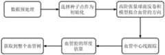
一种基于高角度分辨率扩散成像的脑血管重建方法,所述方法包括以下步骤:A cerebrovascular reconstruction method based on high angular resolution diffusion imaging, the method comprises the following steps:
步骤一:数据预处理Step 1: Data Preprocessing
血管钙化并不是血管腔的一部分,因此在应用血管重建前先将其去除,进行血管分割前对图像进行预处理,得到钙化图,然后将血管钙化的体素强度设置为与大脑组织的强度相等;Vascular calcification is not part of the vessel lumen, so it is removed before applying vessel reconstruction. The image is preprocessed before vessel segmentation to obtain a calcification map, and then the voxel intensity of vessel calcification is set equal to that of the brain tissue. ;
步骤二:选择种子点作为初始化Step 2: Select the seed point as initialization
从用户选择的种子点开始,从血管上的高对比度区域选择,作为初始化步骤,使用最大张量范数准则估计种子点的半径;Starting from a user-selected seed point, selected from a high-contrast region on the blood vessel, as an initialization step, the radius of the seed point is estimated using the maximum tensor norm criterion;
步骤三:获取血管的方向Step 3: Get the direction of the blood vessel
基于高阶张量球面反卷积模型,既能模拟部分血管的不对称,又能模拟血管 n分叉的对称性,其中第四维和更高维方向的张量将用来抑制在不需要的方向上的分量,比如在y结分支的相反方向上;高阶张量球面反卷积算法中有两个重要的部分:第一是单条血管的信号响应函数,第二是血管方向分布函数,血管管道内也具有各向异性,因此高阶张量球面反卷积模型也适用对血管进行拟合;Based on the high-order tensor spherical deconvolution model, it can simulate not only the asymmetry of some blood vessels, but also the symmetry of the n-bifurcation of blood vessels, in which the tensors in the fourth and higher dimensions will be used to suppress the The component in the direction of , such as in the opposite direction of the y-junction branch; there are two important parts in the high-order tensor spherical deconvolution algorithm: the first is the signal response function of a single vessel, and the second is the vessel direction distribution function , there is also anisotropy in the blood vessel pipeline, so the high-order tensor spherical deconvolution model is also suitable for fitting the blood vessels;
在高阶张量球面反卷积模型中,方向分布函数用高阶张量表示,即利用高阶张量方向分布函数与脉冲基函数的卷积模型描述扩散信号衰减情况;为了保证张量模型的非负性,在这里高阶张量血管方向分布函数用卡笛森张量的半正定形式估计,可以推出了球面反卷积的模型,通过平方和多项式理论保证高阶张量非负特性,并通过研究张量空间离散采样对扩散方向计算的影响,建立一个直接从磁共振测量信号估计高阶张量方向分布函数的方法,建立连续高阶张量方向分布函数与脉冲基函数的卷积模型,通过迭代反卷积算法稳定获得高精度血管方向分布函数;当信号受噪声影响时,存在扰动向量,因此,由线性系统标准化方法计算得到解便受到噪声控制,为了得到有效近似,采用最小二乘法求解,该问题的解决将使高阶张量成像模型的优化转换为一个无约束优化问题;In the high-order tensor spherical deconvolution model, the direction distribution function is represented by a high-order tensor, that is, the convolution model of the high-order tensor direction distribution function and the impulse basis function is used to describe the attenuation of the diffusion signal; in order to ensure the tensor model The non-negativity of , where the distribution function of the high-order tensor blood vessel direction is estimated by the positive semi-definite form of the Cartesian tensor, the spherical deconvolution model can be derived, and the non-negative characteristics of the high-order tensor are guaranteed by the sum of squares polynomial theory, and By studying the influence of discrete sampling of tensor space on the calculation of diffusion direction, a method for directly estimating higher-order tensor direction distribution functions from magnetic resonance measurement signals is established, and a convolution model of continuous higher-order tensor direction distribution functions and pulse basis functions is established. , the high-precision blood vessel direction distribution function is stably obtained by the iterative deconvolution algorithm; when the signal is affected by noise, there is a disturbance vector, so the solution calculated by the linear system normalization method is controlled by the noise. In order to obtain an effective approximation, the least square Multiplicative solution, the solution of which will transform the optimization of the higher-order tensor imaging model into an unconstrained optimization problem;
为了估计最终的高阶张量,它描述了血管的方向和球体的半径,高阶张量是在血管内的一个点上计算出来的,张量的模恰好在球体与血管边界吻合时最大;计算出张量后,将其分解为特征值和特征向量,其中为具有特征值的对角矩阵,为张量的特征向量;由于张量沿血管方向最小,沿正交方向较大,所以张量由两个主要特征向量组成,高阶张量的最小特征值对应的特征向量表示血管的方向;In order to estimate the final higher-order tensor, which describes the direction of the vessel and the radius of the sphere, the higher-order tensor is computed at a point inside the vessel, and the modulus of the tensor is maximal exactly when the sphere coincides with the vessel boundary; After the tensor is calculated, it is decomposed into eigenvalues and eigenvectors, among which is the diagonal matrix with eigenvalues and the eigenvector of the tensor; since the tensor is the smallest along the blood vessel direction and larger along the orthogonal direction, the tensor is represented by It consists of two main eigenvectors, and the eigenvector corresponding to the minimum eigenvalue of the high-order tensor represents the direction of the blood vessel;
步骤四:血管中心线跟踪Step 4: Vascular centerline tracking
基于球面反卷积模型的流线型跟踪算法对血管中心线进行跟踪,通过给定适当的起始点与步长,血管的中心线点每前进一步到达的位置可用轨迹递推公式来确定,设定单步前进步长α=0.5,阈值条件的设定也是一致的;当前的中心线方向不仅与该体素内存在的一个或多个方向有关,还与上一个体素的方向有关。为了准确地描绘脑血管的走向,规定当前步的血管中心线的方向即为上一步血管中心线的方向与当前体素内所有扩散特征方向夹角最小的1个特征方向;The streamlined tracking algorithm based on the spherical deconvolution model tracks the centerline of the blood vessel. By giving an appropriate starting point and step size, the position of the centerline point of the blood vessel each step forward can be determined by the trajectory recursion formula. The step-forward progress is α=0.5, and the threshold conditions are set the same; the current centerline direction is not only related to one or more directions existing in the voxel, but also related to the direction of the previous voxel. In order to accurately describe the direction of cerebral blood vessels, it is specified that the direction of the blood vessel centerline of the current step is the one characteristic direction with the smallest angle between the direction of the blood vessel centerline of the previous step and all the diffusion characteristic directions in the current voxel;
当给定一个种子点集,首先取出第一个种子点,记录下其坐标位置,并计算该种子点所存体素的FA值,事先已经通过高阶张量球面反卷积算法获得该体素对应的特征方向;其次判断该体素的位置是否超过边界,若超过,结束跟踪,反之继续判断该体素的FA值是否低于规定的各向异性阈值,若低于,结束跟踪,反之根据轨迹递推公式,由该体素的特征方向,坐标位置计算下一个体素的坐标位置;再次判断下一体素的位置是否超过边界,若超过,结束跟踪,反之获得下一体素的所有特征方向,并分别计算每一个特征方向与上一体素前进方向的夹角,选取夹角最小的一个特征方向作为下4步的前进方向,判断这两个相邻体素前进方向的夹角是否大于规定的角度阈值,若大于,结束跟踪,反之继续据轨迹递推公式计算出再F一个体素的坐标,如此重复;血管跟踪结束有三个条件:1.达到设定的阈值,这意味着该管道到达血管的末端;2.半径达到最大半径时停止血管束,用来避免血管书发散到周围的斑点区域;3.半径达到最小时停止血管树,用来避免将血管周围的噪声跟踪进去;当血管和血管的分支都跟踪出来时,血管的中心线也就可以描绘出来了;When a set of seed points is given, first take out the first seed point, record its coordinate position, and calculate the FA value of the voxel stored in the seed point. The voxel has been obtained through the high-order tensor spherical deconvolution algorithm in advance. The corresponding feature direction; secondly, judge whether the position of the voxel exceeds the boundary, if so, end the tracking, otherwise continue to judge whether the FA value of the voxel is lower than the specified anisotropy threshold, if it is lower, end the tracking, otherwise according to Trajectory recursion formula, calculate the coordinate position of the next voxel from the characteristic direction and coordinate position of the voxel; judge again whether the position of the next voxel exceeds the boundary, if it exceeds, end the tracking, otherwise obtain all the characteristic directions of the next voxel , and calculate the included angle between each feature direction and the forward direction of the upper voxel respectively, select the feature direction with the smallest angle as the forward direction of the next 4 steps, and judge whether the included angle between the forward directions of these two adjacent voxels is greater than the specified If it is greater than the angle threshold, end the tracking, otherwise continue to calculate the coordinates of another voxel according to the trajectory recursion formula, and repeat; there are three conditions for the end of blood vessel tracking: 1. The set threshold is reached, which means that the pipeline Reach the end of the blood vessel; 2. Stop the blood vessel bundle when the radius reaches the maximum radius, to prevent the blood vessel book from spreading to the surrounding spot area; 3. Stop the blood vessel tree when the radius reaches the minimum, to avoid tracking the noise around the blood vessel; when When the blood vessels and the branches of the blood vessels are traced, the center line of the blood vessels can be drawn;
步骤五:血管腔的厚度Step 5: Thickness of Vascular Lumen
要计算的几何测量模型是由一个沿着每个采样方向的空心圆柱组成,公式用一组圆表示,每个圆心坐标代表血管中心线点,定义了圆柱体的半径和高度,定义圆柱的圆被等角离散成极坐标中的点,血管腔的厚度与圆柱的半径直接相关,当模型与数据完全吻合时,得到血管腔的半径,当圆柱的直径小于或大于真实的血管腔厚度时,在一定半径范围内的测量值减小,因此,当圆柱体与容器相切并沿着容器的方向时,测量值最大;The geometric measurement model to be calculated consists of a hollow cylinder along each sampling direction, the formula is represented by a set of circles, each circle center coordinate represents the blood vessel centerline point, defines the radius and height of the cylinder, defines the circle of the cylinder It is equiangularly discretized into points in polar coordinates. The thickness of the vascular lumen is directly related to the radius of the cylinder. When the model is completely consistent with the data, the radius of the vascular lumen is obtained. When the diameter of the cylinder is smaller or larger than the true thickness of the vascular lumen, The measurement decreases within a certain radius, so that the measurement is maximum when the cylinder is tangent to and along the direction of the container;
步骤六:获取整个血管树Step 6: Obtain the entire vascular tree
得到血管的方向、中心线和直径后,在每个中心线点重复,直到所有估计的方向都被跟踪,并根据步骤四中停止的标准到达容器的末端,这样就可以描绘出整个脑动脉。Once the orientation, centerline, and diameter of the vessel are obtained, repeat at each centerline point until all estimated orientations are tracked and reach the end of the vessel according to the criteria stopped in step four, so that the entire cerebral artery can be delineated.
进一步,所述第三步中,将高阶张量球面反卷积模型应用到提取血管的方向,要计算的几何测量模型是由一个沿着每个采样方向的空心圆柱组成,公式用一组圆表示,每个圆心坐标代表血管中心线点,定义了圆柱体的半径和高度,定义圆柱的圆被等角离散成极坐标中的点;Further, in the third step, the high-order tensor spherical deconvolution model is applied to the direction of the extracted blood vessels, and the geometric measurement model to be calculated is composed of a hollow cylinder along each sampling direction. The formula uses a set of Circle representation, each circle center coordinate represents the blood vessel centerline point, defines the radius and height of the cylinder, and the circle defining the cylinder is equiangularly discretized into points in polar coordinates;

其中,g=(g1,g2,g3)T为磁场梯度方向,v是球面上的单位向量,R(v,g)是表示扩散加权信号衰减的轴向对称响应函数,由单个协同定向纤维群测得。其表示如下:where g=(g1 , g2 , g3 )T is the magnetic field gradient direction, v is the unit vector on the sphere, and R(v, g) is the axially symmetric response function representing the attenuation of the diffusion-weighted signal, which is determined by a single synergistic Oriented fiber populations are measured. It is expressed as follows:

其中,μ是给定的正参数,为了保证张量模型的非负性,在这里高阶张量纤维方向分布函数用卡笛森张量的半正定形式估计,模型推到得到如下结果:Among them, μ is a given positive parameter. In order to ensure the non-negativity of the tensor model, the fiber direction distribution function of the high-order tensor is estimated by the positive semi-definite form of the Cartesian tensor, and the model is extrapolated to obtain the following results:

在高阶张量球面反卷积模型中,方向分布函数用高阶张量表示,即利用高阶张量方向分布函数与脉冲基函数的卷积模型描述扩散信号衰减情况,高阶张量模型可以很好的解决一定角度范围内两根或多根血管交叉的问题,为了保证张量模型的非负性,在这里高阶张量血管方向分布函数用卡笛森张量的半正定形式估计,推出了球面反卷积的模型,通过平方和多项式理论保证高阶张量非负特性,并通过研究张量空间离散采样对扩散方向计算的影响,建立一个直接从磁共振测量信号估计高阶张量方向分布函数的方法。建立连续高阶张量方向分布函数与脉冲基函数的卷积模型,通过迭代反卷积算法稳定获得高精度血管方向分布函数,当信号受噪声影响时,存在扰动向量。因此,由线性系统标准化方法计算得到解便受到噪声控制;为了得到有效近似,采用最小二乘法求解,该问题的解决将使高阶张量成像模型的优化转换为一个无约束优化问题,很大程度上减少算法的求解难度;In the high-order tensor spherical deconvolution model, the direction distribution function is represented by a high-order tensor, that is, the convolution model of the high-order tensor direction distribution function and the impulse basis function is used to describe the attenuation of the diffusion signal. The high-order tensor model It can solve the problem of the intersection of two or more blood vessels within a certain angle range. In order to ensure the non-negativity of the tensor model, here the high-order tensor blood vessel direction distribution function is estimated by the semi-positive definite form of the Cartesian tensor. , a spherical deconvolution model was introduced, the non-negative properties of higher-order tensors were guaranteed by the sum-of-squares polynomial theory, and a higher-order tensor directly estimated from the magnetic resonance measurement signal was established by studying the influence of discrete sampling of tensor space on the calculation of the diffusion direction. method for the directional distribution function. The convolution model of the continuous high-order tensor direction distribution function and the pulse basis function is established, and the high-precision blood vessel direction distribution function is stably obtained through the iterative deconvolution algorithm. When the signal is affected by noise, there is a disturbance vector. Therefore, the solution calculated by the linear system normalization method is subject to noise control; in order to obtain an effective approximation, the least squares method is used to solve this problem. To a certain extent reduce the difficulty of solving the algorithm;
为了估计最终的高阶张量,它描述了血管的方向和球体的半径,高阶张量是在血管内的一个点上计算出来的,张量的模恰好在球体与血管边界吻合时最大。计算出张量后,将其分解为特征值和特征向量,其中为具有特征值的对角矩阵,为张量的特征向量,由于张量沿血管方向最小,沿正交方向较大,所以张量由两个主要特征向量组成,高阶张量的最小特征值对应的特征向量表示血管的方向。In order to estimate the final higher-order tensor, which describes the orientation of the vessel and the radius of the sphere, the higher-order tensor is computed at a point inside the vessel, and the modulus of the tensor is maximized exactly when the sphere coincides with the vessel boundary. After the tensor is calculated, it is decomposed into eigenvalues and eigenvectors, where is the diagonal matrix with eigenvalues, and is the eigenvector of the tensor. Since the tensor is the smallest along the blood vessel direction and larger along the orthogonal direction, the tensor is represented by It consists of two main eigenvectors, and the eigenvector corresponding to the smallest eigenvalue of the higher-order tensor represents the direction of the blood vessel.
再进一步,所述第四步中,采用基于球面反卷积模型的流线型跟踪算法对血管中心线进行跟踪,步骤如下:Further, in the described 4th step, adopt the streamline tracking algorithm based on the spherical deconvolution model to track the blood vessel center line, and the steps are as follows:
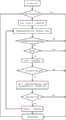
步骤一:定义种子点集Step 1: Define the seed point set
当给定一个种子点集,首先取出第一个种子点,记录下其坐标位置,并计算该种子点所存体素的FA值,事先已经通过高阶张量球面反卷积算法获得该体素对应的特征方向;When a set of seed points is given, first take out the first seed point, record its coordinate position, and calculate the FA value of the voxel stored in the seed point. The voxel has been obtained through the high-order tensor spherical deconvolution algorithm in advance. the corresponding feature direction;
步骤二:判断条件Step 2: Judgment Conditions
判断该体素的位置是否超过边界,若超过,结束跟踪,反之继续判断该体素的FA值是否低于规定的各向异性阈值,若低于,结束跟踪;Determine whether the position of the voxel exceeds the boundary, if so, end the tracking, otherwise continue to judge whether the FA value of the voxel is lower than the specified anisotropy threshold, if it is lower, end the tracking;
步骤三:计算下一体素坐标和夹角Step 3: Calculate the coordinates and angle of the next voxel
根据轨迹递推公式,由该体素的特征方向,坐标位置计算下一个体素的坐标位置。再次判断下一体素的位置是否超过边界,若超过,结束跟踪,反之获得下一体素的所有特征方向,并分别计算每一个特征方向与上一体素前进方向的夹角,选取夹角最小的一个特征方向作为下4步的前进方向,判断这两个相邻体素前进方向的夹角是否大于规定的角度阈值,若大于,结束跟踪,反之继续据轨迹递推公式计算出再F一个体素的坐标,如此反复;According to the trajectory recursion formula, the coordinate position of the next voxel is calculated from the characteristic direction and coordinate position of the voxel. Judge again whether the position of the next voxel exceeds the boundary, if so, end the tracking, otherwise obtain all the characteristic directions of the next voxel, and calculate the angle between each characteristic direction and the forward direction of the previous voxel, and select the one with the smallest angle. The feature direction is used as the forward direction of the next 4 steps, and it is judged whether the included angle between the forward directions of the two adjacent voxels is greater than the specified angle threshold. The coordinates of , and so on;
步骤四:血管跟踪三个结束条件Step 4: Three End Conditions for Vessel Tracking
血管跟踪结束有三个条件:1.达到设定的阈值,这意味着该管道到达血管的末端;2.半径达到最大半径时停止血管束,用来避免血管书发散到周围的斑点区域;3.半径达到最小时停止血管树,用来避免将血管周围的噪声跟踪进去。当血管和血管的分支都跟踪出来时,血管的中心线也就可以描绘出来了。There are three conditions for the end of blood vessel tracking: 1. The set threshold is reached, which means that the tube reaches the end of the blood vessel; 2. The blood vessel bundle is stopped when the radius reaches the maximum radius, which is used to avoid the divergence of the blood vessel book to the surrounding spot area; 3. The vessel tree is stopped when the radius reaches a minimum, to avoid tracking noise around the vessel. When both blood vessels and their branches are traced, the centerline of the blood vessel can also be traced.
本发明的有益效果为:The beneficial effects of the present invention are:
将高阶张量球面反卷积模型应用到血管分割上,基于球面反卷积模型的流线型跟踪算法对血管中心线进行跟踪,能够较好的实现大血管以及血管分支的重建。The high-order tensor spherical deconvolution model is applied to the blood vessel segmentation, and the streamline tracking algorithm based on the spherical deconvolution model tracks the centerline of the blood vessel, which can better realize the reconstruction of large blood vessels and blood vessel branches.
附图说明Description of drawings
图1是本方法的实现步骤流程图。Fig. 1 is a flow chart of the implementation steps of the method.
图2是基于球面反卷积模型的流线型跟踪算法图。Figure 2 is a diagram of a streamlined tracking algorithm based on a spherical deconvolution model.
具体实施方式Detailed ways
下面对本发明作进一步说明。The present invention will be further described below.
参照图1和图2,一种基于高角度分辨率扩散成像的脑血管重建方法,所述方法包括以下步骤:1 and 2, a cerebral blood vessel reconstruction method based on high angular resolution diffusion imaging, the method includes the following steps:
步骤一:数据预处理Step 1: Data Preprocessing
血管钙化并不是血管腔的一部分,因此在应用血管重建前先将其去除,进行血管分割前对图像进行预处理,得到钙化图,然后将血管钙化的体素强度设置为与大脑组织的强度相等;Vascular calcification is not part of the vessel lumen, so it is removed before applying vessel reconstruction. The image is preprocessed before vessel segmentation to obtain a calcification map, and then the voxel intensity of vessel calcification is set equal to that of the brain tissue. ;
步骤二:选择种子点作为初始化Step 2: Select the seed point as initialization
从用户选择的种子点开始,最好是从血管上的高对比度区域选择。作为初始化步骤,我们使用最大张量范数准则估计种子点的半径;Start with a user-selected seed point, preferably from a high-contrast area on the vessel. As an initialization step, we estimate the radius of the seed point using the maximum tensor norm criterion;
步骤三:获取血管的方向Step 3: Get the direction of the blood vessel
基于高阶张量球面反卷积模型,既能模拟部分血管的不对称,又能模拟血管 n分叉的对称性,其中第四维和更高维方向的张量将用来抑制在不需要的方向上的分量,比如在y结分支的相反方向上,高阶张量球面反卷积算法中有两个重要的部分:第一是单条血管的信号响应函数,第二是血管方向分布函数,血管管道内也具有各向异性,因此高阶张量球面反卷积模型也适用对血管进行拟合;Based on the high-order tensor spherical deconvolution model, it can simulate not only the asymmetry of some blood vessels, but also the symmetry of the n-bifurcation of blood vessels, in which the tensors in the fourth and higher dimensions will be used to suppress the The component in the direction of , such as in the opposite direction of the y-junction branch, there are two important parts in the high-order tensor spherical deconvolution algorithm: the first is the signal response function of a single vessel, and the second is the vessel direction distribution function. , there is also anisotropy in the blood vessel pipeline, so the high-order tensor spherical deconvolution model is also suitable for fitting the blood vessels;
在高阶张量球面反卷积模型中,方向分布函数用高阶张量表示,即利用高阶张量方向分布函数与脉冲基函数的卷积模型描述扩散信号衰减情况(磁共振测量信号),高阶张量模型可以很好的解决一定角度范围内两根或多根血管交叉的问题,为了保证张量模型的非负性,在这里高阶张量血管方向分布函数用卡笛森张量的半正定形式估计,可以推出了球面反卷积的模型,通过平方和多项式理论保证高阶张量非负特性,并通过研究张量空间离散采样对扩散方向计算的影响,建立一个直接从磁共振测量信号估计高阶张量方向分布函数的方法,建立连续高阶张量方向分布函数与脉冲基函数的卷积模型,通过迭代反卷积算法稳定获得高精度血管方向分布函数,当信号受噪声影响时,存在扰动向量,因此,由线性系统标准化方法计算得到解便受到噪声控制,为了得到有效近似,采用最小二乘法求解,该问题的解决将使高阶张量成像模型的优化转换为一个无约束优化问题,很大程度上减少算法的求解难度;In the high-order tensor spherical deconvolution model, the direction distribution function is represented by a high-order tensor, that is, the convolution model of the high-order tensor direction distribution function and the impulse basis function is used to describe the attenuation of the diffusion signal (magnetic resonance measurement signal) , the high-order tensor model can well solve the problem of two or more blood vessels crossing within a certain angle range. In order to ensure the non-negativity of the tensor model, here the high-order tensor blood vessel direction distribution function uses the Cartesian tensor The semi-positive definite form estimation of the quantity can be derived from the spherical deconvolution model, the non-negative properties of high-order tensors are guaranteed by the sum of squares polynomial theory, and by studying the influence of discrete sampling in the tensor space on the calculation of the diffusion direction, a model directly derived from magnetic resonance is established. The method of measuring the signal estimation high-order tensor direction distribution function, establishing a convolution model of continuous high-order tensor direction distribution function and pulse basis function, and obtaining high-precision blood vessel direction distribution function stably through an iterative deconvolution algorithm. When it is affected, there is a disturbance vector. Therefore, the solution calculated by the linear system normalization method is controlled by noise. In order to obtain an effective approximation, the least square method is used to solve the problem. The solution of this problem will transform the optimization of the high-order tensor imaging model into a Unconstrained optimization problem, which greatly reduces the difficulty of solving the algorithm;
为了估计最终的高阶张量,它描述了血管的方向和球体的半径,高阶张量是在血管内的一个点上计算出来的,张量的模恰好在球体与血管边界吻合时最大,计算出张量后,将其分解为特征值和特征向量,其中为具有特征值的对角矩阵,为张量的特征向量,由于张量沿血管方向最小,沿正交方向较大,所以张量由两个主要特征向量组成,高阶张量的最小特征值对应的特征向量表示血管的方向;In order to estimate the final higher-order tensor, which describes the orientation of the vessel and the radius of the sphere, the higher-order tensor is computed at a point inside the vessel, and the modulus of the tensor is maximized exactly when the sphere coincides with the vessel boundary, After the tensor is calculated, it is decomposed into eigenvalues and eigenvectors, where is the diagonal matrix with eigenvalues, and is the eigenvector of the tensor. Since the tensor is the smallest along the blood vessel direction and larger along the orthogonal direction, the tensor is represented by It consists of two main eigenvectors, and the eigenvector corresponding to the minimum eigenvalue of the high-order tensor represents the direction of the blood vessel;
步骤四:血管中心线跟踪Step 4: Vascular centerline tracking
与基于DTI的流线型算法描述类似,采用基于球面反卷积模型的流线型跟踪算法对血管中心线进行跟踪,通过给定适当的起始点与步长,血管的中心线点每前进一步到达的位置可用轨迹递推公式来确定,设定单步前进步长α=0.5,阈值条件的设定也是一致的,与DTI模型不同的是,当前的中心线方向不仅与该体素内存在的一个或多个方向有关,还与上一个体素的方向有关,为了准确地描绘脑血管的走向,规定当前步的血管中心线的方向即为上一步血管中心线的方向与当前体素内所有扩散特征方向夹角最小的1个特征方向;Similar to the description of the streamlined algorithm based on DTI, the streamlined tracking algorithm based on the spherical deconvolution model is used to track the centerline of the blood vessel. By giving an appropriate starting point and step size, the position of the centerline point of the blood vessel can be used for each step forward. The trajectory recursion formula is used to determine, set the single-step forward progress α = 0.5, and the threshold conditions are also set in the same way. Unlike the DTI model, the current centerline direction is not only related to one or more In order to accurately describe the direction of cerebral blood vessels, the direction of the blood vessel centerline in the current step is defined as the direction of the blood vessel centerline in the previous step and the direction of all diffusion characteristics in the current voxel. 1 characteristic direction with the smallest included angle;
当给定一个种子点集,首先取出第一个种子点,记录下其坐标位置,并计算该种子点所存体素的FA值,事先已经通过高阶张量球面反卷积算法获得该体素对应的特征方向,其次判断该体素的位置是否超过边界,若超过,结束跟踪,反之继续判断该体素的FA值是否低于规定的各向异性阈值,若低于,结束跟踪,反之根据轨迹递推公式,由该体素的特征方向,坐标位置计算下一个体素的坐标位置,再次判断下一体素的位置是否超过边界,若超过,结束跟踪,反之获得下一体素的所有特征方向,并分别计算每一个特征方向与上一体素前进方向的夹角,选取夹角最小的一个特征方向作为下4步的前进方向,判断这两个相邻体素前进方向的夹角是否大于规定的角度阈值,若大于,结束跟踪,反之继续据轨迹递推公式计算出再F一个体素的坐标,如此重复;血管跟踪结束有三个条件:1.达到设定的阈值,这意味着该管道到达血管的末端;2.半径达到最大半径时停止血管束,用来避免血管书发散到周围的斑点区域;3.半径达到最小时停止血管树,用来避免将血管周围的噪声跟踪进去;当血管和血管的分支都跟踪出来时,血管的中心线也就可以描绘出来了,流程图如图2;When a set of seed points is given, first take out the first seed point, record its coordinate position, and calculate the FA value of the voxel stored in the seed point. The voxel has been obtained through the high-order tensor spherical deconvolution algorithm in advance. Corresponding feature direction, secondly judge whether the position of the voxel exceeds the boundary, if so, end the tracking, otherwise continue to judge whether the FA value of the voxel is lower than the specified anisotropy threshold, if it is lower, end the tracking, otherwise according to The trajectory recursion formula calculates the coordinate position of the next voxel from the characteristic direction and coordinate position of the voxel, and judges again whether the position of the next voxel exceeds the boundary. , and calculate the included angle between each feature direction and the forward direction of the upper voxel respectively, select the feature direction with the smallest angle as the forward direction of the next 4 steps, and judge whether the included angle between the forward directions of these two adjacent voxels is greater than the specified If it is greater than the angle threshold, end the tracking, otherwise continue to calculate the coordinates of another voxel according to the trajectory recursion formula, and repeat; there are three conditions for the end of blood vessel tracking: 1. The set threshold is reached, which means that the pipeline Reach the end of the blood vessel; 2. Stop the blood vessel bundle when the radius reaches the maximum radius, to prevent the blood vessel book from spreading to the surrounding spot area; 3. Stop the blood vessel tree when the radius reaches the minimum, to avoid tracking the noise around the blood vessel; when When the blood vessels and the branches of the blood vessels are traced, the center line of the blood vessels can be drawn, as shown in the flowchart in Figure 2;
步骤五:血管腔的厚度(直径)Step 5: Thickness (diameter) of vessel lumen
要计算的几何测量模型是由一个沿着每个采样方向的空心圆柱组成,公式用一组圆表示,每个圆心坐标代表血管中心线点,定义了圆柱体的半径和高度,定义圆柱的圆被等角离散成极坐标中的点;血管腔的厚度与圆柱的半径直接相关,当模型与数据完全吻合时,得到血管腔的半径;当圆柱的直径小于或大于真实的血管腔厚度时,在一定半径范围内的测量值减小;因此,当圆柱体与容器相切并沿着容器的方向时,测量值最大;The geometric measurement model to be calculated consists of a hollow cylinder along each sampling direction, the formula is represented by a set of circles, each circle center coordinate represents the blood vessel centerline point, defines the radius and height of the cylinder, defines the circle of the cylinder It is equiangularly discretized into points in polar coordinates; the thickness of the vascular lumen is directly related to the radius of the cylinder, when the model is completely consistent with the data, the radius of the vascular lumen is obtained; when the diameter of the cylinder is smaller or larger than the true thickness of the vascular lumen, The measurement decreases within a certain radius; therefore, the measurement is greatest when the cylinder is tangent to and along the direction of the container;
步骤六:获取整个血管树Step 6: Obtain the entire vascular tree
由前面的步骤,可以得到血管的方向,中心线和直径,这些步骤在每个中心线点重复,直到所有估计的方向都被跟踪,并根据步骤四中停止的标准到达容器的末端,这样就可以描绘出整个脑动脉。From the previous steps, the orientation, centerline and diameter of the vessel can be obtained, and these steps are repeated at each centerline point until all estimated orientations are tracked and reach the end of the vessel according to the criteria stopped in step four, so that The entire cerebral artery can be delineated.
Claims (3)


Priority Applications (1)
| Application Number | Priority Date | Filing Date | Title |
|---|---|---|---|
| CN202010492235.2ACN111899213B (en) | 2020-06-03 | 2020-06-03 | Cerebrovascular reconstruction method based on high-angle-resolution diffusion imaging |
Applications Claiming Priority (1)
| Application Number | Priority Date | Filing Date | Title |
|---|---|---|---|
| CN202010492235.2ACN111899213B (en) | 2020-06-03 | 2020-06-03 | Cerebrovascular reconstruction method based on high-angle-resolution diffusion imaging |
Publications (2)
| Publication Number | Publication Date |
|---|---|
| CN111899213Atrue CN111899213A (en) | 2020-11-06 |
| CN111899213B CN111899213B (en) | 2024-03-22 |
Family
ID=73207063
Family Applications (1)
| Application Number | Title | Priority Date | Filing Date |
|---|---|---|---|
| CN202010492235.2AActiveCN111899213B (en) | 2020-06-03 | 2020-06-03 | Cerebrovascular reconstruction method based on high-angle-resolution diffusion imaging |
Country Status (1)
| Country | Link |
|---|---|
| CN (1) | CN111899213B (en) |
Cited By (4)
| Publication number | Priority date | Publication date | Assignee | Title |
|---|---|---|---|---|
| CN112686991A (en)* | 2021-01-08 | 2021-04-20 | 博动医学影像科技(上海)有限公司 | Method and system for reconstructing normal lumen form of blood vessel in hybrid mode |
| CN113408647A (en)* | 2021-07-07 | 2021-09-17 | 中国科学院生物物理研究所 | Extraction method of cerebral small vessel structural features |
| CN115034125A (en)* | 2021-03-08 | 2022-09-09 | 株式会社东芝 | Computing device, computing method, and program |
| CN115147572A (en)* | 2022-05-13 | 2022-10-04 | 浙江工业大学 | Cerebrovascular tracking method based on streamline differential equation |
Citations (2)
| Publication number | Priority date | Publication date | Assignee | Title |
|---|---|---|---|---|
| CN103337071A (en)* | 2013-06-19 | 2013-10-02 | 北京理工大学 | Device and method for structure-reconstruction-based subcutaneous vein three-dimensional visualization |
| CN108154519A (en)* | 2017-12-25 | 2018-06-12 | 吉林大学 | Dividing method, device and the storage medium of eye fundus image medium vessels |
- 2020
- 2020-06-03CNCN202010492235.2Apatent/CN111899213B/enactiveActive
Patent Citations (2)
| Publication number | Priority date | Publication date | Assignee | Title |
|---|---|---|---|---|
| CN103337071A (en)* | 2013-06-19 | 2013-10-02 | 北京理工大学 | Device and method for structure-reconstruction-based subcutaneous vein three-dimensional visualization |
| CN108154519A (en)* | 2017-12-25 | 2018-06-12 | 吉林大学 | Dividing method, device and the storage medium of eye fundus image medium vessels |
Cited By (5)
| Publication number | Priority date | Publication date | Assignee | Title |
|---|---|---|---|---|
| CN112686991A (en)* | 2021-01-08 | 2021-04-20 | 博动医学影像科技(上海)有限公司 | Method and system for reconstructing normal lumen form of blood vessel in hybrid mode |
| CN115034125A (en)* | 2021-03-08 | 2022-09-09 | 株式会社东芝 | Computing device, computing method, and program |
| CN113408647A (en)* | 2021-07-07 | 2021-09-17 | 中国科学院生物物理研究所 | Extraction method of cerebral small vessel structural features |
| CN113408647B (en)* | 2021-07-07 | 2024-04-02 | 中国科学院生物物理研究所 | A method for extracting structural features of small brain blood vessels |
| CN115147572A (en)* | 2022-05-13 | 2022-10-04 | 浙江工业大学 | Cerebrovascular tracking method based on streamline differential equation |
Also Published As
| Publication number | Publication date |
|---|---|
| CN111899213B (en) | 2024-03-22 |
Similar Documents
| Publication | Publication Date | Title |
|---|---|---|
| CN111899213B (en) | Cerebrovascular reconstruction method based on high-angle-resolution diffusion imaging | |
| Cetin et al. | A higher-order tensor vessel tractography for segmentation of vascular structures | |
| CN114303169B (en) | System and method for quantitative measurement of physical properties | |
| Caruyer et al. | Phantomas: a flexible software library to simulate diffusion MR phantoms | |
| Reisert et al. | Fiber continuity: an anisotropic prior for ODF estimation | |
| Descoteaux et al. | Deterministic and probabilistic tractography based on complex fibre orientation distributions | |
| De Craene et al. | 3D strain assessment in ultrasound (straus): A synthetic comparison of five tracking methodologies | |
| US8290247B2 (en) | Method and system for segmentation of tubular structures in 3D images | |
| JP2004535874A (en) | Magnetic resonance angiography and apparatus therefor | |
| Kang et al. | White matter fiber tractography via anisotropic diffusion simulation in the human brain | |
| Wu et al. | Segmentation and reconstruction of vascular structures for 3D real-time simulation | |
| CN104766322B (en) | Based on geodesic cerebrovascular length and flexibility measure | |
| CN104881873B (en) | A kind of multistage adjustment sparse imaging method of mixed weighting for complicated fibre bundle Accurate Reconstruction | |
| Govyadinov et al. | Robust tracing and visualization of heterogeneous microvascular networks | |
| Mohan et al. | Tubular surface segmentation for extracting anatomical structures from medical imagery | |
| Materka et al. | Automated modeling of tubular blood vessels in 3D MR angiography images | |
| Descoteaux et al. | Deterministic and probabilistic Q-ball tractography: from diffusion to sharp fiber distribution | |
| Gasteiger | Visual exploration of cardiovascular hemodynamics | |
| US20160284080A1 (en) | Vasculature modeling | |
| Jackowski et al. | Estimation of anatomical connectivity by anisotropic front propagation and diffusion tensor imaging | |
| Wong et al. | Principal curves for lumen center extraction and flow channel width estimation in 3-D arterial networks: theory, algorithm, and validation | |
| CN115147572B (en) | A method for cerebral vascular tracking based on streamline differential equations | |
| CN114387381B (en) | Front-end visual pathway reconstruction method based on global optimization streamline differential equation | |
| Ho et al. | Fasciculography: robust prior-free real-time normalized volumetric neural tract parcellation | |
| Klein et al. | On the reliability of diffusion neuroimaging |
Legal Events
| Date | Code | Title | Description |
|---|---|---|---|
| PB01 | Publication | ||
| PB01 | Publication | ||
| SE01 | Entry into force of request for substantive examination | ||
| SE01 | Entry into force of request for substantive examination | ||
| GR01 | Patent grant | ||
| GR01 | Patent grant |