CN111888059A - Deep learning and X-ray-based preoperative planning method and device for total hip replacement - Google Patents
Deep learning and X-ray-based preoperative planning method and device for total hip replacementDownload PDFInfo
- Publication number
- CN111888059A CN111888059ACN202010707817.8ACN202010707817ACN111888059ACN 111888059 ACN111888059 ACN 111888059ACN 202010707817 ACN202010707817 ACN 202010707817ACN 111888059 ACN111888059 ACN 111888059A
- Authority
- CN
- China
- Prior art keywords
- image
- determining
- femoral
- neural network
- ray
- Prior art date
- Legal status (The legal status is an assumption and is not a legal conclusion. Google has not performed a legal analysis and makes no representation as to the accuracy of the status listed.)
- Granted
Links
Images


Classifications
- A—HUMAN NECESSITIES
- A61—MEDICAL OR VETERINARY SCIENCE; HYGIENE
- A61F—FILTERS IMPLANTABLE INTO BLOOD VESSELS; PROSTHESES; DEVICES PROVIDING PATENCY TO, OR PREVENTING COLLAPSING OF, TUBULAR STRUCTURES OF THE BODY, e.g. STENTS; ORTHOPAEDIC, NURSING OR CONTRACEPTIVE DEVICES; FOMENTATION; TREATMENT OR PROTECTION OF EYES OR EARS; BANDAGES, DRESSINGS OR ABSORBENT PADS; FIRST-AID KITS
- A61F2/00—Filters implantable into blood vessels; Prostheses, i.e. artificial substitutes or replacements for parts of the body; Appliances for connecting them with the body; Devices providing patency to, or preventing collapsing of, tubular structures of the body, e.g. stents
- A61F2/02—Prostheses implantable into the body
- A61F2/30—Joints
- A61F2/46—Special tools for implanting artificial joints
- A61F2002/4632—Special tools for implanting artificial joints using computer-controlled surgery, e.g. robotic surgery
- A61F2002/4633—Special tools for implanting artificial joints using computer-controlled surgery, e.g. robotic surgery for selection of endoprosthetic joints or for pre-operative planning
Landscapes
- Health & Medical Sciences (AREA)
- Transplantation (AREA)
- Orthopedic Medicine & Surgery (AREA)
- Heart & Thoracic Surgery (AREA)
- Life Sciences & Earth Sciences (AREA)
- Oral & Maxillofacial Surgery (AREA)
- Engineering & Computer Science (AREA)
- Biomedical Technology (AREA)
- Physical Education & Sports Medicine (AREA)
- Vascular Medicine (AREA)
- Cardiology (AREA)
- Animal Behavior & Ethology (AREA)
- General Health & Medical Sciences (AREA)
- Public Health (AREA)
- Veterinary Medicine (AREA)
- Image Analysis (AREA)
- Image Processing (AREA)
Abstract
Translated fromChinese
Description
Translated fromChinese技术领域technical field
本申请涉及医学技术领域,具体而言,涉及一种基于深度学习与X线的全髋关节置换术前规划方法及装置。The present application relates to the field of medical technology, and in particular, to a preoperative planning method and device for total hip arthroplasty based on deep learning and X-rays.
背景技术Background technique
在医学领域中全髋关节置换手术的术前规划主要包括计算所需假体型号及截骨线位置,全髋关节置换手术的术前规划对于手术的成功率起着非常重要的作用,因此如何提供准确的术前规划是非常重要的。目前主要的术前规划方式为人工通过各种工具进行测量,效率低而且准确性无法保证,因此亟需提供一种更便捷更准确的术前规划的方法为全髋关节置换手术提供更好的术前支持。In the medical field, the preoperative planning of total hip replacement surgery mainly includes calculating the required prosthesis size and the position of the osteotomy line. The preoperative planning of total hip replacement surgery plays a very important role in the success rate of the surgery. Therefore, how to It is very important to provide accurate preoperative planning. At present, the main preoperative planning method is to measure manually through various tools, which is inefficient and cannot guarantee the accuracy. Therefore, it is urgent to provide a more convenient and accurate preoperative planning method to provide a better solution for total hip replacement surgery. Preoperative support.
发明内容SUMMARY OF THE INVENTION
本申请的主要目的在于提出一种基于深度学习与X线的全髋关节置换术前规划方法及装置,以提供一种更便捷更准确的术前规划的方式为全髋关节置换手术提供更好的术前支持。The main purpose of this application is to propose a preoperative planning method and device for total hip arthroplasty based on deep learning and X-rays, so as to provide a more convenient and accurate preoperative planning method for total hip arthroplasty. preoperative support.
为了实现上述目的,根据本申请的第一方面,提供了一种基于深度学习与X线的全髋关节置换术前规划方法。In order to achieve the above object, according to the first aspect of the present application, a preoperative planning method for total hip arthroplasty based on deep learning and X-rays is provided.
根据本申请的基于深度学习与X线的全髋关节置换术前规划方法包括:The preoperative planning method for total hip arthroplasty based on deep learning and X-ray according to the present application includes:
获取髋关节的X线图像,所述髋关节的X线图像中包含参照物的图像,所述参照物为已知尺寸的参照物;acquiring an X-ray image of the hip joint, the X-ray image of the hip joint includes an image of a reference object, and the reference object is a reference object of known size;
根据参照物的图像尺寸及其实际尺寸的比例,将髋关节的X线图像进行尺寸的还原;According to the ratio of the image size of the reference object and its actual size, the size of the X-ray image of the hip joint is restored;
基于深度学习模型对还原后的髋关节的X线图像进行识别,确定腿长差、髋臼杯位置以及股骨柄假体的规格型号;Identify the X-ray image of the restored hip joint based on the deep learning model, and determine the leg length difference, the position of the acetabular cup and the specification and model of the femoral stem prosthesis;
根据股骨柄假体的旋转中心和在髋关节的X线图像识别过程中确定的髋臼杯的旋转中心确定截骨线位置。The position of the osteotomy line is determined according to the rotation center of the femoral stem prosthesis and the rotation center of the acetabular cup determined during the X-ray image recognition of the hip joint.
可选的,所述基于深度学习模型对还原后的髋关节的X线图像进行识别确定腿长差,包括:Optionally, identifying the X-ray image of the restored hip joint based on the deep learning model to determine the leg length difference, including:
将髋关节的X线图像转化为灰度图;Convert the X-ray image of the hip joint to a grayscale image;
基于第一神经网络模型对灰度图的每个像素值进行预测,确定泪滴关键点以及股骨小转子关键点位置;Predict the value of each pixel of the grayscale image based on the first neural network model, and determine the key point of the teardrop and the position of the key point of the lesser trochanter;
根据泪滴关键点以及股骨小转子关键点位置,确定腿长差。The leg length difference was determined according to the key point of the teardrop and the key point of the lesser trochanter of the femur.
可选的,所述基于深度学习模型对还原后的髋关节的X线图像进行识别确定髋臼杯位置包括:Optionally, identifying the restored X-ray image of the hip joint based on the deep learning model and determining the position of the acetabular cup include:
将髋关节的X线图像转化为灰度图;Convert the X-ray image of the hip joint to a grayscale image;
基于第二神经网络模型对灰度图的每个像素值进行预测,确定股骨头位置;Predict each pixel value of the grayscale image based on the second neural network model, and determine the position of the femoral head;
根据平面图像的质心公式计算股骨头旋转中心;Calculate the center of rotation of the femoral head according to the centroid formula of the plane image;
根据股骨头的直径推算髋臼杯直径;Calculate the diameter of the acetabular cup from the diameter of the femoral head;
根据骨头旋转中心和髋臼杯直径确定髋臼杯位置。The position of the acetabular cup is determined according to the center of rotation of the bone and the diameter of the acetabular cup.
可选的,所述基于深度学习模型对还原后的髋关节的X线图像进行识别确定股骨柄假体的规格型号包括:Optionally, identifying the X-ray image of the restored hip joint based on the deep learning model and determining the specification and model of the femoral stem prosthesis include:
将髋关节的X线图像转化为灰度图;Convert the X-ray image of the hip joint to a grayscale image;
基于第三神经网络模型对灰度图进行识别,确定髓腔解剖轴线;Identify the grayscale image based on the third neural network model, and determine the anatomical axis of the medullary cavity;
基于第四神经网络模型对灰度图进行识别,确定股骨颈中心轴线;Identify the grayscale image based on the fourth neural network model, and determine the central axis of the femoral neck;
根据髓腔解剖轴线和股骨颈中心轴线确定股骨颈干角;Determine the femoral neck shaft angle according to the anatomical axis of the medullary cavity and the central axis of the femoral neck;
根据股骨颈干角、在确定髓腔解剖轴线过程中确定的髓腔区域以及股骨头旋转中心确定股骨柄假体的规格型号。The size of the femoral stem prosthesis is determined according to the femoral neck shaft angle, the area of the medullary canal determined during the process of determining the anatomical axis of the medullary canal, and the center of rotation of the femoral head.
可选的,所述根据股骨柄假体的旋转中心和在髋关节的X线图像识别过程中确定的髋臼杯的旋转中心确定截骨线位置包括:Optionally, determining the position of the osteotomy line according to the center of rotation of the femoral stem prosthesis and the center of rotation of the acetabular cup determined in the process of identifying the X-ray image of the hip joint includes:
将股骨柄假体的旋转中心与髋臼杯的旋转中心位置重合,确定股骨柄假体实际位置;Coincide the rotation center of the femoral stem prosthesis with the rotation center of the acetabular cup to determine the actual position of the femoral stem prosthesis;
沿股骨柄假体的涂层位置确定截骨线位置。Determine the position of the osteotomy line along the coating position of the femoral stem component.
可选的,所述基于第三神经网络模型对灰度图进行识别,确定髓腔解剖轴线包括:Optionally, identifying the grayscale image based on the third neural network model, and determining the anatomical axis of the medullary cavity includes:
基于第三神经网络模型对灰度图的每个像素值进行预测,确定股骨头区域和骨皮质区域;Predict each pixel value of the grayscale image based on the third neural network model, and determine the femoral head area and the bone cortex area;
根据股骨头区域、骨皮质区域确定髓腔区域;Determine the medullary cavity area according to the femoral head area and the bone cortex area;
对髓腔区域多个中心点坐标进行直线拟合确定髓腔解剖轴线。The anatomical axis of the medullary canal was determined by linear fitting to the coordinates of multiple center points in the medullary canal region.
可选的,所述基于第四神经网络模型对灰度图进行识别,确定股骨颈中心轴线包括:Optionally, identifying the grayscale image based on the fourth neural network model, and determining the central axis of the femoral neck includes:
基于第四神经网络模型对灰度图的每个像素值进行预测,确定股骨头区域和股骨颈基底区域;Predict each pixel value of the grayscale image based on the fourth neural network model, and determine the femoral head area and the femoral neck base area;
根据平面图像的质心公式计算股骨头区域和股骨颈基底区域对应的股骨头中心坐标和股骨颈基底中心坐标;Calculate the femoral head center coordinates and the femoral neck base center coordinates corresponding to the femoral head area and the femoral neck base area according to the centroid formula of the plane image;
根据股骨头中心坐标和股骨颈基底中心坐标确定股骨颈中心轴线。The central axis of the femoral neck is determined according to the central coordinates of the femoral head and the central coordinates of the base of the femoral neck.
可选的,根据截骨线位置,计算术后的腿长差,以及偏距。Optionally, according to the position of the osteotomy line, the postoperative leg length difference and offset distance are calculated.
为了实现上述目的,根据本申请的第二方面,提供了一种基于深度学习与X线的全髋关节置换术前规划装置。In order to achieve the above object, according to the second aspect of the present application, a preoperative planning device for total hip arthroplasty based on deep learning and X-rays is provided.
根据本申请的基于深度学习与X线的全髋关节置换术前规划装置包括:The preoperative planning device for total hip arthroplasty based on deep learning and X-ray according to the present application includes:
比例校准单元,用于根据参照物的图像尺寸及其实际尺寸的比例,将髋关节的X线图像进行尺寸的真实还原;The scale calibration unit is used to restore the real size of the X-ray image of the hip joint according to the ratio of the image size of the reference object and its actual size;
腿长差确定单元,用于基于第一神经网络模型对还原后的髋关节的X线图像进行识别,确定腿长差;The leg length difference determination unit is used for identifying the restored X-ray image of the hip joint based on the first neural network model, and determining the leg length difference;
髋臼杯位置确定单元,用于基于第二神经网络模型对还原后的髋关节的X线图像进行识别,确定髋臼杯位置;An acetabular cup position determination unit, used for identifying the X-ray image of the restored hip joint based on the second neural network model, and determining the position of the acetabular cup;
股骨柄假体规格确定单元,用于基于第三神经网络模型、第四神经网络模型对还原后的髋关节的X线图像进行识别,确定股骨柄假体规格;The femoral stem prosthesis specification determination unit is used for identifying the X-ray image of the restored hip joint based on the third neural network model and the fourth neural network model, and determining the femoral stem prosthesis specification;
截骨线确定单元,用于根据股骨柄假体的旋转中心和在髋关节的X线图像识别过程中确定的髋臼杯的旋转中心确定截骨线位置。The osteotomy line determining unit is used for determining the position of the osteotomy line according to the rotation center of the femoral stem prosthesis and the rotation center of the acetabular cup determined during the X-ray image recognition process of the hip joint.
为了实现上述目的,根据本申请的第三方面,提供了一种计算机可读存储介质,所述计算机可读存储介质存储有计算机指令,所述计算机指令用于使所述计算机执行上述第一方面中任意一项所述的基于深度学习与X线的全髋关节置换术前规划方法。In order to achieve the above object, according to a third aspect of the present application, a computer-readable storage medium is provided, where the computer-readable storage medium stores computer instructions, and the computer instructions are used to cause the computer to execute the above-mentioned first aspect The preoperative planning method for total hip arthroplasty based on deep learning and X-ray described in any one.
为了实现上述目的,根据本申请的第四方面,提供了一种电子设备,包括:至少一个处理器;以及与所述至少一个处理器通信连接的存储器;其中,所述存储器存储有可被所述至少一个处理器执行的计算机程序,所述计算机程序被所述至少一个处理器执行,以使所述至少一个处理器执行上述第一方面中任意一项所述的基于深度学习与X线的全髋关节置换术前规划方法。In order to achieve the above object, according to a fourth aspect of the present application, there is provided an electronic device, comprising: at least one processor; and a memory communicatively connected to the at least one processor; wherein the memory stores data that can be A computer program executed by the at least one processor, where the computer program is executed by the at least one processor, so that the at least one processor executes the deep learning and X-ray-based method according to any one of the first aspects above. Methods of preoperative planning for total hip arthroplasty.
在本申请实施例中,基于深度学习与X线的全髋关节置换术前规划方法及装置中,获取髋关节的X线图像,所述髋关节的X线图像中包含参照物的图像,所述参照物为已知尺寸的参照物;根据参照物的图像尺寸及其实际尺寸的比例,将髋关节的X线图像进行尺寸的还原;基于深度学习模型对还原后的髋关节的X线图像进行识别,确定腿长差、髋臼杯位置以及股骨柄假体的规格型号;根据股骨柄假体的旋转中心和在髋关节的X线图像识别过程中确定的髋臼杯的旋转中心确定截骨线位置。可以看出,本实施例的全髋关节置换术前规划方式中,将髋关节的X线图像进行了真实尺寸的还原,以实际的尺寸进行后续的位置识别更准确;另外,在对X线图像识别的过程中都是基于深度学习模型进行识别的,进一步的保证了根据识别结果确定的腿长差、髋臼杯位置、股骨柄假体的规格型号、截骨线位置的准确性和快速性,从而为全髋关节置换手术提供了更好的术前支持。In the embodiment of the present application, in the preoperative planning method and device for total hip arthroplasty based on deep learning and X-ray, an X-ray image of the hip joint is obtained, and the X-ray image of the hip joint includes an image of a reference object, so The reference object is a reference object with a known size; according to the ratio of the image size of the reference object and its actual size, the X-ray image of the hip joint is restored in size; based on the deep learning model, the X-ray image of the restored hip joint is restored. Identify, determine the leg length difference, the position of the acetabular cup, and the size of the femoral stem prosthesis; determine the cutoff according to the center of rotation of the femoral stem prosthesis and the center of rotation of the acetabular cup determined during the X-ray image recognition process of the hip joint. Bone line location. It can be seen that, in the preoperative planning method for total hip arthroplasty in this embodiment, the X-ray image of the hip joint is restored to its real size, and subsequent position recognition is more accurate with the actual size; The image recognition process is based on the deep learning model, which further ensures the accuracy and speed of the leg length difference, acetabular cup position, femoral stem prosthesis size, and osteotomy line position determined according to the recognition results. sex, thereby providing better preoperative support for total hip replacement surgery.
附图说明Description of drawings
构成本申请的一部分的附图用来提供对本申请的进一步理解,使得本申请的其它特征、目的和优点变得更明显。本申请的示意性实施例附图及其说明用于解释本申请,并不构成对本申请的不当限定。在附图中:The accompanying drawings, which constitute a part of this application, are used to provide a further understanding of the application and make other features, objects and advantages of the application more apparent. The accompanying drawings and descriptions of the exemplary embodiments of the present application are used to explain the present application, and do not constitute an improper limitation of the present application. In the attached image:
图1是根据本申请实施例提供的一种基于深度学习与X线的全髋关节置换术前规划方法流程图;1 is a flowchart of a preoperative planning method for total hip arthroplasty based on deep learning and X-ray provided according to an embodiment of the present application;
图2是根据本申请实施例提供的一种为髋关节的X线图像的示意图;2 is a schematic diagram of an X-ray image of a hip joint provided according to an embodiment of the present application;
图3-4是根据本申请实施例提供的一种确定临床中的实际截骨线位置的示意图;3-4 are schematic diagrams of determining the actual position of the osteotomy line in the clinic according to an embodiment of the present application;
图5是根据本申请实施例提供的一种确定腿长差的方法流程图;5 is a flowchart of a method for determining a leg length difference provided according to an embodiment of the present application;
图6是根据本申请实施例提供的一种自动识别出泪滴关键点以及股骨小转子关键点位置的示意图;6 is a schematic diagram of automatically identifying the key points of teardrops and the key points of the lesser trochanter according to an embodiment of the present application;
图7是根据本申请实施例提供的一种腿长差确定的示意图;7 is a schematic diagram of a leg length difference determination provided according to an embodiment of the present application;
图8是根据本申请实施例提供的一种确定髋臼杯位置的方法流程图;8 is a flowchart of a method for determining the position of an acetabular cup provided according to an embodiment of the present application;
图9为根据本申请实施例提供的一种识别股骨头的示意图;9 is a schematic diagram of identifying a femoral head according to an embodiment of the present application;
图10为根据本申请实施例提供的股骨头旋转中心的示意图;10 is a schematic diagram of a center of rotation of a femoral head provided according to an embodiment of the present application;
图11是根据本申请实施例提供的髋臼杯位置的示意图;11 is a schematic diagram of the position of an acetabular cup provided according to an embodiment of the present application;
图12是根据本申请实施例提供的一种确定股骨柄假体的规格型号的方法流程图;12 is a flowchart of a method for determining the specification and model of a femoral stem prosthesis provided according to an embodiment of the present application;
图13是根据本申请实施例提供的一种识别股骨头区域、骨皮质区域的示意图;13 is a schematic diagram of identifying a femoral head region and a cortical bone region provided according to an embodiment of the present application;
图14是根据本申请实施例提供的一种髓腔区域的示意图;14 is a schematic diagram of a medullary cavity region provided according to an embodiment of the present application;
图15是根据本申请实施例提供的一种确定髓腔解剖轴线的示意图;Fig. 15 is a schematic diagram of determining the anatomical axis of the medullary cavity according to an embodiment of the present application;
图16是根据本申请实施例提供的一种识别股骨头区域、股骨颈基底区域的示意图;16 is a schematic diagram of identifying a femoral head region and a femoral neck base region according to an embodiment of the present application;
图17是根据本申请实施例提供的一种股骨颈中心轴线的示意图;17 is a schematic diagram of a central axis of a femoral neck provided according to an embodiment of the present application;
图18是根据本申请实施例提供的一种基于深度学习与X线的全髋关节置换术前规划装置的组成框图。Fig. 18 is a block diagram of a preoperative planning device for total hip arthroplasty based on deep learning and X-ray provided according to an embodiment of the present application.
具体实施方式Detailed ways
为了使本技术领域的人员更好地理解本申请方案,下面将结合本申请实施例中的附图,对本申请实施例中的技术方案进行清楚、完整地描述,显然,所描述的实施例仅仅是本申请一部分的实施例,而不是全部的实施例。基于本申请中的实施例,本领域普通技术人员在没有做出创造性劳动前提下所获得的所有其他实施例,都应当属于本申请保护的范围。In order to make those skilled in the art better understand the solutions of the present application, the technical solutions in the embodiments of the present application will be clearly and completely described below with reference to the accompanying drawings in the embodiments of the present application. Obviously, the described embodiments are only The embodiments are part of the present application, but not all of the embodiments. Based on the embodiments in the present application, all other embodiments obtained by those of ordinary skill in the art without creative work shall fall within the scope of protection of the present application.
需要说明的是,本申请的说明书和权利要求书及上述附图中的术语“第一”、“第二”等是用于区别类似的对象,而不必用于描述特定的顺序或先后次序。应该理解这样使用的数据在适当情况下可以互换,以便这里描述的本申请的实施例。此外,术语“包括”和“具有”以及他们的任何变形,意图在于覆盖不排他的包含,例如,包含了一系列步骤或单元的过程、方法、系统、产品或设备不必限于清楚地列出的那些步骤或单元,而是可包括没有清楚地列出的或对于这些过程、方法、产品或设备固有的其它步骤或单元。It should be noted that the terms "first", "second" and the like in the description and claims of the present application and the above drawings are used to distinguish similar objects, and are not necessarily used to describe a specific sequence or sequence. It is to be understood that the data so used are interchangeable under appropriate circumstances for the embodiments of the application described herein. Furthermore, the terms "comprising" and "having" and any variations thereof, are intended to cover non-exclusive inclusion, for example, a process, method, system, product or device comprising a series of steps or units is not necessarily limited to those expressly listed Rather, those steps or units may include other steps or units not expressly listed or inherent to these processes, methods, products or devices.
需要说明的是,在不冲突的情况下,本申请中的实施例及实施例中的特征可以相互组合。下面将参考附图并结合实施例来详细说明本申请。It should be noted that the embodiments in the present application and the features of the embodiments may be combined with each other in the case of no conflict. The present application will be described in detail below with reference to the accompanying drawings and in conjunction with the embodiments.
根据本申请实施例,提供了一种基于深度学习与X线的全髋关节置换术前规划方法,如图1所示,该方法包括如下的步骤:According to the embodiment of the present application, a preoperative planning method for total hip arthroplasty based on deep learning and X-ray is provided. As shown in FIG. 1 , the method includes the following steps:
S101.获取髋关节的X线图像,髋关节的X线图像中包含参照物的图像。S101. Acquire an X-ray image of the hip joint, and the X-ray image of the hip joint includes an image of a reference object.
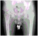
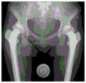
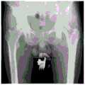
髋关节的X线图像是通过对髋关节进行X光片拍摄时获取的,同时在同张照片里拍摄一个已知尺寸的物体,即参照物。因此得到的髋关节的X线图像中包含参照物的图像。如图2所示,为髋关节的X线图像,其中图像的底部中心部位的标示标准尺寸的图像为参照物的图像。是实际应用中,参照物的选取和拍摄时的排放位置可以根据适应性的调整,本实施例不作限制。An X-ray image of the hip joint is obtained by taking an X-ray of the hip joint while taking an object of known size, the reference object, in the same photo. The X-ray image of the hip joint thus obtained includes the image of the reference object. As shown in FIG. 2 , it is an X-ray image of the hip joint, wherein the image of the standard size at the bottom center of the image is the image of the reference object. In practical applications, the selection of the reference object and the discharge position during shooting can be adjusted according to the adaptability, which is not limited in this embodiment.
S102.根据参照物的图像尺寸及其实际尺寸的比例,将髋关节的X线图像进行尺寸的还原。S102. According to the ratio of the image size of the reference object and its actual size, restore the size of the X-ray image of the hip joint.
参照物的尺寸是已知的,参照物的图像尺寸也可以通过测量得到,根据参照物的图像尺寸及其实际尺寸的比例,可以确定出髋关节的X线图像相对于实际的髋关节尺寸的比例(两者比例相同),然后根据比例将髋关节的X线图像进行真实尺寸的还原。将髋关节的X线图像进行真实尺寸的还原是为和后续的图像识别做基础,使后续根据识别结果确定的腿长差、髋臼杯位置、股骨柄假体的规格型号、截骨线位置与实际的对应位置差距更小,保证识别的准确性。The size of the reference object is known, and the image size of the reference object can also be obtained by measurement. According to the ratio of the image size of the reference object and its actual size, the difference between the X-ray image of the hip joint and the actual size of the hip joint can be determined. Scale (the two are the same scale), and then restore the true size of the X-ray image of the hip joint according to the scale. The restoration of the real size of the X-ray image of the hip joint is the basis for the subsequent image recognition, so that the leg length difference, the position of the acetabular cup, the size of the femoral stem prosthesis, and the position of the osteotomy line can be determined according to the recognition results. The gap with the actual corresponding position is smaller to ensure the accuracy of recognition.
具体的还原操作可以为选取已知尺寸物体的关键部位尺寸。通过计算图像中像素间两点距离,和物体实际尺寸进行比例换算,确定比例,然后根据比例对髋关节的X线图像的比例进行修正。A specific restoration operation may be selecting the size of a key part of an object of known size. By calculating the distance between two points between pixels in the image, and converting the ratio with the actual size of the object, the ratio is determined, and then the ratio of the X-ray image of the hip joint is corrected according to the ratio.
S103.基于深度学习模型对还原后的髋关节的X线图像进行识别,确定腿长差、髋臼杯位置以及股骨柄假体的规格型号。S103. Identify the X-ray image of the restored hip joint based on the deep learning model, and determine the leg length difference, the position of the acetabular cup, and the specification and model of the femoral stem prosthesis.
深度学习模型是神经网络模型,确定腿长差、髋臼杯位置以及股骨柄假体的规格型号可能会用到的模型的输入和输出可能是不同的,但是模型训练的原理是相同的。具体的,神经网络模型训练的原理为:将髋关节的X线图像转化为0-255灰度图,然后将图像进行人工选定标注,将图片的每个像素标注划分为几种属性值(根据实际的需求,属性值的种类数不同,比如可以为两种、三种等等)并分别命名,然后将其输入到神经网络模型中进行卷积池化采样一直迭代学习训练得到神经网络模型。The deep learning model is a neural network model. The input and output of the model that may be used to determine the leg length difference, the position of the acetabular cup, and the size of the femoral stem prosthesis may be different, but the principle of model training is the same. Specifically, the training principle of the neural network model is as follows: convert the X-ray image of the hip joint into a 0-255 grayscale image, then manually select and label the image, and divide each pixel label of the image into several attribute values ( According to actual needs, the types of attribute values are different, such as two, three, etc.) and name them respectively, and then input them into the neural network model for convolution pooling sampling and iterative learning and training to obtain the neural network model .
本步骤中的神经网络模型为分类神经网络,是将图像中的不同的区域进行分类,比如在确定腿长差时,应用神经网络模型主要是为了识别出泪滴和股骨小转子的关键点;再比如在确定髋臼杯位置时,应用神经网络模型主要是为了识别出股骨头区域;再比如在确定股骨柄假体的规格型号时,应用神经网络模型主要是为了识别出股骨头、骨皮质区域以及股骨头、股骨颈基底区域。The neural network model in this step is a classification neural network, which is to classify different areas in the image. For example, when determining the leg length difference, the neural network model is mainly used to identify key points of teardrops and lesser trochanter; For another example, when determining the position of the acetabular cup, the neural network model is mainly used to identify the femoral head area; for example, when determining the size of the femoral stem prosthesis, the neural network model is mainly used to identify the femoral head and bone cortex. area as well as the femoral head and base of the femoral neck.
本实施例中的神经网络可以为卷积神经网络LeNet、卷积神经网络AlexNet、可视化卷积神经网络ZF-Net、卷积神经网络GoogleNet、卷积神经网络VGG、卷积神经网络Inception、卷积神经网络ResNet、卷积神经网络DensNet、卷积神经网络Inception ResNet等。The neural network in this embodiment may be a convolutional neural network LeNet, a convolutional neural network AlexNet, a visualization convolutional neural network ZF-Net, a convolutional neural network GoogleNet, a convolutional neural network VGG, a convolutional neural network Inception, a convolutional neural network Neural Network ResNet, Convolutional Neural Network DensNet, Convolutional Neural Network Inception ResNet, etc.
确定腿长差、髋臼杯位置以及股骨柄假体的规格型号是根据图像的识别结果再进行一些坐标、拟合等计算后确定的。To determine the leg length difference, the position of the acetabular cup, and the size and model of the femoral stem prosthesis, some coordinates and fitting are calculated based on the image recognition results.
S104.根据股骨柄假体的旋转中心和在髋关节的X线图像识别过程中确定的髋臼杯的旋转中心确定截骨线位置。S104. Determine the position of the osteotomy line according to the rotation center of the femoral stem prosthesis and the rotation center of the acetabular cup determined during the X-ray image recognition process of the hip joint.
具体的,“根据股骨柄假体的旋转中心和在髋关节的X线图像识别过程中确定的髋臼杯的旋转中心确定截骨线位置”为通过移动股骨柄假体,将股骨柄假体的旋转中心与之前计算的髋臼杯旋转中心位置重合,得到股骨柄假体实际位置。沿股骨柄假体的涂层位置可确定临床中的实际截骨线位置,如图3-4所示。图3为移动股骨柄假体到预定位置,使股骨柄假体的旋转中心与之前计算的髋臼杯旋转中心位置重合,图4为根据股骨柄假体的外形确定截骨线位置。Specifically, “determine the position of the osteotomy line according to the rotation center of the femoral stem prosthesis and the rotation center of the acetabular cup determined during the X-ray image recognition of the hip joint” is to move the femoral stem prosthesis to move the femoral stem prosthesis The center of rotation of the acetabular cup coincides with the previously calculated center of rotation of the acetabular cup to obtain the actual position of the femoral stem prosthesis. The location of the coating along the femoral stem component can determine the actual position of the osteotomy line in clinical practice, as shown in Figure 3-4. Figure 3 shows moving the femoral stem prosthesis to a predetermined position so that the rotation center of the femoral stem prosthesis coincides with the previously calculated acetabular cup rotation center position, and Figure 4 shows the position of the osteotomy line determined according to the shape of the femoral stem prosthesis.
从以上的描述中,可以看出,本申请实施例的基于深度学习与X线的全髋关节置换术前规划方法中,获取髋关节的X线图像,所述髋关节的X线图像中包含参照物的图像,所述参照物为已知尺寸的参照物;根据参照物的图像尺寸及其实际尺寸的比例,将髋关节的X线图像进行尺寸的还原;基于深度学习模型对还原后的髋关节的X线图像进行识别,确定腿长差、髋臼杯位置以及股骨柄假体的规格型号;根据股骨柄假体的旋转中心和在髋关节的X线图像识别过程中确定的髋臼杯的旋转中心确定截骨线位置。可以看出,本实施例的全髋关节置换术前规划方式中,将髋关节的X线图像进行了真实尺寸的还原,以实际的尺寸进行后续的位置识别更准确;另外,在对X线图像识别的过程中都是基于深度学习模型进行识别的,进一步的保证了根据识别结果确定的腿长差、髋臼杯位置、股骨柄假体的规格型号、截骨线位置的准确性和快速性,从而为全髋关节置换手术提供了更好的术前支持。From the above description, it can be seen that in the preoperative planning method for total hip arthroplasty based on deep learning and X-ray in the embodiment of the present application, the X-ray image of the hip joint is obtained, and the X-ray image of the hip joint includes The image of the reference object, the reference object is a reference object of known size; according to the ratio of the image size of the reference object and its actual size, the X-ray image of the hip joint is restored in size; based on the deep learning model, the restored The X-ray image of the hip joint is identified to determine the leg length difference, the position of the acetabular cup, and the size of the femoral stem prosthesis; according to the rotation center of the femoral stem prosthesis and the acetabulum determined during the identification process of the X-ray image of the hip joint The center of rotation of the cup determines the position of the osteotomy line. It can be seen that, in the preoperative planning method for total hip arthroplasty in this embodiment, the X-ray image of the hip joint is restored to its real size, and subsequent position recognition is more accurate with the actual size; The image recognition process is based on the deep learning model, which further ensures the accuracy and speed of the leg length difference, acetabular cup position, femoral stem prosthesis size, and osteotomy line position determined according to the recognition results. sex, thereby providing better preoperative support for total hip replacement surgery.
进一步的,作为上述实施例的进一步细化,步骤S103对于确定腿长差、髋臼杯位置以及股骨柄假体的规格型号的详细步骤进行分别说明。Further, as a further refinement of the above-mentioned embodiment, step S103 separately describes the detailed steps of determining the leg length difference, the position of the acetabular cup, and the specification and model of the femoral stem prosthesis.
如图5所示,为确定腿长差的流程图,具体包括如下步骤:As shown in Figure 5, the flowchart for determining the leg length difference specifically includes the following steps:
S201.将髋关节的X线图像转化为灰度图。S201. Convert the X-ray image of the hip joint into a grayscale image.
将髋关节的X线图像转化为0-255灰度图。Convert the X-ray image of the hip joint to a 0-255 grayscale image.
S202.基于第一神经网络模型对灰度图的每个像素值进行预测,确定泪滴关键点以及股骨小转子关键点位置。S202. Predict the value of each pixel of the grayscale image based on the first neural network model, and determine the position of the key point of the teardrop and the key point of the lesser trochanter of the femur.
在进行预测之前,首先要根据样本训练得到第一神经网络模型。具体的,将未标记的原始图像(髋关节的X线样本图像对应的灰度图)以及人工识别标记的泪滴关键点及股骨小转子关键点位置的标记传入到卷积神经网络中,将输入的原始图像与特征点的高斯分布函数进行拟合,进行卷积池化采样一直迭代学习训练得到第一神经网络模型。需要说明的是,本步骤中的卷积神经网络可以为卷积神经网络LeNet、卷积神经网络AlexNet、可视化卷积神经网络ZF-Net、卷积神经网络GoogleNet、卷积神经网络VGG、卷积神经网络Inception、卷积神经网络ResNet、卷积神经网络DensNet、卷积神经网络Inception ResNet等。Before making predictions, the first neural network model must be obtained by training the samples. Specifically, the unlabeled original image (the grayscale image corresponding to the X-ray sample image of the hip joint) and the manually identified and marked teardrop key points and the labels of the key points of the lesser trochanter are passed into the convolutional neural network, The input original image is fitted with the Gaussian distribution function of the feature points, and the convolution pooling sampling is performed to iteratively learn and train to obtain the first neural network model. It should be noted that the convolutional neural network in this step can be the convolutional neural network LeNet, the convolutional neural network AlexNet, the visualization convolutional neural network ZF-Net, the convolutional neural network GoogleNet, the convolutional neural network VGG, the convolutional neural network. Neural Network Inception, Convolutional Neural Network ResNet, Convolutional Neural Network DensNet, Convolutional Neural Network Inception ResNet, etc.
得到第一神经网络模型后,将髋关节的X线图像对应的灰度图输入到第一神经网络模型中,可以自动识别出泪滴关键点以及股骨小转子关键点位置。如图6所示,图6为自动识别出泪滴关键点以及股骨小转子关键点位置的示意图。After the first neural network model is obtained, the grayscale image corresponding to the X-ray image of the hip joint is input into the first neural network model, and the key points of the teardrop and the lesser trochanter can be automatically identified. As shown in FIG. 6 , FIG. 6 is a schematic diagram of automatically identifying the key points of the tear drop and the key points of the lesser trochanter of the femur.
S203.根据泪滴关键点以及股骨小转子关键点位置,确定腿长差。S203. Determine the leg length difference according to the key point of the tear drop and the position of the key point of the lesser trochanter.
具体的,如图7所示,其中水平的直线是由两个泪滴关键点确定的,是两个泪滴关键点的连线,其中两条垂直的线段是由股骨小转子关键点和水平直线确定的,两条垂直直线分别记作A和B,A和B的差值为腿长差。Specifically, as shown in Figure 7, the horizontal straight line is determined by the two key points of teardrops, and is the connection line between the two key points of teardrops, and the two vertical line segments are determined by the key point of the lesser trochanter and the horizontal Determined by a straight line, the two vertical straight lines are respectively recorded as A and B, and the difference between A and B is the leg length difference.
如图8所示,为确定髋臼杯位置的流程图,具体包括如下步骤:As shown in Figure 8, the flow chart for determining the position of the acetabular cup specifically includes the following steps:
S301.将髋关节的X线图像转化为灰度图。S301. Convert the X-ray image of the hip joint into a grayscale image.
将髋关节的X线图像转化为0-255灰度图。Convert the X-ray image of the hip joint to a 0-255 grayscale image.
S302.基于第二神经网络模型对灰度图的每个像素值进行预测,确定股骨头位置。S302. Predict the value of each pixel of the grayscale image based on the second neural network model, and determine the position of the femoral head.
在进行预测之前,首先要根据样本训练得到第二神经网络模型。具体的,将未标记的原始图像(髋关节的X线样本图像对应的灰度图)以及人工识别标记像素属性值的标记图像传入到卷积神经网络中,包括两种属性值,分别命名0、1。数值0代表背景像素,1代表股骨头像素;传入到卷积神经网络中,进行卷积池化采样一直迭代学习训练得到第二神经网络模型。需要说明的是,本步骤中的卷积神经网络可以为卷积神经网络LeNet、卷积神经网络AlexNet、可视化卷积神经网络ZF-Net、卷积神经网络GoogleNet、卷积神经网络VGG、卷积神经网络Inception、卷积神经网络ResNet、卷积神经网络DensNet、卷积神经网络InceptionResNet等。Before making predictions, the second neural network model must first be obtained by training the samples. Specifically, the unlabeled original image (the grayscale image corresponding to the X-ray sample image of the hip joint) and the labeled image with the attribute value of the labeled pixel are passed into the convolutional neural network, including two attribute values, named respectively. 0, 1. The value 0 represents the background pixel, and 1 represents the femoral head pixel; it is passed into the convolutional neural network, and the convolution pooling sampling is performed to iteratively learn and train to obtain the second neural network model. It should be noted that the convolutional neural network in this step can be the convolutional neural network LeNet, the convolutional neural network AlexNet, the visualization convolutional neural network ZF-Net, the convolutional neural network GoogleNet, the convolutional neural network VGG, the convolutional neural network. Neural Network Inception, Convolutional Neural Network ResNet, Convolutional Neural Network DensNet, Convolutional Neural Network InceptionResNet, etc.
得到第二神经网络模型后,将髋关节的X线图像对应的灰度图输入到第二神经网络模型中,可以对每个像素值进行预测。自动将X线图像的每个像素值归为一个属性中:0-背景,1-股骨头,完成股骨头区域(即股骨头位置)的自动识别,如图9所示。图9为识别股骨头的示意图。After the second neural network model is obtained, the grayscale image corresponding to the X-ray image of the hip joint is input into the second neural network model, and each pixel value can be predicted. Each pixel value of the X-ray image is automatically classified into an attribute: 0-background, 1-femoral head, and the automatic identification of the femoral head area (ie, the position of the femoral head) is completed, as shown in Figure 9. Figure 9 is a schematic diagram of identifying the femoral head.
S303.根据平面图像的质心公式计算股骨头旋转中心。S303. Calculate the center of rotation of the femoral head according to the centroid formula of the plane image.
因为得到的股骨头区域的图像是二值图像,其质量分布是均匀的,所以质心和形心重合,根据平面图像的质心公式可以计算得到股骨头的中心点坐标,即股骨头旋转中心。假设二值图像为B[i,j],则可根据下列公式求得股骨头的中心点坐标:Because the obtained image of the femoral head area is a binary image, its mass distribution is uniform, so the centroid and centroid coincide, and the center point coordinates of the femoral head can be calculated according to the centroid formula of the plane image, that is, the rotation center of the femoral head. Assuming that the binary image is B[i,j], the coordinates of the center point of the femoral head can be obtained according to the following formula:

其中:


S304.根据股骨头的直径推算髋臼杯直径。S304. Calculate the diameter of the acetabular cup according to the diameter of the femoral head.
根据股骨头区域和股骨头旋转中心确定股骨头的直径,根据股骨头的直径推算髋臼杯直径。根据股骨头的直径推算髋臼杯直径的可以参考现有的任意一种推算方式确定髋臼杯直径。The diameter of the femoral head was determined from the area of the femoral head and the center of rotation of the femoral head, and the diameter of the acetabular cup was calculated from the diameter of the femoral head. To calculate the diameter of the acetabular cup according to the diameter of the femoral head, the diameter of the acetabular cup can be determined by referring to any of the existing calculation methods.
S305.根据骨头旋转中心和髋臼杯直径确定髋臼杯位置。S305. Determine the acetabular cup position according to the bone rotation center and the acetabular cup diameter.
根据股骨头的直径以及股骨头旋转中心位置自动确定髋臼杯位置,如图11所示。图11中线条勾画的区域为髋臼杯位置。The position of the acetabular cup is automatically determined based on the diameter of the femoral head and the position of the center of rotation of the femoral head, as shown in Figure 11. The area delineated by the lines in Figure 11 is the position of the acetabular cup.
如图12所示,为确定股骨柄假体的规格型号的流程图,具体包括如下步骤:As shown in Figure 12, the flow chart for determining the specification and model of the femoral stem prosthesis specifically includes the following steps:
S401.将髋关节的X线图像转化为灰度图。S401. Convert the X-ray image of the hip joint into a grayscale image.
将髋关节的X线图像转化为0-255灰度图。Convert the X-ray image of the hip joint to a 0-255 grayscale image.
S402.基于第三神经网络模型对灰度图进行识别,确定髓腔解剖轴线。S402. Identify the grayscale image based on the third neural network model, and determine the anatomical axis of the medullary cavity.
具体的,确定髓腔解剖轴线包括如下步骤:Specifically, determining the anatomical axis of the medullary cavity includes the following steps:
首先,基于第三神经网络模型对灰度图的每个像素值进行预测,确定股骨头区域和骨皮质区域;First, predict each pixel value of the grayscale image based on the third neural network model to determine the femoral head area and the bone cortex area;
在进行预测之前,首先要根据样本训练得到第三神经网络模型。具体的,将未标记的原始图像(髋关节的X线样本图像对应的灰度图)以及人工识别标记像素属性值的标记图像传入到卷积神经网络中,包括三种属性值,分别命名0、1、2。数值0代表背景像素,1代表股骨头像素,2代表骨皮质;传入到卷积神经网络中,进行卷积池化采样一直迭代学习训练得到第三神经网络模型。需要说明的是,本步骤中的卷积神经网络可以为卷积神经网络LeNet、卷积神经网络AlexNet、可视化卷积神经网络ZF-Net、卷积神经网络GoogleNet、卷积神经网络VGG、卷积神经网络Inception、卷积神经网络ResNet、卷积神经网络DensNet、卷积神经网络Inception ResNet等。Before making predictions, a third neural network model must be obtained by training samples. Specifically, the unlabeled original image (the grayscale image corresponding to the X-ray sample image of the hip joint) and the labeled image with the attribute value of the labeled pixel by manual identification are passed into the convolutional neural network, including three attribute values, named respectively 0, 1, 2. The value 0 represents the background pixel, 1 represents the femoral head pixel, and 2 represents the bone cortex; it is passed into the convolutional neural network, and the convolution pooling sampling is performed to iteratively learn and train to obtain the third neural network model. It should be noted that the convolutional neural network in this step can be the convolutional neural network LeNet, the convolutional neural network AlexNet, the visual convolutional neural network ZF-Net, the convolutional neural network GoogleNet, the convolutional neural network VGG, the convolutional neural network. Neural Network Inception, Convolutional Neural Network ResNet, Convolutional Neural Network DensNet, Convolutional Neural Network Inception ResNet, etc.
得到第三神经网络模型后,将髋关节的X线图像对应的灰度图输入到第三神经网络模型中,可以对每个像素值进行预测。自动将X线图像的每个像素值归为一个属性中:0-背景,1-股骨头,2-骨皮质,完成股骨头区域、骨皮质区域的自动识别,如图13所示。图13为识别股骨头区域、骨皮质区域的示意图。After the third neural network model is obtained, the grayscale image corresponding to the X-ray image of the hip joint is input into the third neural network model, and each pixel value can be predicted. Each pixel value of the X-ray image is automatically classified into an attribute: 0-background, 1-femoral head, 2-cortical bone, to complete the automatic identification of femoral head area and cortical bone area, as shown in Figure 13. 13 is a schematic diagram of identifying the femoral head region and the cortical bone region.
其次,根据股骨头区域、骨皮质区域确定髓腔区域;Secondly, determine the medullary cavity area according to the femoral head area and the bone cortex area;
具体的,截取小转子结束处直到股骨末端部位,使用图像中股骨区域减去骨皮质区域得到的是髓腔区域,如图14所示。Specifically, the end of the lesser trochanter was cut to the end of the femur, and the medullary cavity region was obtained by subtracting the cortical bone region from the femoral region in the image, as shown in Figure 14.
最后,对髓腔区域多个中心点坐标进行直线拟合确定髓腔解剖轴线。Finally, the anatomical axis of the medullary canal is determined by linear fitting on the coordinates of multiple center points in the medullary canal region.
具体的,如图15所示,从小转子结束位置以下,每横行与髓腔交点为四个坐标,从左至右分别命名为A1,A2,B1,B2;依据两点可以求出中点,A1(X1,Y1),A2(X2,Y2)的中点坐标:

S403.基于第四神经网络模型对灰度图进行识别,确定股骨颈中心轴线。S403. Identify the grayscale image based on the fourth neural network model, and determine the central axis of the femoral neck.
具体的,确定股骨颈中心轴线包括如下步骤:Specifically, determining the central axis of the femoral neck includes the following steps:
首先,基于第四神经网络模型对灰度图的每个像素值进行预测,确定股骨头区域和股骨颈基底区域;First, predict each pixel value of the grayscale image based on the fourth neural network model to determine the femoral head area and the femoral neck base area;
在进行预测之前,首先要根据样本训练得到第四神经网络模型。具体的,将未标记的原始图像(髋关节的X线样本图像对应的灰度图)以及人工识别标记像素属性值的标记图像传入到卷积神经网络中,包括三种属性值,分别命名0、1、2。数值0代表背景像素,1代表股骨头像素,2代表股骨颈基底像素;传入到卷积神经网络中,进行卷积池化采样一直迭代学习训练得到第四神经网络模型。需要说明的是,本步骤中的卷积神经网络可以为卷积神经网络LeNet、卷积神经网络AlexNet、可视化卷积神经网络ZF-Net、卷积神经网络GoogleNet、卷积神经网络VGG、卷积神经网络Inception、卷积神经网络ResNet、卷积神经网络DensNet、卷积神经网络Inception ResNet等。Before making predictions, a fourth neural network model must be obtained by training samples. Specifically, the unlabeled original image (the grayscale image corresponding to the X-ray sample image of the hip joint) and the labeled image with the attribute value of the labeled pixel by manual identification are passed into the convolutional neural network, including three attribute values, named respectively 0, 1, 2. The value 0 represents the background pixel, 1 represents the femoral head pixel, and 2 represents the femoral neck base pixel; it is passed into the convolutional neural network, and the convolutional pooling sampling is performed to iteratively learn and train to obtain the fourth neural network model. It should be noted that the convolutional neural network in this step can be the convolutional neural network LeNet, the convolutional neural network AlexNet, the visual convolutional neural network ZF-Net, the convolutional neural network GoogleNet, the convolutional neural network VGG, the convolutional neural network. Neural Network Inception, Convolutional Neural Network ResNet, Convolutional Neural Network DensNet, Convolutional Neural Network Inception ResNet, etc.
在得到第四神经网络模型后,将髋关节的X线图像对应的灰度图输入到第四神经网络模型中,可以对每个像素值进行预测。自动将X线图像的每个像素值归为一个属性中:0-背景,1-股骨头,2-股骨颈基底像素,完成股骨头区域、股骨颈基底区域的自动识别,如图16所示。图16为识别股骨头区域、股骨颈基底区域的示意图。After the fourth neural network model is obtained, the grayscale image corresponding to the X-ray image of the hip joint is input into the fourth neural network model, and each pixel value can be predicted. Automatically classify each pixel value of the X-ray image into an attribute: 0-background, 1-femoral head, 2-femoral neck base pixel, complete the automatic identification of femoral head area and femoral neck base area, as shown in Figure 16 . Fig. 16 is a schematic diagram of identifying the femoral head region and the femoral neck base region.
其次,根据平面图像的质心公式计算股骨头区域和股骨颈基底区域对应的股骨头中心坐标和股骨颈基底中心坐标;Secondly, the center coordinates of the femoral head and the base of the femoral neck corresponding to the femoral head area and the femoral neck base area are calculated according to the centroid formula of the plane image;
股骨头中心坐标和股骨颈基底中心坐标的计算方式类似,都可以参见步骤S303中计算股骨头中心点坐标的实现方式,此处不在赘述。The calculation methods of the center coordinates of the femoral head and the center coordinates of the base of the femoral neck are similar, and reference may be made to the implementation method of calculating the coordinates of the center point of the femoral head in step S303, which is not repeated here.
最后,根据股骨头中心坐标和股骨颈基底中心坐标确定股骨颈中心轴线。Finally, the center axis of the femoral neck is determined according to the center coordinates of the femoral head and the center coordinates of the base of the femoral neck.
具体的,股骨头中心坐标和股骨颈基底中心坐标连线即为股骨颈中心轴线,如图17所示。图17中两条斜向下的线段为股骨颈中心轴线。Specifically, the line connecting the center coordinates of the femoral head and the center coordinates of the base of the femoral neck is the center axis of the femoral neck, as shown in FIG. 17 . The two diagonally downward line segments in Figure 17 are the central axis of the femoral neck.
S404.根据髓腔解剖轴线和股骨颈中心轴线确定股骨颈干角。S404. Determine the femoral neck shaft angle according to the anatomical axis of the medullary cavity and the central axis of the femoral neck.
具体的,髓腔解剖轴线和股骨颈中心轴线形成的夹角为股骨颈干角。Specifically, the angle formed by the anatomical axis of the medullary cavity and the central axis of the femoral neck is the femoral neck shaft angle.
S405.根据股骨颈干角、在确定髓腔解剖轴线过程中确定的髓腔区域以及股骨头旋转中心确定股骨柄假体的规格型号。S405. Determine the specification and model of the femoral stem prosthesis according to the femoral neck shaft angle, the medullary canal area determined in the process of determining the anatomical axis of the medullary canal, and the center of rotation of the femoral head.
具体的,根据股骨颈干角角度值,再结合髓腔形态,股骨头旋转中心位置可对股骨柄假体型号的选择给出推荐。股骨柄假体型号按照股骨柄假体的形状和尺寸等特征进行区分。Specifically, according to the angle value of the femoral neck shaft angle, combined with the shape of the medullary cavity, the position of the center of rotation of the femoral head can give a recommendation for the selection of the femoral stem prosthesis model. Femoral stem prosthesis models are distinguished by the shape and size of the femoral stem prosthesis.
进一步的,作为图1实施例的补充说明,在确定截骨线位置之后,还包括根据截骨线位置,计算术后的腿长差,以及偏距。具体的,偏距包含股骨偏心距:指股骨头旋转中心至股骨干长轴间的垂直距离。还包括联合偏距,具体为股骨和髋臼偏距的累积和。Further, as a supplementary description of the embodiment in FIG. 1 , after the position of the osteotomy line is determined, it also includes calculating the post-operative leg length difference and offset according to the position of the osteotomy line. Specifically, the offset includes the femoral offset: refers to the vertical distance from the center of rotation of the femoral head to the long axis of the femoral shaft. Also included are joint offsets, specifically the cumulative sum of femoral and acetabular offsets.
需要说明的是,在附图的流程图示出的步骤可以在诸如一组计算机可执行指令的计算机系统中执行,并且,虽然在流程图中示出了逻辑顺序,但是在某些情况下,可以以不同于此处的顺序执行所示出或描述的步骤。It should be noted that the steps shown in the flowcharts of the accompanying drawings may be executed in a computer system, such as a set of computer-executable instructions, and, although a logical sequence is shown in the flowcharts, in some cases, Steps shown or described may be performed in an order different from that herein.
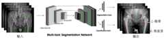
根据本申请实施例,还提供了一种用于实施上述图1-17所述方法的基于深度学习与X线的全髋关节置换术前规划装置,如图18所示,该装置包括:According to the embodiment of the present application, a preoperative planning device for total hip arthroplasty based on deep learning and X-rays for implementing the methods described in Figures 1-17 is also provided. As shown in Figure 18, the device includes:
比例校准单元51,用于根据参照物的图像尺寸及其实际尺寸的比例,将髋关节的X线图像进行尺寸的真实还原;The scale calibration unit 51 is used to restore the real size of the X-ray image of the hip joint according to the ratio of the image size of the reference object and its actual size;
腿长差确定单元52,用于基于第一神经网络模型对还原后的髋关节的X线图像进行识别,确定腿长差;The leg length difference determination unit 52 is used for identifying the X-ray image of the restored hip joint based on the first neural network model, and determining the leg length difference;
髋臼杯位置确定单元53,用于基于第二神经网络模型对还原后的髋关节的X线图像进行识别,确定髋臼杯位置;The acetabular cup position determining unit 53 is used for identifying the restored X-ray image of the hip joint based on the second neural network model, and determining the position of the acetabular cup;
股骨柄假体规格确定单元54,用于基于第三神经网络模型、第四神经网络模型对还原后的髋关节的X线图像进行识别,确定股骨柄假体规格;The femoral stem prosthesis specification determining unit 54 is used for identifying the restored X-ray image of the hip joint based on the third neural network model and the fourth neural network model, and determining the femoral stem prosthesis specification;
截骨线确定单元55,用于根据股骨柄假体的旋转中心和在髋关节的X线图像识别过程中确定的髋臼杯的旋转中心确定截骨线位置。The osteotomy line determining unit 55 is configured to determine the position of the osteotomy line according to the rotation center of the femoral stem prosthesis and the rotation center of the acetabular cup determined during the X-ray image recognition process of the hip joint.
具体的,本申请实施例的装置中各单元、模块实现其功能的具体过程可参见方法实施例中的相关描述,此处不再赘述。Specifically, for the specific process of implementing the functions of each unit and module in the apparatus according to the embodiment of the present application, reference may be made to the relevant description in the method embodiment, which will not be repeated here.
从以上的描述中,可以看出,本申请实施例的基于深度学习与X线的全髋关节置换术前规划装置中,获取髋关节的X线图像,所述髋关节的X线图像中包含参照物的图像,所述参照物为已知尺寸的参照物;根据参照物的图像尺寸及其实际尺寸的比例,将髋关节的X线图像进行尺寸的还原;基于深度学习模型对还原后的髋关节的X线图像进行识别,确定腿长差、髋臼杯位置以及股骨柄假体的规格型号;根据股骨柄假体的旋转中心和在髋关节的X线图像识别过程中确定的髋臼杯的旋转中心确定截骨线位置。可以看出,本实施例的全髋关节置换术前规划方式中,将髋关节的X线图像进行了真实尺寸的还原,以实际的尺寸进行后续的位置识别更准确;另外,在对X线图像识别的过程中都是基于深度学习模型进行识别的,进一步的保证了根据识别结果确定的腿长差、髋臼杯位置、股骨柄假体的规格型号、截骨线位置的准确性和快速性,从而为全髋关节置换手术提供了更好的术前支持。From the above description, it can be seen that in the preoperative planning device for total hip arthroplasty based on deep learning and X-ray in the embodiment of the present application, the X-ray image of the hip joint is obtained, and the X-ray image of the hip joint includes The image of the reference object, the reference object is a reference object of known size; according to the ratio of the image size of the reference object and its actual size, the X-ray image of the hip joint is restored in size; based on the deep learning model, the restored The X-ray image of the hip joint is identified to determine the leg length difference, the position of the acetabular cup, and the size of the femoral stem prosthesis; according to the rotation center of the femoral stem prosthesis and the acetabulum determined during the identification process of the X-ray image of the hip joint The center of rotation of the cup determines the position of the osteotomy line. It can be seen that, in the preoperative planning method for total hip arthroplasty in this embodiment, the X-ray image of the hip joint is restored to its real size, and subsequent position recognition is more accurate with the actual size; The image recognition process is based on the deep learning model, which further ensures the accuracy and speed of the leg length difference, acetabular cup position, femoral stem prosthesis size, and osteotomy line position determined according to the recognition results. sex, thereby providing better preoperative support for total hip replacement surgery.
根据本申请实施例,还提供了一种计算机可读存储介质,其特征在于,所述计算机可读存储介质存储有计算机指令,所述计算机指令用于使所述计算机执行上述方法实施例中的基于深度学习与X线的全髋关节置换术前规划方法。According to an embodiment of the present application, a computer-readable storage medium is further provided, wherein the computer-readable storage medium stores computer instructions, and the computer instructions are used to cause the computer to execute the methods in the foregoing method embodiments. A preoperative planning method for total hip arthroplasty based on deep learning and X-ray.
根据本申请实施例,还提供了一种电子设备,包括:至少一个处理器;以及与所述至少一个处理器通信连接的存储器;其中,所述存储器存储有可被所述至少一个处理器执行的计算机程序,所述计算机程序被所述至少一个处理器执行,以使所述至少一个处理器执行上述方法实施例中的基于深度学习与X线的全髋关节置换术前规划方法。According to an embodiment of the present application, an electronic device is further provided, including: at least one processor; and a memory communicatively connected to the at least one processor; wherein the memory stores data executable by the at least one processor. The computer program is executed by the at least one processor, so that the at least one processor executes the deep learning and X-ray-based preoperative planning method for total hip arthroplasty in the above method embodiments.
显然,本领域的技术人员应该明白,上述的本申请的各模块或各步骤可以用通用的计算装置来实现,它们可以集中在单个的计算装置上,或者分布在多个计算装置所组成的网络上,可选地,它们可以用计算装置可执行的程序代码来实现,从而,可以将它们存储在存储装置中由计算装置来执行,或者将它们分别制作成各个集成电路模块,或者将它们中的多个模块或步骤制作成单个集成电路模块来实现。这样,本申请不限制于任何特定的硬件和软件结合。Obviously, those skilled in the art should understand that the above-mentioned modules or steps of the present application can be implemented by a general-purpose computing device, and they can be centralized on a single computing device, or distributed in a network composed of multiple computing devices Alternatively, they can be implemented with program codes executable by a computing device, so that they can be stored in a storage device and executed by the computing device, or they can be made into individual integrated circuit modules, or they can be integrated into The multiple modules or steps are fabricated into a single integrated circuit module. As such, the present application is not limited to any particular combination of hardware and software.
以上所述仅为本申请的优选实施例而已,并不用于限制本申请,对于本领域的技术人员来说,本申请可以有各种更改和变化。凡在本申请的精神和原则之内,所作的任何修改、等同替换、改进等,均应包含在本申请的保护范围之内。The above descriptions are only preferred embodiments of the present application, and are not intended to limit the present application. For those skilled in the art, the present application may have various modifications and changes. Any modification, equivalent replacement, improvement, etc. made within the spirit and principle of this application shall be included within the protection scope of this application.
Claims (10)
Applications Claiming Priority (2)
| Application Number | Priority Date | Filing Date | Title |
|---|---|---|---|
| CN2020106437135 | 2020-07-06 | ||
| CN202010643713 | 2020-07-06 |
Publications (2)
| Publication Number | Publication Date |
|---|---|
| CN111888059Atrue CN111888059A (en) | 2020-11-06 |
| CN111888059B CN111888059B (en) | 2021-07-27 |
Family
ID=73190359
Family Applications (1)
| Application Number | Title | Priority Date | Filing Date |
|---|---|---|---|
| CN202010707817.8AActiveCN111888059B (en) | 2020-07-06 | 2020-07-21 | Total hip image processing method and device based on deep learning and X-ray |
Country Status (2)
| Country | Link |
|---|---|
| CN (1) | CN111888059B (en) |
| WO (1) | WO2022007972A1 (en) |
Cited By (18)
| Publication number | Priority date | Publication date | Assignee | Title |
|---|---|---|---|---|
| CN112842529A (en)* | 2020-12-31 | 2021-05-28 | 北京长木谷医疗科技有限公司 | Total knee replacement preoperative planning method and device |
| CN113133802A (en)* | 2021-04-20 | 2021-07-20 | 四川大学 | Bone surgery line automatic positioning method based on machine learning |
| CN113592821A (en)* | 2021-07-30 | 2021-11-02 | 瓴域影诺(北京)科技有限公司 | Femoral medullary cavity type detection method and system |
| CN113744214A (en)* | 2021-08-24 | 2021-12-03 | 北京长木谷医疗科技有限公司 | Femoral stem placement method and device based on deep reinforcement learning and electronic equipment |
| CN113907775A (en)* | 2021-10-13 | 2022-01-11 | 瓴域影诺(北京)科技有限公司 | A method and system for judging image quality of hip joint |
| CN113974920A (en)* | 2021-10-08 | 2022-01-28 | 北京长木谷医疗科技有限公司 | Knee joint femoral alignment method and device, electronic device, storage medium |
| CN114419618A (en)* | 2022-01-27 | 2022-04-29 | 北京长木谷医疗科技有限公司 | Deep learning-based preoperative planning system for total hip replacement |
| CN114612391A (en)* | 2022-02-24 | 2022-06-10 | 中国人民解放军总医院第四医学中心 | Calculation method and system for leg length difference after total hip joint operation based on deep learning |
| CN114648492A (en)* | 2022-02-24 | 2022-06-21 | 中国人民解放军总医院第四医学中心 | Calculation method and system for eccentricity after total hip surgery based on deep learning |
| CN114742747A (en)* | 2022-02-24 | 2022-07-12 | 北京长木谷医疗科技有限公司 | Deep learning-based image assessment method and system after hip arthroplasty |
| CN116423054A (en)* | 2023-03-09 | 2023-07-14 | 中铁九桥工程有限公司 | U rib plate welding method and welding system |
| CN116597002A (en)* | 2023-05-12 | 2023-08-15 | 北京长木谷医疗科技股份有限公司 | Automatic femoral stem placement method, device and equipment based on deep reinforcement learning |
| CN116650110A (en)* | 2023-06-12 | 2023-08-29 | 北京长木谷医疗科技股份有限公司 | Method and device for automatic placement of knee prosthesis based on deep reinforcement learning |
| CN116993824A (en)* | 2023-07-19 | 2023-11-03 | 北京长木谷医疗科技股份有限公司 | Acetabular rotation center calculating method, device, equipment and readable storage medium |
| CN117058087A (en)* | 2023-08-09 | 2023-11-14 | 联影智能医疗科技(北京)有限公司 | Medical image-based hip joint prosthesis matching method and program product |
| CN118121299A (en)* | 2024-03-26 | 2024-06-04 | 北京和华瑞博医疗科技有限公司 | Data processing method, apparatus, device, medium, and program product |
| CN118806432A (en)* | 2024-07-31 | 2024-10-22 | 北京长木谷医疗科技股份有限公司 | A method and device for planning periacetabular osteotomy based on reinforcement learning |
| CN119385684A (en)* | 2024-12-27 | 2025-02-07 | 中国人民解放军总医院第四医学中心 | A method for predicting the position of the center of rotation of the hip joint during hip replacement surgery |
Families Citing this family (3)
| Publication number | Priority date | Publication date | Assignee | Title |
|---|---|---|---|---|
| CN114431957B (en)* | 2022-04-12 | 2022-07-29 | 北京长木谷医疗科技有限公司 | Total knee joint replacement postoperative revision preoperative planning system based on deep learning |
| CN115830247B (en)* | 2023-02-14 | 2023-07-14 | 北京壹点灵动科技有限公司 | Fitting method and device for hip joint rotation center, processor and electronic equipment |
| CN117437459B (en)* | 2023-10-08 | 2024-03-22 | 昆山市第一人民医院 | Method for realizing user knee joint patella softening state analysis based on decision network |
Citations (8)
| Publication number | Priority date | Publication date | Assignee | Title |
|---|---|---|---|---|
| CN101815477A (en)* | 2007-09-28 | 2010-08-25 | 株式会社力克赛 | Preoperative planning device for artificial knee joint replacement surgery and surgical auxiliary tool |
| CN103209652A (en)* | 2010-08-13 | 2013-07-17 | 史密夫和内修有限公司 | Surgical guides |
| CN106456196A (en)* | 2014-02-11 | 2017-02-22 | 史密夫和内修有限公司 | Anterior and posterior referencing sizing guides and cutting blocks and methods |
| CN107106307A (en)* | 2015-01-06 | 2017-08-29 | 沃尔德玛链接有限公司 | It is determined that being adapted to the measurer of the femoral implant size of the knee-joint prosthesis of patient |
| CN107252338A (en)* | 2009-05-29 | 2017-10-17 | 史密夫和内修有限公司 | Method and apparatus for performing arthroplasty of knee |
| CN110648337A (en)* | 2019-09-23 | 2020-01-03 | 武汉联影医疗科技有限公司 | Hip joint segmentation method, hip joint segmentation device, electronic apparatus, and storage medium |
| CN110730639A (en)* | 2017-03-14 | 2020-01-24 | S·B·墨菲 | System and method for determining leg length changes during hip surgery |
| CN111179350A (en)* | 2020-02-13 | 2020-05-19 | 张逸凌 | Hip joint image processing method based on deep learning and computing equipment |
Family Cites Families (1)
| Publication number | Priority date | Publication date | Assignee | Title |
|---|---|---|---|---|
| US6917827B2 (en)* | 2000-11-17 | 2005-07-12 | Ge Medical Systems Global Technology Company, Llc | Enhanced graphic features for computer assisted surgery system |
- 2020
- 2020-07-21CNCN202010707817.8Apatent/CN111888059B/enactiveActive
- 2021
- 2021-07-21WOPCT/CN2021/107720patent/WO2022007972A1/ennot_activeCeased
Patent Citations (8)
| Publication number | Priority date | Publication date | Assignee | Title |
|---|---|---|---|---|
| CN101815477A (en)* | 2007-09-28 | 2010-08-25 | 株式会社力克赛 | Preoperative planning device for artificial knee joint replacement surgery and surgical auxiliary tool |
| CN107252338A (en)* | 2009-05-29 | 2017-10-17 | 史密夫和内修有限公司 | Method and apparatus for performing arthroplasty of knee |
| CN103209652A (en)* | 2010-08-13 | 2013-07-17 | 史密夫和内修有限公司 | Surgical guides |
| CN106456196A (en)* | 2014-02-11 | 2017-02-22 | 史密夫和内修有限公司 | Anterior and posterior referencing sizing guides and cutting blocks and methods |
| CN107106307A (en)* | 2015-01-06 | 2017-08-29 | 沃尔德玛链接有限公司 | It is determined that being adapted to the measurer of the femoral implant size of the knee-joint prosthesis of patient |
| CN110730639A (en)* | 2017-03-14 | 2020-01-24 | S·B·墨菲 | System and method for determining leg length changes during hip surgery |
| CN110648337A (en)* | 2019-09-23 | 2020-01-03 | 武汉联影医疗科技有限公司 | Hip joint segmentation method, hip joint segmentation device, electronic apparatus, and storage medium |
| CN111179350A (en)* | 2020-02-13 | 2020-05-19 | 张逸凌 | Hip joint image processing method based on deep learning and computing equipment |
Non-Patent Citations (1)
| Title |
|---|
| XIANG LI: "《Evaluation of Biological Properties of Electron Beam Melted Ti6Al4V Implant with Biomimetic Coating In Vitro and In Vivo》", 《PLOS ONE》* |
Cited By (28)
| Publication number | Priority date | Publication date | Assignee | Title |
|---|---|---|---|---|
| WO2022142741A1 (en)* | 2020-12-31 | 2022-07-07 | 北京长木谷医疗科技有限公司 | Total knee arthroplasty preoperative planning method and device |
| CN112842529A (en)* | 2020-12-31 | 2021-05-28 | 北京长木谷医疗科技有限公司 | Total knee replacement preoperative planning method and device |
| CN113133802A (en)* | 2021-04-20 | 2021-07-20 | 四川大学 | Bone surgery line automatic positioning method based on machine learning |
| CN113592821A (en)* | 2021-07-30 | 2021-11-02 | 瓴域影诺(北京)科技有限公司 | Femoral medullary cavity type detection method and system |
| CN113744214A (en)* | 2021-08-24 | 2021-12-03 | 北京长木谷医疗科技有限公司 | Femoral stem placement method and device based on deep reinforcement learning and electronic equipment |
| WO2023024883A1 (en)* | 2021-08-24 | 2023-03-02 | 北京长木谷医疗科技有限公司 | Femoral stem placement method and apparatus based on deep reinforcement learning, and electronic device |
| CN113974920A (en)* | 2021-10-08 | 2022-01-28 | 北京长木谷医疗科技有限公司 | Knee joint femoral alignment method and device, electronic device, storage medium |
| CN113974920B (en)* | 2021-10-08 | 2022-10-11 | 北京长木谷医疗科技有限公司 | Knee joint femur force line determining method and device, electronic equipment and storage medium |
| CN113907775A (en)* | 2021-10-13 | 2022-01-11 | 瓴域影诺(北京)科技有限公司 | A method and system for judging image quality of hip joint |
| CN113907775B (en)* | 2021-10-13 | 2024-12-06 | 瓴域影诺(北京)科技有限公司 | A method and system for judging the quality of hip joint images |
| CN114419618A (en)* | 2022-01-27 | 2022-04-29 | 北京长木谷医疗科技有限公司 | Deep learning-based preoperative planning system for total hip replacement |
| CN114419618B (en)* | 2022-01-27 | 2024-02-02 | 北京长木谷医疗科技股份有限公司 | Preoperative planning system for total hip replacement based on deep learning |
| CN114648492A (en)* | 2022-02-24 | 2022-06-21 | 中国人民解放军总医院第四医学中心 | Calculation method and system for eccentricity after total hip surgery based on deep learning |
| CN114742747A (en)* | 2022-02-24 | 2022-07-12 | 北京长木谷医疗科技有限公司 | Deep learning-based image assessment method and system after hip arthroplasty |
| CN114612391A (en)* | 2022-02-24 | 2022-06-10 | 中国人民解放军总医院第四医学中心 | Calculation method and system for leg length difference after total hip joint operation based on deep learning |
| WO2023160272A1 (en)* | 2022-02-24 | 2023-08-31 | 北京长木谷医疗科技有限公司 | Deep learning-based hip replacement postoperative image evaluation method and system |
| CN116423054A (en)* | 2023-03-09 | 2023-07-14 | 中铁九桥工程有限公司 | U rib plate welding method and welding system |
| CN116597002A (en)* | 2023-05-12 | 2023-08-15 | 北京长木谷医疗科技股份有限公司 | Automatic femoral stem placement method, device and equipment based on deep reinforcement learning |
| CN116597002B (en)* | 2023-05-12 | 2024-01-30 | 北京长木谷医疗科技股份有限公司 | Automatic femoral stem placement method, device and equipment based on deep reinforcement learning |
| CN116650110B (en)* | 2023-06-12 | 2024-05-07 | 北京长木谷医疗科技股份有限公司 | Automatic knee joint prosthesis placement method and device based on deep reinforcement learning |
| CN116650110A (en)* | 2023-06-12 | 2023-08-29 | 北京长木谷医疗科技股份有限公司 | Method and device for automatic placement of knee prosthesis based on deep reinforcement learning |
| CN116993824A (en)* | 2023-07-19 | 2023-11-03 | 北京长木谷医疗科技股份有限公司 | Acetabular rotation center calculating method, device, equipment and readable storage medium |
| CN117058087A (en)* | 2023-08-09 | 2023-11-14 | 联影智能医疗科技(北京)有限公司 | Medical image-based hip joint prosthesis matching method and program product |
| CN118121299A (en)* | 2024-03-26 | 2024-06-04 | 北京和华瑞博医疗科技有限公司 | Data processing method, apparatus, device, medium, and program product |
| CN118806432A (en)* | 2024-07-31 | 2024-10-22 | 北京长木谷医疗科技股份有限公司 | A method and device for planning periacetabular osteotomy based on reinforcement learning |
| CN118806432B (en)* | 2024-07-31 | 2025-08-22 | 北京长木谷医疗科技股份有限公司 | Acetabular periarticular osteotomy planning method and device based on reinforcement learning |
| CN119385684A (en)* | 2024-12-27 | 2025-02-07 | 中国人民解放军总医院第四医学中心 | A method for predicting the position of the center of rotation of the hip joint during hip replacement surgery |
| CN119385684B (en)* | 2024-12-27 | 2025-03-14 | 中国人民解放军总医院第四医学中心 | Method for predicting rotation center position of hip joint in hip joint replacement |
Also Published As
| Publication number | Publication date |
|---|---|
| CN111888059B (en) | 2021-07-27 |
| WO2022007972A1 (en) | 2022-01-13 |
Similar Documents
| Publication | Publication Date | Title |
|---|---|---|
| CN111888059A (en) | Deep learning and X-ray-based preoperative planning method and device for total hip replacement | |
| Rouzrokh et al. | A deep learning tool for automated radiographic measurement of acetabular component inclination and version after total hip arthroplasty | |
| US10991070B2 (en) | Method of providing surgical guidance | |
| CN114742747B (en) | Evaluation method and system for hip replacement postoperative image based on deep learning | |
| EP4018947A2 (en) | Planning systems and methods for planning a surgical correction of abnormal bones | |
| CN114419618A (en) | Deep learning-based preoperative planning system for total hip replacement | |
| CN110751179B (en) | Ultrasound device | |
| CN115252233B (en) | Automatic planning method for acetabular cup in total hip arthroplasty based on deep learning | |
| US8050469B2 (en) | Automated measurement of objects using deformable models | |
| CN105005995B (en) | A Method for Computing 3D Point Cloud Model Skeleton | |
| CN115393272B (en) | Three-dimensional preoperative planning system and method for knee and patella replacement based on deep learning | |
| CN102132320A (en) | Image processing, in particular methods and devices for medical image processing | |
| CN115456990B (en) | CT image-based rib counting method, device, equipment and storage medium | |
| CN108597017A (en) | A kind of textured bone template construction method based on measurement parameter | |
| CN112348737A (en) | Method for generating simulation image, electronic device and storage medium | |
| CN114711794A (en) | Knee joint tibia replacement postoperative evaluation system based on deep learning | |
| US7792360B2 (en) | Method, a computer program, and apparatus, an image analysis system and an imaging system for an object mapping in a multi-dimensional dataset | |
| WO2023165260A1 (en) | Deep learning-based knee joint femoral replacement postoperative evaluation system | |
| CN115063471A (en) | Knee joint clearance measurement method and system | |
| CN114612391A (en) | Calculation method and system for leg length difference after total hip joint operation based on deep learning | |
| CN114372970A (en) | Operation reference information generation method and device | |
| CN113706600A (en) | Method, apparatus and medium for measuring critical dimensions of a body part | |
| CN113907775A (en) | A method and system for judging image quality of hip joint | |
| CN119385683B (en) | Wound repair preoperative planning method and device based on artificial intelligence | |
| Kotcheff et al. | Shape model analysis of THR radiographs |
Legal Events
| Date | Code | Title | Description |
|---|---|---|---|
| PB01 | Publication | ||
| PB01 | Publication | ||
| SE01 | Entry into force of request for substantive examination | ||
| SE01 | Entry into force of request for substantive examination | ||
| CB03 | Change of inventor or designer information | Inventor after:Zhang Yiling Inventor after:Liu Xingyu Inventor before:Zhang Yiling Inventor before:Liu Xingyu Inventor before:An Yicheng Inventor before:Chen Peng | |
| CB03 | Change of inventor or designer information | ||
| GR01 | Patent grant | ||
| GR01 | Patent grant | ||
| CP01 | Change in the name or title of a patent holder | Address after:101102 room 402, 4th floor, building 28, yard 18, Kechuang 13th Street, Beijing Economic and Technological Development Zone, Daxing District, Beijing Patentee after:Beijing Changmugu Medical Technology Co.,Ltd. Address before:101102 room 402, 4th floor, building 28, yard 18, Kechuang 13th Street, Beijing Economic and Technological Development Zone, Daxing District, Beijing Patentee before:BEIJING CHANGMUGU MEDICAL TECHNOLOGY Co.,Ltd. | |
| CP01 | Change in the name or title of a patent holder |