CN111867645A - Therapeutic agent for intervertebral disc degeneration and intervertebral disc cell culture material - Google Patents
Therapeutic agent for intervertebral disc degeneration and intervertebral disc cell culture materialDownload PDFInfo
- Publication number
- CN111867645A CN111867645ACN201980011055.3ACN201980011055ACN111867645ACN 111867645 ACN111867645 ACN 111867645ACN 201980011055 ACN201980011055 ACN 201980011055ACN 111867645 ACN111867645 ACN 111867645A
- Authority
- CN
- China
- Prior art keywords
- lascol
- collagen
- intervertebral disc
- nucleus pulposus
- cells
- Prior art date
- Legal status (The legal status is an assumption and is not a legal conclusion. Google has not performed a legal analysis and makes no representation as to the accuracy of the status listed.)
- Granted
Links
- 206010061246Intervertebral disc degenerationDiseases0.000titleclaimsabstractdescription43
- 239000003814drugSubstances0.000titleclaimsabstractdescription39
- 229940124597therapeutic agentDrugs0.000titleclaimsabstractdescription38
- 208000021600intervertebral disc degenerative diseaseDiseases0.000titleclaimsdescription39
- 208000018180degenerative disc diseaseDiseases0.000titleclaimsdescription38
- 239000000463materialSubstances0.000titleclaimsdescription35
- 238000004113cell cultureMethods0.000titleclaimsdescription15
- 229920001436collagenPolymers0.000claimsabstractdescription76
- 102000008186CollagenHuman genes0.000claimsabstractdescription74
- 108010035532CollagenProteins0.000claimsabstractdescription74
- 102000004190EnzymesHuman genes0.000claimsabstractdescription33
- 108090000790EnzymesProteins0.000claimsabstractdescription33
- 230000007850degenerationEffects0.000claimsabstractdescription6
- 239000000499gelSubstances0.000claimsdescription65
- 230000003797telogen phaseEffects0.000claimsdescription57
- DHMQDGOQFOQNFH-UHFFFAOYSA-NGlycineChemical compoundNCC(O)=ODHMQDGOQFOQNFH-UHFFFAOYSA-N0.000claimsdescription30
- 150000001413amino acidsChemical group0.000claimsdescription27
- 241000282414Homo sapiensSpecies0.000claimsdescription24
- 239000000126substanceSubstances0.000claimsdescription22
- 238000000354decomposition reactionMethods0.000claimsdescription17
- 239000000017hydrogelSubstances0.000claimsdescription15
- KIUKXJAPPMFGSW-DNGZLQJQSA-N(2S,3S,4S,5R,6R)-6-[(2S,3R,4R,5S,6R)-3-Acetamido-2-[(2S,3S,4R,5R,6R)-6-[(2R,3R,4R,5S,6R)-3-acetamido-2,5-dihydroxy-6-(hydroxymethyl)oxan-4-yl]oxy-2-carboxy-4,5-dihydroxyoxan-3-yl]oxy-5-hydroxy-6-(hydroxymethyl)oxan-4-yl]oxy-3,4,5-trihydroxyoxane-2-carboxylic acidChemical compoundCC(=O)N[C@H]1[C@H](O)O[C@H](CO)[C@@H](O)[C@@H]1O[C@H]1[C@H](O)[C@@H](O)[C@H](O[C@H]2[C@@H]([C@@H](O[C@H]3[C@@H]([C@@H](O)[C@H](O)[C@H](O3)C(O)=O)O)[C@H](O)[C@@H](CO)O2)NC(C)=O)[C@@H](C(O)=O)O1KIUKXJAPPMFGSW-DNGZLQJQSA-N0.000claimsdescription14
- 229920002674hyaluronanPolymers0.000claimsdescription14
- 229960003160hyaluronic acidDrugs0.000claimsdescription14
- 239000002904solventSubstances0.000claimsdescription14
- 239000004471GlycineSubstances0.000claimsdescription11
- 229920000642polymerPolymers0.000claimsdescription10
- 229920001661ChitosanPolymers0.000claimsdescription5
- 108010010803GelatinProteins0.000claimsdescription5
- 229920000615alginic acidPolymers0.000claimsdescription5
- 235000010443alginic acidNutrition0.000claimsdescription5
- 239000000783alginic acidSubstances0.000claimsdescription5
- 229960001126alginic acidDrugs0.000claimsdescription5
- 150000004781alginic acidsChemical class0.000claimsdescription5
- 239000008273gelatinSubstances0.000claimsdescription5
- 229920000159gelatinPolymers0.000claimsdescription5
- 235000019322gelatineNutrition0.000claimsdescription5
- 235000011852gelatine dessertsNutrition0.000claimsdescription5
- 238000004519manufacturing processMethods0.000claimsdescription5
- 125000002924primary amino groupChemical group[H]N([H])*0.000claimsdescription4
- 239000003795chemical substances by applicationSubstances0.000claimsdescription3
- 238000000034methodMethods0.000abstractdescription33
- 238000012423maintenanceMethods0.000abstractdescription7
- 230000008929regenerationEffects0.000abstractdescription6
- 238000011069regeneration methodMethods0.000abstractdescription6
- 238000005520cutting processMethods0.000abstractdescription2
- 238000002054transplantationMethods0.000abstract1
- 210000004027cellAnatomy0.000description128
- 241000700159RattusSpecies0.000description32
- 229940088598enzymeDrugs0.000description30
- 238000001356surgical procedureMethods0.000description23
- 239000004365ProteaseSubstances0.000description22
- 150000003839saltsChemical class0.000description20
- 239000007864aqueous solutionSubstances0.000description19
- FWBHETKCLVMNFS-UHFFFAOYSA-N4',6-Diamino-2-phenylindolChemical compoundC1=CC(C(=N)N)=CC=C1C1=CC2=CC=C(C(N)=N)C=C2N1FWBHETKCLVMNFS-UHFFFAOYSA-N0.000description15
- 239000000243solutionSubstances0.000description15
- 102000016284AggrecansHuman genes0.000description14
- 108010067219AggrecansProteins0.000description14
- 102000035195PeptidasesHuman genes0.000description14
- 108091005804PeptidasesProteins0.000description14
- 235000019419proteasesNutrition0.000description14
- 238000010186stainingMethods0.000description14
- 210000001519tissueAnatomy0.000description14
- 238000012744immunostainingMethods0.000description13
- 239000000047productSubstances0.000description13
- 102000016611ProteoglycansHuman genes0.000description12
- 108010067787ProteoglycansProteins0.000description12
- 235000001014amino acidNutrition0.000description12
- 238000003860storageMethods0.000description12
- OARRHUQTFTUEOS-UHFFFAOYSA-NsafraninChemical compound[Cl-].C=12C=C(N)C(C)=CC2=NC2=CC(C)=C(N)C=C2[N+]=1C1=CC=CC=C1OARRHUQTFTUEOS-UHFFFAOYSA-N0.000description11
- 108010049870Bone Morphogenetic Protein 7Proteins0.000description10
- 102100022544Bone morphogenetic protein 7Human genes0.000description10
- 102000012422Collagen Type IHuman genes0.000description10
- 108010022452Collagen Type IProteins0.000description10
- 101100481408Danio rerio tie2 geneProteins0.000description10
- 101100481410Mus musculus Tek geneProteins0.000description10
- 101100328884Caenorhabditis elegans sqt-3 geneProteins0.000description9
- 238000003776cleavage reactionMethods0.000description9
- 230000000694effectsEffects0.000description9
- 230000007017scissionEffects0.000description9
- 244000298697Actinidia deliciosaSpecies0.000description8
- 235000009436Actinidia deliciosaNutrition0.000description8
- 241000251468ActinopterygiiSpecies0.000description8
- 210000000988bone and boneAnatomy0.000description8
- 235000019688fishNutrition0.000description8
- 230000035882stressEffects0.000description8
- 101100328886Caenorhabditis elegans col-2 geneProteins0.000description7
- 102000010834Extracellular Matrix ProteinsHuman genes0.000description7
- 108010037362Extracellular Matrix ProteinsProteins0.000description7
- 210000002744extracellular matrixAnatomy0.000description7
- 239000003102growth factorSubstances0.000description7
- 238000012545processingMethods0.000description7
- 235000018102proteinsNutrition0.000description7
- 102000004169proteins and genesHuman genes0.000description7
- 108090000623proteins and genesProteins0.000description7
- 210000000130stem cellAnatomy0.000description7
- 235000009434Actinidia chinensisNutrition0.000description6
- FAPWRFPIFSIZLT-UHFFFAOYSA-MSodium chlorideChemical compound[Na+].[Cl-]FAPWRFPIFSIZLT-UHFFFAOYSA-M0.000description6
- 230000002980postoperative effectEffects0.000description6
- 102000005927Cysteine ProteasesHuman genes0.000description5
- 108010005843Cysteine ProteasesProteins0.000description5
- 241000282412HomoSpecies0.000description5
- 239000000872bufferSubstances0.000description5
- 239000000945fillerSubstances0.000description5
- 230000008595infiltrationEffects0.000description5
- 238000001764infiltrationMethods0.000description5
- 238000002347injectionMethods0.000description5
- 239000007924injectionSubstances0.000description5
- 241000271566AvesSpecies0.000description4
- 108090000625Cathepsin KProteins0.000description4
- 102000004171Cathepsin KHuman genes0.000description4
- 102000000503Collagen Type IIHuman genes0.000description4
- 108010041390Collagen Type IIProteins0.000description4
- 108090000270FicainProteins0.000description4
- KZNQNBZMBZJQJO-UHFFFAOYSA-NN-glycyl-L-prolineNatural productsNCC(=O)N1CCCC1C(O)=OKZNQNBZMBZJQJO-UHFFFAOYSA-N0.000description4
- 241000283973Oryctolagus cuniculusSpecies0.000description4
- 108090000526PapainProteins0.000description4
- 241000276707TilapiaSpecies0.000description4
- 230000015572biosynthetic processEffects0.000description4
- 238000012258culturingMethods0.000description4
- 238000009826distributionMethods0.000description4
- 238000002474experimental methodMethods0.000description4
- JYPCXBJRLBHWME-UHFFFAOYSA-Nglycyl-L-prolyl-L-arginineNatural productsNCC(=O)N1CCCC1C(=O)NC(CCCN=C(N)N)C(O)=OJYPCXBJRLBHWME-UHFFFAOYSA-N0.000description4
- 238000002595magnetic resonance imagingMethods0.000description4
- 239000003550markerSubstances0.000description4
- 239000011159matrix materialSubstances0.000description4
- 210000003458notochordAnatomy0.000description4
- 238000002360preparation methodMethods0.000description4
- FWMNVWWHGCHHJJ-SKKKGAJSSA-N4-amino-1-[(2r)-6-amino-2-[[(2r)-2-[[(2r)-2-[[(2r)-2-amino-3-phenylpropanoyl]amino]-3-phenylpropanoyl]amino]-4-methylpentanoyl]amino]hexanoyl]piperidine-4-carboxylic acidChemical compoundC([C@H](C(=O)N[C@H](CC(C)C)C(=O)N[C@H](CCCCN)C(=O)N1CCC(N)(CC1)C(O)=O)NC(=O)[C@H](N)CC=1C=CC=CC=1)C1=CC=CC=C1FWMNVWWHGCHHJJ-SKKKGAJSSA-N0.000description3
- 241000972773AulopiformesSpecies0.000description3
- 241000283690Bos taurusSpecies0.000description3
- 101000613575Homo sapiens Paired box protein Pax-1Proteins0.000description3
- 208000003618Intervertebral Disc DisplacementDiseases0.000description3
- 206010050296Intervertebral disc protrusionDiseases0.000description3
- 241000124008MammaliaSpecies0.000description3
- 241001465754MetazoaSpecies0.000description3
- 208000002193PainDiseases0.000description3
- 102100040851Paired box protein Pax-1Human genes0.000description3
- BNBBNGZZKQUWCD-IUCAKERBSA-NPro-Arg-GlyChemical compoundNC(N)=NCCC[C@@H](C(=O)NCC(O)=O)NC(=O)[C@@H]1CCCN1BNBBNGZZKQUWCD-IUCAKERBSA-N0.000description3
- 230000002378acidificating effectEffects0.000description3
- KRKNYBCHXYNGOX-UHFFFAOYSA-Ncitric acidChemical compoundOC(=O)CC(O)(C(O)=O)CC(O)=OKRKNYBCHXYNGOX-UHFFFAOYSA-N0.000description3
- 239000000835fiberSubstances0.000description3
- 235000019836ficinNutrition0.000description3
- POTUGHMKJGOKRI-UHFFFAOYSA-NficinChemical compoundFI=CI=NPOTUGHMKJGOKRI-UHFFFAOYSA-N0.000description3
- 239000012530fluidSubstances0.000description3
- 108010077515glycylprolineProteins0.000description3
- 239000012535impuritySubstances0.000description3
- 238000001727in vivoMethods0.000description3
- 239000007788liquidSubstances0.000description3
- 238000005259measurementMethods0.000description3
- 230000036407painEffects0.000description3
- 235000019834papainNutrition0.000description3
- 229940055729papainDrugs0.000description3
- 229920002981polyvinylidene fluoridePolymers0.000description3
- 235000019515salmonNutrition0.000description3
- 239000011780sodium chlorideSubstances0.000description3
- XLYOFNOQVPJJNP-UHFFFAOYSA-NwaterSubstancesOXLYOFNOQVPJJNP-UHFFFAOYSA-N0.000description3
- 241001116389AloeSpecies0.000description2
- 241000473391Archosargus rhomboidalisSpecies0.000description2
- 208000008035Back PainDiseases0.000description2
- 108010004032BromelainsProteins0.000description2
- 108090000712Cathepsin BProteins0.000description2
- 102000004225Cathepsin BHuman genes0.000description2
- 108090000619Cathepsin HProteins0.000description2
- 102000004175Cathepsin HHuman genes0.000description2
- 108090000624Cathepsin LProteins0.000description2
- 102000004172Cathepsin LHuman genes0.000description2
- 108090000613Cathepsin SProteins0.000description2
- 102100035654Cathepsin SHuman genes0.000description2
- 241000251730ChondrichthyesSpecies0.000description2
- 241000938605CrocodyliaSpecies0.000description2
- 241000252233Cyprinus carpioSpecies0.000description2
- 239000006144Dulbecco’s modified Eagle's mediumSubstances0.000description2
- KCXVZYZYPLLWCC-UHFFFAOYSA-NEDTAChemical compoundOC(=O)CN(CC(O)=O)CCN(CC(O)=O)CC(O)=OKCXVZYZYPLLWCC-UHFFFAOYSA-N0.000description2
- WSFSSNUMVMOOMR-UHFFFAOYSA-NFormaldehydeChemical compoundO=CWSFSSNUMVMOOMR-UHFFFAOYSA-N0.000description2
- 241000287828Gallus gallusSpecies0.000description2
- VEXZGXHMUGYJMC-UHFFFAOYSA-NHydrochloric acidChemical compoundClVEXZGXHMUGYJMC-UHFFFAOYSA-N0.000description2
- FLNPJLDPGMLWAU-UWVGGRQHSA-NLeu-Met-GlyChemical compoundOC(=O)CNC(=O)[C@H](CCSC)NC(=O)[C@@H](N)CC(C)CFLNPJLDPGMLWAU-UWVGGRQHSA-N0.000description2
- 239000002033PVDF binderSubstances0.000description2
- 102000057297Pepsin AHuman genes0.000description2
- 108090000284Pepsin AProteins0.000description2
- WCUXLLCKKVVCTQ-UHFFFAOYSA-MPotassium chlorideChemical compound[Cl-].[K+]WCUXLLCKKVVCTQ-UHFFFAOYSA-M0.000description2
- WIPAMEKBSHNFQE-IUCAKERBSA-NPro-Met-GlyChemical compoundCSCC[C@@H](C(=O)NCC(=O)O)NC(=O)[C@@H]1CCCN1WIPAMEKBSHNFQE-IUCAKERBSA-N0.000description2
- 208000012287ProlapseDiseases0.000description2
- 208000008765SciaticaDiseases0.000description2
- 239000003929acidic solutionSubstances0.000description2
- 230000009471actionEffects0.000description2
- 239000000853adhesiveSubstances0.000description2
- 230000002776aggregationEffects0.000description2
- 238000004220aggregationMethods0.000description2
- 235000011399aloe veraNutrition0.000description2
- 235000019835bromelainNutrition0.000description2
- 230000015556catabolic processEffects0.000description2
- 239000003054catalystSubstances0.000description2
- 239000002771cell markerSubstances0.000description2
- 230000008859changeEffects0.000description2
- 238000006243chemical reactionMethods0.000description2
- 235000013330chicken meatNutrition0.000description2
- 239000000512collagen gelSubstances0.000description2
- 229940096422collagen type iDrugs0.000description2
- 238000007796conventional methodMethods0.000description2
- 210000004748cultured cellAnatomy0.000description2
- 230000007423decreaseEffects0.000description2
- 230000003247decreasing effectEffects0.000description2
- 239000007857degradation productSubstances0.000description2
- 238000004925denaturationMethods0.000description2
- 230000036425denaturationEffects0.000description2
- 210000004207dermisAnatomy0.000description2
- 208000037265diseases, disorders, signs and symptomsDiseases0.000description2
- 238000011049fillingMethods0.000description2
- 108010025801glycyl-prolyl-arginineProteins0.000description2
- 230000006698inductionEffects0.000description2
- KWGKDLIKAYFUFQ-UHFFFAOYSA-Mlithium chlorideChemical compound[Li+].[Cl-]KWGKDLIKAYFUFQ-UHFFFAOYSA-M0.000description2
- 239000012528membraneSubstances0.000description2
- 238000002156mixingMethods0.000description2
- 239000000203mixtureSubstances0.000description2
- 210000003205muscleAnatomy0.000description2
- 229940111202pepsinDrugs0.000description2
- 229920001223polyethylene glycolPolymers0.000description2
- 108090000765processed proteins & peptidesProteins0.000description2
- 230000001172regenerating effectEffects0.000description2
- 210000003491skinAnatomy0.000description2
- 238000002415sodium dodecyl sulfate polyacrylamide gel electrophoresisMethods0.000description2
- 208000005198spinal stenosisDiseases0.000description2
- 238000003786synthesis reactionMethods0.000description2
- 210000002435tendonAnatomy0.000description2
- 238000012360testing methodMethods0.000description2
- 241000252073AnguilliformesSpecies0.000description1
- 241000894006BacteriaSpecies0.000description1
- 102100024506Bone morphogenetic protein 2Human genes0.000description1
- 101710155556Calcium-dependent proteaseProteins0.000description1
- VEXZGXHMUGYJMC-UHFFFAOYSA-MChloride anionChemical compound[Cl-]VEXZGXHMUGYJMC-UHFFFAOYSA-M0.000description1
- 241000252210CyprinidaeSpecies0.000description1
- 238000000116DAPI stainingMethods0.000description1
- 230000004568DNA-bindingEffects0.000description1
- 241000723298Dicentrarchus labraxSpecies0.000description1
- 241000196324EmbryophytaSpecies0.000description1
- 102100024785Fibroblast growth factor 2Human genes0.000description1
- 108090000379Fibroblast growth factor 2Proteins0.000description1
- -1For exampleProteins0.000description1
- 241000233866FungiSpecies0.000description1
- NSVOVKWEKGEOQB-LURJTMIESA-NGly-Pro-GlyChemical compoundNCC(=O)N1CCC[C@H]1C(=O)NCC(O)=ONSVOVKWEKGEOQB-LURJTMIESA-N0.000description1
- 108010090254Growth Differentiation Factor 5Proteins0.000description1
- 102100035379Growth/differentiation factor 5Human genes0.000description1
- 101000762366Homo sapiens Bone morphogenetic protein 2Proteins0.000description1
- MHAJPDPJQMAIIY-UHFFFAOYSA-NHydrogen peroxideChemical compoundOOMHAJPDPJQMAIIY-UHFFFAOYSA-N0.000description1
- 102000004218Insulin-Like Growth Factor IHuman genes0.000description1
- 108090000723Insulin-Like Growth Factor IProteins0.000description1
- 208000018650Intervertebral disc diseaseDiseases0.000description1
- 241000270322LepidosauriaSpecies0.000description1
- 241000721654Lepomis macrochirusSpecies0.000description1
- 208000008930Low Back PainDiseases0.000description1
- 241001417534LutjanidaeSpecies0.000description1
- MVBZBRKNZVJEKK-DTWKUNHWSA-NMet-Gly-ProChemical compoundCSCC[C@@H](C(=O)NCC(=O)N1CCC[C@@H]1C(=O)O)NMVBZBRKNZVJEKK-DTWKUNHWSA-N0.000description1
- 241000699670Mus sp.Species0.000description1
- 239000004677NylonSubstances0.000description1
- CTQNGGLPUBDAKN-UHFFFAOYSA-NO-XyleneChemical compoundCC1=CC=CC=C1CCTQNGGLPUBDAKN-UHFFFAOYSA-N0.000description1
- BGWKULMLUIUPKY-BQBZGAKWSA-NPro-Ser-GlyChemical compoundOC(=O)CNC(=O)[C@H](CO)NC(=O)[C@@H]1CCCN1BGWKULMLUIUPKY-BQBZGAKWSA-N0.000description1
- 102000007056Recombinant Fusion ProteinsHuman genes0.000description1
- 108010008281Recombinant Fusion ProteinsProteins0.000description1
- KDGARKCAKHBEDB-NKWVEPMBSA-NSer-Gly-ProChemical compoundC1C[C@@H](N(C1)C(=O)CNC(=O)[C@H](CO)N)C(=O)OKDGARKCAKHBEDB-NKWVEPMBSA-N0.000description1
- 241000282887SuidaeSpecies0.000description1
- 102000046299Transforming Growth Factor beta1Human genes0.000description1
- 101800002279Transforming growth factor beta-1Proteins0.000description1
- 241001482311TrionychidaeSpecies0.000description1
- SJRUJQFQVLMZFW-WPRPVWTQSA-NVal-Pro-GlyChemical compoundCC(C)[C@H](N)C(=O)N1CCC[C@H]1C(=O)NCC(O)=OSJRUJQFQVLMZFW-WPRPVWTQSA-N0.000description1
- 108010019530Vascular Endothelial Growth FactorsProteins0.000description1
- 102000005789Vascular Endothelial Growth FactorsHuman genes0.000description1
- 241000251539Vertebrata <Metazoa>Species0.000description1
- 241000700605VirusesSpecies0.000description1
- 238000010521absorption reactionMethods0.000description1
- 108090000350actinidainProteins0.000description1
- 230000001070adhesive effectEffects0.000description1
- 239000002671adjuvantSubstances0.000description1
- 230000004520agglutinationEffects0.000description1
- 230000032683agingEffects0.000description1
- 238000004458analytical methodMethods0.000description1
- 238000005452bendingMethods0.000description1
- 230000036760body temperatureEffects0.000description1
- 125000003178carboxy groupChemical group[H]OC(*)=O0.000description1
- 230000010261cell growthEffects0.000description1
- 230000001413cellular effectEffects0.000description1
- 150000001805chlorine compoundsChemical class0.000description1
- 210000001612chondrocyteAnatomy0.000description1
- 239000007979citrate bufferSubstances0.000description1
- 230000006835compressionEffects0.000description1
- 238000007906compressionMethods0.000description1
- 238000011109contaminationMethods0.000description1
- 238000006731degradation reactionMethods0.000description1
- 238000011161developmentMethods0.000description1
- 230000018109developmental processEffects0.000description1
- 238000010586diagramMethods0.000description1
- 238000000502dialysisMethods0.000description1
- 230000004069differentiationEffects0.000description1
- 238000009792diffusion processMethods0.000description1
- 230000029087digestionEffects0.000description1
- 201000010099diseaseDiseases0.000description1
- 238000006073displacement reactionMethods0.000description1
- VHJLVAABSRFDPM-QWWZWVQMSA-NdithiothreitolChemical compoundSC[C@@H](O)[C@H](O)CSVHJLVAABSRFDPM-QWWZWVQMSA-N0.000description1
- 229940079593drugDrugs0.000description1
- 238000001035dryingMethods0.000description1
- 238000006911enzymatic reactionMethods0.000description1
- 238000000605extractionMethods0.000description1
- 210000003195fasciaAnatomy0.000description1
- 238000005227gel permeation chromatographyMethods0.000description1
- 238000002695general anesthesiaMethods0.000description1
- 238000010353genetic engineeringMethods0.000description1
- 238000010438heat treatmentMethods0.000description1
- 210000005260human cellAnatomy0.000description1
- GPRLSGONYQIRFK-UHFFFAOYSA-NhydronChemical compound[H+]GPRLSGONYQIRFK-UHFFFAOYSA-N0.000description1
- 238000004191hydrophobic interaction chromatographyMethods0.000description1
- 238000010166immunofluorescenceMethods0.000description1
- 230000000415inactivating effectEffects0.000description1
- 238000004255ion exchange chromatographyMethods0.000description1
- 230000003902lesionEffects0.000description1
- 210000000944nerve tissueAnatomy0.000description1
- 201000001119neuropathyDiseases0.000description1
- 230000007823neuropathyEffects0.000description1
- 229920001778nylonPolymers0.000description1
- 230000008520organizationEffects0.000description1
- 239000003002pH adjusting agentSubstances0.000description1
- 238000010979pH adjustmentMethods0.000description1
- 239000012188paraffin waxSubstances0.000description1
- 208000033808peripheral neuropathyDiseases0.000description1
- 239000008363phosphate bufferSubstances0.000description1
- 238000007539photo-oxidation reactionMethods0.000description1
- 238000010149post-hoc-testMethods0.000description1
- 239000000843powderSubstances0.000description1
- 238000001556precipitationMethods0.000description1
- 230000008569processEffects0.000description1
- 102000004196processed proteins & peptidesHuman genes0.000description1
- 230000002062proliferating effectEffects0.000description1
- 108010014614prolyl-glycyl-prolineProteins0.000description1
- 108010031719prolyl-serineProteins0.000description1
- 230000001737promoting effectEffects0.000description1
- 230000009257reactivityEffects0.000description1
- 230000004043responsivenessEffects0.000description1
- 238000005185salting outMethods0.000description1
- 230000035939shockEffects0.000description1
- 238000012453sprague-dawley rat modelMethods0.000description1
- 238000007619statistical methodMethods0.000description1
- 208000024891symptomDiseases0.000description1
- 230000001360synchronised effectEffects0.000description1
- 230000001225therapeutic effectEffects0.000description1
- 238000002560therapeutic procedureMethods0.000description1
- 230000017423tissue regenerationEffects0.000description1
- 238000007492two-way ANOVAMethods0.000description1
- 239000011800void materialSubstances0.000description1
- 239000008096xyleneSubstances0.000description1
Images


Classifications
- A—HUMAN NECESSITIES
- A61—MEDICAL OR VETERINARY SCIENCE; HYGIENE
- A61L—METHODS OR APPARATUS FOR STERILISING MATERIALS OR OBJECTS IN GENERAL; DISINFECTION, STERILISATION OR DEODORISATION OF AIR; CHEMICAL ASPECTS OF BANDAGES, DRESSINGS, ABSORBENT PADS OR SURGICAL ARTICLES; MATERIALS FOR BANDAGES, DRESSINGS, ABSORBENT PADS OR SURGICAL ARTICLES
- A61L27/00—Materials for grafts or prostheses or for coating grafts or prostheses
- A61L27/50—Materials characterised by their function or physical properties, e.g. injectable or lubricating compositions, shape-memory materials, surface modified materials
- A61L27/54—Biologically active materials, e.g. therapeutic substances
- A—HUMAN NECESSITIES
- A61—MEDICAL OR VETERINARY SCIENCE; HYGIENE
- A61K—PREPARATIONS FOR MEDICAL, DENTAL OR TOILETRY PURPOSES
- A61K38/00—Medicinal preparations containing peptides
- A61K38/16—Peptides having more than 20 amino acids; Gastrins; Somatostatins; Melanotropins; Derivatives thereof
- A61K38/17—Peptides having more than 20 amino acids; Gastrins; Somatostatins; Melanotropins; Derivatives thereof from animals; from humans
- A61K38/39—Connective tissue peptides, e.g. collagen, elastin, laminin, fibronectin, vitronectin, cold insoluble globulin [CIG]
- A—HUMAN NECESSITIES
- A61—MEDICAL OR VETERINARY SCIENCE; HYGIENE
- A61L—METHODS OR APPARATUS FOR STERILISING MATERIALS OR OBJECTS IN GENERAL; DISINFECTION, STERILISATION OR DEODORISATION OF AIR; CHEMICAL ASPECTS OF BANDAGES, DRESSINGS, ABSORBENT PADS OR SURGICAL ARTICLES; MATERIALS FOR BANDAGES, DRESSINGS, ABSORBENT PADS OR SURGICAL ARTICLES
- A61L27/00—Materials for grafts or prostheses or for coating grafts or prostheses
- A61L27/14—Macromolecular materials
- A61L27/22—Polypeptides or derivatives thereof, e.g. degradation products
- A61L27/24—Collagen
- A—HUMAN NECESSITIES
- A61—MEDICAL OR VETERINARY SCIENCE; HYGIENE
- A61L—METHODS OR APPARATUS FOR STERILISING MATERIALS OR OBJECTS IN GENERAL; DISINFECTION, STERILISATION OR DEODORISATION OF AIR; CHEMICAL ASPECTS OF BANDAGES, DRESSINGS, ABSORBENT PADS OR SURGICAL ARTICLES; MATERIALS FOR BANDAGES, DRESSINGS, ABSORBENT PADS OR SURGICAL ARTICLES
- A61L27/00—Materials for grafts or prostheses or for coating grafts or prostheses
- A61L27/50—Materials characterised by their function or physical properties, e.g. injectable or lubricating compositions, shape-memory materials, surface modified materials
- A61L27/52—Hydrogels or hydrocolloids
- A—HUMAN NECESSITIES
- A61—MEDICAL OR VETERINARY SCIENCE; HYGIENE
- A61P—SPECIFIC THERAPEUTIC ACTIVITY OF CHEMICAL COMPOUNDS OR MEDICINAL PREPARATIONS
- A61P19/00—Drugs for skeletal disorders
- A61P19/08—Drugs for skeletal disorders for bone diseases, e.g. rachitism, Paget's disease
- A—HUMAN NECESSITIES
- A61—MEDICAL OR VETERINARY SCIENCE; HYGIENE
- A61P—SPECIFIC THERAPEUTIC ACTIVITY OF CHEMICAL COMPOUNDS OR MEDICINAL PREPARATIONS
- A61P43/00—Drugs for specific purposes, not provided for in groups A61P1/00-A61P41/00
- C—CHEMISTRY; METALLURGY
- C07—ORGANIC CHEMISTRY
- C07K—PEPTIDES
- C07K14/00—Peptides having more than 20 amino acids; Gastrins; Somatostatins; Melanotropins; Derivatives thereof
- C07K14/435—Peptides having more than 20 amino acids; Gastrins; Somatostatins; Melanotropins; Derivatives thereof from animals; from humans
- C07K14/78—Connective tissue peptides, e.g. collagen, elastin, laminin, fibronectin, vitronectin or cold insoluble globulin [CIG]
- C—CHEMISTRY; METALLURGY
- C12—BIOCHEMISTRY; BEER; SPIRITS; WINE; VINEGAR; MICROBIOLOGY; ENZYMOLOGY; MUTATION OR GENETIC ENGINEERING
- C12M—APPARATUS FOR ENZYMOLOGY OR MICROBIOLOGY; APPARATUS FOR CULTURING MICROORGANISMS FOR PRODUCING BIOMASS, FOR GROWING CELLS OR FOR OBTAINING FERMENTATION OR METABOLIC PRODUCTS, i.e. BIOREACTORS OR FERMENTERS
- C12M1/00—Apparatus for enzymology or microbiology
- C—CHEMISTRY; METALLURGY
- C12—BIOCHEMISTRY; BEER; SPIRITS; WINE; VINEGAR; MICROBIOLOGY; ENZYMOLOGY; MUTATION OR GENETIC ENGINEERING
- C12N—MICROORGANISMS OR ENZYMES; COMPOSITIONS THEREOF; PROPAGATING, PRESERVING, OR MAINTAINING MICROORGANISMS; MUTATION OR GENETIC ENGINEERING; CULTURE MEDIA
- C12N5/00—Undifferentiated human, animal or plant cells, e.g. cell lines; Tissues; Cultivation or maintenance thereof; Culture media therefor
- C12N5/06—Animal cells or tissues; Human cells or tissues
- C12N5/0602—Vertebrate cells
- C12N5/0652—Cells of skeletal and connective tissues; Mesenchyme
- C12N5/0655—Chondrocytes; Cartilage
- A—HUMAN NECESSITIES
- A61—MEDICAL OR VETERINARY SCIENCE; HYGIENE
- A61L—METHODS OR APPARATUS FOR STERILISING MATERIALS OR OBJECTS IN GENERAL; DISINFECTION, STERILISATION OR DEODORISATION OF AIR; CHEMICAL ASPECTS OF BANDAGES, DRESSINGS, ABSORBENT PADS OR SURGICAL ARTICLES; MATERIALS FOR BANDAGES, DRESSINGS, ABSORBENT PADS OR SURGICAL ARTICLES
- A61L2400/00—Materials characterised by their function or physical properties
- A61L2400/06—Flowable or injectable implant compositions
- A—HUMAN NECESSITIES
- A61—MEDICAL OR VETERINARY SCIENCE; HYGIENE
- A61L—METHODS OR APPARATUS FOR STERILISING MATERIALS OR OBJECTS IN GENERAL; DISINFECTION, STERILISATION OR DEODORISATION OF AIR; CHEMICAL ASPECTS OF BANDAGES, DRESSINGS, ABSORBENT PADS OR SURGICAL ARTICLES; MATERIALS FOR BANDAGES, DRESSINGS, ABSORBENT PADS OR SURGICAL ARTICLES
- A61L2430/00—Materials or treatment for tissue regeneration
- A61L2430/38—Materials or treatment for tissue regeneration for reconstruction of the spine, vertebrae or intervertebral discs
- C—CHEMISTRY; METALLURGY
- C07—ORGANIC CHEMISTRY
- C07K—PEPTIDES
- C07K1/00—General methods for the preparation of peptides, i.e. processes for the organic chemical preparation of peptides or proteins of any length
- C07K1/12—General methods for the preparation of peptides, i.e. processes for the organic chemical preparation of peptides or proteins of any length by hydrolysis, i.e. solvolysis in general
- C—CHEMISTRY; METALLURGY
- C12—BIOCHEMISTRY; BEER; SPIRITS; WINE; VINEGAR; MICROBIOLOGY; ENZYMOLOGY; MUTATION OR GENETIC ENGINEERING
- C12N—MICROORGANISMS OR ENZYMES; COMPOSITIONS THEREOF; PROPAGATING, PRESERVING, OR MAINTAINING MICROORGANISMS; MUTATION OR GENETIC ENGINEERING; CULTURE MEDIA
- C12N2533/00—Supports or coatings for cell culture, characterised by material
- C12N2533/50—Proteins
- C12N2533/54—Collagen; Gelatin
- C—CHEMISTRY; METALLURGY
- C12—BIOCHEMISTRY; BEER; SPIRITS; WINE; VINEGAR; MICROBIOLOGY; ENZYMOLOGY; MUTATION OR GENETIC ENGINEERING
- C12N—MICROORGANISMS OR ENZYMES; COMPOSITIONS THEREOF; PROPAGATING, PRESERVING, OR MAINTAINING MICROORGANISMS; MUTATION OR GENETIC ENGINEERING; CULTURE MEDIA
- C12N2533/00—Supports or coatings for cell culture, characterised by material
- C12N2533/70—Polysaccharides
- C12N2533/80—Hyaluronan
- C—CHEMISTRY; METALLURGY
- C12—BIOCHEMISTRY; BEER; SPIRITS; WINE; VINEGAR; MICROBIOLOGY; ENZYMOLOGY; MUTATION OR GENETIC ENGINEERING
- C12N—MICROORGANISMS OR ENZYMES; COMPOSITIONS THEREOF; PROPAGATING, PRESERVING, OR MAINTAINING MICROORGANISMS; MUTATION OR GENETIC ENGINEERING; CULTURE MEDIA
- C12N5/00—Undifferentiated human, animal or plant cells, e.g. cell lines; Tissues; Cultivation or maintenance thereof; Culture media therefor
- C12N5/0068—General culture methods using substrates
Landscapes
- Health & Medical Sciences (AREA)
- Life Sciences & Earth Sciences (AREA)
- Chemical & Material Sciences (AREA)
- Engineering & Computer Science (AREA)
- General Health & Medical Sciences (AREA)
- Medicinal Chemistry (AREA)
- Organic Chemistry (AREA)
- Bioinformatics & Cheminformatics (AREA)
- Zoology (AREA)
- Biomedical Technology (AREA)
- Veterinary Medicine (AREA)
- Public Health (AREA)
- Animal Behavior & Ethology (AREA)
- Genetics & Genomics (AREA)
- Epidemiology (AREA)
- Biotechnology (AREA)
- Wood Science & Technology (AREA)
- Transplantation (AREA)
- Dermatology (AREA)
- Oral & Maxillofacial Surgery (AREA)
- Biochemistry (AREA)
- Rheumatology (AREA)
- Biophysics (AREA)
- Proteomics, Peptides & Aminoacids (AREA)
- Molecular Biology (AREA)
- Gastroenterology & Hepatology (AREA)
- General Engineering & Computer Science (AREA)
- Microbiology (AREA)
- Pharmacology & Pharmacy (AREA)
- Toxicology (AREA)
- Cell Biology (AREA)
- Nuclear Medicine, Radiotherapy & Molecular Imaging (AREA)
- General Chemical & Material Sciences (AREA)
- Physical Education & Sports Medicine (AREA)
- Chemical Kinetics & Catalysis (AREA)
- Dispersion Chemistry (AREA)
- Sustainable Development (AREA)
- Immunology (AREA)
- Orthopedic Medicine & Surgery (AREA)
- Peptides Or Proteins (AREA)
Abstract
Description
Translated fromChinese技术领域technical field
本发明涉及治疗脊椎椎间盘退变时使用的治疗剂及其方法。此外,本发明也包括椎间盘细胞培养材料。The present invention relates to therapeutic agents and methods for use in the treatment of vertebral disc degeneration. In addition, the present invention also includes intervertebral disc cell culture materials.
背景技术Background technique
人的脊椎是由椎体、椎弓和棘突等构成的椎骨纵向连成的。在椎骨间配置有作为缓冲材料的椎间盘,使得人们能进行整个脊椎的弯曲、伸展、旋转的动作。椎间盘由纤维和髓核构成,纤维是被称为纤维环的呈层状的环状纤维,髓核是位于该纤维中、包含产生胶原和蛋白聚糖的软骨细胞的冻胶状的髓核。The human spine is composed of vertebrae, vertebral bodies, vertebral arches and spinous processes. An intervertebral disc as a buffer material is arranged between the vertebrae, so that people can perform the actions of bending, extending and rotating the entire spine. The intervertebral disc is composed of fibers, which are lamellar annular fibers called annulus fibrosus, and a nucleus pulposus, which is a jelly-like nucleus pulposus containing chondrocytes that produce collagen and proteoglycans, located in the fibers.
在该椎间盘处由于某种原因使得髓核和纤维环的正常位置关系出现破绽的状态称为椎间盘脱出症。由于丧失正常的缓冲性,因而产生背部疼痛等局部疼痛。进而,如果脱出的髓核压迫附近的神经组织,则产生向神经支配区域的放射性疼痛,作为其代表性症状可例举坐骨神经痛。A state in which the normal positional relationship between the nucleus pulposus and the annulus fibrosus is broken for some reason at the intervertebral disc is called intervertebral disc herniation. Local pain such as back pain occurs due to the loss of normal cushioning. Furthermore, when the protruded nucleus pulposus presses the adjacent nerve tissue, pain radiating to the innervated area occurs, and sciatica is a typical symptom.
例如,治疗椎间盘脱出症时,有将漏出的髓核以外科手术方式切除的手术疗法。但是,在手术疗法后,椎骨间因髓核的损失而变窄,随着时间的推移,会发展出脊柱的老龄化变化,即退变。For example, in the treatment of a herniated disc, there is a surgical treatment in which the leaked nucleus pulposus is surgically removed. However, following surgical therapy, the intervertebral spaces narrow due to the loss of the nucleus pulposus, which over time develops aging changes in the spine, known as degeneration.
如果椎骨间距缩短,则冲击吸收性减小,诱发局部疼痛。此外,由于脊柱的稳定性减小,因而发生椎骨的错位,引起椎管狭窄,会带来坐骨神经痛等神经病变。因此,长期以来都要求开发出能维持椎间盘的填充剂。但是,单单只是填充的话,该填充剂会突出来,再次发生与脱出症同样的病变。于是,迫切希望开发出可期待通过固定于髓核所在部分使组织再生来长期维持椎骨间距的填充剂。If the vertebral spacing is shortened, the shock absorption is reduced and local pain is induced. In addition, because the stability of the spine is reduced, the dislocation of the vertebrae occurs, resulting in spinal stenosis and neuropathy such as sciatica. Therefore, it has long been required to develop a filler that can maintain the intervertebral disc. However, if the filler is only filled, the filler will protrude, and the same lesions as prolapse will occur again. Therefore, the development of a filler that can be expected to maintain the intervertebral distance for a long time by fixing to the part where the nucleus pulposus is located to regenerate the tissue is eagerly desired.
髓核的细胞外基质主要是聚集蛋白聚糖和II型胶原,构成的细胞来源于脊索。纤维环的细胞外基质主要是聚集蛋白聚糖和I型胶原,构成的细胞来源于间充质。于是,以往采取的是使用胶原作为受伤髓核的代用材料的方式(专利文献1)。The extracellular matrix of the nucleus pulposus is mainly aggrecan and type II collagen, and the cells are derived from the notochord. The extracellular matrix of the annulus fibrosus is mainly aggrecan and type I collagen, and the cells are derived from the mesenchyme. Therefore, conventionally, a method of using collagen as a substitute material for the injured nucleus pulposus has been adopted (Patent Document 1).
另一方面,专利文献2公开了一种基质,其是用于治疗患有退变椎间盘疾病的患者的基质,包含可注射流体和多个活细胞;所述可注射流体含有通过光氧化催化剂反应和可见光的照射而交联的、具有消化耐性的、可改造的胶原;所述活细胞分散在所述可注射流体内以形成用于治疗退变椎间盘疾病的可注射细胞基质,具有在机体内合成蛋白聚糖的固有能力。专利文献2的发明不单只是维持椎骨间的距离,还促进髓核细胞的再生。On the other hand,
胶原是具有机体亲和性且容易获取的材料。已知胶原有多种类型。胶原具有α链的三螺旋结构。专利文献3中记载了通过用规定的酶将该α链的末端切断而制成的低粘合性胶原(Low Adhesive Scaffold Collagen:以下也称为“LASCol”)。此外,LASCol作为细胞培养用支架的材料是已知的(专利文献4)。Collagen is a body-friendly and readily available material. Various types of collagen are known. Collagen has a triple helical structure of alpha chains.
与现有的使用胶原的支架相比,通过采用使用LASCol的支架,欲培养的细胞形成凝集块(球状体),可在更接近于机体内的三维状态下对欲培养的细胞进行培养。此外,该LASCol对于干细胞的分化诱导的促进有效果(专利文献4)。By using the scaffold using LASCol, the cells to be cultured form aggregates (spheroids) compared with the conventional scaffolds using collagen, and the cells to be cultured can be cultured in a three-dimensional state closer to the body. Furthermore, this LASCol is effective in promoting differentiation induction of stem cells (Patent Document 4).
现有技术文献prior art literature
专利文献Patent Literature
专利文献1:日本专利特表2006-508771号公报Patent Document 1: Japanese Patent Publication No. 2006-508771
专利文献2:日本专利特表2003-530364号公报Patent Document 2: Japanese Patent Publication No. 2003-530364
专利文献3:国际公开第2015/167003号Patent Document 3: International Publication No. 2015/167003
专利文献4:国际公开第2015/167004号Patent Document 4: International Publication No. 2015/167004
非专利文献Non-patent literature
非专利文献1:K.Morimoto等,Bioscience,Biotechnology,and Biochemistry,第68卷,第861-867页,2004Non-patent document 1: K. Morimoto et al., Bioscience, Biotechnology, and Biochemistry, Vol. 68, pp. 861-867, 2004
非专利文献2:Masuda,K.,Imai,Y.,Okuma,M.,等(2006):成骨蛋白1向退变的椎间盘中的注射在兔肛门穿刺模型中诱导椎间盘高度的恢复和结构变化(Osteogenicprotein-1 injestion into a degenerated disc induces the restoration of discheight and structural changes in the rabbit anular puncture model.)Spine,31,742-754Non-Patent Document 2: Masuda, K., Imai, Y., Okuma, M., et al. (2006): Injection of
发明内容SUMMARY OF THE INVENTION
发明所要解决的技术问题The technical problem to be solved by the invention
专利文献2中,代替髓核注入的基质是由供体脊椎动物得到的,例如是由猪脊柱的椎间盘无菌切除的髓核组织。此外,该基质中也有产生蛋白聚糖的活细胞混入。对于来自供体的髓核组织,无法否定供体所具有的病毒及其它任何污染的危险性。此外,各种不同活细胞的导入对接受移植的人有何种影响也不清楚,不清楚的点较多。In
另一方面,专利文献1中,虽然使用具有机体亲和性、也有用于人体的实践经验的胶原材料,但没有髓核细胞再生的效果。因此,作为代替髓核注入的填充材料,希望是更安全的、能使髓核细胞再生的材料。On the other hand, in
解决技术问题所采用的技术方案Technical solutions adopted to solve technical problems
本发明是鉴于上述课题而想到的发明,提供一种用于治疗椎间盘退变的、代替髓核注入纤维环中的组合物(椎间盘退变的治疗剂)。这里,椎间盘退变包括脱出症。更具体而言,是包含LASCol的用于治疗椎间盘退变的治疗剂。另外,本发明也可以是使用LASCol的椎间盘退变的治疗方法。The present invention was conceived in view of the above-mentioned problems, and provides a composition (a therapeutic agent for intervertebral disc degeneration) which is injected into the annulus fibrosus instead of the nucleus pulposus for the treatment of intervertebral disc degeneration. Here, disc degeneration includes prolapse. More specifically, it is a therapeutic agent for treating intervertebral disc degeneration comprising LASCol. In addition, the present invention may also be a method of treating intervertebral disc degeneration using LASCol.
此外,本发明也提供包含LASCol的椎间盘细胞培养材料。这里,椎间盘细胞培养材料是指能培养髓核细胞和/或纤维环细胞的培养材料。此外,本发明也提供治疗剂和培养材料的制造方法、使用椎间盘退变的治疗剂的人以外的动物的治疗方法、以及在治疗剂的作用下再生的髓核细胞和纤维环细胞。In addition, the present invention also provides an intervertebral disc cell culture material comprising LASCol. Here, the intervertebral disc cell culture material refers to a culture material capable of culturing nucleus pulposus cells and/or annulus fibrosus cells. In addition, the present invention also provides a method for producing a therapeutic agent and a culture material, a method for treating animals other than humans using the therapeutic agent for intervertebral disc degeneration, and nucleus pulposus cells and annulus fibrosus cells regenerated under the action of the therapeutic agent.
发明效果Invention effect
本发明的椎间盘退变的治疗剂含有LASCol,可长期维持抽走了髓核的椎骨间的距离。LASCol本身以生来就存在于椎间盘的胶原作为材料,在椎间盘中的亲和性高,有助于高安全性。而且,LASCol可从周围细胞游走至产生作为髓核成分的蛋白聚糖的细胞并将其浸润,即使不从外部注入髓核细胞或活细胞,也能获得髓核再生的效果。The therapeutic agent for intervertebral disc degeneration of the present invention contains LASCol, and can maintain the distance between the vertebrae from which the nucleus pulposus has been extracted for a long time. LASCol itself is made of collagen, which is naturally present in the intervertebral disc, and has a high affinity for the intervertebral disc, contributing to high safety. Furthermore, LASCol can migrate from surrounding cells to cells that produce proteoglycans, which are components of the nucleus pulposus, and infiltrate them, and the effect of regenerating the nucleus pulposus can be obtained without external injection of nucleus pulposus cells or living cells.
附图说明Description of drawings
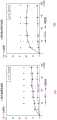
图1是表示LASCol浓度和储能模量的关系的图。FIG. 1 is a graph showing the relationship between the LASCol concentration and the storage modulus.
图2是表示LASCol的应变和应力的关系的图。FIG. 2 is a graph showing the relationship between strain and stress of LASCol.
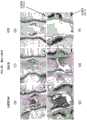
图3是表示培养的大鼠纤维环细胞凝集形成球状体的照片。Fig. 3 is a photograph showing the aggregation of cultured rat annulus fibrosus cells to form spheroids.
图4是表示大鼠的椎间盘髓核细胞(图4(a))、大鼠的椎间盘纤维环细胞(图4(b))的培养时间和球状体数的演变的图。4 is a graph showing the evolution of the culture time and the number of spheroids of rat intervertebral disc nucleus pulposus cells ( FIG. 4( a )) and rat intervertebral disc annulus fibrosus cells ( FIG. 4( b )).
图5是表示人的髓核细胞(图5(a))、人的纤维环细胞(图5(b))的培养时间和球状体数的演变的图。FIG. 5 is a graph showing the evolution of the culture time and the number of spheroids of human nucleus pulposus cells ( FIG. 5( a )) and human annulus fibrosus cells ( FIG. 5( b )).
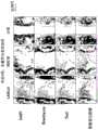
图6是表示人椎间盘髓核细胞在LASCol凝胶上培养而成的LASCol凝胶群的染色结果的照片。Fig. 6 is a photograph showing the result of staining of a LASCol gel population obtained by culturing human intervertebral disc nucleus pulposus cells on a LASCol gel.
图7是表示人椎间盘髓核细胞在AC凝胶上培养而成的端胶原凝胶群(AC凝胶群)的染色结果的照片。Fig. 7 is a photograph showing the results of staining of Telcollage gel population (AC gel population) obtained by culturing human intervertebral disc nucleus pulposus cells on AC gel.
图8是表示人椎间盘纤维环细胞的LASCol凝胶群的染色结果的照片。Fig. 8 is a photograph showing the results of staining of LASCol gel populations of human intervertebral disc annulus fibrosus cells.
图9是表示人椎间盘纤维环细胞的AC凝胶群的染色结果的照片。FIG. 9 is a photograph showing the staining results of AC gel populations of human intervertebral disc annulus fibrosus cells.
图10是进行了椎骨间距的维持试验的大鼠尾部的X光照片(图10(a))和MRI的T2加权像的照片(图10(b))。FIG. 10 is an X-ray photograph ( FIG. 10( a )) and a photograph of a T2-weighted image of MRI ( FIG. 10( b )) of the tail of the rat subjected to the vertebral distance maintenance test.
图11是表示使用大鼠的椎骨间距的维持试验的结果的图。FIG. 11 is a graph showing the results of a maintenance test using a rat's intervertebral distance.
图12是表示LASCol的浓度不同对椎骨间距(%DHI)的影响的图。Fig. 12 is a graph showing the effect of different LASCol concentrations on intervertebral distance (%DHI).
图13是表示在LASCol中添加生长因子的效果的图。FIG. 13 is a graph showing the effect of adding growth factors to LASCol.
图14是将术后1周的大鼠尾椎髓核区域的组织标本用番红-O染色的照片。Fig. 14 is a photograph of a tissue specimen of a rat caudal nucleus pulposus region stained with Safranin-
图15是将术后2周的大鼠尾椎髓核区域的组织标本用番红-O染色的照片。Fig. 15 is a photograph of a tissue specimen of a rat caudal nucleus pulposus region stained with Safranin-
图16是将术后4周的大鼠尾椎髓核区域的组织标本用番红-O染色的照片。Fig. 16 is a photograph of a tissue specimen of a rat caudal nucleus pulposus region stained with Safranin-
图17是将术后8周的大鼠尾椎髓核区域的组织标本用番红-O染色的照片。Fig. 17 is a photograph of a tissue specimen of a rat caudal nucleus pulposus region stained with Safranin-
图18是表示在术后每周测定图14至图17中呈蛋白聚糖阳性(番红-O阳性)的红色部分面积的结果的图。Fig. 18 is a graph showing the results of measuring the area of the red portion in Figs. 14 to 17 that were positive for proteoglycan (safranin-O positive) every week after surgery.
图19是表示对图14至图17中浸润于髓核区域的细胞数进行计数的结果的图。FIG. 19 is a diagram showing the results of counting the number of cells infiltrating the nucleus pulposus region in FIGS. 14 to 17 .
图20是对术后1周的髓核区域进行多重荧光免疫染色、对所得图像作黑白处理的照片。Fig. 20 is a photograph of the
图21是对术后2周的髓核区域进行多重荧光免疫染色、对所得图像作黑白处理的照片。Fig. 21 is a photograph of the
图22是对术后4周的髓核区域进行多重荧光免疫染色、对所得图像作黑白处理的照片。Figure 22 is a photograph of multiplexed fluorescent immunostaining of the
图23是对术后8周的髓核区域进行多重荧光免疫染色、对所得图像作黑白处理的照片。Figure 23 is a photograph of multiplex fluorescent immunostaining of the nucleus pulposus region at 8 weeks after surgery, and the resulting image was processed in black and white.
图24是将刚刚术后、术后3天、术后1周的大鼠尾椎髓核区域的组织标本用番红-O染色的照片。Fig. 24 is a photograph of a tissue specimen of a rat caudal nucleus pulposus region immediately after surgery, 3 days after surgery, and 1 week after surgery, stained with Safranin-O.
图25是对刚刚术后的髓核区域进行多重荧光免疫染色、对所得图像作黑白处理的照片。Fig. 25 is a photograph of the nucleus pulposus region immediately after surgery by multiplex fluorescent immunostaining and black-and-white processing of the resulting image.
图26是对术后3天的髓核区域进行多重荧光免疫染色、对所得图像作黑白处理的照片。Fig. 26 is a photograph of the
图27是对术后1周的髓核区域进行多重荧光免疫染色、对所得图像作黑白处理的照片。Fig. 27 is a photograph of the
图28是对术后1周的髓核区域进行多重荧光免疫染色、对所得图像作黑白处理的照片。FIG. 28 is a photograph of the
具体实施方式Detailed ways
以下,对本发明的椎间盘退变的治疗剂及椎间盘细胞培养材料,示出附图和实施例进行说明。另外,以下的说明是对本发明的一种实施方式和一个实施例进行例示,本发明并不限定于以下的说明。以下的说明可在不脱离本发明的思想的范围内进行改变。Hereinafter, the therapeutic agent for intervertebral disc degeneration and the intervertebral disc cell culture material of the present invention will be described with reference to the accompanying drawings and Examples. In addition, the following description illustrates an embodiment and an example of the present invention, and the present invention is not limited to the following description. The following description may be changed within a range not departing from the spirit of the present invention.
作为本发明的椎间盘退变的治疗剂及椎间盘细胞培养材料使用的LASCol包括胶原或端胶原的分解产物。该分解产物具有胶原所具有的与细胞的粘合性减弱、变为低粘合性的性质。此外,作为本发明的椎间盘退变的治疗剂,可以包含水凝胶、明胶凝胶、壳聚糖凝胶、透明质酸·胶原水凝胶、透明质酸聚合物、透明质酸·PEG聚合物、胶原·透明质酸·PEG水凝胶、被称为高纯度褐藻酸凝胶(UPAL)的物质、具有人体亲和性的溶剂(将这些统称为“辅助物质”)。当然,也可以仅为LASCol。此外,也可以添加缓冲液、pH调整液、盐、细胞生长因子。The LASCol used as the therapeutic agent for intervertebral disc degeneration and the intervertebral disc cell culture material of the present invention includes a degradation product of collagen or telogen. This degradation product has the property that the adhesiveness to cells which collagen has is weakened and becomes low adhesiveness. In addition, the therapeutic agent for intervertebral disc degeneration of the present invention may contain hydrogel, gelatin gel, chitosan gel, hyaluronic acid/collagen hydrogel, hyaluronic acid polymer, and hyaluronic acid/PEG polymer. substances, collagen, hyaluronic acid, and PEG hydrogels, substances called high-purity alginic acid gels (UPAL), and solvents with human affinity (these are collectively referred to as "auxiliary substances"). Of course, only LASCol may be used. In addition, buffers, pH adjustment solutions, salts, and cell growth factors may also be added.
LASCol是通过将胶原或端胶原用酶分解而得到的。并且根据分解时的条件不同,所包含的肽序列不同。即,LASCol根据分解的条件不同,可获得不同种类的LASCol。LASCol is obtained by enzymatically decomposing collagen or telogen. In addition, the peptide sequences contained differ depending on the decomposition conditions. That is, different types of LASCol can be obtained depending on the decomposition conditions of LASCol.
可用于本发明的LASCol的特征是,由胶原或端胶原的三螺旋结构域的如下述(A:序列编号1)所示的氨基末端的氨基酸序列中Y1和Y2之间的化学键切断而成的α链的组合构成。The LASCol that can be used in the present invention is characterized in that it is formed by cleavage of a chemical bond betweenY1 andY2 in the amino-terminal amino acid sequence of the triple helical domain of collagen or telogen as shown in the following (A: SEQ ID NO: 1). Combination of formed α chains.
(A)-Y1-Y2-Y3-G-Y4-Y5-G-Y6-Y7-G-Y8-Y9-G-(序列编号1):(A)-Y1 -Y2 -Y3 -GY4 -Y5 -GY6 -Y7 -GY8 -Y9 -G- (serial number 1):
(其中,G是甘氨酸,Y1~Y9是任意氨基酸)。(wherein, G is glycine, and Y1 to Y9 are arbitrary amino acids).
已知胶原的三螺旋结构域是连续的序列-G-X-Y-(G是甘氨酸,X和Y是任意氨基酸)。上述序列中,“-Y3-G-Y4-Y5-”中的“G”表示三螺旋结构域的N末端侧的甘氨酸。由上述序列可知,Y1和Y2之间的化学键切断是指在三螺旋结构域的外侧进行切断。如下所述,如果分解条件不同,则在三螺旋结构域的内侧发生切断。本发明中使用的LASCol之一是在三螺旋结构域的外侧发生了切断的LASCol。以下将其称为LASCol-A。The triple helical domain of collagen is known to be the contiguous sequence -GXY- (G is glycine, X and Y are arbitrary amino acids). In the above sequence, "G" in "-Y3 -GY4 -Y5 -" represents glycine on the N-terminal side of the triple helical domain. As can be seen from the above sequence, the cleavage of the chemical bond between Y1 and Y2 refers to the cleavage on the outside of the triple helix domain. As described below, if the decomposition conditions are different, cleavage occurs inside the triple helix domain. One of the LASCols used in the present invention is a LASCol cleaved outside the triple helical domain. It is hereinafter referred to as LASCol-A.
另外,已知根据分解的条件,可获得以下LASCol。由胶原或端胶原的三螺旋结构域的如下述(B:序列编号2)所示的氨基末端的氨基酸序列中X1和X2之间的化学键、X2和G之间的化学键、G和X3之间的化学键、X4和G之间的化学键、或X6和G之间的化学键切断而成的α链的组合构成。In addition, it is known that the following LASCol can be obtained depending on the decomposition conditions. The chemical bond between X1 and X2 , the chemical bond between X2 and G, G and The chemical bond between X3 , the chemical bond between X4 and G, or the chemical bond between X6 and G is composed of a combination of α chains cut.
(B)-G-X1-X2-G-X3-X4-G-X5-X6-G-(序列编号2):(其中,G是甘氨酸,X1~X6是任意氨基酸)。将其称为LASCol-B。LASCol-B是三螺旋结构域的内侧发生切断。序列编号2中,“-G-X1-X2-G-”的“G”是三螺旋结构域的N末端侧的甘氨酸。当然,也可以是包含其它肽的LASCol。作为椎间盘退变的治疗剂,在现在已知的LASCol中最合适的是LASCol-A。但是,不排除其它LASCol。(B)-GX1 -X2 -GX3 -X4 -GX5 -X6 -G- (SEQ ID NO: 2): (wherein G is glycine, and X1 to X6 are any amino acids). Call it LASCol-B. LASCol-B is cleaved on the inside of the triple helix domain. In SEQ ID NO: 2, "G" of "-GX1 -X2 -G-" is a glycine on the N-terminal side of the triple helical domain. Of course, LASCol containing other peptides is also possible. Among the currently known LASCols, LASCol-A is the most suitable as a therapeutic agent for intervertebral disc degeneration. However, other LASCols are not excluded.
作为本发明的椎间盘退变的治疗剂使用的LASCol可在酸性状态下以溶液形式保存。并且通过调整pH和浓度、将温度升至体温而变成凝胶状。通过变成凝胶状,LASCol在纤维环内的扩散得到抑制,导致细胞的诱导和/或细胞外基质的产生,通过实现组织再生来发挥维持椎骨间距的效果(称为“椎骨间维持能力”)。The LASCol used as the therapeutic agent for intervertebral disc degeneration of the present invention can be stored as a solution in an acidic state. And it becomes gel-like by adjusting pH and concentration and raising the temperature to body temperature. By becoming gelatinous, the diffusion of LASCol within the annulus fibrosus is inhibited, resulting in the induction of cells and/or the production of extracellular matrix, which exerts the effect of maintaining the intervertebral spacing by achieving tissue regeneration (called "intervertebral maintenance capacity"). ).
变成凝胶状时的弹性模量与溶液中的LASCol的浓度和pH、温度成比例。在下述实施例中,示出调整pH和浓度、在液体状态下吸到注射器中、通过注射给药至纤维环内、在纤维环内变成凝胶状的例子。但是,作为本发明的椎间盘退变的治疗剂使用的LASCol也可以制成膜状或海绵状而埋入患处。另外,膜状、海绵状是指将LASCol以规定形状干燥的产物(也称为形状体)。The elastic modulus when it becomes a gel is proportional to the concentration of LASCol in the solution, pH, and temperature. In the following examples, examples of adjusting pH and concentration, sucking into a syringe in a liquid state, administering into an annulus fibrosus by injection, and becoming a gel in an annulus fibrosus are shown. However, the LASCol used as the therapeutic agent for intervertebral disc degeneration of the present invention may be embedded in the affected area in the form of a film or sponge. In addition, film-like and sponge-like refer to a product (also referred to as a shaped body) obtained by drying LASCol in a predetermined shape.
此外,作为椎间盘退变的治疗剂,也可以将LASCol和辅助物质一起使用。这是因为,通过由辅助物质负责椎骨间维持能力这一机械强度,由LASCol承担从周围细胞游走至产生作为髓核成分的蛋白聚糖的细胞并将其浸润、从而使髓核细胞再生的任务,从而成为椎间盘退变的治疗剂。In addition, as a therapeutic agent for disc degeneration, LASCol can also be used together with auxiliary substances. This is because LASCol assumes the role of migrating from surrounding cells to cells producing proteoglycan, which is a component of the nucleus pulposus, and infiltrating them, thereby regenerating the nucleus pulposus cells by the mechanical strength that the auxiliary substance is responsible for the ability to maintain the intervertebral bone. task and thus become a therapeutic agent for intervertebral disc degeneration.
如下所述在本发明中使用的LASCol的浓度如果在3.5mg/ml(以下述的“实用弹性模量”计为20Pa)以上,则可以呈凝胶状。因此,通过将该浓度以上的LASCol与辅助物质混合,可获得使髓核细胞再生的椎间盘退变的治疗剂及椎间盘细胞培养材料。As described below, the concentration of LASCol used in the present invention is 3.5 mg/ml (20 Pa in terms of "practical elastic modulus" described below) or more, and the gel may be formed. Therefore, by mixing LASCol at a concentration higher than this concentration with an auxiliary substance, a therapeutic agent for intervertebral disc degeneration which regenerates nucleus pulposus cells and an intervertebral disc cell culture material can be obtained.
此外,如果浓度在7mg/ml以上,则单独使用时具有椎骨间维持能力,如果在21mg/ml以上,则即使是单独使用时也具有端胶原以上的椎骨间维持能力。In addition, when the concentration is 7 mg/ml or more, it has intervertebral maintenance ability when used alone, and when it is 21 mg/ml or more, it has intervertebral maintenance ability even when used alone.
因此,可用于本发明的LASCol能够以3.5mg/ml以上的浓度使用,优选以7mg/ml以上、更优选以21mg/ml以上的浓度使用。另外,作为凝胶的浓度上限至少在42mg/ml以上,但即使是在其以上的浓度,也能用作椎间盘退变的治疗剂。Therefore, LASCol that can be used in the present invention can be used at a concentration of 3.5 mg/ml or more, preferably at a concentration of 7 mg/ml or more, and more preferably at a concentration of 21 mg/ml or more. In addition, the upper limit of the concentration of the gel is at least 42 mg/ml or more, but it can be used as a therapeutic agent for intervertebral disc degeneration even at a concentration higher than that.
作为有关LASCol的制备方法的发现,LASCol-B和LASCol-A大致相同。于是,关于在两者中共通的发现,仅针对LASCol进行说明。此外,以下说明中,“分解产物”是指LASCol。As a finding regarding the preparation method of LASCol, LASCol-B and LASCol-A are approximately the same. Therefore, regarding the findings common to both, only LASCol will be described. In addition, in the following description, "decomposition product" means LASCol.
<LASCol的材料><Material of LASCol>
作为LASCol的材料的胶原或端胶原无特别限定,可以是公知的胶原和端胶原。The collagen or telogen as a material of LASCol is not particularly limited, and known collagen and telogen can be used.
作为胶原,可使用哺乳类(例如牛、猪、兔、人、兔或小鼠等)、鸟类(例如鸡等)、或者鱼类(例如鲨鱼、鲤鱼、鳗鱼、金枪鱼(例如黄肌金枪鱼)、罗非鱼、鲷鱼、鲑鱼等)或爬行类(例如鳖)的胶原。As collagen, mammals (eg, cows, pigs, rabbits, humans, rabbits, mice, etc.), birds (eg, chickens, etc.), or fishes (eg, sharks, carps, eels, tuna (eg, yellow muscle tuna)) can be used , tilapia, snapper, salmon, etc.) or reptiles (such as soft-shelled turtle).
本发明中使用的胶原例如可使用来源于上述哺乳类或鸟类的真皮、肌腱、骨或筋膜等的胶原,来源于上述鱼类的皮肤或鳞等的胶原,来源于上述爬行类的真皮、肌腱、骨等的胶原。As the collagen used in the present invention, for example, collagen derived from the dermis, tendon, bone, or fascia of the above-mentioned mammals or birds, collagen derived from the skin or scale of the above-mentioned fish, and the dermis of the above-mentioned reptiles can be used. , tendon, bone, etc. collagen.
此外,作为LASCol的制备中使用的端胶原,可使用将上述哺乳类、鸟类、鱼类或爬行类的胶原用蛋白酶(例如胃蛋白酶等)处理而得的、从胶原分子的氨基末端和/或羧基末端将端肽部分除去而成的端胶原。In addition, as telogen used in the preparation of LASCol, collagen obtained by treating the above-mentioned mammalian, avian, fish, or reptile collagen with a protease (for example, pepsin, etc.), obtained from the amino terminus of collagen molecule and/or Or telogen in which the telopeptide moiety has been removed from the carboxyl terminus.
其中,可优选使用鸡、猪、牛、人或大鼠的胶原或端胶原。此外,可更优选使用猪、牛或人的胶原或端胶原作为LASCol的材料。Among them, chicken, porcine, bovine, human or rat collagen or telogen can be preferably used. In addition, porcine, bovine or human collagen or telogen can be more preferably used as the material of LASCol.
此外,作为LASCol的材料,也可以使用鱼类的胶原或端胶原。如果使用鱼类,则可简便、安全地提供材料,并且可大量获取,可提供无病毒的对人而言更加安全的胶原或端胶原的分解产物(LASCol)。In addition, as a material of LASCol, fish collagen or telogen can also be used. If fish is used, the material can be easily and safely provided, and can be obtained in large quantities, which can provide a virus-free collagen or telogen breakdown product (LASCol) which is safer for humans.
另外,使用鱼类的胶原或端胶原作为LASCol的材料的情况下,优选使用鲨鱼、鲤鱼、鳗鱼、金枪鱼(例如黄肌金枪鱼)、罗非鱼、黑鲈、蓝鳃太阳鱼、鲷鱼或鲑鱼的胶原或端胶原,更优选使用金枪鱼、罗非鱼、鲷鱼或鲑鱼的胶原或端胶原。In addition, in the case of using fish collagen or terminal collagen as the material of LASCol, it is preferable to use shark, carp, eel, tuna (for example, yellow tuna), tilapia, sea bass, bluegill, sea bream or salmon collagen or telogen, more preferably tuna, tilapia, sea bream or salmon collagen or telogen.
使用端胶原作为LASCol的材料的情况下,优选使用热变性的温度优选在15℃以上、更优选在20℃以上的端胶原。例如,使用鱼类的端胶原作为分解产物的材料的情况下,金枪鱼(例如黄肌金枪鱼)或鲤鱼、罗非鱼等的端胶原的热变性温度在25℃以上,因此优选使用这些端胶原。When telogen is used as the material of LASCol, it is preferable to use telogen whose temperature of thermal denaturation is preferably 15°C or higher, more preferably 20°C or higher. For example, when telogen from fish is used as the material for the decomposition product, telogen from tuna (eg, yellow muscle tuna), carp, tilapia, etc. has a thermal denaturation temperature of 25° C. or higher, so these telogens are preferably used.
如果是上述构成,则可将本实施方式的椎间盘退变的治疗剂变成凝胶状的温度调节至优选15℃以上、更优选20℃以上。其结果是,如果是上述构成,则可实现储藏时的稳定性、使用时的稳定性优良的椎间盘退变的治疗剂。With the above configuration, the temperature at which the therapeutic agent for intervertebral disc degeneration of the present embodiment becomes gel-like can be adjusted to preferably 15°C or higher, more preferably 20°C or higher. As a result, with the above configuration, a therapeutic agent for intervertebral disc degeneration which is excellent in stability during storage and stability during use can be realized.
这些胶原或端胶原可通过公知的方法获取。例如,可通过将哺乳类、鸟类或鱼类的富含胶原的组织投入pH2~4左右的酸性溶液,使胶原溶出。然后,向该溶出液中添加胃蛋白酶等蛋白酶,将胶原分子的氨基末端和/或羧基末端的端肽部分除去。然后,向该溶出液中添加氯化钠等盐,可使端胶原沉淀。These collagens or Telogen can be obtained by known methods. For example, collagen can be eluted by throwing collagen-rich tissues of mammals, birds, or fish into an acidic solution having a pH of about 2 to 4. Then, a protease such as pepsin is added to the eluate to remove the amino-terminal and/or carboxy-terminal telopeptide portion of the collagen molecule. Then, a salt such as sodium chloride is added to the eluate to precipitate telogen.
为获得LASCol,要使酶与胶原或端胶原作用,将这些材料分解。但是,通过制备三螺旋结构域内的化学键已被切断的胶原或端胶原的分解产物(例如化学合成法、重组蛋白的表达),也可以获得LASCol。To obtain LASCol, enzymes are allowed to interact with collagen or telogen, breaking down these materials. However, LASCol can also be obtained by preparing a decomposition product of collagen or Telogen in which the chemical bonds within the triple helical domain have been cleaved (eg, chemical synthesis, recombinant protein expression).
以下,对如上所述通过酶(例如蛋白酶)将胶原或端胶原分解而获得LASCol的方法进行说明。Hereinafter, a method for obtaining LASCol by decomposing collagen or telogen with an enzyme (eg, protease) as described above will be described.
作为上述酶无特别限定,优选使用例如半胱氨酸蛋白酶。Although it does not specifically limit as said enzyme, For example, cysteine protease is used preferably.
作为半胱氨酸蛋白酶,优选使用酸性氨基酸量比碱性氨基酸量更多的半胱氨酸蛋白酶、在酸性区域的氢离子浓度下有活性的半胱氨酸蛋白酶。As the cysteine protease, it is preferable to use a cysteine protease having a larger amount of acidic amino acids than the amount of basic amino acids, and a cysteine protease that is active at the hydrogen ion concentration in the acidic region.
作为这样的半胱氨酸蛋白酶,可例举猕猴桃蛋白酶[EC 3.4.22.14]、木瓜蛋白酶[EC 3.4.22.2]、无花果蛋白酶[EC 3.4.22.3]、菠萝蛋白酶[EC 3.4.22.32]、组织蛋白酶B[EC 3.4.22.1]、组织蛋白酶L[EC 3.4.22.15]、组织蛋白酶S[EC 3.4.22.27]、组织蛋白酶K[EC 3.4.22.38]、组织蛋白酶H[EC 3.4.22.16]、芦荟蛋白酶、钙依赖性蛋白酶等。另外,方括号内为酶编号。Examples of such cysteine proteases include kiwi protease [EC 3.4.22.14], papain [EC 3.4.22.2], ficin [EC 3.4.22.3], bromelain [EC 3.4.22.32], cathepsin B [EC 3.4.22.1], Cathepsin L [EC 3.4.22.15], Cathepsin S [EC 3.4.22.27], Cathepsin K [EC 3.4.22.38], Cathepsin H [EC 3.4.22.16], Aloe , calcium-dependent proteases, etc. In addition, the enzyme numbers are in square brackets.
其中,优选使用猕猴桃蛋白酶、木瓜蛋白酶、无花果蛋白酶、组织蛋白酶K、芦荟蛋白酶或菠萝蛋白酶,更优选使用猕猴桃蛋白酶、木瓜蛋白酶、无花果蛋白酶、组织蛋白酶K。Among them, kiwi protease, papain, ficin, cathepsin K, aloe protease, or bromelain are preferably used, and kiwi protease, papain, ficin, and cathepsin K are more preferably used.
上述酶可通过公知的方法获取。例如,可通过如下方式获取:通过化学合成制备酶;从细菌、真菌、各种动植物的细胞或组织提取酶;通过基因工程方式制备酶;等等。当然,也可以使用市售的酶。The above-mentioned enzymes can be obtained by known methods. For example, it can be obtained by: preparing enzymes by chemical synthesis; extracting enzymes from cells or tissues of bacteria, fungi, various animals and plants; preparing enzymes by genetic engineering; and the like. Of course, commercially available enzymes can also be used.
通过用酶(例如蛋白酶)分解胶原或端胶原来进行切断的情况下,可按照例如以下(i)~(iii)的方法进行切断工序。以下(i)~(iii)的方法只不过是切断工序的一例,LASCol的制造方法不限于这些(i)~(iii)的方法。When the cleavage is performed by decomposing collagen or telogen with an enzyme (eg, protease), the cleaving step can be performed according to, for example, the following methods (i) to (iii). The following methods (i) to (iii) are merely examples of the cutting step, and the method for producing LASCol is not limited to these methods (i) to (iii).
另外,通过以下(i)和(ii)的方法,可获得LASCol-B。此外,通过以下(iii)的方法,可获得LASCol-A和LASCol-B。In addition, LASCol-B can be obtained by the following methods (i) and (ii). Furthermore, by the method of the following (iii), LASCol-A and LASCol-B can be obtained.
(i)在高浓度盐的存在下,使胶原或端胶原与酶接触的方法。(i) A method of contacting collagen or telogen with an enzyme in the presence of a high concentration of salt.
(ii)使与高浓度盐接触后的酶与胶原或端胶原接触的方法。(ii) A method of bringing the enzyme contacted with a high-concentration salt into contact with collagen or telogen.
(iii)在低浓度盐的存在下,使胶原或端胶原与酶接触的方法。(iii) A method of contacting collagen or telogen with an enzyme in the presence of a low concentration of salt.
作为上述(i)的方法的具体例,可例举例如在含高浓度盐的水溶液中使胶原或端胶原与酶接触的方法。As a specific example of the method of said (i), the method of contacting collagen or telogen with an enzyme in a high-concentration salt-containing aqueous solution may, for example, be mentioned.
作为上述(ii)的方法的具体例,可例举例如事先使含高浓度盐的水溶液与酶接触,然后使该酶与胶原或端胶原接触的方法。As a specific example of the method of the above (ii), for example, a method of contacting an aqueous solution containing a high-concentration salt with an enzyme and then contacting the enzyme with collagen or telogen.
作为上述(iii)的方法的具体例,可例举例如在含低浓度盐的水溶液中使胶原或端胶原与酶接触的方法。作为上述水溶液的具体构成无特别限定,例如可使用水。As a specific example of the method of said (iii), the method of contacting collagen or telogen with an enzyme in an aqueous solution containing a low-concentration salt can be mentioned, for example. The specific structure of the above-mentioned aqueous solution is not particularly limited, and for example, water can be used.
作为上述盐的具体构成无特别限定,优选使用氯化物。作为氯化物无特别限定,例如可使用NaCl、KCl、LiCl或MgCl2。The specific structure of the above-mentioned salt is not particularly limited, but chlorides are preferably used. The chloride is not particularly limited, and for example, NaCl, KCl, LiCl, or MgCl2 can be used.
上述含高浓度盐的水溶液中的盐的浓度无特别限定,但可以说越高越好。例如,该浓度优选在200mM以上,更优选在500mM以上,更优选在1000mM以上,更优选在1500mM以上,最优选在2000mM以上。The concentration of the salt in the high-concentration salt-containing aqueous solution is not particularly limited, but it can be said that the higher the better. For example, the concentration is preferably 200 mM or higher, more preferably 500 mM or higher, more preferably 1000 mM or higher, more preferably 1500 mM or higher, and most preferably 2000 mM or higher.
上述含低浓度盐的水溶液中的盐的浓度无特别限定,但可以说越低越好。例如,该浓度优选低于200mM,更优选在150mM以下,更优选在100mM以下,更优选在50mM以下,最优选为接近0mM。The concentration of the salt in the above-mentioned low-concentration salt-containing aqueous solution is not particularly limited, but it can be said that the lower the better. For example, the concentration is preferably below 200 mM, more preferably below 150 mM, more preferably below 100 mM, more preferably below 50 mM, and most preferably close to 0 mM.
溶解于上述水溶液(例如水)的胶原或端胶原的量无特别限定,例如相对于1000重量份~10000重量份的水溶液,优选溶解1重量份的胶原或端胶原。The amount of collagen or telogen dissolved in the above-mentioned aqueous solution (eg, water) is not particularly limited.
如果是上述构成,则向水溶液中添加酶的情况下,可使该酶与胶原或端胶原高效地接触。于是,其结果是,可通过酶高效地分解胶原或端胶原。With the above configuration, when an enzyme is added to the aqueous solution, the enzyme can be efficiently brought into contact with collagen or telogen. Consequently, as a result, collagen or terminal collagen can be efficiently decomposed by enzymes.
添加至上述水溶液中的酶的量无特别限定,例如相对于100重量份的胶原或端胶原,优选添加10重量份~20重量份的酶。The amount of the enzyme to be added to the above-mentioned aqueous solution is not particularly limited, but for example, 10 to 20 parts by weight of the enzyme is preferably added relative to 100 parts by weight of collagen or telogen.
如果是上述构成,则水溶液中的酶的浓度高,因此可通过酶(例如蛋白酶)高效地分解胶原或端胶原。With the above configuration, since the concentration of the enzyme in the aqueous solution is high, collagen or telogen can be efficiently decomposed by an enzyme (eg, protease).
在水溶液中使胶原或端胶原与酶接触时的其它条件(例如水溶液的pH、温度、接触时间等)也无特别限定,可以适当设定,但优选以下范围。另外,以下对这些条件的优选范围进行例示。Other conditions (for example, pH of the aqueous solution, temperature, contact time, etc.) for contacting collagen or Telogen with the enzyme in an aqueous solution are not particularly limited and can be appropriately set, but the following ranges are preferred. In addition, preferable ranges of these conditions are illustrated below.
1)水溶液的pH优选为pH2.0~7.0,更优选为pH3.0~6.5。为了将水溶液的pH保持在上述范围,可向水溶液中添加公知的缓冲剂。如果是上述pH,则可将胶原或端胶原均匀地溶解于水溶液中,其结果是,可高效地进行酶反应。1) The pH of the aqueous solution is preferably pH 2.0 to 7.0, and more preferably pH 3.0 to 6.5. In order to keep the pH of the aqueous solution within the above-mentioned range, a known buffer may be added to the aqueous solution. At the above pH, collagen or telogen can be uniformly dissolved in the aqueous solution, and as a result, the enzymatic reaction can be efficiently performed.
2)温度无特别限定,可根据所用的酶选择温度。例如,该温度优选为15℃~40℃,更优选为20℃~35℃。2) The temperature is not particularly limited, and the temperature can be selected according to the enzyme used. For example, the temperature is preferably 15°C to 40°C, and more preferably 20°C to 35°C.
3)接触时间无特别限定,可根据酶的量和/或胶原或端胶原的量选择接触时间。例如,该时间优选为1小时~60天,更优选为1天~7天,进一步优选为3天~7天。3) The contact time is not particularly limited, and the contact time can be selected according to the amount of the enzyme and/or the amount of collagen or telogen. For example, the time period is preferably 1 hour to 60 days, more preferably 1 day to 7 days, and still more preferably 3 days to 7 days.
另外,也可以经过选自在水溶液中使胶原或端胶原与酶接触后、根据需要再调整pH的工序,使酶失活的工序以及除去杂质的工序中的至少一道工序。In addition, at least one step selected from the step of bringing the collagen or Telogen in contact with the enzyme in an aqueous solution, and then adjusting the pH as necessary, the step of inactivating the enzyme, and the step of removing impurities may be performed.
此外,上述除去杂质的工序可通过用于分离物质的常规方法来进行。上述除去杂质的工序可通过例如透析、盐析、凝胶渗透色谱、等电点沉淀、离子交换色谱或疏水相互作用色谱等来进行。In addition, the above-mentioned step of removing impurities can be carried out by a conventional method for separating substances. The above-mentioned step of removing impurities can be performed by, for example, dialysis, salting out, gel permeation chromatography, isoelectric point precipitation, ion exchange chromatography, hydrophobic interaction chromatography, or the like.
本发明的椎间盘退变的治疗剂主要通过外科手术通过注射等给药至椎间盘中。另外,此时希望椎间盘退变的治疗剂中所含的LASCol具有规定以上的弹性模量(下述的实用弹性模量)。这是因为,如果是弹性模量低的状态,则LASCol可能会从椎间盘流出。The therapeutic agent for intervertebral disc degeneration of the present invention is mainly administered into the intervertebral disc by injection or the like through surgery. In addition, in this case, it is desirable that the LASCol contained in the therapeutic agent for intervertebral disc degeneration has a predetermined elastic modulus (the following practical elastic modulus). This is because LASCol may flow out of the intervertebral disc if the elastic modulus is low.
本发明的椎间盘退变的治疗剂或椎间盘细胞培养材料以凝胶状态或干燥状态(包括粉末和形状体)等供给。此外,以规定的浓度使用本发明的椎间盘退变的治疗剂或椎间盘细胞培养材料也包括如下情况:附加或通知了向干燥状态的LASCol中添加一定的溶剂的指令,其结果是,成为本发明的优选浓度的LASCol。The therapeutic agent for intervertebral disc degeneration or the intervertebral disc cell culture material of the present invention is supplied in a gel state or a dry state (including powder and shaped body). In addition, the use of the therapeutic agent for intervertebral disc degeneration or the intervertebral disc cell culture material of the present invention at a predetermined concentration also includes a case where an instruction to add a certain solvent to dry LASCol is added or notified, and the result is the present invention The preferred concentration of LASCol.
本说明书中,“给药”是指经由患处(椎间盘)给予患者治疗剂。此外,用本发明治疗的椎间盘病变除了作为代表例的椎间盘脱出症外,还可应用于腰痛症、椎管狭窄症、脊柱变性这样的与椎间盘退变有关的疾病等。换言之,本发明可以说是使用本发明的椎间盘退变的治疗剂的椎间盘退变的治疗方法。此外,也可以说是使用本发明的椎间盘细胞的培养材料的椎间盘细胞的培养方法。In the present specification, "administration" refers to administration of a therapeutic agent to a patient via an affected area (intervertebral disc). In addition, the intervertebral disc disease treated by the present invention can be applied to diseases related to intervertebral disc degeneration, such as low back pain, spinal stenosis, and spinal degeneration, in addition to disc herniation as a representative example. In other words, the present invention can be said to be a method of treating intervertebral disc degeneration using the agent for treating intervertebral disc degeneration of the present invention. In addition, it can also be said to be a method for culturing intervertebral disc cells using the intervertebral disc cell culture material of the present invention.
实施例Example
<含LASCol的溶液的制备><Preparation of LASCol-containing solution>
准备氯化钠浓度为0mM和1500mM的50mM柠檬酸缓冲液(pH3.0)。另外,使用水作为该水溶液的溶剂。Prepare 50 mM citrate buffer (pH 3.0) with sodium chloride concentrations of 0 mM and 1500 mM. In addition, water is used as a solvent of this aqueous solution.
为了使猕猴桃蛋白酶活化,将猕猴桃蛋白酶溶解于含10mM的二硫苏糖醇和5mM的EDTA(乙二胺四乙酸)的50mM磷酸缓冲液(pH6.5)中,在25℃下静置90分钟。另外,作为猕猴桃蛋白酶,采用通过公知方法纯化的猕猴桃蛋白酶(例如参照非专利文献1)。In order to activate the kiwi protease, the kiwi protease was dissolved in 50 mM phosphate buffer (pH 6.5) containing 10 mM dithiothreitol and 5 mM EDTA (ethylenediaminetetraacetic acid), and allowed to stand at 25°C for 90 minutes. In addition, as the kiwifruit protease, a kiwifruit protease purified by a known method is used (for example, refer to Non-Patent Document 1).
接着,将来源于猪的I型胶原溶解于含盐的50mM柠檬酸缓冲液(pH3.0)中。使含猕猴桃蛋白酶的水溶液与含来源于猪的I型胶原的该溶液在20℃下接触10天以上,制成I型胶原的分解产物。另外,来源于猪的I型胶原基于公知方法纯化(例如参照非专利文献1)。Next, porcine-derived collagen type I was dissolved in 50 mM citric acid buffer (pH 3.0) containing salt. The aqueous solution containing kiwi protease and the solution containing porcine-derived type I collagen were brought into contact at 20°C for 10 days or more to prepare a decomposition product of type I collagen. In addition, porcine-derived collagen type I was purified based on a known method (for example, see Non-Patent Document 1).
将上述分解产物进行十二烷基硫酸钠-聚丙烯酰胺凝胶电泳(SDS-PAGE),将I型胶原的分解产物分离。The above decomposition products were subjected to sodium dodecyl sulfate-polyacrylamide gel electrophoresis (SDS-PAGE) to separate the decomposition products of type I collagen.
接着,将I型胶原的分解产物通过常规方法转印至PVDF(聚偏氟乙烯,Polyvinylidene Difluoride)膜。然后,通过埃德曼降解法确定转印至PVDF膜的α1链的分解产物的氨基末端的氨基酸序列。Next, the decomposition product of type I collagen was transferred to a PVDF (Polyvinylidene Difluoride) membrane by a conventional method. Then, the amino-terminal amino acid sequence of the decomposition product of the α1 chain transferred to the PVDF membrane was determined by the Edman degradation method.
另外,实际的埃德曼分析是委托阿普罗科技株式会社(アプロサイエンス株式会社)或近畿大学医学院分析仪器共同研究室按照公知方法进行的。In addition, the actual Edman analysis was performed by a well-known method by entrusting Apollo Science and Technology Co., Ltd. or the Joint Laboratory of Analytical Instruments, Kinki University School of Medicine.
表1所示为盐浓度为0mM和1500mM时的α1链的分解产物的氨基末端及其附近的氨基酸序列。Table 1 shows the amino terminus of the decomposition product of the α1 chain and the amino acid sequences in the vicinity thereof when the salt concentration is 0 mM and 1500 mM.
如表1所示,如果盐浓度低(0mM),则在以“GPMGPSGPRG…”表示的三螺旋结构域的外侧发生切断,如果盐浓度高(1500mM),则在三螺旋结构域的内侧发生切断。序列编号3中,从左起第3个甘氨酸(G)开始朝着C末端开始出现三螺旋结构域。0mM时生成的是LASCol-A的溶液,1500mM时生成的是LASCol-B的溶液。在以下实施例中,使用LASCol-A的溶液作为LASCol的溶液。As shown in Table 1, when the salt concentration is low (0 mM), cleavage occurs on the outside of the triple helix domain represented by "GPMGPSGPRG...", and when the salt concentration is high (1500 mM), cleavage occurs on the inside of the triple helix domain. . In SEQ ID NO: 3, a triple helical domain appears from the third glycine (G) from the left toward the C-terminus. A solution of LASCol-A was produced at 0 mM, and a solution of LASCol-B was produced at 1500 mM. In the following examples, a solution of LASCol-A was used as a solution of LASCol.
[表1][Table 1]
另外,LASCol-A的α2链也发生切断。表2中,序列编号5表示α2链的氨基末端部分。序列编号5中,从“··GPMGLMG…”左端的甘氨酸(G)开始朝着C末端开始出现三螺旋结构域。另外,作为LASCol-A制备条件的盐浓度为0mM时的α2链的末端示于序列编号6。其相当于参照序列编号2将G和X3之间的化学键切断。In addition, the α2 chain of LASCol-A was also cleaved. In Table 2, SEQ ID NO: 5 represents the amino terminal portion of the α2 chain. In SEQ ID NO: 5, a triple helical domain appears from the glycine (G) at the left end of "··GPGMGLMG..." toward the C-terminus. In addition, the end of the α2 chain is shown in SEQ ID NO: 6 when the salt concentration is 0 mM as a preparation condition of LASCol-A. It corresponds to cleavage of the chemical bond between G and X3 with reference to SEQ ID NO: 2.
即,LASCol-A中,在α1链上在三螺旋结构域的外侧发生切断,而在α2链上在三螺旋结构域的内侧发生切断。LASCol-A可以具有序列编号3或序列编号6的任一种切断。That is, in LASCol-A, the α1 chain is cleaved outside the triple helical domain, and the α2 chain is cleaved inside the triple helix domain. LASCol-A may have either SEQ ID NO: 3 or SEQ ID NO: 6 cleavage.
[表2][Table 2]
图1所示为含LASCol的溶液的弹性特性(复数模量中的储能模量G’)。横轴为时间(分钟),纵轴为储能模量G’(Pa)。图1(a)和图1(b)中,横轴相同,但纵轴不同。图1(b)的纵轴的标尺比图1(a)大。图1(a)和图1(b)各自的曲线表示LASCol浓度的差异。不同浓度的LASCol溶液用5mM的盐酸溶液配制,以使得终浓度为2.1mg/mL、3.5mg/mL、4.9mg/mL(以上图1(a))、21mg/ml(图1(b)。Figure 1 shows the elastic properties (storage modulus G' in complex modulus) of solutions containing LASCol. The horizontal axis is time (minutes), and the vertical axis is storage modulus G' (Pa). In Figs. 1(a) and 1(b), the horizontal axis is the same, but the vertical axis is different. The scale of the vertical axis of Fig. 1(b) is larger than that of Fig. 1(a). The respective curves of Figure 1(a) and Figure 1(b) represent differences in LASCol concentrations. LASCol solutions of different concentrations were prepared with 5 mM hydrochloric acid solution so that the final concentrations were 2.1 mg/mL, 3.5 mg/mL, 4.9 mg/mL (Figure 1(a) above), 21 mg/ml (Figure 1(b).
这些LASCol在酸性溶液中在5℃~10℃保存。在该状态下,LASCol以液态保存。图1是在LASCol中添加pH调整剂和浓度调整液,将pH调整至大致为7.4后,安装于动态粘弹性测定装置(流变仪,HAAKE MARS III,赛默飞世尔科技公司(サ一モフィツシャ一サィェンティフィツク社)),升温至37℃后测得的结果。测定条件为:频率1Hz,振幅6°/秒,应变量1%。另外,升温在数秒内完成。These LASCols are stored at 5°C to 10°C in an acidic solution. In this state, LASCol is stored in a liquid state. Figure 1 shows that after adding a pH adjuster and a concentration adjusting solution to LASCol to adjust the pH to approximately 7.4, it was installed in a dynamic viscoelasticity measuring device (Rheometer, HAAKE MARS III, Thermo Fisher Scientific (サOne The results measured after heating up to 37°C. The measurement conditions were as follows:
参照图1(a),无论在何种浓度下,刚开始测定时的储能模量G’都较低。然后,无论在何种浓度下,储能模量G’都升高,在约10分钟后接近饱和值。另一方面,图1(b)中,在测定开始1分钟时,储能模量G’升至饱和值,然后缓慢下降并饱和。由图1和图2可见,通过提高浓度,到储能模量G’升高为止的时间也缩短。Referring to Fig. 1(a), regardless of the concentration, the storage modulus G' at the beginning of the measurement is low. Then, regardless of the concentration, the storage modulus G' increased, approaching the saturation value after about 10 minutes. On the other hand, in Fig. 1(b), the storage modulus G' rises to a
由此可知,如果调整pH和浓度、使温度升高,则含LASCol的溶液的储能模量G’升至与浓度相对应的规定值。此外也可知,如果将LASCol配制为规定浓度、在达到37℃之后经过30分钟,则储能模量大致达到稳定值。于是,将此时的储能模量称为LASCol的“实用弹性模量”。From this, it can be seen that when the pH and the concentration are adjusted and the temperature is raised, the storage modulus G' of the LASCol-containing solution increases to a predetermined value corresponding to the concentration. In addition, it was found that when LASCol was prepared at a predetermined concentration and 30 minutes passed after reaching 37° C., the storage modulus reached a substantially stable value. Therefore, the storage modulus at this time is referred to as the "practical elastic modulus" of LASCol.
这表明,通过暴露于合适的条件下,LASCol的性状从无法测定弹性模量的溶胶变为能够对弹性模量进行定量的凝胶,特别是通过注入机体,可用作可注射凝胶。This suggests that, upon exposure to appropriate conditions, the behavior of LASCol changes from a sol in which elastic modulus cannot be measured to a gel capable of quantifying elastic modulus, especially by injection into the body, which can be used as an injectable gel.
图2所示为在流变仪中在37℃下经过30分钟后的“应变(流变仪的驱动侧的旋转方向的位移)”和“应力(流变仪的被动侧所受的应力)”的关系。左纵轴为应变φ(弧度),右纵轴为应力M(μNm),横轴为仪器步数,无单位,500步相当于1秒。即,图2的各图是用1秒的时间测定从5×10⁻⁴弧度到-5×10⁻⁴弧度的往返而得的图。Figure 2 shows "Strain (displacement in the rotational direction of the driving side of the rheometer)" and "Stress (stress on the passive side of the rheometer)" after 30 minutes at 37°C in the rheometer "Relationship. The left vertical axis is the strain φ (radian), the right vertical axis is the stress M (μNm), and the horizontal axis is the number of instrument steps, unitless, 500 steps are equivalent to 1 second. That is, each graph of FIG. 2 is a graph obtained by measuring the round trip from 5×10⁻⁴ radians to −5×10⁻⁴ radians in 1 second.
图2(a)是LASCol浓度为2.1mg/ml的情况,图2(b)是LASCol浓度为3.5mg/ml的情况,图2(c)是LASCol浓度为5.6mg/ml的情况。实用弹性模量分别为8Pa、20Pa、70Pa。LASCol浓度为2.1mg/ml(图2(a))时,对于应变几乎没有应力响应性。即,LASCol可以说是接近于液体的状态。如果LASCol浓度升至3.5mg/ml(图2(b)),则可见与应变相当的应力反应性。Fig. 2(a) is the case where the LASCol concentration is 2.1 mg/ml, Fig. 2(b) is the case where the LASCol concentration is 3.5 mg/ml, and Fig. 2(c) is the case where the LASCol concentration is 5.6 mg/ml. The practical elastic modulus is 8Pa, 20Pa, 70Pa respectively. At a LASCol concentration of 2.1 mg/ml (Fig. 2(a)), there was little stress responsiveness to strain. That is, LASCol can be said to be in a state close to a liquid. If the LASCol concentration was raised to 3.5 mg/ml (Fig. 2(b)), a stress reactivity comparable to the strain was seen.
如果LASCol的濃度进一步升高(图2(c)),则应力与施加的应变同步。另外,应变与应力的相位有错位的原因在于,凝胶具有损耗模量。因此可以判断,在图2(b)的LASCol浓度为3.5mg/ml的时候呈凝胶状。这以实用弹性模量计相当于20Pa。If the concentration of LASCol was further increased (Fig. 2(c)), the stress was synchronized with the applied strain. In addition, the reason why the phases of strain and stress are shifted is that the gel has a loss modulus. Therefore, it can be judged that when the concentration of LASCol in Fig. 2(b) is 3.5 mg/ml, it is in the form of a gel. This corresponds to 20 Pa in terms of practical elastic modulus.
用作椎间盘退变的治疗剂的情况下,认为变成凝胶状时的储能模量的下限为20Pa。LASCol也具有作为细胞支架的功能,因此需要一定程度上贮留在一处。这是因为,认为在弹性低于20Pa的情况下,LASCol没有作为凝胶的行为,因此难以贮留在椎间盘中。When used as a therapeutic agent for intervertebral disc degeneration, it is considered that the lower limit of the storage elastic modulus when it becomes a gel is 20 Pa. LASCol also functions as a cytoscaffold and thus needs to be stored in one place to some extent. This is because it is considered that LASCol does not behave as a gel when the elasticity is lower than 20 Pa, and thus it is difficult to store in the intervertebral disc.
以下,对本发明的椎间盘退变的治疗剂的实施例进行说明。另外,以下实验全都是在获得神户大学医学伦理委员会以及神户大学动物实验委员会的批准后进行的。Hereinafter, examples of the therapeutic agent for intervertebral disc degeneration of the present invention will be described. In addition, the following experiments were all performed after obtaining approval from the Kobe University Medical Ethics Committee and the Kobe University Animal Experiment Committee.
通过腰椎椎间盘切除或椎体间固定术从患者取出人椎间盘的髓核和纤维环的样品。患者数(n数)为15。患者的年龄为46.1±24.1岁,男性8名,女性7名。此外,表示椎间盘退变程度的Pfirrmann分级中的中央值为2。Samples of the nucleus pulposus and annulus fibrosus of human discs were removed from patients by lumbar discectomy or interbody fixation. The number of patients (n number) was 15. The age of the patients was 46.1±24.1 years, and there were 8 males and 7 females. In addition, the median value in the Pfirrmann grading, which represents the degree of intervertebral disc degeneration, is 2.
此外,也取出12周龄的斯普拉格-道利大鼠(Sprague-Dawley rat:以下称为“SD大鼠”)的椎间盘髓核细胞和纤维环细胞。In addition, intervertebral disc nucleus pulposus cells and annulus fibrosus cells of 12-week-old Sprague-Dawley rats (hereinafter referred to as "SD rats") were also taken out.
接着,准备固定有7.0mg/ml的LASCol凝胶的24孔板、和固定有2.1mg/ml的端胶原凝胶的24孔板。对于大鼠和人,培养的样品细胞分别为n=6。各样品在加入各板中的添加有10%FBS的DMEM(达尔伯克氏改良伊格尔培养基)中培养192小时。Next, a 24-well plate immobilized with 7.0 mg/ml LASCol gel and a 24-well plate immobilized with 2.1 mg/ml Telcollagen gel were prepared. Cultured sample cells were n=6 for rat and human, respectively. Each sample was cultured for 192 hours in DMEM (Dulbecco's Modified Eagle's Medium) supplemented with 10% FBS added to each plate.
另外,在LASCol凝胶上培养的细胞称为LASCol凝胶群,在端胶原凝胶上培养的细胞称为AC凝胶群。In addition, cells cultured on LASCol gels are called LASCol gel populations, and cells cultured on Telogen gels are called AC gel populations.
其结果是,在端胶原凝胶上,人椎间盘髓核细胞和人纤维环细胞均显著增殖。另一方面,在LASCol凝胶上,这些细胞明显未增殖。然而,在LASCol中观察到大量的作为细胞凝集的球状体。另外,图3是表示培养的细胞成为球状体的状态的照片。As a result, both human intervertebral disc nucleus pulposus cells and human annulus fibrosus cells proliferated significantly on the telogen gel. On the LASCol gel, on the other hand, these cells were clearly not proliferating. However, a large number of spheroids as cell aggregates were observed in LASCol. In addition, FIG. 3 is a photograph showing a state in which the cultured cells became spheroids.
参照图3。图3(a)所示为LASCol凝胶上的球状体,图3(b)所示为端胶原凝胶上的球状体。照片中的比例尺表示100μm。图3(a)中,有大量细胞密集形成球状体(三角箭头),与之相对,图3(b)中,只有数个细胞聚集(三角箭头)。Refer to Figure 3. Figure 3(a) shows spheroids on LASCol gel and Figure 3(b) shows spheroids on Telcollage gel. The scale bar in the photos indicates 100 μm. In Fig. 3(a), a large number of cells were densely formed into spheroids (triangular arrows), whereas in Fig. 3(b), only a few cells were aggregated (triangular arrows).
图4和图5分别是表示大鼠的椎间盘髓核细胞(图4(a))、纤维环细胞(图4(b))、人的椎间盘髓核细胞(图5(a))、纤维环细胞(图5(b))的培养时间和球状体数的演变的图。各自的横轴为培养时间(小时),纵轴为球状体数。另外,球状体是3个以上细胞凝集而成的,对100倍倍率的视野内的个数进行计数。Fig. 4 and Fig. 5 respectively show rat intervertebral disc nucleus pulposus cells (Fig. 4(a)), annulus fibrosus cells (Fig. 4(b)), human intervertebral disc nucleus pulposus cells (Fig. 5(a)), annulus fibrosus Graph of the evolution of culture time and spheroid number of cells (Fig. 5(b)). The respective horizontal axis represents the culture time (hours), and the vertical axis represents the number of spheroids. In addition, the spheroid was formed by agglutination of three or more cells, and the number of the spheroids in the field of view at a magnification of 100 was counted.
例如,将图3(b)的三角箭头所示的状态的细胞作为球状体计数。当然,如图3(a)所示有多个细胞聚集的情况也是球状体。图4和图5的任一图中,实线都是LASCol凝胶群,虚线都是AC凝胶群。各图中,以平均±标准偏差记载,统计学分析采用双向ANOVA和Tukey-Kramer事后检验。For example, cells in the state indicated by the triangular arrows in Fig. 3(b) are counted as spheroids. Of course, as shown in Fig. 3(a), the case where many cells are aggregated is also a spheroid. In any of FIGS. 4 and 5 , the solid line is the LASCol gel group, and the broken line is the AC gel group. In each figure, the mean ± standard deviation is shown, and statistical analysis was performed using two-way ANOVA and Tukey-Kramer post hoc test.
参照图4(a)、图4(b)的大鼠细胞的结果。在任一细胞种类中,LASCol凝胶群中的球状体都有所增加。但是,AC凝胶群中未见增加。球状体数从培养开始后12小时起到192小时为止,在LASCol凝胶群中明显更多。Refer to the results of rat cells in Fig. 4(a) and Fig. 4(b). Spheroids in the LASCol gel population increased in either cell type. However, no increase was seen in the AC gel group. The number of spheroids was significantly higher in the LASCol gel group from 12 hours to 192 hours after the start of culture.
参照图5(a)、图5(b)的人细胞的结果。在任一细胞种类中,LASCol凝胶群中的球状体数都有所增加。但是,AC凝胶群中未见球状体数的增加。就髓核细胞而言,球状体数从培养开始后48小时起到192小时为止,在LASCol凝胶群中明显更多,对于纤维环细胞而言,球状体数从培养开始后24小时起到192小时为止,在LASCol凝胶群中明显更多。Refer to the results of human cells in Fig. 5(a) and Fig. 5(b). In either cell type, the number of spheroids in the LASCol gel population increased. However, no increase in the number of spheroids was observed in the AC gel group. For nucleus pulposus cells, the number of spheroids was significantly higher from 48 hours to 192 hours after the start of culture, and it was significantly higher in the LASCol gel group, and for annulus fibrosus cells, the number of spheroids increased from 24 hours after the start of culture. By 192 hours, there were significantly more in the LASCol gel population.
综上所述,可以说如果在LASCol凝胶环境中培养细胞,则与在端胶原凝胶环境中培养的情况相比,无论是髓核细胞还是纤维环细胞,球状体数都增多,且构成球状体的细胞也增多。如上所述,LASCol变成凝胶状的浓度在3.5mg/ml以上(以实用弹性模量计为20Pa以上)。因此可以说,如果LASCol浓度在3.5mg/ml以上(至少7.0mg/ml以上),则可发挥在机体内培养细胞并形成球状体的能力。From the above, it can be said that when cells are cultured in the LASCol gel environment, the number of spheroids in both nucleus pulposus cells and annulus fibrosus cells is increased compared with the case where the cells are cultured in the telogen gel environment, and the composition The cells of the spheroids also increased. As described above, the concentration at which LASCol becomes gel-like is 3.5 mg/ml or more (20 Pa or more in practical elastic modulus). Therefore, it can be said that if the concentration of LASCol is 3.5 mg/ml or more (at least 7.0 mg/ml or more), the ability to culture cells in vivo and form spheroids can be exhibited.
接着,进行培养细胞多重荧光免疫染色,其是针对每种细胞表型分别进行免疫染色。对于髓核细胞,使用与DNA结合的DAPI(4’,6-二脒基-2-苯基吲哚)、髓核·脊索标志物Brachyury、祖细胞标志物Tie2、以及细胞外基质聚集蛋白聚糖(Aggrecan)。对于纤维环细胞,使用DAPI、纤维环标志物PAX1、聚集蛋白聚糖。Next, multiplex fluorescent immunostaining of cultured cells was performed, which was immunostained separately for each cell phenotype. For nucleus pulposus cells, DNA-binding DAPI (4',6-diamidino-2-phenylindole), nucleus pulposus and notochord marker Brachyury, progenitor cell marker Tie2, and extracellular matrix aggregated protein were used. Sugar (Aggrecan). For annulus cells, DAPI, annulus marker PAX1, aggrecan were used.
图6是表示人椎间盘髓核细胞的LASCol凝胶群的染色结果的照片,是进行了图像处理、使得只有被染色的部分为黑色、其余为白色的照片。右下的比例尺分别为100μm。图6(a)至图6(d)分别是DAPI、Brachyury、Tie2、聚集蛋白聚糖的结果。图6(e)是将所有图合并而成的图(记作“合并(Merge)”)。6 is a photograph showing a result of staining of human intervertebral disc nucleus pulposus cells on a LASCol gel population, and is a photograph after image processing so that only the stained portion is black and the rest is white. The scale bars in the lower right are 100 μm, respectively. Figures 6(a) to 6(d) are the results of DAPI, Brachyury, Tie2, and aggrecan, respectively. Fig. 6(e) is a graph obtained by merging all graphs (referred to as "Merge").
通过DAPI(图6(a)),确认到多个球状体的形成。此外,通过髓核·脊索标志物Brachyury(图6(b))、祖细胞标志物Tie2(图6(c))、构成细胞外基质的聚集蛋白聚糖(图6(d)),观察到与球状体一致的强表达。Formation of a plurality of spheroids was confirmed by DAPI ( FIG. 6( a )). In addition, it was observed by the nucleus pulposus and notochord marker Brachyury (Fig. 6(b)), the progenitor cell marker Tie2 (Fig. 6(c)), and the extracellular matrix aggrecan (Fig. 6(d)). Strong expression consistent with spheroids.
图7是表示人椎间盘髓核细胞的AC凝胶群的染色结果的照片。与图6同样是进行了图像处理、使得只有被染色的部分为黑色、其余为白色的照片。右下的比例尺分别为100μm。通过DAPI(图7(a)),确认到一致的细胞分布。未确认到图6(a)那样的细胞凝集而成的球状体的形成。通过Brachyury(图7(b))和Tie2(图7(c)),几乎未确认到表达。即,未能确认髓核·脊索和祖细胞的存在。通过聚集蛋白聚糖(图7(d)),确认到与细胞的分布(图7a))一致的弱表达。Fig. 7 is a photograph showing the staining results of AC gel populations of human intervertebral disc nucleus pulposus cells. Similar to FIG. 6 , the image processing is performed so that only the dyed part is black and the rest is white. The scale bars in the lower right are 100 μm, respectively. Consistent cell distribution was confirmed by DAPI (Fig. 7(a)). The formation of spheroids in which cells aggregated as shown in Fig. 6(a) was not confirmed. Almost no expression was confirmed by Brachyury (Fig. 7(b)) and Tie2 (Fig. 7(c)). That is, the existence of nucleus pulposus, notochord and progenitor cells could not be confirmed. Weak expression consistent with the distribution of cells (Fig. 7a)) was confirmed by aggrecan (Fig. 7(d)).
图8是表示人椎间盘纤维环细胞的LASCol凝胶群的染色结果的照片。与图6同样是进行了图像处理、使得只有被染色的部分为黑色、其余为白色的照片。右下的比例尺分别为100μm。通过DAPI(图8(a)),确认到多个球状体的形成。通过纤维环标志物PAX1(图8(b))和聚集蛋白聚糖(图8(c)),确认到与球状体(图8(a))一致的强表达。Fig. 8 is a photograph showing the results of staining of LASCol gel populations of human intervertebral disc annulus fibrosus cells. Similar to FIG. 6 , the image processing is performed so that only the dyed part is black and the rest is white. The scale bars in the lower right are 100 μm, respectively. Formation of a plurality of spheroids was confirmed by DAPI ( FIG. 8( a )). Strong expression consistent with spheroids (Fig. 8(a)) was confirmed by the annulus markers PAX1 (Fig. 8(b)) and aggrecan (Fig. 8(c)).
图9是表示人椎间盘纤维环细胞的AC凝胶群的染色结果的照片。与图6同样是进行了图像处理、使得只有被染色的部分为黑色、其余为白色的照片。右下的比例尺分别为100μm。通过DAPI(图9(a)),确认到一致的细胞分布,但未见球状体形成。通过纤维环标志物PAX1(图9(b))和聚集蛋白聚糖(图9(c)),确认到与细胞分布(图9(a))一致的弱表达。FIG. 9 is a photograph showing the staining results of AC gel populations of human intervertebral disc annulus fibrosus cells. Similar to FIG. 6 , the image processing is performed so that only the dyed part is black and the rest is white. The scale bars in the lower right are 100 μm, respectively. By DAPI (Fig. 9(a)), uniform cell distribution was confirmed, but no spheroid formation was seen. Weak expression consistent with cell distribution (Fig. 9(a)) was confirmed by the annulus markers PAX1 (Fig. 9(b)) and aggrecan (Fig. 9(c)).
综上所述,LASCol促进髓核细胞和纤维环细胞的球状体,藉此,使组织再生的髓核细胞、祖细胞、纤维环细胞游走、浸润、固定,表达组织特异性的细胞外基质(聚集蛋白聚糖)。关于这一点,在端胶原中,虽然细胞数有所增加,但几乎未能检出髓核细胞、祖细胞、纤维环细胞,几乎不表达组织的细胞外基质。即,LASCol可用作维持并进一步提高髓核细胞和纤维环细胞的功能的培养材料。In conclusion, LASCol promotes the spheroids of nucleus pulposus cells and annulus fibrosus cells, thereby allowing the regenerated nucleus pulposus cells, progenitor cells, and annulus fibrosus cells to migrate, infiltrate, fix, and express tissue-specific extracellular matrix. (Aggrecan). In this regard, although the number of cells increased, nucleus pulposus cells, progenitor cells, and annulus fibrosus cells were hardly detected, and the extracellular matrix of the tissue was hardly expressed. That is, LASCol can be used as a culture material that maintains and further improves the functions of nucleus pulposus cells and annulus fibrosus cells.
这就表示,即使在LASCol单独使用时的椎骨间维持能力低的情况下,如果混合使用辅助物质,则可在维持椎骨间的同时期待髓核细胞、祖细胞、纤维环细胞的再生。另外还表示,在下述实施例中,LASCol实际上能够在去除了髓核细胞的椎骨间从周围细胞游走至产生作为髓核成分的蛋白聚糖的细胞并将其浸润。This means that even when LASCol alone has a low ability to maintain intervertebral bone, if adjuvant substances are used in combination, regeneration of nucleus pulposus cells, progenitor cells, and annulus fibrosus cells can be expected while maintaining the intervertebral bone. In addition, in the following examples, LASCol can actually migrate from surrounding cells to and infiltrate cells that produce proteoglycans, which are components of the nucleus pulposus, between the vertebrae from which the nucleus pulposus cells have been removed.
接着,对使用大鼠的体内实验结果进行说明。实验中使用12周龄的SD大鼠。在全身麻醉下在SD大鼠尾巴的皮肤上划出小切口,从第8-9、9-10和10-11节尾椎骨之间的椎间盘进行髓核摘除后,将21.0mg/ml的LASCol以15μl的量、7.0mg/ml的端胶原、作为对照的溶剂注入各椎间盘的髓核部分。小切口用尼龙线缝合。Next, the results of in vivo experiments using rats will be described. 12-week-old SD rats were used in the experiments. A small incision was made in the skin of the tail of SD rats under general anesthesia, and 21.0 mg/ml of LASCol was administered after nucleus extraction from the intervertebral disc between the 8-9, 9-10, and 10-11 coccygeal vertebrae. The nucleus pulposus portion of each intervertebral disc was injected in an amount of 15 μl, 7.0 mg/ml of Telcollage, and a solvent as a control. The small incision is sutured with nylon thread.
拍摄术后的0、7、14、28以及56天后的单纯X射线照片。以术前值修正后的椎骨间距值(椎间盘高度指数百分数:后面称为“%DHI”)通过Masuda等人的方法(非专利文献2)算出。将注入了LASCol、端胶原和溶剂的各组称为LASCol给药组、AC给药组和对照组。Simple radiographs were taken at 0, 7, 14, 28, and 56 days after surgery. The intervertebral distance value corrected from the preoperative value (intervertebral disc height index percentage: hereinafter referred to as "%DHI") was calculated by the method of Masuda et al. (Non-Patent Document 2). Each of the groups injected with LASCol, telogen, and solvent was referred to as a LASCol-administered group, an AC-administered group, and a control group.
图10例示单纯X射线照片(图10(a))和术后4周的MRI(核磁共振成像,MagneticResonance Imaging)的T2加权像(图10(b))。图10(a)是术后的各经过天数的单纯X射线照片。箭头表示LASCol(第8、第9节尾椎骨间)、端胶原(记作“AC”;第9、第10节尾椎骨间)和对照(记作“对照”;第10、第11节尾椎骨间)的给药部位。FIG. 10 illustrates a simple X-ray photograph ( FIG. 10( a )) and a T2-weighted image of MRI (Magnetic Resonance Imaging) at 4 weeks postoperatively ( FIG. 10( b )). Fig. 10(a) is a simple X-ray photograph for each post-operative day. Arrows indicate LASCol (between
参照图10(b)。MRI的T2加权像中,LASCol给药组、AC给药组、对照组均比正常椎间盘(图10(b)中记作“正常”)的亮度低。但是,LASCol给药组与AC给药组和对照组相比亮度高。因此可以说,在除去髓核后,LASCol给药组与AC给药组、对照组相比退变减轻。Refer to Fig. 10(b). In the T2-weighted image of MRI, the luminance of the LASCol-administered group, the AC-administered group, and the control group was lower than that of the normal intervertebral disc (represented as "normal" in Fig. 10(b) ). However, the brightness of the LASCol-administered group was higher than that of the AC-administered group and the control group. Therefore, it can be said that the degeneration of the LASCol-administered group was less than that of the AC-administered group and the control group after the nucleus pulposus was removed.
图11是表示由该单纯X射线照片求得的%DHI和经过时间的图。参照图11,横轴表示经过时间(术后周数),纵轴为%DHI。此外,术后1周、术后2周以及术后4周以n=8的数据为基础,术后8周的LASCol给药组和AC给药组是n=8的数据,而对照是n=6的数据。FIG. 11 is a graph showing %DHI and elapsed time obtained from the simple X-ray photograph. Referring to Fig. 11 , the horizontal axis represents the elapsed time (postoperative weeks), and the vertical axis represents %DHI. In addition, 1 week postoperatively, 2 weeks postoperatively, and 4 weeks postoperatively based on the data of n=8, the data of n=8 for the LASCol-administered group and the AC-administered group at 8 weeks postoperatively, and n=8 for the control group = 6 data.
参照图11,在所有情况下,到术后7天为止%DHI都有所下降。但是,其下降程度只不过是LASCol以比对照更高的值显著下降。此外,在LASCol给药组和AC给药组之间以及AC给药组和对照组之间未确认到显著差异。然后,LASCol给药组、AC给药组和对照组的%DHI全部呈下降趋势。Referring to Figure 11, in all cases, the %DHI decreased by 7 days postoperatively. However, the magnitude of the decrease was simply that LASCol decreased significantly at a higher value than the control. Furthermore, no significant difference was confirmed between the LASCol-administered group and the AC-administered group, and between the AC-administered group and the control group. Then, the %DHI of the LASCol-administered group, the AC-administered group and the control group all showed a downward trend.
接着,考察LASCol浓度的差异对%DHI的效果。作为浓度差异的样品,准备7mg/ml、14mg/ml、21mg/ml、42mg/ml的LASCol。此外,作为对比,也准备7mg/ml的端胶原和对照溶剂。Next, the effect of the difference in LASCol concentration on %DHI was examined. As samples for concentration differences, 7 mg/ml, 14 mg/ml, 21 mg/ml, and 42 mg/ml of LASCol were prepared. In addition, as a comparison, 7 mg/ml of Telogen and a control solvent were also prepared.
使用7mg/ml浓度的LASCol的大鼠称作“7mgLASCol给药组”,使用14mg/ml浓度的LASCol的大鼠称作“14mgLASCol给药组”,使用21mg/ml浓度的LASCol的大鼠称作“21mgLASCol给药组”,使用42mg/ml浓度的LASCol的大鼠称作“42mgLASCol给药组”。此外,施用7mg/ml的端胶原的大鼠称作7mgAC给药组。Rats using LASCol at a concentration of 7 mg/ml are referred to as "7 mg LASCol-administered group", rats using LASCol at a concentration of 14 mg/ml are referred to as "14 mg LASCol-administered group", and rats using LASCol at a concentration of 21 mg/ml are referred to as In the "21 mg LASCol-administered group", the rats using LASCol at a concentration of 42 mg/ml were referred to as the "42 mg LASCol-administered group". In addition, the rat to which 7 mg/ml of Telogen was administered was referred to as the 7 mg AC-administered group.
结果示于图12(a)。横轴为术后的经过时间(术后周数),纵轴为%DHI。此外,各组所用的大鼠个数示于图12(b)。在术后8周的时候,42mgLASCol给药组、21mgLASCol给药组和7mgAC给药组与对照相比显示出明显更高的%DHI。因此可以说,至少21mg/ml以上的LASCol单独具有椎骨间维持能力。The results are shown in Fig. 12(a). The horizontal axis is the elapsed time after surgery (postoperative weeks), and the vertical axis is %DHI. In addition, the number of rats used in each group is shown in Fig. 12(b). At 8 weeks postoperatively, the 42 mg LASCol-administered, 21 mg LASCol-administered, and 7 mg AC-administered groups showed significantly higher %DHI compared to controls. Therefore, it can be said that at least 21 mg/ml or more of LASCol alone has the ability to maintain the intervertebral bone.
接着,考察在LASCol添加生长因子时的效果。通过混合生长因子,可期待髓核部分的细胞再生。作为生长因子,准备加入了OP-1(成骨蛋白-1)的生长因子。另外,作为生长因子,除了OP-1外也可以使用bFGF、TGF-β1、GDF-5、BMP2、VEGF、IGF-1。关于OP-1的浓度,相对于21mg/ml浓度的LASCol15μl混合2μg的OP-1。将其称为OP-1+LASCol给药组。Next, the effect of adding growth factors to LASCol was examined. By mixing growth factors, cell regeneration of the nucleus pulposus can be expected. As a growth factor, a growth factor to which OP-1 (osteogenic protein-1) was added was prepared. In addition, as growth factors, bFGF, TGF-β1, GDF-5, BMP2, VEGF, and IGF-1 can be used in addition to OP-1. Regarding the concentration of OP-1, 2 μg of OP-1 was mixed with 15 μl of LASCol at a concentration of 21 mg/ml. This is called the OP-1+LASCol administration group.
图13(a)所示为OP-1+LASCol给药组、21mgLASCol给药组、7mgAC给药组和对照的%DHI的结果。横轴为术后周数,纵轴为%DHI。此外,各组所用的大鼠个数示于图13(b)。Figure 13(a) shows the results of %DHI of the OP-1+LASCol-administered group, the 21 mg LASCol-administered group, the 7 mg AC-administered group, and the control. The horizontal axis is postoperative weeks, and the vertical axis is %DHI. In addition, the number of rats used in each group is shown in Fig. 13(b).
参照图13(a),OP-1+LASCol给药组、21mgLASCol给药组中,在到术后4周为止的期间,与对照相比%DHI都明显更高。因此,由该结果也可以说,至少21mg/ml以上的LASCol单独具有与端胶原同等水平以上的椎骨间维持能力。Referring to Fig. 13(a) , in both the OP-1+LASCol-administered group and the 21 mg LASCol-administered group, the %DHI was significantly higher than that of the control during the period up to 4 weeks after the operation. Therefore, from this result, it can also be said that at least 21 mg/ml or more of LASCol alone has the ability to maintain intervertebral bones at a level equal to or higher than that of telogen.
因此认为,作为椎间盘退变的治疗剂单独应用于人的情况下,LASCol优选为21mg/ml以上的浓度。Therefore, it is considered that LASCol is preferably at a concentration of 21 mg/ml or more when it is used alone as a therapeutic agent for intervertebral disc degeneration in humans.
另外,因为给药至大鼠尾骨的髓核区域的LASCol的量为微量(15μl),所以42mg/ml这一浓度作为操作浓度接近上限。但是,认为在应用于人的情况下,通过在给药方法和形态上下工夫,能够以包括干燥状态在内的更高浓度使用。In addition, since the amount of LASCol administered to the nucleus pulposus region of the rat coccyx was a small amount (15 μl), the concentration of 42 mg/ml was close to the upper limit as the operating concentration. However, when applied to humans, it is considered that it can be used at a higher concentration including a dry state by elaborating on the administration method and form.
图14是将术后1周的大鼠尾椎髓核区域的组织标本用番红-O染色的照片。更具体而言,将位于大鼠尾巴的椎间盘及其椎间盘两侧的尾椎骨去除,用福尔马林固定,石蜡包埋,制作切面,用番红-O染色。利用番红-O,作为代表性的细胞外基质的蛋白聚糖被染成红色。图14的各照片是将染色的照片图像处理成黑白而成的照片。各照片都是在左右方存在尾椎骨,将尾椎骨之间作为标本。图14(e)所示为尾椎骨和髓核区域。其它照片和后面的图15~图17的照片也同样。Fig. 14 is a photograph of a tissue specimen of a rat caudal nucleus pulposus region stained with Safranin-
图14(a)和图14(b)所示为将大鼠尾椎髓核摘除后,在髓核区域(称为有髓核的部分)填充21mg/ml浓度的LASCol的情况(这是LASCol给药组)。图14(c)和图14(d)所示为在髓核区域填充7mg/ml浓度的端胶原的情况(这是AC给药组)。图14(e)和图14(f)所示为在髓核区域填充溶剂的情况(图14中记作“对照”)。Figures 14(a) and 14(b) show the case where 21 mg/ml of LASCol was filled in the nucleus pulposus (the part called the nucleus pulposus) after the caudal nucleus pulposus of the rat was removed (this is the LASCol G). Fig. 14(c) and Fig. 14(d) show the case where the nucleus pulposus region was filled with telogen at a concentration of 7 mg/ml (this is the AC-administered group). Fig. 14(e) and Fig. 14(f) show the case where the nucleus pulposus region is filled with solvent (referred to as "control" in Fig. 14).
各照片中,右下的比例尺表示100μm,图14(a)和图14(b)、图14(c)和图14(d)、图14(e)和图14(f)分别是同一部分的不同倍率的照片。在图14(e)和图14(f)中示出比例尺。其它照片和后面的图15~图17的照片也同样。In each photograph, the scale bar in the lower right indicates 100 μm, and Fig. 14(a) and Fig. 14(b), Fig. 14(c) and Fig. 14(d), Fig. 14(e) and Fig. 14(f) are the same part, respectively photos of different magnifications. Scale bars are shown in Figures 14(e) and 14(f). The same applies to the other photographs and the photographs of the following FIGS. 15 to 17 .
参照图14(a)和图14(b),给予LASCol的情况下,在髓核区域观测到被深深地染成红色的部分,是蛋白聚糖丰富的区域(箭头)。此外,在蛋白聚糖丰富的区域确认到细胞的浸润。Referring to FIGS. 14( a ) and 14 ( b ), when LASCol was administered, a deeply stained red portion was observed in the nucleus pulposus region, which is a proteoglycan-rich region (arrow). In addition, cell infiltration was confirmed in the proteoglycan-rich region.
参照图14(c)和图14(d),给予端胶原的情况下,在髓核区域也确认到被染成红色的部分(箭头)。但是,该色调比图14(a)和图14(b)的给予LASCol的情况要浅,浸润的细胞也极少。Referring to FIGS. 14( c ) and 14 ( d ), when telogen was administered, a red-stained portion (arrow) was also observed in the nucleus pulposus region. However, the color tone was lighter than in the case of LASCol administration in FIGS. 14( a ) and 14 ( b ), and there were very few infiltrated cells.
参照图14(e)和图14(f),给予溶剂的情况下,在髓核区域没有被染成红色的部分,也未确认到细胞。Referring to Fig. 14(e) and Fig. 14(f) , when the solvent was administered, no cells were observed in the part where the nucleus pulposus region was not stained red.
图15、图16、图17分别是将术后2周、术后4周、术后8周时的大鼠尾椎髓核区域的组织标本用番红-O染色的照片。各照片中,(a)和(b)所示为将大鼠尾椎髓核摘除后,在髓核区域填充LASCol的情况。(c)和(d)所示为在髓核区域填充端胶原的情况。(e)和(f)所示为在髓核区域填充溶剂的情况(各图中记作“对照”)。15 , 16 , and 17 are photographs of safranin-O staining of tissue specimens of rat caudal nucleus pulposus regions at 2 weeks, 4 weeks, and 8 weeks after surgery, respectively. In each of the photographs, (a) and (b) show the cases in which LASCol was filled in the nucleus pulposus region after the rat caudal nucleus pulposus was removed. (c) and (d) show the filling of terminal collagen in the nucleus pulposus region. (e) and (f) show the case where the nucleus pulposus region was filled with solvent (represented as "control" in each figure).
参照图17(a)和图17(b),给予LASCol的情况下,髓核区域被深深地染红。这表示蛋白聚糖丰富。此外,也确认到向髓核区域的细胞浸润。17(a) and 17(b), when LASCol was administered, the nucleus pulposus region was deeply stained red. This indicates that proteoglycans are abundant. In addition, cell infiltration into the nucleus pulposus region was also confirmed.
参照图17(c)和图17(d),给予端胶原的情况下,在髓核区域也确认到被染成红色的部分。但是,该色调比图17(a)和图17(b)的给予LASCol的情况要浅,浸润的细胞也极少。Referring to FIGS. 17( c ) and 17 ( d ), when telogen was administered, a red-stained portion was also confirmed in the nucleus pulposus region. However, the color tone was lighter than in the case of administration of LASCol shown in FIGS. 17( a ) and 17 ( b ), and there were very few infiltrated cells.
参照图17(e)和图17(f),给予溶剂的情况下,在髓核区域没有被染成红色的部分,也未确认到细胞。其结果是,得到了与术后1周大致相同的结果。Referring to Fig. 17(e) and Fig. 17(f) , when the solvent was administered, no cells were observed in the portion where the nucleus pulposus region was not stained red. As a result, almost the same results as one week after the operation were obtained.
图18所示为对图14~图17进行更加定量地考察的结果。横轴为术后周数,纵轴为呈蛋白聚糖阳性的红色部分的面积(×103μm2)。在各术后周数处,将LASCol给药组、AC给药组和对照组一并记载。在术后1周部分,LASCol给药组以“L”表示,端胶原给药组以“AC”表示,对照组以“Cont”表示。FIG. 18 shows the results of a more quantitative examination of FIGS. 14 to 17 . The horizontal axis is the number of weeks after the operation, and the vertical axis is the area of the proteoglycan-positive red portion (×103 μm2 ). At each postoperative week number, the LASCol-administered group, the AC-administered group, and the control group were recorded together. At 1 week after operation, the LASCol-administered group is indicated by "L", the Telogen administration group is indicated by "AC", and the control group is indicated by "Cont".
在术后1周,LASCol组为(3.33±0.89)×105μm2,端胶原组为(1.27±0.29)×105μm2,对照组为(7.47±3.67)×103μm2。在术后2周,分别为(2.83±0.30)×105μm2、(1.36±0.37)×105μm2、(5.03±2.70)×103μm2。在术后4周,分别为(2.64±0.73)×105μm2、(6.55±1.53)×104μm2、(3.50±1.39)×103μm2。在术后8周,分别为(2.44±0.59)×105μm2、(7.80±5.03)×104μm2、(2.52±1.68)×103μm2。One week after the operation, the LASCol group was (3.33±0.89)×105 μm2 , the Telogen group was (1.27±0.29)×105 μm2 , and the control group was (7.47±3.67)×103 μm2 . At 2 weeks after operation, they were (2.83±0.30)×105 μm2 , (1.36±0.37)×105 μm2 , and (5.03±2.70)×103 μm2 , respectively. At 4 weeks after operation, they were (2.64±0.73)×105 μm2 , (6.55±1.53)×104 μm2 , and (3.50±1.39)×103 μm2 , respectively. At 8 weeks after operation, they were (2.44±0.59)×105 μm2 , (7.80±5.03)×104 μm2 , and (2.52±1.68)×103 μm2 , respectively.
在任一时间点,LASCol给药组与其它2组相比面积明显更大,端胶原给药组比对照组明显更大。此外,从术后1周起到8周为止,在任一给药组中都未确认到显著变化。At any time point, the LASCol-administered group was significantly larger than the other two groups, and the telogen-administered group was significantly larger than the control group. In addition, no significant change was observed in any of the administration groups from 1 week to 8 weeks after the operation.
图19所示为对图14至图17中浸润于髓核区域的细胞数进行计数的结果。横轴为术后周数,纵轴为浸润于髓核区域的每块椎间盘的细胞数(个/椎间盘)。在各术后周数处,将LASCol给药组、AC给药组和对照组一并记载。在术后1周部分,LASCol给药组以“L”表示,端胶原给药组以“AC”表示,对照组以“Cont”表示。FIG. 19 shows the results of counting the number of cells infiltrating the nucleus pulposus region shown in FIGS. 14 to 17 . The horizontal axis is the number of weeks after surgery, and the vertical axis is the number of cells per disc infiltrated in the nucleus pulposus region (cells/disc). At each postoperative week number, the LASCol-administered group, the AC-administered group, and the control group were recorded together. At 1 week after operation, the LASCol-administered group is indicated by "L", the Telogen administration group is indicated by "AC", and the control group is indicated by "Cont".
在术后1周,LASCol给药组的细胞数平均为66.3±9.4个,端胶原给药组为20.4±7.1个,对照组为2.3±2.2个。在术后2周,分别为62.4±17.4个、19.2±5.6个、1.0±0.7个。在术后4周,分别为77.8±23.2个、23.3±5.3个、2.0±2.2个。在术后8周,分别为65.8±18.0个、28.8±8.7个、2.6±2.4个。At 1 week after the operation, the average number of cells in the LASCol-administered group was 66.3±9.4, that in the Telogen-administered group was 20.4±7.1, and that in the control group was 2.3±2.2. At 2 weeks after operation, the numbers were 62.4±17.4, 19.2±5.6, and 1.0±0.7, respectively. At 4 weeks after operation, the numbers were 77.8±23.2, 23.3±5.3, and 2.0±2.2, respectively. At 8 weeks after operation, the numbers were 65.8±18.0, 28.8±8.7, and 2.6±2.4, respectively.
在任一时间点,LASCol给药组与其它2组相比细胞数明显更多,端胶原给药组比对照组明显更多。此外,从术后1周起到8周为止,在任一组中都未确认到显著变化。At any time point, the number of cells in the LASCol-administered group was significantly higher than that in the other two groups, and the telogen-administered group was significantly higher than that in the control group. In addition, no significant change was observed in either group from 1 week to 8 weeks after the operation.
图20是对术后1周的髓核区域进行多重荧光免疫染色、对所得图像作黑白处理的照片。多重荧光免疫用DAPI、Brachyury、Tie2和聚集蛋白聚糖进行。更具体而言,将位于大鼠尾巴的椎间盘及其椎间盘两侧的尾椎骨去除,用福尔马林固定,石蜡包埋,制作切面。接着,用二甲苯进行脱石蜡处理,通过使用缓冲液的温浴法激活,用过氧化氢水溶液封闭。然后,用各标志物进行一次抗体反应,对每种动物分别进行标记二次抗体反应,封闭。Fig. 20 is a photograph of the
在LASCol给药组中,在髓核区域确认到Brachyury、Tie2阳性细胞,也可见强烈的聚集蛋白聚糖表达。另一方面,在端胶原给药组和对照组中,几乎没有Brachyury、Tie2阳性细胞,聚集蛋白聚糖表达也弱。In the LASCol-administered group, Brachyury and Tie2 positive cells were observed in the nucleus pulposus region, and strong aggrecan expression was also observed. On the other hand, Brachyury- and Tie2-positive cells were almost absent in the Telcoll-administered group and the control group, and the expression of aggrecan was also weak.
图21、图22、图23分别是对术后2周、术后4周、术后8周的髓核区域进行多重荧光免疫染色、对所得图像作黑白处理的结果。多重荧光免疫染色的结果与图20有相同趋势。Fig. 21, Fig. 22, Fig. 23 are the results of performing multiple fluorescent immunostaining on the nucleus pulposus region at 2 weeks, 4 weeks, and 8 weeks after surgery, respectively, and processing the obtained images in black and white. The results of the multiplex fluorescent immunostaining showed the same trend as in Figure 20.
图24所示为刚刚术后、术后3天、术后1周的髓核部分的番红-O染色的结果照片。图24(a)~图24(c)为LASCol给药组(记作“LASCol”),图24(d)~图24(f)为端胶原给药组(记作“AC”),图24(g)~图24(i)为对照组(记作“对照”)。Fig. 24 shows photographs showing the results of safranin-O staining of the nucleus pulposus immediately after surgery, 3 days after surgery, and 1 week after surgery. Figures 24(a) to 24(c) are the LASCol-administered group (denoted as "LASCol"), and Figures 24(d) to 24(f) are the telogen-administered group (denoted as "AC"). 24(g) to 24(i) are control groups (referred to as "control").
在LASCol给药组中,在刚刚术后在髓核区域确认到被染成绿色的LASCol凝胶。在术后3天、1周,在凝胶内确认到大量的细胞浸润。在端胶原给药组中,从刚刚术后到术后1周也在髓核区域确认到绿色的胶原凝胶,但在术后3天、1周,仅在凝胶表面确认到少量细胞。在对照组中,在刚刚术后和术后3天,髓核区域为空隙。在术后1周,髓核区域呈压垮状态,也未确认到细胞浸润。如此,LASCol在给药后3天确认到细胞浸润。In the LASCol-administered group, a green-stained LASCol gel was observed in the nucleus pulposus immediately after surgery. Three days and one week after the operation, a large amount of cell infiltration was observed in the gel. In the Telcoll-administered group, green collagen gel was observed in the nucleus pulposus region from immediately after the operation to 1 week after the operation, but only a few cells were observed on the surface of the
图25~图30所示为髓核区域的多重荧光免疫染色的照片。图25为刚刚术后的情况,图26为术后3天的情况,图27~图29为术后1周的情况。分别将LASCol给药组记作“LASCol”,将端胶原给药组记作“AC”,将对照组记作“对照”。Figures 25 to 30 show photographs of multiplex fluorescent immunostaining of the nucleus pulposus region. Fig. 25 shows the situation immediately after the operation, Fig. 26 shows the
参照图25(刚刚术后)。图25(a)~图25(d)为LASCol给药组,图25(e)~图25(h)为端胶原给药组,图25(i)~图25(l)为对照组。是针对各组的DAPI、Col1(与I型胶原反应)、Col2(与II型胶原反应)的染色结果。合并表示图25内的合并。See Figure 25 (just after surgery). Figures 25(a) to 25(d) are the LASCol-administered groups, Figures 25(e) to 25(h) are the Telogen-administered groups, and Figures 25(i) to 25(l) are the control groups. It is the staining result of DAPI, Col1 (reacting with type I collagen), and Col2 (reacting with type II collagen) for each group. Merge means merge in FIG. 25 .
在LASCol给药组、端胶原给药组中,在髓核区域有被Col1深深地染色的区域,表示注入的各胶原凝胶的存在。此外,LASCol、端胶原均为Col2阴性。此外,3组中均几乎没有通过DAPI染色而被染色的部分,可知髓核细胞已被大致除去。In the LASCol-administered group and the Telogen-administered group, there was a region deeply stained with Col1 in the nucleus pulposus region, indicating the presence of each injected collagen gel. In addition, LASCol and terminal collagen were both Col2 negative. In addition, in all of the three groups, there was almost no portion stained by DAPI staining, and it was found that the nucleus pulposus cells were substantially removed.
参照图26(术后3天)。图26(a)~图26(d)为LASCol给药组,图26(e)~图26(h)为端胶原给药组,图26(i)~图26(l)为对照组。是针对各组的DAPI、Col1(与I型胶原反应)、Col2(与II型胶原反应)的染色结果。合并表示图26内的合并。See Figure 26 (3 days after surgery). Figures 26(a) to 26(d) are the LASCol-administered groups, Figures 26(e) to 26(h) are the Telogen-administered groups, and Figures 26(i) to 26(l) are the control groups. It is the staining result of DAPI, Col1 (reacting with type I collagen), and Col2 (reacting with type II collagen) for each group. Merge means merge in FIG. 26 .
在LASCol给药组中,通过DAPI在细胞聚团的部分确认到Col1阳性区域,表示已有细胞浸润至LASCol凝胶内。在髓核区域内几乎没有Col2阳性部分。在端胶原组中,确认到Col1阳性的端胶原,但只是贮留在凝胶表面,没有向凝胶内的细胞浸润。In the LASCol-administered group, a Col1-positive area was confirmed in the cell aggregated portion by DAPI, indicating that cells had already infiltrated into the LASCol gel. There is almost no Col2-positive fraction within the nucleus pulposus region. In the telogen group, Col1-positive telogen was observed, but it was only stored on the surface of the gel and did not infiltrate cells in the gel.
参照图27(术后1周)。图27(a)~图27(d)为LASCol给药组,图27(e)~图27(h)为端胶原给药组,图27(i)~图27(l)为对照组。是针对各组的DAPI、Col1(与I型胶原反应)、Col2(与II型胶原反应)的染色结果。合并是图27内的合并。See Figure 27 (1 week after surgery). Figures 27(a) to 27(d) are the LASCol-administered groups, Figures 27(e) to 27(h) are the Telogen-administered groups, and Figures 27(i) to 27(l) are the control groups. It is the staining result of DAPI, Col1 (reacting with type I collagen), and Col2 (reacting with type II collagen) for each group. The merge is the merge within FIG. 27 .
在LASCol给药组,细胞聚团的部分成为Col2阳性。另一方面,Col1仅可见微弱表达。在端胶原给药组中,可知残存有Col1阳性的端胶原凝胶。未确认到Col2阳性区域。在对照组中,在纤维环细胞区域确认到一部分Col1阳性部分。In the LASCol-administered group, the part of the cell aggregates became Col2 positive. On the other hand, only weak expression of Col1 was seen. In the Telcoll-administered group, it was found that Col1-positive Telcollage gel remained. No Col2-positive area was identified. In the control group, a part of the Col1-positive part was confirmed in the annulus fibrosus cell region.
参照图28(术后1周)。图28(a)~图28(e)为LASCol给药组,图28(f)~图28(j)为端胶原给药组,图28(k)~图28(o)为对照组。是针对各组的DAPI、Brachyury、Tie2、聚集蛋白聚糖的染色结果。合并是图28内的合并。See Figure 28 (1 week postoperatively). Figures 28(a) to 28(e) are the LASCol-administered groups, Figures 28(f) to 28(j) are the Telogen-administered groups, and Figures 28(k) to 28(o) are the control groups. It is the staining result of DAPI, Brachyury, Tie2, and aggrecan for each group. The merge is the merge within FIG. 28 .
在LASCol给药组中,通过DAPI可见细胞的聚团。此外,在髓核区域内可见少量的Brachyury、Tie2阳性细胞。在端胶原给药组中,确认到被认为是残存细胞的细胞,Brachyury、Tie2均为阴性。在对照组中,由于纤维环区域的压迫,髓核区域被压垮。In the LASCol-administered group, cell aggregation was seen by DAPI. In addition, a small number of Brachyury and Tie2 positive cells were seen in the nucleus pulposus region. In the telogen administration group, cells considered to be residual cells were confirmed, and both Brachyury and Tie2 were negative. In the control group, the nucleus pulposus area was crushed due to compression of the annulus fibrosus area.
综上所述,本发明的椎间盘退变的治疗用的治疗剂可通过填充于髓核细胞流出的髓核区域来使髓核再生。此外,单独使用时,在21mg/ml以上的浓度下具有椎骨间维持能力。因此,可在维持椎骨间距的同时使髓核再生。此外,如果是至少在3.5mg/ml以上的浓度,则LASCol呈凝胶状,因此通过与具有椎骨间维持能力的其它辅助物质共同使用,可期待同样的治疗效果。本治疗剂不使用活细胞或供体的髓核细胞,因此安全性高。As described above, the therapeutic agent for the treatment of intervertebral disc degeneration of the present invention can regenerate the nucleus pulposus by filling the nucleus pulposus region where the nucleus pulposus cells flow out. In addition, when used alone, it has the ability to maintain intervertebral bones at a concentration of 21 mg/ml or more. Therefore, the nucleus pulposus can be regenerated while maintaining the vertebral spacing. In addition, if the concentration is at least 3.5 mg/ml or more, since LASCol is gel-like, the same therapeutic effect can be expected by co-use with other auxiliary substances having intervertebral maintenance ability. This therapeutic agent does not use living cells or donor nucleus pulposus cells, so it is highly safe.
此外,本发明的椎间盘细胞(髓核细胞和/或纤维环细胞)的培养材料可促进髓核细胞和纤维环细胞的球状体,并且可培养出每一个球状体的细胞数也多的球状体。In addition, the culture material for intervertebral disc cells (nucleus cells and/or annulus fibrosus cells) of the present invention can promote spheroids of nucleus pulposus cells and annulus fibrosus cells, and spheroids with a large number of cells per spheroid can be cultured .
产业上利用的可能性possibility of industrial use
本发明的椎间盘退变的治疗剂可适合用于椎间盘退变的治疗。本发明的椎间盘细胞的培养材料可培养髓核细胞和/或纤维环细胞。The therapeutic agent for intervertebral disc degeneration of the present invention can be suitably used for the treatment of intervertebral disc degeneration. The culture material for intervertebral disc cells of the present invention can culture nucleus pulposus cells and/or annulus fibrosus cells.
序列表sequence listing
<110> 国立大学法人神户大学<110> Kobe University, a national university corporation
学校法人近畿大学Kinki University
公益财团法人神户医疗产业都市推进机构Kobe Medical Industry Urban Promotion Organization
<120> 含LASCOL的椎间盘退变和脱出的治疗剂<120> Therapeutic agent for intervertebral disc degeneration and prolapse containing LASCOL
<130> KOKUTI21901<130> KOKUTI21901
<160> 6<160> 6
<170> PatentIn version 3.5<170> PatentIn version 3.5
<210> 1<210> 1
<211> 13<211> 13
<212> PRT<212> PRT
<213> 人工序列<213> Artificial sequences
<220><220>
<223> 蛋白质的部分序列<223> Partial sequence of protein
<220><220>
<221> misc_feature<221> misc_feature
<222> (1)..(3)<222> (1)..(3)
<223> Xaa可以是任意天然存在的氨基酸<223> Xaa can be any naturally occurring amino acid
<220><220>
<221> misc_feature<221> misc_feature
<222> (5)..(6)<222> (5)..(6)
<223> Xaa可以是任意天然存在的氨基酸<223> Xaa can be any naturally occurring amino acid
<220><220>
<221> misc_feature<221> misc_feature
<222> (8)..(9)<222> (8)..(9)
<223> Xaa可以是任意天然存在的氨基酸<223> Xaa can be any naturally occurring amino acid
<220><220>
<221> misc_feature<221> misc_feature
<222> (11)..(12)<222> (11)..(12)
<223> Xaa可以是任意天然存在的氨基酸<223> Xaa can be any naturally occurring amino acid
<400> 1<400> 1
Xaa Xaa Xaa Gly Xaa Xaa Gly Xaa Xaa Gly Xaa Xaa GlyXaa Xaa Xaa Gly Xaa Xaa Gly Xaa Xaa Gly Xaa Xaa Gly
1 5 101 5 10
<210> 2<210> 2
<211> 10<211> 10
<212> PRT<212> PRT
<213> 人工序列<213> Artificial sequences
<220><220>
<223> 蛋白质的部分序列<223> Partial sequence of protein
<220><220>
<221> misc_feature<221> misc_feature
<222> (2)..(3)<222> (2)..(3)
<223> Xaa可以是任意天然存在的氨基酸<223> Xaa can be any naturally occurring amino acid
<220><220>
<221> misc_feature<221> misc_feature
<222> (5)..(6)<222> (5)..(6)
<223> Xaa可以是任意天然存在的氨基酸<223> Xaa can be any naturally occurring amino acid
<220><220>
<221> misc_feature<221> misc_feature
<222> (8)..(9)<222> (8)..(9)
<223> Xaa可以是任意天然存在的氨基酸<223> Xaa can be any naturally occurring amino acid
<400> 2<400> 2
Gly Xaa Xaa Gly Xaa Xaa Gly Xaa Xaa GlyGly Xaa Xaa Gly Xaa Xaa Gly Xaa Xaa Gly
1 5 101 5 10
<210> 3<210> 3
<211> 12<211> 12
<212> PRT<212> PRT
<213> 人工序列<213> Artificial sequences
<220><220>
<223> 蛋白质的部分序列<223> Partial sequence of protein
<400> 3<400> 3
Val Pro Gly Pro Met Gly Pro Ser Gly Pro Arg GlyVal Pro Gly Pro Met Gly Pro Ser Gly Pro Arg Gly
1 5 101 5 10
<210> 4<210> 4
<211> 8<211> 8
<212> PRT<212> PRT
<213> 人工序列<213> Artificial sequences
<220><220>
<223> 蛋白质的部分序列<223> Partial sequence of protein
<400> 4<400> 4
Met Gly Pro Ser Gly Pro Arg GlyMet Gly Pro Ser Gly Pro Arg Gly
1 51 5
<210> 5<210> 5
<211> 14<211> 14
<212> PRT<212> PRT
<213> 人工序列<213> Artificial sequences
<220><220>
<223> 蛋白质的部分序列<223> Partial sequence of protein
<400> 5<400> 5
Gly Pro Gly Pro Met Gly Leu Met Gly Pro Arg Gly Pro ProGly Pro Gly Pro Met Gly Leu Met Gly Pro Arg Gly Pro Pro
1 5 101 5 10
<210> 6<210> 6
<211> 8<211> 8
<212> PRT<212> PRT
<213> 人工序列<213> Artificial sequences
<220><220>
<223> 蛋白质的部分序列<223> Partial sequence of protein
<400> 6<400> 6
Leu Met Gly Pro Arg Gly Pro ProLeu Met Gly Pro Arg Gly Pro Pro
1 51 5
Claims (10)
Applications Claiming Priority (3)
| Application Number | Priority Date | Filing Date | Title |
|---|---|---|---|
| JP2018015541 | 2018-01-31 | ||
| JP2018-015541 | 2018-01-31 | ||
| PCT/JP2019/003494WO2019151444A1 (en) | 2018-01-31 | 2019-01-31 | Therapeutic agent for intervertebral disc degeneration and material for culturing intervertebral disc cells |
Publications (2)
| Publication Number | Publication Date |
|---|---|
| CN111867645Atrue CN111867645A (en) | 2020-10-30 |
| CN111867645B CN111867645B (en) | 2022-05-27 |
Family
ID=67478324
Family Applications (1)
| Application Number | Title | Priority Date | Filing Date |
|---|---|---|---|
| CN201980011055.3AActiveCN111867645B (en) | 2018-01-31 | 2019-01-31 | Therapeutic agent for intervertebral disc degeneration and intervertebral disc cell culture material |
Country Status (4)
| Country | Link |
|---|---|
| US (1) | US11951231B2 (en) |
| JP (1) | JP6984829B2 (en) |
| CN (1) | CN111867645B (en) |
| WO (1) | WO2019151444A1 (en) |
Cited By (1)
| Publication number | Priority date | Publication date | Assignee | Title |
|---|---|---|---|---|
| CN115501341A (en)* | 2021-06-07 | 2022-12-23 | 苏州市立医院 | Application of Brachyury gene/protein promoter in preparation of medicines for preventing and/or treating intervertebral disc degeneration |
Families Citing this family (1)
| Publication number | Priority date | Publication date | Assignee | Title |
|---|---|---|---|---|
| BR112022011557A2 (en) | 2019-12-12 | 2022-08-30 | Ineb Inst De Engenharia Biomedica | DECELLULARIZED FETAL MATERIAL FROM THE PULPUS NUCLEUS AND METHODS FOR OBTAINING PHARMACEUTICAL COMPOSITIONS TO BE USED IN THE TREATMENT OF INTERVERTEBRAL DISC DEGENERATION AND BACK PAIN |
Citations (15)
| Publication number | Priority date | Publication date | Assignee | Title |
|---|---|---|---|---|
| US6340369B1 (en)* | 1999-08-13 | 2002-01-22 | Bret A. Ferree | Treating degenerative disc disease with harvested disc cells and analogues of the extracellular matrix |
| JP2003530364A (en)* | 2000-04-07 | 2003-10-14 | サルザー バイオロジクス インコーポレイテッド | Methods and compositions for treating intervertebral disc degeneration |
| WO2005020862A1 (en)* | 2001-08-31 | 2005-03-10 | University Of Southern California | Crosslinking reagent for treating vertebral disc disorders |
| US20050119754A1 (en)* | 2002-09-18 | 2005-06-02 | Trieu Hai H. | Compositions and methods for treating intervertebral discs with collagen-based materials |
| JP2006508771A (en)* | 2002-12-07 | 2006-03-16 | エスディージーアイ・ホールディングス・インコーポレーテッド | Method and device for intervertebral disc expansion |
| US20060275273A1 (en)* | 2004-02-20 | 2006-12-07 | Seyedin Mitchell S | Intervertebral Disc Repair, Methods and Devices Therefor |
| CN1875988A (en)* | 2006-04-26 | 2006-12-13 | 周立军 | Method for preparing low molecular weighted glucidamin collagen composition, its product and use |
| CN101198297A (en)* | 2002-09-18 | 2008-06-11 | 华沙整形外科股份有限公司 | Compositions and methods for treating intervertebral discs with collagen-based materials |
| WO2010129692A1 (en)* | 2009-05-05 | 2010-11-11 | Cornell University | Composite tissue-engineered intervertebral disc with self-assembled annular alignment |
| CN101954123A (en)* | 2010-10-21 | 2011-01-26 | 中国人民解放军第三军医大学第二附属医院 | Artificial intervertebral disc complex tissue and preparation method thereof |
| WO2015167003A1 (en)* | 2014-04-30 | 2015-11-05 | 学校法人近畿大学 | Collagen or atelocollagen hydrolysate, production method for said hydrolysate, and use for said hydrolysate |
| WO2015167004A1 (en)* | 2014-04-30 | 2015-11-05 | 学校法人近畿大学 | Composition for inducing differentiation |
| CN105030828A (en)* | 2008-06-25 | 2015-11-11 | 麦瑟布莱斯特公司 | Repair and/or reconstitution of invertebral discs |
| WO2017163603A1 (en)* | 2016-03-23 | 2017-09-28 | 国立大学法人北海道大学 | Composition for treating intervertebral disc |
| CN107496060A (en)* | 2017-08-09 | 2017-12-22 | 国家纳米科学中心 | A kind of three-dimensional artificial total spinal disc and its preparation method and application |
Family Cites Families (1)
| Publication number | Priority date | Publication date | Assignee | Title |
|---|---|---|---|---|
| US20040054414A1 (en) | 2002-09-18 | 2004-03-18 | Trieu Hai H. | Collagen-based materials and methods for augmenting intervertebral discs |
- 2019
- 2019-01-31WOPCT/JP2019/003494patent/WO2019151444A1/ennot_activeCeased
- 2019-01-31JPJP2019569584Apatent/JP6984829B2/enactiveActive
- 2019-01-31USUS16/966,226patent/US11951231B2/enactiveActive
- 2019-01-31CNCN201980011055.3Apatent/CN111867645B/enactiveActive
Patent Citations (18)
| Publication number | Priority date | Publication date | Assignee | Title |
|---|---|---|---|---|
| US6340369B1 (en)* | 1999-08-13 | 2002-01-22 | Bret A. Ferree | Treating degenerative disc disease with harvested disc cells and analogues of the extracellular matrix |
| JP2003530364A (en)* | 2000-04-07 | 2003-10-14 | サルザー バイオロジクス インコーポレイテッド | Methods and compositions for treating intervertebral disc degeneration |
| US20050002909A1 (en)* | 2000-04-07 | 2005-01-06 | Centerpulse Biologics Inc | Methods and compositions for treating intervertebral disc degeneration |
| US20100021439A1 (en)* | 2000-04-07 | 2010-01-28 | Jeffrey William Moehlenbruck | Methods and compositions for treating intervertebral disc degeneration |
| WO2005020862A1 (en)* | 2001-08-31 | 2005-03-10 | University Of Southern California | Crosslinking reagent for treating vertebral disc disorders |
| CN101198297A (en)* | 2002-09-18 | 2008-06-11 | 华沙整形外科股份有限公司 | Compositions and methods for treating intervertebral discs with collagen-based materials |
| US20050119754A1 (en)* | 2002-09-18 | 2005-06-02 | Trieu Hai H. | Compositions and methods for treating intervertebral discs with collagen-based materials |
| JP2006508771A (en)* | 2002-12-07 | 2006-03-16 | エスディージーアイ・ホールディングス・インコーポレーテッド | Method and device for intervertebral disc expansion |
| US20060275273A1 (en)* | 2004-02-20 | 2006-12-07 | Seyedin Mitchell S | Intervertebral Disc Repair, Methods and Devices Therefor |
| WO2007131424A1 (en)* | 2006-04-26 | 2007-11-22 | Lijun Zhou | Method for preparing low molecular weight proteoglycan and collagen compositions, its products and uses |
| CN1875988A (en)* | 2006-04-26 | 2006-12-13 | 周立军 | Method for preparing low molecular weighted glucidamin collagen composition, its product and use |
| CN105030828A (en)* | 2008-06-25 | 2015-11-11 | 麦瑟布莱斯特公司 | Repair and/or reconstitution of invertebral discs |
| WO2010129692A1 (en)* | 2009-05-05 | 2010-11-11 | Cornell University | Composite tissue-engineered intervertebral disc with self-assembled annular alignment |
| CN101954123A (en)* | 2010-10-21 | 2011-01-26 | 中国人民解放军第三军医大学第二附属医院 | Artificial intervertebral disc complex tissue and preparation method thereof |
| WO2015167003A1 (en)* | 2014-04-30 | 2015-11-05 | 学校法人近畿大学 | Collagen or atelocollagen hydrolysate, production method for said hydrolysate, and use for said hydrolysate |
| WO2015167004A1 (en)* | 2014-04-30 | 2015-11-05 | 学校法人近畿大学 | Composition for inducing differentiation |
| WO2017163603A1 (en)* | 2016-03-23 | 2017-09-28 | 国立大学法人北海道大学 | Composition for treating intervertebral disc |
| CN107496060A (en)* | 2017-08-09 | 2017-12-22 | 国家纳米科学中心 | A kind of three-dimensional artificial total spinal disc and its preparation method and application |
Non-Patent Citations (7)
| Title |
|---|
| CHENGJIE LIAN等: "Collagen type II is downregulated in the degenerative nucleus pulposus and contributes to the degeneration and apoptosis of human nucleus pulposus cells", 《MOL MED REP.》* |
| LEE, KI等: "Tissue Engineering of the Intervertebral DiscWith Cultured Nucleus Pulposus Ce11s Using Ateloco11agerScaffold and Growth Factors", 《SPINE》* |
| SAORI KUNII等: "Low adhesive scaffold collagen promotes the osteogenic differentiation of rat marrow mesenchymal cells", 《BONE ABSTRACTS》* |
| TAKEOKA, YOSHIKI等: "Reduced nucleotomy-induced intervertebral disc disruption through spontaneous spheroid formation by the Low Adhesive Scaffold Collagen (LASCol)", 《BIOMATERIALS》* |
| TANG, X等: "A new non-enzymatic method for isolating human intervertebral disc cells preserves the phenotype of nucleus pulposus cells", 《CYTOTECHNOLOGY》* |
| 任大江等: "椎间盘组织工程学研究进展", 《中国脊柱脊髓杂志》* |
| 武岡由樹ら: "細胞低接着性コラーゲン(Low Adhesive Scaffold Collagen: LASCo1)による脊椎椎間板再生の可能性", 《JOURNAL OF SPINE RESEARCH》* |
Cited By (1)
| Publication number | Priority date | Publication date | Assignee | Title |
|---|---|---|---|---|
| CN115501341A (en)* | 2021-06-07 | 2022-12-23 | 苏州市立医院 | Application of Brachyury gene/protein promoter in preparation of medicines for preventing and/or treating intervertebral disc degeneration |
Also Published As
| Publication number | Publication date |
|---|---|
| JP6984829B2 (en) | 2021-12-22 |
| CN111867645B (en) | 2022-05-27 |
| US20210046214A1 (en) | 2021-02-18 |
| WO2019151444A1 (en) | 2019-08-08 |
| JPWO2019151444A1 (en) | 2021-01-07 |
| US11951231B2 (en) | 2024-04-09 |
Similar Documents
| Publication | Publication Date | Title |
|---|---|---|
| Helm et al. | Utilization of type I collagen gel, demineralized bone matrix, and bone morphogenetic protein-2 to enhance autologous bone lumbar spinal fusion | |
| Okubo et al. | Osteoinduction by recombinant human bone morphogenetic protein‐2 at intramuscular, intermuscular, subcutaneous and intrafatty sites | |
| Wang et al. | Injectable nanostructured colloidal gels resembling native nucleus pulposus as carriers of mesenchymal stem cells for the repair of degenerated intervertebral discs | |
| KR102751476B1 (en) | Autologous bone graft substitute | |
| US7754683B2 (en) | Bioactive peptide of bone morphogenetic protein-2 | |
| KR102437057B1 (en) | Fgf-18 in graft transplantation and tissue engineering procedures | |
| JP2012082180A (en) | Self-organizing peptide hydrogel for bone regeneration | |
| Quinlan et al. | Functionalization of a collagen–hydroxyapatite scaffold with osteostatin to facilitate enhanced bone regeneration | |
| CN111867645B (en) | Therapeutic agent for intervertebral disc degeneration and intervertebral disc cell culture material | |
| US20220257836A1 (en) | Biologic filler for restoring and regenerating tissue | |
| CN114853852A (en) | Polypeptide and application thereof in promoting bone repair | |
| Hu et al. | Bone marrow-derived mesenchymal stem cells assembled with low-dose BMP-2 in a three-dimensional hybrid construct enhances posterolateral spinal fusion in syngeneic rats | |
| Della Bella et al. | Osteoinductivity of nanostructured hydroxyapatite‐functionalized gelatin modulated by human and endogenous mesenchymal stromal cells | |
| JP2009519008A5 (en) | ||
| CN109863163B (en) | Polypeptides promoting bone regeneration or bone formation and uses thereof | |
| EP2686027B1 (en) | Pharmaceutical formulation for use in spinal fusion | |
| JP6042038B2 (en) | Bone formation promoter | |
| US9981010B2 (en) | Methods and compositions for bone formation | |
| JP7012970B2 (en) | Nerve injury treatment | |
| EP4005597A1 (en) | Biograft material | |
| CN101098717A (en) | Supplementary Matrix for Fracture Repair | |
| WO2024038721A1 (en) | Regrowth material for alveolar bone and periodontal ligaments, and method for manufacturing regrowth material for alveolar bone and periodontal ligaments | |
| Tian | Fibrillized peptide hydrogels and microgels for 3D cell culture and tissue engineering | |
| HK1234669A1 (en) | Bone formation promoter |
Legal Events
| Date | Code | Title | Description |
|---|---|---|---|
| PB01 | Publication | ||
| PB01 | Publication | ||
| SE01 | Entry into force of request for substantive examination | ||
| SE01 | Entry into force of request for substantive examination | ||
| GR01 | Patent grant | ||
| GR01 | Patent grant | ||
| TR01 | Transfer of patent right | ||
| TR01 | Transfer of patent right | Effective date of registration:20230607 Address after:Osaka, Japan Patentee after:KINKI University Address before:Japan Hyogo Prefecture Patentee before:NATIONAL UNIVERSITY CORPORATION KOBE University Patentee before:KINKI University Patentee before:FOUNDATION FOR BIOMEDICAL RESEARCH AND INNOVATION |