CN111803128A - A breast tissue elasticity imaging method, device, equipment and medium - Google Patents
A breast tissue elasticity imaging method, device, equipment and mediumDownload PDFInfo
- Publication number
- CN111803128A CN111803128ACN202010680159.8ACN202010680159ACN111803128ACN 111803128 ACN111803128 ACN 111803128ACN 202010680159 ACN202010680159 ACN 202010680159ACN 111803128 ACN111803128 ACN 111803128A
- Authority
- CN
- China
- Prior art keywords
- dimensional
- image
- elastography
- elastic
- tissue
- Prior art date
- Legal status (The legal status is an assumption and is not a legal conclusion. Google has not performed a legal analysis and makes no representation as to the accuracy of the status listed.)
- Pending
Links
Images


Classifications
- A—HUMAN NECESSITIES
- A61—MEDICAL OR VETERINARY SCIENCE; HYGIENE
- A61B—DIAGNOSIS; SURGERY; IDENTIFICATION
- A61B8/00—Diagnosis using ultrasonic, sonic or infrasonic waves
- A61B8/08—Clinical applications
- A61B8/0825—Clinical applications for diagnosis of the breast, e.g. mammography
- A—HUMAN NECESSITIES
- A61—MEDICAL OR VETERINARY SCIENCE; HYGIENE
- A61B—DIAGNOSIS; SURGERY; IDENTIFICATION
- A61B8/00—Diagnosis using ultrasonic, sonic or infrasonic waves
- A61B8/48—Diagnostic techniques
- A61B8/483—Diagnostic techniques involving the acquisition of a 3D volume of data
- A—HUMAN NECESSITIES
- A61—MEDICAL OR VETERINARY SCIENCE; HYGIENE
- A61B—DIAGNOSIS; SURGERY; IDENTIFICATION
- A61B8/00—Diagnosis using ultrasonic, sonic or infrasonic waves
- A61B8/48—Diagnostic techniques
- A61B8/485—Diagnostic techniques involving measuring strain or elastic properties
- A—HUMAN NECESSITIES
- A61—MEDICAL OR VETERINARY SCIENCE; HYGIENE
- A61B—DIAGNOSIS; SURGERY; IDENTIFICATION
- A61B8/00—Diagnosis using ultrasonic, sonic or infrasonic waves
- A61B8/52—Devices using data or image processing specially adapted for diagnosis using ultrasonic, sonic or infrasonic waves
- A61B8/5215—Devices using data or image processing specially adapted for diagnosis using ultrasonic, sonic or infrasonic waves involving processing of medical diagnostic data
- A61B8/5223—Devices using data or image processing specially adapted for diagnosis using ultrasonic, sonic or infrasonic waves involving processing of medical diagnostic data for extracting a diagnostic or physiological parameter from medical diagnostic data
- A—HUMAN NECESSITIES
- A61—MEDICAL OR VETERINARY SCIENCE; HYGIENE
- A61B—DIAGNOSIS; SURGERY; IDENTIFICATION
- A61B8/00—Diagnosis using ultrasonic, sonic or infrasonic waves
- A61B8/52—Devices using data or image processing specially adapted for diagnosis using ultrasonic, sonic or infrasonic waves
- A61B8/5215—Devices using data or image processing specially adapted for diagnosis using ultrasonic, sonic or infrasonic waves involving processing of medical diagnostic data
- A61B8/5238—Devices using data or image processing specially adapted for diagnosis using ultrasonic, sonic or infrasonic waves involving processing of medical diagnostic data for combining image data of patient, e.g. merging several images from different acquisition modes into one image
- A61B8/5246—Devices using data or image processing specially adapted for diagnosis using ultrasonic, sonic or infrasonic waves involving processing of medical diagnostic data for combining image data of patient, e.g. merging several images from different acquisition modes into one image combining images from the same or different imaging techniques, e.g. color Doppler and B-mode
- A61B8/5253—Devices using data or image processing specially adapted for diagnosis using ultrasonic, sonic or infrasonic waves involving processing of medical diagnostic data for combining image data of patient, e.g. merging several images from different acquisition modes into one image combining images from the same or different imaging techniques, e.g. color Doppler and B-mode combining overlapping images, e.g. spatial compounding
Landscapes
- Health & Medical Sciences (AREA)
- Life Sciences & Earth Sciences (AREA)
- Engineering & Computer Science (AREA)
- Medical Informatics (AREA)
- Surgery (AREA)
- Pathology (AREA)
- Radiology & Medical Imaging (AREA)
- Biophysics (AREA)
- Biomedical Technology (AREA)
- Heart & Thoracic Surgery (AREA)
- Physics & Mathematics (AREA)
- Molecular Biology (AREA)
- Nuclear Medicine, Radiotherapy & Molecular Imaging (AREA)
- Animal Behavior & Ethology (AREA)
- General Health & Medical Sciences (AREA)
- Public Health (AREA)
- Veterinary Medicine (AREA)
- Computer Vision & Pattern Recognition (AREA)
- Physiology (AREA)
- Ultra Sonic Daignosis Equipment (AREA)
Abstract
Description
Translated fromChinese技术领域technical field
本发明实施例涉及医学图像处理技术领域,尤其涉及一种乳腺组织弹性成像方法、装置、设备和介质。Embodiments of the present invention relate to the technical field of medical image processing, and in particular, to a breast tissue elasticity imaging method, apparatus, device, and medium.
背景技术Background technique
三维超声成像是一种通过超声探头与计算机处理相结合,从而获得三维图像信息的技术,其中利用二维超声探头进行一系列二维成像,然后将二维图像重建为三维图像的三维重建技术,具有视野全面、定位准确、图像直观、图像标准化程度高等特点,因此得到广泛的应用,三维全乳超声就是其中之一。超声弹性成像是一种通过加压、剪切波等方法诱发组织形变,对加压前后组织形变程度进行成像的方法,从而显示感兴趣区域内应变分布,评估组织的硬度,弥补了传统超声的不足,可提高超声诊断的准确率。Three-dimensional ultrasound imaging is a technology that obtains three-dimensional image information by combining an ultrasound probe with computer processing, in which a series of two-dimensional imaging is performed using a two-dimensional ultrasound probe, and then the two-dimensional image is reconstructed into a three-dimensional reconstruction technology. It has the characteristics of comprehensive field of view, accurate positioning, intuitive image, and high degree of image standardization, so it has been widely used, and three-dimensional whole breast ultrasound is one of them. Ultrasound elastography is a method of inducing tissue deformation through compression, shear wave and other methods, and imaging the degree of tissue deformation before and after compression, so as to display the strain distribution in the region of interest and evaluate the hardness of the tissue, which makes up for the traditional ultrasound. Insufficient, can improve the accuracy of ultrasound diagnosis.
和传统的二维超声相比,三维全乳超声无法提供弹性信息,也就是无法评估组织病变的组织硬度。目前,在临床上通常将三维超声的灰度图像与二维弹性成像结果并列显示于两台电脑(如图1所示,左侧为三维全乳超声图像,右侧为二维超声弹性图像),结合分析组织病变结果;或者,通过手持面阵探头直接进行三维弹性成像。Compared with traditional two-dimensional ultrasound, three-dimensional whole breast ultrasound cannot provide elasticity information, that is, it cannot assess the tissue stiffness of tissue lesions. At present, in clinical practice, the grayscale images of 3D ultrasound and the results of 2D elastography are usually displayed side by side on two computers (as shown in Figure 1, the left side is the 3D whole breast ultrasound image, and the right side is the 2D ultrasound elasticity image). , combined with analyzing the results of tissue lesions; or, directly perform 3D elastography with a handheld area array probe.
但是,由于三维超声图像和二维弹性图像需要用不同的仪器采集,采用两台显示器同时查看三维超声的灰度图像与二维弹性图像的方法,检查项目繁琐,花费时间长;而且,实际的肿瘤是三维的,二维的弹性图像并不能真实全面的反映整个肿瘤的状态,如果在检查过程中受成像原理的限制没有获取到某些重要的图像信息,可能会影响医生的临床诊断,医生只能凭借肉眼,无法精确定位到二维弹性图像对应的三维超声图像对应的“断层”。采用手持面阵探头直接进行三维弹性成像的方法,对整个组织成像需要手动操作,得到的各个视野之间的关系难以标准化,且视野范围较小,远小于组织整体的超声图像的成像视野范围。However, since the 3D ultrasound image and the 2D elastic image need to be collected with different instruments, the method of using two monitors to view the grayscale image of the 3D ultrasound and the 2D elastic image at the same time is cumbersome and takes a long time. The tumor is three-dimensional, and two-dimensional elastic images cannot truly and comprehensively reflect the state of the entire tumor. If some important image information is not obtained due to the limitation of the imaging principle during the examination process, it may affect the doctor's clinical diagnosis. Only with the naked eye, it is impossible to accurately locate the "fault" corresponding to the 3D ultrasound image corresponding to the 2D elastic image. Using a handheld area array probe to directly perform 3D elastography requires manual operation to image the entire tissue. The relationship between the obtained fields of view is difficult to standardize, and the field of view is small, much smaller than the imaging field of view of the overall tissue ultrasound image.
发明内容SUMMARY OF THE INVENTION
本发明实施例提供一种乳腺组织弹性成像方法、装置、设备和介质,以实现根据三维全乳超声灰度图像直接生成三维超声弹性图像,提高了三维弹性图像的获取效率,有助于疾病的诊断。The embodiments of the present invention provide a breast tissue elastography method, device, equipment and medium, so as to directly generate a three-dimensional ultrasonic elastography image according to a three-dimensional whole breast ultrasonic grayscale image, improve the acquisition efficiency of the three-dimensional elastography image, and facilitate the diagnosis of diseases. diagnosis.
第一方面,本发明实施例提供了一种乳腺组织弹性成像方法,该方法包括:In a first aspect, an embodiment of the present invention provides a breast tissue elastography method, the method comprising:
获取三维全乳超声图像,并对所述三维全乳超声图像进行预处理得到多个预处理图像;acquiring a three-dimensional whole breast ultrasound image, and preprocessing the three-dimensional whole breast ultrasound image to obtain a plurality of preprocessed images;
将所述预处理图像输入至预先训练好的弹性成像模型得到与所述预处理图像对应的弹性图像;Inputting the preprocessed image into a pretrained elastography model to obtain an elastography image corresponding to the preprocessed image;
将所述弹性图像进行融合得到与所述三维全乳超声图像对应的目标三维组织弹性图像。The elasticity image is fused to obtain a target three-dimensional tissue elasticity image corresponding to the three-dimensional whole breast ultrasound image.
可选的,所述对所述三维全乳超声图像进行预处理得到预处理图像,包括:Optionally, the preprocessing of the three-dimensional whole breast ultrasound image to obtain a preprocessed image includes:
将所述三维全乳超声图像分解为多帧二维组织超声图像。The three-dimensional whole breast ultrasound image is decomposed into multiple frames of two-dimensional tissue ultrasound images.
可选的,所述将所述预处理图像输入至预先训练好的弹性成像模型得到与所述预处理图像对应的弹性图像,包括:Optionally, inputting the pre-processed image into a pre-trained elastography model to obtain an elastic image corresponding to the pre-processed image includes:
将所述二维组织超声图像逐帧输入至所述弹性成像模型,得到与所述二维组织超声图像对应的二维弹性图像,其中,所述弹性成像模型为二维弹性成像模型。The two-dimensional tissue ultrasound image is input into the elastography model frame by frame to obtain a two-dimensional elasticity image corresponding to the two-dimensional tissue ultrasound image, wherein the elastography model is a two-dimensional elastography model.
可选的,所述二维弹性成像模型是以二维超声灰度图像和与所述二维超声灰度图像对应的二维弹性图像为训练样本训练完成的。Optionally, the two-dimensional elastography model is trained by using a two-dimensional ultrasonic grayscale image and a two-dimensional elastic image corresponding to the two-dimensional ultrasonic grayscale image as training samples.
可选的,所述对所述三维全乳超声图像进行预处理得到预处理图像,包括:Optionally, the preprocessing of the three-dimensional whole breast ultrasound image to obtain a preprocessed image includes:
将所述三维全乳超声图像分割为预设尺寸的多个三维全乳超声图像块。The three-dimensional whole breast ultrasound image is divided into a plurality of three-dimensional whole breast ultrasound image blocks of preset size.
可选的,所述将所述预处理图像输入至预先训练好的弹性成像模型得到与所述预处理图像对应的弹性图像,包括:Optionally, inputting the pre-processed image into a pre-trained elastography model to obtain an elastic image corresponding to the pre-processed image includes:
分别将所述多个三维全乳超声图像块输入至所述弹性成像模型,得到与所述三维全乳超声图像块对应的三维弹性图像块,其中,所述弹性成像模型为三维弹性成像模型。The plurality of three-dimensional whole breast ultrasound image blocks are respectively input into the elastography model to obtain three-dimensional elastic image blocks corresponding to the three-dimensional whole breast ultrasound image blocks, wherein the elastography model is a three-dimensional elastography model.
可选的,所述三维弹性成像模型是以三维超声灰度图像块和与所述三维超声灰度图像块对应得三维弹性图像块为训练样本训练完成的。Optionally, the three-dimensional elastography model is trained using three-dimensional ultrasonic grayscale image blocks and three-dimensional elastic image blocks corresponding to the three-dimensional ultrasonic grayscale image blocks as training samples.
第二方面,本发明实施例还提供了一种乳腺组织弹性成像装置,该装置包括:In a second aspect, an embodiment of the present invention further provides a breast tissue elastography device, the device comprising:
图像预处理模块,用于获取三维全乳超声图像,并对所述三维全乳超声图像进行预处理得到预处理图像;an image preprocessing module, used for acquiring a three-dimensional whole breast ultrasound image, and preprocessing the three-dimensional whole breast ultrasound image to obtain a preprocessed image;
图像生成模块,用于将所述预处理图像输入至预先训练好的弹性成像模型得到与所述预处理图像对应的弹性图像;an image generation module, configured to input the preprocessed image into a pretrained elastography model to obtain an elastography image corresponding to the preprocessed image;
目标图像获取模块,用于将所述弹性图像进行融合得到与所述三维全乳超声图像对应的目标三维组织弹性图像。A target image acquisition module, configured to fuse the elasticity image to obtain a target three-dimensional tissue elasticity image corresponding to the three-dimensional whole breast ultrasound image.
可选的,所述图像预处理模块具体用于:Optionally, the image preprocessing module is specifically used for:
将所述三维全乳超声图像分解为多帧二维组织超声图像。The three-dimensional whole breast ultrasound image is decomposed into multiple frames of two-dimensional tissue ultrasound images.
可选的,所述图像生成模块具体用于:Optionally, the image generation module is specifically used for:
将所述二维组织超声图像逐帧输入至所述弹性成像模型,得到与所述二维组织超声图像对应的二维弹性图像,其中,所述弹性成像模型为二维弹性成像模型。The two-dimensional tissue ultrasound image is input into the elastography model frame by frame to obtain a two-dimensional elasticity image corresponding to the two-dimensional tissue ultrasound image, wherein the elastography model is a two-dimensional elastography model.
可选的,所述二维弹性成像模型是以二维超声灰度图像和与所述二维超声灰度图像对应的二维弹性图像为训练样本训练完成的。Optionally, the two-dimensional elastography model is trained by using a two-dimensional ultrasonic grayscale image and a two-dimensional elastic image corresponding to the two-dimensional ultrasonic grayscale image as training samples.
可选的,所述图像预处理模块具体还用于:Optionally, the image preprocessing module is further used for:
将所述三维全乳超声图像分割为预设尺寸的多个三维全乳超声图像块。The three-dimensional whole breast ultrasound image is divided into a plurality of three-dimensional whole breast ultrasound image blocks of preset size.
可选的,所述图像生成模块具体还用于:Optionally, the image generation module is further used for:
分别将所述多个三维全乳超声图像块输入至所述弹性成像模型,得到与所述三维全乳超声图像块对应的三维弹性图像块,其中,所述弹性成像模型为三维弹性成像模型。The plurality of three-dimensional whole breast ultrasound image blocks are respectively input into the elastography model to obtain three-dimensional elastic image blocks corresponding to the three-dimensional whole breast ultrasound image blocks, wherein the elastography model is a three-dimensional elastography model.
可选的,所述三维弹性成像模型是以三维超声灰度图像块和与所述三维超声灰度图像块对应得三维弹性图像块为训练样本训练完成的。Optionally, the three-dimensional elastography model is trained using three-dimensional ultrasonic grayscale image blocks and three-dimensional elastic image blocks corresponding to the three-dimensional ultrasonic grayscale image blocks as training samples.
第三方面,本发明实施例还提供了一种计算机设备,该计算机设备包括:In a third aspect, an embodiment of the present invention further provides a computer device, the computer device comprising:
一个或多个处理器;one or more processors;
存储装置,用于存储一个或多个程序;a storage device for storing one or more programs;
当所述一个或多个程序被所述一个或多个处理器执行,使得所述一个或多个处理器实现本发明实施例中任一所述的乳腺组织弹性成像方法。When the one or more programs are executed by the one or more processors, the one or more processors implement the breast tissue elastography method according to any one of the embodiments of the present invention.
第四方面,本发明实施例还提供了一种计算机可读存储介质,其上存储有计算机程序,该程序被处理器执行时实现如发明实施例中任一所述的乳腺组织弹性成像方法。In a fourth aspect, an embodiment of the present invention further provides a computer-readable storage medium, on which a computer program is stored, and when the program is executed by a processor, implements the breast tissue elasticity imaging method according to any one of the embodiments of the present invention.
本发明实施例,通过将获取三维全乳超声图像进行预处理得到多个预处理图像,将预处理图像输入至预先训练好的弹性成像模型得到与预处理图像对应的弹性图像,进一步的将得到的多个弹性图像进行融合得到与三维预防超声图像对应的目标三维组织弹性图像,解决了三维全乳超声图像不含有弹性信息以及直接采用面阵探头进行三维弹性成像视野范围小的问题;可以实现根据三维超声灰度图像直接生成三维超声弹性图像,无需二次采集图像,也无需手动操作进行三维成像,提高了三维弹性图像的获取效率,有助于疾病的诊断。In the embodiment of the present invention, a plurality of pre-processed images are obtained by pre-processing the acquired three-dimensional whole breast ultrasound image, and the pre-processed images are input into the pre-trained elasticity imaging model to obtain elasticity images corresponding to the pre-processed images. The target 3D tissue elasticity image corresponding to the 3D preventive ultrasound image is obtained by fusing multiple elastic images obtained by the 3D whole breast ultrasound image, which solves the problem that the 3D whole breast ultrasound image does not contain elastic information and the area array probe is directly used for the 3D elastic imaging field of view. The three-dimensional ultrasound elasticity image is directly generated according to the three-dimensional ultrasound grayscale image, without the need for secondary image acquisition or manual operation for three-dimensional imaging, which improves the acquisition efficiency of the three-dimensional elasticity image and is helpful for disease diagnosis.
附图说明Description of drawings
图1是现有技术中临床通过图像进行组织评估的示意图;Fig. 1 is a schematic diagram of clinical tissue assessment through images in the prior art;
图2是本发明实施例一中的乳腺组织弹性成像方法的流程图;2 is a flowchart of a breast tissue elastography method in Embodiment 1 of the present invention;
图3是本发明实施例二中的乳腺组织弹性成像方法的流程图;3 is a flowchart of a breast tissue elastography method in Embodiment 2 of the present invention;
图4是本发明实施例三中的乳腺组织弹性成像方法的流程图;4 is a flowchart of a breast tissue elastography method in Embodiment 3 of the present invention;
图5是本发明实施例四中的乳腺组织弹性成像装置的结构示意图;5 is a schematic structural diagram of a breast tissue elastography device in Embodiment 4 of the present invention;
图6是本发明实施例五中的计算机设备的结构示意图。FIG. 6 is a schematic structural diagram of a computer device in Embodiment 5 of the present invention.
具体实施方式Detailed ways
为使本发明的目的、技术方案和优点更加清楚,以下将参照本发明实施例中的附图,通过实施方式清楚、完整地描述本发明的技术方案,显然,所描述的实施例是本发明一部分实施例,而不是全部的实施例。基于本发明中的实施例,本领域普通技术人员在没有做出创造性劳动前提下所获得的所有其他实施例,都属于本发明保护的范围。下述各实施例中,每个实施例中同时提供了可选特征和示例,实施例中记载的各个特征可进行组合,形成多个可选方案,不应将每个编号的实施例仅视为一个技术方案。In order to make the objectives, technical solutions and advantages of the present invention clearer, the following will refer to the accompanying drawings in the embodiments of the present invention, and describe the technical solutions of the present invention clearly and completely through the implementation manner. Obviously, the described embodiments are the present invention. Some examples, but not all examples. Based on the embodiments of the present invention, all other embodiments obtained by those of ordinary skill in the art without creative efforts shall fall within the protection scope of the present invention. In the following embodiments, optional features and examples are provided in each embodiment at the same time, and the various features described in the embodiments can be combined to form multiple optional solutions, and each numbered embodiment should not be regarded as only for a technical solution.
实施例一Example 1
图2为本发明实施例一提供的乳腺组织弹性成像方法的流程图,本实施例可适用于获取乳腺组织三维弹性图像的情况,该方法可以由乳腺组织弹性成像装置实现,该装置配置于计算机设备中,具体可通过设备中的软件和/或硬件来实施。2 is a flowchart of a breast tissue elastography method provided in Embodiment 1 of the present invention. This embodiment can be applied to the situation of obtaining a three-dimensional elastography image of breast tissue. The method can be implemented by a breast tissue elastography device, which is configured in a computer In the device, the specific implementation may be implemented by software and/or hardware in the device.
如图2所示,乳腺组织弹性成像方法具体包括:As shown in Figure 2, the breast tissue elastography method specifically includes:
S110、获取三维全乳超声图像,并对所述三维全乳超声图像进行预处理得到多个预处理图像。S110. Acquire a three-dimensional whole breast ultrasound image, and perform preprocessing on the three-dimensional whole breast ultrasound image to obtain a plurality of preprocessed images.
其中,该成像方法的成像对象是乳腺组织。三维全乳超声图像则是通过将超声声束射入目标组织处,得到反射信号并对反射信号进行处理所得到的超声灰度图像。在三维全乳房超声成像中,三维全乳超声图像是通过一个机械臂探头末端的二维超声探头,由机械臂带动进行平移,平移过程中,每0.5mm就由二维探头获取一个二维超声图像,最后在三维空间中将各二维超声图像叠加合成三维全乳超灰度图像。Wherein, the imaging object of the imaging method is breast tissue. The three-dimensional whole breast ultrasound image is an ultrasound grayscale image obtained by injecting the ultrasound beam into the target tissue, obtaining the reflected signal and processing the reflected signal. In 3D whole breast ultrasound imaging, the 3D whole breast ultrasound image is translated by a 2D ultrasound probe at the end of a robotic arm probe. During the translation process, a 2D ultrasound is acquired by the 2D probe every 0.5mm. Finally, the two-dimensional ultrasound images are superimposed in the three-dimensional space to synthesize a three-dimensional whole breast super grayscale image.
进一步的,对三维全乳超声图像进行预处理则是为了步骤S120进行准备,预处理操作可以是对图像进行分解、分割等操作。预处理图像的形式取决于S120中弹性成像模型的输入形式。例如,可以是叠加成三维全乳超声图像的二维超声图像。至于弹性成像模型的输入形式则取决于在弹性成像模型在训练过程中输入的样本类型。也就是说弹性成像模型在训练阶段输入的样本的类型即为预处理图像的类型。Further, the preprocessing of the three-dimensional whole breast ultrasound image is to prepare for step S120, and the preprocessing operation may be operations such as decomposing and segmenting the image. The form of the preprocessed image depends on the input form of the elastography model in S120. For example, it may be a two-dimensional ultrasound image superimposed into a three-dimensional whole breast ultrasound image. As for the input form of the elastography model, it depends on the type of samples input during the training process of the elastography model. That is to say, the type of samples input by the elastography model during the training phase is the type of the preprocessed image.
S120、将所述预处理图像输入至预先训练好的弹性成像模型得到与所述预处理图像对应的弹性图像。S120. Input the pre-processed image into a pre-trained elastography model to obtain an elastic image corresponding to the pre-processed image.
弹性成像模型可以是经过深度学习建立的一个模型,在模型训练的过程中通过对样本数据进行一系列卷积和池化等操作,提取不同尺度有效特征,不断训练迭代模型中的权重参数,目标是使损失函数的值最小,输出的结果与实际结果最接近,最终得到理想的模型。例如:U-Net,FCN,GAN等模型。The elastic imaging model can be a model established by deep learning. In the process of model training, a series of operations such as convolution and pooling are performed on the sample data to extract effective features of different scales, and the weight parameters in the iterative model are continuously trained, and the target It is to minimize the value of the loss function, the output result is the closest to the actual result, and finally the ideal model is obtained. For example: U-Net, FCN, GAN and other models.
已经训练好的弹性成像模型可以根据输入的图像,直接输出一个对应的弹性图像结果,因此,当输入为预处理图像时,输出的结果是与预处理图像对应的弹性图像。The trained elastography model can directly output a corresponding elastic image result according to the input image. Therefore, when the input is a preprocessed image, the output result is an elastic image corresponding to the preprocessed image.
S130、将所述弹性图像进行融合得到与所述三维全乳超声图像对应的目标三维组织弹性图像。S130 , fuse the elasticity images to obtain a target three-dimensional tissue elasticity image corresponding to the three-dimensional whole breast ultrasound image.
与多个预处理图像对应的,经过弹性成像模型得到的弹性图像也有多个,将弹性图像进行融合得到与三维全乳超声图像对应的目标三维组织弹性图像。具体的,融合可以是在维度上进行融合,即将二维弹性图像融合为三维弹性图像,也可以是空间位置上的融合,即将组织的不同部位的三维弹性图像融合成为一个整体的组织三维弹性图像,至于是哪一种融合方式依然是取决于弹性成像模型的输入输出图像的类型。Corresponding to the multiple preprocessed images, there are also multiple elastic images obtained through the elastic imaging model, and the elastic images are fused to obtain the target three-dimensional tissue elastic image corresponding to the three-dimensional whole breast ultrasound image. Specifically, fusion may be dimensional fusion, that is, fusion of two-dimensional elastic images into three-dimensional elastic images, or fusion in spatial position, that is, fusion of three-dimensional elastic images of different parts of the tissue into a whole three-dimensional elastic image of tissue , which fusion method is still depends on the type of input and output images of the elastography model.
本实施例的技术方案,通过将获取三维全乳超声图像进行预处理得到多个预处理图像,将预处理图像输入至预先训练好的弹性成像模型得到与预处理图像对应的弹性图像,进一步的将得到的多个弹性图像进行融合得到与三维全乳超声图像对应的目标三维组织弹性图像,解决了三维全乳超声图像不含有弹性信息以及直接采用三维面阵探头进行三维弹性成像视野范围小的问题;可以实现根据三维超声灰度图像直接生成三维超声弹性图像,无需二次采集图像,也无需手动操作进行三维成像,提高了三维弹性图像的获取效率,有助于疾病的诊断。In the technical solution of this embodiment, a plurality of pre-processed images are obtained by pre-processing the acquired three-dimensional whole breast ultrasound image, and the pre-processed images are input into the pre-trained elasticity imaging model to obtain elasticity images corresponding to the pre-processed images, and further Fusion of the obtained multiple elasticity images obtains the target 3D tissue elasticity image corresponding to the 3D whole breast ultrasound image, which solves the problem that the 3D whole breast ultrasound image does not contain elasticity information and the direct use of the 3D area array probe for 3D elasticity imaging has a small field of view. Problem; 3D ultrasonic elasticity images can be directly generated from 3D ultrasonic grayscale images, without the need for secondary image acquisition or manual operation for 3D imaging, which improves the acquisition efficiency of 3D elasticity images and is helpful for disease diagnosis.
实施例二Embodiment 2
图3为本发明实施例二提供的乳腺组织弹性成像方法的流程图,在上述实施例的基础上,本实施例进一步的说明弹性成像模型的输入的二维组织超声图像以及输出为相应的二维弹性图像的情况。FIG. 3 is a flowchart of the breast tissue elastography method provided in the second embodiment of the present invention. On the basis of the above embodiment, this embodiment further describes the input two-dimensional tissue ultrasound image of the elastography model and the output as the corresponding two-dimensional tissue ultrasound image. the case of dimensional elastic images.
如图3所示,乳腺组织弹性成像方法具体包括:As shown in Figure 3, the breast tissue elastography method specifically includes:
S210、获取三维全乳超声图像,并将所述三维全乳超声图像分解为多帧二维组织超声图像。S210. Acquire a three-dimensional whole breast ultrasound image, and decompose the three-dimensional whole breast ultrasound image into multiple frames of two-dimensional tissue ultrasound images.
三维组织超声成像中,三维全乳超声图像可以是通过一个机械臂探头末端的二维超声探头,由机械臂带动进行平移,平移过程中,每间隔预设距离(如0.5mm)就由二维探头获取一个二维超声图像,最后在三维空间中将各二维超声图像叠加合成三维全乳超声灰度图像。三维全乳超声图像也可以是通过手持面阵超声探头直接获取的三维全乳超声图像。在本实施例中,弹性成像模型的输入是二维组织超声图像,那么就需要将三维全乳超声图像在空间上分为多个二维组织超声图像。In three-dimensional tissue ultrasound imaging, the three-dimensional whole breast ultrasound image can be translated by a two-dimensional ultrasound probe at the end of a robotic arm probe, driven by the robotic arm. The probe acquires a two-dimensional ultrasound image, and finally superimposes each two-dimensional ultrasound image in a three-dimensional space to synthesize a three-dimensional whole breast ultrasound grayscale image. The three-dimensional whole breast ultrasound image may also be a three-dimensional whole breast ultrasound image obtained directly by a handheld area array ultrasound probe. In this embodiment, the input of the elastography model is a two-dimensional tissue ultrasound image, so the three-dimensional whole breast ultrasound image needs to be spatially divided into a plurality of two-dimensional tissue ultrasound images.
S220、将所述二维组织超声图像逐帧输入至所述弹性成像模型,得到与所述二维组织超声图像对应的二维弹性图像,其中,所述弹性成像模型为二维弹性成像模型。S220. Input the two-dimensional tissue ultrasound image frame by frame to the elastography model to obtain a two-dimensional elasticity image corresponding to the two-dimensional tissue ultrasound image, where the elastography model is a two-dimensional elastography model.
在本实施例中,弹性成像模型为二维弹性成像模型,该模型是以二维超声灰度图像和与所述二维超声灰度图像对应的二维弹性图像为训练样本训练完成的,也就是说,该模型的输入为二维组织超声图像,输出为对应的二维弹性图像。In this embodiment, the elastography model is a two-dimensional elastography model, and the model is trained using a two-dimensional ultrasonic grayscale image and a two-dimensional elastic image corresponding to the two-dimensional ultrasonic grayscale image as training samples. That is, the input to the model is a two-dimensional tissue ultrasound image, and the output is a corresponding two-dimensional elastic image.
S230、将所述二维弹性图像进行融合得到与所述三维全乳超声图像对应的目标三维组织弹性图像。S230. Fusion of the two-dimensional elasticity image to obtain a target three-dimensional tissue elasticity image corresponding to the three-dimensional whole breast ultrasound image.
在本实施例中,弹性图像为二维弹性图像,按照二维组织超声图像的空间顺序将相应的二维弹性图像融合为一个三维组织弹性图像即为目标三维组织弹性图像。In this embodiment, the elasticity image is a two-dimensional elasticity image, and the corresponding two-dimensional elasticity images are fused into a three-dimensional tissue elasticity image according to the spatial order of the two-dimensional tissue ultrasound images, which is the target three-dimensional tissue elasticity image.
本实施例的技术方案,通过将获取三维全乳超声图像进行预处理得到多个二维组织超声图像,将二维组织超声图像输入至预先训练好的二维弹性成像模型得到与二维组织超声图像对应的二维弹性图像,进一步的将得到的多个二维弹性图像进行融合得到与三维全乳超声图像对应的目标三维组织弹性图像,解决了三维全乳超声图像不含有弹性信息以及直接采用三维面阵探头进行三维弹性成像视野范围小的问题;可以实现根据三维超声灰度图像直接生成三维超声弹性图像,无需二次采集图像,也无需手动操作进行三维成像,提高了三维弹性图像的获取效率,有助于疾病的诊断。In the technical solution of this embodiment, a plurality of two-dimensional tissue ultrasound images are obtained by preprocessing the acquired three-dimensional whole breast ultrasound images, and the two-dimensional tissue ultrasound images are input into the pre-trained two-dimensional elastography model to obtain the same two-dimensional tissue ultrasound images. The two-dimensional elasticity image corresponding to the image is further fused to obtain the target three-dimensional tissue elasticity image corresponding to the three-dimensional whole breast ultrasound image, which solves the problem that the three-dimensional whole breast ultrasound image does not contain elasticity information and directly uses The 3D area array probe has a small field of view for 3D elastography; it can directly generate 3D ultrasonic elastography images based on 3D ultrasonic grayscale images, without the need for secondary image acquisition or manual operation for 3D imaging, which improves the acquisition of 3D elastography images. Efficiency and help in the diagnosis of diseases.
实施例三Embodiment 3
图4为本发明实施例三提供的乳腺组织弹性成像方法的流程图,在上述实施例的基础上,本实施例进一步的说明弹性成像模型的输入的三维全乳超声图像块以及输出为相应的三维弹性图像块的情况。4 is a flowchart of a breast tissue elastography method provided in Embodiment 3 of the present invention. On the basis of the above embodiments, this embodiment further describes the input 3D whole breast ultrasound image blocks of the elastography model and the output as corresponding The case of a 3D elastic image patch.
如图4所示,乳腺组织弹性成像方法具体包括:As shown in Figure 4, the breast tissue elastography method specifically includes:
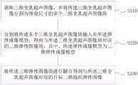
S310、获取三维全乳超声图像,并将所述三维全乳超声图像分割为预设尺寸的多个三维全乳超声图像块。S310. Acquire a three-dimensional whole breast ultrasound image, and divide the three-dimensional whole breast ultrasound image into a plurality of three-dimensional whole breast ultrasound image blocks of a preset size.
在本实施例中,弹性成像模型的输入为三维全乳超声图像的小块,因此,需要将三维全乳超声图像分割为预设尺寸的多个三维全乳超声图像块。即本实施例中的预处理图像为三维全乳超声图像块。In this embodiment, the input of the elastography model is a small block of a three-dimensional whole breast ultrasound image. Therefore, the three-dimensional whole breast ultrasound image needs to be divided into a plurality of three-dimensional whole breast ultrasound image blocks of a preset size. That is, the preprocessed image in this embodiment is a three-dimensional whole breast ultrasound image block.
其中,预设尺寸是与面阵探头视野相当的尺寸。这是因为“手持面阵探头”可以直接向空间发射二维超声波束,接收并直接生成三维超声图像以及相应的三维弹性图像。在本实施例中,弹性成像模型的训练样本即为“手持面阵探头”生成三维超声图像以及相应的三维弹性图像。The preset size is a size equivalent to the field of view of the area array probe. This is because the "hand-held area array probe" can directly transmit two-dimensional ultrasonic beams into space, receive and directly generate three-dimensional ultrasonic images and corresponding three-dimensional elastic images. In this embodiment, the training sample of the elasticity imaging model is the "hand-held area array probe" to generate a three-dimensional ultrasound image and a corresponding three-dimensional elasticity image.
S320、分别将所述多个三维全乳超声图像块输入至所述弹性成像模型,得到与所述三维全乳超声图像块对应的三维弹性图像块,其中,所述弹性成像模型为三维弹性成像模型。S320. Input the plurality of three-dimensional whole breast ultrasound image blocks into the elastography model respectively, to obtain three-dimensional elastography image blocks corresponding to the three-dimensional whole breast ultrasound image blocks, wherein the elastography model is three-dimensional elastography Model.
三维弹性成像模型是以三维超声灰度图像块和与所述三维超声灰度图像块对应得三维弹性图像块为训练样本训练完成的。训练样本是通过面阵超声探头获取的。面阵探头受探测视野的限制,训练样本本身就是以这种小区域的方式成像然后再拼接融合成完整的全乳图像。因此,取同样范围的小区域图像数据做训练数据,这个区域内的图像最大限度的保留了探头探测到的信息的完整性。The three-dimensional elastography model is trained using three-dimensional ultrasonic grayscale image blocks and three-dimensional elastic image blocks corresponding to the three-dimensional ultrasonic grayscale image blocks as training samples. The training samples were acquired with an area array ultrasound probe. The area array probe is limited by the detection field of view, and the training sample itself is imaged in such a small area and then spliced and fused into a complete whole breast image. Therefore, the image data of a small area of the same range is taken as the training data, and the images in this area retain the integrity of the information detected by the probe to the greatest extent.
S330、将所述三维弹性图像块进行融合得到与所述三维全乳超声图像对应的目标三维组织弹性图像。S330 , fuse the three-dimensional elasticity image blocks to obtain a target three-dimensional tissue elasticity image corresponding to the three-dimensional whole breast ultrasound image.
将得到的多个三维弹性图像块进行拼接融合,便可以得到目标三维组织弹性图像。By splicing and fusing the obtained multiple 3D elastic image blocks, the target 3D tissue elastic image can be obtained.
通常,在手持面阵超声探头的使用中,因为面阵的尺寸限制,其所形成的三维空间范围较小。对于较大的组织部位,如整个乳腺,需要多次移动面阵探头,才可以扫查整个乳腺区域。而多次移动探头之后获取的各个不同的三维视野之间的空间关系,因为是手动的原因,很难由固定的轨迹,很难标准化。而通过本实施例的技术方案,便可以克服上述技术缺陷,直接通过三维全乳超声图像得到相应的三维弹性图像。Generally, in the use of a hand-held area array ultrasonic probe, due to the size limitation of the area array, the three-dimensional space formed by the area array is small. For larger tissue sites, such as the entire breast, the area array probe needs to be moved several times to scan the entire breast area. However, the spatial relationship between different three-dimensional fields of view obtained after moving the probe for many times is difficult to be standardized by a fixed trajectory because it is manual. With the technical solution of this embodiment, the above-mentioned technical defects can be overcome, and the corresponding three-dimensional elastic image can be obtained directly from the three-dimensional whole breast ultrasound image.
本实施例的技术方案,通过将获取三维全乳超声图像进行预处理得到多个三维弹性图像块,将三维弹性图像块输入至预先训练好的三维弹性成像模型得到与三维弹性图像块对应的三维弹性图像块,进一步的将得到的多个三维弹性图像进行拼接融合得到与三维全乳超声图像对应的目标三维组织弹性图像,解决了三维全乳超声图像不含有弹性信息以及通过手持面阵超声探头直接进行三维弹性成像视野范围小的问题;可以实现根据三维超声灰度图像直接生成三维超声弹性图像,无需二次采集图像,也无需手动操作进行三维成像,提高了三维弹性图像的获取效率,有助于疾病的诊断。In the technical solution of this embodiment, a plurality of 3D elastic image blocks are obtained by preprocessing the acquired 3D whole breast ultrasound image, and the 3D elastic image blocks corresponding to the 3D elastic image blocks are obtained by inputting the 3D elastic image blocks into the pre-trained 3D elastography model. Elasticity image block, and further splicing and merging the obtained multiple 3D elastic images to obtain the target 3D tissue elastic image corresponding to the 3D whole breast ultrasound image, which solves the problem that the 3D whole breast ultrasound image does not contain elastic information and the hand-held area array ultrasound probe is used. The problem of directly performing 3D elastography has a small field of view; it can directly generate 3D ultrasonic elastography images from 3D ultrasonic grayscale images, without the need for secondary image acquisition or manual operations for 3D imaging, which improves the acquisition efficiency of 3D elastography images. Aid in the diagnosis of disease.
实施例四Embodiment 4
图5示出了本发明实施例四提供的一种乳腺组织弹性成像装置的结构示意图,本发明实施例可适用于获取乳腺组织的三维弹性图像的情况。FIG. 5 shows a schematic structural diagram of a breast tissue elasticity imaging device according to Embodiment 4 of the present invention, and the embodiment of the present invention is applicable to a situation in which a three-dimensional elasticity image of breast tissue is obtained.
如图5所示,本发明实施例中乳腺组织弹性成像装置,包括:图像预处理模块510、图像生成模块520和目标图像获取模块530。As shown in FIG. 5 , the breast tissue elasticity imaging device in the embodiment of the present invention includes: an
其中,图像预处理模块510,用于获取三维全乳超声图像,并对所述三维全乳超声图像进行预处理得到多个预处理图像;图像生成模块520,用于将所述预处理图像输入至预先训练好的弹性成像模型得到与所述预处理图像对应的弹性图像;目标图像获取模块530,用于将所述弹性图像进行融合得到与所述三维全乳超声图像对应的目标三维组织弹性图像。Among them, the
本实施例的技术方案,通过将获取三维全乳超声图像进行预处理得到多个预处理图像,将预处理图像输入至预先训练好的弹性成像模型得到与预处理图像对应的弹性图像,进一步的将得到的多个弹性图像进行融合得到与三维全乳超声图像对应的目标三维组织弹性图像,解决了三维全乳超声图像不含有弹性信息以及直接采用三维面阵探头进行三维弹性成像视野范围小的问题;可以实现根据三维超声灰度图像直接生成三维超声弹性图像,无需二次采集图像,也无需手动操作进行三维成像,提高了三维弹性图像的获取效率,有助于疾病的诊断。In the technical solution of this embodiment, a plurality of pre-processed images are obtained by pre-processing the acquired three-dimensional whole breast ultrasound image, and the pre-processed images are input into the pre-trained elasticity imaging model to obtain elasticity images corresponding to the pre-processed images, and further Fusion of the obtained multiple elasticity images obtains the target 3D tissue elasticity image corresponding to the 3D whole breast ultrasound image, which solves the problem that the 3D whole breast ultrasound image does not contain elasticity information and the direct use of the 3D area array probe for 3D elasticity imaging has a small field of view. Problem; 3D ultrasonic elasticity images can be directly generated from 3D ultrasonic grayscale images, without the need for secondary image acquisition or manual operation for 3D imaging, which improves the acquisition efficiency of 3D elasticity images and is helpful for disease diagnosis.
可选的,所述图像预处理模块具体用于:Optionally, the image preprocessing module is specifically used for:
将所述三维全乳超声图像分解为多帧二维组织超声图像。The three-dimensional whole breast ultrasound image is decomposed into multiple frames of two-dimensional tissue ultrasound images.
可选的,所述图像生成模块具体用于:Optionally, the image generation module is specifically used for:
将所述二维组织超声图像逐帧输入至所述弹性成像模型,得到与所述二维组织超声图像对应的二维弹性图像,其中,所述弹性成像模型为二维弹性成像模型。The two-dimensional tissue ultrasound image is input into the elastography model frame by frame to obtain a two-dimensional elasticity image corresponding to the two-dimensional tissue ultrasound image, wherein the elastography model is a two-dimensional elastography model.
可选的,所述二维弹性成像模型是以二维超声灰度图像和与所述二维超声灰度图像对应的二维弹性图像为训练样本训练完成的。Optionally, the two-dimensional elastography model is trained by using a two-dimensional ultrasonic grayscale image and a two-dimensional elastic image corresponding to the two-dimensional ultrasonic grayscale image as training samples.
可选的,所述图像预处理模块具体还用于:Optionally, the image preprocessing module is further used for:
将所述三维全乳超声图像分割为预设尺寸的多个三维全乳超声图像块。The three-dimensional whole breast ultrasound image is divided into a plurality of three-dimensional whole breast ultrasound image blocks of preset size.
可选的,所述图像生成模块具体还用于:Optionally, the image generation module is further used for:
分别将所述多个三维全乳超声图像块输入至所述弹性成像模型,得到与所述三维全乳超声图像块对应的三维弹性图像块,其中,所述弹性成像模型为三维弹性成像模型。The plurality of three-dimensional whole breast ultrasound image blocks are respectively input into the elastography model to obtain three-dimensional elastic image blocks corresponding to the three-dimensional whole breast ultrasound image blocks, wherein the elastography model is a three-dimensional elastography model.
可选的,所述三维弹性成像模型是以三维超声灰度图像块和与所述三维超声灰度图像块对应得三维弹性图像块为训练样本训练完成的。Optionally, the three-dimensional elastography model is trained using three-dimensional ultrasonic grayscale image blocks and three-dimensional elastic image blocks corresponding to the three-dimensional ultrasonic grayscale image blocks as training samples.
本发明实施例所提供的乳腺组织弹性成像装置可执行本发明任意实施例所提供的乳腺组织弹性成像方法,具备执行方法相应的功能模块和有益效果。The breast tissue elastography apparatus provided by the embodiment of the present invention can execute the breast tissue elastography method provided by any embodiment of the present invention, and has corresponding functional modules and beneficial effects for executing the method.
实施例五Embodiment 5
图6是本发明实施例五中的计算机设备的结构示意图。图6示出了适于用来实现本发明实施方式的示例性计算机设备612的框图。图6显示的计算机设备612仅仅是一个示例,不应对本发明实施例的功能和使用范围带来任何限制。FIG. 6 is a schematic structural diagram of a computer device in Embodiment 5 of the present invention. Figure 6 shows a block diagram of an
如图6所示,计算机设备612以通用计算设备的形式表现。计算机设备612的组件可以包括但不限于:一个或者多个处理器或者处理单元616,系统存储器628,连接不同系统组件(包括系统存储器628和处理单元616)的总线618。As shown in FIG. 6,
总线618表示几类总线结构中的一种或多种,包括存储器总线或者存储器控制器,外围总线,图形加速端口,处理器或者使用多种总线结构中的任意总线结构的局域总线。举例来说,这些体系结构包括但不限于工业标准体系结构(ISA)总线,微通道体系结构(MAC)总线,增强型ISA总线、视频电子标准协会(VESA)局域总线以及外围组件互连(PCI)总线。Bus 618 represents one or more of several types of bus structures, including a memory bus or memory controller, a peripheral bus, a graphics acceleration port, a processor, or a local bus using any of a variety of bus structures. By way of example, these architectures include, but are not limited to, Industry Standard Architecture (ISA) bus, Micro Channel Architecture (MAC) bus, Enhanced ISA bus, Video Electronics Standards Association (VESA) local bus, and Peripheral Component Interconnect ( PCI) bus.
计算机设备612典型地包括多种计算机系统可读介质。这些介质可以是任何能够被计算机设备612访问的可用介质,包括易失性和非易失性介质,可移动的和不可移动的介质。
系统存储器628可以包括易失性存储器形式的计算机系统可读介质,例如随机存取存储器(RAM)630和/或高速缓存存储器632。计算机设备612可以进一步包括其它可移动/不可移动的、易失性/非易失性计算机系统存储介质。仅作为举例,存储系统634可以用于读写不可移动的、非易失性磁介质(图6未显示,通常称为“硬盘驱动器”)。尽管图6中未示出,可以提供用于对可移动非易失性磁盘(例如“软盘”)读写的磁盘驱动器,以及对可移动非易失性光盘(例如CD-ROM,DVD-ROM或者其它光介质)读写的光盘驱动器。在这些情况下,每个驱动器可以通过一个或者多个数据介质接口与总线618相连。存储器628可以包括至少一个程序产品,该程序产品具有一组(例如至少一个)程序模块,这些程序模块被配置以执行本发明各实施例的功能。
具有一组(至少一个)程序模块642的程序/实用工具640,可以存储在例如存储器628中,这样的程序模块642包括但不限于操作系统、一个或者多个应用程序、其它程序模块以及程序数据,这些示例中的每一个或某种组合中可能包括网络环境的实现。程序模块642通常执行本发明所描述的实施例中的功能和/或方法。A program/
计算机设备612也可以与一个或多个外部设备614(例如键盘、指向设备、显示器624等)通信,还可与一个或者多个使得用户能与该计算机设备612交互的设备通信,和/或与使得该计算机设备612能与一个或多个其它计算设备进行通信的任何设备(例如网卡,调制解调器等等)通信。这种通信可以通过输入/输出(I/O)接口622进行。并且,计算机设备612还可以通过网络适配器620与一个或者多个网络(例如局域网(LAN),广域网(WAN)和/或公共网络,例如因特网)通信。如图所示,网络适配器620通过总线618与计算机设备612的其它模块通信。应当明白,尽管图6中未示出,可以结合计算机设备612使用其它硬件和/或软件模块,包括但不限于:微代码、设备驱动器、冗余处理单元、外部磁盘驱动阵列、RAID系统、磁带驱动器以及数据备份存储系统等。
处理单元616通过运行存储在系统存储器628中的程序,从而执行各种功能应用以及数据处理,例如实现本发明实施例所提供的乳腺组织弹性成像方法,该方法主要包括:The
获取三维全乳超声图像,并对所述三维全乳超声图像进行预处理得到多个预处理图像;acquiring a three-dimensional whole breast ultrasound image, and preprocessing the three-dimensional whole breast ultrasound image to obtain a plurality of preprocessed images;
将所述预处理图像输入至预先训练好的弹性成像模型得到与所述预处理图像对应的弹性图像;Inputting the preprocessed image into a pretrained elastography model to obtain an elastography image corresponding to the preprocessed image;
将所述弹性图像进行融合得到与所述三维全乳超声图像对应的目标三维组织弹性图像。The elasticity image is fused to obtain a target three-dimensional tissue elasticity image corresponding to the three-dimensional whole breast ultrasound image.
实施例六Embodiment 6
本发明实施例六还提供了一种计算机可读存储介质,其上存储有计算机程序,该程序被处理器执行时实现如本发明实施例所提供的乳腺组织弹性成像方法,该方法主要包括:Embodiment 6 of the present invention also provides a computer-readable storage medium, on which a computer program is stored, and when the program is executed by a processor, implements the breast tissue elasticity imaging method provided by the embodiment of the present invention, and the method mainly includes:
获取三维全乳超声图像,并对所述三维全乳超声图像进行预处理得到多个预处理图像;acquiring a three-dimensional whole breast ultrasound image, and preprocessing the three-dimensional whole breast ultrasound image to obtain a plurality of preprocessed images;
将所述预处理图像输入至预先训练好的弹性成像模型得到与所述预处理图像对应的弹性图像;Inputting the preprocessed image into a pretrained elastography model to obtain an elastography image corresponding to the preprocessed image;
将所述弹性图像进行融合得到与所述三维全乳超声图像对应的目标三维组织弹性图像。The elasticity image is fused to obtain a target three-dimensional tissue elasticity image corresponding to the three-dimensional whole breast ultrasound image.
本发明实施例的计算机存储介质,可以采用一个或多个计算机可读的介质的任意组合。计算机可读介质可以是计算机可读信号介质或者计算机可读存储介质。计算机可读存储介质例如可以是——但不限于——电、磁、光、电磁、红外线、或半导体的系统、装置或器件,或者任意以上的组合。计算机可读存储介质的更具体的例子(非穷举的列表)包括:具有一个或多个导线的电连接、便携式计算机磁盘、硬盘、随机存取存储器(RAM)、只读存储器(ROM)、可擦式可编程只读存储器(EPROM或闪存)、光纤、便携式紧凑磁盘只读存储器(CD-ROM)、光存储器件、磁存储器件、或者上述的任意合适的组合。在本文件中,计算机可读存储介质可以是任何包含或存储程序的有形介质,该程序可以被指令执行系统、装置或者器件使用或者与其结合使用。The computer storage medium in the embodiments of the present invention may adopt any combination of one or more computer-readable mediums. The computer-readable medium may be a computer-readable signal medium or a computer-readable storage medium. The computer-readable storage medium can be, for example, but not limited to, an electrical, magnetic, optical, electromagnetic, infrared, or semiconductor system, apparatus or device, or a combination of any of the above. More specific examples (a non-exhaustive list) of computer readable storage media include: electrical connections having one or more wires, portable computer disks, hard disks, random access memory (RAM), read only memory (ROM), Erasable programmable read only memory (EPROM or flash memory), optical fiber, portable compact disk read only memory (CD-ROM), optical storage devices, magnetic storage devices, or any suitable combination of the foregoing. In this document, a computer-readable storage medium can be any tangible medium that contains or stores a program that can be used by or in conjunction with an instruction execution system, apparatus, or device.
计算机可读的信号介质可以包括在基带中或者作为载波一部分传播的数据信号,其中承载了计算机可读的程序代码。这种传播的数据信号可以采用多种形式,包括但不限于电磁信号、光信号或上述的任意合适的组合。计算机可读的信号介质还可以是计算机可读存储介质以外的任何计算机可读介质,该计算机可读介质可以发送、传播或者传输用于由指令执行系统、装置或者器件使用或者与其结合使用的程序。A computer-readable signal medium may include a propagated data signal in baseband or as part of a carrier wave, with computer-readable program code embodied thereon. Such propagated data signals may take a variety of forms, including but not limited to electromagnetic signals, optical signals, or any suitable combination of the foregoing. A computer-readable signal medium can also be any computer-readable medium other than a computer-readable storage medium that can transmit, propagate, or transport the program for use by or in connection with the instruction execution system, apparatus, or device .
计算机可读介质上包含的程序代码可以用任何适当的介质传输,包括——但不限于无线、电线、光缆、RF等等,或者上述的任意合适的组合。Program code embodied on a computer readable medium may be transmitted using any suitable medium, including - but not limited to wireless, wireline, optical fiber cable, RF, etc., or any suitable combination of the foregoing.
可以以一种或多种程序设计语言或其组合来编写用于执行本发明操作的计算机程序代码,所述程序设计语言包括面向对象的程序设计语言—诸如Java、Smalltalk、C++,还包括常规的过程式程序设计语言—诸如”C”语言或类似的程序设计语言。程序代码可以完全地在用户计算机上执行、部分地在用户计算机上执行、作为一个独立的软件包执行、部分在用户计算机上部分在远程计算机上执行、或者完全在远程计算机或服务器上执行。在涉及远程计算机的情形中,远程计算机可以通过任意种类的网络——包括局域网(LAN)或广域网(WAN)—连接到用户计算机,或者,可以连接到外部计算机(例如利用因特网服务提供商来通过因特网连接)。Computer program code for carrying out operations of the present invention may be written in one or more programming languages, including object-oriented programming languages—such as Java, Smalltalk, C++, but also conventional Procedural programming language - such as the "C" language or similar programming language. The program code may execute entirely on the user's computer, partly on the user's computer, as a stand-alone software package, partly on the user's computer and partly on a remote computer, or entirely on the remote computer or server. In the case of a remote computer, the remote computer may be connected to the user's computer through any kind of network, including a local area network (LAN) or a wide area network (WAN), or may be connected to an external computer (eg, using an Internet service provider through Internet connection).
注意,上述仅为本发明的较佳实施例及所运用技术原理。本领域技术人员会理解,本发明不限于这里所述的特定实施例,对本领域技术人员来说能够进行各种明显的变化、重新调整和替代而不会脱离本发明的保护范围。因此,虽然通过以上实施例对本发明进行了较为详细的说明,但是本发明不仅仅限于以上实施例,在不脱离本发明构思的情况下,还可以包括更多其他等效实施例,而本发明的范围由所附的权利要求范围决定。Note that the above are only preferred embodiments of the present invention and applied technical principles. Those skilled in the art will understand that the present invention is not limited to the specific embodiments described herein, and various obvious changes, readjustments and substitutions can be made by those skilled in the art without departing from the protection scope of the present invention. Therefore, although the present invention has been described in detail through the above embodiments, the present invention is not limited to the above embodiments, and can also include more other equivalent embodiments without departing from the concept of the present invention. The scope is determined by the scope of the appended claims.
Claims (10)
Priority Applications (1)
| Application Number | Priority Date | Filing Date | Title |
|---|---|---|---|
| CN202010680159.8ACN111803128A (en) | 2020-07-15 | 2020-07-15 | A breast tissue elasticity imaging method, device, equipment and medium |
Applications Claiming Priority (1)
| Application Number | Priority Date | Filing Date | Title |
|---|---|---|---|
| CN202010680159.8ACN111803128A (en) | 2020-07-15 | 2020-07-15 | A breast tissue elasticity imaging method, device, equipment and medium |
Publications (1)
| Publication Number | Publication Date |
|---|---|
| CN111803128Atrue CN111803128A (en) | 2020-10-23 |
Family
ID=72866256
Family Applications (1)
| Application Number | Title | Priority Date | Filing Date |
|---|---|---|---|
| CN202010680159.8APendingCN111803128A (en) | 2020-07-15 | 2020-07-15 | A breast tissue elasticity imaging method, device, equipment and medium |
Country Status (1)
| Country | Link |
|---|---|
| CN (1) | CN111803128A (en) |
Cited By (3)
| Publication number | Priority date | Publication date | Assignee | Title |
|---|---|---|---|---|
| CN115393301A (en)* | 2022-08-16 | 2022-11-25 | 中山大学附属第一医院 | A radiomics analysis method and device for two-dimensional shear wave elastography images of the liver |
| WO2024044824A1 (en)* | 2022-09-01 | 2024-03-07 | The University Of Adelaide | Scale-dependent elastography method for detection of small inclusions in biological tissue |
| CN118172311A (en)* | 2024-02-29 | 2024-06-11 | 中山大学附属第一医院 | A method and device for analyzing two-dimensional shear wave elastic images of hepatocellular carcinoma |
Citations (13)
| Publication number | Priority date | Publication date | Assignee | Title |
|---|---|---|---|---|
| US20080306384A1 (en)* | 2007-06-08 | 2008-12-11 | The Johns Hopkins University | Apparatus and method for computing 3D ultrasound elasticity images |
| US20090326378A1 (en)* | 2008-06-26 | 2009-12-31 | Medison Co., Ltd. | Formation Of An Enhanced Elastic Image In An Ultrasound System |
| CN102908166A (en)* | 2012-08-28 | 2013-02-06 | 华南理工大学 | Scanning device and method for three-dimensional ultrasound elasticity imaging |
| CN102908168A (en)* | 2012-10-24 | 2013-02-06 | 华南理工大学 | A-mode ultrasonic elastic imaging system based on mechanical scanning and method thereof |
| CN103269430A (en)* | 2013-04-16 | 2013-08-28 | 上海上安机电设计事务所有限公司 | Three-dimensional scene generation method based on building information model (BIM) |
| KR20150056013A (en)* | 2013-11-14 | 2015-05-22 | 계명대학교 산학협력단 | Method and apparatus for acquisition and analysis of breast ultrasonography imaging and elastography imaging |
| US20170153321A1 (en)* | 2015-12-01 | 2017-06-01 | Samsung Medison Co., Ltd. | Method and apparatus for acquiring image using ultrasound |
| CN107451983A (en)* | 2017-07-18 | 2017-12-08 | 中山大学附属第六医院 | The three-dimensional fusion method and system of CT images |
| CN107483910A (en)* | 2017-07-14 | 2017-12-15 | 清华大学 | A long-distance naked-eye stereoscopic display method and system thereof |
| CN108986909A (en)* | 2018-06-29 | 2018-12-11 | 清华大学 | Soft tissue elasticity and viscoelastic characterizations' method and device based on ultrasonic elastograph imaging |
| US20190336108A1 (en)* | 2017-01-05 | 2019-11-07 | Koninklijke Philips N.V. | Ultrasound imaging system with a neural network for deriving imaging data and tissue information |
| CN111091603A (en)* | 2019-11-04 | 2020-05-01 | 深圳先进技术研究院 | Ultrasonic imaging method and device, readable storage medium and terminal equipment |
| US20200168227A1 (en)* | 2017-09-29 | 2020-05-28 | Shanghai Cambricon Information Technology Co., Ltd | Image processing apparatus and method |
- 2020
- 2020-07-15CNCN202010680159.8Apatent/CN111803128A/enactivePending
Patent Citations (13)
| Publication number | Priority date | Publication date | Assignee | Title |
|---|---|---|---|---|
| US20080306384A1 (en)* | 2007-06-08 | 2008-12-11 | The Johns Hopkins University | Apparatus and method for computing 3D ultrasound elasticity images |
| US20090326378A1 (en)* | 2008-06-26 | 2009-12-31 | Medison Co., Ltd. | Formation Of An Enhanced Elastic Image In An Ultrasound System |
| CN102908166A (en)* | 2012-08-28 | 2013-02-06 | 华南理工大学 | Scanning device and method for three-dimensional ultrasound elasticity imaging |
| CN102908168A (en)* | 2012-10-24 | 2013-02-06 | 华南理工大学 | A-mode ultrasonic elastic imaging system based on mechanical scanning and method thereof |
| CN103269430A (en)* | 2013-04-16 | 2013-08-28 | 上海上安机电设计事务所有限公司 | Three-dimensional scene generation method based on building information model (BIM) |
| KR20150056013A (en)* | 2013-11-14 | 2015-05-22 | 계명대학교 산학협력단 | Method and apparatus for acquisition and analysis of breast ultrasonography imaging and elastography imaging |
| US20170153321A1 (en)* | 2015-12-01 | 2017-06-01 | Samsung Medison Co., Ltd. | Method and apparatus for acquiring image using ultrasound |
| US20190336108A1 (en)* | 2017-01-05 | 2019-11-07 | Koninklijke Philips N.V. | Ultrasound imaging system with a neural network for deriving imaging data and tissue information |
| CN107483910A (en)* | 2017-07-14 | 2017-12-15 | 清华大学 | A long-distance naked-eye stereoscopic display method and system thereof |
| CN107451983A (en)* | 2017-07-18 | 2017-12-08 | 中山大学附属第六医院 | The three-dimensional fusion method and system of CT images |
| US20200168227A1 (en)* | 2017-09-29 | 2020-05-28 | Shanghai Cambricon Information Technology Co., Ltd | Image processing apparatus and method |
| CN108986909A (en)* | 2018-06-29 | 2018-12-11 | 清华大学 | Soft tissue elasticity and viscoelastic characterizations' method and device based on ultrasonic elastograph imaging |
| CN111091603A (en)* | 2019-11-04 | 2020-05-01 | 深圳先进技术研究院 | Ultrasonic imaging method and device, readable storage medium and terminal equipment |
Non-Patent Citations (1)
| Title |
|---|
| R.R.WILDEBOER ET AL: "Synthetic Elastography from B-Mode ultrasound through Deep Learning", 《2019 IEEE INTERNATIONAL ULTRASONICS SYMPOSIUM(IUS)》, 9 December 2019 (2019-12-09), pages 108 - 109* |
Cited By (5)
| Publication number | Priority date | Publication date | Assignee | Title |
|---|---|---|---|---|
| CN115393301A (en)* | 2022-08-16 | 2022-11-25 | 中山大学附属第一医院 | A radiomics analysis method and device for two-dimensional shear wave elastography images of the liver |
| CN115393301B (en)* | 2022-08-16 | 2024-03-12 | 中山大学附属第一医院 | Image histology analysis method and device for liver two-dimensional shear wave elastic image |
| WO2024044824A1 (en)* | 2022-09-01 | 2024-03-07 | The University Of Adelaide | Scale-dependent elastography method for detection of small inclusions in biological tissue |
| CN118172311A (en)* | 2024-02-29 | 2024-06-11 | 中山大学附属第一医院 | A method and device for analyzing two-dimensional shear wave elastic images of hepatocellular carcinoma |
| CN118172311B (en)* | 2024-02-29 | 2025-03-28 | 中山大学附属第一医院 | A method and device for analyzing two-dimensional shear wave elastic images of hepatocellular carcinoma |
Similar Documents
| Publication | Publication Date | Title |
|---|---|---|
| CN109961491B (en) | Multi-mode image truncation compensation method, device, computer equipment and medium | |
| US10950026B2 (en) | Systems and methods for displaying a medical image | |
| CN110599526B (en) | Image registration method, computer device, and storage medium | |
| EP3926537A1 (en) | Medical image segmentation method, image segmentation method, related device and system | |
| CN111814768B (en) | Image recognition method, device, medium and equipment based on AI composite model | |
| CN111803128A (en) | A breast tissue elasticity imaging method, device, equipment and medium | |
| CN110728673A (en) | Target part analysis method and device, computer equipment and storage medium | |
| US9142030B2 (en) | Systems, methods and computer readable storage media storing instructions for automatically segmenting images of a region of interest | |
| CN108742679B (en) | Nodule detection apparatus and method | |
| CN111062935B (en) | Mammary gland tumor detection method, storage medium and terminal equipment | |
| CN110246135B (en) | Follicle monitoring method, device, system and storage medium | |
| WO2022206025A1 (en) | Biomechanical modeling method and apparatus, electronic device and storage medium | |
| CN109087357B (en) | Scanning and positioning method, apparatus, computer equipment and computer-readable storage medium | |
| CN117115355A (en) | Three-dimensional ultrasonic modeling method, system, electronic device and readable storage medium | |
| US20120078101A1 (en) | Ultrasound system for displaying slice of object and method thereof | |
| Sun et al. | Automatic Robotic Ultrasound for 3D Musculoskeletal Reconstruction: A Comprehensive Framework | |
| US20230137369A1 (en) | Aiding a user to perform a medical ultrasound examination | |
| CN110555897A (en) | Image generation method, device, equipment and storage medium | |
| CN119833083A (en) | Image comparison system based on natural language | |
| JP7453400B2 (en) | Ultrasonic systems and methods of controlling them | |
| WO2022127318A1 (en) | Scanning positioning method and apparatus, storage medium and electronic device | |
| US20250137971A1 (en) | Ultrasound to 3d registration using a-mode signals | |
| CN112674799B (en) | Ultrasonic elastography method, electronic device and storage medium | |
| CN112614123B (en) | Ultrasonic image recognition method and related device | |
| CN111627081B (en) | CT image reconstruction method, device, equipment and medium |
Legal Events
| Date | Code | Title | Description |
|---|---|---|---|
| PB01 | Publication | ||
| PB01 | Publication | ||
| SE01 | Entry into force of request for substantive examination | ||
| SE01 | Entry into force of request for substantive examination | ||
| RJ01 | Rejection of invention patent application after publication | Application publication date:20201023 | |
| RJ01 | Rejection of invention patent application after publication |