CN111801107A - Bacteriophages for the regulation of inflammatory bowel disease - Google Patents
Bacteriophages for the regulation of inflammatory bowel diseaseDownload PDFInfo
- Publication number
- CN111801107A CN111801107ACN201880071574.4ACN201880071574ACN111801107ACN 111801107 ACN111801107 ACN 111801107ACN 201880071574 ACN201880071574 ACN 201880071574ACN 111801107 ACN111801107 ACN 111801107A
- Authority
- CN
- China
- Prior art keywords
- kp2m
- kp2w
- kp3w
- bacteriophage
- lytic
- Prior art date
- Legal status (The legal status is an assumption and is not a legal conclusion. Google has not performed a legal analysis and makes no representation as to the accuracy of the status listed.)
- Pending
Links
- 241001515965unidentified phageSpecies0.000titleclaimsabstractdescription256
- 208000022559Inflammatory bowel diseaseDiseases0.000titleclaimsabstractdescription80
- 239000000203mixtureSubstances0.000claimsabstractdescription126
- 230000001580bacterial effectEffects0.000claimsabstractdescription65
- 241000894006BacteriaSpecies0.000claimsdescription132
- 238000000034methodMethods0.000claimsdescription53
- 230000002101lytic effectEffects0.000claimsdescription49
- 208000011231Crohn diseaseDiseases0.000claimsdescription39
- 241000588747Klebsiella pneumoniaeSpecies0.000claimsdescription39
- 239000008194pharmaceutical compositionSubstances0.000claimsdescription39
- 208000015181infectious diseaseDiseases0.000claimsdescription38
- 206010009900Colitis ulcerativeDiseases0.000claimsdescription36
- 201000006704Ulcerative ColitisDiseases0.000claimsdescription36
- 241000124008MammaliaSpecies0.000claimsdescription23
- 210000000936intestineAnatomy0.000claimsdescription20
- 239000012472biological sampleSubstances0.000claimsdescription7
- 229940126409proton pump inhibitorDrugs0.000claimsdescription7
- 239000000612proton pump inhibitorSubstances0.000claimsdescription7
- 208000010157sclerosing cholangitisDiseases0.000claimsdescription7
- 238000002560therapeutic procedureMethods0.000claimsdescription7
- 206010008609Cholangitis sclerosingDiseases0.000claimsdescription6
- 229920003229poly(methyl methacrylate)Polymers0.000claimsdescription6
- 201000000742primary sclerosing cholangitisDiseases0.000claimsdescription6
- 238000012258culturingMethods0.000claimsdescription4
- 241000588748KlebsiellaSpecies0.000abstractdescription53
- 230000002934lysing effectEffects0.000abstractdescription29
- 208000037265diseases, disorders, signs and symptomsDiseases0.000abstractdescription18
- 201000010099diseaseDiseases0.000abstractdescription15
- 230000001225therapeutic effectEffects0.000abstractdescription8
- 241000699670Mus sp.Species0.000description51
- 238000003556assayMethods0.000description40
- 210000004027cellAnatomy0.000description29
- 229920001817AgarPolymers0.000description28
- -1Mg2+ ionsChemical class0.000description28
- 239000008272agarSubstances0.000description28
- 238000004458analytical methodMethods0.000description24
- 239000007788liquidSubstances0.000description24
- 239000000523sampleSubstances0.000description24
- 235000010419agarNutrition0.000description23
- 108090000623proteins and genesProteins0.000description18
- 210000003296salivaAnatomy0.000description17
- 239000010865sewageSubstances0.000description17
- 239000000546pharmaceutical excipientSubstances0.000description16
- 208000024891symptomDiseases0.000description16
- 241000588915Klebsiella aerogenesSpecies0.000description15
- 241000736262MicrobiotaSpecies0.000description15
- 230000006872improvementEffects0.000description14
- 238000011282treatmentMethods0.000description14
- 230000012010growthEffects0.000description13
- 239000002609mediumSubstances0.000description13
- 229940092559enterobacter aerogenesDrugs0.000description11
- 230000002550fecal effectEffects0.000description11
- 210000000214mouthAnatomy0.000description11
- 210000004400mucous membraneAnatomy0.000description11
- 108020004414DNAProteins0.000description10
- 150000007523nucleic acidsChemical group0.000description10
- 239000003826tabletSubstances0.000description10
- 206010061218InflammationDiseases0.000description9
- 239000000872bufferSubstances0.000description9
- 210000001072colonAnatomy0.000description9
- 239000002552dosage formSubstances0.000description9
- 239000003814drugSubstances0.000description9
- 230000007613environmental effectEffects0.000description9
- 210000003608feceAnatomy0.000description9
- 230000002068genetic effectEffects0.000description9
- 230000004054inflammatory processEffects0.000description9
- 229920001223polyethylene glycolPolymers0.000description9
- 241000894007speciesSpecies0.000description9
- 239000000725suspensionSubstances0.000description9
- 238000013268sustained releaseMethods0.000description9
- 239000012730sustained-release formSubstances0.000description9
- 229920002472StarchPolymers0.000description8
- 239000011575calciumSubstances0.000description8
- 230000000112colonic effectEffects0.000description8
- 210000001035gastrointestinal tractAnatomy0.000description8
- 230000000968intestinal effectEffects0.000description8
- 230000009467reductionEffects0.000description8
- 235000002639sodium chlorideNutrition0.000description8
- 239000007787solidSubstances0.000description8
- 238000012360testing methodMethods0.000description8
- 10802000446516S ribosomal RNAProteins0.000description7
- 239000002775capsuleSubstances0.000description7
- 150000001875compoundsChemical class0.000description7
- 238000009472formulationMethods0.000description7
- 238000001727in vivoMethods0.000description7
- 238000012216screeningMethods0.000description7
- 238000012163sequencing techniqueMethods0.000description7
- 239000000243solutionSubstances0.000description7
- 239000008107starchSubstances0.000description7
- 235000019698starchNutrition0.000description7
- 229940032147starchDrugs0.000description7
- LFQSCWFLJHTTHZ-UHFFFAOYSA-NEthanolChemical compoundCCOLFQSCWFLJHTTHZ-UHFFFAOYSA-N0.000description6
- PEDCQBHIVMGVHV-UHFFFAOYSA-NGlycerineChemical compoundOCC(O)COPEDCQBHIVMGVHV-UHFFFAOYSA-N0.000description6
- 239000002202Polyethylene glycolSubstances0.000description6
- FAPWRFPIFSIZLT-UHFFFAOYSA-MSodium chlorideChemical compound[Na+].[Cl-]FAPWRFPIFSIZLT-UHFFFAOYSA-M0.000description6
- 230000009089cytolysisEffects0.000description6
- LOKCTEFSRHRXRJ-UHFFFAOYSA-Idipotassium trisodium dihydrogen phosphate hydrogen phosphate dichlorideChemical compoundP(=O)(O)(O)[O-].[K+].P(=O)(O)([O-])[O-].[Na+].[Na+].[Cl-].[K+].[Cl-].[Na+]LOKCTEFSRHRXRJ-UHFFFAOYSA-I0.000description6
- 230000000694effectsEffects0.000description6
- 230000006698inductionEffects0.000description6
- 238000002955isolationMethods0.000description6
- 230000003287optical effectEffects0.000description6
- 230000008506pathogenesisEffects0.000description6
- 239000002953phosphate buffered salineSubstances0.000description6
- 150000003839saltsChemical group0.000description6
- 210000000813small intestineAnatomy0.000description6
- 239000006228supernatantSubstances0.000description6
- XLYOFNOQVPJJNP-UHFFFAOYSA-NwaterSubstancesOXLYOFNOQVPJJNP-UHFFFAOYSA-N0.000description6
- 108091028043Nucleic acid sequenceProteins0.000description5
- VYPSYNLAJGMNEJ-UHFFFAOYSA-NSilicium dioxideChemical compoundO=[Si]=OVYPSYNLAJGMNEJ-UHFFFAOYSA-N0.000description5
- 238000009825accumulationMethods0.000description5
- 235000010980celluloseNutrition0.000description5
- 229920002678cellulosePolymers0.000description5
- 239000001913celluloseSubstances0.000description5
- 239000003795chemical substances by applicationSubstances0.000description5
- 238000013270controlled releaseMethods0.000description5
- 229940079593drugDrugs0.000description5
- 244000005709gut microbiomeSpecies0.000description5
- 230000001320lysogenic effectEffects0.000description5
- 108020004707nucleic acidsProteins0.000description5
- 102000039446nucleic acidsHuman genes0.000description5
- 102000004169proteins and genesHuman genes0.000description5
- 210000002784stomachAnatomy0.000description5
- 239000004094surface-active agentSubstances0.000description5
- IJGRMHOSHXDMSA-UHFFFAOYSA-NAtomic nitrogenChemical compoundN#NIJGRMHOSHXDMSA-UHFFFAOYSA-N0.000description4
- HEDRZPFGACZZDS-UHFFFAOYSA-NChloroformChemical compoundClC(Cl)ClHEDRZPFGACZZDS-UHFFFAOYSA-N0.000description4
- KCXVZYZYPLLWCC-UHFFFAOYSA-NEDTAChemical compoundOC(=O)CN(CC(O)=O)CCN(CC(O)=O)CC(O)=OKCXVZYZYPLLWCC-UHFFFAOYSA-N0.000description4
- 108010010803GelatinProteins0.000description4
- SIKJAQJRHWYJAI-UHFFFAOYSA-NIndoleChemical compoundC1=CC=C2NC=CC2=C1SIKJAQJRHWYJAI-UHFFFAOYSA-N0.000description4
- UIIMBOGNXHQVGW-UHFFFAOYSA-MSodium bicarbonateChemical compound[Na+].OC([O-])=OUIIMBOGNXHQVGW-UHFFFAOYSA-M0.000description4
- 210000001744T-lymphocyteAnatomy0.000description4
- 150000001413amino acidsChemical group0.000description4
- 239000003242anti bacterial agentSubstances0.000description4
- 229940088710antibiotic agentDrugs0.000description4
- 239000003937drug carrierSubstances0.000description4
- 239000008273gelatinSubstances0.000description4
- 229920000159gelatinPolymers0.000description4
- 229940014259gelatinDrugs0.000description4
- 235000019322gelatineNutrition0.000description4
- 235000011852gelatine dessertsNutrition0.000description4
- 239000008187granular materialSubstances0.000description4
- 210000003405ileumAnatomy0.000description4
- 210000002865immune cellAnatomy0.000description4
- 230000028993immune responseEffects0.000description4
- 238000011534incubationMethods0.000description4
- 230000001939inductive effectEffects0.000description4
- 210000002429large intestineAnatomy0.000description4
- 230000001717pathogenic effectEffects0.000description4
- 229920000036polyvinylpyrrolidonePolymers0.000description4
- 239000001267polyvinylpyrrolidoneSubstances0.000description4
- 235000013855polyvinylpyrrolidoneNutrition0.000description4
- 238000012545processingMethods0.000description4
- 235000018102proteinsNutrition0.000description4
- 239000007858starting materialSubstances0.000description4
- 235000000346sugarNutrition0.000description4
- 150000008163sugarsChemical class0.000description4
- 208000004998Abdominal PainDiseases0.000description3
- 229920000936AgarosePolymers0.000description3
- OYPRJOBELJOOCE-UHFFFAOYSA-NCalciumChemical compound[Ca]OYPRJOBELJOOCE-UHFFFAOYSA-N0.000description3
- 206010012735DiarrhoeaDiseases0.000description3
- 208000027244DysbiosisDiseases0.000description3
- 241000605909FusobacteriumSpecies0.000description3
- 201000008225Klebsiella pneumoniaDiseases0.000description3
- GUBGYTABKSRVRQ-QKKXKWKRSA-NLactoseNatural productsOC[C@H]1O[C@@H](O[C@H]2[C@H](O)[C@@H](O)C(O)O[C@@H]2CO)[C@H](O)[C@@H](O)[C@H]1OGUBGYTABKSRVRQ-QKKXKWKRSA-N0.000description3
- 206010035717Pneumonia klebsiellaDiseases0.000description3
- 229920002732PolyanhydridePolymers0.000description3
- 229920000954PolyglycolidePolymers0.000description3
- 229920001213Polysorbate 20Polymers0.000description3
- 238000011529RT qPCRMethods0.000description3
- DBMJMQXJHONAFJ-UHFFFAOYSA-MSodium laurylsulphateChemical compound[Na+].CCCCCCCCCCCCOS([O-])(=O)=ODBMJMQXJHONAFJ-UHFFFAOYSA-M0.000description3
- 229930006000SucroseNatural products0.000description3
- CZMRCDWAGMRECN-UGDNZRGBSA-NSucroseChemical compoundO[C@H]1[C@H](O)[C@@H](CO)O[C@@]1(CO)O[C@@H]1[C@H](O)[C@@H](O)[C@H](O)[C@@H](CO)O1CZMRCDWAGMRECN-UGDNZRGBSA-N0.000description3
- RTAQQCXQSZGOHL-UHFFFAOYSA-NTitaniumChemical compound[Ti]RTAQQCXQSZGOHL-UHFFFAOYSA-N0.000description3
- ZMANZCXQSJIPKH-UHFFFAOYSA-NTriethylamineChemical compoundCCN(CC)CCZMANZCXQSJIPKH-UHFFFAOYSA-N0.000description3
- 208000025865UlcerDiseases0.000description3
- 241001148134VeillonellaSpecies0.000description3
- 241000700605VirusesSpecies0.000description3
- 239000002253acidSubstances0.000description3
- 239000002671adjuvantSubstances0.000description3
- 230000003321amplificationEffects0.000description3
- 210000000436anusAnatomy0.000description3
- 229910052791calciumInorganic materials0.000description3
- 230000003247decreasing effectEffects0.000description3
- 235000005911dietNutrition0.000description3
- 230000007140dysbiosisEffects0.000description3
- 210000003238esophagusAnatomy0.000description3
- 238000001704evaporationMethods0.000description3
- 230000008020evaporationEffects0.000description3
- 239000000499gelSubstances0.000description3
- 210000003128headAnatomy0.000description3
- 239000008101lactoseSubstances0.000description3
- 208000019423liver diseaseDiseases0.000description3
- 210000004698lymphocyteAnatomy0.000description3
- 239000011777magnesiumSubstances0.000description3
- 229910001425magnesium ionInorganic materials0.000description3
- 239000003550markerSubstances0.000description3
- 239000000463materialSubstances0.000description3
- 239000011159matrix materialSubstances0.000description3
- 239000003094microcapsuleSubstances0.000description3
- 239000002480mineral oilSubstances0.000description3
- 235000010446mineral oilNutrition0.000description3
- 210000004877mucosaAnatomy0.000description3
- 230000035772mutationEffects0.000description3
- 239000002088nanocapsuleSubstances0.000description3
- 239000002105nanoparticleSubstances0.000description3
- 229910052757nitrogenInorganic materials0.000description3
- 238000003199nucleic acid amplification methodMethods0.000description3
- 239000002245particleSubstances0.000description3
- 244000052769pathogenSpecies0.000description3
- 229920000747poly(lactic acid)Polymers0.000description3
- 229920001606poly(lactic acid-co-glycolic acid)Polymers0.000description3
- 229920000642polymerPolymers0.000description3
- 235000010486polyoxyethylene sorbitan monolaurateNutrition0.000description3
- 239000000256polyoxyethylene sorbitan monolaurateSubstances0.000description3
- 229940068977polysorbate 20Drugs0.000description3
- 239000013641positive controlSubstances0.000description3
- 239000000843powderSubstances0.000description3
- 238000002360preparation methodMethods0.000description3
- 230000002265preventionEffects0.000description3
- 239000006041probioticSubstances0.000description3
- 235000018291probioticsNutrition0.000description3
- 230000008569processEffects0.000description3
- 239000000047productSubstances0.000description3
- 230000000770proinflammatory effectEffects0.000description3
- 230000000069prophylactic effectEffects0.000description3
- 210000000664rectumAnatomy0.000description3
- 210000003705ribosomeAnatomy0.000description3
- 239000011780sodium chlorideSubstances0.000description3
- 235000019333sodium laurylsulphateNutrition0.000description3
- 235000010356sorbitolNutrition0.000description3
- 238000010186stainingMethods0.000description3
- 239000005720sucroseSubstances0.000description3
- 239000006188syrupSubstances0.000description3
- 235000020357syrupNutrition0.000description3
- 239000010936titaniumSubstances0.000description3
- 229910052719titaniumInorganic materials0.000description3
- 235000015112vegetable and seed oilNutrition0.000description3
- 239000008158vegetable oilSubstances0.000description3
- 208000016261weight lossDiseases0.000description3
- 230000004580weight lossEffects0.000description3
- IXPNQXFRVYWDDI-UHFFFAOYSA-N1-methyl-2,4-dioxo-1,3-diazinane-5-carboximidamideChemical compoundCN1CC(C(N)=N)C(=O)NC1=OIXPNQXFRVYWDDI-UHFFFAOYSA-N0.000description2
- QKNYBSVHEMOAJP-UHFFFAOYSA-N2-amino-2-(hydroxymethyl)propane-1,3-diol;hydron;chlorideChemical compoundCl.OCC(N)(CO)COQKNYBSVHEMOAJP-UHFFFAOYSA-N0.000description2
- GUBGYTABKSRVRQ-XLOQQCSPSA-NAlpha-LactoseChemical compoundO[C@@H]1[C@@H](O)[C@@H](O)[C@@H](CO)O[C@H]1O[C@@H]1[C@@H](CO)O[C@H](O)[C@H](O)[C@H]1OGUBGYTABKSRVRQ-XLOQQCSPSA-N0.000description2
- 108091093088AmpliconProteins0.000description2
- 241000379991AnaerococcusSpecies0.000description2
- 206010002556Ankylosing SpondylitisDiseases0.000description2
- 241000416162Astragalus gummiferSpecies0.000description2
- 206010003827Autoimmune hepatitisDiseases0.000description2
- 206010009691ClubbingDiseases0.000description2
- 229920002261Corn starchPolymers0.000description2
- FBPFZTCFMRRESA-FSIIMWSLSA-ND-GlucitolNatural productsOC[C@H](O)[C@H](O)[C@@H](O)[C@H](O)COFBPFZTCFMRRESA-FSIIMWSLSA-N0.000description2
- FBPFZTCFMRRESA-JGWLITMVSA-ND-glucitolChemical compoundOC[C@H](O)[C@@H](O)[C@H](O)[C@H](O)COFBPFZTCFMRRESA-JGWLITMVSA-N0.000description2
- 241000701832Enterobacteria phage T3Species0.000description2
- 241000588921EnterobacteriaceaeSpecies0.000description2
- 206010015084EpiscleritisDiseases0.000description2
- 241000588724Escherichia coliSpecies0.000description2
- 208000010201ExanthemaDiseases0.000description2
- DHMQDGOQFOQNFH-UHFFFAOYSA-NGlycineChemical compoundNCC(O)=ODHMQDGOQFOQNFH-UHFFFAOYSA-N0.000description2
- AEMRFAOFKBGASW-UHFFFAOYSA-NGlycolic acidPolymersOCC(O)=OAEMRFAOFKBGASW-UHFFFAOYSA-N0.000description2
- 239000012981Hank's balanced salt solutionSubstances0.000description2
- 208000035186Hemolytic Autoimmune AnemiaDiseases0.000description2
- 241000282412HomoSpecies0.000description2
- 101150085950IL10 geneProteins0.000description2
- 102100037850Interferon gammaHuman genes0.000description2
- 108010074328Interferon-gammaProteins0.000description2
- XEEYBQQBJWHFJM-UHFFFAOYSA-NIronChemical compound[Fe]XEEYBQQBJWHFJM-UHFFFAOYSA-N0.000description2
- ROHFNLRQFUQHCH-YFKPBYRVSA-NL-leucineChemical compoundCC(C)C[C@H](N)C(O)=OROHFNLRQFUQHCH-YFKPBYRVSA-N0.000description2
- TWRXJAOTZQYOKJ-UHFFFAOYSA-LMagnesium chlorideChemical compound[Mg+2].[Cl-].[Cl-]TWRXJAOTZQYOKJ-UHFFFAOYSA-L0.000description2
- CSNNHWWHGAXBCP-UHFFFAOYSA-LMagnesium sulfateChemical compound[Mg+2].[O-][S+2]([O-])([O-])[O-]CSNNHWWHGAXBCP-UHFFFAOYSA-L0.000description2
- 241001465754MetazoaSpecies0.000description2
- 241000699666Mus <mouse, genus>Species0.000description2
- 241000566145OtusSpecies0.000description2
- ISWSIDIOOBJBQZ-UHFFFAOYSA-NPhenolChemical compoundOC1=CC=CC=C1ISWSIDIOOBJBQZ-UHFFFAOYSA-N0.000description2
- 241000702072PodoviridaeSpecies0.000description2
- 206010037660PyrexiaDiseases0.000description2
- 206010038063Rectal haemorrhageDiseases0.000description2
- 206010039705ScleritisDiseases0.000description2
- 108010071390Serum AlbuminProteins0.000description2
- 102000007562Serum AlbuminHuman genes0.000description2
- 241000702202SiphoviridaeSpecies0.000description2
- 229920001615TragacanthPolymers0.000description2
- 150000007513acidsChemical class0.000description2
- 239000004480active ingredientSubstances0.000description2
- 108700023471alginate-polylysine-alginateProteins0.000description2
- 229920000615alginic acidPolymers0.000description2
- 208000007502anemiaDiseases0.000description2
- 206010003246arthritisDiseases0.000description2
- 201000000448autoimmune hemolytic anemiaDiseases0.000description2
- 239000011324beadSubstances0.000description2
- NKWPZUCBCARRDP-UHFFFAOYSA-Lcalcium bicarbonateChemical compound[Ca+2].OC([O-])=O.OC([O-])=ONKWPZUCBCARRDP-UHFFFAOYSA-L0.000description2
- 229910000020calcium bicarbonateInorganic materials0.000description2
- 229910001424calcium ionInorganic materials0.000description2
- 239000001506calcium phosphateSubstances0.000description2
- 229910000389calcium phosphateInorganic materials0.000description2
- 235000011010calcium phosphatesNutrition0.000description2
- 239000001768carboxy methyl celluloseSubstances0.000description2
- 239000000969carrierSubstances0.000description2
- 230000036755cellular responseEffects0.000description2
- 230000001684chronic effectEffects0.000description2
- 239000011248coating agentSubstances0.000description2
- 238000000576coating methodMethods0.000description2
- 206010009887colitisDiseases0.000description2
- 238000007796conventional methodMethods0.000description2
- 239000008120corn starchSubstances0.000description2
- 229940099112cornstarchDrugs0.000description2
- 239000002577cryoprotective agentSubstances0.000description2
- OPTASPLRGRRNAP-UHFFFAOYSA-NcytosineChemical compoundNC=1C=CNC(=O)N=1OPTASPLRGRRNAP-UHFFFAOYSA-N0.000description2
- 238000001514detection methodMethods0.000description2
- 230000037213dietEffects0.000description2
- 239000003085diluting agentSubstances0.000description2
- 239000004205dimethyl polysiloxaneSubstances0.000description2
- 235000013870dimethyl polysiloxaneNutrition0.000description2
- 230000009266disease activityEffects0.000description2
- 208000035475disorderDiseases0.000description2
- 239000008298dragéeSubstances0.000description2
- 210000001198duodenumAnatomy0.000description2
- 230000002497edematous effectEffects0.000description2
- 201000005884exanthemDiseases0.000description2
- 238000002474experimental methodMethods0.000description2
- 238000000605extractionMethods0.000description2
- 239000000945fillerSubstances0.000description2
- OVBPIULPVIDEAO-LBPRGKRZSA-Nfolic acidChemical compoundC=1N=C2NC(N)=NC(=O)C2=NC=1CNC1=CC=C(C(=O)N[C@@H](CCC(O)=O)C(O)=O)C=C1OVBPIULPVIDEAO-LBPRGKRZSA-N0.000description2
- 230000002496gastric effectEffects0.000description2
- 229910052739hydrogenInorganic materials0.000description2
- 229920003088hydroxypropyl methyl cellulosePolymers0.000description2
- 235000010979hydroxypropyl methyl celluloseNutrition0.000description2
- 239000001866hydroxypropyl methyl celluloseSubstances0.000description2
- UFVKGYZPFZQRLF-UHFFFAOYSA-Nhydroxypropyl methyl celluloseChemical compoundOC1C(O)C(OC)OC(CO)C1OC1C(O)C(O)C(OC2C(C(O)C(OC3C(C(O)C(O)C(CO)O3)O)C(CO)O2)O)C(CO)O1UFVKGYZPFZQRLF-UHFFFAOYSA-N0.000description2
- 238000000338in vitroMethods0.000description2
- 238000000099in vitro assayMethods0.000description2
- 238000005462in vivo assayMethods0.000description2
- PZOUSPYUWWUPPK-UHFFFAOYSA-NindoleNatural productsCC1=CC=CC2=C1C=CN2PZOUSPYUWWUPPK-UHFFFAOYSA-N0.000description2
- RKJUIXBNRJVNHR-UHFFFAOYSA-NindolenineNatural productsC1=CC=C2CC=NC2=C1RKJUIXBNRJVNHR-UHFFFAOYSA-N0.000description2
- 230000008595infiltrationEffects0.000description2
- 238000001764infiltrationMethods0.000description2
- 230000002757inflammatory effectEffects0.000description2
- 238000001802infusionMethods0.000description2
- 150000002500ionsChemical class0.000description2
- PHTQWCKDNZKARW-UHFFFAOYSA-NisoamylolChemical compoundCC(C)CCOPHTQWCKDNZKARW-UHFFFAOYSA-N0.000description2
- 150000002632lipidsChemical class0.000description2
- 238000009630liquid cultureMethods0.000description2
- 239000008176lyophilized powderSubstances0.000description2
- 210000002540macrophageAnatomy0.000description2
- HQKMJHAJHXVSDF-UHFFFAOYSA-Lmagnesium stearateChemical compound[Mg+2].CCCCCCCCCCCCCCCCCC([O-])=O.CCCCCCCCCCCCCCCCCC([O-])=OHQKMJHAJHXVSDF-UHFFFAOYSA-L0.000description2
- 238000005259measurementMethods0.000description2
- 230000000813microbial effectEffects0.000description2
- 238000002156mixingMethods0.000description2
- 230000007935neutral effectEffects0.000description2
- 210000000440neutrophilAnatomy0.000description2
- 235000015097nutrientsNutrition0.000description2
- 239000008188pelletSubstances0.000description2
- 230000035515penetrationEffects0.000description2
- 239000012071phaseSubstances0.000description2
- PHEDXBVPIONUQT-RGYGYFBISA-Nphorbol 13-acetate 12-myristateChemical compoundC([C@]1(O)C(=O)C(C)=C[C@H]1[C@@]1(O)[C@H](C)[C@H]2OC(=O)CCCCCCCCCCCCC)C(CO)=C[C@H]1[C@H]1[C@]2(OC(C)=O)C1(C)CPHEDXBVPIONUQT-RGYGYFBISA-N0.000description2
- 239000006187pillSubstances0.000description2
- 229920000435poly(dimethylsiloxane)Polymers0.000description2
- 201000006292polyarteritis nodosaDiseases0.000description2
- 229920001592potato starchPolymers0.000description2
- 229940116317potato starchDrugs0.000description2
- 230000000529probiotic effectEffects0.000description2
- 230000002062proliferating effectEffects0.000description2
- QELSKZZBTMNZEB-UHFFFAOYSA-NpropylparabenChemical compoundCCCOC(=O)C1=CC=C(O)C=C1QELSKZZBTMNZEB-UHFFFAOYSA-N0.000description2
- 238000012175pyrosequencingMethods0.000description2
- 206010037844rashDiseases0.000description2
- 238000001878scanning electron micrographMethods0.000description2
- 239000000377silicon dioxideSubstances0.000description2
- 235000010413sodium alginateNutrition0.000description2
- 239000000661sodium alginateSubstances0.000description2
- 229940005550sodium alginateDrugs0.000description2
- 235000017557sodium bicarbonateNutrition0.000description2
- 229910000030sodium bicarbonateInorganic materials0.000description2
- 239000000600sorbitolSubstances0.000description2
- 238000001179sorption measurementMethods0.000description2
- 230000006641stabilisationEffects0.000description2
- 238000011105stabilizationMethods0.000description2
- 150000003431steroidsChemical class0.000description2
- NCEXYHBECQHGNR-QZQOTICOSA-NsulfasalazineChemical compoundC1=C(O)C(C(=O)O)=CC(\N=N\C=2C=CC(=CC=2)S(=O)(=O)NC=2N=CC=CC=2)=C1NCEXYHBECQHGNR-QZQOTICOSA-N0.000description2
- 229960001940sulfasalazineDrugs0.000description2
- 239000000375suspending agentSubstances0.000description2
- 230000009885systemic effectEffects0.000description2
- 229940124597therapeutic agentDrugs0.000description2
- 210000001519tissueAnatomy0.000description2
- 235000010487tragacanthNutrition0.000description2
- 239000000196tragacanthSubstances0.000description2
- 229940116362tragacanthDrugs0.000description2
- QORWJWZARLRLPR-UHFFFAOYSA-Htricalcium bis(phosphate)Chemical compound[Ca+2].[Ca+2].[Ca+2].[O-]P([O-])([O-])=O.[O-]P([O-])([O-])=OQORWJWZARLRLPR-UHFFFAOYSA-H0.000description2
- 231100000397ulcerToxicity0.000description2
- 210000002845virionAnatomy0.000description2
- HDTRYLNUVZCQOY-UHFFFAOYSA-Nα-D-glucopyranosyl-α-D-glucopyranosideNatural productsOC1C(O)C(O)C(CO)OC1OC1C(O)C(O)C(O)C(CO)O1HDTRYLNUVZCQOY-UHFFFAOYSA-N0.000description1
- JNYAEWCLZODPBN-JGWLITMVSA-N(2r,3r,4s)-2-[(1r)-1,2-dihydroxyethyl]oxolane-3,4-diolChemical compoundOC[C@@H](O)[C@H]1OC[C@H](O)[C@H]1OJNYAEWCLZODPBN-JGWLITMVSA-N0.000description1
- LNAZSHAWQACDHT-XIYTZBAFSA-N(2r,3r,4s,5r,6s)-4,5-dimethoxy-2-(methoxymethyl)-3-[(2s,3r,4s,5r,6r)-3,4,5-trimethoxy-6-(methoxymethyl)oxan-2-yl]oxy-6-[(2r,3r,4s,5r,6r)-4,5,6-trimethoxy-2-(methoxymethyl)oxan-3-yl]oxyoxaneChemical compoundCO[C@@H]1[C@@H](OC)[C@H](OC)[C@@H](COC)O[C@H]1O[C@H]1[C@H](OC)[C@@H](OC)[C@H](O[C@H]2[C@@H]([C@@H](OC)[C@H](OC)O[C@@H]2COC)OC)O[C@@H]1COCLNAZSHAWQACDHT-XIYTZBAFSA-N0.000description1
- 108091032973(ribonucleotides)n+mProteins0.000description1
- 102000040650(ribonucleotides)n+mHuman genes0.000description1
- IIZPXYDJLKNOIY-JXPKJXOSSA-N1-palmitoyl-2-arachidonoyl-sn-glycero-3-phosphocholineChemical compoundCCCCCCCCCCCCCCCC(=O)OC[C@H](COP([O-])(=O)OCC[N+](C)(C)C)OC(=O)CCC\C=C/C\C=C/C\C=C/C\C=C/CCCCCIIZPXYDJLKNOIY-JXPKJXOSSA-N0.000description1
- MIJDSYMOBYNHOT-UHFFFAOYSA-N2-(ethylamino)ethanolChemical compoundCCNCCOMIJDSYMOBYNHOT-UHFFFAOYSA-N0.000description1
- NLHHRLWOUZZQLW-UHFFFAOYSA-NAcrylonitrileChemical compoundC=CC#NNLHHRLWOUZZQLW-UHFFFAOYSA-N0.000description1
- 102000002260Alkaline PhosphataseHuman genes0.000description1
- 108020004774Alkaline PhosphataseProteins0.000description1
- 235000019489Almond oilNutrition0.000description1
- 239000005995Aluminium silicateSubstances0.000description1
- QGZKDVFQNNGYKY-UHFFFAOYSA-OAmmoniumChemical compound[NH4+]QGZKDVFQNNGYKY-UHFFFAOYSA-O0.000description1
- 235000019737Animal fatNutrition0.000description1
- 239000004475ArginineSubstances0.000description1
- 208000036487ArthropathiesDiseases0.000description1
- 208000023275Autoimmune diseaseDiseases0.000description1
- 102100040355Autophagy-related protein 16-1Human genes0.000description1
- 208000008035Back PainDiseases0.000description1
- 208000035143Bacterial infectionDiseases0.000description1
- 208000027496Behcet diseaseDiseases0.000description1
- 208000009137Behcet syndromeDiseases0.000description1
- 241000186000BifidobacteriumSpecies0.000description1
- 108091003079Bovine Serum AlbuminProteins0.000description1
- 108010074051C-Reactive ProteinProteins0.000description1
- 102100032752C-reactive proteinHuman genes0.000description1
- 238000011740C57BL/6 mouseMethods0.000description1
- 229920002134Carboxymethyl cellulosePolymers0.000description1
- 208000005623CarcinogenesisDiseases0.000description1
- 229920000623Cellulose acetate phthalatePolymers0.000description1
- 108091026890Coding regionProteins0.000description1
- 208000002881ColicDiseases0.000description1
- 102000029816CollagenaseHuman genes0.000description1
- 108060005980CollagenaseProteins0.000description1
- 206010009944Colon cancerDiseases0.000description1
- 206010010774ConstipationDiseases0.000description1
- FBPFZTCFMRRESA-KVTDHHQDSA-ND-MannitolChemical compoundOC[C@@H](O)[C@@H](O)[C@H](O)[C@H](O)COFBPFZTCFMRRESA-KVTDHHQDSA-N0.000description1
- 102000053602DNAHuman genes0.000description1
- 230000007067DNA methylationEffects0.000description1
- 102000007260Deoxyribonuclease IHuman genes0.000description1
- 108010008532Deoxyribonuclease IProteins0.000description1
- 238000002965ELISAMethods0.000description1
- 108010067770Endopeptidase KProteins0.000description1
- 241000792859EnemaSpecies0.000description1
- 241000588914EnterobacterSpecies0.000description1
- 241000194031Enterococcus faeciumSpecies0.000description1
- 206010015226Erythema nodosumDiseases0.000description1
- 241000588722EscherichiaSpecies0.000description1
- 241000210654Escherichia virus RB43Species0.000description1
- 241000701533Escherichia virus T4Species0.000description1
- 206010016654FibrosisDiseases0.000description1
- 206010016717FistulaDiseases0.000description1
- 208000005577GastroenteritisDiseases0.000description1
- 241000193789GemellaSpecies0.000description1
- 208000034826Genetic Predisposition to DiseaseDiseases0.000description1
- WQZGKKKJIJFFOK-GASJEMHNSA-NGlucoseNatural productsOC[C@H]1OC(O)[C@H](O)[C@@H](O)[C@@H]1OWQZGKKKJIJFFOK-GASJEMHNSA-N0.000description1
- 239000004471GlycineSubstances0.000description1
- 208000009329Graft vs Host DiseaseDiseases0.000description1
- 206010018691GranulomaDiseases0.000description1
- 208000031886HIV InfectionsDiseases0.000description1
- 241000258937HemipteraSpecies0.000description1
- 102000001554HemoglobinsHuman genes0.000description1
- 108010054147HemoglobinsProteins0.000description1
- 208000032843HemorrhageDiseases0.000description1
- 101000964092Homo sapiens Autophagy-related protein 16-1Proteins0.000description1
- 101000669513Homo sapiens Metalloproteinase inhibitor 1Proteins0.000description1
- 101001125026Homo sapiens Nucleotide-binding oligomerization domain-containing protein 2Proteins0.000description1
- 101000617830Homo sapiens Sterol O-acyltransferase 1Proteins0.000description1
- 108010003272Hyaluronate lyaseProteins0.000description1
- 102000001974HyaluronidasesHuman genes0.000description1
- UFHFLCQGNIYNRP-UHFFFAOYSA-NHydrogenChemical compound[H][H]UFHFLCQGNIYNRP-UHFFFAOYSA-N0.000description1
- 206010020751HypersensitivityDiseases0.000description1
- 206010021245Idiopathic thrombocytopenic purpuraDiseases0.000description1
- DGAQECJNVWCQMB-PUAWFVPOSA-MIlexoside XXIXChemical compoundC[C@@H]1CC[C@@]2(CC[C@@]3(C(=CC[C@H]4[C@]3(CC[C@@H]5[C@@]4(CC[C@@H](C5(C)C)OS(=O)(=O)[O-])C)C)[C@@H]2[C@]1(C)O)C)C(=O)O[C@H]6[C@@H]([C@H]([C@@H]([C@H](O6)CO)O)O)O.[Na+]DGAQECJNVWCQMB-PUAWFVPOSA-M0.000description1
- 102100034343IntegraseHuman genes0.000description1
- 108010061833IntegrasesProteins0.000description1
- 102000015696InterleukinsHuman genes0.000description1
- 108010063738InterleukinsProteins0.000description1
- 206010022941IridocyclitisDiseases0.000description1
- 206010022971Iron DeficienciesDiseases0.000description1
- 102000042838JAK familyHuman genes0.000description1
- 108091082332JAK familyProteins0.000description1
- UETNIIAIRMUTSM-UHFFFAOYSA-NJacareubinNatural productsCC1(C)OC2=CC3Oc4c(O)c(O)ccc4C(=O)C3C(=C2C=C1)OUETNIIAIRMUTSM-UHFFFAOYSA-N0.000description1
- 208000012659Joint diseaseDiseases0.000description1
- 241001014264Klebsiella variicolaSpecies0.000description1
- ODKSFYDXXFIFQN-BYPYZUCNSA-PL-argininium(2+)Chemical compoundNC(=[NH2+])NCCC[C@H]([NH3+])C(O)=OODKSFYDXXFIFQN-BYPYZUCNSA-P0.000description1
- HNDVDQJCIGZPNO-YFKPBYRVSA-NL-histidineChemical compoundOC(=O)[C@@H](N)CC1=CN=CN1HNDVDQJCIGZPNO-YFKPBYRVSA-N0.000description1
- 239000004395L-leucineSubstances0.000description1
- 235000019454L-leucineNutrition0.000description1
- QIVBCDIJIAJPQS-VIFPVBQESA-NL-tryptophaneChemical compoundC1=CC=C2C(C[C@H](N)C(O)=O)=CNC2=C1QIVBCDIJIAJPQS-VIFPVBQESA-N0.000description1
- 235000010643Leucaena leucocephalaNutrition0.000description1
- 240000007472Leucaena leucocephalaSpecies0.000description1
- 102000001109Leukocyte L1 Antigen ComplexHuman genes0.000description1
- 108010069316Leukocyte L1 Antigen ComplexProteins0.000description1
- 208000008930Low Back PainDiseases0.000description1
- 208000005777Lupus NephritisDiseases0.000description1
- 239000006142Luria-Bertani AgarSubstances0.000description1
- 239000006137Luria-Bertani brothSubstances0.000description1
- 108010053229Lysyl endopeptidaseProteins0.000description1
- 206010025476MalabsorptionDiseases0.000description1
- 208000004155Malabsorption SyndromesDiseases0.000description1
- 208000002720MalnutritionDiseases0.000description1
- 229910021380Manganese ChlorideInorganic materials0.000description1
- GLFNIEUTAYBVOC-UHFFFAOYSA-LManganese chlorideChemical compoundCl[Mn]ClGLFNIEUTAYBVOC-UHFFFAOYSA-L0.000description1
- 229930195725MannitolNatural products0.000description1
- 108010000684Matrix MetalloproteinasesProteins0.000description1
- 102000002274Matrix MetalloproteinasesHuman genes0.000description1
- 229920000168Microcrystalline cellulosePolymers0.000description1
- 102000016943MuramidaseHuman genes0.000description1
- 108010014251MuramidaseProteins0.000description1
- 241000238367Mya arenariaSpecies0.000description1
- 241000701553MyoviridaeSpecies0.000description1
- 108010062010N-Acetylmuramoyl-L-alanine AmidaseProteins0.000description1
- OVBPIULPVIDEAO-UHFFFAOYSA-NN-Pteroyl-L-glutaminsaeureNatural productsC=1N=C2NC(N)=NC(=O)C2=NC=1CNC1=CC=C(C(=O)NC(CCC(O)=O)C(O)=O)C=C1OVBPIULPVIDEAO-UHFFFAOYSA-N0.000description1
- CHJJGSNFBQVOTG-UHFFFAOYSA-NN-methyl-guanidineNatural productsCNC(N)=NCHJJGSNFBQVOTG-UHFFFAOYSA-N0.000description1
- 206010028813NauseaDiseases0.000description1
- 102100029441Nucleotide-binding oligomerization domain-containing protein 2Human genes0.000description1
- 208000022873Ocular diseaseDiseases0.000description1
- 206010030111Oedema mucosalDiseases0.000description1
- 208000007027Oral CandidiasisDiseases0.000description1
- 238000010222PCR analysisMethods0.000description1
- 108020002230Pancreatic RibonucleaseProteins0.000description1
- 102000005891Pancreatic ribonucleaseHuman genes0.000description1
- 208000031481Pathologic ConstrictionDiseases0.000description1
- 201000011152PemphigusDiseases0.000description1
- 239000001888PeptoneSubstances0.000description1
- 108010080698PeptonesProteins0.000description1
- 244000046052Phaseolus vulgarisSpecies0.000description1
- 235000010627Phaseolus vulgarisNutrition0.000description1
- NBIIXXVUZAFLBC-UHFFFAOYSA-LPhosphate ion(2-)Chemical compoundOP([O-])([O-])=ONBIIXXVUZAFLBC-UHFFFAOYSA-L0.000description1
- ZYFVNVRFVHJEIU-UHFFFAOYSA-NPicoGreenChemical compoundCN(C)CCCN(CCCN(C)C)C1=CC(=CC2=[N+](C3=CC=CC=C3S2)C)C2=CC=CC=C2N1C1=CC=CC=C1ZYFVNVRFVHJEIU-UHFFFAOYSA-N0.000description1
- 229920001054Poly(ethylene‐co‐vinyl acetate)Polymers0.000description1
- 229920002594Polyethylene Glycol 8000Polymers0.000description1
- 239000004353Polyethylene glycol 8000Substances0.000description1
- 229920001710PolyorthoesterPolymers0.000description1
- 239000004743PolypropyleneSubstances0.000description1
- 241001619461Poria <basidiomycete fungus>Species0.000description1
- ZLMJMSJWJFRBEC-UHFFFAOYSA-NPotassiumChemical compound[K]ZLMJMSJWJFRBEC-UHFFFAOYSA-N0.000description1
- 201000004681PsoriasisDiseases0.000description1
- 201000001263Psoriatic ArthritisDiseases0.000description1
- 208000036824Psoriatic arthropathyDiseases0.000description1
- 239000012980RPMI-1640 mediumSubstances0.000description1
- 102000018120RecombinasesHuman genes0.000description1
- 108010091086RecombinasesProteins0.000description1
- PLXBWHJQWKZRKG-UHFFFAOYSA-NResazurinChemical compoundC1=CC(=O)C=C2OC3=CC(O)=CC=C3[N+]([O-])=C21PLXBWHJQWKZRKG-UHFFFAOYSA-N0.000description1
- 206010039361SacroiliitisDiseases0.000description1
- BUGBHKTXTAQXES-UHFFFAOYSA-NSeleniumChemical compound[Se]BUGBHKTXTAQXES-UHFFFAOYSA-N0.000description1
- UIIMBOGNXHQVGW-DEQYMQKBSA-MSodium bicarbonate-14CChemical compound[Na+].O[14C]([O-])=OUIIMBOGNXHQVGW-DEQYMQKBSA-M0.000description1
- 229920002125Sokalan®Polymers0.000description1
- 235000021355Stearic acidNutrition0.000description1
- 102100021993Sterol O-acyltransferase 1Human genes0.000description1
- 241000194017StreptococcusSpecies0.000description1
- 101000697584Streptomyces lavendulae Streptothricin acetyltransferaseProteins0.000description1
- QAOWNCQODCNURD-UHFFFAOYSA-LSulfateChemical compound[O-]S([O-])(=O)=OQAOWNCQODCNURD-UHFFFAOYSA-L0.000description1
- NINIDFKCEFEMDL-UHFFFAOYSA-NSulfurChemical compound[S]NINIDFKCEFEMDL-UHFFFAOYSA-N0.000description1
- 208000031981Thrombocytopenic Idiopathic PurpuraDiseases0.000description1
- 102000003929TransaminasesHuman genes0.000description1
- 108090000340TransaminasesProteins0.000description1
- HDTRYLNUVZCQOY-WSWWMNSNSA-NTrehaloseNatural productsO[C@@H]1[C@@H](O)[C@@H](O)[C@@H](CO)O[C@@H]1O[C@@H]1[C@H](O)[C@@H](O)[C@@H](O)[C@@H](CO)O1HDTRYLNUVZCQOY-WSWWMNSNSA-N0.000description1
- 239000007984Tris EDTA bufferSubstances0.000description1
- 239000007983Tris bufferSubstances0.000description1
- QIVBCDIJIAJPQS-UHFFFAOYSA-NTryptophanNatural productsC1=CC=C2C(CC(N)C(O)=O)=CNC2=C1QIVBCDIJIAJPQS-UHFFFAOYSA-N0.000description1
- 206010067584Type 1 diabetes mellitusDiseases0.000description1
- 206010046851UveitisDiseases0.000description1
- 206010047700VomitingDiseases0.000description1
- 241000143019Yersinia phage phiYeO3-12Species0.000description1
- HCHKCACWOHOZIP-UHFFFAOYSA-NZincChemical compound[Zn]HCHKCACWOHOZIP-UHFFFAOYSA-N0.000description1
- 238000002835absorbanceMethods0.000description1
- 238000010521absorption reactionMethods0.000description1
- DPXJVFZANSGRMM-UHFFFAOYSA-Nacetic acid;2,3,4,5,6-pentahydroxyhexanal;sodiumChemical compound[Na].CC(O)=O.OCC(O)C(O)C(O)C(O)C=ODPXJVFZANSGRMM-UHFFFAOYSA-N0.000description1
- 230000002378acidificating effectEffects0.000description1
- 230000001154acute effectEffects0.000description1
- 208000012873acute gastroenteritisDiseases0.000description1
- 230000000274adsorptive effectEffects0.000description1
- 235000013334alcoholic beverageNutrition0.000description1
- 229940072056alginateDrugs0.000description1
- 235000010443alginic acidNutrition0.000description1
- 239000000783alginic acidSubstances0.000description1
- 229960001126alginic acidDrugs0.000description1
- 150000004781alginic acidsChemical class0.000description1
- 208000026935allergic diseaseDiseases0.000description1
- 239000008168almond oilSubstances0.000description1
- HDTRYLNUVZCQOY-LIZSDCNHSA-Nalpha,alpha-trehaloseChemical compoundO[C@@H]1[C@@H](O)[C@H](O)[C@@H](CO)O[C@@H]1O[C@@H]1[C@H](O)[C@@H](O)[C@H](O)[C@@H](CO)O1HDTRYLNUVZCQOY-LIZSDCNHSA-N0.000description1
- XAGFODPZIPBFFR-UHFFFAOYSA-NaluminiumChemical compound[Al]XAGFODPZIPBFFR-UHFFFAOYSA-N0.000description1
- 229910052782aluminiumInorganic materials0.000description1
- 235000012211aluminium silicateNutrition0.000description1
- 229940024606amino acidDrugs0.000description1
- 239000003708ampulSubstances0.000description1
- 210000002255anal canalAnatomy0.000description1
- 201000003465angular cheilitisDiseases0.000description1
- ZRALSGWEFCBTJO-UHFFFAOYSA-Nanhydrous guanidineNatural productsNC(N)=NZRALSGWEFCBTJO-UHFFFAOYSA-N0.000description1
- 150000001450anionsChemical class0.000description1
- 208000022531anorexiaDiseases0.000description1
- 201000004612anterior uveitisDiseases0.000description1
- 229940124599anti-inflammatory drugDrugs0.000description1
- 208000002399aphthous stomatitisDiseases0.000description1
- 230000006907apoptotic processEffects0.000description1
- 239000008135aqueous vehicleSubstances0.000description1
- ODKSFYDXXFIFQN-UHFFFAOYSA-NarginineNatural productsOC(=O)C(N)CCCNC(N)=NODKSFYDXXFIFQN-UHFFFAOYSA-N0.000description1
- 238000003149assay kitMethods0.000description1
- 208000006673asthmaDiseases0.000description1
- QVGXLLKOCUKJST-UHFFFAOYSA-Natomic oxygenChemical compound[O]QVGXLLKOCUKJST-UHFFFAOYSA-N0.000description1
- 201000003710autoimmune thrombocytopenic purpuraDiseases0.000description1
- 230000004900autophagic degradationEffects0.000description1
- 208000022362bacterial infectious diseaseDiseases0.000description1
- 230000009286beneficial effectEffects0.000description1
- WQZGKKKJIJFFOK-VFUOTHLCSA-Nbeta-D-glucoseChemical compoundOC[C@H]1O[C@@H](O)[C@H](O)[C@@H](O)[C@@H]1OWQZGKKKJIJFFOK-VFUOTHLCSA-N0.000description1
- 210000000013bile ductAnatomy0.000description1
- 239000011230binding agentSubstances0.000description1
- 230000003115biocidal effectEffects0.000description1
- 229920002988biodegradable polymerPolymers0.000description1
- 239000004621biodegradable polymerSubstances0.000description1
- 230000004071biological effectEffects0.000description1
- 238000001574biopsyMethods0.000description1
- 208000034158bleedingDiseases0.000description1
- 231100000319bleedingToxicity0.000description1
- 230000000740bleeding effectEffects0.000description1
- 210000004369bloodAnatomy0.000description1
- 239000008280bloodSubstances0.000description1
- 238000004820blood countMethods0.000description1
- 210000004556brainAnatomy0.000description1
- 235000020299breveNutrition0.000description1
- 201000009267bronchiectasisDiseases0.000description1
- 239000006189buccal tabletSubstances0.000description1
- 239000000337buffer saltSubstances0.000description1
- 239000004067bulking agentSubstances0.000description1
- 235000010410calcium alginateNutrition0.000description1
- 239000000648calcium alginateSubstances0.000description1
- 229960002681calcium alginateDrugs0.000description1
- OKHHGHGGPDJQHR-YMOPUZKJSA-Lcalcium;(2s,3s,4s,5s,6r)-6-[(2r,3s,4r,5s,6r)-2-carboxy-6-[(2r,3s,4r,5s,6r)-2-carboxylato-4,5,6-trihydroxyoxan-3-yl]oxy-4,5-dihydroxyoxan-3-yl]oxy-3,4,5-trihydroxyoxane-2-carboxylateChemical compound[Ca+2].O[C@@H]1[C@H](O)[C@H](O)O[C@@H](C([O-])=O)[C@H]1O[C@H]1[C@@H](O)[C@@H](O)[C@H](O[C@H]2[C@H]([C@@H](O)[C@H](O)[C@H](O2)C([O-])=O)O)[C@H](C(O)=O)O1OKHHGHGGPDJQHR-YMOPUZKJSA-L0.000description1
- 230000036952cancer formationEffects0.000description1
- 229940041514candida albicans extractDrugs0.000description1
- 208000020670canker soreDiseases0.000description1
- 150000001720carbohydratesChemical class0.000description1
- 235000014633carbohydratesNutrition0.000description1
- 235000010948carboxy methyl celluloseNutrition0.000description1
- 239000008112carboxymethyl-celluloseSubstances0.000description1
- 231100000504carcinogenesisToxicity0.000description1
- 235000010418carrageenanNutrition0.000description1
- 239000000679carrageenanSubstances0.000description1
- 229920001525carrageenanPolymers0.000description1
- 229940113118carrageenanDrugs0.000description1
- 230000015556catabolic processEffects0.000description1
- 125000002091cationic groupChemical class0.000description1
- 150000001768cationsChemical class0.000description1
- 210000004534cecumAnatomy0.000description1
- 230000006037cell lysisEffects0.000description1
- 230000001413cellular effectEffects0.000description1
- 229940081734cellulose acetate phthalateDrugs0.000description1
- 238000005119centrifugationMethods0.000description1
- 208000007287cheilitisDiseases0.000description1
- 229940112822chewing gumDrugs0.000description1
- 235000015218chewing gumNutrition0.000description1
- 210000000349chromosomeAnatomy0.000description1
- 208000037976chronic inflammationDiseases0.000description1
- 230000006020chronic inflammationEffects0.000description1
- 229920001688coating polymerPolymers0.000description1
- 229940110456cocoa butterDrugs0.000description1
- 235000019868cocoa butterNutrition0.000description1
- 229960002424collagenaseDrugs0.000description1
- 208000029742colonic neoplasmDiseases0.000description1
- 239000003086colorantSubstances0.000description1
- 238000004040coloringMethods0.000description1
- 235000008504concentrateNutrition0.000description1
- 239000012141concentrateSubstances0.000description1
- 238000010276constructionMethods0.000description1
- 229940104302cytosineDrugs0.000description1
- 206010061428decreased appetiteDiseases0.000description1
- 230000003111delayed effectEffects0.000description1
- 230000002939deleterious effectEffects0.000description1
- 238000000326densiometryMethods0.000description1
- 201000001981dermatomyositisDiseases0.000description1
- 238000013461designMethods0.000description1
- 238000011161developmentMethods0.000description1
- 230000000378dietary effectEffects0.000description1
- 235000020805dietary restrictionsNutrition0.000description1
- 230000029087digestionEffects0.000description1
- 238000007865dilutingMethods0.000description1
- 238000010790dilutionMethods0.000description1
- 239000012895dilutionSubstances0.000description1
- SWSQBOPZIKWTGO-UHFFFAOYSA-NdimethylaminoamidineNatural productsCN(C)C(N)=NSWSQBOPZIKWTGO-UHFFFAOYSA-N0.000description1
- 239000007884disintegrantSubstances0.000description1
- 108010007093dispaseProteins0.000description1
- 239000002270dispersing agentSubstances0.000description1
- 239000006185dispersionSubstances0.000description1
- 230000002183duodenal effectEffects0.000description1
- 239000003995emulsifying agentSubstances0.000description1
- 239000000839emulsionSubstances0.000description1
- 239000007920enemaSubstances0.000description1
- 229940079360enema for constipationDrugs0.000description1
- 239000003623enhancerSubstances0.000description1
- 230000002708enhancing effectEffects0.000description1
- 239000002662enteric coated tabletSubstances0.000description1
- 239000002702enteric coatingSubstances0.000description1
- 238000009505enteric coatingMethods0.000description1
- 230000000369enteropathogenic effectEffects0.000description1
- 239000003344environmental pollutantSubstances0.000description1
- 230000007608epigenetic mechanismEffects0.000description1
- 210000002919epithelial cellAnatomy0.000description1
- 230000008029eradicationEffects0.000description1
- 150000002148estersChemical class0.000description1
- 238000011156evaluationMethods0.000description1
- 230000029142excretionEffects0.000description1
- 239000003925fatSubstances0.000description1
- 206010016256fatigueDiseases0.000description1
- 235000019197fatsNutrition0.000description1
- 239000012091fetal bovine serumSubstances0.000description1
- 230000004761fibrosisEffects0.000description1
- 238000001914filtrationMethods0.000description1
- 230000003890fistulaEffects0.000description1
- 239000000796flavoring agentSubstances0.000description1
- 229960000304folic acidDrugs0.000description1
- 235000019152folic acidNutrition0.000description1
- 239000011724folic acidSubstances0.000description1
- 235000013305foodNutrition0.000description1
- 235000012041food componentNutrition0.000description1
- 235000003599food sweetenerNutrition0.000description1
- 239000012634fragmentSubstances0.000description1
- 239000007789gasSubstances0.000description1
- 230000027119gastric acid secretionEffects0.000description1
- 238000003304gavageMethods0.000description1
- 239000007903gelatin capsuleSubstances0.000description1
- 210000004907glandAnatomy0.000description1
- 239000003365glass fiberSubstances0.000description1
- 206010018388glossodyniaDiseases0.000description1
- 239000008103glucoseSubstances0.000description1
- 235000001727glucoseNutrition0.000description1
- 125000005456glyceride groupChemical group0.000description1
- 208000024908graft versus host diseaseDiseases0.000description1
- 238000000227grindingMethods0.000description1
- 239000007902hard capsuleSubstances0.000description1
- 230000036541healthEffects0.000description1
- HNDVDQJCIGZPNO-UHFFFAOYSA-NhistidineNatural productsOC(=O)C(N)CC1=CN=CN1HNDVDQJCIGZPNO-UHFFFAOYSA-N0.000description1
- 230000013632homeostatic processEffects0.000description1
- 244000005702human microbiomeSpecies0.000description1
- 229960002773hyaluronidaseDrugs0.000description1
- 239000001257hydrogenSubstances0.000description1
- 230000009610hypersensitivityEffects0.000description1
- 239000012729immediate-release (IR) formulationSubstances0.000description1
- 230000036737immune functionEffects0.000description1
- 230000003832immune regulationEffects0.000description1
- 238000003018immunoassayMethods0.000description1
- 239000012535impuritySubstances0.000description1
- 238000000126in silico methodMethods0.000description1
- 230000002779inactivationEffects0.000description1
- 239000003701inert diluentSubstances0.000description1
- 230000002458infectious effectEffects0.000description1
- 230000028709inflammatory responseEffects0.000description1
- 239000004615ingredientSubstances0.000description1
- 238000002347injectionMethods0.000description1
- 239000007924injectionSubstances0.000description1
- 238000011081inoculationMethods0.000description1
- 230000003993interactionEffects0.000description1
- 229960003130interferon gammaDrugs0.000description1
- 229940047122interleukinsDrugs0.000description1
- 210000004347intestinal mucosaAnatomy0.000description1
- 230000006799invasive growth in response to glucose limitationEffects0.000description1
- PGHMRUGBZOYCAA-ADZNBVRBSA-NionomycinChemical compoundO1[C@H](C[C@H](O)[C@H](C)[C@H](O)[C@H](C)/C=C/C[C@@H](C)C[C@@H](C)C(/O)=C/C(=O)[C@@H](C)C[C@@H](C)C[C@@H](CCC(O)=O)C)CC[C@@]1(C)[C@@H]1O[C@](C)([C@@H](C)O)CC1PGHMRUGBZOYCAA-ADZNBVRBSA-N0.000description1
- PGHMRUGBZOYCAA-UHFFFAOYSA-NionomycinNatural productsO1C(CC(O)C(C)C(O)C(C)C=CCC(C)CC(C)C(O)=CC(=O)C(C)CC(C)CC(CCC(O)=O)C)CCC1(C)C1OC(C)(C(C)O)CC1PGHMRUGBZOYCAA-UHFFFAOYSA-N0.000description1
- 229910052742ironInorganic materials0.000description1
- 230000007794irritationEffects0.000description1
- JJWLVOIRVHMVIS-UHFFFAOYSA-NisopropylamineChemical compoundCC(C)NJJWLVOIRVHMVIS-UHFFFAOYSA-N0.000description1
- 210000001630jejunumAnatomy0.000description1
- NLYAJNPCOHFWQQ-UHFFFAOYSA-NkaolinChemical compoundO.O.O=[Al]O[Si](=O)O[Si](=O)O[Al]=ONLYAJNPCOHFWQQ-UHFFFAOYSA-N0.000description1
- 238000002372labellingMethods0.000description1
- 239000012633leachableSubstances0.000description1
- 235000010445lecithinNutrition0.000description1
- 239000000787lecithinSubstances0.000description1
- 229940067606lecithinDrugs0.000description1
- 229960003136leucineDrugs0.000description1
- 238000011866long-term treatmentMethods0.000description1
- 210000003750lower gastrointestinal tractAnatomy0.000description1
- 239000007937lozengeSubstances0.000description1
- 239000000314lubricantSubstances0.000description1
- 230000028744lysogenyEffects0.000description1
- 239000004325lysozymeSubstances0.000description1
- 235000010335lysozymeNutrition0.000description1
- 229960000274lysozymeDrugs0.000description1
- 229910001629magnesium chlorideInorganic materials0.000description1
- 235000019359magnesium stearateNutrition0.000description1
- 229940057948magnesium stearateDrugs0.000description1
- 229910052943magnesium sulfateInorganic materials0.000description1
- 235000019341magnesium sulphateNutrition0.000description1
- 230000014759maintenance of locationEffects0.000description1
- 206010025482malaiseDiseases0.000description1
- 230000001071malnutritionEffects0.000description1
- 235000000824malnutritionNutrition0.000description1
- 239000011565manganese chlorideSubstances0.000description1
- 235000002867manganese chlorideNutrition0.000description1
- 239000000594mannitolSubstances0.000description1
- 235000010355mannitolNutrition0.000description1
- 238000004519manufacturing processMethods0.000description1
- 238000004949mass spectrometryMethods0.000description1
- 229920000609methyl cellulosePolymers0.000description1
- 125000002496methyl groupChemical group[H]C([H])([H])*0.000description1
- 235000010270methyl p-hydroxybenzoateNutrition0.000description1
- 239000004292methyl p-hydroxybenzoateSubstances0.000description1
- 239000001923methylcelluloseSubstances0.000description1
- 229960002900methylcelluloseDrugs0.000description1
- 235000010981methylcelluloseNutrition0.000description1
- 229960002216methylparabenDrugs0.000description1
- 244000005700microbiomeSpecies0.000description1
- 229940016286microcrystalline celluloseDrugs0.000description1
- 235000019813microcrystalline celluloseNutrition0.000description1
- 239000008108microcrystalline celluloseSubstances0.000description1
- 230000004048modificationEffects0.000description1
- 238000012986modificationMethods0.000description1
- 230000004899motilityEffects0.000description1
- 229940051866mouthwashDrugs0.000description1
- 210000003097mucusAnatomy0.000description1
- 201000006417multiple sclerosisDiseases0.000description1
- 210000003205muscleAnatomy0.000description1
- 206010028417myasthenia gravisDiseases0.000description1
- 230000008693nauseaEffects0.000description1
- 208000004235neutropeniaDiseases0.000description1
- 239000002687nonaqueous vehicleSubstances0.000description1
- 239000002773nucleotideSubstances0.000description1
- 125000003729nucleotide groupChemical group0.000description1
- 235000015816nutrient absorptionNutrition0.000description1
- 208000015380nutritional deficiency diseaseDiseases0.000description1
- QIQXTHQIDYTFRH-UHFFFAOYSA-Noctadecanoic acidChemical compoundCCCCCCCCCCCCCCCCCC(O)=OQIQXTHQIDYTFRH-UHFFFAOYSA-N0.000description1
- OQCDKBAXFALNLD-UHFFFAOYSA-Noctadecanoic acidNatural productsCCCCCCCC(C)CCCCCCCCC(O)=OOQCDKBAXFALNLD-UHFFFAOYSA-N0.000description1
- CXQXSVUQTKDNFP-UHFFFAOYSA-NoctamethyltrisiloxaneChemical compoundC[Si](C)(C)O[Si](C)(C)O[Si](C)(C)CCXQXSVUQTKDNFP-UHFFFAOYSA-N0.000description1
- 210000000056organAnatomy0.000description1
- 239000001301oxygenSubstances0.000description1
- 229910052760oxygenInorganic materials0.000description1
- LXCFILQKKLGQFO-UHFFFAOYSA-Np-hydroxybenzoic acid methyl esterNatural productsCOC(=O)C1=CC=C(O)C=C1LXCFILQKKLGQFO-UHFFFAOYSA-N0.000description1
- 238000007911parenteral administrationMethods0.000description1
- 230000001575pathological effectEffects0.000description1
- 201000001976pemphigus vulgarisDiseases0.000description1
- 235000019319peptoneNutrition0.000description1
- 208000008494pericarditisDiseases0.000description1
- 238000001066phage therapyMethods0.000description1
- 210000003800pharynxAnatomy0.000description1
- 238000004987plasma desorption mass spectroscopyMethods0.000description1
- 229920003023plasticPolymers0.000description1
- 239000004033plasticSubstances0.000description1
- 210000002706plastidAnatomy0.000description1
- 238000007747platingMethods0.000description1
- 230000010287polarizationEffects0.000description1
- 231100000719pollutantToxicity0.000description1
- 229920000191poly(N-vinyl pyrrolidone)Polymers0.000description1
- 229920001308poly(aminoacid)Polymers0.000description1
- 229920001200poly(ethylene-vinyl acetate)Polymers0.000description1
- 229920002401polyacrylamidePolymers0.000description1
- 229920006267polyester filmPolymers0.000description1
- 229940085678polyethylene glycol 8000Drugs0.000description1
- 235000019446polyethylene glycol 8000Nutrition0.000description1
- 239000004633polyglycolic acidSubstances0.000description1
- 229920002338polyhydroxyethylmethacrylatePolymers0.000description1
- 229920000193polymethacrylatePolymers0.000description1
- 239000004926polymethyl methacrylateSubstances0.000description1
- 208000005987polymyositisDiseases0.000description1
- 235000010482polyoxyethylene sorbitan monooleateNutrition0.000description1
- 239000000244polyoxyethylene sorbitan monooleateSubstances0.000description1
- 229920001155polypropylenePolymers0.000description1
- 229940068968polysorbate 80Drugs0.000description1
- 229920000053polysorbate 80Polymers0.000description1
- 229920002451polyvinyl alcoholPolymers0.000description1
- 239000011591potassiumSubstances0.000description1
- 229910052700potassiumInorganic materials0.000description1
- 230000003389potentiating effectEffects0.000description1
- 238000011045prefiltrationMethods0.000description1
- 239000003755preservative agentSubstances0.000description1
- 230000002335preservative effectEffects0.000description1
- MFDFERRIHVXMIY-UHFFFAOYSA-NprocaineChemical compoundCCN(CC)CCOC(=O)C1=CC=C(N)C=C1MFDFERRIHVXMIY-UHFFFAOYSA-N0.000description1
- 229960004919procaineDrugs0.000description1
- 235000020991processed meatNutrition0.000description1
- 230000000750progressive effectEffects0.000description1
- 230000035755proliferationEffects0.000description1
- 230000000644propagated effectEffects0.000description1
- 235000010232propyl p-hydroxybenzoateNutrition0.000description1
- 239000004405propyl p-hydroxybenzoateSubstances0.000description1
- 229960003415propylparabenDrugs0.000description1
- 208000009954pyoderma gangrenosumDiseases0.000description1
- 230000000306recurrent effectEffects0.000description1
- 235000020989red meatNutrition0.000description1
- 230000002829reductive effectEffects0.000description1
- 235000021067refined foodNutrition0.000description1
- 210000003289regulatory T cellAnatomy0.000description1
- 230000001105regulatory effectEffects0.000description1
- 230000004044responseEffects0.000description1
- 230000004043responsivenessEffects0.000description1
- 230000002441reversible effectEffects0.000description1
- 206010039073rheumatoid arthritisDiseases0.000description1
- 229940100486rice starchDrugs0.000description1
- 230000000276sedentary effectEffects0.000description1
- 229910052711seleniumInorganic materials0.000description1
- 239000011669seleniumSubstances0.000description1
- 238000000926separation methodMethods0.000description1
- 238000002864sequence alignmentMethods0.000description1
- 238000013207serial dilutionMethods0.000description1
- 210000002966serumAnatomy0.000description1
- 235000012239silicon dioxideNutrition0.000description1
- 229960001866silicon dioxideDrugs0.000description1
- 239000002002slurrySubstances0.000description1
- 230000000391smoking effectEffects0.000description1
- 239000011734sodiumSubstances0.000description1
- 229910052708sodiumInorganic materials0.000description1
- WXMKPNITSTVMEF-UHFFFAOYSA-Msodium benzoateChemical compound[Na+].[O-]C(=O)C1=CC=CC=C1WXMKPNITSTVMEF-UHFFFAOYSA-M0.000description1
- 239000004299sodium benzoateSubstances0.000description1
- 235000010234sodium benzoateNutrition0.000description1
- 229960003885sodium benzoateDrugs0.000description1
- 235000019812sodium carboxymethyl celluloseNutrition0.000description1
- 229920001027sodium carboxymethylcellulosePolymers0.000description1
- 229920003109sodium starch glycolatePolymers0.000description1
- 239000008109sodium starch glycolateSubstances0.000description1
- 229940079832sodium starch glycolateDrugs0.000description1
- SZHIIIPPJJXYRY-UHFFFAOYSA-Msodium;2-methylprop-2-ene-1-sulfonateChemical compound[Na+].CC(=C)CS([O-])(=O)=OSZHIIIPPJJXYRY-UHFFFAOYSA-M0.000description1
- 239000007901soft capsuleSubstances0.000description1
- 210000004872soft tissueAnatomy0.000description1
- 239000012439solid excipientSubstances0.000description1
- 239000007790solid phaseSubstances0.000description1
- 229940100515sorbitanDrugs0.000description1
- 239000007921spraySubstances0.000description1
- 239000003381stabilizerSubstances0.000description1
- 230000000087stabilizing effectEffects0.000description1
- 239000008117stearic acidSubstances0.000description1
- 239000008223sterile waterSubstances0.000description1
- 230000004936stimulating effectEffects0.000description1
- 230000000638stimulationEffects0.000description1
- 238000003860storageMethods0.000description1
- 239000000126substanceSubstances0.000description1
- NCEXYHBECQHGNR-UHFFFAOYSA-NsulfasalazineNatural productsC1=C(O)C(C(=O)O)=CC(N=NC=2C=CC(=CC=2)S(=O)(=O)NC=2N=CC=CC=2)=C1NCEXYHBECQHGNR-UHFFFAOYSA-N0.000description1
- 229910052717sulfurInorganic materials0.000description1
- 239000011593sulfurSubstances0.000description1
- 235000001508sulfurNutrition0.000description1
- 150000003467sulfuric acid derivativesChemical class0.000description1
- 239000000829suppositorySubstances0.000description1
- 239000002511suppository baseSubstances0.000description1
- 238000011477surgical interventionMethods0.000description1
- 239000003765sweetening agentSubstances0.000description1
- 201000000596systemic lupus erythematosusDiseases0.000description1
- 239000000454talcSubstances0.000description1
- 229910052623talcInorganic materials0.000description1
- 229940033134talcDrugs0.000description1
- 235000012222talcNutrition0.000description1
- 230000008685targetingEffects0.000description1
- 210000004876tela submucosaAnatomy0.000description1
- 230000004797therapeutic responseEffects0.000description1
- 239000002562thickening agentSubstances0.000description1
- 230000007838tissue remodelingEffects0.000description1
- 230000000699topical effectEffects0.000description1
- 201000002516toxic megacolonDiseases0.000description1
- 238000012546transferMethods0.000description1
- LENZDBCJOHFCAS-UHFFFAOYSA-NtrisChemical compoundOCC(N)(CO)COLENZDBCJOHFCAS-UHFFFAOYSA-N0.000description1
- 230000036269ulcerationEffects0.000description1
- 210000002438upper gastrointestinal tractAnatomy0.000description1
- 238000003828vacuum filtrationMethods0.000description1
- 239000003981vehicleSubstances0.000description1
- 230000008673vomitingEffects0.000description1
- 235000012431wafersNutrition0.000description1
- 239000002699waste materialSubstances0.000description1
- 230000003442weekly effectEffects0.000description1
- 239000000080wetting agentSubstances0.000description1
- 229940100445wheat starchDrugs0.000description1
- 238000012070whole genome sequencing analysisMethods0.000description1
- 229920001285xanthan gumPolymers0.000description1
- 239000012138yeast extractSubstances0.000description1
- 239000011701zincSubstances0.000description1
- 229910052725zincInorganic materials0.000description1
- UHVMMEOXYDMDKI-JKYCWFKZSA-Lzinc;1-(5-cyanopyridin-2-yl)-3-[(1s,2s)-2-(6-fluoro-2-hydroxy-3-propanoylphenyl)cyclopropyl]urea;diacetateChemical compound[Zn+2].CC([O-])=O.CC([O-])=O.CCC(=O)C1=CC=C(F)C([C@H]2[C@H](C2)NC(=O)NC=2N=CC(=CC=2)C#N)=C1OUHVMMEOXYDMDKI-JKYCWFKZSA-L0.000description1
Images


Classifications
- A—HUMAN NECESSITIES
- A61—MEDICAL OR VETERINARY SCIENCE; HYGIENE
- A61K—PREPARATIONS FOR MEDICAL, DENTAL OR TOILETRY PURPOSES
- A61K9/00—Medicinal preparations characterised by special physical form
- A61K9/0012—Galenical forms characterised by the site of application
- A61K9/0053—Mouth and digestive tract, i.e. intraoral and peroral administration
- A—HUMAN NECESSITIES
- A61—MEDICAL OR VETERINARY SCIENCE; HYGIENE
- A61K—PREPARATIONS FOR MEDICAL, DENTAL OR TOILETRY PURPOSES
- A61K35/00—Medicinal preparations containing materials or reaction products thereof with undetermined constitution
- A61K35/66—Microorganisms or materials therefrom
- A61K35/76—Viruses; Subviral particles; Bacteriophages
- C—CHEMISTRY; METALLURGY
- C12—BIOCHEMISTRY; BEER; SPIRITS; WINE; VINEGAR; MICROBIOLOGY; ENZYMOLOGY; MUTATION OR GENETIC ENGINEERING
- C12N—MICROORGANISMS OR ENZYMES; COMPOSITIONS THEREOF; PROPAGATING, PRESERVING, OR MAINTAINING MICROORGANISMS; MUTATION OR GENETIC ENGINEERING; CULTURE MEDIA
- C12N7/00—Viruses; Bacteriophages; Compositions thereof; Preparation or purification thereof
- C—CHEMISTRY; METALLURGY
- C12—BIOCHEMISTRY; BEER; SPIRITS; WINE; VINEGAR; MICROBIOLOGY; ENZYMOLOGY; MUTATION OR GENETIC ENGINEERING
- C12N—MICROORGANISMS OR ENZYMES; COMPOSITIONS THEREOF; PROPAGATING, PRESERVING, OR MAINTAINING MICROORGANISMS; MUTATION OR GENETIC ENGINEERING; CULTURE MEDIA
- C12N2795/00—Bacteriophages
- C12N2795/00011—Details
- C12N2795/10011—Details dsDNA Bacteriophages
- C12N2795/10021—Viruses as such, e.g. new isolates, mutants or their genomic sequences
- C—CHEMISTRY; METALLURGY
- C12—BIOCHEMISTRY; BEER; SPIRITS; WINE; VINEGAR; MICROBIOLOGY; ENZYMOLOGY; MUTATION OR GENETIC ENGINEERING
- C12N—MICROORGANISMS OR ENZYMES; COMPOSITIONS THEREOF; PROPAGATING, PRESERVING, OR MAINTAINING MICROORGANISMS; MUTATION OR GENETIC ENGINEERING; CULTURE MEDIA
- C12N2795/00—Bacteriophages
- C12N2795/00011—Details
- C12N2795/10011—Details dsDNA Bacteriophages
- C12N2795/10032—Use of virus as therapeutic agent, other than vaccine, e.g. as cytolytic agent
Landscapes
- Health & Medical Sciences (AREA)
- Life Sciences & Earth Sciences (AREA)
- Chemical & Material Sciences (AREA)
- General Health & Medical Sciences (AREA)
- Medicinal Chemistry (AREA)
- Virology (AREA)
- Engineering & Computer Science (AREA)
- Bioinformatics & Cheminformatics (AREA)
- Organic Chemistry (AREA)
- Zoology (AREA)
- Genetics & Genomics (AREA)
- Microbiology (AREA)
- Wood Science & Technology (AREA)
- Animal Behavior & Ethology (AREA)
- Veterinary Medicine (AREA)
- Public Health (AREA)
- Epidemiology (AREA)
- Pharmacology & Pharmacy (AREA)
- Mycology (AREA)
- Biomedical Technology (AREA)
- Biotechnology (AREA)
- Immunology (AREA)
- General Engineering & Computer Science (AREA)
- Biochemistry (AREA)
- Nutrition Science (AREA)
- Physiology (AREA)
- Medicines Containing Material From Animals Or Micro-Organisms (AREA)
- Micro-Organisms Or Cultivation Processes Thereof (AREA)
- Measuring Or Testing Involving Enzymes Or Micro-Organisms (AREA)
Abstract
Description
Translated fromChinese背景background
本申请要求2017年9月8日提交的美国临时申请第62/555,790号的优先权,其内容通过引用以其整体并入本文。This application claims priority to US Provisional Application No. 62/555,790, filed September 8, 2017, the contents of which are incorporated herein by reference in their entirety.
本申请包含序列表,该序列表已经以ASCII格式电子提交并且特此通过引用以其整体并入。创建于2017年8月31日的所述ASCII副本被命名为14283_6000-00000_SL.txt,并且大小为32,895,979字节。This application contains a Sequence Listing, which has been submitted electronically in ASCII format and is hereby incorporated by reference in its entirety. Said ASCII copy, created on August 31, 2017, is named 14283_6000-00000_SL.txt and is 32,895,979 bytes in size.
本公开内容涉及细菌噬菌体组合物及其治疗用途。在特定方面中,本公开内容涉及能够裂解克雷伯氏菌属(Klebsiella)细菌菌株的裂解性细菌噬菌体。在某些实施方案中,裂解性细菌噬菌体能够裂解与炎性肠病(IBD)(例如,溃疡性结肠炎、克罗恩病)相关的克雷伯氏菌属细菌菌株,从而调节疾病。The present disclosure relates to bacteriophage compositions and their therapeutic uses. In certain aspects, the present disclosure relates to lytic bacteriophages capable of lysing Klebsiella bacterial strains. In certain embodiments, the lytic bacteriophage is capable of lysing strains of Klebsiella bacteria associated with inflammatory bowel disease (IBD) (eg, ulcerative colitis, Crohn's disease), thereby modulating the disease.
肠道微生物群失调(gut microbiota dysbiosis)与IBD的发病机制相关(Said等人)。增加的来自肠道微生物群的刺激可能导致炎症并改变患者的免疫响应。在人类中,口腔微生物群包括超过700种细菌物种或种系型(phylotype)(Said等人)。口腔摄入的细菌通常不定殖于健康的肠(Chen等人,2016;Bik等人,2010;美国临时申请第62/415,759号,TH1Cell Stimulatory Bacteria Colonizing in Human Oral Cavity;还参见实施例1)。然而,口腔来源的细菌可能在患有IBD的患者的肠道微生物群中增加(Gevers等人,2014)。在患有IBD的患者中观察到口腔微生物群失调,并且其可能导致疾病病因学(Said等人)。Gut microbiota dysbiosis has been implicated in the pathogenesis of IBD (Said et al.). Increased stimulation from the gut microbiota may lead to inflammation and alter the patient's immune response. In humans, the oral microbiota includes more than 700 bacterial species or phylotypes (Said et al.). Bacteria ingested orally do not normally colonize the healthy intestine (Chen et al., 2016; Bik et al., 2010; US Provisional Application No. 62/415,759, TH1 Cell Stimulatory Bacteria Colonizing in Human Oral Cavity; see also Example 1). However, bacteria of oral origin may be increased in the gut microbiota of patients with IBD (Gevers et al., 2014). Oral microbiota dysbiosis is observed in patients with IBD and may contribute to disease etiology (Said et al.).
IBD,诸如克罗恩病(CD)和溃疡性结肠炎(UC)可能导致腹泻、营养素吸收不良、营养不良、贫血和体重减轻。严重的肠道炎症可能延伸越过肠粘膜,并引起溃疡、出血、中毒性巨结肠、狭窄(stricture)和瘘管。此外,慢性炎症与结肠癌相关。肠外并发症包括但不限于关节炎、皮疹、肝病和眼部疾患,诸如巩膜外层炎和葡萄膜炎。IBD, such as Crohn's disease (CD) and ulcerative colitis (UC) can lead to diarrhea, nutrient malabsorption, malnutrition, anemia and weight loss. Severe intestinal inflammation may extend across the intestinal mucosa and cause ulcers, bleeding, toxic megacolon, strictures, and fistulas. In addition, chronic inflammation is associated with colon cancer. Extraintestinal complications include, but are not limited to, arthritis, rash, liver disease, and ocular disorders such as episcleritis and uveitis.
根据美国克罗恩病&结肠炎基金会(Crohn's&Colitis Foundation of America),对于IBD“不存在单一的理想疗法”。单独的饮食限制尚未显示出改善IBD。对于许多患者,手术干预是必要的。即使用目前可用的IBD治疗,许多患者在缓解期之间仍经历IBD复发(flare)。因此,对于有效、可靠且长期治疗和/或预防IBD存在极大的未被满足的需求。According to the Crohn's & Colitis Foundation of America, "there is no single ideal therapy" for IBD. Dietary restriction alone has not been shown to improve IBD. For many patients, surgical intervention is necessary. Even with currently available IBD treatments, many patients experience IBD flares between periods of remission. Therefore, there is a great unmet need for effective, reliable and long-term treatment and/or prevention of IBD.
本公开内容提供了细菌噬菌体组合物及其治疗用途。在具体实施方案中,本发明提供了能够裂解一种或更多种与IBD相关的克雷伯氏菌属的种、株或亚株,诸如肺炎克雷伯氏菌(Klebsiella pneumoniae)(例如,肺炎克雷伯氏菌2H7菌株,本文的“KP2”)或Klebsiella aeromobilis(例如,Klebsiella aeromobilis/产气肠杆菌(Enterobacteraerogenes)11E12,本文的“KP3”)的裂解性细菌噬菌体。本公开内容提供了用于调节IBD的方法。本公开内容还提供了用于选择待用本文提供的方法治疗的患者的方法。The present disclosure provides bacteriophage compositions and therapeutic uses thereof. In specific embodiments, the present invention provides the ability to lyse one or more species, strains or substrains of Klebsiella associated with IBD, such as Klebsiella pneumoniae (eg, Lytic bacteriophage of Klebsiella pneumoniae 2H7 strain, "KP2" herein) or Klebsiella aeromobilis (eg, Klebsiella aeromobilis/Enterobacteraerogenes 11E12, "KP3" herein). The present disclosure provides methods for regulating IBD. The present disclosure also provides methods for selecting patients to be treated with the methods provided herein.
附图简述Brief Description of Drawings
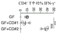
图1A-图1C。从人类唾液微生物群中分离诱导TH1细胞且促炎性的肺炎克雷伯氏菌菌株。(图1A)来自接种有来自患有CD的患者的唾液样品的被唾液微生物群定殖的无菌(ex-GF)小鼠的结肠固有层(LP)的CD4+TCRβ+T细胞内的IFN-γ+的频率。每个点代表个体小鼠。(图1B)被8-混合物、Veillo+Fuso、Kp-2H7或7-混合物定殖的B6小鼠的结肠LP中TH1细胞的百分比。(图1C)Kp-2H7-或Ec-2B1-单定殖的WT或Il1-/-小鼠的近端结肠的SEM图像。符号代表个体小鼠。误差棒指示平均值±SEM。数据代表了具有相似结果的至少两个独立实验。*P<0.02;**P<0.001。1A-1C. Isolation of a TH1 cell-inducing and pro-inflammatory Klebsiella pneumoniae strain from the human salivary microbiota. (FIG. 1A) IFN in CD4+ TCRβ+ T cells from colonic lamina propria (LP) of germ-free (ex-GF) mice colonized with salivary microbiota vaccinated with saliva samples from patients with CD The frequency of -γ+ . Each dot represents an individual mouse. (FIG. IB) Percentage of TH1 cells in colonic LPs of B6 mice colonized with 8-mix, Veillo+Fuso, Kp-2H7 or 7-mix. (FIG. 1C) SEM images of proximal colons of Kp-2H7- or Ec-2B1-single-colonized WT or Il1-/- mice. Symbols represent individual mice. Error bars indicate mean ± SEM. Data are representative of at least two independent experiments with similar results. *P<0.02;**P<0.001.
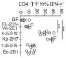
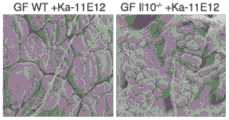
图2A-图2C。产气肠杆菌作为另一种TH1细胞诱导性口腔细菌物种。(图2A)来自接种有来自健康供体和UC患者的唾液样品的ex-GF小鼠的结肠LP CD4+TCRβ+T细胞内的IFNγ+的频率。每个点代表个体小鼠。(图2B)被13-混合物、Efae-11A1、Ka-11E12或11-混合物定殖的B6小鼠的结肠LP中TH1细胞的百分比。(图2C)Ka-11E12-单定殖的WT或Il10-/-小鼠的近端结肠的SEM图像。2A-2C. Enterobacter aerogenes as another TH1 cell-inducible oral bacterial species. (FIG. 2A) Frequency of IFNγ+ in colonic LP CD4+ TCRβ+ T cells from ex-GF mice vaccinated with saliva samples from healthy donors and UC patients. Each dot represents an individual mouse. (FIG. 2B) Percentage of TH1 cells in colonic LPs of B6 mice colonized with 13-mix, Efae-11A1, Ka-11E12 or 11-mix. (FIG. 2C) SEM images of the proximal colon of Ka-11E12-single-colonized WT or Il10-/- mice.
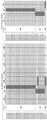
图3。针对KP2和针对KP3分离的噬菌体的宿主范围分析,包括针对如所指示的其他克雷伯氏菌属菌株的活性。测量两种噬菌体滴度-1×10⁹PFU/mL和1×106PFU/mL的活性。S-敏感的,R-耐受的,NT-未测试的,PFU-空斑形成单位image 3. Host range analysis of phage isolated against KP2 and against KP3, including activity against other Klebsiella strains as indicated. Two phage titers - 1 x10⁹ PFU/mL and1 x 106 PFU/mL activity were measured. S-sensitive, R-resistant, NT-untested, PFU-plaque forming unit
图4。与未感染的对照相比,用单一噬菌体感染的KP2培养物中和用6种噬菌体的混合物感染的KP2培养物中的噬菌体耐受突变体的出现。KP2在没有噬菌体情况下的生长曲线由黑线图示,而剩余的线表示KP2在具有如所指示的单一噬菌体情况下的生长曲线。虚线表示在存在六种噬菌体的混合物时KP2的生长曲线。在个体噬菌体存在下产生的KP2生长曲线表明在1小时-2小时后耐受菌株的再生长或突变体菌株的出现。相比之下,在混合物存在下的KP2的生长显示出耐受突变体在9小时后出现,使得“到产生突变体的时间(time tomutant)”参数显著改善。在研究过程中,对KP3菌株进行的类似分析产生了三倍的“到产生突变体的时间”,或甚至没有突变体出现。Figure 4. The emergence of phage-tolerant mutants in KP2 cultures infected with a single phage and in KP2 cultures infected with a mixture of 6 phages compared to uninfected controls. The growth curve of KP2 without phage is illustrated by the black line, while the remaining lines represent the growth curve of KP2 with a single phage as indicated. The dotted line represents the growth curve of KP2 in the presence of a mixture of six phages. KP2 growth curves generated in the presence of individual phage indicated regrowth of tolerant strains or emergence of mutant strains after 1-2 hours. In contrast, growth of KP2 in the presence of the mixture showed the emergence of tolerant mutants after 9 hours, resulting in a significant improvement in the "time to mutant" parameter. Over the course of the study, a similar analysis of the KP3 strain yielded three times the "time to mutant", or even no mutants.
图5。针对耐受原始KP2噬菌体感染的突变体KP2菌株分离的噬菌体的宿主范围分析和同源性百分比。(左侧)对针对耐受细菌突变体菌株MKP2_2161_1(对噬菌体KP2W-P0104耐受)和耐受细菌突变体菌株MKP2_coc_1(对噬菌体KP2W-P0101、KP2W-P0102、KP2W-P0103、KP2W-P0104、KP2W-P0106、KP2W-M0104耐受)以及针对KP2菌株分离的噬菌体在另外四个KP突变体MKP2_2161_C(对KP2W-P0104的感染耐受)、MKP2_251_B、MKP2_251_C和MKP2_251_D(对KP2W-M0104的感染耐受)上进行宿主范围分析。通过滴加试验(drop assay)将每种噬菌体(10μL)添加到48孔板中的不同肺炎克雷伯氏菌菌株的菌苔(bacterial lawn)中。将板在有氧条件下孵育2小时-3小时(37℃),之后菌苔上的空斑变得可见。具有MKP2_2161_1、MKP2_coc_1、KP2的菌苔及其对应的噬菌体的板用作阳性对照。用两种噬菌体滴度1×10⁶PFU/mL和1×109PFU/mL对每种噬菌体完成宿主范围分析。R-对测试的噬菌体的感染耐受,S-对测试的噬菌体的感染敏感,NT-未测试的。然而,在KP2上分离的噬菌体感染部分或所有KP2突变体菌株以及商业菌株KP4。(右侧)噬菌体基因组之间的同源性百分比通过组合所有非重叠的BLASTN比对区段(BLAST HSP),将它们的“相同匹配数”的值相加,并将该和除以查询序列的长度来确定。在一些实施方案中,这产生非对称矩阵。Figure 5. Host range analysis and percent homology of phages isolated against mutant KP2 strains resistant to infection with the original KP2 phage. (left) against tolerant bacterial mutant strain MKP2_2161_1 (to phage KP2W-P0104) and tolerant bacterial mutant strain MKP2_coc_1 (to phage KP2W-P0101, KP2W-P0102, KP2W-P0103, KP2W-P0104, KP2W -P0106, KP2W-M0104 tolerance) and phages isolated against the KP2 strain in another four KP mutants MKP2_2161_C (infection tolerance to KP2W-P0104), MKP2_251_B, MKP2_251_C and MKP2_251_D (infection tolerance to KP2W-M0104) Perform a host range analysis on . Each phage (10 [mu]L) was added to bacterial lawns of different K. pneumoniae strains in 48-well plates by drop assay. Plates were incubated under aerobic conditions for 2-3 hours (37°C), after which plaques on the lawn became visible. Plates with lawns of MKP2_2161_1, MKP2_coc_1, KP2 and their corresponding phages were used as positive controls. Host range analysis was done for each phage with two phage titers,1 x 106 PFU/mL and 1 x10⁹ PFU/mL. R - resistant to infection by tested phage, S - susceptible to infection by tested phage, NT - not tested. However, phages isolated on KP2 infected some or all KP2 mutant strains as well as the commercial strain KP4. (Right) Percent homology between phage genomes by combining all non-overlapping BLASTN aligned segments (BLAST HSP), summing their "number of identical matches" values, and dividing this sum by the query sequence length to be determined. In some embodiments, this produces an asymmetric matrix.
图6。针对KP3分离的噬菌体,包括短尾噬菌体科(podoviridae)类别的19种噬菌体(由P指示)和长尾噬菌体科(siphoviridae)类别的2种噬菌体(由S指示)的同源性百分比。噬菌体基因组之间的同源性百分比通过组合所有非重叠的BLASTN比对区段(BLAST HSP),将它们的“相同匹配数”的值相加,并将该和除以查询序列的长度来确定。在一些实施方案中,这产生非对称矩阵。
表1中公开了示例性细菌噬菌体和细菌的序列。KP2基因组已经被测序并且是本领域已知的,参见例如GenBank BDQR01000001.1(https://www.ncbi.nlm.nih.gov/nuccore/BDQR01000001.1)、GenBank GCA_002260905.1(https://www.ncbi.nlm.nih.gov/assembly/GCA_002260905.1)、BioSample:SAMD00083910(https://www.ncbi.nlm.nih.gov/biosample/SAMD00083910/)、BioProject PRJDB5883(https://www.ncbi.nlm.nih.gov/bioproject/PRJDB5883)和Atarashi等人,Ectopic colonizationof oral bacteria in the intestine drives TH1 cell induction and inflammation,Science(2017)。在一些实施方案中,基因组序列,例如KP2基因组序列,可以包含与相同细菌的另一个基因组序列相比的少量测序差异,例如少于0.5%、少于1%、少于1.5%、少于2%、少于2.5%或少于3%,并且测序差异不应导致细菌功能的差异,例如细菌噬菌体的感染性。Sequences of exemplary bacteriophages and bacteria are disclosed in Table 1. The KP2 genome has been sequenced and is known in the art, see eg GenBank BDQR01000001.1 (https://www.ncbi.nlm.nih.gov/nuccore/BDQR01000001.1), GenBank GCA_002260905.1 (https://www.ncbi.nlm.nih.gov/nuccore/BDQR01000001.1) www.ncbi.nlm.nih.gov/assembly/GCA_002260905.1), BioSample: SAMD00083910 (https://www.ncbi.nlm.nih.gov/biosample/SAMD00083910/), BioProject PRJDB5883 (https://www. ncbi.nlm.nih.gov/bioproject/PRJDB5883) and Atarashi et al., Ectopic colonization of oral bacteria in the intestine drives TH1 cell induction and inflammation, Science (2017). In some embodiments, a genome sequence, eg, a KP2 genome sequence, may contain a small number of sequencing differences compared to another genome sequence of the same bacterium, eg, less than 0.5%, less than 1%, less than 1.5%, less than 2 %, less than 2.5%, or less than 3%, and sequencing differences should not result in differences in bacterial function, such as bacteriophage infectivity.
表1.序列简述Table 1. Brief description of sequences


实施方案的描述Description of the implementation
本公开内容涉及细菌噬菌体、其药物组合物、调节IBD的方法(Jostins等人,2012),以及选择对本文陈述的方法的治疗有响应的患者的方法。在一些实施方案中,本文描述的细菌噬菌体能够裂解与IBD相关的克雷伯氏菌属细菌。肺炎克雷伯氏菌已与IBD关联。与对照和其他病理状况相比,IBD患者表现出针对某些荚膜类型(例如,存在于KP2中的K2、K17、K26、K36、K50和K21)的全身抗体升高。这些发现表明克雷伯氏菌属参与IBD,但无法解释克雷伯氏菌属是否积极参与该疾病的发病机制,并且无法提供菌株特异性的解决方案,因为不同的菌株共有相似的荚膜类型。实施例1显示了单一致病有机体(pathobiont)菌株(KP2或KP3)的肠施用和肠道的免疫响应(固有层中的TH1极化)以及体内结肠炎诱导(在1110-/-小鼠中)之间的关系。The present disclosure relates to bacteriophages, pharmaceutical compositions thereof, methods of modulating IBD (Jostins et al., 2012), and methods of selecting patients responsive to treatment by the methods set forth herein. In some embodiments, the bacteriophages described herein are capable of lysing Klebsiella bacteria associated with IBD. Klebsiella pneumoniae has been associated with IBD. Compared to controls and other pathological conditions, IBD patients exhibit elevated systemic antibodies to certain capsule types (eg, K2, K17, K26, K36, K50, and K21 present in KP2). These findings suggest that Klebsiella is involved in IBD, but cannot explain whether Klebsiella is actively involved in the pathogenesis of the disease, and do not provide a strain-specific solution because different strains share similar capsule types . Example 1 shows intestinal administration of a single pathogenic organism (pathobiont) strain (KP2 or KP3) and intestinal immune response (TH1 polarization in the lamina propria) and in vivo colitis induction (in 1110-/- mice). )The relationship between.
定义definition
为使本公开内容可以更容易地理解,首先定义一些术语。这些定义应该根据本公开内容的剩余部分并且如本领域普通技术人员所理解的进行阅读。除非另有限定,本文使用的所有技术和科学术语具有与由本领域的普通技术人员通常所理解的相同的含义。在整个详细描述中陈述另外的定义。To make this disclosure easier to understand, some terms are first defined. These definitions should be read in light of the remainder of this disclosure and as understood by those of ordinary skill in the art. Unless otherwise defined, all technical and scientific terms used herein have the same meaning as commonly understood by one of ordinary skill in the art. Additional definitions are set forth throughout the detailed description.
如本文使用的,术语“治疗”和“调节”及其同源词是指IBD或其至少一种可辨别的症状的改善。在一些实施方案中,“治疗”和“调节”及其同源词是指至少一个可测量的物理参数的改善,该物理参数不一定是患者能够辨别的。在一些实施方案中,“治疗”和“调节”及其同源词是指相对于未治疗的对照,在物理上(例如,可辨别症状的稳定)、生理上(例如,物理参数的稳定)或两方面都抑制或减少IBD或者减缓IBD的进展。在某些实施方案中,“治疗”和“调节”及其同源词是指相对于未治疗的对照,减缓IBD的进展或逆转IBD的进展。如本文使用的,“预防”及其同源词是指相对于未治疗的对照,延迟IBD或与IBD相关的症状的发作或降低获得IBD或与IBD相关的症状的风险。As used herein, the terms "treating" and "modulating" and their cognates refer to amelioration of IBD or at least one identifiable symptom thereof. In some embodiments, "treating" and "modulating" and their cognates refer to an improvement in at least one measurable physical parameter that is not necessarily discernible by a patient. In some embodiments, "treating" and "modulating" and their cognates refer to physical (eg, stabilization of discernible symptoms), physiological (eg, stabilization of physical parameters) relative to an untreated control Either inhibit or reduce IBD or slow the progression of IBD, or both. In certain embodiments, "treating" and "modulating" and their cognates refer to slowing the progression of IBD or reversing the progression of IBD relative to an untreated control. As used herein, "prevention" and its cognates refer to delaying the onset of or reducing the risk of acquiring IBD or IBD-related symptoms relative to an untreated control.
在某些实施方案中,本文描述的细菌噬菌体被施用以改善受试者的IBD,并且导致该状况或紊乱的一种或更多种症状或物理参数与未治疗的受试者或对照受试者的水平相比改善至少约10%、20%、30%、40%、50%、60%、70%、80%、90%、95%或更多。在一些实施方案中,通过比较受试者在施用细菌噬菌体之前和之后的症状或物理参数来测量改善。在一些实施方案中,可测量的物理参数是粪便中细菌CFU的减少。In certain embodiments, the bacteriophages described herein are administered to ameliorate IBD in a subject, and one or more symptoms or physical parameters that cause the condition or disorder are compared to untreated subjects or control subjects improvement of at least about 10%, 20%, 30%, 40%, 50%, 60%, 70%, 80%, 90%, 95% or more compared to the level of the patient. In some embodiments, improvement is measured by comparing a subject's symptoms or physical parameters before and after administration of the bacteriophage. In some embodiments, the measurable physical parameter is a reduction in bacterial CFU in feces.
可测量的物理参数可以是本领域已知的例如针对克罗恩病(Sandborn等人,2013;Best等人,1976;Warrel-Oxford Textbook of Medicine,第5版)或针对溃疡性结肠炎(Tursi等人,2010;Warrel-Oxford Textbook of Medicine,第5版)的任何合适的临床参数。克罗恩病参数包括但不限于使用克罗恩病活动指数(CDAI)(CDAI评分≤150分)和CDAI-100响应(CDAI评分从基线减低≥100分)评估临床缓解;如通过血清C-反应蛋白水平、血细胞分类计数(differential blood count)和血清白蛋白水平的降低测量的炎症减轻;如通过显微检查内镜程序期间收集的活检评估的,降低的粪便钙防卫蛋白水平以及末端回肠和直肠的粘膜层的外观的改善(例如,中性粒细胞、巨噬细胞和其他免疫细胞浸润、肉芽肿尺寸和数目的减少、溃疡、水肿粘膜的数目和尺寸的减少)。此外,可以基于对临床症状的评估来评价疾病的改善,所述临床症状包括腹泻、腹痛、体重减轻、发热、呕吐、疝痛(colic)、直肠出血、贫血、肠外表现(对柳氮磺胺吡啶(sulphasalazine)超敏的皮疹、结节性红斑、坏疽性脓皮病、口疮性溃疡(oral aphthous ulcer)、舌痛、口角炎、巩膜外层炎或前葡萄膜炎、关节炎、急性关节病、下背痛、骶髂关节炎、强直性脊柱炎,肝病:碱性磷酸酶或转氨酶的少量升高、原发性硬化性胆管炎、慢性肝病(范围从自身免疫性肝炎到胆管周围向心性纤维化伴胆管闭塞(concentric periductular fibrosis with obliteration of bile ducts)的典型图像),伴或不伴渗液的心包炎(pericarditis with or without an effusion)、自身免疫性溶血性贫血、淀粉样蛋白、快速进展的支气管扩张)。Measurable physical parameters can be those known in the art, eg, for Crohn's disease (Sandborn et al, 2013; Best et al, 1976; Warrel-Oxford Textbook of Medicine, 5th edition) or for ulcerative colitis (Tursi et al., 2010; Warrel-Oxford Textbook of Medicine, 5th edition) for any suitable clinical parameters. Crohn's disease parameters include, but are not limited to, assessment of clinical remission using the Crohn's Disease Activity Index (CDAI) (CDAI score ≤150 points) and CDAI-100 response (CDAI score reduction from baseline ≥100 points); as assessed by serum C- Reduced inflammation as measured by reductions in reactive protein levels, differential blood counts, and serum albumin levels; decreased fecal calprotectin levels and terminal ileal and Improvement in the appearance of the mucosal layer of the rectum (eg, infiltration of neutrophils, macrophages and other immune cells, reduction in size and number of granulomas, ulceration, reduction in number and size of edematous mucosa). In addition, improvement in disease can be assessed based on assessment of clinical symptoms including diarrhea, abdominal pain, weight loss, fever, vomiting, colic, rectal bleeding, anemia, extraintestinal manifestations (for sulfasalazine sulphasalazine hypersensitivity rash, erythema nodosum, pyoderma gangrenosum, oral aphthous ulcer, tongue pain, angular stomatitis, episcleritis or anterior uveitis, arthritis, acute arthropathy , low back pain, sacroiliitis, ankylosing spondylitis, liver disease: small elevations in alkaline phosphatase or transaminases, primary sclerosing cholangitis, chronic liver disease (ranging from autoimmune hepatitis to peribiliary concentricity Typical image of concentric periductular fibrosis with obliteration of bile ducts, pericarditis with or without an effusion, autoimmune hemolytic anemia, amyloid, rapid progressive bronchiectasis).
溃疡性结肠炎疾病参数包括但不限于溃疡性结肠炎疾病活动指数(UCDAI)的改善(UCDAI从基线下降50%或更多)、复发性UC的活动的改善;缓解,被视为UCDAI≤2;血红蛋白水平、血清白蛋白水平的改善,C-反应蛋白水平的降低;中性粒细胞白细胞增多的改善(血细胞分类计数(blood differential count));内镜评分的改善(水肿粘膜面积的减少,中性粒细胞、巨噬细胞和其他免疫细胞浸润的减少,溃疡、水肿粘膜的数目和尺寸的减少);以下临床症状的改善:直肠出血、发热、大便频率、腹泻、粘液通过、腹痛、便秘、厌食、恶心、体重减轻、不适、疲乏、慢性缺铁症状、轻微肛周疾病诸如肛裂(fissure)、上文描述的肠外表现以及另外的口腔念珠菌病、杵状指(趾)(finger clubbing)。Ulcerative colitis disease parameters including, but not limited to, improvement in Ulcerative Colitis Disease Activity Index (UCDAI) (50% or more decrease in UCDAI from baseline), improvement in activity of recurrent UC; remission, considered UCDAI ≤ 2 ; improvement in hemoglobin level, serum albumin level, decrease in C-reactive protein level; improvement in neutropenia (blood differential count); improvement in endoscopic score (reduction in edema mucosal area, Decreased infiltration of neutrophils, macrophages, and other immune cells, reduction in number and size of ulcers, edematous mucosa); improvement in the following clinical symptoms: rectal bleeding, fever, stool frequency, diarrhea, mucus passage, abdominal pain, constipation , anorexia, nausea, weight loss, malaise, fatigue, chronic iron deficiency symptoms, minor perianal disorders such as fissures, extraintestinal manifestations described above and additional oral candidiasis, digital clubbing ( finger clubbing).
需要治疗的个体可以包括已具有IBD的个体,以及处于患该疾病的风险的个体或可能最终获得该疾病的个体。通过以下评估治疗需要:例如,与IBD的发展相关的一种或更多种危险因素的存在、IBD的存在或进展或者具有IBD的受试者对治疗的接受可能性。例如,“治疗”IBD可以包括减少或消除相关的症状,而不一定包括消除潜在的疾病病因学,例如遗传不稳定性基因座。Individuals in need of treatment can include individuals who already have IBD, as well as individuals who are at risk of developing the disease or who may eventually acquire the disease. The need for treatment is assessed by, for example, the presence of one or more risk factors associated with the development of IBD, the presence or progression of IBD, or the likelihood that a subject with IBD will receive treatment. For example, "treating" IBD may include reducing or eliminating associated symptoms, but not necessarily eliminating underlying disease etiologies, such as genetic instability loci.
在一些实施方案中,患有IBD的个体处于缓解中和/或当前无症状的,并且本文描述的细菌噬菌体可以在缓解期期间被施用以降低突然复发(flare-up)的可能性。在一些实施方案中,处于IBD缓解中和/或目前IBD无症状的个体正在经历例如抗生素和/或类固醇的治疗,并且本文描述的细菌噬菌体可以与此类治疗共施用,以降低突然复发的可能性。In some embodiments, the individual with IBD is in remission and/or is currently asymptomatic, and the bacteriophage described herein can be administered during the remission period to reduce the likelihood of flare-up. In some embodiments, individuals in remission of IBD and/or currently asymptomatic with IBD are undergoing treatment such as antibiotics and/or steroids, and the bacteriophages described herein can be co-administered with such treatment to reduce the likelihood of sudden relapse sex.
克雷伯氏菌属是属于肠杆菌科(Enterobacteriaceae)的细菌属。克雷伯氏菌属是革兰氏阴性的、不运动的和棒状的。在一些实施方案中,克雷伯氏菌属的物种例如肺炎克雷伯氏菌可能与人类疾病相关。如本文使用的,克雷伯氏菌属包括目前被分类的、先前被分类的或将要被重新分类为克雷伯氏菌属细菌的细菌,例如Klebsiella aeromobilis,其也被称为产气肠杆菌(Tindall等人,2017;Diene等人,2013)。在一些实施方案中,克雷伯氏菌属是指肺炎克雷伯氏菌。在一些实施方案中,克雷伯氏菌属是指Klebsiella aeromobilis/产气肠杆菌细菌。在一些实施方案中,克雷伯氏菌属是指天然存在的克雷伯氏菌属。在一些实施方案中,克雷伯氏菌属是指天然存在的、变异(variant)或突变的克雷伯氏菌属(例如,抗生素耐受的、噬菌体耐受的、院内感染的(nosocomial))。在一些实施方案中,变异或突变的克雷伯氏菌属对至少1种、至少2种、至少3种、至少4种或至少5种抗生素是耐受的。在一些实施方案中,突变体细菌菌株可以在细菌噬菌体存在下产生,并且变得对所述细菌噬菌体耐受。Klebsiella is a bacterial genus belonging to the family Enterobacteriaceae. Klebsiella is gram-negative, immotile and rod-shaped. In some embodiments, Klebsiella species such as Klebsiella pneumoniae may be associated with human disease. As used herein, Klebsiella includes bacteria that are currently classified, previously classified, or to be reclassified as Klebsiella bacteria, such as Klebsiella aeromobilis, which is also known as Enterobacter aerogenes (Tindall et al., 2017; Diene et al., 2013). In some embodiments, Klebsiella refers to Klebsiella pneumoniae. In some embodiments, Klebsiella refers to Klebsiella aeromobilis/Enterobacter aerogenes bacteria. In some embodiments, Klebsiella refers to naturally occurring Klebsiella. In some embodiments, Klebsiella refers to naturally occurring, variant, or mutated Klebsiella (eg, antibiotic-resistant, phage-resistant, nosocomial) ). In some embodiments, the mutated or mutated Klebsiella is resistant to at least 1, at least 2, at least 3, at least 4, or at least 5 antibiotics. In some embodiments, mutant bacterial strains can be produced in the presence of a bacteriophage and become tolerant to the bacteriophage.
如本文使用的,细菌的“菌株”是指细菌的遗传变体或亚型。在一些实施方案中,细菌的“菌株”包括来自纯的所述细菌的培养物中的单一分离的后代。如本文使用的,细菌的“菌株”可以指所述细菌的一种或更多种遗传变体或亚型。例如,如本文使用的,肺炎克雷伯氏菌的“菌株”可以指肺炎克雷伯氏菌的一种或更多种遗传变体或亚型,包括但不限于KP1(ATCC BAA-2552;也被称为变栖克雷伯氏菌(Klebsiella variicola))、KP2(本文描述的肺炎克雷伯氏菌菌株2H7)、KP4(ATCC 23356)、KP5(ATCC 13882)、KP6(ATCC BAA-1705)、KP7(ATCC 700603)和KP8(ATCC 700721)。类似地,如本文使用的,能够裂解肺炎克雷伯氏菌的“菌株”的细菌噬菌体是指能够裂解肺炎克雷伯氏菌的一种或更多种遗传变体或亚型(包括但不限于KP1、KP2、KP3、KP4、KP5、KP6、KP7和KP8)的细菌噬菌体。在一些实施方案中,能够裂解肺炎克雷伯氏菌的“菌株”的细菌噬菌体是指能够裂解肺炎克雷伯氏菌的一种或更多种遗传变体或亚型KP1、KP2、KP4、KP5、KP6、KP7和KP8的细菌噬菌体。在一些实施方案中,能够裂解肺炎克雷伯氏菌的“菌株”的细菌噬菌体是指能够裂解肺炎克雷伯氏菌的一种或更多种遗传变体或亚型KP2、KP4、KP5、KP6、KP7和KP8的细菌噬菌体。在一些实施方案中,能够裂解Klebsiella aeromobilis/产气肠杆菌的“菌株”的细菌噬菌体是指能够裂解Klebsiellaaeromobilis/产气肠杆菌KP3的一种或更多种遗传变体或亚型,例如本文描述的菌株11E12的细菌噬菌体。在一些实施方案中,本文描述的KP2细菌噬菌体能够裂解肺炎克雷伯氏菌的KP2菌株。在一些实施方案中,本文描述的KP3细菌噬菌体能够裂解Klebsiellaaeromobilis的KP3菌株。As used herein, a "strain" of a bacterium refers to a genetic variant or subtype of a bacterium. In some embodiments, a "strain" of a bacterium includes a single isolated progeny from a pure culture of the bacterium. As used herein, a "strain" of a bacterium can refer to one or more genetic variants or subtypes of the bacterium. For example, as used herein, a "strain" of Klebsiella pneumoniae may refer to one or more genetic variants or subtypes of Klebsiella pneumoniae, including but not limited to KP1 (ATCC BAA-2552; Also known as Klebsiella variicola), KP2 (Klebsiella pneumoniae strain 2H7 described herein), KP4 (ATCC 23356), KP5 (ATCC 13882), KP6 (ATCC BAA-1705 ), KP7 (ATCC 700603) and KP8 (ATCC 700721). Similarly, as used herein, a bacteriophage capable of lysing a "strain" of Klebsiella pneumonia refers to one or more genetic variants or subtypes (including but not Bacteriophages restricted to KP1, KP2, KP3, KP4, KP5, KP6, KP7 and KP8). In some embodiments, a bacteriophage capable of lysing a "strain" of Klebsiella pneumonia refers to one or more genetic variants or subtypes KP1, KP2, KP4, Bacteriophages of KP5, KP6, KP7 and KP8. In some embodiments, a bacteriophage capable of lysing a "strain" of Klebsiella pneumonia refers to one or more genetic variants or subtypes KP2, KP4, KP5, Bacteriophages of KP6, KP7 and KP8. In some embodiments, a bacteriophage capable of lysing a "strain" of Klebsiella aeromobilis/Enterobacter aerogenes refers to one or more genetic variants or subtypes of Klebsiella aeromobilis/Enterobacter aerogenes KP3, such as described herein bacteriophage of strain 11E12. In some embodiments, the KP2 bacteriophages described herein are capable of lysing a KP2 strain of Klebsiella pneumoniae. In some embodiments, the KP3 bacteriophages described herein are capable of lysing the KP3 strain of Klebsiella aeromobilis.
在一些实施方案中,如本文使用的,“突变体”细菌是指包括与相应的野生型细菌菌株大于约85%、大于约90%、大于约95%、大于约97%或大于约99%的同源性的细菌。例如,突变体KP2细菌包含与野生型KP2细菌(表1,重叠群(contig))大于约85%、大于约90%、大于约95%、大于约97%或大于约99%的同源性。突变体KP3细菌包含与野生型KP3细菌(表1)大于约85%、大于约90%、大于约95%、大于约97%或大于约99%的同源性。In some embodiments, as used herein, a "mutant" bacterium is meant to comprise greater than about 85%, greater than about 90%, greater than about 95%, greater than about 97%, or greater than about 99% with the corresponding wild-type bacterial strain homologous bacteria. For example, mutant KP2 bacteria comprise greater than about 85%, greater than about 90%, greater than about 95%, greater than about 97%, or greater than about 99% homology to wild-type KP2 bacteria (Table 1, contigs). . Mutant KP3 bacteria comprise greater than about 85%, greater than about 90%, greater than about 95%, greater than about 97%, or greater than about 99% homology to wild-type KP3 bacteria (Table 1).
在一些实施方案中,“突变体”KP2细菌是能够被本文公开的KP2细菌噬菌体裂解的细菌。在一些实施方案中,“突变体”KP2细菌是指相比于野生型KP2包括小于约85%同源性的细菌,但能够被本文公开的KP2细菌噬菌体裂解。在一些实施方案中,“突变体”KP3细菌是能够被本文公开的KP3细菌噬菌体裂解的细菌。在一些实施方案中,“突变体”KP3细菌是指相比于野生型KP3包括小于约85%同源性的细菌,但能够被本文公开的KP3细菌噬菌体裂解。In some embodiments, a "mutant" KP2 bacterium is a bacterium capable of being lysed by the KP2 bacteriophage disclosed herein. In some embodiments, a "mutant" KP2 bacterium refers to a bacterium that includes less than about 85% homology compared to wild-type KP2, but is capable of being lysed by the KP2 bacteriophage disclosed herein. In some embodiments, a "mutant" KP3 bacterium is a bacterium capable of being lysed by the KP3 bacteriophage disclosed herein. In some embodiments, a "mutant" KP3 bacterium refers to a bacterium that includes less than about 85% homology compared to wild-type KP3, but is capable of being lysed by the KP3 bacteriophage disclosed herein.
在一些实施方案中,如本文描述的KP2细菌,例如KP2细菌菌株,和/或突变体KP2细菌例如环境分离株和/或临床分离株,对于独特的基因组区域是阳性的,该独特的基因组区域可以通过PCR使用以下引物来鉴定:5'AGCACTAGCGGCTGTGGTAT3’和5'ACTTACTCGGGCCCTTGATT3’。参见,例如Atarashi等人,2017。在一些实施方案中,突变体KP2细菌例如使用所述引物鉴定的环境分离株和/或临床分离株,包括与KP2细菌(表1,重叠群)大于约95%、大于约96%、大于约97%、大于约98%或大于约99%同源性,例如,编码区的同源性%通过ANI(平均核苷酸同一性)计算。参见,例如Han等人,ANItools web:a web toolfor fast genome comparison within multiple bacterial strains(2016)。在一些实施方案中,通过所述引物鉴定的且与KP2细菌(表1,重叠群)至少约99%同源的突变体KP2细菌在本文中被称为“KP2样”。在一些实施方案中,突变体和/或KP2样细菌能够被本文公开的至少一种KP2细菌噬菌体裂解。In some embodiments, KP2 bacteria as described herein, eg, KP2 bacterial strains, and/or mutant KP2 bacteria, eg, environmental isolates and/or clinical isolates, are positive for a unique genomic region that is It can be identified by PCR using the following primers: 5'AGCACTAGCGGCTGTGGTAT3' and 5'ACTTACTCGGGCCCTTGATT3'. See, eg, Atarashi et al., 2017. In some embodiments, mutant KP2 bacteria, eg, environmental isolates and/or clinical isolates identified using the primers, include greater than about 95%, greater than about 96%, greater than about 97%, greater than about 98%, or greater than about 99% homology, eg, % homology of a coding region is calculated by ANI (average nucleotide identity). See, eg, Han et al., ANItools web: a web tool for fast genome comparison within multiple bacterial strains (2016). In some embodiments, mutant KP2 bacteria identified by the primers that are at least about 99% homologous to KP2 bacteria (Table 1, contigs) are referred to herein as "KP2-like." In some embodiments, the mutant and/or KP2-like bacteria are capable of being lysed by at least one KP2 bacteriophage disclosed herein.
如本文使用的,“细菌噬菌体(bacteriophage)”和“噬菌体(phage)”可互换使用并且是指能够感染细菌的分离的病毒。在一些实施方案中,噬菌体包含DNA或RNA基因组。噬菌体可以从天然环境或人造环境中分离。在一些实施方案中,噬菌体选自肌尾噬菌体科(Myoviridae)、短尾噬菌体科(Podoviridae)和长尾噬菌体科(Siphoviridae)。如本文使用的,“KP细菌噬菌体”意图指能够裂解肺炎克雷伯氏菌或Klebsiella aeromobilis/产气肠杆菌细菌的细菌噬菌体。例如,“KP2细菌噬菌体”是指能够裂解肺炎克雷伯氏菌菌株KP2细菌,包括野生型细菌和突变体KP2细菌的细菌噬菌体。“KP3细菌噬菌体”是指能够裂解Klebsiella aeromobilis/产气肠杆菌菌株KP3细菌,包括野生型细菌和突变体KP3细菌的细菌噬菌体。As used herein, "bacteriophage" and "phage" are used interchangeably and refer to an isolated virus capable of infecting bacteria. In some embodiments, the phage comprises a DNA or RNA genome. Phages can be isolated from natural or artificial environments. In some embodiments, the phage is selected from the families Myoviridae, Podoviridae, and Siphoviridae. As used herein, "KP bacteriophage" is intended to refer to a bacteriophage capable of lysing Klebsiella pneumoniae or Klebsiella aeromobilis/Enterobacter aerogenes bacteria. For example, "KP2 bacteriophage" refers to a bacteriophage capable of lysing Klebsiella pneumoniae strain KP2 bacteria, including wild-type bacteria and mutant KP2 bacteria. "KP3 bacteriophage" refers to a bacteriophage capable of lysing Klebsiella aeromobilis/Enterobacter aerogenes strain KP3 bacteria, including wild-type bacteria and mutant KP3 bacteria.
已知给定细菌噬菌体的不同分离株可能在核酸序列水平上不同。在一些实施方案中,细菌噬菌体被认为是“功能等同的”,只要它们表现出相似的表型,例如相似的宿主范围、相似的裂解能力和/或阈值序列相似性(例如,大于约85%、大于约90%、大于约95%、大于约97%或大于约99%)。如本文使用的,术语“细菌噬菌体”包括亲本细菌噬菌体以及其后代或衍生物。It is known that different isolates of a given bacteriophage may differ at the nucleic acid sequence level. In some embodiments, bacteriophages are considered "functionally equivalent" as long as they exhibit similar phenotypes, eg, similar host ranges, similar lytic capacity, and/or threshold sequence similarity (eg, greater than about 85% , greater than about 90%, greater than about 95%, greater than about 97%, or greater than about 99%). As used herein, the term "bacteriophage" includes the parental bacteriophage as well as progeny or derivatives thereof.
如本文使用的,“宿主范围”是指易受特定噬菌体感染的细菌。噬菌体的宿主范围可以包括但不限于细菌的一个菌株、一个物种、一个属或多个属。该术语包括吸附性、非增殖性感染(例如限制性、流产性、溶源性)和增殖性感染的噬菌体。在一些实施方案中,噬菌体可以识别两种或更多种菌株。在一些实施方案中,噬菌体可以识别野生型菌株和噬菌体耐受性突变体菌株。As used herein, "host range" refers to bacteria susceptible to infection by a particular phage. The host range of a phage can include, but is not limited to, a strain, species, genus, or genera of bacteria. The term includes adsorptive, non-proliferative (eg, restrictive, abortive, lysogenic) and proliferative infectious phages. In some embodiments, the phage can recognize two or more strains. In some embodiments, the phage can recognize wild-type strains and phage-tolerant mutant strains.
可以使用本领域已知的方法例如,空斑测定、液体培养基测定、固体培养基测定制备不同的噬菌体分离株并进行表型分析。在一些实施方案中,定量和分离噬菌体的固体培养基测定基于空斑测定(Abedon&Yin,2009),范围从铺板效率(EOP)(Kutter,2009)到斑点测试(spot testing)(Hyman&Abedon,2010)。在一些实施方案中,用于空斑测定的板格式可以被修改,例如,从皮氏培养皿到48孔板。Different phage isolates can be prepared and phenotyped using methods known in the art, eg, plaque assays, liquid media assays, solid media assays. In some embodiments, solid medium assays to quantify and isolate phage are based on plaque assays (Abedon & Yin, 2009), ranging from efficiency of plating (EOP) (Kutter, 2009) to spot testing (Hyman & Abedon, 2010). In some embodiments, the plate format for the plaque assay can be modified, eg, from a Petri dish to a 48-well plate.
在一些实施方案中,使用双层空斑测定对细菌噬菌体分离物进行表型分析。例如,4mL BHIS的起始培养物可以用来自板的5-10个菌落接种。该培养物可以在37℃孵育1.5小时-2小时。100μL体积的该培养物可以与100μL的含噬菌体的样品(或仅培养基对照)混合,并孵育15分钟。此后,可以添加3mL BHIS顶部琼脂(补充有1mM Ca2+和Mg2+离子的预熔化的0.4%BHIS琼脂),并可将混合物倾倒在BHIS底部琼脂板(1.5%BHIS琼脂)上。可允许板在室温胶凝化,并然后在37℃孵育2小时-3小时,直到鉴定出空斑。In some embodiments, bacteriophage isolates are phenotyped using a double-layer plaque assay. For example, a 4 mL starter culture of BHIS can be inoculated with 5-10 colonies from the plate. The culture can be incubated at 37°C for 1.5 hours to 2 hours. A 100 [mu]L volume of this culture can be mixed with 100 [mu]L of the phage-containing sample (or medium only control) and incubated for 15 minutes. Thereafter, 3 mL of BHIS top agar (pre-melted 0.4% BHIS agar supplemented with 1 mM Ca2+ andMg2+ ions) can be added and the mixture can be poured onto BHIS bottom agar plates (1.5% BHIS agar). Plates can be allowed to gel at room temperature and then incubated at 37°C for 2-3 hours until plaques are identified.
在一些实施方案中,使用改良的点滴测定(spot drop assay)对细菌噬菌体分离物进行表型分析。例如,4mL BHIS的起始培养物可以用来自板的5-10个菌落接种。该培养物可以在37℃孵育1.5小时-2小时。100μL体积的该培养物可以与3mL的BHIS顶部琼脂(补充有1mM Ca2+和Mg2+离子的预熔化的0.4%BHIS琼脂)混合,并可将100μL的该混合物分散到已经含有1mL/孔的BHIS底部琼脂的Nunc 48孔板的每孔。在室温固化后,板可以在37度孵育30分钟。在此阶段,可将10μL的含有噬菌体的样品或仅培养基的对照滴在孔的中间,让其吸收,并且然后可以孵育2小时-3小时,直到空斑可见用于计数。In some embodiments, bacteriophage isolates are phenotyped using a modified spot drop assay. For example, a 4 mL starter culture of BHIS can be inoculated with 5-10 colonies from the plate. The culture can be incubated at 37°C for 1.5 hours to 2 hours. A volume of 100 μL of this culture can be mixed with 3 mL of BHIS top agar (pre-melted 0.4% BHIS agar supplemented with 1 mMCa and Mgions ) and 100 μL of this mixture can be dispensed to already contain 1 mL/well. BHIS bottom agar for each well of a Nunc 48-well plate. After curing at room temperature, plates can be incubated at 37 degrees for 30 minutes. At this stage, 10 μL of the phage-containing sample or the medium-only control can be dropped in the middle of the well, allowed to absorb, and then incubated for 2-3 hours until plaques are visible for counting.
在一些实施方案中,使用液体培养基测定对细菌噬菌体进行表型分析。在一些实施方案中,基于液体的噬菌体感染测定遵循感染的时间过程,并且与固相空斑测定相比,可以提供不只是定量的感染终点。在一些实施方案中,通过将噬菌体与细菌混合在液体培养基中,然后跟踪培养物随着时间推移的浊度,人们可以辨别不同细菌菌株如何与噬菌体相互作用之间的更细微的差异(例如,细胞裂解时间的延迟)。在一些实施方案中,液体培养基测定允许通过使用96孔板和在读板器中读取光密度进行高通量测量。In some embodiments, bacteriophages are phenotyped using a liquid culture assay. In some embodiments, liquid-based phage infection assays follow a time course of infection and can provide more than quantitative endpoints of infection compared to solid-phase plaque assays. In some embodiments, by mixing phage with bacteria in a liquid medium and then tracking the turbidity of the culture over time, one can discern finer differences between how different bacterial strains interact with the phage (e.g. , the delay in cell lysis time). In some embodiments, liquid culture assays allow for high-throughput measurements by using 96-well plates and reading optical density in a plate reader.
例如,细菌菌株可以生长1.5小时-2小时,直到OD600为约1.7-2。然后可以使用BHIS培养基将该培养物稀释至起始光密度,通常在0.05和0.2OD600之间。然后可将200μL体积的培养物分配到Nunclon平底96孔板的孔中。可将10μL的含有噬菌体的样品或10μL培养基作为对照添加到每个孔中。孔可以用50μL的矿物油覆盖以限制蒸发,并且可以添加薄的无菌光学透明聚酯膜以保持培养物无菌。光密度测量可以例如在连接到Tecan EVO75自动机器的Tecan Infinite M200读板器上每15分钟进行一次。在测量之间,该板可以在37℃例如在EVO75培养箱内在振荡的同时孵育。For example, bacterial strains can be grown for 1.5-2 hours until anOD600 of about 1.7-2. The culture can then be diluted to the starting optical density using BHIS medium, typically between 0.05 and 0.2OD600 . A volume of 200 μL of the culture can then be dispensed into the wells of a Nunclon flat bottom 96-well plate. 10 μL of phage-containing sample or 10 μL of medium can be added to each well as a control. Wells can be covered with 50 μL of mineral oil to limit evaporation, and a thin sterile optically clear polyester film can be added to keep cultures sterile. Densitometric measurements can be performed, for example, every 15 minutes on a Tecan Infinite M200 plate reader connected to a Tecan EVO75 robotic machine. Between measurements, the plate can be incubated at 37°C while shaking, eg, in an EVO75 incubator.
在一些实施方案中,感染性(infectivity)仅通过固体测定中的空斑存在来确定。在一些实施方案中,感染性仅通过液体测定中细菌培养物光密度的降低来确定。在一些实施方案中,感染性通过液体测定和固体测定两者中细菌培养物光密度的降低和空斑存在来确定。In some embodiments, infectivity is determined solely by the presence of plaques in solid assays. In some embodiments, infectivity is determined solely by a decrease in the optical density of bacterial cultures in a liquid assay. In some embodiments, infectivity is determined by the reduction in optical density of bacterial cultures and the presence of plaques in both liquid and solid assays.
如本文使用的,“裂解性”细菌噬菌体是指在过程中附着于细菌宿主并将其遗传物质插入细菌宿主细胞的强毒性细菌噬菌体。此后,噬菌体通常遵循两个生命周期中的一个,裂解性(强毒性)或溶源性(温和性)。裂解性噬菌体接管细胞的机器来制造噬菌体组分。然后它们破坏或裂解细胞,释放新的噬菌体颗粒。溶源性噬菌体将它们的核酸掺入到宿主细胞的染色体中,并且随其作为一个单位复制而不破坏细胞。在某些条件下,溶源性噬菌体可以被诱导以跟踪裂解循环。在一些实施方案中,在感染后,释放新的细菌噬菌体颗粒。在一些实施方案中,在感染后,宿主细菌细胞被裂解和破坏。在一些实施方案中,少于90%的宿主细菌细胞被裂解和破坏。参见,例如Abedon等人,2011;Sulakvelidze等人,2001;Green等人,2017。As used herein, a "lytic" bacteriophage refers to a highly virulent bacteriophage that, in the process, attaches to a bacterial host and inserts its genetic material into the bacterial host cell. Thereafter, phages typically follow one of two life cycles, lytic (highly virulent) or lysogenic (mild). Lytic phages take over the cell's machinery to make phage components. They then destroy or lyse cells, releasing new phage particles. Lysogenic phages incorporate their nucleic acid into the host cell's chromosome, and as it replicates as a unit, do not destroy the cell. Under certain conditions, lysogenic phages can be induced to follow lytic cycles. In some embodiments, after infection, new bacteriophage particles are released. In some embodiments, following infection, the host bacterial cells are lysed and destroyed. In some embodiments, less than 90% of the host bacterial cells are lysed and destroyed. See, eg, Abedon et al, 2011; Sulakvelidze et al, 2001; Green et al, 2017.
在一些实施方案中,通过本领域已知的和本文描述的方法,例如通过光密度(OD)或qPCR来测量裂解%。In some embodiments, % lysis is measured by methods known in the art and described herein, eg, by optical density (OD) or qPCR.
如本文使用的,“同源性%”是指当使用序列比对程序比对时第一核酸或氨基酸序列与第二核酸或氨基酸序列之间的核酸序列同一性或氨基酸序列同一性的水平。当第一序列和第二序列中的一个位置被相同的核酸或氨基酸占据时(例如,如果第一核酸序列和第二核酸序列中的一个位置被胞嘧啶占据),则第一序列和第二序列在该位置是同源的。As used herein, "% homology" refers to the level of nucleic acid sequence identity or amino acid sequence identity between a first nucleic acid or amino acid sequence and a second nucleic acid or amino acid sequence when aligned using a sequence alignment program. When a position in the first sequence and the second sequence is occupied by the same nucleic acid or amino acid (eg, if a position in the first nucleic acid sequence and the second nucleic acid sequence is occupied by a cytosine), the first sequence and the second sequence are The sequences are homologous at this position.
通常,两个序列之间的同源性根据两个序列共有的匹配或同源位置的数目相比于所比较的位置的总数来计算。在一些实施方案中,第一序列和第二序列以最大同源性%的方式比对。在一些实施方案中,同源性%是指相对于两个序列中较短序列的同一性%。在一些实施方案中,核酸序列的同源性%包括内含子区域和/或基因间区域。示例性的同源性%水平包括但不限于第一序列和第二序列之间的80%、81%、82%、83%、84%、85%、86%、87%、88%、89%、90%、91%、92%、93%、94%、95%、96%、97%、98%、99%或更大的序列同一性。Typically, homology between two sequences is calculated based on the number of matches or homologous positions shared by the two sequences compared to the total number of positions compared. In some embodiments, the first sequence and the second sequence are aligned in % maximum homology. In some embodiments, % homology refers to % identity relative to the shorter of two sequences. In some embodiments, the % homology of a nucleic acid sequence includes intronic regions and/or intergenic regions. Exemplary % homology levels include, but are not limited to, 80%, 81%, 82%, 83%, 84%, 85%, 86%, 87%, 88%, 89% between the first sequence and the second sequence %, 90%, 91%, 92%, 93%, 94%, 95%, 96%, 97%, 98%, 99% or greater sequence identity.
可以被用于确定两个序列之间的同源性%的示例性序列比对程序包括但不限于FASTA包(包括严格的(SSEARCH、LALIGN、GGSEARCH和GLSEARCH)和启发式(FASTA、FASTX/Y、TFASTX/Y和FASTS/M/F)算法)、EMBOSS包(Needle、stretcher、water和matcher)、BLAST程序(包括但不限于BLASTN、BLASTX、TBLASTX、BLASTP、TBLASTN)、megablast和BLAT。在一些实施方案中,序列比对程序是BLASTN。例如,95%同源性是指由BLASTN通过如下步骤确定的95%序列同一性:通过组合所有非重叠比对区段(BLAST HSP),将它们的相同匹配数相加,并将该和除以较短序列的长度。Exemplary sequence alignment programs that can be used to determine % homology between two sequences include, but are not limited to, the FASTA packages (including stringent (SSEARCH, LALIGN, GGSEARCH, and GLSEARCH) and heuristics (FASTA, FASTX/Y) , TFASTX/Y and FASTS/M/F) algorithms), EMBOSS packages (Needle, stretcher, water and matcher), BLAST programs (including but not limited to BLASTN, BLASTX, TBLASTX, BLASTP, TBLASTN), megablast and BLAT. In some embodiments, the sequence alignment program is BLASTN. For example, 95% homology refers to 95% sequence identity determined by BLASTN by combining all non-overlapping aligned segments (BLAST HSP), adding their identical match numbers, and dividing the sum by with shorter sequence lengths.
在一些实施方案中,序列比对程序是基本的局部比对程序,例如BLAST。在一些实施方案中,序列比对程序是成对全局比对程序。在一些实施方案中,成对全局比对程序被用于蛋白-蛋白比对。在一些实施方案中,成对全局比对程序是Needle。在一些实施方案中,序列比对程序是多重比对程序。在一些实施方案中,多重比对程序是MAFFT。在一些实施方案中,序列比对程序是全基因组比对程序。在一些实施方案中,使用BLASTN进行全基因组比对。在一些实施方案中,在不对缺省参数进行任何改变的情况下使用BLASTN。In some embodiments, the sequence alignment program is a basic local alignment program, such as BLAST. In some embodiments, the sequence alignment program is a pairwise global alignment program. In some embodiments, a pairwise global alignment program is used for protein-protein alignments. In some embodiments, the pairwise global alignment program is Needle. In some embodiments, the sequence alignment program is a multiple alignment program. In some embodiments, the multiple alignment program is MAFFT. In some embodiments, the sequence alignment program is a genome-wide alignment program. In some embodiments, genome-wide alignments are performed using BLASTN. In some embodiments, BLASTN is used without any changes to the default parameters.
如本文使用的,“药物组合物”是指本发明的细菌噬菌体与其他组分诸如生理上合适的载体和/或赋形剂的制剂。As used herein, "pharmaceutical composition" refers to the formulation of the bacteriophage of the present invention with other components such as physiologically suitable carriers and/or excipients.
“生理上可接受的载体”和“药学上可接受的载体”在本文中可互换使用,指不会对生物体引起显著刺激并且不会消除所施用的细菌噬菌体组合物的生物学活性和特性的载体或稀释剂。助剂(adjuvant)被包括在这些措辞之下。"Physiologically acceptable carrier" and "pharmaceutically acceptable carrier" are used interchangeably herein and refer to a bacteriophage composition that does not cause significant irritation to the organism and does not abrogate the biological activity and Characteristic carrier or diluent. Adjuvant is included under these terms.
术语“赋形剂”是指被添加至药物组合物中以进一步促进活性成分施用的惰性物质。实例包括,但不限于,碳酸氢钙、磷酸钙、多种糖和淀粉类型、纤维素衍生物、明胶、植物油、聚乙二醇和表面活性剂,包括例如聚山梨醇酯20。The term "excipient" refers to an inert substance added to a pharmaceutical composition to further facilitate administration of the active ingredient. Examples include, but are not limited to, calcium bicarbonate, calcium phosphate, various sugars and starch types, cellulose derivatives, gelatin, vegetable oils, polyethylene glycols, and surfactants, including, for example,
术语“治疗有效剂量”和“治疗有效量”用于指与未治疗的对照相比导致IBD症状的阻止、IBD症状发作的延迟或者IBD的症状改善的化合物的量。治疗有效量可以例如与未治疗的对照相比足以治疗、预防IBD、降低IBD的严重程度、延迟IBD的发作和/或降低发生IBD的一种或更多种症状的风险。The terms "therapeutically effective dose" and "therapeutically effective amount" are used to refer to the amount of compound that results in the prevention of IBD symptoms, a delay in the onset of IBD symptoms, or an improvement in the symptoms of IBD compared to an untreated control. A therapeutically effective amount can be sufficient to treat, prevent, reduce the severity of IBD, delay the onset of IBD, and/or reduce the risk of developing one or more symptoms of IBD, eg, as compared to an untreated control.
如本文使用的,“肠(intestine)”是指哺乳动物的胃肠道并且包括负责食物转移和消化、营养素吸收和废弃物排泄的器官、腺体、管道和系统。在人类中,“肠”包括胃肠道,其起于口并终于肛门并且还包括食道、胃、小肠和大肠。上胃肠道包括食道、胃和小肠的十二指肠。下胃肠道包括小肠的剩余部分,即空肠和回肠,以及全部的大肠,即盲肠、结肠、直肠和肛管。细菌可以存在整个肠中。如本文使用的,口包括口中的组织,例如哺乳动物的口中的任何软组织、咽等。As used herein, "intestine" refers to the mammalian gastrointestinal tract and includes the organs, glands, ducts, and systems responsible for food transfer and digestion, nutrient absorption, and waste excretion. In humans, "intestine" includes the gastrointestinal tract, which begins in the mouth and ends in the anus and also includes the esophagus, stomach, small and large intestines. The upper gastrointestinal tract includes the esophagus, stomach, and duodenum of the small intestine. The lower gastrointestinal tract includes the remainder of the small intestine, ie, the jejunum and ileum, and the entire large intestine, ie, the cecum, colon, rectum, and anal canal. Bacteria can exist throughout the intestine. As used herein, the mouth includes tissue in the mouth, such as any soft tissue in the mouth of a mammal, the pharynx, and the like.
在一些实施方案中,肠的不同部分可能与疾病相关。例如,在CD中,细菌噬菌体可以被配制成靶向存在于小肠、结肠、回肠、回盲连接部(ileocecal junction)、食道、口和/或肛门中的细菌。例如,在UC中,细菌噬菌体可以被配制成靶向存在于哺乳动物的结肠、直肠、肛门和大肠中的细菌。In some embodiments, different parts of the intestine may be associated with disease. For example, in CD, bacteriophage can be formulated to target bacteria present in the small intestine, colon, ileum, ileocecal junction, esophagus, mouth and/or anus. For example, in UC, bacteriophages can be formulated to target bacteria present in the colon, rectum, anus, and large intestine of mammals.
除非另有明确相反指示,否则如本文使用的冠词“一(a)”和“一(an)”应该被理解为意指“至少一个”。The articles "a (a)" and "an (an)" as used herein should be understood to mean "at least one" unless expressly indicated to the contrary.
当在列表中的要素之间使用时,短语“和/或”意图意指(1)仅单个列出的要素存在,或(2)列表中的多于一个要素存在。例如,“A、B和/或C”指示,选择可以为单独的A;单独的B;单独的C;A和B;A和C;B和C;或A、B和C。对于列表中的要素,短语“和/或”可以与列表中的“至少一个”或“一个或更多个”互换使用。When used between elements of a list, the phrase "and/or" is intended to mean that (1) only a single listed element is present, or (2) more than one element of the list is present. For example, "A, B and/or C" indicates that the selection can be A alone; B alone; C alone; A and B; A and C; B and C; or A, B and C. The phrase "and/or" can be used interchangeably with "at least one" or "one or more" of the list of elements.
所有范围都包括端点。引用的所有参考文献被出于任何目的并入(当存在不一致时,由本说明书控制)。单数形式包括复数。All ranges include endpoints. All references cited are incorporated for any purpose (in the event of inconsistencies, the specification controls). The singular form includes the plural.
细菌噬菌体Bacteriophage
本文描述的细菌噬菌体能够裂解一种或更多种与IBD(例如溃疡性结肠炎、克罗恩病)相关的克雷伯氏菌属细菌菌株。在一些实施方案中,细菌噬菌体能够裂解一种或更多种与IBD相关的肺炎克雷伯氏菌细菌菌株。在一些实施方案中,细菌噬菌体能够裂解一种或更多种与IBD相关的Klebsiella aeromobilis细菌菌株。在一些实施方案中,细菌噬菌体能够通过裂解哺乳动物中的一种或更多种克雷伯氏菌属细菌来调节IBD。在一些实施方案中,细菌噬菌体能够通过裂解哺乳动物的肠中的一种或更多种克雷伯氏菌属细菌来调节IBD。在一些实施方案中,细菌噬菌体能够通过裂解哺乳动物的口中的一种或更多种克雷伯氏菌属细菌来调节IBD。The bacteriophages described herein are capable of lysing one or more strains of Klebsiella bacteria associated with IBD (eg, ulcerative colitis, Crohn's disease). In some embodiments, the bacteriophage is capable of lysing one or more strains of Klebsiella pneumoniae bacteria associated with IBD. In some embodiments, the bacteriophage is capable of lysing one or more strains of Klebsiella aeromobilis bacteria associated with IBD. In some embodiments, the bacteriophage is capable of modulating IBD by lysing one or more Klebsiella bacteria in a mammal. In some embodiments, the bacteriophage is capable of modulating IBD by lysing one or more Klebsiella bacteria in the gut of a mammal. In some embodiments, the bacteriophage is capable of modulating IBD by lysing one or more Klebsiella bacteria in the mouth of a mammal.
在一些实施方案中,细菌噬菌体能够通过裂解一种或更多种克雷伯氏菌属细菌来调节与质子泵抑制剂(PPI)疗法相关的IBD(Juillerat等人,2012)。在一些实施方案中,噬菌体能够通过裂解一种或更多种克雷伯氏菌属细菌来调节与原发性硬化性胆管炎相关的IBD(Hirschfield等人,2013;Palmela等人,2017)。In some embodiments, bacteriophages are capable of modulating IBD associated with proton pump inhibitor (PPI) therapy by lysing one or more Klebsiella bacteria (Juillerat et al., 2012). In some embodiments, phages are capable of modulating IBD associated with primary sclerosing cholangitis by lysing one or more Klebsiella bacteria (Hirschfield et al., 2013; Palmela et al., 2017).
在一些实施方案中,细菌噬菌体选自KP2W-P0101、KP2W-P0102、KP2W-P0103、KP2W-P0104、KP2W-P0105、KP2W-P0106、KP2W-P0107、KP2W-P0108和KP2W-P0109。在一些实施方案中,细菌噬菌体另外选自包括与KP2W-P0101的序列至少约83%、至少约85%、至少约88%、至少约90%、至少约95%、至少约97%或至少约99%同源性(通过BLASTN测量的)的噬菌体。此段落中提到的细菌噬菌体被称为“第1组”细菌噬菌体,并且能够感染本文描述的细菌菌株,参见例如图3和图5。In some embodiments, the bacteriophage is selected from the group consisting of KP2W-P0101, KP2W-P0102, KP2W-P0103, KP2W-P0104, KP2W-P0105, KP2W-P0106, KP2W-P0107, KP2W-P0108, and KP2W-P0109. In some embodiments, the bacteriophage is additionally selected from the group consisting of at least about 83%, at least about 85%, at least about 88%, at least about 90%, at least about 95%, at least about 97%, or at least about Phage with 99% homology (measured by BLASTN). The bacteriophages mentioned in this paragraph are referred to as "
在一些实施方案中,细菌噬菌体选自KP2M-P0201、KP2W-P0202、KP2M-P0203、KP2M-P0204、KP2M-P0205、KP2M-P0206、KP2M-P0207、KP2M-P0208、KP2M-P0209、KP2M-P0210、KP2M-P0211、KP2M-P0212。在一些实施方案中,细菌噬菌体另外选自包括与KP2M-P0201的序列至少约85%、至少约86%、至少约88%、至少约90%、至少约95%、至少约97%或至少约99%同源性(通过BLASTN测量的)的噬菌体。此段落中提到的细菌噬菌体被称为“第2组”细菌噬菌体,并且能够感染本文描述的细菌菌株,参见例如图3和图5。In some embodiments, the bacteriophage is selected from the group consisting of KP2M-P0201, KP2W-P0202, KP2M-P0203, KP2M-P0204, KP2M-P0205, KP2M-P0206, KP2M-P0207, KP2M-P0208, KP2M-P0209, KP2M-P0210, KP2M-P0211, KP2M-P0212. In some embodiments, the bacteriophage is further selected from the group consisting of at least about 85%, at least about 86%, at least about 88%, at least about 90%, at least about 95%, at least about 97%, or at least about Phage with 99% homology (measured by BLASTN). The bacteriophages mentioned in this paragraph are referred to as "group 2" bacteriophages and are capable of infecting the bacterial strains described herein, see eg Figures 3 and 5.
在一些实施方案中,细菌噬菌体选自KP2W-M0102、KP2W-M0103、KP2W-M0104、KP2M-M0105、KP2W-M0106、KP2W-M0107、KP2M-M0108、KP2M-M0109、KP2M-M0110、KP2M-M0111、KP2M-M0112。在一些实施方案中,细菌噬菌体另外选自包括与KP2W-M0102的序列至少约86%、至少约88%、至少约90%、至少约95%、至少约97%或至少约99%同源性(通过BLASTN测量的)的噬菌体。此段落中提到的细菌噬菌体被称为“第3组”细菌噬菌体,并且能够感染本文描述的细菌菌株,参见例如图3和图5。In some embodiments, the bacteriophage is selected from the group consisting of KP2W-M0102, KP2W-M0103, KP2W-M0104, KP2M-M0105, KP2W-M0106, KP2W-M0107, KP2M-M0108, KP2M-M0109, KP2M-M0110, KP2M-M0111, KP2M-M0112. In some embodiments, the bacteriophage is further selected from the group comprising at least about 86%, at least about 88%, at least about 90%, at least about 95%, at least about 97%, or at least about 99% homology to a sequence of KP2W-M0102 (measured by BLASTN). The bacteriophages mentioned in this paragraph are referred to as "group 3" bacteriophages and are capable of infecting the bacterial strains described herein, see eg Figures 3 and 5.
在一些实施方案中,细菌噬菌体选自KP3W-P0101、KP3W-P0102、KP3W-P0103、KP3W-P0104、KP3W-P0105、KP3W-P0106、KP3W-P0107、KP3W-P0108、KP3W-P0109、KP3W-P0110、KP3W-P0111、KP3W-P0112、KP3W-P0113、KP3W-P0114、KP3W-P0201、KP3W-P0202和KP3W-P0203。在一些实施方案中,细菌噬菌体包括与KP3W-P0101的序列至少约78%、至少约80%、至少约82%、至少约85%、至少约88%、至少约90%、至少约95%、至少约97%或至少约99%同源性(通过BLASTN测量的)。在一些实施方案中,细菌噬菌体包括与KP3W-P0201的序列至少约95%、至少约96%、至少约97%或至少约99%同源性(通过BLASTN测量的)。此段落中提到的细菌噬菌体被称为“第4组”细菌噬菌体,并且能够感染本文描述的细菌菌株。参见,例如图3和图6。In some embodiments, the bacteriophage is selected from the group consisting of KP3W-P0101, KP3W-P0102, KP3W-P0103, KP3W-P0104, KP3W-P0105, KP3W-P0106, KP3W-P0107, KP3W-P0108, KP3W-P0109, KP3W-P0110, KP3W-P0111, KP3W-P0112, KP3W-P0113, KP3W-P0114, KP3W-P0201, KP3W-P0202 and KP3W-P0203. In some embodiments, the bacteriophage comprises at least about 78%, at least about 80%, at least about 82%, at least about 85%, at least about 88%, at least about 90%, at least about 95%, At least about 97% or at least about 99% homology (measured by BLASTN). In some embodiments, the bacteriophage comprises at least about 95%, at least about 96%, at least about 97%, or at least about 99% homology (measured by BLASTN) to the sequence of KP3W-P0201. The bacteriophages mentioned in this paragraph are referred to as "group 4" bacteriophages and are capable of infecting the bacterial strains described herein. See, eg, Figures 3 and 6.
在一些实施方案中,细菌噬菌体选自KP3W-S0101和KP3W-S0102。在一些实施方案中,细菌噬菌体另外选自包括与KP3W-P0201的序列至少约88%、至少约90%、至少约95%、至少约97%或至少约99%同源性(通过BLASTN测量的)的噬菌体。此段落中提到的细菌噬菌体被称为“第5组”细菌噬菌体,并且能够感染本文描述的细菌菌株。参见,例如图3和图6。In some embodiments, the bacteriophage is selected from KP3W-S0101 and KP3W-S0102. In some embodiments, the bacteriophage is additionally selected from the group comprising at least about 88%, at least about 90%, at least about 95%, at least about 97%, or at least about 99% homology to a sequence of KP3W-P0201 (measured by BLASTN ) of bacteriophages. The bacteriophages mentioned in this paragraph are referred to as "
在一些实施方案中,细菌噬菌体能够感染KP2,但不能感染ATCC BAA-2552(KP1)、KP3、ATCC 23356(KP4)、ATCC 13882(KP5)、ATCC BAA-1705(KP6)、ATCC 700603(KP7)或ATCC700721(KP8)中的任一种。参见,例如图3和图5。In some embodiments, the bacteriophage is capable of infecting KP2, but not ATCC BAA-2552 (KP1), KP3, ATCC 23356 (KP4), ATCC 13882 (KP5), ATCC BAA-1705 (KP6), ATCC 700603 (KP7) or any of ATCC700721(KP8). See, eg, Figures 3 and 5.
在一些实施方案中,细菌噬菌体能够感染KP3,但不能感染ATCC BAA-2552(KP1)、KP2、ATCC 23356(KP4)、ATCC 13882(KP5)、ATCC BAA-1705(KP6)、ATCC 700603(KP7)或ATCC700721(KP8)中的任一种。参见,例如图3和图5。In some embodiments, the bacteriophage is capable of infecting KP3, but not ATCC BAA-2552 (KP1), KP2, ATCC 23356 (KP4), ATCC 13882 (KP5), ATCC BAA-1705 (KP6), ATCC 700603 (KP7) or any of ATCC700721(KP8). See, eg, Figures 3 and 5.
在一些实施方案中,细菌噬菌体能够感染KP2和KP4,但不能感染ATCC BAA-2552(KP1)、KP3、ATCC 13882(KP5)、ATCC BAA-1705(KP6)、ATCC 700603(KP7)或ATCC 700721(KP8)中的任一种。参见,例如图3和图5。In some embodiments, the bacteriophage is capable of infecting KP2 and KP4, but not ATCC BAA-2552 (KP1), KP3, ATCC 13882 (KP5), ATCC BAA-1705 (KP6), ATCC 700603 (KP7), or ATCC 700721 ( Any of KP8). See, eg, Figures 3 and 5.
在一些实施方案中,在噬菌体感染期间,产生了对噬菌体耐受的突变体细菌菌株。在一些实施方案中,噬菌体靶向突变体细菌是有益的。为了产生所述噬菌体,可以将细菌与本文公开的一种或更多种噬菌体一起孵育,以产生突变体细菌菌株。然后可以对环境样品和临床样品进行筛选以获得能够感染突变体细菌菌株的噬菌体,以便鉴定该新的噬菌体。然后对噬菌体的滴度进行选择,以允许新噬菌体对细菌细胞有效附着、渗透、扩增并从细菌细胞释放。本文描述了示例性突变体KP2菌株。参见,例如表1(重叠群)。在一些实施方案中,突变体细菌菌株反映在受试者用本文公开的噬菌体治疗时可能在体内发生的突变。在一些实施方案中,可以产生能够感染和裂解突变体细菌菌株的细菌噬菌体。因此,在一些实施方案中,本文提供的细菌噬菌体能够处理未修饰的KP2和/或KP3细菌,以及耐受性KP2和/或KP3突变体。In some embodiments, during phage infection, mutant bacterial strains that are tolerant to the phage are generated. In some embodiments, phage targeting of mutant bacteria is beneficial. To generate the phage, bacteria can be incubated with one or more of the phages disclosed herein to generate mutant bacterial strains. Environmental and clinical samples can then be screened for phage capable of infecting mutant bacterial strains in order to identify the new phage. The titer of the phage is then selected to allow efficient attachment, penetration, expansion and release of new phage to bacterial cells. Exemplary mutant KP2 strains are described herein. See, eg, Table 1 (contigs). In some embodiments, the mutant bacterial strain reflects mutations that may occur in vivo when a subject is treated with a phage disclosed herein. In some embodiments, bacteriophages capable of infecting and lysing mutant bacterial strains can be produced. Thus, in some embodiments, the bacteriophages provided herein are capable of processing unmodified KP2 and/or KP3 bacteria, as well as tolerant KP2 and/or KP3 mutants.
在一些实施方案中,细菌噬菌体能够感染KP2和选自MKP2_2161_1、MKP2_2161_C、MKP2_251_B、MKP2_251_C、MKP2_251_D和MKP2_coc_1的至少一种细菌、至少两种细菌、至少三种细菌、至少四种细菌或至少五种细菌。参见,例如图5。In some embodiments, the bacteriophage is capable of infecting KP2 and at least one bacteria, at least two bacteria, at least three bacteria, at least four bacteria, or at least five bacteria selected from the group consisting of MKP2_2161_1, MKP2_2161_C, MKP2_251_B, MKP2_251_C, MKP2_251_D, and MKP2_coc_1 . See, eg, Figure 5.
在一些实施方案中,细菌噬菌体能够感染KP2、KP4和选自MKP2_2161_1、MKP2_2161_C、MKP2_251_B、MKP2_251_C、MKP2_251_D和MKP2_coc_1的至少一种细菌、至少两种细菌、至少三种细菌、至少四种细菌或至少五种细菌。参见,例如图5。In some embodiments, the bacteriophage is capable of infecting KP2, KP4, and at least one bacterium, at least two bacteria, at least three bacteria, at least four bacteria, or at least five bacteria selected from the group consisting of MKP2_2161_1, MKP2_2161_C, MKP2_251_B, MKP2_251_C, MKP2_251_D, and MKP2_coc_1 species of bacteria. See, eg, Figure 5.
在一些实施方案中,细菌噬菌体能够感染选自MKP2_2161_1、MKP2_2161_C、MKP2_251_B、MKP2_251_C、MKP2_251_D和MKP2_coc_1的至少一种细菌。参见,例如图5。In some embodiments, the bacteriophage is capable of infecting at least one bacterium selected from the group consisting of MKP2_2161_1, MKP2_2161_C, MKP2_251_B, MKP2_251_C, MKP2_251_D, and MKP2_coc_1. See, eg, Figure 5.
如本文使用的,裂解性细菌噬菌体“混合物(cocktail)”是指包含如本文描述的裂解性细菌噬菌体的至少两种不同分离株的组合物。如本文使用的,“一(a)”或“一(one)”细菌噬菌体是指细菌噬菌体的分离株或类型,并且不一定意图指单一的细菌噬菌体颗粒。As used herein, a lytic bacteriophage "cocktail" refers to a composition comprising at least two different isolates of a lytic bacteriophage as described herein. As used herein, "a" or "one" bacteriophage refers to an isolate or type of bacteriophage, and is not necessarily intended to refer to a single bacteriophage particle.
在一些实施方案中,混合物包含本文描述的至少两种不同的KP2细菌噬菌体,并且能够感染野生型KP2细菌。在一些实施方案中,混合物包含本文描述的至少两种不同的KP2细菌噬菌体,并且能够感染突变体KP2细菌。在一些实施方案中,混合物包含本文描述的至少两种不同的KP2细菌噬菌体,并且能够感染野生型KP2细菌和突变体KP2细菌。In some embodiments, the mixture comprises at least two different KP2 bacteriophages described herein and is capable of infecting wild-type KP2 bacteria. In some embodiments, the mixture comprises at least two different KP2 bacteriophages described herein and is capable of infecting mutant KP2 bacteria. In some embodiments, the mixture comprises at least two different KP2 bacteriophages described herein and is capable of infecting wild-type KP2 bacteria and mutant KP2 bacteria.
在一些实施方案中,混合物包含本文描述的至少两种不同的KP3细菌噬菌体,并且能够感染野生型KP3细菌。在一些实施方案中,混合物包含本文描述的至少两种不同的KP3细菌噬菌体,并且能够感染突变体KP3细菌。在一些实施方案中,混合物包含本文描述的至少两种不同的KP3细菌噬菌体,并且能够感染野生型KP3细菌和突变体KP3细菌。In some embodiments, the mixture comprises at least two different KP3 bacteriophages described herein and is capable of infecting wild-type KP3 bacteria. In some embodiments, the mixture comprises at least two different KP3 bacteriophages described herein and is capable of infecting mutant KP3 bacteria. In some embodiments, the mixture comprises at least two different KP3 bacteriophages described herein and is capable of infecting wild-type KP3 bacteria and mutant KP3 bacteria.
在一些实施方案中,混合物包含至少两种不同类型的KP2细菌噬菌体,并且没有KP3细菌噬菌体。在一些实施方案中,混合物包含至少两种不同类型的KP3细菌噬菌体,并且没有KP2细菌噬菌体。在一些实施方案中,混合物包含至少一种类型的KP2细菌噬菌体和至少一种类型的KP3细菌噬菌体。在一些实施方案中,混合物包含仅一种类型的KP2细菌噬菌体和仅一种类型的KP3细菌噬菌体。在一些实施方案中,与单个噬菌体相比,包含至少两种噬菌体的混合物扩大了宿主范围。在一些实施方案中,与单个噬菌体相比,包含至少两种噬菌体的混合物减少了噬菌体耐受细菌的出现。In some embodiments, the mixture comprises at least two different types of KP2 bacteriophage and no KP3 bacteriophage. In some embodiments, the mixture comprises at least two different types of KP3 bacteriophage and no KP2 bacteriophage. In some embodiments, the mixture comprises at least one type of KP2 bacteriophage and at least one type of KP3 bacteriophage. In some embodiments, the mixture comprises only one type of KP2 bacteriophage and only one type of KP3 bacteriophage. In some embodiments, the mixture comprising at least two phages expands the host range compared to a single phage. In some embodiments, the mixture comprising at least two phage reduces the emergence of phage-resistant bacteria compared to a single phage.
在一些实施方案中,混合物包含本文描述的至少一种KP2细菌噬菌体和至少一种KP3细菌噬菌体,并且能够感染野生型KP2细菌和野生型KP3细菌。在一些实施方案中,混合物包含本文描述的至少一种KP2细菌噬菌体和至少一种KP3细菌噬菌体,并且能够感染野生型KP2细菌、突变体噬菌体耐受KP2细菌和/或野生型KP3细菌。在一些实施方案中,混合物包含本文描述的至少一种KP2细菌噬菌体和至少一种KP3细菌噬菌体,并且能够感染野生型KP2细菌、突变体噬菌体耐受KP2细菌、野生型KP3细菌和/或突变体噬菌体耐受KP3细菌。In some embodiments, the mixture comprises at least one KP2 bacteriophage and at least one KP3 bacteriophage described herein, and is capable of infecting wild-type KP2 bacteria and wild-type KP3 bacteria. In some embodiments, the mixture comprises at least one KP2 bacteriophage and at least one KP3 bacteriophage described herein, and is capable of infecting wild-type KP2 bacteria, mutant phage-tolerant KP2 bacteria, and/or wild-type KP3 bacteria. In some embodiments, the mixture comprises at least one KP2 bacteriophage and at least one KP3 bacteriophage described herein, and is capable of infecting wild-type KP2 bacteria, mutant phage-tolerant KP2 bacteria, wild-type KP3 bacteria, and/or mutants Phages are resistant to KP3 bacteria.
在一些实施方案中,混合物包含KP2W-P0105+KP2M-P0203;KP2M-P0203+KP2W-M0104;KP2W-P0105+KP2W-M0104;KP2W-P0102+KP2M-P0209;KP2M-P0209+KP2W-M0107;KP2W-P0102+KP2W-M0107;KP2W-P0109+KP2M-P0211;KP2M-P0211+KP2M-M0112;或KP2W-P0109+KP2M-M0112。在一些实施方案中,混合物包含选自KP2W-P0105、KP2W-P0102和KP2W-P0109的至少一种细菌噬菌体;选自KP2M-P0203、KP2M-P0209和KP2M-P0211的至少一种细菌噬菌体;和选自KP2W-M0104、KP2W-M0107和KP2M-M0112的至少一种细菌噬菌体。在一些实施方案中,混合物包含KP2W-P0105+KP2M-P0203+KP2W-M0104;KP2W-P0102+KP2M-P0209+KP2W-M0107;KP2W-P0109+KP2M-P0211+KP2M-M0112;KP2W-P0105+KP2M-P0209+KP2M-M0112;KP2W-P0102+KP2M-P0211+KP2W-M0104;或KP2W-P0109+KP2M-P0203+KP2W-M0107。In some embodiments, the mixture comprises KP2W-P0105+KP2M-P0203; KP2M-P0203+KP2W-M0104; KP2W-P0105+KP2W-M0104; KP2W-P0102+KP2M-P0209; P0102+KP2W-M0107; KP2W-P0109+KP2M-P0211; KP2M-P0211+KP2M-M0112; or KP2W-P0109+KP2M-M0112. In some embodiments, the mixture comprises at least one bacteriophage selected from KP2W-P0105, KP2W-P0102, and KP2W-P0109; at least one bacteriophage selected from KP2M-P0203, KP2M-P0209, and KP2M-P0211; and optionally At least one bacteriophage from KP2W-M0104, KP2W-M0107 and KP2M-M0112. In some embodiments, the mixture comprises KP2W-P0105+KP2M-P0203+KP2W-M0104; KP2W-P0102+KP2M-P0209+KP2W-M0107; KP2W-P0109+KP2M-P0211+KP2M-M0112; KP2W-P0105+KP2M- P0209+KP2M-M0112; KP2W-P0102+KP2M-P0211+KP2W-M0104; or KP2W-P0109+KP2M-P0203+KP2W-M0107.
在一些实施方案中,细菌噬菌体或混合物不会显著影响受试者的微生物群。在一些实施方案中,细菌噬菌体或混合物不会显著影响受试者的共生细菌。在一些实施方案中,细菌噬菌体或混合物不会显著影响受试者的益生细菌。在一些实施方案中,细菌噬菌体或混合物天然地不能影响微生物群、共生细菌或益生细菌,因为细菌噬菌体的天然宿主范围局限于,例如KP2或KP3。In some embodiments, the bacteriophage or mixture does not significantly affect the subject's microbiota. In some embodiments, the bacteriophage or mixture does not significantly affect the commensal bacteria of the subject. In some embodiments, the bacteriophage or mixture does not significantly affect the subject's probiotic bacteria. In some embodiments, the bacteriophage or mixture is naturally incapable of affecting the microbiota, commensal bacteria, or probiotic bacteria because the natural host range of the bacteriophage is limited to, eg, KP2 or KP3.
在一些实施方案中,对细菌特异的细菌噬菌体能够识别由所述细菌专有地表达的特定受体和/或附着位点。在一些实施方案中,KP2细菌噬菌体对KP2细菌是“特异的”。例如,KP2细菌噬菌体能够在体外或体内测定中感染KP2细菌和/或在KP2细菌中有效地产生更多的病毒体,例如,在空斑测定、OD测定、噬菌体吸附测定或任何其他表明细菌噬菌体可以进入细菌、在细菌中繁殖和离开细菌的测定中。在一些实施方案中,KP3细菌噬菌体对KP3细菌是“特异的”。例如,KP3细菌噬菌体能够在体外或体内测定中感染KP3细菌和/或在KP3细菌中有效地产生更多的病毒体,例如,在空斑测定、OD测定、噬菌体吸附测定或任何其他表明噬菌体可以进入细菌、在细菌中繁殖和离开细菌的测定中。In some embodiments, bacteriophages specific for bacteria are capable of recognizing specific receptors and/or attachment sites that are exclusively expressed by the bacteria. In some embodiments, the KP2 bacteriophage is "specific" for KP2 bacteria. For example, KP2 bacteriophages are capable of infecting KP2 bacteria and/or efficiently producing more virions in KP2 bacteria in either in vitro or in vivo assays, e.g., plaque assays, OD assays, phage adsorption assays, or any other indicating bacteriophage In assays that can enter bacteria, multiply in bacteria, and leave bacteria. In some embodiments, the KP3 bacteriophage is "specific" for KP3 bacteria. For example, KP3 bacteriophage is capable of infecting KP3 bacteria and/or efficiently producing more virions in KP3 bacteria in in vitro or in vivo assays, e.g., in plaque assays, OD assays, phage adsorption assays, or any other indication that phage can In the assay of entering bacteria, multiplying in bacteria and leaving bacteria.
细菌裂解Bacterial lysis
在一些实施方案中,本文提供的细菌噬菌体能够裂解诱导免疫响应和/或炎性响应的有害的克雷伯氏菌属细菌。在一些实施方案中,本文提供的细菌噬菌体能够裂解例如通过增强促炎性活性破坏肠中的免疫内稳态平衡,最终导致慢性肠炎症的有害的克雷伯氏菌属细菌。在一些实施方案中,本文提供的细菌噬菌体能够裂解诱导T辅助1(TH1)细胞响应的有害的克雷伯氏菌属细菌。在一些实施方案中,被本文提供的细菌噬菌体裂解的克雷伯氏菌属细菌在内腔中。在一些实施方案中,被本文提供的细菌噬菌体裂解的克雷伯氏菌属细菌与上皮细胞相关。在一些实施方案中,被本文提供的细菌噬菌体裂解的克雷伯氏菌属细菌处于回肠中的粘膜层和粘膜下层之间,例如在克罗恩病中(Chiodini等人,2015)。In some embodiments, the bacteriophages provided herein are capable of lysing harmful Klebsiella bacteria that induce immune and/or inflammatory responses. In some embodiments, the bacteriophages provided herein are capable of lysing harmful Klebsiella bacteria that disrupt immune homeostasis in the gut, eg, by enhancing proinflammatory activity, ultimately leading to chronic intestinal inflammation. In some embodiments, the bacteriophages provided herein are capable of lysing harmful Klebsiella bacteria that induce T helper 1 (TH1) cell responses. In some embodiments, the Klebsiella bacterium lysed by the bacteriophage provided herein is in the lumen. In some embodiments, the Klebsiella bacteria lysed by the bacteriophages provided herein are associated with epithelial cells. In some embodiments, Klebsiella bacteria lysed by the bacteriophages provided herein are located between the mucosa and submucosa in the ileum, such as in Crohn's disease (Chiodini et al., 2015).
在一些实施方案中,本文提供的细菌噬菌体能够裂解与IBD例如溃疡性结肠炎或克罗恩病相关的有害的KP2和/或KP3细菌,并且可以被施用以改善疾病或其至少一种症状。在一些实施方案中,与溃疡性结肠炎相比,与克罗恩病相关的KP2细菌存在更高的发生率。然而,在其中受试者具有与KP2细菌存在相关的溃疡性结肠炎的实施方案中,本文提供的细菌噬菌体可以被施用以裂解这些细菌并改善溃疡性结肠炎或其至少一种症状。In some embodiments, the bacteriophages provided herein are capable of lysing harmful KP2 and/or KP3 bacteria associated with IBD, such as ulcerative colitis or Crohn's disease, and can be administered to ameliorate the disease or at least one symptom thereof. In some embodiments, there is a higher incidence of KP2 bacteria associated with Crohn's disease compared to ulcerative colitis. However, in embodiments wherein the subject has ulcerative colitis associated with the presence of KP2 bacteria, the bacteriophages provided herein can be administered to lyse these bacteria and ameliorate ulcerative colitis or at least one symptom thereof.
抑制胃酸分泌的药物(例如质子泵抑制剂)和原发性硬化性胆管炎(PSC)可能改变IBD的进程。在一些实施方案中,本文提供的细菌噬菌体能够裂解与质子泵抑制剂疗法或PSC相关的有害的KP2和/或KP3细菌,并且可以被施用以改善与质子泵抑制剂疗法或PSC相关的IBD的至少一种症状。Drugs that inhibit gastric acid secretion (eg, proton pump inhibitors) and primary sclerosing cholangitis (PSC) may alter the course of IBD. In some embodiments, the bacteriophages provided herein are capable of lysing deleterious KP2 and/or KP3 bacteria associated with proton pump inhibitor therapy or PSC, and can be administered to ameliorate proton pump inhibitor therapy or PSC-associated IBD at least one symptom.
药物组合物pharmaceutical composition
包含本文描述的细菌噬菌体的药物组合物可以被用于调节IBD。提供了这样的药物组合物,其包含单独的或与预防剂、治疗剂组合的一种或更多种细菌噬菌体和/或药学上可接受的载体。在某些实施方案中,药物组合物包含本文描述的一种细菌噬菌体。在替代实施方案中,药物组合物包含本文描述的两种或更多种细菌噬菌体。Pharmaceutical compositions comprising bacteriophages described herein can be used to modulate IBD. Pharmaceutical compositions are provided comprising one or more bacteriophages and/or a pharmaceutically acceptable carrier, alone or in combination with prophylactic, therapeutic agents. In certain embodiments, the pharmaceutical composition comprises a bacteriophage described herein. In alternative embodiments, the pharmaceutical composition comprises two or more bacteriophages described herein.
本文描述的药物组合物可以使用一种或更多种生理上可接受的载体(包括赋形剂和辅剂)以常规方式来配制,该载体有利于将活性成分加工为用于药物使用的组合物。配制药物组合物的方法是本领域已知的(参见,例如,“Remington′s PharmaceuticalSciences”,Mack Publishing Co.,Easton,PA)。在一些实施方案中,对药物组合物进行压片、冻干、直接压制、常规混合、溶解、制粒、研磨、乳化、包封、包埋或喷雾干燥以形成片剂、粒剂、纳米颗粒、纳米胶囊、微胶囊、微片剂、小药丸(pellet)、或粉末,它们可以是肠溶包衣或未包衣的。适当的制剂取决于施用途径。The pharmaceutical compositions described herein may be formulated in conventional manner using one or more physiologically acceptable carriers (including excipients and adjuvants) that facilitate processing of the active ingredients into combinations for pharmaceutical use thing. Methods of formulating pharmaceutical compositions are known in the art (see, eg, "Remington's Pharmaceutical Sciences", Mack Publishing Co., Easton, PA). In some embodiments, the pharmaceutical composition is compressed, lyophilized, directly compressed, conventionally mixed, dissolved, granulated, ground, emulsified, encapsulated, embedded or spray dried to form tablets, granules, nanoparticles , nanocapsules, microcapsules, microtablets, pellets, or powders, which may be enteric-coated or uncoated. Appropriate formulations depend on the route of administration.
本文描述的细菌噬菌体可以被配制为呈任何合适的剂型(例如,用于口服施用的液体、胶囊、小药囊(sachet)、硬胶囊、软胶囊、片剂、肠溶包衣片剂、悬浮粉末、粒剂或基质持续释放制剂)和用于任何合适类型的施用(例如,口服、局部、可注射、立即释放、脉冲释放、延迟释放或持续释放)的药物组合物。在一些实施方案中,细菌噬菌体被配制为用于作为口服冲洗剂、锭剂(lozenge)、条剂或口香糖施用。组合物可以每天、每周或每月一次或更多次被施用。The bacteriophages described herein can be formulated in any suitable dosage form (eg, liquid, capsule, sachet, hard capsule, soft capsule, tablet, enteric-coated tablet, suspension for oral administration) powder, granule or matrix sustained release formulations) and pharmaceutical compositions for any suitable type of administration (eg, oral, topical, injectable, immediate release, pulsed release, delayed release or sustained release). In some embodiments, the bacteriophage is formulated for administration as an oral rinse, lozenge, stick, or chewing gum. The composition may be administered one or more times daily, weekly or monthly.
细菌噬菌体可以被配制为包含一种或更多种药学上可接受的载体、增稠剂、稀释剂、缓冲液、缓冲剂、表面活性剂、中性或阳离子脂质、脂质复合物、脂质体、渗透增强剂、载体化合物和其他药学上可接受的载体或剂的药物组合物。例如,药物组合物可以包括添加,但不限于,碳酸氢钙、碳酸氢钠、磷酸钙、多种糖和淀粉类型、纤维素衍生物、明胶、植物油、聚乙二醇和表面活性剂,包括例如聚山梨醇酯20。在一些实施方案中,细菌噬菌体可以被配制在碳酸氢钠溶液,例如,1摩尔碳酸氢钠溶中(例如,以缓冲酸性细胞环境,诸如胃)。细菌噬菌体可以作为中性或盐形式被施用和配制。药学上可接受的盐包括由阴离子形成的盐,诸如来源于盐酸、磷酸、乙酸、草酸、酒石酸等的那些,以及由阳离子形成的盐,诸如来源于钠、钾、铵、钙、铁的氢氧化物、异丙胺、三乙胺、2-乙基氨基乙醇、组氨酸、普鲁卡因等的那些。Bacteriophage can be formulated to contain one or more pharmaceutically acceptable carriers, thickeners, diluents, buffers, buffers, surfactants, neutral or cationic lipids, lipid complexes, lipids Pharmaceutical compositions of plastids, penetration enhancers, carrier compounds and other pharmaceutically acceptable carriers or agents. For example, pharmaceutical compositions can include, but are not limited to, calcium bicarbonate, sodium bicarbonate, calcium phosphate, various sugars and starch types, cellulose derivatives, gelatin, vegetable oils, polyethylene glycols, and surfactants, including, for example,
细菌噬菌体可以被口服施用并且被配制为片剂、丸剂、糖衣丸(dragees)、胶囊、液体、凝胶、糖浆剂(syrup)、浆体、悬浮液等。用于口服使用的药物组合物可以使用固体赋形剂制备,任选地研磨所得的混合物,并(如果需要)在添加合适的辅剂之后,加工颗粒的混合物,以获得片剂或糖衣丸芯。合适的赋形剂包括,但不限于,填充剂诸如糖,包括乳糖、蔗糖、甘露醇或山梨醇;纤维素组合物诸如玉米淀粉、小麦淀粉、大米淀粉、马铃薯淀粉、明胶、黄蓍胶、甲基纤维素、羟丙基甲基纤维素、羧甲基纤维素钠;和/或生理上可接受的聚合物诸如聚乙烯吡咯烷酮(PVP)或聚乙二醇(PEG)。还可以添加崩解剂,诸如交联的聚乙烯吡咯烷酮、琼脂、海藻酸或其盐诸如藻酸钠。Bacteriophage can be administered orally and formulated as tablets, pills, dragees, capsules, liquids, gels, syrups, slurries, suspensions, and the like. Pharmaceutical compositions for oral use can be prepared using a solid excipient, optionally grinding the resulting mixture, and processing the mixture of granules, after adding suitable auxiliaries, if desired, to obtain tablets or dragee cores . Suitable excipients include, but are not limited to, fillers such as sugars, including lactose, sucrose, mannitol or sorbitol; cellulosic compositions such as corn starch, wheat starch, rice starch, potato starch, gelatin, tragacanth, Methylcellulose, hydroxypropylmethylcellulose, sodium carboxymethylcellulose; and/or physiologically acceptable polymers such as polyvinylpyrrolidone (PVP) or polyethylene glycol (PEG). Disintegrants such as cross-linked polyvinylpyrrolidone, agar, alginic acid or salts thereof such as sodium alginate may also be added.
在一些实施方案中,细菌噬菌体被配制为用于递送至哺乳动物的口。在一些实施方案中,药物组合物包含细菌噬菌体和药学上可接受的赋形剂,其中细菌噬菌体和药学上可接受的赋形剂在自然界中不一起存在。在一些实施方案中,药物组合物包含细菌噬菌体和药学上可接受的赋形剂,其中药学上可接受的赋形剂是非天然存在的赋形剂。In some embodiments, the bacteriophage is formulated for delivery to the mouth of a mammal. In some embodiments, the pharmaceutical composition comprises a bacteriophage and a pharmaceutically acceptable excipient, wherein the bacteriophage and the pharmaceutically acceptable excipient do not occur together in nature. In some embodiments, the pharmaceutical composition comprises a bacteriophage and a pharmaceutically acceptable excipient, wherein the pharmaceutically acceptable excipient is a non-naturally occurring excipient.
在一些实施方案中,细菌噬菌体被配制为用于递送至哺乳动物的肠。在一些实施方案中,药物组合物包含细菌噬菌体和药学上可接受的赋形剂,其中细菌噬菌体和药学上可接受的赋形剂在自然界中不一起存在。在一些实施方案中,药物组合物包含细菌噬菌体和药学上可接受的赋形剂,其中药学上可接受的赋形剂是非天然存在的赋形剂。In some embodiments, the bacteriophage is formulated for delivery to the intestine of a mammal. In some embodiments, the pharmaceutical composition comprises a bacteriophage and a pharmaceutically acceptable excipient, wherein the bacteriophage and the pharmaceutically acceptable excipient do not occur together in nature. In some embodiments, the pharmaceutical composition comprises a bacteriophage and a pharmaceutically acceptable excipient, wherein the pharmaceutically acceptable excipient is a non-naturally occurring excipient.
片剂或胶囊可以用药学上可接受的赋形剂通过常规方法制备,所述药学上可接受的赋形剂诸如粘合剂(例如,预胶化的玉米淀粉、聚乙烯吡咯烷酮、羟丙基甲基纤维素、羧甲基纤维素、聚乙二醇、蔗糖、葡萄糖、山梨醇、淀粉、树胶、高岭土和黄芪胶(tragacanth));填充剂(例如,乳糖、微晶纤维素或磷酸氢钙);润滑剂(例如,钙、铝、锌、硬脂酸、聚乙二醇、月桂基硫酸钠、淀粉、苯甲酸钠、L-亮氨酸、硬脂酸镁、滑石或二氧化硅);崩解剂(例如,淀粉、马铃薯淀粉、羟基乙酸淀粉钠、糖、纤维素衍生物、二氧化硅粉末);或润湿剂(例如,月桂基硫酸钠)。片剂可以通过本领域熟知的方法包衣。可以存在包衣壳,并且常见的膜包括,但不限于,聚丙交酯、聚乙醇酸、聚酸酐、其他生物可降解聚合物、藻酸盐-聚赖氨酸-藻酸盐(APA)、藻酸盐-聚亚甲基-共-胍-藻酸盐(A-PMCG-A)、羟基甲基丙烯酸-甲基丙烯酸甲酯(HEMA-MMA)、多层HEMA-MMA-MAA、聚丙烯腈氯乙烯(polyacrylonitrilevinylchloride)(PAN-PVC)、丙烯腈/甲代烯丙基磺酸钠(acrylonitrile/sodium methallylsulfonate)(AN-69)、聚乙二醇/聚五甲基环五硅氧烷/聚二甲基硅氧烷(PEG/PD5/PDMS)、聚N,N-二甲基丙烯酰胺(PDMAAm)、硅质包封物、硫酸纤维素/藻酸钠/聚亚甲基-共-胍(CS/A/PMCG)、邻苯二甲酸乙酸纤维素、藻酸钙、k-卡拉胶-槐豆胶凝胶珠、结冷胶(gellan)-黄原胶珠、聚(丙交酯-共-乙交酯)、卡拉胶、淀粉聚酸酐、淀粉聚甲基丙烯酸酯、聚氨基酸和肠溶包衣聚合物。Tablets or capsules can be prepared by conventional methods with pharmaceutically acceptable excipients such as binders (eg, pregelatinized cornstarch, polyvinylpyrrolidone, hydroxypropyl methylcellulose, carboxymethylcellulose, polyethylene glycol, sucrose, glucose, sorbitol, starch, gums, kaolin, and tragacanth); fillers (eg, lactose, microcrystalline cellulose, or hydrogen phosphate) calcium); lubricants (eg, calcium, aluminum, zinc, stearic acid, polyethylene glycol, sodium lauryl sulfate, starch, sodium benzoate, L-leucine, magnesium stearate, talc, or silicon dioxide) ; disintegrating agents (eg, starch, potato starch, sodium starch glycolate, sugars, cellulose derivatives, silicon dioxide powder); or wetting agents (eg, sodium lauryl sulfate). Tablets can be coated by methods well known in the art. A coating shell may be present, and common films include, but are not limited to, polylactide, polyglycolic acid, polyanhydrides, other biodegradable polymers, alginate-polylysine-alginate (APA), Alginate-polymethylene-co-guanidine-alginate (A-PMCG-A), hydroxymethacrylic acid-methyl methacrylate (HEMA-MMA), multilayer HEMA-MMA-MAA, polypropylene Polyacrylonitrilevinylchloride (PAN-PVC), acrylonitrile/sodium methallylsulfonate (AN-69), polyethylene glycol/polypentamethylcyclopentasiloxane/ Polydimethylsiloxane (PEG/PD5/PDMS), Poly-N,N-Dimethacrylamide (PDMAAm), Silica Encapsulate, Cellulose Sulfate/Sodium Alginate/Polymethylene-Co- Guanidine (CS/A/PMCG), cellulose acetate phthalate, calcium alginate, k-carrageenan-locust bean gel beads, gellan-xanthan beads, poly(lactide) - co-glycolide), carrageenan, starch polyanhydrides, starch polymethacrylates, polyamino acids and enteric coating polymers.
在一些实施方案中,将细菌噬菌体进行肠溶包衣以释放到肠或肠的特定区域。从胃至结肠的典型pH曲线是约1-4(胃)、5.5-6(十二指肠)、7.3-8.0(回肠)和5.5-6.5(结肠)。在一些疾病中,pH曲线可以被修改。在一些实施方案中,包衣在特定pH环境中降解以指定释放部位。In some embodiments, the bacteriophage is enteric-coated for release into the intestine or a specific region of the intestine. Typical pH profiles from stomach to colon are about 1-4 (stomach), 5.5-6 (duodenum), 7.3-8.0 (ileum) and 5.5-6.5 (colon). In some diseases, the pH profile can be modified. In some embodiments, the coating degrades in a specific pH environment to specify the release site.
用于口服施用的液体制剂可以采用溶液、糖浆剂、悬浮液或在使用之前用水或其他合适的媒介物构建的干燥产品的形式。此类液体制剂可以用药学上可接受的剂通过常规方法制备,所述药学上可接受的剂诸如悬浮剂(例如,山梨醇糖浆剂、纤维素衍生物或氢化可食用脂肪);乳化剂(例如,卵磷脂或阿拉伯胶);非水性媒介物(例如,杏仁油、油酯、乙醇或分级的植物油);和防腐剂(例如,对羟基苯甲酸甲酯或对羟基苯甲酸丙酯或山梨酸)。制剂还可以适当地包含缓冲盐、调味剂、着色剂和甜味剂。用于口服施用的制剂可以合适地被配制为用于细菌噬菌体的缓慢释放、控制释放或持续释放。Liquid preparations for oral administration can take the form of solutions, syrups, suspensions or a dry product for constitution with water or other suitable vehicle before use. Such liquid preparations can be prepared by conventional methods using pharmaceutically acceptable agents such as suspending agents (eg, sorbitol syrups, cellulose derivatives or hydrogenated edible fats); emulsifying agents ( For example, lecithin or acacia); a non-aqueous vehicle (for example, almond oil, oily esters, ethanol, or fractionated vegetable oils); and a preservative (for example, methyl or propyl paraben or sorbitan acid). The formulations may also contain buffer salts, flavoring, coloring and sweetening agents as appropriate. Formulations for oral administration may suitably be formulated for slow, controlled or sustained release of bacteriophage.
在某些实施方案中,细菌噬菌体可以例如与惰性稀释剂或可吸收的可食用载体一起口服施用。化合物也可以被包封在硬壳或软壳明胶胶囊中,压制为片剂,或直接掺入到受试者的饮食中。对于口服治疗施用,化合物可以与赋形剂掺混并以可摄入片剂、含服片剂、糖锭剂(troches)、胶囊、酏剂、悬浮液、糖浆剂、薄饼(wafer)等形式使用。为了通过除了肠胃外施用以外的方式施用化合物,可能需要将化合物用材料包衣或将化合物与材料共施用,以防止其失活。在一些实施方案中,细菌噬菌体被口服施用以递送至口,并且能够裂解存在于口中的细菌。在一些实施方案中,细菌噬菌体被口服施用以递送至肠,并且能够裂解存在于肠中的细菌。In certain embodiments, bacteriophage can be administered orally, eg, with an inert diluent or an ingestible edible carrier. The compounds can also be enclosed in hard- or soft-shell gelatin capsules, compressed into tablets, or incorporated directly into a subject's diet. For oral therapeutic administration, the compounds can be incorporated with excipients and in the form of ingestible tablets, buccal tablets, troches, capsules, elixirs, suspensions, syrups, wafers, and the like use. In order to administer the compound by means other than parenteral administration, it may be desirable to coat the compound with a material or to co-administer the compound with a material to prevent its inactivation. In some embodiments, the bacteriophage is administered orally for delivery to the mouth and is capable of lysing bacteria present in the mouth. In some embodiments, bacteriophage is administered orally for delivery to the intestine and is capable of lysing bacteria present in the intestine.
在一些实施方案中,组合物被配制为经由纳米颗粒、纳米胶囊、微胶囊或微片剂(其被肠溶包衣或未包衣)用于肠内施用、空肠内施用、十二指肠内施用、回肠内施用、胃分流施用或结肠内施用。药物组合物也可以使用例如常规栓剂基质诸如可可脂或其他甘油酯被配制为直肠组合物诸如栓剂或保留灌肠剂。组合物可以是在油性或水性媒介物中的悬浮液、溶液或乳状液,并且可以包含悬浮剂、稳定剂和/或分散剂。In some embodiments, the composition is formulated for enteral administration, intrajejunal administration, duodenal administration via nanoparticles, nanocapsules, microcapsules or microtablets (which are enteric coated or uncoated) Intraoral administration, intraileal administration, gastric bypass administration or intracolonic administration. Pharmaceutical compositions can also be formulated in rectal compositions such as suppositories or retention enemas using, for example, conventional suppository bases such as cocoa butter or other glycerides. The compositions can be suspensions, solutions or emulsions in oily or aqueous vehicles, and can contain suspending, stabilizing and/or dispersing agents.
在一些实施方案中,药学上可接受的组合物呈单一剂型。单一剂型可以呈液体或固体形式。单一剂型可以被直接施用至患者而不经修改,或者可以在施用之前被稀释或重构。在某些实施方案中,单一剂型可以以大药丸(bolus)形式被施用,例如单次注射、单次口服剂量,所述单次口服剂量包括的口服剂量包含多于一个片剂、胶囊、丸剂等。在替代实施方案中,单一剂型可以例如通过输注在一定时间段内被施用。药物组合物的单一剂型可以通过以下来制备:将药物组合物分配为较小的等分试样、分配到单剂量容器、分配为单剂量液体形式或单剂量固体形式,诸如片剂、粒剂、纳米颗粒、纳米胶囊、微胶囊、微片剂、小药丸或粉末,其可以是肠溶包衣或未包衣的。呈固体形式的单一剂量可以在施用至患者之前通过添加液体(通常是无菌水或盐水溶液)来重构。In some embodiments, the pharmaceutically acceptable composition is in a single dosage form. A single dosage form can be in liquid or solid form. A single dosage form may be administered directly to a patient without modification, or may be diluted or reconstituted prior to administration. In certain embodiments, a single dosage form may be administered in the form of a bolus, eg, a single injection, a single oral dose, which includes an oral dose comprising more than one tablet, capsule, pill Wait. In alternative embodiments, a single dosage form may be administered over a period of time, eg, by infusion. A single dosage form of a pharmaceutical composition can be prepared by dispensing the pharmaceutical composition into smaller aliquots, into single-dose containers, into single-dose liquid forms or single-dose solid forms such as tablets, granules , nanoparticles, nanocapsules, microcapsules, microtablets, pellets or powders, which may be enteric coated or uncoated. A single dose in solid form can be reconstituted by adding a liquid (usually sterile water or saline solution) prior to administration to a patient.
在其他实施方案中,组合物可以在控制释放或持续释放系统中递送。在一个实施方案中,可以使用泵来实现控制释放或持续释放。在另一个实施方案中,可以使用聚合物材料来实现本公开内容的疗法的控制释放或持续释放(参见,例如,美国专利第5,989,463号)。用于持续释放制剂的聚合物的实例包括但不限于,聚(甲基丙烯酸2-羟基乙酯)、聚(甲基丙烯酸甲酯)、聚(丙烯酸)、聚(乙烯-共-乙酸乙烯酯)、聚(甲基丙烯酸)、聚乙交酯(PLG)、聚酸酐、聚(N-乙烯吡咯烷酮)、聚(乙烯醇)、聚丙烯酰胺、聚(乙二醇)、聚丙交酯(PLA)、聚(丙交酯-共-乙交酯)(PLGA)和聚原酸酯。用于持续释放制剂的聚合物可以是惰性的,不含可浸出(leachable)的杂质,在储存时是稳定的、无菌的且可生物降解的。在一些实施方案中,控制释放或持续释放系统可以被置于预防或治疗靶附近,因此仅需要全身剂量的一部分。可以使用本领域技术人员已知的任何合适的技术。In other embodiments, the composition can be delivered in a controlled release or sustained release system. In one embodiment, a pump can be used to achieve controlled or sustained release. In another embodiment, polymeric materials can be used to achieve controlled or sustained release of the therapeutics of the present disclosure (see, eg, US Pat. No. 5,989,463). Examples of polymers used in sustained release formulations include, but are not limited to, poly(2-hydroxyethyl methacrylate), poly(methyl methacrylate), poly(acrylic acid), poly(ethylene-co-vinyl acetate) ), poly(methacrylic acid), polyglycolide (PLG), polyanhydride, poly(N-vinylpyrrolidone), poly(vinyl alcohol), polyacrylamide, poly(ethylene glycol), polylactide (PLA) ), poly(lactide-co-glycolide) (PLGA), and polyorthoesters. The polymers used in sustained release formulations can be inert, free from leachable impurities, stable, sterile, and biodegradable on storage. In some embodiments, a controlled release or sustained release system can be placed near a prophylactic or therapeutic target, thus requiring only a fraction of the systemic dose. Any suitable technique known to those skilled in the art can be used.
可以调整剂量方案以提供治疗响应。剂量可以取决于几个因素,包括疾病的严重程度和响应性、施用途径、治疗时程(几天至几个月至几年)、以及到疾病改善的时间。例如,可以一次施用单个大药丸,可以跨预定时间段内施用几个分开的剂量,或者可以如治疗情况所指示的降低或增加剂量。Dosage regimens can be adjusted to provide a therapeutic response. The dosage may depend on several factors, including the severity and responsiveness of the disease, the route of administration, the duration of treatment (days to months to years), and the time to disease amelioration. For example, a single bolus may be administered at a time, several divided doses may be administered over a predetermined period of time, or the dose may be decreased or increased as indicated by the therapeutic situation.
在一些实施方案中,成分以单位剂型单独地或混合在一起供应。药物组合物可以被包装于药物(agent)的量的气密密封的容器,诸如安瓿或小药囊中。在一个实施方案中,一种或更多种药物组合物作为在气密密封的容器中的干燥灭菌的冻干粉末或无水浓缩物供应,并且可以被重构(例如,用水或盐水)为适当的浓度以用于施用至受试者。在实施方案中,一种或更多种预防剂或治疗剂或药物组合物作为在气密密封的容器中的干燥灭菌的冻干粉末供应,并且在施用之前被重构。冻干剂型可以包括冷冻保护剂,主要是0%-10%的蔗糖(最佳为0.5%-1.0%)。其他合适的冷冻保护剂包括海藻糖和乳糖。其他合适的疏松剂(bulking agent)包括甘氨酸和精氨酸(其任一种可以以0%-0.05%的浓度被包含)以及聚山梨醇酯-80(最佳地以0.005%-0.01%的浓度被包含)。另外的表面活性剂包括但不限于聚山梨醇酯20和BRIJ表面活性剂。药物组合物可以被制备为可注射溶液,并且还可以包含可用作助剂的剂,诸如用于增加吸收或分散的那些,例如,透明质酸酶。In some embodiments, the ingredients are supplied individually or mixed together in unit dosage form. The pharmaceutical composition can be packaged in an agent quantity in a hermetically sealed container, such as an ampule or sachet. In one embodiment, the one or more pharmaceutical compositions are supplied as dry sterilized lyophilized powders or anhydrous concentrates in air-tight containers and can be reconstituted (eg, with water or saline) at an appropriate concentration for administration to a subject. In embodiments, the one or more prophylactic or therapeutic agents or pharmaceutical compositions are supplied as a dry sterilized lyophilized powder in a hermetically sealed container and reconstituted prior to administration. Lyophilized dosage forms may include cryoprotectants, primarily 0%-10% sucrose (optimally 0.5%-1.0%). Other suitable cryoprotectants include trehalose and lactose. Other suitable bulking agents include glycine and arginine (any of which may be included at a concentration of 0%-0.05%) and polysorbate-80 (optimally at a concentration of 0.005%-0.01%) concentration is included). Additional surfactants include, but are not limited to,
治疗方法treatment method
提供了使用细菌噬菌体、细菌噬菌体混合物和/或包含细菌噬菌体或细菌噬菌体混合物的药物组合物治疗哺乳动物中的IBD的方法。在一些实施方案中,治疗IBD的方法包括向受试者施用本文描述的KP2和/或KP3细菌噬菌体。在一些实施方案中,治疗克罗恩病的方法包括向受试者施用本文描述的KP2和/或KP3细菌噬菌体。在一些实施方案中,治疗溃疡性结肠炎的方法包括向受试者施用本文描述的KP2和/或KP3细菌噬菌体。在一些实施方案中,治疗与原发性硬化性胆管炎相关的IBD的方法包括向受试者施用本文描述的KP2和/或KP3细菌噬菌体。在一些实施方案中,IBD可能与质子泵抑制剂疗法相关,并且受试者可以在质子泵抑制剂疗法之前、质子泵抑制剂疗法期间或质子泵抑制剂疗法之后施用本文描述的KP2和/或KP3细菌噬菌体。Methods of treating IBD in mammals using bacteriophages, mixtures of bacteriophages, and/or pharmaceutical compositions comprising bacteriophages or mixtures of bacteriophages are provided. In some embodiments, a method of treating IBD comprises administering to a subject a KP2 and/or KP3 bacteriophage described herein. In some embodiments, a method of treating Crohn's disease comprises administering to a subject a KP2 and/or KP3 bacteriophage described herein. In some embodiments, a method of treating ulcerative colitis comprises administering to a subject a KP2 and/or KP3 bacteriophage described herein. In some embodiments, a method of treating IBD associated with primary sclerosing cholangitis comprises administering to a subject a KP2 and/or KP3 bacteriophage described herein. In some embodiments, the IBD may be associated with proton pump inhibitor therapy, and the subject may be administered KP2 and/or as described herein before, during, or after proton pump inhibitor therapy KP3 bacteriophage.
在一些实施方案中,治疗IBD的方法包括向被确定感染克雷伯氏菌属细菌的哺乳动物施用本文描述的细菌噬菌体、细菌噬菌体混合物和/或包含细菌噬菌体或细菌噬菌体混合物的药物组合物。在一些实施方案中,治疗IBD的方法包括向被确定感染肺炎克雷伯氏菌细菌的哺乳动物施用本文描述的细菌噬菌体、细菌噬菌体混合物和/或包含细菌噬菌体或细菌噬菌体混合物的药物组合物。在一些实施方案中,治疗IBD的方法包括向被确定感染肺炎克雷伯氏菌KP2细菌的哺乳动物施用本文描述的细菌噬菌体、细菌噬菌体混合物和/或包含细菌噬菌体或细菌噬菌体混合物的药物组合物。在一些实施方案中,治疗IBD的方法包括向被确定感染Klebsiella aeromobilis细菌的哺乳动物施用本文描述的细菌噬菌体、细菌噬菌体混合物和/或包含细菌噬菌体或细菌噬菌体混合物的药物组合物。在一些实施方案中,治疗IBD的方法包括向被确定感染Klebsiella aeromobilis KP3细菌的哺乳动物施用本文描述的细菌噬菌体、细菌噬菌体混合物和/或包含细菌噬菌体或细菌噬菌体混合物的药物组合物。In some embodiments, a method of treating IBD comprises administering a bacteriophage, mixture of bacteriophages, and/or a pharmaceutical composition comprising a bacteriophage or mixture of bacteriophages described herein to a mammal determined to be infected with Klebsiella bacteria. In some embodiments, a method of treating IBD comprises administering a bacteriophage, a mixture of bacteriophages, and/or a pharmaceutical composition comprising a bacteriophage or a mixture of bacteriophages described herein to a mammal determined to be infected with Klebsiella pneumoniae bacteria. In some embodiments, a method of treating IBD comprises administering a bacteriophage, a mixture of bacteriophages, and/or a pharmaceutical composition comprising a bacteriophage or a mixture of bacteriophages described herein to a mammal determined to be infected with Klebsiella pneumoniae KP2 bacteria . In some embodiments, a method of treating IBD comprises administering a bacteriophage, mixture of bacteriophages, and/or a pharmaceutical composition comprising a bacteriophage or mixture of bacteriophages described herein to a mammal determined to be infected with Klebsiella aeromobilis bacteria. In some embodiments, a method of treating IBD comprises administering to a mammal determined to be infected with Klebsiella aeromobilis KP3 bacteria a bacteriophage, a mixture of bacteriophages, and/or a pharmaceutical composition comprising a bacteriophage or a mixture of bacteriophages described herein.
在一些实施方案中,细菌噬菌体和治疗方法被预防性地使用。例如,所述方法包括向被确定对IBD易感的哺乳动物施用本文描述的细菌噬菌体、细菌噬菌体混合物和/或包含细菌噬菌体或细菌噬菌体混合物的药物组合物。In some embodiments, bacteriophages and methods of treatment are used prophylactically. For example, the methods include administering to a mammal determined to be susceptible to IBD a bacteriophage, a mixture of bacteriophages, and/or a pharmaceutical composition comprising a bacteriophage or a mixture of bacteriophages described herein.
在一些实施方案中,通过获得IBD的遗传易感性来测量对IBD的易感性,如通过以下测量的:肠中与免疫功能相关的基因或基因座的突变的存在,所述基因或基因座包括参与炎症和免疫调节、组织重塑、肿瘤发生和凋亡的基因座和基因(例如,NOD2、HLA-27、ATG16L1、白细胞介素、CCL和CCR基因、SOD基因、MMP、JAK、STAT、TIMP基因和如Suzuki等人,2012中描述的许多其他基因)以及与自噬相关的基因(Jostins等人,2012);其他免疫状况(同胞病(sibling disease)),诸如自身免疫性紊乱、白塞氏病、移植物抗宿主病、系统性红斑狼疮、皮肌炎、多肌炎、自身免疫性慢性活动性肝炎、寻常型天疱疮、结节性多动脉炎、自身免疫性溶血性贫血、特发性血小板减少性紫癜、重症肌无力、狼疮性肾炎、血小板输注无效(platelet transfusion refractoriness)、1型糖尿病、强直性脊柱炎、多发性硬化、银屑病、银屑病性关节炎、哮喘和类风湿性关节炎(Suzuki等人,2012);急性肠胃炎(Raposo等人,2017)的存在;已表明通过微生物群的调节影响免疫响应和炎性状态的饮食成分,例如高碳水化合物和动物脂肪饮食、高摄入量的红肉和加工肉类、蛋白、酒精饮料、硫和硫酸盐、加工食品(Raposo等人,2017);生活习惯(西方卫生标准和改善的卫生条件);尤其是在儿童中使用时导致微生物群系组成的改变(Raposo等人,2017)的药物(抗生素和类固醇);依赖营养素例如叶酸和硒的表观遗传机制(DNA甲基化(Raposo等人,2017));污染物、久坐的生活方式和吸烟(Raposo等人,2017);地理学(Hold等人,2014);家族中IBD的历史(Baumgart和Sandborn,2012;Neurath,2014)。In some embodiments, susceptibility to IBD is measured by obtaining a genetic susceptibility to IBD, as measured by the presence of a mutation in a gene or locus associated with immune function in the intestine, the gene or locus comprising Loci and genes involved in inflammation and immune regulation, tissue remodeling, tumorigenesis and apoptosis (eg, NOD2, HLA-27, ATG16L1, interleukins, CCL and CCR genes, SOD genes, MMP, JAK, STAT, TIMP genes and many others as described in Suzuki et al., 2012) and genes related to autophagy (Jostins et al., 2012); other immune conditions (sibling diseases) such as autoimmune disorders, Behcet disease, graft-versus-host disease, systemic lupus erythematosus, dermatomyositis, polymyositis, autoimmune chronic active hepatitis, pemphigus vulgaris, polyarteritis nodosa, autoimmune hemolytic anemia, Idiopathic thrombocytopenic purpura, myasthenia gravis, lupus nephritis, platelet transfusion refractoriness, type 1 diabetes, ankylosing spondylitis, multiple sclerosis, psoriasis, psoriatic arthritis, Asthma and rheumatoid arthritis (Suzuki et al, 2012); presence of acute gastroenteritis (Raposo et al, 2017); dietary components such as high carbohydrates that have been shown to influence immune responses and inflammatory states through modulation of the microbiota and animal fat diets, high intakes of red and processed meats, protein, alcoholic beverages, sulfur and sulfates, processed foods (Raposo et al., 2017); lifestyle habits (Western hygiene standards and improved hygiene); Drugs (antibiotics and steroids) that lead to altered microbiome composition (Raposo et al., 2017), especially when used in children; epigenetic mechanisms that depend on nutrients such as folic acid and selenium (DNA methylation (Raposo et al., 2017) 2017)); pollutants, sedentary lifestyles and smoking (Raposo et al., 2017); geography (Hold et al., 2014); family history of IBD (Baumgart and Sandborn, 2012; Neurath, 2014).
在一些实施方案中,细菌噬菌体被施用多于一次以实现期望的治疗效果。例如,当宿主细菌被破坏时,感染所述细菌的细菌噬菌体不能再复制,因为其宿主已经被根除并且可能从消化道中消除,并且细菌噬菌体可能需要被再施用,例如,每天至少两次、每天至少一次、每周至少一次或每月至少一次。In some embodiments, the bacteriophage is administered more than once to achieve the desired therapeutic effect. For example, when the host bacterium is destroyed, the bacteriophage that infects the bacterium cannot replicate anymore because its host has been eradicated and may be eliminated from the digestive tract, and the bacteriophage may need to be re-administered, e.g., at least twice a day, daily At least once, at least once a week, or at least once a month.
在一些实施方案中,本文描述的噬菌体可以与一种或更多种已知且适合IBD的药物,包括抗炎药、饮食治疗剂、益生菌等组合施用。In some embodiments, the phages described herein can be administered in combination with one or more drugs known and suitable for IBD, including anti-inflammatory drugs, dietary therapeutics, probiotics, and the like.
确定待用噬菌体治疗的受试者的方法Methods of determining subjects to be treated with bacteriophage
本文提供了选择对治疗有响应的受试者的方法。在一些实施方案中,选择对本文陈述的方法的治疗有响应的受试者的方法包括(1)例如从受试者的肠或口中获得生物样品,(2)培养从生物样品中获得的细菌,(3)用本文描述的细菌噬菌体接种培养的细菌,和(4)确定被细菌噬菌体裂解的培养的细菌的量。在一些实施方案中,通过如本文描述的液体培养基测定和/或固体培养基测定来确定裂解。在一些实施方案中,将使用从肠样品培养的细菌的裂解的量与对照,例如从不同的未感染的受试者获得的样品或来自同一受试者感染之前的样品进行比较。在一些实施方案中,如果噬菌体在体外和/或在体内介导任何裂解,则本文描述的噬菌体适合于治疗KP感染。在一些实施方案中,如果能够检测到从受试者培养的细菌的一部分被细菌噬菌体裂解,则确定该受试者感染了克雷伯氏菌属。在一些实施方案中,如果能够检测到培养的细菌的一部分被KP2细菌噬菌体裂解,则确定该受试者感染了肺炎克雷伯氏菌KP2,参见例如表5。在一些实施方案中,如果能够检测到培养的细菌的一部分被KP3细菌噬菌体裂解,则确定该受试者感染了Klebsiella aeromobilis KP3,例如KP3W-P0101、KP3W-P0102、KP3W-P0103、KP3W-P0104、KP3W-P0105、KP3W-P0106、KP3W-P0107、KP3W-P0108、KP3W-P0109、KP3W-P0110、KP3W-P0111、KP3W-P0112、KP3W-P0113、KP3W-P0114、KP3W-P0201、KP3W-P0202、KP3W-P0203、KP3W-S0101、KP3W-S0101或其变体或突变体。Provided herein are methods of selecting subjects who are responsive to treatment. In some embodiments, the method of selecting a subject responsive to treatment by the methods set forth herein comprises (1) obtaining a biological sample, eg, from the intestine or mouth of the subject, (2) culturing bacteria obtained from the biological sample , (3) inoculating the cultured bacteria with the bacteriophages described herein, and (4) determining the amount of cultured bacteria lysed by the bacteriophages. In some embodiments, lysis is determined by a liquid medium assay and/or a solid medium assay as described herein. In some embodiments, the amount of lysis using bacteria cultured from an intestinal sample is compared to a control, eg, a sample obtained from a different uninfected subject or a sample from the same subject prior to infection. In some embodiments, the phages described herein are suitable for the treatment of KP infections if the phage mediates any lysis in vitro and/or in vivo. In some embodiments, the subject is determined to be infected with Klebsiella if a portion of the bacteria cultured from the subject can be detected to be lysed by bacteriophage. In some embodiments, the subject is determined to be infected with Klebsiella pneumoniae KP2 if a portion of the cultured bacteria can be detected to be lysed by KP2 bacteriophage, see eg, Table 5. In some embodiments, the subject is determined to be infected with Klebsiella aeromobilis KP3 if a portion of the cultured bacteria can be detected to be lysed by KP3 bacteriophage, eg, KP3W-P0101, KP3W-P0102, KP3W-P0103, KP3W-P0104, KP3W-P0105, KP3W-P0106, KP3W-P0107, KP3W-P0108, KP3W-P0109, KP3W-P0110, KP3W-P0111, KP3W-P0112, KP3W-P0113, KP3W-P0114, KP3W-P0201, KP3W-P0203W- P0203, KP3W-S0101, KP3W-S0101 or a variant or mutant thereof.
在一些实施方案中,生物样品是口腔样品或吐出物(expectorate)、活检物或粪便。在一些实施方案中,确定被细菌噬菌体裂解的培养的细菌的量包括对细菌样品进行KP2-特异性qPCR,对细菌样品进行KP3-特异性qPCR,和/或使用本文描述的空斑测定或液体OD方法对噬菌体敏感性进行直接测试。在一些实施方案中,阳性样品或待治疗的受试者通过空斑测定中的空斑存在来确定。在一些实施方案中,阳性样品或待治疗的受试者通过液体OD测定中OD的降低来确定。在一些实施方案中,基于血清型的筛选使用例如免疫测定、ELISA的测定确定针对KP2、KP3和/或它们各自的荚膜类型的抗体的存在来进行。在一些实施方案中,用可检测标志物,例如发光标志物标记噬菌体,并且通过检测标志物的增加(例如在发光测定中)来确定细菌的感染。In some embodiments, the biological sample is an oral sample or expectorate, biopsy or stool. In some embodiments, determining the amount of cultured bacteria lysed by bacteriophage comprises performing KP2-specific qPCR on bacterial samples, KP3-specific qPCR on bacterial samples, and/or using the plaque assays or liquids described herein The OD method is a direct test of phage susceptibility. In some embodiments, a positive sample or subject to be treated is determined by the presence of plaque in a plaque assay. In some embodiments, a positive sample or subject to be treated is determined by a decrease in OD in a liquid OD assay. In some embodiments, serotype-based screening is performed using assays such as immunoassays, ELISA to determine the presence of antibodies to KP2, KP3 and/or their respective capsular types. In some embodiments, the phage is labeled with a detectable marker, eg, a luminescent marker, and bacterial infection is determined by detecting an increase in the marker (eg, in a luminescent assay).
参考文献references
1.Abedon,S.T.,&Yin,J.(2009).Bacteriophage plaques:theory andanalysis.Methods in molecular biology(Clifton,N.J.),501,161-74.doi:10.1007/978-1-60327-164-6_171. Abedon, S.T., & Yin, J. (2009). Bacteriophage plaques: theory and analysis. Methods in molecular biology (Clifton, N.J.), 501, 161-74. doi: 10.1007/978-1-60327-164-6_17
2.Altschul et al.,1997.Gapped BLAST and PSI-BLAST:a new generation ofprotein database search programs,Nucleic Acids Res.25:3389-3402.2. Altschul et al., 1997. Gapped BLAST and PSI-BLAST: a new generation of protein database search programs, Nucleic Acids Res. 25: 3389-3402.
3.Baym et al.(2015.Inexpensive multiplexed library preparation formegabase-sized genomes.PLoS ONE,10(5),1-15)3. Baym et al. (2015. Inexpensive multiplexed library preparation formegabase-sized genomes. PLoS ONE, 10(5), 1-15)
4.Y.Chen et al.,Dysbiosis of small intestinal microbiota in livercirrhosis and its association with etiology.Scientific reports 6,34055(Sep30,2016).4. Y. Chen et al., Dysbiosis of small intestinal microbiota in livercirrhosis and its association with etiology.
5.Cole et al.2014.Ribosomal Database Project:data and tools for highthroughput rRNA analysis Nucl.Acids Res.42:D633-D6425. Cole et al. 2014. Ribosomal Database Project: data and tools for highthroughput rRNA analysis Nucl. Acids Res. 42: D633-D642
6.A.Davin-Regli,J.M.Pages,Enterobacter aerogenes and Enterobactercloacae;versatile bacterial pathogens confronting antibiotictreatment.Frontiers in microbiology 6,392(2015).6. A. Davin-Regli, J. M. Pages, Enterobacter aerogenes and Enterobacter cloacae; versatile bacterial pathogens confronting antibiotic treatment. Frontiers in
7.Diene et al.The rhizome of the multidrug-resistant Enterobacteraerogenes genome reveals how new″killer bugs″are created because of asympatric lifestyle.Mol Biol Evol.2013Feb;30(2):369-83.PubMed PMID:23071100.7. Diene et al. The rhizome of the multidrug-resistant Enterobacteraerogenes genome reveals how new "killer bugs" are created because of asympatric lifestyle. Mol Biol Evol. 2013 Feb;30(2):369-83.PubMed PMID:23071100.
8.D.Gevers et al.,The treatment-naive microbiome in new-onset Crohn′sdisease.Cell hostµbe 15,382(Mar 12,2014).8. D. Gevers et al., The treatment-naive microbiome in new-onset Crohn's disease. Cell host &
9.J.H.Grose et al.,Understanding the enormous diversity ofbacteriophages:the tailed phages that infect the bacterial familyEnterobacteriaceae.Virology 0,421(November 2014).9. J.H. Grose et al., Understanding the enormous diversity of bacteriophages: the tailed phages that infect the bacterial family Enterobacteriaceae.
10.K.E.Holt et al.,Genomic analysis of diversity,populationstructure,virulence,and antimicrobial resistance in Klebsiella pneumoniae,anurgent threat to public health.Proceedings of the National Academy ofSciences of the United States of America 112,E3574(Jul 07,2015).10. K.E. Holt et al., Genomic analysis of diversity, population structure, virus, and antimicrobial resistance in Klebsiella pneumoniae, anurgent threat to public health. Proceedings of the National Academy of Sciences of the United States of America 112, E3574 (Jul 07, 2015 ).
11.Hyman,P.,&Abedon,S.T.(2010).Bacteriophage host range and bacterialresistance.Advances in applied microbiology(lst ed.,Vol.70,pp.217-48).Elsevier Inc.doi:10.1016/S0065-2164(10)70007-111. Hyman, P., & Abedon, S.T. (2010). Bacteriophage host range and bacterial resistance. Advances in applied microbiology (lst ed., Vol. 70, pp. 217-48). Elsevier Inc. doi: 10.1016/S0065-2164 (10)70007-1
12.Kazutaka Katoh,Daron M.Standley;MAFFT Multiple Sequence AlignmentSoftware Version 7:Improvements in Performance and Usability.Mol Biol Evol2013;30(4):772-78012. Kazutaka Katoh, Daron M. Standley; MAFFT Multiple Sequence Alignment Software Version 7: Improvements in Performance and Usability. Mol Biol Evol 2013; 30(4): 772-780
13.Kutter,E.(2009).Phage host range and efficiency of plating.Methodsin molecular biology(Clifton,N.J.),501,141-9.doi:10.1007/978-1-60327-164-6_1413. Kutter, E. (2009). Phage host range and efficiency of plating. Methods in molecular biology (Clifton, N.J.), 501, 141-9. doi: 10.1007/978-1-60327-164-6_14
14.C.A.Lozupone et al.,Alterations in the gut microbiota associatedwith HIV-1 infection.Cell hostµbe 14,329(Sep 11,2013).14. C.A. Lozupone et al., Alterations in the gut microbiota associated with HIV-1 infection. Cell host & microbe 14, 329 (Sep 11, 2013).
15.S.Mondot et al.,Highlighting new phylogenetic specificities ofCrohn′s disease microbiota.Inflammatory bowel diseases 17,185(Jan,2011).15. S. Mondot et al., Highlighting new phylogenetic specificities of Crohn's disease microbiota. Inflammatory bowel diseases 17, 185 (Jan, 2011).
16.E.G.Pamer,Resurrecting the intestinal microbiota to combatantibiotic-resistant pathogens.Science 352,535(Apr 29,2016).16. E.G. Pamer, Resurrecting the intestinal microbiota to combat antibiotic-resistant pathogens. Science 352, 535 (Apr 29, 2016).
17.N.Qin et al.,Alterations of the human gut microbiome in livercirrhosis.Nature 513,59(Sep 04,2014).17. N. Qin et al., Alterations of the human gut microbiome in livercirrhosis. Nature 513, 59 (Sep 04, 2014).
18.H.S.Said et al.,Dysbiosis of salivary microbiota in inflammatorybowel disease and its association with oral immunological biomarkers.DNAresearch:an international journal for rapid publication of reports on genesand genomes 21,15(Feb,2014).18. H.S. Said et al., Dysbiosis of salivary microbiota in inflammatorybowel disease and its association with oral immunological biomarkers. DNAresearch: an international journal for rapid publication of reports on genes and genomes 21, 15 (Feb, 2014).
19.C.L.Sears,W.S.Garrett,Microbes,microbiota,and colon cancer.Cellhostµbe 15,317(Mar 12,2014).19. C.L. Sears, W.S. Garrett, Microbes, microbiota, and colon cancer. Cellhost &
20.E.S.Snitkin et al.,Tracking a hospital outbreak of carbapenem-resistant Klebsiella pneumoniae with whole-genome sequencing.Sciencetranslational medicine 4,148ra116(Aug 22,2012).20. E.S. Snitkin et al., Tracking a hospital outbreak of carbapenem-resistant Klebsiella pneumoniae with whole-genome sequencing. Science translational medicine 4, 148ra116 (Aug 22, 2012).
21.Y.Taur,E.G.Pamer,The intestinal microbiota and susceptibility toinfection in immunocompromised patients.Current opinion in infectiousdiseases 26,332(Aug,2013).21. Y. Taur, E. G. Pamer, The intestinal microbiota and susceptibility toinfection in immunocompromised patients. Current opinion in infectious diseases 26, 332 (Aug, 2013).
22.Tindall et al.Enterobacter aerogenes Hormaeche and Edwards 1960(Approved Lists 1980)and Klebsiella mobilis Bascomb et al.1971(Approved Lists1980)share the same nomenclatural type(ATCC 13048)on the Approved Lists andare homotypic synonyms,with consequences for the name Klebsiella mobilisBascomb et al.1971(Approved Lists 1980).Int J Syst Evol Microbiol.2017 Feb;67(2):502-504.PubMed PMID:27902205.22. Tindall et al. Enterobacter aerogenes Hormaeche and Edwards 1960 (Approved Lists 1980) and Klebsiella mobilis Bascomb et al. 1971 (Approved Lists 1980) share the same nomenclatural type (ATCC 13048) on the Approved Lists and are homotypic synonyms, with consequences for the name Klebsiella mobilisBascomb et al. 1971 (Approved Lists 1980). Int J Syst Evol Microbiol. 2017 Feb; 67(2): 502-504. PubMed PMID: 27902205.
23.I.Vujkovic-Cvijin et al.,Dysbiosis of the gut microbiota isassociated with HIV disease progression and tryptophan catabolism.Sciencetranslational medicinc 5,193ra91(Jul 10,2013).23. I. Vujkovic-Cvijin et al., Dysbiosis of the gut microbiota is associated with HIV disease progression and tryptophan catabolism.
24.Zhou et al.2011 PHAST:A Fast Phage Search Tool.Nucl.Acids Res.39suppl.2:W347-W35224. Zhou et al. 2011 PHAST: A Fast Phage Search Tool. Nucl. Acids Res. 39suppl. 2: W347-W352
25.U.S.Patent No.9,415,079.Composition for inducing proliferation oraccumulation of regulatory T cells.25. U.S. Patent No. 9,415,079. Composition for inducing proliferation or accumulation of regulatory T cells.
实施例Example
实施例1:评估分离的肺炎克雷伯氏菌2H7(“KP2”)和产气肠杆菌11E12菌株(“KP3”)的致病潜力和致结肠炎潜力Example 1: Evaluation of the pathogenic and colitisogenic potential of isolated Klebsiella pneumoniae 2H7 ("KP2") and Enterobacter aerogenes 11E12 strains ("KP3")
肺炎克雷伯氏菌2H7菌株Klebsiella pneumoniae 2H7 strain
候选致病性肺炎克雷伯氏菌菌株ID 2H7(“KP2”)从人类唾液样品中分离。在琉球大学医学院(the Hospital of University of the Ryukyus)使用Said等人描述的研究方案收集人类唾液样品。KP2菌株的来源是来自患有克罗恩病的患者[IBD121,52岁的日本男性,IOIBD评分1(缓解期),“CD患者#2,CD#2”]的唾液样品。来自患有CD的患者[IBD029,50岁的日本男性,IOIBD评分3(活跃期),“CD患者#1,CD#1”;IBD121,52岁的日本男性,IOIBD评分1(缓解期),“CD患者#2,CD#2”],患有UC的患者(IBD096,23岁的日本女性,UC-DAI轻度,“UC患者#1,UC#1”;IBD118,65岁的日本男性,UC-DAI中度,“UC患者#2,UC#2”)和来自健康捐赠者(H11,22岁的日本女性;H17,23岁的日本男性)的唾液样品基于对唾液微生物群的16SrRNA测序数据的主坐标分析被选择为每组(健康、CD和UC)的代表。将唾液样品悬浮在等体积(w/v)的含40%甘油的PBS中,在液氮中快速冷冻,并且储存在-80℃直到使用。将冷冻的储备液(stock)解冻并且口服接种到GF小鼠中(每个头部250μL)。A candidate pathogenic Klebsiella pneumoniae strain ID 2H7 ("KP2") was isolated from human saliva samples. Human saliva samples were collected at the Hospital of University of the Ryukyus using the study protocol described by Said et al. The source of the KP2 strain was a saliva sample from a patient with Crohn's disease [IBD121, 52-year-old Japanese male, IOIBD score 1 (in remission), "CD patient #2, CD #2"]. From patients with CD [IBD029, 50-year-old Japanese male, IOIBD score 3 (active), "
来自患有克罗恩病的患者的唾液样品首先通过管饲移植到C57BL/6(B6)无菌(GF)小鼠中。小鼠购自Sankyo Laboratories Japan,并且维持在GF条件下。将每组小鼠图养在单独的无菌隔离器中3周,在3周时检查小肠和结肠固有层(LP)的免疫细胞。Saliva samples from patients with Crohn's disease were first transplanted into C57BL/6(B6) germ-free (GF) mice by gavage. Mice were purchased from Sankyo Laboratories Japan and maintained under GF conditions. Mice in each group were housed in individual sterile isolators for 3 weeks, at which time the small intestine and colonic lamina propria (LP) were examined for immune cells.
为了分离LP淋巴细胞,收集小肠和大肠并纵向切开,用pH 7.4的磷酸盐缓冲盐水(PBS)洗涤以去除所有腔内容物,并且在含有5mM乙二胺四乙酸(EDTA)的Hanks平衡盐溶液(HBSS)中在37℃振荡20min。在使用镊子去除上皮细胞和肌肉层后,将LP层切割成小片并用含有4%胎牛血清、0.5mg/mL胶原酶D、0.5mg/mL分散酶和40μg/mL DNA酶I的RPMI1640在37℃在振荡水浴中孵育1h。将消化的组织用含有5mM EDTA的HBSS洗涤,重悬在5mL的40%Percoll中,并且覆盖在15mL Falcon管中的2.5mL的80%Percoll上。通过在25℃以800×g离心20min来进行Percoll梯度分离。从Percoll梯度的界面收集淋巴细胞,并且将其悬浮在冰冷的PBS中,并在Golgistop存在下用50ng/mL乙酸肉豆蔻佛波醇(PMA)和750ng/mL离子霉素在37℃刺激4h。在使用存活/死亡(LIVE/DEAD)可固定染色试剂盒标记死细胞后,细胞被透化并且使用Foxp3染色缓冲液套装用抗CD3、抗CD4、抗TCRβ、抗IL-17A、抗IFN-γ、抗T-Bet和抗RORyt染色。所有数据都是在BD LSRFortessa或FACSAria III上收集的,并且用Flowjo软件进行分析。CD4+T细胞被定义为活淋巴细胞门控内的CD4+TCRβ+亚群。To isolate LP lymphocytes, the small and large intestines were collected and dissected longitudinally, washed with phosphate buffered saline (PBS) pH 7.4 to remove all luminal contents, and washed in Hanks Balanced Salts containing 5 mM ethylenediaminetetraacetic acid (EDTA) The solution (HBSS) was shaken at 37°C for 20 min. After removing the epithelial and muscle layers using forceps, the LP layer was cut into small pieces and treated with RPMI1640 containing 4% fetal bovine serum, 0.5 mg/mL collagenase D, 0.5 mg/mL dispase, and 40 μg/mL DNase I at 37 Incubate for 1 h in a shaking water bath. Digested tissue was washed with HBSS containing 5 mM EDTA, resuspended in 5 mL of 40% Percoll, and overlaid on 2.5 mL of 80% Percoll in a 15 mL Falcon tube. Percoll gradient separation was performed by centrifugation at 800 xg for 20 min at 25°C. Lymphocytes were collected from the interface of the Percoll gradient and suspended in ice-cold PBS and stimulated with 50 ng/mL phorbol myristate acetate (PMA) and 750 ng/mL ionomycin in the presence of Golgistop for 4 h at 37°C. After labeling of dead cells using a live/dead (LIVE/DEAD) fixable staining kit, cells were permeabilized and stained with anti-CD3, anti-CD4, anti-TCRβ, anti-IL-17A, anti-IFN-γ using Foxp3 staining buffer set , anti-T-Bet and anti-RORyt staining. All data were collected on BD LSRFortessa or FACSAria III and analyzed with Flowjo software. CD4+ T cells were defined as the CD4+ TCRβ+ subset within the live lymphocyte gate.
在接受来自CD患者#1的唾液样品的小鼠(GF+CD#1小鼠)中,肠免疫细胞特征不存在显著变化。相比之下,在接受来自CD患者#2的唾液样品的组(GF+CD#2小鼠)中,在肠LP中存在干扰素-γ(IFN-γ)+CD4+T细胞(TH1细胞)的显著积累。(图1A)。通过16S rRNA基因测序比较施用至GF小鼠之前的唾液微生物群和随后所定殖的动物(ex-GF小鼠)的粪便微生物群的群落组成。为了这个目的,将来自小鼠的盲肠内容物悬浮在10mL含有10mM Tris-HCl(pH 8.0)和10mM EDTA、含有100μg/mL RNA酶A的Tris-EDTA缓冲液中,并且添加溶菌酶至最终浓度为15mg/mL。将悬浮液在温和混合下在37℃孵育1h。添加纯化的消化肽酶(achromopeptidase)至最终浓度为2,000单位/mL,并且将样品进一步在37℃孵育30min。然后,将十二烷基硫酸钠(最终浓度,1%)添加到悬浮液中并充分混合。随后,将蛋白酶K添加(最终浓度,1mg/mL)到悬浮液中,并且将混合物在55℃孵育1h。将高分子量DNA用苯酚:氯仿:异戊醇(25:24:1)提取,用聚乙二醇8000(PEG 8000)沉淀,用70%乙醇洗涤,并重悬在TE中。使用Ex Taq和(1)454引物A(5′-CCATCTCATCCCTGCGTGTCTCCGACTCAG(454衔接子序列)+条形码(10个碱基)+AGRGTTTGATYMTGGCTCAG-3′(27Fmod))和(2)454引物B(5′-CCTATCCCCTGTGTGCCTTGGCAGTCTCAG(454衔接子序列)+TGCTGCCTCCCGTAGGAGT-3′(338R))对16S rRNA基因的V1-V2区进行PCR。由每个样品产生的扩增子(~330bp)随后使用AMPure XP纯化。使用Quant-iT Picogreen dsDNA测定试剂盒和TBS-380微型荧光计对DNA进行定量。然后,将扩增的DNA用作模板以便根据制造商的说明使用GS Junior Titanium emPCR Kit-Lib-L、GS Junior Titanium SequencingKit和GS Junior Titanium PicoTiterPlate Kit进行454GS Junior焦磷酸测序。质量过滤器-通过的读段通过去除不具有两个引物序列、具有的平均质量值<25且可能是嵌合的读段来获得。在过滤器通过的读段中,3000个读段在剔除每个样品的两个引物序列后使用并进行OTU分析,截止相似性为96%同一性。将来自每个OTU的代表性序列与来自RDP的数据库和参考基因组数据库进行局部序列排比检索(blast),该参考基因组数据库从NCBI和人类微生物组项目数据库(Human MicrobiomeProject databases)中可公开获得的基因组序列构建。In mice that received saliva samples from CD patient #1 (GF+
尽管两个患者的唾液样品具有相似的微生物群落,但是GF+CD#2小鼠和GF+CD#1小鼠之间的粪便微生物群组成显著不同。在ex-GF小鼠的粪便微生物群中观察到的大多数细菌物种被发现是唾液微生物群的次要组分,表明构成口腔微生物群的一小部分的细菌物种可以定殖于肠中,并且进一步限制的亚群可以诱导肠TH1细胞的积累。Although saliva samples from both patients had similar microbial communities, the fecal microbiota composition was significantly different between GF+CD#2 and GF+
为了分离CD#2样品中的TH1-细胞诱导性细菌,将来自GF+CD#2小鼠的盲肠内容物用PBS连续稀释,并且接种到非选择性琼脂板(Schaedler、BHI、GAM、CM0151、BL和TS)上。在厌氧条件(80%N2、10%H2、10%CO2)下在厌氧室中于37℃培养2天或4天后,挑取单独的菌落,并且提取DNA并对16S rRNA基因片段进行测序。从培养基中挑取针对GF+CD#2小鼠的224个具有不同菌落外观的菌落。如果菌种库(culture collection)中的个体分离株的16SrRNA基因序列共享>96%的同一性,则它们被归类为“菌株”。将得到的菌株序列与核糖体数据库项目(Ribosomal Database Project)(RDP)数据库中的序列以及盲肠样品中观察到的OTU进行比较,以确定密切相关的物种或菌株和相应的OTU。To isolate TH1-cell-inducing bacteria from CD#2 samples, cecal contents from GF+CD#2 mice were serially diluted with PBS and plated on non-selective agar plates (Schaedler, BHI, GAM, CM0151, BL and TS). After culturing for 2 or 4 days at 37°C in an anaerobic chamber under anaerobic conditions (80% N2 , 10% H2 , 10% CO2 ), individual colonies were picked and DNA was extracted and assayed for the 16S rRNA gene Fragments are sequenced. 224 colonies with different colony appearances against GF+CD#2 mice were picked from the medium. Individual isolates in a culture collection were classified as "strains" if their 16S rRNA gene sequences shared >96% identity. The resulting strain sequences were compared to sequences in the Ribosomal Database Project (RDP) database and to OTUs observed in cecal samples to identify closely related species or strains and corresponding OTUs.
对16S rRNA基因的测序揭示,这些菌落包括八种菌株,包括孪生球菌属(Gemella)、双歧杆菌属(Bifidobacterium)、链球菌属(Streptococcus)、埃希氏菌属(Escherichia)、梭杆菌属(Fusobacterium)、韦荣球菌属(Veillonella)、厌氧球菌属(Anaerococcus)和克雷伯氏菌属。培养这八种菌株并作为混合物(8-混合物)引入到GF小鼠中。为了制备细菌混合物,使细菌菌株单独在EG肉汤中生长至汇合,并且以等体积的培养基混合。将分离株的混合物口服施用至GF小鼠(每个头部为150μL培养基中的约1×10⁸CFU的总细菌)。将所有接受给定的细菌菌株混合物的小鼠维持在单一的限菌(gnotobiotic)隔离器中。使肺炎克雷伯氏菌菌株在Luria-Bertani(LB)肉汤或在LB琼脂板上在37℃生长。Sequencing of the 16S rRNA gene revealed that the colonies included eight strains, including Gemella, Bifidobacterium, Streptococcus, Escherichia, Fusobacterium (Fusobacterium), Veillonella (Veillonella), Anaerococcus (Anaerococcus) and Klebsiella. These eight strains were grown and introduced into GF mice as a mixture (8-mix). To prepare bacterial mixtures, bacterial strains were grown to confluency in EG broth individually and mixed in equal volumes of medium. A mixture of isolates was orally administered to GF mice (approximately1 x 108 CFU of total bacteria in 150 [mu]L medium per head). All mice receiving a given mixture of bacterial strains were maintained in a single gnotobiotic isolator. Klebsiella pneumoniae strains were grown in Luria-Bertani (LB) broth or on LB agar plates at 37°C.
在接种8-混合物的小鼠中,在结肠LP中观察到对TH1细胞的有效诱导,其量级与GF+CD#2小鼠中观察到的量级相当。因为梭杆菌属(Fu)和韦荣球菌属(Ve)已牵涉IBD发病机制(Gevers等人,2014),所以用两种菌株(菌株ID Fu-21f和Ve-2E1)对小鼠定殖。然而,出乎意料地,用Fu-21f和Ve-2E1菌株定殖仅导致TH1细胞频率的微不足道(marginal)的升高。然而,令人惊讶地,口服施用单独的肺炎克雷伯氏菌菌株ID 2H7(“KP2”)诱导了TH1细胞,而剩余7种菌株的混合物(7-混合物)未能诱导TH1细胞,这表明KP2菌株是在GF+CD#2小鼠中观察到的TH1细胞积累的主要贡献者。(图1B)。In mice vaccinated with 8-mix, a potent induction of TH1 cells was observed in the colonic LP at an order of magnitude comparable to that observed in GF+CD#2 mice. Because Fusobacterium (Fu) and Veillonella (Ve) have been implicated in IBD pathogenesis (Gevers et al., 2014), mice were colonized with two strains (strain ID Fu-21f and Ve-2E1). However, unexpectedly, colonization with Fu-21f and Ve-2E1 strains resulted in only marginal increases in TH1 cell frequency. Surprisingly, however, oral administration of Klebsiella pneumoniae strain ID 2H7 ("KP2") alone induced TH1 cells, whereas the mixture of the remaining seven strains (7-mix) failed to induce TH1 cells, suggesting that The KP2 strain was a major contributor to the accumulation of TH1 cells observed in GF+CD#2 mice. (Fig. 1B).
检查了KP2的致结肠炎潜力。尽管单定殖于B6 GF小鼠中或定殖于Amp处理的B6SPF小鼠中的KP2诱导TH1细胞,但KP2没有在野生型(WT)宿主的肠中诱导任何炎症变化。由于微生物和遗传因素之间的相互作用实质上促成了IBD的发病机制,因此对KP2在遗传上易患结肠炎的I110-/-B6小鼠(Jackson Laboratories)中的定殖的影响进行了检查。对GFI110-/-小鼠或Amp-处理的SPF I110-/-小鼠口服施用KP2或大肠杆菌(Escherichiacoli)菌株ID 2B1(Ec-2B1),Ec-2B1也是从GF+CD#2小鼠中分离的,但具有弱的诱导TH1细胞的能力。肺炎克雷伯氏菌和大肠杆菌两者都属于肠杆菌科(Enterobacteriaceae),该科已牵涉IBD发病机制(Gevers等人,2014)。组织学分析示出,KP2在ex-GF小鼠或Amp处理的SPFI110-/-小鼠的近端结肠中诱导比Ec-2B1更严重的炎症,这表明KP2通过诱导TH1细胞响应作为肠致病有机体起作用。(图1C)。KP2也可以以物种特异性和宿主基因型特异性的方式诱导另外的促炎作用。The colitisogenic potential of KP2 was examined. Although KP2 alone in B6 GF mice or in Amp-treated B6SPF mice induced TH1 cells, KP2 did not induce any inflammatory changes in the intestine of wild-type (WT) hosts. Since interactions between microbial and genetic factors substantially contribute to the pathogenesis of IBD, the effect of KP2 colonization in genetically predisposed I110-/-B6 mice (Jackson Laboratories) was examined . Oral administration of KP2 or Escherichiacoli strain ID 2B1 (Ec-2B1) to GFI110-/- mice or Amp-treated SPF I110-/- mice, also from GF+CD#2 mice isolated, but with a weak ability to induce TH1 cells. Both Klebsiella pneumoniae and E. coli belong to the Enterobacteriaceae family, which has been implicated in IBD pathogenesis (Gevers et al., 2014). Histological analysis showed that KP2 induced more severe inflammation than Ec-2B1 in the proximal colon of ex-GF mice or Amp-treated SPFI110-/- mice, suggesting that KP2 acts as an enteropathogenic effect by inducing a TH1 cell response Organisms work. (Figure 1C). KP2 can also induce additional pro-inflammatory effects in a species-specific and host genotype-specific manner.
产气肠杆菌11E12菌株Enterobacter aerogenes strain 11E12
候选致病性产气肠杆菌菌株ID 11E12(“KP3”)从人类唾液样品中分离。在琉球大学医学院(the Hospital of University of the Ryukyus)使用Said等人描述的研究方案收集人类唾液样品。KP3菌株的来源是患有溃疡性结肠炎的患者[IBD118,65岁的日本男性,UC-DAI中度,“UC患者2,UC#2”]的唾液。将测试的唾液样品悬浮在等体积(w/v)的含40%甘油的PBS中,在液氮中快速冷冻,并且储存在-80℃直到使用。如对于KP2描述的,将冷冻的储备液解冻并且口服接种到GF小鼠中(每个头部250μL)。A candidate pathogenic E. aerogenes strain ID 11E12 ("KP3") was isolated from human saliva samples. Human saliva samples were collected at the Hospital of University of the Ryukyus using the study protocol described by Said et al. The source of the KP3 strain was the saliva of a patient with ulcerative colitis [IBD118, 65-year-old Japanese male, UC-DAI moderate, "UC patient 2, UC#2"]. Tested saliva samples were suspended in an equal volume (w/v) of 40% glycerol in PBS, snap frozen in liquid nitrogen, and stored at -80°C until use. Frozen stocks were thawed and orally inoculated into GF mice (250 [mu]L per head) as described for KP2.
将来自患有活跃性溃疡性结肠炎的两个患者(UC患者#1和UC患者#2)的唾液样品施用至GF WT B6小鼠,并且如对于肺炎克雷伯氏菌KP2菌株描述的进行分析。在接种有来自UC患者#2的唾液样品的小鼠(GF+UC#2小鼠)的结肠LP中观察到显著的TH1细胞积累,并且水平与在GF+CD#2小鼠中观察到的水平相当。(图2A)。Saliva samples from two patients with active ulcerative colitis (
为了分离在UC#2样品中造成所观察到的TH1细胞诱导的菌株,从GF+UC#2小鼠中收集盲肠内容物,并且按照对于CD#2样品的培养描述的在体外培养。分离了13种菌株,并且它们大致代表了GF+UC#2小鼠的微生物群结构。关于结肠TH1细胞的诱导,在GF小鼠中口服施用13种菌株的混合物(13-混合物)重现了在GF+UC#2小鼠中观察到的表型。在13种菌株中,屎肠球菌(Enterococcus faecium)菌株ID 11A1(Ef-11A1)和Klebsiella aeromobilis11E12(“KP3”)已被报告为重要的多药耐受性致病有机体,并且牵涉IBD发病机制(Taur等人,2013;Davin-Regli等人,2015;Mondot等人,2011;Diene等人,2013)。用Ef-11A1、KP3或11种其他菌株的混合物(11-混合物)管饲GF小鼠。KP3在结肠中诱导显著的TH1细胞积累,其水平与在GF+13-混合物和GF+UC#2小鼠中观察到的水平相当,而Ef-11A1或11-混合物未能如此。因此,KP3可能与TH1细胞的诱导相关,尽管KP3是GF+UC#2小鼠中肠道微生物群的次要成员。(图2B)。To isolate the strain responsible for the observed induction of TH1 cells in UC#2 samples, cecal contents were collected from GF+UC#2 mice and cultured in vitro as described for the culture of CD#2 samples. Thirteen strains were isolated and they roughly represent the microbiota structure of GF+UC#2 mice. Regarding the induction of colonic TH1 cells, oral administration of a mixture of 13 strains (13-mix) in GF mice recapitulated the phenotype observed in GF+UC#2 mice. Among 13 strains, Enterococcus faecium strain ID 11A1 (Ef-11A1) and Klebsiella aeromobilis 11E12 ("KP3") have been reported as important multidrug-tolerant pathogenic organisms and implicated in IBD pathogenesis ( Taur et al., 2013; Davin-Regli et al., 2015; Mondot et al., 2011; Diene et al., 2013). GF mice were gavaged with Ef-11A1, KP3 or a mixture of 11 other strains (11-mix). KP3 induced a significant accumulation of TH1 cells in the colon at levels comparable to those observed in GF+13-mix and GF+UC#2 mice, whereas Ef-11A1 or 11-mix failed. Therefore, KP3 may be involved in the induction of TH1 cells, although KP3 is a minor member of the gut microbiota in GF+UC#2 mice. (Fig. 2B).
接下来,如对于KP2描述的,评估分离的KP3菌株在Il1-/-小鼠中的致结肠炎潜力。KP3定殖导致Il10-/-小鼠的炎症,这表明口服获得的K.aeromobilis菌株也可以作为肠内的致病有机体起作用。(图2C)。Next, the isolated KP3 strains were assessed for their colitisogenic potential in Il1-/- mice as described for KP2. KP3 colonization resulted in inflammation in Il10-/- mice, suggesting that orally obtained K. aeromobilis strains can also function as pathogenic organisms in the gut. (Fig. 2C).
实施例2:细菌噬菌体的样品来源和处理。Example 2: Sample source and processing of bacteriophage.
污水(sewage)在Sheba Hospital的国家病毒学中心(National VirologyCenter)获得。牙科污水从Poria Hospital的牙科门诊(dental clinic)收集。粪便样品由Ichilov Hospital的健康捐赠者提供。Sewage was obtained at the National Virology Center at Sheba Hospital. Dental sewage was collected from the dental clinic of Poria Hospital. Fecal samples were provided by healthy donors at Ichilov Hospital.
将在不同时间从不同地点获得的构成5-6个样品的多批污水离心,并且使用真空过滤系统过滤上清液(Merck Millipore玻璃纤维预过滤器APFD、APFB,然后是Expressplus PES 47mm圆盘0.45μm过滤器)。在此过程期间,将400mL的5-6个样品混合到一个池中。将合并的污水样品混合物通过Pellicon过滤系统浓缩(从2L到20mL)。将浓缩的污水样品用0.45μM过滤器过滤,传递至加条形码的管中并在4℃储存。因此,库中的每个污水噬菌体分离株包括来自不同地理来源的5-6个样品的噬菌体。Batches of effluent comprising 5-6 samples obtained from different locations at different times were centrifuged and the supernatants were filtered using a vacuum filtration system (Merck Millipore glass fiber pre-filters APFD, APFB, then Expressplus PES 47mm discs 0.45 μm filter). During this process, 400 mL of 5-6 samples were mixed into one pool. The combined sewage sample mixture was concentrated (from 2 L to 20 mL) through a Pellicon filtration system. Concentrated sewage samples were filtered through 0.45 μM filters, transferred to barcoded tubes and stored at 4°C. Thus, each sewage phage isolate in the library included phages from 5-6 samples from different geographic origins.
对4-5个不同捐赠者的牙科污水样品以类似于常规污水的方式处理,不同之处是各批次的初始体积<400mL。将浓缩的牙科污水样品用0.45um过滤器过滤,传递至加条形码的管,并在4℃储存。Dental sewage samples from 4-5 different donors were processed in a manner similar to conventional sewage, except that the initial volume of each batch was <400 mL. The concentrated dental sewage samples were filtered through 0.45um filters, transferred to barcoded tubes, and stored at 4°C.
粪便样品按如下单独处理:称量个体捐赠者的每个粪便样品5g,并且将其与SM缓冲液(50mM Tris、200mM NaCl、10mM MgSO4、0.01%明胶)一起插入BagMixer塑料袋中。将粪便物质以2分钟2个循环重悬在BagMixer 400P stomacher中。将得到的悬浮液离心,并且将剩余的上清液通过0.45μm过滤器过滤以去除细菌级分,并保持在40℃。Fecal samples were processed individually as follows: 5 g of each fecal sample from an individual donor was weighed and inserted into a BagMixer plastic bag along with SM buffer (50 mM Tris, 200 mM NaCl, 10 mM MgSO4, 0.01% gelatin). Fecal matter was resuspended in BagMixer 400P stomacher for 2 cycles of 2 minutes. The resulting suspension was centrifuged and the remaining supernatant was filtered through a 0.45 μm filter to remove the bacterial fraction and kept at 40°C.
实施例3:对环境样品和临床样品筛选以获得识别肺炎克雷伯氏菌临床菌株的噬菌体。Example 3: Screening of environmental and clinical samples for phage recognizing clinical strains of Klebsiella pneumoniae.
如本文描述的分离KP2和KP3并测序,以产生参考基因组。随后使用bowtie2将Illumina读段与参考基因组进行比对。>98%的总体对齐确认随后的菌株是纯的。对KP2和KP3基因组运行计算机模拟(in-silico)16S PCR,得到NCBI BLAST(Altschul等人,1997)和核糖体数据库项目(Cole等人,2014)两者分别与肺炎克雷伯氏菌和产气肠杆菌的16S相匹配的扩增子。通过菌落质谱法进一步确认了KP3为产气肠杆菌的指定。KP2 and KP3 were isolated and sequenced as described herein to generate a reference genome. The Illumina reads were then aligned to the reference genome using bowtie2. A >98% overall alignment confirmed that the subsequent strains were pure. In silico 16S PCR was run on the KP2 and KP3 genomes, yielding NCBI BLAST (Altschul et al., 1997) and Ribosomal Database projects (Cole et al., 2014) for both Klebsiella pneumoniae and Klebsiella pneumoniae, respectively. 16S-matched amplicon of Enterobacter aeruginosa. The assignment of KP3 to Enterobacter aerogenes was further confirmed by colony mass spectrometry.
使用10个牙科污水样品、41个污水样品和70个粪便样品来组装微型混合物或3样品混合物(总计39种混合物)。通过滴斑测定(drop plaque assay)(细菌噬菌体方法和方案MRJ Clokie AM Kropinsky)通过在48孔板中的KP2和KP3菌苔上测试10μL每种混合物来进行筛选。将板在有氧条件下(37℃)孵育2小时-3小时,之后菌苔上的空斑变得可见。通过该程序,对于KP2在1/39孔中检测到空斑,并且对于KP3在11/39孔中检测到空斑,如表2所示。对于KP3观察到的空斑如下分离:从5/11孔中各自挑取一个空斑,从4/11孔中根据形态学挑取两个不同的空斑,从2/11孔中根据形态学挑取三个不同的空斑。对于KP2分离单个空斑。将空斑挑取(使用无菌的20μL吸头)到噬菌体缓冲液(DDW中的50mM Tris-HCL(pH 7.5)、100mM NaCl、5mM MgCl2*6H2O、0.1mM MnCl2*4H2O)中,并且进行再次分离,共3轮(参见表2)。10 dental sewage samples, 41 sewage samples, and 70 fecal samples were used to assemble a micromix or a 3-sample mixture (39 total mixtures). Screening was performed by the drop plaque assay (Bacteriophage Methods and Protocols MRJ Clokie AM Kropinsky) by testing 10 [mu]L of each mixture on KP2 and KP3 lawns in 48 well plates. Plates were incubated under aerobic conditions (37°C) for 2-3 hours after which plaques on the lawn became visible. With this procedure, plaques were detected in 1/39 wells for KP2 and 11/39 wells for KP3, as shown in Table 2. The plaques observed for KP3 were separated as follows: one plaque each was picked from well 5/11, two different plaques were picked based on morphology from well 4/11, and two different plaques were picked based on morphology from well 2/11 Pick three different plaques. Individual plaques were isolated for KP2. Pick plaques (using sterile 20 μL tips) into phage buffer (50 mM Tris-HCl (pH 7.5) in DDW, 100 mMNaCl ,5 mMMgCl2 *6H2O, 0.1 mM MnCl2*4H2O ) and separated again for 3 rounds (see Table 2).
表2.从环境样品中获得的KP2和KP3噬菌体。Table 2. KP2 and KP3 phages obtained from environmental samples.

为了扩大识别KP2的噬菌体库,对环境样品和临床样品进行富集以获得如下期望的噬菌体:使用10个牙科污水样品、41个污水样品和70个粪便样品来组装6样品混合物(总计21种混合物)。通过在21×4mL培养管加100μL的每种混合物中培养KP2,并在37℃孵育3小时来进行富集。在此时间段期间,感染KP2的任何噬菌体都将被扩增以能够更容易地通过空斑测定来检测。经由富集发现的KP2噬菌体可能是被扩增的存在于原始样品中的野生型噬菌体,或者是在定向富集过程期间获得了允许更有效感染的特定突变的噬菌体。To expand the KP2-recognizing phage library, environmental and clinical samples were enriched to obtain the following desired phages: 10 dental sewage samples, 41 sewage samples, and 70 fecal samples were used to assemble a 6-sample mixture (21 mixtures in total). ). Enrichment was performed by incubating KP2 in 21 x 4 mL culture tubes plus 100 μL of each mixture and incubating at 37 °C for 3 h. During this time period, any phage that infects KP2 will be amplified to enable easier detection by plaque assays. KP2 phages discovered via enrichment may be amplified wild-type phages present in the original sample, or phages that acquired specific mutations that allow for more efficient infection during the directed enrichment process.
在孵育后,将管离心,将上清液通过0.45μm注射器式过滤器过滤,并且将管保持在4℃。对富集的环境样品的筛选通过滴斑测定在48孔板中的KP2菌苔上各测试10μL来进行。将板在有氧条件下在37℃孵育2小时-3小时,之后菌苔上的空斑变得可见。通过该程序,在4/21富集物中检测到空斑,如表3所示。从3/21孔中各自挑取一个空斑,并且从1/21孔中通过形态学挑取两个不同的空斑。将空斑挑取(使用无菌的20μL吸头)到噬菌体缓冲液中,并进行再次分离,共3轮。After incubation, the tubes were centrifuged, the supernatant was filtered through a 0.45 μm syringe filter, and the tubes were kept at 4°C. Screening of enriched environmental samples was performed by a drop spot assay on 10 μL each on KP2 lawns in 48-well plates. Plates were incubated aerobically at 37°C for 2-3 hours after which plaques on the lawn became visible. With this procedure, plaques were detected in the 4/21 enrichment, as shown in Table 3. One plaque each was picked from 3/21 wells, and two different plaques were morphologically picked from 1/21 wells. Plaques were picked (using sterile 20 μL tips) into phage buffer and re-isolated for 3 rounds.
表3.通过富集环境和临床样品获得的另外的KP2噬菌体。SWG-污水、粪便-从健康捐赠者或IBD捐赠者获得的粪便临床样品。Table 3. Additional KP2 phages obtained by enriching environmental and clinical samples. SWG - Sewage, Feces - Fecal clinical samples obtained from healthy donors or IBD donors.
实施例4:噬菌体扩增和噬菌体滴度的确定。Example 4: Phage Amplification and Determination of Phage Titer.
在25种噬菌体(在KP2上分离的6种噬菌体加在KP3上分离的19种噬菌体)中,部分在液体肉汤中扩增,而其余通过双琼脂覆盖空斑测定从软琼脂中扩增。从液体肉汤中的扩增通过将50μL的分离的噬菌体样品稀释到OD=1.7的4mL对数期培养物中或稀释到OD=0.5的4mL对数期培养物中并在37℃孵育过夜来进行。将管离心,将上清液通过0.45μm注射器式过滤器过滤,并且添加1mM的二价离子Ca2+和Mg2+。Of the 25 phages (6 phage isolated on KP2 plus 19 phage isolated on KP3), some were amplified in liquid broth, while the rest were amplified from soft agar by double-agar overlay plaque assay. Amplification from liquid broth was performed by diluting 50 μL of isolated phage sample into 4 mL log phase culture at OD=1.7 or into 4 mL log phase culture at OD=0.5 and incubating overnight at 37°C conduct. The tube was centrifuged, the supernatant was filtered through a 0.45 μm syringe filter, and 1 mM of divalent ions Ca2+ and Mg2+ were added.
通过滴斑测定如下确定噬菌体滴度:通过对宿主的5-10个菌落接种4mL液体BHIS(BACTOTM脑心浸液BHI,酵母提取物0.5%和刃天青)并在37℃孵育到OD为1.7(1.5小时-2小时),制备宿主培养物。将100μL的宿主培养物添加到4mL的具有二价离子Ca2+Mg2+的熔化的顶部琼脂(BHIS顶部琼脂:BHIS培养基,0.4%琼脂糖)中,并分配(100μL/孔)到具有底部琼脂(BHIS底部琼脂:BHIS培养基,1.6%琼脂糖)铺垫的48孔板。将板孵育30分钟(37℃),并且然后滴加噬菌体样品的系列稀释液(10μL)。将板孵育2小时-3小时,之后对空斑计数(10-50个空斑)并且确定噬菌体滴度(空斑数量×10×计数的稀释度的倒数=PFU/mL)。Phage titers were determined by spotting assay as follows: by inoculating 5-10 colonies of the host with 4 mL of liquid BHIS (BACTO™ Brain Heart Infusion BHI, yeast extract 0.5% and resazurin) and incubating at 37°C to an OD of 1.7 (1.5 hours-2 hours), prepare host culture. 100 μL of the host culture was added to 4 mL of molten top agar with divalent Ca2+ Mg2+ (BHIS top agar: BHIS medium, 0.4% agarose) and dispensed (100 μL/well) into the 48-well plate plated with bottom agar (BHIS bottom agar: BHIS medium, 1.6% agarose). The plate was incubated for 30 minutes (37°C) and then serial dilutions (10 μL) of phage samples were added dropwise. Plates were incubated for 2-3 hours, after which plaques were counted (10-50 plaques) and phage titers were determined (number of plaques x 10 x reciprocal dilution of counts = PFU/mL).
在其中从液体BHIS产生的噬菌体滴度低于1×10⁹PFU/mL的情况下,在软琼脂中进行噬菌体的扩增。将OD=1.7的宿主培养物稀释至OD=1,并且将100μL稀释的宿主培养物的等分试样添加到100μL的噬菌体(所述噬菌体的滴度足以导致汇合的空斑的产生),并且在室温孵育15分钟。然后将每种宿主-噬菌体混合物用3mL软质的熔化的琼脂稀释,并且倾倒在BHIS底部琼脂的上部。使板变硬,并且然后在37℃孵育2小时-3小时或直到完全裂解。将板用涂布器从顶部琼脂刮擦,并且将琼脂转移到具有BHIS和10μL氯仿的无菌管中。允许管静置30分钟,以允许噬菌体从软琼脂中洗脱出来。将管离心,并且将上清液用0.45μM注射器式过滤器过滤以消除残留的琼脂。将浓度为1mM的二价离子Ca2+Mg2+添加到管中,储存在4℃。In cases where the titer of phage produced from liquid BHIS was below 1 x 10⁹ PFU/mL, amplification of phage was performed in soft agar. The OD=1.7 host culture was diluted to OD=1, and a 100 μL aliquot of the diluted host culture was added to 100 μL of phage (at a titer sufficient to result in the production of confluent plaques), and Incubate at room temperature for 15 minutes. Each host-phage mixture was then diluted with 3 mL of soft melted agar and poured on top of BHIS bottom agar. Plates were allowed to harden and then incubated at 37°C for 2-3 hours or until complete lysis. The plate was scraped from the top agar with a spreader, and the agar was transferred to a sterile tube with BHIS and 10 μL of chloroform. Tubes were allowed to sit for 30 minutes to allow phage to elute from the soft agar. The tube was centrifuged and the supernatant was filtered through a 0.45 μM syringe filter to eliminate residual agar. Add divalent ion Ca2+ Mg2+ at a concentration of 1 mM to the tube and store at 4 °C.
实施例5:分离的噬菌体的宿主范围分析。Example 5: Host range analysis of isolated phage.
对在KP2和KP3菌株上分离的噬菌体以及商业噬菌体(ATCC 23356-B1)在来自ATCC:BAA-2552(KP1)、23356(KP4)、13882(KP5)、BAA-1705(KP6)、700603(KP7)、700721(KP8)的六种肺炎克雷伯氏菌菌株上进行宿主范围分析。通过滴加试验将每种噬菌体(10μL)添加到在48孔板中的肺炎克雷伯氏菌菌株的菌苔中。将板在氧条件下孵育2小时-3小时(37℃),之后菌苔上的空斑变得可见。具有KP2和KP3及其各自的噬菌体的菌苔的板用作阳性对照。用两种噬菌体滴度:1×10⁶PFU/mL和1×109PFU/mL对每种噬菌体进行宿主范围分析。如图3中可以看出的,其中深灰色指示易感性,并且灰色指示耐受性,所有噬菌体对其宿主表现出高特异性。对于所测试的两种滴度情况都如此。该观察结果的例外是噬菌体KP2W-M0104,其以1×109PFU/mL的滴度感染23356菌株,然而对于KP2W-M0104以1×106PFU/mL的较低滴度在23356上没有观察到空斑。因此,在KP2和KP3上分离的噬菌体各自对其宿主是特异性的,这意味着它们识别由这些细菌菌株(KP2、KP3)专有地表达的特定受体/附着位点。此外,商业噬菌体ATCC 23356-B1既不识别KP2也不识别KP3,而是仅识别在其上分离到它的菌株KP4,这表明新分离的噬菌体具有新的功能。Phages isolated on KP2 and KP3 strains as well as commercial phages (ATCC 23356-B1) were obtained from ATCC: BAA-2552 (KP1), 23356 (KP4), 13882 (KP5), BAA-1705 (KP6), 700603 (KP7) ), 700721 (KP8) and six Klebsiella pneumoniae strains for host range analysis. Each phage (10 [mu]L) was added to the lawn of Klebsiella pneumoniae strains in 48-well plates by dropwise assay. Plates were incubated under oxygen conditions for 2-3 hours (37°C), after which plaques on the lawn became visible. Plates with lawns of KP2 and KP3 and their respective phages were used as positive controls. Host range analysis of each phage was performed with two phage titers:1 x 10⁶ PFU/mL and 1 x10⁹ PFU/mL. As can be seen in Figure 3, where dark grey indicates susceptibility and grey indicates tolerance, all phages showed high specificity for their hosts. This was the case for both titers tested. The exception to this observation was phage KP2W-M0104, which
实施例6:KP2、KP3和突变体的生长曲线。Example 6: Growth curves of KP2, KP3 and mutants.
KP2和KP3菌株的生长曲线是针对单独培养的菌株、在个体噬菌体存在下培养的菌株,或在可获得的针对每种细菌菌株的所有噬菌体的混合物的存在下培养的菌株通过使用读板器监测在600nm处的OD而产生的。简言之,将KP2和KP3菌株在4mL液体BHIS中培养至OD600=1.7。然后,将培养物稀释至OD600=0.2,并分配(0.2mL/孔)到96孔板中。每种细菌菌株被一式两份地针对分别的个体噬菌体(KP2噬菌体(n=6)和KP3噬菌体(n=19))进行测试,或是在含有所有分别的噬菌体(以相等比率且总数量等于个体噬菌体的总数量)的噬菌体混合物存在下被一式两份地测试,带有无噬菌体对照。噬菌体滴度在10μL/孔的噬菌体或噬菌体混合物的体积为1×106PFU/mL。将孔用矿物油(50μL/孔)填充,并且将板用光学密封部件密封以避免孵育期间样品蒸发。将板插入自动机器读板器(Freedom EVO 75,Tecan),孵育(37℃)板并每15分钟在OD=600nm读取一次,持续22小时。对KP2菌株的结果的分析呈现在图4中。在没有噬菌体的情况下KP2生长曲线由黑线图示,而剩余的线表示具有如所指示的单种噬菌体情况下的KP2生长曲线。虚线表示在六种噬菌体的混合物存在下的KP2生长曲线。在个体噬菌体存在下产生的KP2生长曲线证明了在1小时-2小时后耐受菌株的再生长或突变体菌株的出现。相比之下,KP2在混合物存在下的生长在9小时后显示出耐受突变体的出现,使得“到产生突变体的时间”参数显著改善。在研究过程期间,对KP3菌株进行的类似分析产生了三倍的“到产生突变体的时间”,或甚至没有突变体出现。Growth curves for the KP2 and KP3 strains were monitored for strains grown alone, in the presence of individual phages, or in the presence of a mixture of all phages available for each bacterial strain by using a plate reader OD at 600 nm. Briefly, KP2 and KP3 strains were grown to OD600 = 1.7 in 4 mL of liquid BHIS. The cultures were then diluted to OD600=0.2 and distributed (0.2 mL/well) into 96-well plates. Each bacterial strain was tested in duplicate against the respective individual phages (KP2 phage (n=6) and KP3 phage (n=19)), or in the presence of all the respective phages (in equal ratios and in total numbers equal to Total number of individual phages) in the presence of phage mixtures were tested in duplicate with no phage controls. The volume of phage or phage mixture at 10 μL/well of phage titer was 1×10⁶ PFU/mL. Wells were filled with mineral oil (50 μL/well) and the plate was sealed with optical seals to avoid sample evaporation during incubation. The plate was inserted into an automated machine plate reader (Freedom EVO 75, Tecan), incubated (37°C) and read every 15 minutes at OD=600 nm for 22 hours. An analysis of the results for the KP2 strain is presented in FIG. 4 . The KP2 growth curve in the absence of phage is illustrated by the black line, while the remaining lines represent the KP2 growth curve with a single phage as indicated. The dotted line represents the KP2 growth curve in the presence of a mixture of six phages. KP2 growth curves generated in the presence of individual phages demonstrated regrowth of tolerant strains or emergence of mutant strains after 1-2 hours. In contrast, growth of KP2 in the presence of the mixture showed the emergence of tolerant mutants after 9 h, resulting in a significant improvement in the "time to mutant" parameter. During the course of the study, a similar analysis of the KP3 strain yielded three times the "time to mutant", or even no mutants.
实施例7:创建用于筛选另外的噬菌体的KP2突变体Example 7: Creation of KP2 mutants for screening additional phage
在噬菌体感染细菌期间,产生了对噬菌体耐受的突变体细菌菌株。为了靶向该耐受突变体菌株,采用以下策略:首先,收集并表征由于与下列噬菌体KP2W-P0101、KP2W-P0102、KP2W-P0103、KP2W-P0104和KP2W-M0104(噬菌体滴度为1×10⁶PFU/mL)的混合物孵育而产生的突变体KP2菌株以及针对KP2W-P0104噬菌体产生的突变体。为了这个目的,将KP2菌株在4mL BHIS中在振荡下于37℃培养至OD600nm=1.5。然后,将培养物用液体BHIS稀释至OD=0.2,并分配(200μL/孔)到96孔板中。添加10μL体积的KP2W-P0104噬菌体以创建MKP2_2161_1突变体,并且添加10μL的上文描述的噬菌体(以相等量)的混合物以产生MKP2_coc_1突变体。将每个孔用矿物油(50μL/孔)覆盖,并且用胶带密封以避免样品蒸发。每15min在读板器中监测吸光度(600nm),持续24小时。对单独的KP2或感染了KP2W-P0104的KP2或感染了所有六种噬菌体的KP2的生长曲线的分析揭露了突变体候选物。将含有突变体候选物的孔的内容物铺板在BHIS琼脂板上,并且在37℃孵育过夜。接下来,分离单个菌落并且在液体BHIS中繁殖。候选物通过16S焦磷酸测序被证实为肺炎克雷伯氏菌,随后通过滴加试验和全基因组测序确认对相应噬菌体的耐受性。发现MKP2_2161_1对除KP2W-M0104以外的所有噬菌体分离株耐受,并且发现MKP2_coc_1对所有测试的噬菌体耐受。将这两个突变体MKP2_2161_1和MKP2_Coc_1用于噬菌体选择,以进一步扩展我们的噬菌体库。During phage infection of bacteria, phage-tolerant mutant bacterial strains are generated. To target this tolerant mutant strain, the following strategy was employed: First, collect and characterize results from the6 PFU/mL) of the mutant KP2 strain produced by incubation and the mutant produced against the KP2W-P0104 phage. For this purpose, the KP2 strain was grown in 4 mL of BHIS at 37°C with shaking toOD600nm = 1.5. The cultures were then diluted to OD=0.2 with liquid BHIS and dispensed (200 μL/well) into 96-well plates. A volume of 10 μL of KP2W-P0104 phage was added to create the MKP2_2161_1 mutant, and 10 μL of a mixture of the phage described above (in equal amounts) was added to create the MKP2_coc_1 mutant. Each well was covered with mineral oil (50 μL/well) and sealed with tape to avoid sample evaporation. Absorbance (600 nm) was monitored in a plate reader every 15 min for 24 hours. Analysis of the growth curves of KP2 alone or KP2W-P0104 infected or KP2 infected with all six phages revealed mutant candidates. The contents of wells containing mutant candidates were plated on BHIS agar plates and incubated overnight at 37°C. Next, individual colonies were isolated and propagated in liquid BHIS. The candidate was confirmed to be Klebsiella pneumoniae by 16S pyrosequencing, followed by a drop test and whole-genome sequencing to confirm tolerance to the corresponding phage. MKP2_2161_1 was found to be resistant to all phage isolates except KP2W-M0104, and MKP2_coc_1 was found to be resistant to all phages tested. These two mutants, MKP2_2161_1 and MKP2_Coc_1, were used for phage selection to further expand our phage library.
实施例8:MKP2_2161_1和MKP2_coc_1的富集Example 8: Enrichment of MKP2_2161_1 and MKP2_coc_1
为了分离能够感染耐受性KP2突变体MKP2_2161_1和MKP2_coc_1的噬菌体,将以下环境样品和临床样品针对MKP2_2161_1和MKP2_coc_1进行富集:使用19个牙科污水样品、58个污水样品和100个粪便样品来组装7-9样品混合物(总计19种混合物)。富集通过在19×4mL培养管中用19种混合物中的每一种在37℃培养每种突变体(MKP2_2161_1和MKP2_Coc_1)3小时来进行,加150μL的每种混合物。在孵育后,将管离心,将上清液通过0.45μm注射器式过滤器过滤并保持在40℃。富集的样品的筛选通过滴斑测定在48孔板中的KP2、MKP2_2161_1和MKP2_Coc_1菌苔上测试10μL的每个富集的样品来进行。将板在有氧条件下在37℃孵育2小时-3小时,之后菌苔上的空斑变得可见。该程序在8/19富集中产生空斑,如表4所示。在MKP2_2161_1菌苔上测试的针对MKP2_2161_1的富集产生9种噬菌体,并且在MKP2_Coc_1上测试的富集产生来自8个不同来源的17种噬菌体。测试了针对MKP2_Coc_1的富集物对MKP2_2161_1和MKP2_Coc_1苔的感染性,并且产生了来自8个不同来源的10个空斑。在KP2苔上测试的针对MKP2_2161_1的富集和在KP2苔上测试的针对MKP2_Coc_1的富集产生了来自6个不同来源的10个空斑(表4)。使用无菌的20μL吸头将空斑挑取到噬菌体缓冲液中,并进行再次分离,共3轮。To isolate phages capable of infecting tolerant KP2 mutants MKP2_2161_1 and MKP2_coc_1, the following environmental and clinical samples were enriched for MKP2_2161_1 and MKP2_coc_1: 19 dental sewage samples, 58 sewage samples and 100 stool samples were used to assemble7 - 9 sample mixes (19 mixes in total). Enrichment was performed by incubating each mutant (MKP2_2161_1 and MKP2_Coc_1) with each of the 19 mixtures for 3 hours at 37°C in 19 x 4 mL culture tubes, adding 150 μL of each mixture. After incubation, the tubes were centrifuged and the supernatant was filtered through a 0.45 μm syringe filter and kept at 40°C. Screening of enriched samples was performed by drop
表4:从噬菌体分离株的富集获得的针对KP突变体的噬菌体。Table 4: Phage against KP mutants obtained from enrichment of phage isolates.
针对MKP2_2161_1的富集/再次分离(一轮或二轮,或没有富集/再次分离(0))产生9种噬菌体,并且针对MKP2_Coc_1的富集/再次分离(范围从一轮至三轮,或没有富集/再次分离(0))产生来自8个不同来源的17种噬菌体。在MKP2_2161_1、MKP2_Coc_1和KP2苔上测试富集物。SWG-污水,DSWG-牙科污水,粪便-从健康捐赠者或IBD捐赠者获得的粪便临床样品。Enrichment/re-isolation for MKP2_2161_1 (one or two rounds, or no enrichment/re-isolation (0)) yielded 9 phages, and enrichment/re-isolation for MKP2_Coc_1 (ranging from one to three rounds, or No enrichment/re-isolation (0)) yielded 17 phages from 8 different sources. Enrichments were tested on MKP2_2161_1, MKP2_Coc_1 and KP2 moss. SWG - Sewage, DSWG - Dental Sewage, Feces - Fecal clinical samples obtained from healthy donors or IBD donors.

实施例9:新分离的噬菌体的宿主范围分析Example 9: Host range analysis of newly isolated phage
对在MKP2_2161_1、MKP2_Coc_1和KP2菌株上分离的噬菌体在另外四种KP突变体:MKP2_2161_C(针对KP2W-P0104产生的)、MKP2_251_B、MKP2_251_C和MKP2_251_D(针对KP2W-M0104产生的)以及在KP4菌株上进行宿主范围分析。通过滴加试验将每种噬菌体(10μL)添加到48孔板中的不同肺炎克雷伯氏菌菌株的菌苔中。将板在有氧条件孵育2小时-3小时(37℃),之后菌苔上的空斑变得可见。具有MKP2_2161_1、MKP2_Coc_1、KP2及其各自的噬菌体的菌苔的板用作阳性对照。用两种噬菌体滴度1×10⁶PFU/mL和1×109PFU/mL对每种噬菌体进行宿主范围分析。表5、表6和表7示出了针对MKP2_2161_1和MKP2_Coc_1分离的噬菌体没有感染KP2。然而,针对KP2分离的噬菌体感染了部分或所有的KP2突变体菌株以及商业菌株KP4。Phages isolated on MKP2_2161_1, MKP2_Coc_1 and KP2 strains were hosted on four additional KP mutants: MKP2_2161_C (generated against KP2W-P0104), MKP2_251_B, MKP2_251_C and MKP2_251_D (generated against KP2W-M0104) and on KP4 strain Scope analysis. Each phage (10 μL) was added to the lawns of different K. pneumoniae strains in 48-well plates by dropwise assay. Plates were incubated under aerobic conditions for 2-3 hours (37°C), after which plaques on the lawn became visible. Plates with lawns of MKP2_2161_1, MKP2_Coc_1, KP2 and their respective phages were used as positive controls. Host range analysis of each phage was performed with two phage titers,1 x 106 PFU/mL and 1 x10⁹ PFU/mL. Tables 5, 6 and 7 show that phage isolated against MKP2_2161_1 and MKP2_Coc_1 did not infect KP2. However, phage isolated against KP2 infected some or all of the KP2 mutant strains as well as the commercial strain KP4.
表5.分离的噬菌体的特性。Table 5. Characteristics of isolated phages.

表6.针对MKP2_2161_1、MKP2_coc_1和KP2分离的噬菌体的宿主范围分析,包括针对其他KP2突变体和克雷伯氏菌属菌株KP4的活性。噬菌体滴度为1×10⁹PFU/mL。R-对测试的噬菌体的感染耐受,S-对测试的噬菌体的感染敏感,NT-未测试的。Table 6. Host range analysis of phage isolated against MKP2_2161_1, MKP2_coc_1 and KP2, including activity against other KP2 mutants and Klebsiella strain KP4. The phage titer was 1×10⁹ PFU/mL. R - resistant to infection by tested phage, S - susceptible to infection by tested phage, NT - not tested.
1×10⁹PFU/mL的感染性Infectivity of 1×10⁹ PFU/mL

表7.针对MKP2_21611、MKP2_coc_1和KP2分离的噬菌体的宿主范围分析,包括针对其他KP2突变体和克雷伯氏菌菌株KP4的活性。噬菌体滴度为1×10⁶PFU/mL。R-对测试的噬菌体的感染耐受,S-对测试的噬菌体的感染敏感,NT-未测试的。Table 7. Host range analysis of phage isolated against MKP2_21611, MKP2_coc_1 and KP2, including activity against other KP2 mutants and Klebsiella strain KP4. The phage titer was 1×10⁶ PFU/mL. R - resistant to infection by tested phage, S - susceptible to infection by tested phage, NT - not tested.
1×10^6PFU/mL的感染性Infectivity of 1×10^6PFU/mL

实施例10:噬菌体DNA的提取和测序。Example 10: Extraction and sequencing of phage DNA.
使用QIAGEN QIAamp DNA迷你试剂盒和以下方案,从0.2mL的至少1×10⁹PFU/mL的噬菌体中提取噬菌体DNA。在提取后,按照Baym等人(2015)的方案创建Illumina文库并使用成对末端40bp读段进行测序,每个位置的平均覆盖度为~250读段。Phage DNA was extracted from 0.2 mL of phage at least 1 x 10⁹ PFU/mL using the QIAGEN QIAamp DNA Mini Kit and the following protocol. After extraction, Illumina libraries were created following the protocol of Baym et al. (2015) and sequenced using paired-end 40 bp reads with an average coverage of ~250 reads per position.
实施例11:噬菌体DNA的组装和分析。Example 11: Assembly and analysis of phage DNA.
读段使用SPAdes 3.9.0组装,并且使用PHAST(You Zhou等人,2011)注释。噬菌体结构基因使用VIRFAM(Lopes A等人,Automated detection and classification ofhead-to-tail connection proteins in bacteriophages,BMC Genomics.2014Nov 27;15:1027)来分析,VIRFAM将噬菌体结构基因分簇成3个不同的家族:2型肌尾噬菌体科(诸如已知的噬菌体T4或RB43)、3型短尾噬菌体科(诸如已知的噬菌体phiYeO3-12、T3或T7)和1型长尾噬菌体科(诸如已知的噬菌体T1、TLS或RTP)。Reads were assembled using SPAdes 3.9.0 and annotated using PHAST (You Zhou et al., 2011). Phage structural genes were analyzed using VIRFAM (Lopes A et al., Automated detection and classification of head-to-tail connection proteins in bacteriophages, BMC Genomics. 2014 Nov 27;15:1027), which clusters phage structural genes into 3 distinct Families: Type 2 Myophage family (such as known bacteriophage T4 or RB43), Type 3 short tail bacteriophage (such as known bacteriophage phiYeO3-12, T3 or T7) and
如表5中可以看出的,针对KP3菌株分离的大多数噬菌体属于3型短尾噬菌体科(基因组为~40kb),并且仅两种噬菌体属于长尾噬菌体科(基因组大小为~50kb)。除了针对KP2菌株分离的5种噬菌体之外,针对KP2菌株分离的所有噬菌体以及针对KP2突变体KP2-Coc-M(也被称为MKP2-Coc_1)分离的所有噬菌体和针对突变体KP2 MKP2-2161_1分离的一种噬菌体都属于3型短尾噬菌体科。针对KP2分离的五种噬菌体和针对突变KP2 MKP2-2161_1分离的的所有噬菌体(除了一种噬菌体之外)都属于肌尾噬菌体科,基因组为~180kb,是短尾噬菌体科基因组的四倍大。As can be seen in Table 5, most of the phages isolated against the KP3 strain belonged to the type 3 Brachyophage family (genome size ˜40 kb), and only two phages belonged to the Longtail phage family (genome size ˜50 kb). In addition to the 5 phages isolated against the KP2 strain, all phages isolated against the KP2 strain and all phages isolated against the KP2 mutant KP2-Coc-M (also known as MKP2-Coc_1) and against the mutant KP2 MKP2-2161_1 All of the isolated phages belonged to the type 3 Brachyphage family. Five phages isolated against KP2 and all phages (except one phage) isolated against mutant KP2 MKP2-2161_1 belonged to the family Myophagidae, with a genome of ~180 kb, which is four times the size of the Brachyophage family.
如图5中可以看出的,识别KP2的短尾病毒聚集为两个聚类,它们之间的同一性百分比超过83%和85%。识别KP2的肌病毒聚集为单独的聚类,具有>86%的同源性。As can be seen in Figure 5, the KP2-recognizing breve viruses clustered into two clusters with a percent identity between them exceeding 83% and 85%. Myoviruses recognizing KP2 clustered as separate clusters with >86% homology.
针对KP3的短尾病毒聚集为两个单独的区段(图6),分别具有>78%和>96%的同一性,并且来自两个不同聚类的成员之间的同源性百分比范围从38%至略高于50%。长尾病毒形成独特的聚类,两个之间具有>88%的同一性。The breveviruses against KP3 clustered into two separate segments (Fig. 6) with >78% and >96% identity, respectively, and the percent homology between members from the two different clusters ranged from 38% to just over 50%. Long-tailed viruses form unique clusters with >88% identity between the two.
尽管与我们的噬菌体属于相同分类组的更广为人知的噬菌体(例如,T4、T7和T1)通常已知为强毒性(仅裂解性)噬菌体,但仍使用PHAST(Zhou等人,2011)对这些噬菌体序列进行注释以检测与溶源性相关的特定基因。筛选揭示没有整合酶、重组酶或已知的其他基因参与溶原化循环(lysogenic cycle),这表明噬菌体也仅是裂解性的,如对于治疗目的噬菌体所要求的。用噬菌体引物对两种细菌菌株KP2和KP3的PCR分析未产生任何PCR产物。Although more widely known phages (e.g., T4, T7 and T1) belonging to the same taxonomic group as ours are generally known as highly virulent (lytic-only) phages, PHAST (Zhou et al., 2011) was used to characterize these phages Sequences were annotated to detect specific genes associated with lysogeny. Screening revealed no integrase, recombinase or other genes known to be involved in the lysogenic cycle, suggesting that the phage is also only lytic, as is required for phage for therapeutic purposes. PCR analysis of the two bacterial strains KP2 and KP3 with phage primers did not yield any PCR products.
实施例12.体内测试。Example 12. In vivo testing.
评估噬菌体处理的功效。体内平台被用于测试1)强毒性肺炎克雷伯氏菌菌株在胃肠道中的定殖,以及2)噬菌体疗法对它们的根除。在一些实施方案中,测试了包含针对一种细菌菌株的若干种噬菌体的噬菌体混合物。在一些实施方案中,噬菌体混合物中的一种或更多种噬菌体延迟了细菌突变体和/或靶细菌突变体的出现。在一些实施方案中,该体内平台可以被用于测试例如致病性KP2菌株的胃肠道定殖。在一些实施方案中,该体内平台可以被用于比较用诱导定殖的两种不同抗生素的混合物预处理的KP2菌株的单次和双次施用后的接种效率。将动物用细菌培养物接种,并且用噬菌体或噬菌体混合物处理,如表8中描述的。评估细菌负荷和炎症,如表9中描述的。Efficacy of phage treatment was assessed. The in vivo platform was used to test 1) the colonization of the gastrointestinal tract by virulent Klebsiella pneumoniae strains, and 2) their eradication by phage therapy. In some embodiments, phage cocktails comprising several phages against one bacterial strain are tested. In some embodiments, one or more phages in the phage cocktail delay the emergence of bacterial mutants and/or target bacterial mutants. In some embodiments, the in vivo platform can be used to test, for example, the gastrointestinal colonization of pathogenic KP2 strains. In some embodiments, this in vivo platform can be used to compare the inoculation efficiency after single and double administration of KP2 strains pretreated with a mixture of two different antibiotics that induce colonization. Animals were inoculated with bacterial cultures and treated with bacteriophage or a mixture of bacteriophages as described in Table 8. Bacterial load and inflammation were assessed as described in Table 9.
表8.实验程序。Table 8. Experimental procedure.

表9.研究设计。Table 9. Study Design.

实施例13.KP2样临床菌株的分离。Example 13. Isolation of KP2-like clinical strains.
将10μL体积的悬浮在缓冲液中的粪便在4mL蛋白胨水(M028,Hylabs)中在37℃培养过夜,之后将10μl的培养物划线到MacConkey和Chromagar Orientation板(Hylabs)上。使用UMI琼脂管(TT-147,Hylabs)测试具有在MacConkey板上生长并呈现粉红色粘液菌落形态的能力的菌落或在Chromagar板上呈现蓝色的菌落的运动性和形成吲哚气体的能力。针对不运动的菌落和不产生吲哚的菌落进行16S rRNA区的PCR。对PCR产物测序以确定与肺炎克雷伯氏菌的同一性。使用针对211bp独特区域的PCR将阳性分离物鉴定为KP2样(正向引物:5'AGCACTAGCGGCTGTGGTAT3’和反向引物:5'ACTTACTCGGGCCCTTGATT3’)。参见,例如Atarashi等人,2017。A 10 μL volume of stool suspended in buffer was incubated in 4 mL peptone water (M028, Hylabs) overnight at 37° C., after which 10 μl of the culture was streaked onto MacConkey and Chromagar Orientation plates (Hylabs). Colonies with the ability to grow on MacConkey plates and assume pink slime colony morphology or colonies that appear blue on Chromagar plates were tested for motility and ability to form indole gas using UMI agar tubes (TT-147, Hylabs). PCR of the 16S rRNA region was performed on non-motile colonies and colonies that did not produce indole. The PCR product was sequenced to determine identity to Klebsiella pneumoniae. Positive isolates were identified as KP2-like using PCR against a unique region of 211 bp (forward primer: 5'AGCACTAGCGGCTGTGGTAT3' and reverse primer: 5'ACTTACTCGGGCCCTTGATT3'). See, eg, Atarashi et al., 2017.
对KP2噬菌体在18个KP2样临床菌株上进行宿主范围分析。通过滴加试验如下将每种噬菌体添加到在BHIS琼脂板中的不同肺炎克雷伯氏菌菌株的菌苔中:用来自板的5-10个菌落接种4mL BHIS的起始培养物。将该培养物在37℃孵育1.5小时-2小时。将150μL体积的该培养物与4mL的BHIS顶部琼脂(补充有1mM Ca2+和Mg2+离子的预熔化的0.4%琼脂BHIS)混合,并倾倒在含有BHIS底部琼脂(具有1.5%琼脂糖的BHIS)的皮氏培养皿上。在室温固化后,将板在37度孵育30分钟。在此阶段,滴加5μL的含有106pfu/ml噬菌体的样品,并且使其吸收。将板在有氧条件下在37℃孵育过夜,之后菌苔上的空斑变得可见。以1×106PFU/mL的滴度对每种噬菌体进行宿主范围分析(表X)。对于能够在特定临床株系菌苔上形成至少10个空斑的噬菌体,确定该细菌宿主对该噬菌体敏感。Host-range analysis of KP2 phages on 18 KP2-like clinical strains. Each phage was added to the lawns of different K. pneumoniae strains in BHIS agar plates by dropwise assay as follows: 4 mL of BHIS starter culture was inoculated with 5-10 colonies from the plate. The culture was incubated at 37°C for 1.5 hours-2 hours. A 150 μL volume of this culture was mixed with 4 mL of BHIS top agar (pre-melted 0.4% agar BHIS supplemented with 1 mMCa and Mgions ) and poured onto BHIS bottom agar (with 1.5% agarose). BHIS) on a petri dish. After curing at room temperature, the plates were incubated at 37 degrees for 30 minutes. At this stage, 5 μL of the sample containing 106 pfu/ml of phage was added dropwise and allowed to absorb. Plates were incubated aerobically at 37°C overnight after which plaques on the lawn became visible. Host range analysis was performed on each phage at a titer of1 x 106 PFU/mL (Table X). For phages capable of forming at least 10 plaques on the lawn of a particular clinical strain, the bacterial host was determined to be susceptible to the phage.
表10.对KP2噬菌体(滴度1×10⁶PFU/mL)在针对19个KP2样临床菌株上进行宿主范围分析。Table 10. Host range analysis of KP2 phage (titer1 x 106 PFU/mL) on 19 KP2-like clinical strains.
R-对测试的噬菌体的感染耐受,S-对测试的噬菌体的感染敏感。R - resistant to infection by the tested phage, S - susceptible to infection by the tested phage.

Claims (30)
Applications Claiming Priority (3)
| Application Number | Priority Date | Filing Date | Title |
|---|---|---|---|
| US201762555790P | 2017-09-08 | 2017-09-08 | |
| US62/555,790 | 2017-09-08 | ||
| PCT/IB2018/001128WO2019048930A2 (en) | 2017-09-08 | 2018-09-07 | Bacteriophage for modulating inflammatory bowel disease |
Publications (1)
| Publication Number | Publication Date |
|---|---|
| CN111801107Atrue CN111801107A (en) | 2020-10-20 |
Family
ID=64316588
Family Applications (1)
| Application Number | Title | Priority Date | Filing Date |
|---|---|---|---|
| CN201880071574.4APendingCN111801107A (en) | 2017-09-08 | 2018-09-07 | Bacteriophages for the regulation of inflammatory bowel disease |
Country Status (7)
| Country | Link |
|---|---|
| US (1) | US20220031776A1 (en) |
| EP (2) | EP3678639B1 (en) |
| JP (1) | JP2020533408A (en) |
| CN (1) | CN111801107A (en) |
| AU (1) | AU2018330848A1 (en) |
| CA (1) | CA3074406A1 (en) |
| WO (1) | WO2019048930A2 (en) |
Cited By (2)
| Publication number | Priority date | Publication date | Assignee | Title |
|---|---|---|---|---|
| CN115558650A (en)* | 2022-10-09 | 2023-01-03 | 优宜邦生物科技(上海)有限公司 | Klebsiella variicola bacteriophage with wide host spectrum and application and preparation thereof |
| CN118318913A (en)* | 2024-05-09 | 2024-07-12 | 江苏省农业科学院 | Novel feed additive containing bacteriophage and application thereof |
Families Citing this family (5)
| Publication number | Priority date | Publication date | Assignee | Title |
|---|---|---|---|---|
| WO2019226950A1 (en)* | 2018-05-23 | 2019-11-28 | Brigham Young University | Bacteriophage compositions and kits and related methods |
| US11986502B2 (en) | 2018-05-23 | 2024-05-21 | Optium, LLC | Bacteriophage compositions and kits and related methods |
| WO2020178636A1 (en)* | 2019-03-07 | 2020-09-10 | Keio University | Bacteriophage for modulating inflammatory bowel disease |
| WO2022157776A1 (en)* | 2021-01-21 | 2022-07-28 | Biomx Ltd. | Klebsiella bacteriophage and uses thereof |
| MX2023012454A (en)* | 2021-04-22 | 2023-10-31 | Ferring Bv | BACTERIOPHAGE THERAPY AGAINST ADHERENT-INVASIVE <i>ESCHERICHIA COLI. |
Citations (2)
| Publication number | Priority date | Publication date | Assignee | Title |
|---|---|---|---|---|
| US20160143965A1 (en)* | 2013-04-30 | 2016-05-26 | Ferring B.V. | Bacteriophage therapy |
| CN106754745A (en)* | 2016-11-29 | 2017-05-31 | 江苏省农业科学院 | A kind of Friedlander's bacillus bacteriophage and its application |
Family Cites Families (2)
| Publication number | Priority date | Publication date | Assignee | Title |
|---|---|---|---|---|
| US5989463A (en) | 1997-09-24 | 1999-11-23 | Alkermes Controlled Therapeutics, Inc. | Methods for fabricating polymer-based controlled release devices |
| WO2011151941A1 (en) | 2010-06-04 | 2011-12-08 | 国立大学法人東京大学 | Composition having activity of inducing proliferation or accumulation of regulatory t cell |
- 2018
- 2018-09-07CNCN201880071574.4Apatent/CN111801107A/enactivePending
- 2018-09-07USUS16/644,641patent/US20220031776A1/ennot_activeAbandoned
- 2018-09-07WOPCT/IB2018/001128patent/WO2019048930A2/ennot_activeCeased
- 2018-09-07CACA3074406Apatent/CA3074406A1/ennot_activeAbandoned
- 2018-09-07JPJP2020535321Apatent/JP2020533408A/ennot_activeWithdrawn
- 2018-09-07EPEP18803755.0Apatent/EP3678639B1/enactiveActive
- 2018-09-07AUAU2018330848Apatent/AU2018330848A1/ennot_activeAbandoned
- 2018-09-07EPEP21198738.3Apatent/EP4032526A1/ennot_activeWithdrawn
Patent Citations (2)
| Publication number | Priority date | Publication date | Assignee | Title |
|---|---|---|---|---|
| US20160143965A1 (en)* | 2013-04-30 | 2016-05-26 | Ferring B.V. | Bacteriophage therapy |
| CN106754745A (en)* | 2016-11-29 | 2017-05-31 | 江苏省农业科学院 | A kind of Friedlander's bacillus bacteriophage and its application |
Non-Patent Citations (4)
| Title |
|---|
| KOJI ATARASHI等: "Ectopic colonization of oral bacteria in the intestine drives TH1 cell induction and inflammation", 《SCIENCE》* |
| MATTHIEU GALTIER等: "Bacteriophages targeting adherent invasing Escherichia coli strains as a promising new treatment for Crohn"s disease", 《JOURNAL OF CROHN"S AND COLITIS》* |
| TAHA RASHID等: "The Role of Klebsiella in Crohn"s Disease with a potential for the use of Antimicrobial Measures", 《INTERNATIONAL JOURNAL OF RHEUMATOLOGY》* |
| 高晶等: "肺炎克雷伯菌噬菌体的生物学特性和基因组学研究", 《中华医学会会议论文集》* |
Cited By (3)
| Publication number | Priority date | Publication date | Assignee | Title |
|---|---|---|---|---|
| CN115558650A (en)* | 2022-10-09 | 2023-01-03 | 优宜邦生物科技(上海)有限公司 | Klebsiella variicola bacteriophage with wide host spectrum and application and preparation thereof |
| CN115558650B (en)* | 2022-10-09 | 2023-05-30 | 优宜邦生物科技(上海)有限公司 | Klebsiella variabilis phage with wide host spectrum, application and preparation thereof |
| CN118318913A (en)* | 2024-05-09 | 2024-07-12 | 江苏省农业科学院 | Novel feed additive containing bacteriophage and application thereof |
Also Published As
| Publication number | Publication date |
|---|---|
| WO2019048930A3 (en) | 2019-04-18 |
| AU2018330848A1 (en) | 2020-03-12 |
| JP2020533408A (en) | 2020-11-19 |
| WO2019048930A2 (en) | 2019-03-14 |
| CA3074406A1 (en) | 2019-03-14 |
| EP4032526A1 (en) | 2022-07-27 |
| US20220031776A1 (en) | 2022-02-03 |
| EP3678639B1 (en) | 2022-02-16 |
| EP3678639A2 (en) | 2020-07-15 |
Similar Documents
| Publication | Publication Date | Title |
|---|---|---|
| EP3678639B1 (en) | Bacteriophage for modulating inflammatory bowel disease | |
| US11197897B2 (en) | Methods and compositions for reducing vancomycin-resistant enterococci infection or colonization | |
| US10953053B2 (en) | Methods and compositions for preventing infection by a Vibrio species | |
| US12390499B2 (en) | Bacteriophage for modulating inflammatory bowel disease | |
| JP2020525012A5 (en) | ||
| JP2019510052A5 (en) | ||
| CN118510527A (en) | Probiotic lactobacillus strain composition and its application including for intestinal leakage and inflammation | |
| US12011466B2 (en) | Modified Escherichia coli strain Nissle and treatment of gastrointestinal disorder | |
| CN118374460B (en) | A Pseudomonas aeruginosa phage and its application | |
| WO2019191508A1 (en) | Biomarker and treatment target for alcoholic hepatitis | |
| US20230181659A1 (en) | Phage-mediated delivery of genes to gut microbiome | |
| US20240350563A1 (en) | Biotherapeutic enterococcus isolates | |
| Nohomovich | The Microbiome of Acute Bacterial Gastroenteritis and the Functional Role of Intestinal Bacteriophages | |
| Semmler et al. | Genome dynamics and molecular infection epidemiology of multi-drug resistant | |
| Liu | Genomic and metagenomic studies of the gut and skin microbiome with probiotic applications |
Legal Events
| Date | Code | Title | Description |
|---|---|---|---|
| PB01 | Publication | ||
| PB01 | Publication | ||
| SE01 | Entry into force of request for substantive examination | ||
| SE01 | Entry into force of request for substantive examination | ||
| WD01 | Invention patent application deemed withdrawn after publication | Application publication date:20201020 | |
| WD01 | Invention patent application deemed withdrawn after publication |