CN111772567A - Novel lumbar decompression and intervertebral fusion large-channel endoscope device system - Google Patents
Novel lumbar decompression and intervertebral fusion large-channel endoscope device systemDownload PDFInfo
- Publication number
- CN111772567A CN111772567ACN202010832533.1ACN202010832533ACN111772567ACN 111772567 ACN111772567 ACN 111772567ACN 202010832533 ACN202010832533 ACN 202010832533ACN 111772567 ACN111772567 ACN 111772567A
- Authority
- CN
- China
- Prior art keywords
- channel
- instrument
- device system
- intervertebral fusion
- decompression
- Prior art date
- Legal status (The legal status is an assumption and is not a legal conclusion. Google has not performed a legal analysis and makes no representation as to the accuracy of the status listed.)
- Pending
Links
- 230000006837decompressionEffects0.000titleclaimsabstractdescription30
- 230000004927fusionEffects0.000titleclaimsabstractdescription29
- 238000003384imaging methodMethods0.000claimsabstractdescription27
- 238000004140cleaningMethods0.000claimsabstractdescription19
- 238000011010flushing procedureMethods0.000claimsabstractdescription12
- 210000000988bone and boneAnatomy0.000claimsdescription3
- 210000005036nerveAnatomy0.000claimsdescription3
- 239000000523sampleSubstances0.000claimsdescription3
- 230000003287optical effectEffects0.000claims1
- 208000014674injuryDiseases0.000abstractdescription7
- 230000008733traumaEffects0.000abstractdescription5
- 230000000149penetrating effectEffects0.000abstractdescription3
- 238000000034methodMethods0.000description7
- 238000010586diagramMethods0.000description6
- 238000001356surgical procedureMethods0.000description6
- 230000006378damageEffects0.000description4
- 230000003902lesionEffects0.000description4
- 238000005516engineering processMethods0.000description3
- 208000027418Wounds and injuryDiseases0.000description2
- 230000000740bleeding effectEffects0.000description2
- 238000000605extractionMethods0.000description2
- 210000003205muscleAnatomy0.000description2
- 210000001519tissueAnatomy0.000description2
- 208000028389Nerve injuryDiseases0.000description1
- 208000007103SpondylolisthesisDiseases0.000description1
- 238000010276constructionMethods0.000description1
- 230000007547defectEffects0.000description1
- 239000003814drugSubstances0.000description1
- 238000001839endoscopyMethods0.000description1
- 238000002357laparoscopic surgeryMethods0.000description1
- 210000004705lumbosacral regionAnatomy0.000description1
- 238000012986modificationMethods0.000description1
- 230000004048modificationEffects0.000description1
- 230000008764nerve damageEffects0.000description1
- 230000000750progressive effectEffects0.000description1
- 208000005198spinal stenosisDiseases0.000description1
- 230000000472traumatic effectEffects0.000description1
Images


Classifications
- A—HUMAN NECESSITIES
- A61—MEDICAL OR VETERINARY SCIENCE; HYGIENE
- A61B—DIAGNOSIS; SURGERY; IDENTIFICATION
- A61B1/00—Instruments for performing medical examinations of the interior of cavities or tubes of the body by visual or photographical inspection, e.g. endoscopes; Illuminating arrangements therefor
- A61B1/313—Instruments for performing medical examinations of the interior of cavities or tubes of the body by visual or photographical inspection, e.g. endoscopes; Illuminating arrangements therefor for introducing through surgical openings, e.g. laparoscopes
- A61B1/317—Instruments for performing medical examinations of the interior of cavities or tubes of the body by visual or photographical inspection, e.g. endoscopes; Illuminating arrangements therefor for introducing through surgical openings, e.g. laparoscopes for bones or joints, e.g. osteoscopes, arthroscopes
- A—HUMAN NECESSITIES
- A61—MEDICAL OR VETERINARY SCIENCE; HYGIENE
- A61B—DIAGNOSIS; SURGERY; IDENTIFICATION
- A61B1/00—Instruments for performing medical examinations of the interior of cavities or tubes of the body by visual or photographical inspection, e.g. endoscopes; Illuminating arrangements therefor
- A61B1/00112—Connection or coupling means
- A61B1/00121—Connectors, fasteners and adapters, e.g. on the endoscope handle
- A61B1/00126—Connectors, fasteners and adapters, e.g. on the endoscope handle optical, e.g. for light supply cables
- A—HUMAN NECESSITIES
- A61—MEDICAL OR VETERINARY SCIENCE; HYGIENE
- A61B—DIAGNOSIS; SURGERY; IDENTIFICATION
- A61B1/00—Instruments for performing medical examinations of the interior of cavities or tubes of the body by visual or photographical inspection, e.g. endoscopes; Illuminating arrangements therefor
- A61B1/00112—Connection or coupling means
- A61B1/00121—Connectors, fasteners and adapters, e.g. on the endoscope handle
- A61B1/00128—Connectors, fasteners and adapters, e.g. on the endoscope handle mechanical, e.g. for tubes or pipes
- A—HUMAN NECESSITIES
- A61—MEDICAL OR VETERINARY SCIENCE; HYGIENE
- A61B—DIAGNOSIS; SURGERY; IDENTIFICATION
- A61B1/00—Instruments for performing medical examinations of the interior of cavities or tubes of the body by visual or photographical inspection, e.g. endoscopes; Illuminating arrangements therefor
- A61B1/012—Instruments for performing medical examinations of the interior of cavities or tubes of the body by visual or photographical inspection, e.g. endoscopes; Illuminating arrangements therefor characterised by internal passages or accessories therefor
- A61B1/018—Instruments for performing medical examinations of the interior of cavities or tubes of the body by visual or photographical inspection, e.g. endoscopes; Illuminating arrangements therefor characterised by internal passages or accessories therefor for receiving instruments
Landscapes
- Health & Medical Sciences (AREA)
- Life Sciences & Earth Sciences (AREA)
- Surgery (AREA)
- Engineering & Computer Science (AREA)
- Biophysics (AREA)
- Medical Informatics (AREA)
- Nuclear Medicine, Radiotherapy & Molecular Imaging (AREA)
- Optics & Photonics (AREA)
- Pathology (AREA)
- Radiology & Medical Imaging (AREA)
- Veterinary Medicine (AREA)
- Biomedical Technology (AREA)
- Heart & Thoracic Surgery (AREA)
- Physics & Mathematics (AREA)
- Molecular Biology (AREA)
- Animal Behavior & Ethology (AREA)
- General Health & Medical Sciences (AREA)
- Public Health (AREA)
- Mechanical Engineering (AREA)
- Orthopedic Medicine & Surgery (AREA)
- Physical Education & Sports Medicine (AREA)
- Surgical Instruments (AREA)
- Endoscopes (AREA)
Abstract
Description
Translated fromChinese技术领域technical field
本发明涉及医疗器械领域技术领域,更具体的说是涉及一种新型腰椎减压、椎间融合的大通道内镜装置系统。The invention relates to the technical field of the field of medical devices, and more particularly to a novel large-channel endoscopic device system for lumbar vertebra decompression and intervertebral fusion.
背景技术Background technique
随着精准医学的发展,脊柱手术精准化和微创化越来越受到青睐。目前临床上应用较广泛且成熟的脊柱微创系统主要是用于腰椎的椎间孔镜及各种可扩展通道技术。其中,孔镜系统主要优势是具备内窥镜和高清摄像系统,类似于腹腔镜原理,通过经皮穿刺逐级置入工作套管至病灶处可快速有效完成椎间盘髓核突出摘除术,经皮切开约7mm且不损伤肌肉组织,创伤小、出血少,病人术后24小时内即可出院。孔镜手术终究属于阶梯治疗的一部分,还有一大部分并不能由单纯孔镜技术就能解决,诸如腰椎滑脱、椎管狭窄等患者需要采用减压融合内固定术。With the development of precision medicine, precise and minimally invasive spine surgery has become more and more popular. At present, the widely used and mature minimally invasive spine systems are mainly used for lumbar foramenoscopy and various expandable channel technologies. Among them, the main advantage of the hole endoscope system is that it has an endoscope and a high-definition camera system. Similar to the principle of laparoscopy, it can quickly and effectively complete the excision of the nucleus pulposus of the intervertebral disc by placing the working sleeve step by step through percutaneous puncture. The incision is about 7mm without damaging the muscle tissue, with little trauma and less bleeding, and the patient can be discharged within 24 hours after the operation. After all, foramen surgery is a part of step-by-step treatment, and a large part cannot be solved by simple foramenoscopy. Patients such as lumbar spondylolisthesis and spinal stenosis require decompression, fusion and internal fixation.
为了进一步实现常规腰椎减压融合术的微创操作,Schwender等报道了采用后外侧经肌肉间隙入路的可扩张通道微创技术,通过皮肤上30-40mm大小的切口,经过腰背部肌肉间隙放置通道并通过通道的扩张显示脊柱周围的结构然后进行常规的操作,该项技术不需要任何的摄像和图像显示系统,并且通过切口内部的扩张器扩大切口内部的空间,从而进行常规减压融合手术的基本操作。但相对孔镜技术该技术创伤较大,并且由于存在一定程度上的牵拉造成神经损伤和出血的并发症,因此被认为是缩小后的开放手术。In order to further realize the minimally invasive operation of conventional lumbar decompression and fusion, Schwender et al. reported a minimally invasive technique of an expandable channel using a posterolateral transmuscular space approach, which was placed through a 30-40mm incision on the skin and placed through the lumbar back muscle space. The channel and the expansion of the channel show the structures around the spine and then perform conventional operations. This technique does not require any camera and image display system, and expands the space inside the incision through the dilator inside the incision, so as to perform conventional decompression and fusion surgery basic operation. However, this technique is more traumatic than the hole-endoscopy technique, and is considered to be an open surgery after reduction due to the complications of nerve damage and bleeding due to a certain degree of traction.
但是,现有的孔镜工作套管系统为单纯髓核摘除术设计诞生的,其椎管减压范围十分有限,最主要的是其不能完成椎间融合术。而通道技术的通道体内扩展牵拉组织的创伤较孔镜增大,损伤风险增大,而且不具备孔镜系统的摄像功能,最主要的是侧方通道入路能很好的完成椎间隙融合术,但不能实现有效的椎管减压术。However, the existing foramoscope working cannula system is designed for simple nucleus pulposus extraction, and its range of spinal canal decompression is very limited, and the most important thing is that it cannot complete intervertebral fusion. However, the trauma of expanding and pulling the tissue in the channel of the channel technology is larger than that of the hole mirror, and the risk of injury is increased, and it does not have the camera function of the hole mirror system. The most important thing is that the lateral channel approach can well complete the intervertebral space fusion. However, effective spinal decompression cannot be achieved.
因此,鉴于目前腰椎微创系统不能兼顾减压术和椎间融合术于一体的缺陷,如何提供一种既具有孔镜系统的高清摄像系统的放大手术视野从而减小通道的扩展范围和损害,也能通过椎间融合器置入,以达到以最小的工作套管经皮穿刺置入病灶实现腰椎管减压手术的同时可以进行椎间融合术的一种新型腰椎减压、椎间融合的大通道内镜装置系统是本领域技术人员亟需解决的问题。Therefore, in view of the defect that the current lumbar spine minimally invasive system cannot take into account both decompression and interbody fusion, how to provide an enlarged surgical field of view of a high-definition camera system with both a hole mirror system to reduce the expansion range and damage of the channel, It can also be inserted through an intervertebral fusion device to achieve a new type of lumbar decompression and intervertebral fusion that can perform lumbar spinal canal decompression surgery with the smallest working cannula percutaneously inserted into the lesion. The large-channel endoscopic device system is an urgent problem to be solved by those skilled in the art.
发明内容SUMMARY OF THE INVENTION
有鉴于此,本发明提供了一种新型腰椎减压、椎间融合的大通道内镜装置系统,解决了现有技术中孔镜工作套管系统为单纯髓核摘除,其椎管减压范围十分有限,不能完成椎间融合术,而通道技术的通道体内扩展牵拉组织的创伤较孔镜大,损伤风险大,而且不具备孔镜系统的摄像功能,不能实现有效的椎管减压术。In view of this, the present invention provides a new type of large-channel endoscopic device system for lumbar vertebral decompression and intervertebral fusion, which solves the problem that the prior art mesoscopic working sleeve system is simple nucleus pulposus extraction, and the range of spinal canal decompression is solved. It is very limited and cannot complete intervertebral fusion. The trauma of expanding and pulling tissue in the channel of the channel technology is larger than that of the hole mirror, and the risk of injury is higher. Moreover, it does not have the camera function of the hole mirror system, so it cannot achieve effective spinal canal decompression. .
为了实现上述目的,本发明采用如下技术方案:In order to achieve the above object, the present invention adopts the following technical solutions:
一种新型腰椎减压、椎间融合的大通道内镜装置系统,包括:A novel large-channel endoscopic device system for lumbar decompression and interbody fusion, comprising:
内窥镜,所述内窥镜包括镜身、外工作管、接口部和设置在外工作管内的通道部;an endoscope, the endoscope includes a mirror body, an outer working tube, an interface portion, and a channel portion arranged in the outer working tube;
所述通道部包括器械通道、光源通道、成像光通道和清洁通道,所述器械通道与所述外工作管偏心布置,所述成像光通道设置在所述器械通道的下方,且所述成像光通道延伸至所述镜身内,所述镜身与所述外工作管的末端连接,所述清洁通道设有两个,两个清洁通道对称的设于所述成像光通道两侧,所述光源通道设置在所述清洁通道的上方;The channel part includes an instrument channel, a light source channel, an imaging light channel and a cleaning channel, the instrument channel and the outer working tube are arranged eccentrically, the imaging light channel is arranged below the instrument channel, and the imaging light The channel extends into the mirror body, the mirror body is connected with the end of the outer working tube, the cleaning channel is provided with two, and the two cleaning channels are symmetrically arranged on both sides of the imaging light channel, and the light source a channel is arranged above the cleaning channel;
所述接口部包括摄像头连接口、光源接口和冲水接口,所述摄像头连接口设于所述镜身后端底部且与所述成像光通道连通,所述光源接口设置于所述镜身前端底部且与所述光源通道连通,所述冲水接口设有两个,两个所述冲水接口设于所述镜身前端两侧且分别与两个所述清洁通道连通;The interface part includes a camera connection port, a light source interface and a flushing interface, the camera connection port is arranged at the bottom of the rear end of the mirror body and communicated with the imaging light channel, and the light source interface is arranged at the bottom of the front end of the mirror body and communicated with the light source channel, the flushing interface is provided with two, and the two flushing interfaces are arranged on both sides of the front end of the mirror body and are respectively communicated with the two cleaning channels;
器械组件,所述器械组件穿设于所述器械通道内。An instrument assembly, the instrument assembly is penetrated in the instrument channel.
进一步地,还包括工作鞘组件,所述工作鞘组件包括工作套管和定位手柄,所述定位手柄固定于所述工作套管的末端,所述工作套管套设于所述外工作管上。Further, it also includes a working sheath assembly, the working sheath assembly includes a working sleeve and a positioning handle, the positioning handle is fixed to the end of the working sleeve, and the working sleeve is sleeved on the outer working tube .
进一步地,所述工作套管的表面为螺纹型结构。Further, the surface of the working sleeve is a threaded structure.
进一步地,所述工作套管的前端设有延长导向部。Further, the front end of the working sleeve is provided with an extension guide portion.
进一步地,所述器械通道的末端设有用于与所述器械组件连接的螺纹段。Further, the end of the instrument channel is provided with a threaded segment for connecting with the instrument assembly.
进一步地,所述器械组件为椎间孔扩孔钻、细齿扩孔钻、弧形渐变锥型导杆、螺纹型中空骨钻、可弯曲神经探棒、镜下扩孔钻和镜下蓝钳中的任意一种。Further, the instrument components are intervertebral foramen reamer, fine-tooth reamer, curved tapered guide rod, threaded hollow bone drill, flexible nerve probe, endoscopic reamer and endoscopic blue. any of the pliers.
进一步地,所述镜身包括水平部和倾斜部,所述水平部和所述倾斜部一体成型,所述水平部套设于所述外工作管上,所述倾斜部的一端与所述水平部的中部连接,且所述成像光通道延伸至所述倾斜部内。Further, the mirror body includes a horizontal portion and an inclined portion, the horizontal portion and the inclined portion are integrally formed, the horizontal portion is sleeved on the outer working tube, and one end of the inclined portion is connected to the horizontal portion. The middle part of the part is connected, and the imaging light channel extends into the inclined part.
进一步地,所述水平部的首端距所述外工作管的首端为125mm-208mm 之间。Further, the head end of the horizontal portion is between 125mm-208mm from the head end of the outer working pipe.
进一步地,所述成像光通道的直径为1.1mm-1.3mm之间。Further, the diameter of the imaging light channel is between 1.1mm-1.3mm.
经由上述的技术方案可知,与现有技术相比,本发明公开提供了一种新型腰椎减压、椎间融合的大通道内镜装置系统,通过器械通道与外工作管的偏心布置,与同心布置的器械通道与外工作管,能够有效的增加光源通道、成像光通道和清洁通道直径,进而增加外工作管的有效利用,减小通道的扩展范围和损害,也能通过椎间融合器植入,以达到以最小的工作套管经皮穿刺置入病灶实现腰椎管减压手术,同时可以达到孔镜下的清晰术野;通过工作套管的表面为螺纹型结构,能够实现椎板间最佳固定和调整,稳定控制内镜刺入病灶的深度,使该新型腰椎减压、椎间融合的大通道内镜装置系统与目前孔镜系统相比具有椎间隙融合优势,较目前扩展通道系统具备较小创伤和清晰术野的优势。It can be seen from the above technical solutions that, compared with the prior art, the present invention provides a novel large-channel endoscopic device system for lumbar vertebral decompression and intervertebral fusion. The arranged instrument channel and outer working tube can effectively increase the diameter of the light source channel, imaging light channel and cleaning channel, thereby increasing the effective use of the outer working tube, reducing the expansion range and damage of the channel, and can also be implanted through the intervertebral cage. In order to achieve lumbar spinal canal decompression surgery with the smallest working cannula percutaneously inserted into the lesion, and at the same time to achieve a clear surgical field under the hole endoscope; the surface of the working cannula is a threaded structure, which can achieve interlaminar decompression. Optimal fixation and adjustment, stable control of the depth of the endoscope penetrating into the lesion, makes this new type of lumbar decompression and intervertebral fusion large-channel endoscopic device system with the advantage of intervertebral space fusion compared with the current foramoscope system, and compared with the current extended channel The system has the advantages of less trauma and clear surgical field.
附图说明Description of drawings
为了更清楚地说明本发明实施例或现有技术中的技术方案,下面将对实施例或现有技术描述中所需要使用的附图作简单地介绍,显而易见地,下面描述中的附图仅仅是本发明的实施例,对于本领域普通技术人员来讲,在不付出创造性劳动的前提下,还可以根据提供的附图获得其他的附图。In order to explain the embodiments of the present invention or the technical solutions in the prior art more clearly, the following briefly introduces the accompanying drawings that need to be used in the description of the embodiments or the prior art. Obviously, the accompanying drawings in the following description are only It is an embodiment of the present invention. For those of ordinary skill in the art, other drawings can also be obtained according to the provided drawings without creative work.
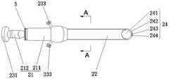
图1附图为本发明提供的一种新型腰椎减压、椎间融合的大通道内镜装置系统的结构示意图;1 is a schematic structural diagram of a novel large-channel endoscopic device system for lumbar decompression and intervertebral fusion provided by the present invention;
图2附图为本发明提供的内窥镜的结构示意图;2 is a schematic structural diagram of an endoscope provided by the present invention;
图3附图为本发明提供的A-A的剖面结构示意图;Fig. 3 accompanying drawing is the sectional structure schematic diagram of A-A provided by the present invention;
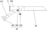
图4附图为本发明提供的内窥镜的另一种视角下的结构示意图;4 is a schematic structural diagram of the endoscope provided by the present invention from another viewing angle;
图5附图为本发明提供的内窥镜的另一种视角下的结构示意图;5 is a schematic structural diagram of the endoscope provided by the present invention from another viewing angle;
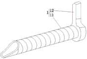
图6附图为本发明提供的工作鞘组件的结构示意图。FIG. 6 is a schematic structural diagram of the working sheath assembly provided by the present invention.
其中:1为工作鞘组件;11为工作套管;12为定位手柄;2为内窥镜; 21为镜身;211为水平部;212为倾斜部;22为外工作管;23为接口部;231 为摄像头连接口;232为光源接口;233为冲水接口;24为通道部;241为器械通道;242为光源通道;243为成像光通道;244清洁通道;3为螺纹段。12 is the positioning handle; 2 is the endoscope; 21 is the mirror body; 211 is the horizontal part; 212 is the inclined part; 22 is the outer working tube; 23 is the
具体实施方式Detailed ways
下面将结合本发明实施例中的附图,对本发明实施例中的技术方案进行清楚、完整地描述,显然,所描述的实施例仅仅是本发明一部分实施例,而不是全部的实施例。基于本发明中的实施例,本领域普通技术人员在没有做出创造性劳动前提下所获得的所有其他实施例,都属于本发明保护的范围。The technical solutions in the embodiments of the present invention will be clearly and completely described below with reference to the accompanying drawings in the embodiments of the present invention. Obviously, the described embodiments are only a part of the embodiments of the present invention, but not all of the embodiments. Based on the embodiments of the present invention, all other embodiments obtained by those of ordinary skill in the art without creative efforts shall fall within the protection scope of the present invention.
在本发明的描述中,需要说明的是,术语“中心”、“上”、“下”、“左”、“右”、“竖直”、“水平”、“内”、“外”等指示的方位或位置关系为基于附图所示的方位或位置关系,仅是为了便于描述本发明和简化描述,而不是指示或暗示所指的机构或元件必须具有特定的方位、以特定的方位构造和操作,因此不能理解为对本发明的限制。此外,术语“第一”、“第二”仅用于描述目的,而不能理解为指示或暗示相对重要性。In the description of the present invention, it should be noted that the terms "center", "upper", "lower", "left", "right", "vertical", "horizontal", "inner", "outer", etc. The indicated orientation or positional relationship is based on the orientation or positional relationship shown in the accompanying drawings, which is only for the convenience of describing the present invention and simplifying the description, rather than indicating or implying that the indicated mechanism or element must have a specific orientation or a specific orientation. construction and operation, and therefore should not be construed as limiting the invention. Furthermore, the terms "first" and "second" are used for descriptive purposes only and should not be construed to indicate or imply relative importance.
参见附图1-6,本发明实施例公开了一种新型腰椎减压、椎间融合的大通道内镜装置系统,包括:工作鞘组件1、内窥镜2和器械组件(图中未标出)。Referring to Figures 1-6, an embodiment of the present invention discloses a novel large-channel endoscopic device system for lumbar vertebral decompression and intervertebral fusion, comprising: a working
内窥镜2包括镜身21、外工作管22、接口部23和设置在外工作管22内的通道部24。The
其中,通道部24包括器械通道241、光源通道242、成像光通道243和清洁通道244,器械通道241与外工作管22偏心布置,成像光通道243设置在器械通道241的下方,其中,成像光通道243的直径为1.1mm-1.3mm之间,在本实施例中,像光通道243的直径优选为1.2mm,且成像光通道243延伸至镜身21内,镜身21与外工作管22的末端连接,清洁通道244设有两个,两个清洁通道244对称的设于成像光通道(243)两侧,光源通道242设置在清洁通道244的上方。The
接口部23包括摄像头连接口231、光源接口232和冲水接口233,摄像头连接口231设于镜身21后端底部且与成像光通道243连通,光源接口232 设置于镜身21前端底部且与光源通道242连通,冲水接口233设有两个,两个冲水接口233设于镜身21前端两侧且分别与两个清洁通道244连通;The
器械组件穿设于器械通道241内,器械通道241的末端设有用于器械组件连接的螺纹段3,在本实施例中,器械组件为椎间孔扩孔钻、细齿扩孔钻、弧形渐变锥型导杆、螺纹型中空骨钻、可弯曲神经探棒、镜下扩孔钻或镜下蓝钳中的任意一种。The instrument assembly passes through the
工作鞘组件1包括工作套管11和定位手柄12,定位手柄12固定于工作套管11的末端,工作套管11套设于外工作管22上,其中,工作套管11的表面为螺纹型结构,螺纹型结构能够实现椎板间最佳固定和调整,稳定控制内镜刺入病灶的深度,工作套管11的前端设有延长导向部,导向部为倾斜剖面。The working
具体地,镜身21包括水平部211和倾斜部212,水平部211和倾斜部212 一体成型,水平部211套设于外工作管22上,倾斜部212的一端与水平部211 的中部连接,且成像光通道243延伸至倾斜部212内,其中,水平部211的首端距外工作管22的首端为125mm-208mm之间,此段外工作管22为工作长度,在本实施例中工作长度优选为171mm,在另一些实施例中工作长度还可以为125mm或208mm,也可以根据实际需求具体设置,在此不做具体限制。Specifically, the
本说明书中各个实施例采用递进的方式描述,每个实施例重点说明的都是与其他实施例的不同之处,各个实施例之间相同相似部分互相参见即可。对于实施例公开的装置而言,由于其与实施例公开的方法相对应,所以描述的比较简单,相关之处参见方法部分说明即可。The various embodiments in this specification are described in a progressive manner, and each embodiment focuses on the differences from other embodiments, and the same and similar parts between the various embodiments can be referred to each other. As for the device disclosed in the embodiment, since it corresponds to the method disclosed in the embodiment, the description is relatively simple, and the relevant part can be referred to the description of the method.
对所公开的实施例的上述说明,使本领域专业技术人员能够实现或使用本发明。对这些实施例的多种修改对本领域的专业技术人员来说将是显而易见的,本文中所定义的一般原理可以在不脱离本发明的精神或范围的情况下,在其它实施例中实现。因此,本发明将不会被限制于本文所示的这些实施例,而是要符合与本文所公开的原理和新颖特点相一致的最宽的范围。The above description of the disclosed embodiments enables any person skilled in the art to make or use the present invention. Various modifications to these embodiments will be readily apparent to those skilled in the art, and the generic principles defined herein may be implemented in other embodiments without departing from the spirit or scope of the invention. Thus, the present invention is not intended to be limited to the embodiments shown herein, but is to be accorded the widest scope consistent with the principles and novel features disclosed herein.
Claims (9)
Translated fromChinesePriority Applications (1)
| Application Number | Priority Date | Filing Date | Title |
|---|---|---|---|
| CN202010832533.1ACN111772567A (en) | 2020-08-18 | 2020-08-18 | Novel lumbar decompression and intervertebral fusion large-channel endoscope device system |
Applications Claiming Priority (1)
| Application Number | Priority Date | Filing Date | Title |
|---|---|---|---|
| CN202010832533.1ACN111772567A (en) | 2020-08-18 | 2020-08-18 | Novel lumbar decompression and intervertebral fusion large-channel endoscope device system |
Publications (1)
| Publication Number | Publication Date |
|---|---|
| CN111772567Atrue CN111772567A (en) | 2020-10-16 |
Family
ID=72762193
Family Applications (1)
| Application Number | Title | Priority Date | Filing Date |
|---|---|---|---|
| CN202010832533.1APendingCN111772567A (en) | 2020-08-18 | 2020-08-18 | Novel lumbar decompression and intervertebral fusion large-channel endoscope device system |
Country Status (1)
| Country | Link |
|---|---|
| CN (1) | CN111772567A (en) |
Cited By (4)
| Publication number | Priority date | Publication date | Assignee | Title |
|---|---|---|---|---|
| CN112263285A (en)* | 2020-11-10 | 2021-01-26 | 广东省人民医院 | Percutaneous two-channel endoscope sleeve device |
| CN114209386A (en)* | 2021-08-13 | 2022-03-22 | 北京积水潭医院 | Visual wire saw guide operation system and method for orthopedic surgery |
| CN114431821A (en)* | 2022-01-14 | 2022-05-06 | 四川耐特光电科技有限公司 | Electronic intervertebral foramen mirror |
| WO2023092831A1 (en)* | 2021-11-25 | 2023-06-01 | 北京大学第三医院 | Spine intervertebral fusion endoscope structure |
Citations (3)
| Publication number | Priority date | Publication date | Assignee | Title |
|---|---|---|---|---|
| CN204618185U (en)* | 2015-04-23 | 2015-09-09 | 上海凯利泰医疗科技股份有限公司 | arthroscope threaded sleeve |
| CN107874735A (en)* | 2017-10-25 | 2018-04-06 | 珠海康弘发展有限公司 | A kind of percutaneous spinal endoscopes of big passage for being applied to fusion device under mirror |
| CN110384525A (en)* | 2019-06-17 | 2019-10-29 | 中国人民解放军联勤保障部队第九二〇医院 | It is a kind of through canalis spinalis mirror approach Micro-operation system |
- 2020
- 2020-08-18CNCN202010832533.1Apatent/CN111772567A/enactivePending
Patent Citations (3)
| Publication number | Priority date | Publication date | Assignee | Title |
|---|---|---|---|---|
| CN204618185U (en)* | 2015-04-23 | 2015-09-09 | 上海凯利泰医疗科技股份有限公司 | arthroscope threaded sleeve |
| CN107874735A (en)* | 2017-10-25 | 2018-04-06 | 珠海康弘发展有限公司 | A kind of percutaneous spinal endoscopes of big passage for being applied to fusion device under mirror |
| CN110384525A (en)* | 2019-06-17 | 2019-10-29 | 中国人民解放军联勤保障部队第九二〇医院 | It is a kind of through canalis spinalis mirror approach Micro-operation system |
Cited By (4)
| Publication number | Priority date | Publication date | Assignee | Title |
|---|---|---|---|---|
| CN112263285A (en)* | 2020-11-10 | 2021-01-26 | 广东省人民医院 | Percutaneous two-channel endoscope sleeve device |
| CN114209386A (en)* | 2021-08-13 | 2022-03-22 | 北京积水潭医院 | Visual wire saw guide operation system and method for orthopedic surgery |
| WO2023092831A1 (en)* | 2021-11-25 | 2023-06-01 | 北京大学第三医院 | Spine intervertebral fusion endoscope structure |
| CN114431821A (en)* | 2022-01-14 | 2022-05-06 | 四川耐特光电科技有限公司 | Electronic intervertebral foramen mirror |
Similar Documents
| Publication | Publication Date | Title |
|---|---|---|
| CN111772567A (en) | Novel lumbar decompression and intervertebral fusion large-channel endoscope device system | |
| CN103690205B (en) | The surgical apparatus of minimally-invasive spinal fusion and the surgery systems for including it | |
| JP4726377B2 (en) | Device that provides posterior or anterior transsacral access to the vertebra | |
| US7641657B2 (en) | Method and apparatus for providing posterior or anterior trans-sacral access to spinal vertebrae | |
| KR101310665B1 (en) | Facet joint reamer | |
| JP2015500680A (en) | Spine treatment lateral approach access device | |
| CN103976779B (en) | Foramen intervertebrale lens lancing system | |
| JP2000511788A (en) | Percutaneous surgical device and method | |
| JP2011519691A (en) | Method and apparatus for lateral access to intervertebral disc space | |
| US20240252033A1 (en) | Image guided spinal decompression with contralateral oblique view | |
| CN111358600A (en) | Self-locking intervertebral fusion device system under spinal endoscope | |
| CN101327146B (en) | A double-slot channel-connected percutaneous pedicle screw internal fixation system | |
| CN101528110A (en) | Percutaneous access and visualization of the spine | |
| CN110384525A (en) | It is a kind of through canalis spinalis mirror approach Micro-operation system | |
| CN111759365A (en) | Foraminoplasty surgical instruments | |
| US20240358364A1 (en) | Retractor tube device | |
| CN114305310B (en) | Microscopic endoscope and application method thereof | |
| CN214966253U (en) | Common spinal endoscope rear approach trepan casing | |
| US20240074787A1 (en) | Working channel for use in a method and system for percutaneous procedures | |
| CN215584235U (en) | Flexible eccentric minimally invasive channel system for anterior cervical approach | |
| CN209490040U (en) | It is capable of the PPH auxiliary instrument of expansion space | |
| JP3249918U (en) | Surgical instruments | |
| CN215777987U (en) | Detection device and micro-endoscope | |
| Song et al. | Minimally invasive cervical stenosis decompression | |
| CN219962947U (en) | Step-by-step expansion channel tube for spinal surgery |
Legal Events
| Date | Code | Title | Description |
|---|---|---|---|
| PB01 | Publication | ||
| PB01 | Publication | ||
| SE01 | Entry into force of request for substantive examination | ||
| SE01 | Entry into force of request for substantive examination | ||
| RJ01 | Rejection of invention patent application after publication | ||
| RJ01 | Rejection of invention patent application after publication | Application publication date:20201016 |