CN111754520A - A brain hematoma segmentation method and system based on deep learning - Google Patents
A brain hematoma segmentation method and system based on deep learningDownload PDFInfo
- Publication number
- CN111754520A CN111754520ACN202010517019.9ACN202010517019ACN111754520ACN 111754520 ACN111754520 ACN 111754520ACN 202010517019 ACN202010517019 ACN 202010517019ACN 111754520 ACN111754520 ACN 111754520A
- Authority
- CN
- China
- Prior art keywords
- image
- self
- brain
- unit
- attention
- Prior art date
- Legal status (The legal status is an assumption and is not a legal conclusion. Google has not performed a legal analysis and makes no representation as to the accuracy of the status listed.)
- Granted
Links
Images


Classifications
- G—PHYSICS
- G06—COMPUTING OR CALCULATING; COUNTING
- G06T—IMAGE DATA PROCESSING OR GENERATION, IN GENERAL
- G06T7/00—Image analysis
- G06T7/10—Segmentation; Edge detection
- G06T7/11—Region-based segmentation
- G—PHYSICS
- G06—COMPUTING OR CALCULATING; COUNTING
- G06N—COMPUTING ARRANGEMENTS BASED ON SPECIFIC COMPUTATIONAL MODELS
- G06N3/00—Computing arrangements based on biological models
- G06N3/02—Neural networks
- G06N3/04—Architecture, e.g. interconnection topology
- G06N3/045—Combinations of networks
- G—PHYSICS
- G06—COMPUTING OR CALCULATING; COUNTING
- G06N—COMPUTING ARRANGEMENTS BASED ON SPECIFIC COMPUTATIONAL MODELS
- G06N3/00—Computing arrangements based on biological models
- G06N3/02—Neural networks
- G06N3/08—Learning methods
- G06N3/084—Backpropagation, e.g. using gradient descent
- G—PHYSICS
- G06—COMPUTING OR CALCULATING; COUNTING
- G06T—IMAGE DATA PROCESSING OR GENERATION, IN GENERAL
- G06T2207/00—Indexing scheme for image analysis or image enhancement
- G06T2207/10—Image acquisition modality
- G06T2207/10072—Tomographic images
- G06T2207/10081—Computed x-ray tomography [CT]
- G—PHYSICS
- G06—COMPUTING OR CALCULATING; COUNTING
- G06T—IMAGE DATA PROCESSING OR GENERATION, IN GENERAL
- G06T2207/00—Indexing scheme for image analysis or image enhancement
- G06T2207/20—Special algorithmic details
- G06T2207/20081—Training; Learning
- G—PHYSICS
- G06—COMPUTING OR CALCULATING; COUNTING
- G06T—IMAGE DATA PROCESSING OR GENERATION, IN GENERAL
- G06T2207/00—Indexing scheme for image analysis or image enhancement
- G06T2207/20—Special algorithmic details
- G06T2207/20084—Artificial neural networks [ANN]
- G—PHYSICS
- G06—COMPUTING OR CALCULATING; COUNTING
- G06T—IMAGE DATA PROCESSING OR GENERATION, IN GENERAL
- G06T2207/00—Indexing scheme for image analysis or image enhancement
- G06T2207/20—Special algorithmic details
- G06T2207/20212—Image combination
- G06T2207/20221—Image fusion; Image merging
- G—PHYSICS
- G06—COMPUTING OR CALCULATING; COUNTING
- G06T—IMAGE DATA PROCESSING OR GENERATION, IN GENERAL
- G06T2207/00—Indexing scheme for image analysis or image enhancement
- G06T2207/30—Subject of image; Context of image processing
- G06T2207/30004—Biomedical image processing
- G06T2207/30101—Blood vessel; Artery; Vein; Vascular
Landscapes
- Engineering & Computer Science (AREA)
- Physics & Mathematics (AREA)
- Theoretical Computer Science (AREA)
- General Physics & Mathematics (AREA)
- General Health & Medical Sciences (AREA)
- General Engineering & Computer Science (AREA)
- Biophysics (AREA)
- Computational Linguistics (AREA)
- Data Mining & Analysis (AREA)
- Evolutionary Computation (AREA)
- Artificial Intelligence (AREA)
- Molecular Biology (AREA)
- Computing Systems (AREA)
- Biomedical Technology (AREA)
- Life Sciences & Earth Sciences (AREA)
- Mathematical Physics (AREA)
- Software Systems (AREA)
- Health & Medical Sciences (AREA)
- Computer Vision & Pattern Recognition (AREA)
- Image Analysis (AREA)
- Image Processing (AREA)
Abstract
Translated fromChinese
Description
Translated fromChinese技术领域technical field
本发明涉及图像分割技术领域,特别是涉及一种基于深度学习的脑血肿分割方法及系统。The invention relates to the technical field of image segmentation, in particular to a method and system for segmentation of brain hematoma based on deep learning.
背景技术Background technique
脑卒中属于脑血管类疾病,主要是由非外脑性实质内血管破裂引起的出血所导致,其中导致脑出血的原因有很多种,例如高血压,高血脂,糖尿病以及其他的心血管疾病,在生活中发病率极高,根据世界卫生组织发布数据,每年的死亡人口中,约有百分之三十到四十是由于脑部出血所引起,目前脑出血已经成为当今人类死亡率最高的疾病之一。然而目前关于定量测量疑似血肿区域体积的方法是很少的,快速、准确、可重复的体积估算对于许多医学诊断、治疗、评估是至关重要的,是决定病人是否需要动手术的一个重要指标,因此,精确的体积测量具有重要的临床应用价值。Stroke is a cerebrovascular disease, mainly caused by hemorrhage caused by rupture of non-external cerebral parenchyma blood vessels. There are many reasons for cerebral hemorrhage, such as hypertension, hyperlipidemia, diabetes and other cardiovascular diseases. The incidence of morbidity in life is extremely high. According to data released by the World Health Organization, about 30 to 40% of the annual deaths are caused by cerebral hemorrhage. At present, cerebral hemorrhage has become the highest mortality rate in human beings today. one of the diseases. However, there are currently few methods for quantitatively measuring the volume of suspected hematoma areas. Rapid, accurate, and repeatable volume estimation is crucial for many medical diagnosis, treatment, and evaluation, and is an important indicator for deciding whether a patient needs surgery. , therefore, accurate volume measurement has important clinical application value.
图像分割的目的就是改变对一幅图像中感兴趣的疑似病灶区域的描述,使其分析起来更加容易并且更有意义。然而医学图像不同于一般的图像,常会伴随着出现弱边界、低对比度、强噪音等现象,正因为医学图像自身所具有多样性和特殊性,才导致了分割的复杂性。目前,国内外大部分医院对患者颅内疑似血肿体积的临床测量主要是通过手动分割和人工计算两大歩骤来实现的。手动分割极其浪费时间且非常艰辛,精确度和可重复性比较差。The purpose of image segmentation is to change the description of the suspected lesion area of interest in an image, making it easier and more meaningful to analyze. However, medical images are different from ordinary images, often accompanied by weak boundaries, low contrast, strong noise and other phenomena. It is precisely because of the diversity and particularity of medical images that the complexity of segmentation is caused. At present, the clinical measurement of the suspected intracranial hematoma volume in most hospitals at home and abroad is mainly achieved through two steps of manual segmentation and manual calculation. Manual segmentation is extremely time-consuming and laborious, with poor accuracy and repeatability.
发明内容SUMMARY OF THE INVENTION
本发明的目的是提供一种基于深度学习的脑血肿分割方法及系统。The purpose of the present invention is to provide a brain hematoma segmentation method and system based on deep learning.
为实现上述目的,本发明提供了如下方案:For achieving the above object, the present invention provides the following scheme:
一种基于深度学习的脑血肿分割方法,包括:A brain hematoma segmentation method based on deep learning, including:
构建神经网络模型,所述神经网络模型包括若干顺序连接的图像信息压缩模块和若干顺序连接的图像信息融合模块,其中,所述图像信息压缩模块包括依次顺序连接的第一自注意力卷积单元、第二自注意力卷积单元和池化层,所述图像信息融合模块包括依次顺序连接的上采样单元、特征图拼接单元和第三自注意力卷积单元,所述第一自注意力卷积单元用于降低其输入图像的通道数,所述第二自注意力卷积单元用于对其输入的图像进行特征提取,所述池化层用于对其输入的图像进行降维处理,所述上采样单元用于对其输入的图像进行上采样,所述特征图拼接单元用于将其输入的采样图像与同一级第二注意力卷积单元输出的降维图像在通道方向拼接,所述第三自注意力卷积单元用于对拼接后的图像进行多尺度融合;同一级的第二注意力卷积单元和特征图拼接单元满足如下条件:所述第二注意力卷积单元输出图像的维度与所述特征图拼接单元输入图像的维度相同;Construct a neural network model, the neural network model includes several sequentially connected image information compression modules and several sequentially connected image information fusion modules, wherein the image information compression module includes sequentially connected first self-attention convolution units , the second self-attention convolution unit and the pooling layer, the image information fusion module includes an upsampling unit, a feature map splicing unit and a third self-attention convolution unit connected in sequence, the first self-attention The convolution unit is used to reduce the number of channels of its input image, the second self-attention convolution unit is used to perform feature extraction on its input image, and the pooling layer is used to perform dimensionality reduction processing on its input image , the up-sampling unit is used for up-sampling the input image, and the feature map stitching unit is used for stitching the input sample image and the dimension-reduced image output by the second attention convolution unit of the same level in the channel direction , the third self-attention convolution unit is used to perform multi-scale fusion on the spliced image; the second attention convolution unit and the feature map splicing unit at the same level satisfy the following conditions: the second attention convolution unit The dimension of the unit output image is the same as the dimension of the input image of the feature map stitching unit;
获取脑CT样本图像;Obtain brain CT sample images;
以所述脑CT样本图像为输入,以所述脑CT样本图像中各像素点的出血情况为标签,对所述神经网络模型进行训练;Taking the brain CT sample image as an input, and using the bleeding situation of each pixel in the brain CT sample image as a label, the neural network model is trained;
采用训练好的神经网络模型对待分割脑CT图像进行脑出血识别。Intracerebral hemorrhage recognition was performed by using the trained neural network model in the brain CT images to be segmented.
可选的,所述第三自注意力卷积单元包括顺序连接的第一自注意力卷积子单元和第二自注意力卷积子单元,所述第一自注意力卷积单元用于降低其输入图像的通道数,所述第二自注意力卷积子单元与所述第二自注意力卷积单元的结构参数相同。Optionally, the third self-attention convolution unit includes a sequentially connected first self-attention convolution sub-unit and a second self-attention convolution sub-unit, and the first self-attention convolution unit is used for The number of channels of its input image is reduced, and the second self-attention convolution sub-unit has the same structural parameters as the second self-attention convolution unit.
可选的,所述采用训练好的神经网络模型对待分割脑CT图像进行脑出血识别,具体包括:Optionally, the use of the trained neural network model to identify cerebral hemorrhage in the to-be-segmented brain CT image specifically includes:
将所述待分割脑CT图像输入训练好的神经网络模型,确定出血区域的位置。Input the brain CT image to be segmented into the trained neural network model to determine the location of the bleeding area.
可选的,所述采用训练好的神经网络模型对待分割脑CT图像进行脑出血识别,还包括:Optionally, the use of the trained neural network model to identify cerebral hemorrhage in the brain CT image to be segmented also includes:
统计出血像素点,根据出血像素点计算出血面积。The bleeding pixels are counted, and the bleeding area is calculated according to the bleeding pixels.
可选的,所述采用训练好的神经网络模型对待分割脑CT图像进行脑出血识别,还包括:Optionally, the use of the trained neural network model to identify cerebral hemorrhage in the brain CT image to be segmented also includes:
根据所述脑CT图像的切片厚度、切片层数以及每层脑CT图像的出血面积,计算脑血肿的体积。The volume of the cerebral hematoma is calculated according to the slice thickness of the brain CT image, the number of slice layers and the bleeding area of each slice of the brain CT image.
本发明还提供了一种基于深度学习的脑血肿分割系统,包括:The present invention also provides a deep learning-based brain hematoma segmentation system, including:
神经网络模型构建模块,用于构建神经网络模型,所述神经网络模型包括若干顺序连接的图像信息压缩模块和若干顺序连接的图像信息融合模块,其中,所述图像信息压缩模块包括依次顺序连接的第一自注意力卷积单元、第二自注意力卷积单元和池化层,所述图像信息融合模块包括依次顺序连接的上采样单元、特征图拼接单元和第三自注意力卷积单元,所述第一自注意力卷积单元用于降低其输入图像的通道数,所述第二自注意力卷积单元用于对其输入的图像进行特征提取,所述池化层用于对其输入的图像进行降维处理,所述上采样单元用于对其输入的图像进行上采样,所述特征图拼接单元用于将其输入的采样图像与同一级第二注意力卷积单元输出的降维图像在通道方向拼接,所述第三自注意力卷积单元用于对拼接后的图像进行多尺度融合;同一级的第二注意力卷积单元和特征图拼接单元满足如下条件:所述第二注意力卷积单元输出图像的维度与所述特征图拼接单元输入图像的维度相同;A neural network model building module is used to build a neural network model, the neural network model includes a number of sequentially connected image information compression modules and a number of sequentially connected image information fusion modules, wherein the image information compression modules The first self-attention convolution unit, the second self-attention convolution unit and the pooling layer, the image information fusion module includes an upsampling unit, a feature map stitching unit and a third self-attention convolution unit connected in sequence , the first self-attention convolution unit is used to reduce the number of channels of its input image, the second self-attention convolution unit is used to perform feature extraction on the input image, and the pooling layer is used to The input image is subjected to dimensionality reduction processing, the upsampling unit is used for upsampling the input image, and the feature map stitching unit is used for outputting the input sampled image and the second attention convolution unit of the same level The dimension-reduced images are spliced in the channel direction, and the third self-attention convolution unit is used to perform multi-scale fusion on the spliced image; the second attention convolution unit and the feature map splicing unit at the same level satisfy the following conditions: The dimension of the output image of the second attention convolution unit is the same as the dimension of the input image of the feature map stitching unit;
样本图像获取模块,用于获取脑CT样本图像;The sample image acquisition module is used to acquire brain CT sample images;
神经网络模型训练模块,用于以所述脑CT样本图像为输入,以所述脑CT样本图像中各像素点出血情况为标签,对所述神经网络模型进行训练;A neural network model training module for training the neural network model with the brain CT sample image as an input and the bleeding situation of each pixel in the brain CT sample image as a label;
脑出血识别模块,用于采用训练好的神经网络模型对待分割脑CT图像进行脑出血识别。The intracerebral hemorrhage identification module is used to identify the intracerebral hemorrhage in the to-be-segmented CT image of the brain by using the trained neural network model.
可选的,所述第三自注意力卷积单元包括顺序连接的第一自注意力卷积单元和第二自注意力卷积子单元,所述第一自注意力卷积单元用于降低其输入图像的通道数,所述第二自注意力卷积子单元与所述第二自注意力卷积单元的结构参数相同。Optionally, the third self-attention convolution unit includes a sequentially connected first self-attention convolution unit and a second self-attention convolution subunit, and the first self-attention convolution unit is used to reduce The number of channels of its input image, the second self-attention convolution subunit and the second self-attention convolution unit have the same structural parameters.
可选的,所述脑出血识别模块,具体包括:Optionally, the intracerebral hemorrhage identification module specifically includes:
将所述待分割脑CT图像输入训练好的神经网络模型,确定出血区域的位置。Input the brain CT image to be segmented into the trained neural network model to determine the location of the bleeding area.
可选的,所述脑出血识别模块还包括:Optionally, the cerebral hemorrhage identification module further includes:
统计出血像素点,根据出血像素点计算出血面积。The bleeding pixels are counted, and the bleeding area is calculated according to the bleeding pixels.
可选的,所述脑出血识别模块还包括:Optionally, the cerebral hemorrhage identification module further includes:
根据所述脑CT图像的切片厚度、切片层数以及每层脑CT图像的出血面积,计算脑血肿的体积。The volume of the cerebral hematoma is calculated according to the slice thickness of the brain CT image, the number of slice layers and the bleeding area of each slice of the brain CT image.
根据本发明提供的具体实施例,本发明公开了以下技术效果:本发明提供的基于深度学习的脑血肿分割方法及系统先通过设计的自注意力卷积单元对输入CT图像进行深度特征提取,并结合二维池化层降低计算量;对经过反复特征提取、维度压缩后输出的特征图,使用双线性插值法重新上采样;使用特征图拼接的方式,降低了因池化层所带来的信息损失,之后采用卷积单元对拼接的多尺度特征进行深度信息融合;重复多次上采样、特征图拼接与多尺度融合运算获得模型预测的分割图像;基于模型预测的分割图像与真实标签图像之间的损失值训练该神经网络模型。采用训练好的神经网络模型对待待分割脑CT图像的出血区域进行识别。本发明不仅识别精度高,而且,相较于现有技术中的手动识别和人工识别,具有效率高的优势。According to the specific embodiments provided by the present invention, the present invention discloses the following technical effects: the deep learning-based cerebral hematoma segmentation method and system provided by the present invention first perform depth feature extraction on the input CT image through the designed self-attention convolution unit, Combined with the two-dimensional pooling layer to reduce the amount of calculation; for the feature map output after repeated feature extraction and dimension compression, the bilinear interpolation method is used to re-upsample; the feature map splicing method is used to reduce the cost caused by the pooling layer. Then, the convolution unit is used to perform depth information fusion on the spliced multi-scale features; repeat upsampling, feature map splicing and multi-scale fusion operations to obtain the segmented image predicted by the model; The loss value between the label images trains this neural network model. The trained neural network model is used to identify the hemorrhagic region of the brain CT image to be segmented. The invention not only has high recognition accuracy, but also has the advantage of high efficiency compared with manual recognition and manual recognition in the prior art.
附图说明Description of drawings
为了更清楚地说明本发明实施例或现有技术中的技术方案,下面将对实施例中所需要使用的附图作简单地介绍,显而易见地,下面描述中的附图仅仅是本发明的一些实施例,对于本领域普通技术人员来讲,在不付出创造性劳动的前提下,还可以根据这些附图获得其他的附图。In order to more clearly illustrate the embodiments of the present invention or the technical solutions in the prior art, the accompanying drawings required in the embodiments will be briefly introduced below. Obviously, the drawings in the following description are only some of the present invention. In the embodiments, for those of ordinary skill in the art, other drawings can also be obtained according to these drawings without any creative effort.
图1为本发明实施例1提供的基于深度学习的脑血肿分割方法流程图;1 is a flowchart of a deep learning-based brain hematoma segmentation method provided in Embodiment 1 of the present invention;
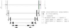
图2为本发明实施例1提供的神经网络模型的工作流程示意图;2 is a schematic diagram of the workflow of the neural network model provided in Embodiment 1 of the present invention;
图3为本发明实施例1提供的自注意力卷积单元的结构示意图;3 is a schematic structural diagram of a self-attention convolution unit provided in Embodiment 1 of the present invention;
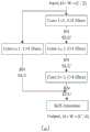
图4(a)为本发明实施例1提供的第一自注意力卷积单元结构示意图,图4(b)为本发明实施例1提供的第二自注意力卷积单元与第二自注意力卷积子单元的结构示意图,图4(c)为本发明实施例1提供的第一自注意力卷积子单元的结构示意图;FIG. 4(a) is a schematic structural diagram of the first self-attention convolution unit provided in Embodiment 1 of the present invention, and FIG. 4(b) is a second self-attention convolution unit and a second self-attention provided by Embodiment 1 of the present invention. A schematic structural diagram of a force convolution subunit, FIG. 4(c) is a schematic structural diagram of the first self-attention convolution subunit provided in Embodiment 1 of the present invention;
图5为本发明实施例2提供的基于深度学习的脑血肿分割系统结构图。FIG. 5 is a structural diagram of a brain hematoma segmentation system based on deep learning according to Embodiment 2 of the present invention.
具体实施方式Detailed ways
下面将结合本发明实施例中的附图,对本发明实施例中的技术方案进行清楚、完整地描述,显然,所描述的实施例仅仅是本发明一部分实施例,而不是全部的实施例。基于本发明中的实施例,本领域普通技术人员在没有做出创造性劳动前提下所获得的所有其他实施例,都属于本发明保护的范围。The technical solutions in the embodiments of the present invention will be clearly and completely described below with reference to the accompanying drawings in the embodiments of the present invention. Obviously, the described embodiments are only a part of the embodiments of the present invention, but not all of the embodiments. Based on the embodiments of the present invention, all other embodiments obtained by those of ordinary skill in the art without creative efforts shall fall within the protection scope of the present invention.
为使本发明的上述目的、特征和优点能够更加明显易懂,下面结合附图和具体实施方式对本发明作进一步详细的说明。In order to make the above objects, features and advantages of the present invention more clearly understood, the present invention will be described in further detail below with reference to the accompanying drawings and specific embodiments.
图1为本发明实施例1提供的基于深度学习的脑血肿分割方法流程图,如图1所示,本实施例提供的基于深度学习的脑血肿分割方法流程图包括以下步骤:1 is a flowchart of a method for segmentation of brain hematoma based on deep learning provided in Embodiment 1 of the present invention. As shown in FIG. 1 , the flowchart of the method for segmentation of brain hematoma based on deep learning provided by this embodiment includes the following steps:
步骤101:构建神经网络模型,所述神经网络模型包括若干顺序连接的图像信息压缩模块和若干顺序连接的图像信息融合模块,其中,所述图像信息融合模块用于对压缩后的图像进行特征提取,所述图像信息压缩模块包括依次顺序连接的第一自注意力卷积单元、第二自注意力卷积单元和池化层,所述图像信息融合模块包括依次顺序连接的上采样单元、特征图拼接单元和第三自注意力卷积单元,所述第一自注意力卷积单元用于降低其输入图像的通道数,所述第二自注意力卷积单元用于对其输入的图像进行特征提取,所述池化层用于对其输入的图像进行降维处理,所述上采样单元用于对其输入的图像进行上采样,所述特征图拼接单元用于将其输入的采样图像与同一级第二自注意力卷积单元输出的降维图像在通道方向拼接,所述第三自注意力卷积单元用于对拼接后的图像进行多尺度融合;同一级的第二自注意力卷积单元和特征图拼接单元满足如下条件:所述第二自注意力卷积单元输出图像的维度与所述特征图拼接单元输入图像的维度相同;Step 101: Build a neural network model, the neural network model includes several sequentially connected image information compression modules and several sequentially connected image information fusion modules, wherein the image information fusion module is used to perform feature extraction on the compressed image , the image information compression module includes a first self-attention convolution unit, a second self-attention convolution unit and a pooling layer connected in sequence, and the image information fusion module includes an upsampling unit, a feature The image stitching unit and the third self-attention convolution unit, the first self-attention convolution unit is used for reducing the number of channels of its input image, and the second self-attention convolution unit is used for the input image Perform feature extraction, the pooling layer is used to reduce the dimension of the input image, the upsampling unit is used to upsample the input image, and the feature map stitching unit is used to sample the input. The image is spliced in the channel direction with the dimension-reduced image output by the second self-attention convolution unit of the same level, and the third self-attention convolution unit is used to perform multi-scale fusion on the spliced image; the second self-attention convolution unit of the same level The attention convolution unit and the feature map splicing unit satisfy the following conditions: the dimension of the output image of the second self-attention convolution unit is the same as the dimension of the input image of the feature map splicing unit;
步骤102:获取脑CT样本图像;Step 102: obtaining a brain CT sample image;
步骤103:以所述脑CT样本图像为输入,以所述脑CT样本图像中各像素点出血情况为标签,比如出血的概率,对所述神经网络模型进行训练;Step 103: Using the brain CT sample image as input, and using the bleeding situation of each pixel in the brain CT sample image as a label, such as the probability of bleeding, train the neural network model;
步骤104:采用训练好的神经网络模型对待分割脑CT图像进行脑出血识别。Step 104: Use the trained neural network model to identify cerebral hemorrhage in the CT image of the brain to be segmented.
作为一种实施方式,所述第三自注意力卷积单元包括顺序连接的第一自注意力卷积单元和第二自注意力卷积子单元,所述第一自注意力卷积单元用于降低其输入图像的通道数,所述第二自注意力卷积子单元与所述第二自注意力卷积单元的结构参数相同。As an embodiment, the third self-attention convolution unit includes a sequentially connected first self-attention convolution unit and a second self-attention convolution subunit, and the first self-attention convolution unit uses In order to reduce the number of channels of its input image, the second self-attention convolution sub-unit has the same structural parameters as the second self-attention convolution unit.
如图2所示,本实施例构建的神经网络模型第一步对脑CT图像进行信息压缩,具体可以为:首先通过设计的第一自注意力卷积单元降低输入模型CT图像的通道数,再用第二自注意力卷积单元深度提取输入图像的特征。其中,如图3所示,自注意力卷积单元为在每个卷积单元结束前添加自注意力集中层,获取特征图像素间的远距离依赖关系,增加模型全局感受野,提高分割精度。然后,使用二维池化层对第二自注意力卷积单元输出的特征图进行降维处理,减少模型的可训练参数,降低计算量。之后反复堆叠五次第一自注意力卷积单元、第二自注意力卷积单元与池化层,将输入模型长宽为256×256的CT图像压缩为长宽只有8×8的特征图。当然,第一自注意力卷积单元、第二自注意力卷积单元与池化层的堆叠次数不仅限于本实施例所述的五次,输入输出图像的长宽也不仅限于256×256和8×8,在其他的实施例中,可以选取其他的堆叠次数以及输入输出图像的长宽。第二步:重新提取压缩信息并预测分割图像。具体可以为:使用双线性插值法对压缩的特征图重新上采样,提高特征图的分辨率。将使用双线性插值上采样特征图与第一步降维压缩的特征图,在通道方向拼接,减少池化层所带来的信息损失。使用第三自注意力卷积单元对拼接后多尺度特征图进行融合。重复五次上采样、特征图拼接、第三自注意力卷积单元,将8×8的特征图恢复为256×256大小,作为最终模型预测的分割图像。计算模型预测的分割图像与真实标签图像之间的损失值,使用反向传播算法优化模型可训练参数。当损失值小于指定值时,模型可以准确分割CT图像中脑血肿区域。当然,上采样、特征图拼接、第三自注意力卷积单元的重复次数不仅限于五次,在其他的实施例中也可以选择其他次数。特征图恢复后的大小也不仅限于256×256,在其他的实施例中也可以选择其他的长宽规模。在本实施例中,第一自注意力卷积单元、第二自注意力卷积单元以及第一自注意力卷积子单元、第二自注意力卷积子单元的具体结构以及参数设置可以如图4所示,具体的,第一自注意力卷积单元如图4(a)所示,第二自注意力卷积单元与第二自注意力卷积子单元如图4(b)所示,第一自注意力卷积子单元如图4(c)所示,其中,C:通道数,H:高度,W:宽度,Input:输入,Output:输出,BN:批量归一化层,ELU:激活函数,Conv:卷积层。As shown in FIG. 2 , the first step of the neural network model constructed in this embodiment compresses the information of the brain CT image. Specifically, the first step is to reduce the number of channels of the input model CT image through the designed first self-attention convolution unit. The second self-attention convolution unit is then used to extract the features of the input image depthwise. Among them, as shown in Figure 3, the self-attention convolution unit adds a self-attention concentration layer before the end of each convolution unit, obtains the long-distance dependency between the pixels of the feature map, increases the global receptive field of the model, and improves the segmentation accuracy . Then, a two-dimensional pooling layer is used to reduce the dimension of the feature map output by the second self-attention convolution unit to reduce the trainable parameters of the model and reduce the amount of computation. After that, the first self-attention convolution unit, the second self-attention convolution unit and the pooling layer are repeatedly stacked five times, and the CT image with a length and width of 256×256 of the input model is compressed into a feature map with a length and width of only 8×8 . Of course, the stacking times of the first self-attention convolution unit, the second self-attention convolution unit and the pooling layer are not limited to the five times described in this embodiment, and the length and width of the input and output images are not limited to 256×256 and 256×256. 8×8, in other embodiments, other stacking times and the length and width of the input and output images can be selected. Step 2: Re-extract the compressed information and predict the segmented image. Specifically, the compressed feature map may be re-upsampled using a bilinear interpolation method to improve the resolution of the feature map. The bilinear interpolation upsampling feature map and the feature map compressed by the first step of dimensionality reduction are spliced in the channel direction to reduce the information loss caused by the pooling layer. The concatenated multi-scale feature maps are fused using a third self-attention convolution unit. Repeat upsampling, feature map stitching, and the third self-attention convolution unit five times to restore the 8×8 feature map to a size of 256×256 as the segmented image predicted by the final model. Calculate the loss value between the segmented image predicted by the model and the ground-truth label image, and use the back-propagation algorithm to optimize the model trainable parameters. When the loss value is less than the specified value, the model can accurately segment the brain hematoma region in the CT image. Of course, the repetition times of upsampling, feature map splicing, and the third self-attention convolution unit are not limited to five times, and other times may also be selected in other embodiments. The size of the restored feature map is not limited to 256×256, and other length and width scales can also be selected in other embodiments. In this embodiment, the specific structures and parameter settings of the first self-attention convolution unit, the second self-attention convolution unit, and the first self-attention convolution subunit and the second self-attention convolution subunit can be As shown in Figure 4, specifically, the first self-attention convolution unit is shown in Figure 4(a), the second self-attention convolution unit and the second self-attention convolution sub-unit are shown in Figure 4(b) As shown, the first self-attention convolution subunit is shown in Figure 4(c), where C: number of channels, H: height, W: width, Input: input, Output: output, BN: batch normalization layer, ELU: activation function, Conv: convolutional layer.
作为一种可选的实施方式,如图4所示,通过三个自注意力卷积单元增强模型的特征提取能力。其中,二维卷积层计算如公式(1)所示,Activation表示经过卷积后采用的非线性激活函数,WL为在第L层神经网络上卷积权重,bL为卷积层添加的偏置项,WL-1(m,n)是特征图在特征图坐标(x,y)的权重。Conv2DL和Conv2DL-1分别表示第L层和第L-1层的卷积特征块,Conv2DL(x,y)为Conv2DL在二维卷积特征块坐标(x,y)上的元素,Conv2DL-1(x+m,y+n)为Conv2DL(x,y)在经过二维卷积核WL-1(m,n)卷积后Conv2DL-1在坐标(x+m,y+n)上的元素值。As an optional implementation, as shown in FIG. 4 , the feature extraction capability of the model is enhanced through three self-attention convolution units. Among them, the calculation of the two-dimensional convolution layer is shown in formula (1), Activation represents the nonlinear activation function used after convolution, WL is the convolution weight on the L-th layer of neural network, and bL is the convolution layer added The bias term of , WL-1 (m, n) is the weight of the feature map at the feature map coordinates (x, y). Conv2DL and Conv2DL-1 represent the convolutional feature blocks of the Lth layer and L-1th layer, respectively, and Conv2DL (x, y) is the element of Conv2DL on the two-dimensional convolution feature block coordinates (x, y) , Conv2DL-1 (x+m, y+n) is Conv2DL (x, y) after the two-dimensional convolution kernel WL-1 (m, n) convolution Conv2DL-1 at the coordinate (x+ m,y+n) on the element value.

在每个卷积模块结束前,使用自注意力集中机制增强了特征图像素间的远距离依赖关系,自注意力集中机制网络层示意图如图3所示。将特征图经过三组卷积核为1×1的卷积层,分别得到H、P和Q,记输入特征图为Finput。Before the end of each convolution module, the self-attention mechanism is used to enhance the long-distance dependencies between the pixels of the feature map. The schematic diagram of the network layer of the self-attention mechanism is shown in Figure 3. Pass the feature map through three groups of convolutional layers with a convolution kernel of 1×1 to obtain H, P and Q respectively, and denote the input feature map as Finput .
H=ReLU(FinputWH+bH) (2)H=ReLU(Finput WH +bH ) (2)
H的计算如公式(2)所示,其中WH表示卷积层的权重参数矩阵,bH为卷积层添加的偏置项,为了引入非线性学习注意力,使用ReLU作为卷积层的激活函数。P和Q的计算方式以此类推。The calculation of H is shown in formula (2), where WH represents the weight parameter matrix of the convolution layer, and bH is the bias term added by the convolution layer. In order to introduce nonlinear learning attention, ReLU is used as the convolution layer. activation function. P and Q are calculated in the same way.
将P与转置后的H做矩阵乘法,通过softmax激活函数做归一化处理,输出得到注意力特征图Fattention。Softmax激活函数与注意力特征图Fattention的计算分别如公式(3)和(4)所示。Matrix multiplication of P and transposed H is performed, normalized by the softmax activation function, and the output is the attention feature map Fattention . The calculation of the Softmax activation function and the attention feature map Fattention are shown in formulas (3) and (4), respectively.

Fattention=softmax(PHT) (4)Fattention = softmax(PHT ) (4)
最后将得到注意力特征图Fattention与Q做矩阵乘法,形成自注意力集中网络层的最终输出Foutput。Finally, the obtained attention feature map Fattention and Q are matrix multiplied to form the final output Foutput of the self-attention concentration network layer.
Foutput=FattentionQ (5)Foutput =Fattention Q (5)
二维池化层选用最大池化层通过增大步长的方式,对二维特征图在长度和宽度的通道上进行尺寸的压缩,从而减小计算量,池化层的卷积核大小为Kp×Kp,步长设置为L×L,则

双线性插值法是对中心像素点邻域内相同颜色分量求平均,并将该平均值作为中心像素点缺失颜色的灰度值。通过双线性插值法对压缩后的信息上采样,重新提高了特征图的分辨率。The bilinear interpolation method averages the same color components in the neighborhood of the central pixel, and uses the average as the gray value of the missing color of the central pixel. The compressed information is up-sampled by bilinear interpolation, which re-improves the resolution of the feature map.
特征图融合:为了获取不同尺度的特征信息,从而提高模型的分割精度,将特征压缩路径与特征上采样路径上的特征图进行拼接。特征压缩路径与上采样路径的特征图尺寸分别记为B×H×W×CD和B×H×W×CU,其中B为每一个训练批次放入模型的图像数量,H、W和C分别为特征图的高度、宽度与通道数。为保证两个特征图在通道方向拼接融合,特征图的大小必须保持一致,两个特征图拼接后的尺寸为B×H×W×(CD+CU)。Feature map fusion: In order to obtain feature information of different scales and improve the segmentation accuracy of the model, the feature map on the feature compression path and the feature upsampling path are spliced. The feature map sizes of the feature compression path and the upsampling path are denoted as B×H×W×CD andB ×H ×W×CU respectively, where B is the number of images put into the model for each training batch, H, W and C are the height, width and number of channels of the feature map, respectively. In order to ensure that the two feature maps are spliced and merged in the channel direction, the size of the feature maps must be consistent, and the size of the two feature maps after splicing is B×H×W×(CD + CU ).
定义图像分割损失函数如公式(6)所示。针对在CT图像中,出血区域与非出血样本的不均衡性,使用交叉熵(Cross-Entropy)配合Dice作为损失函数,计算模型的预测图像与真实标签图像每个像素点之间的误差值。最后通过反向传播算法优化模型的可训练参数。The image segmentation loss function is defined as shown in formula (6). Aiming at the imbalance between the bleeding area and the non-bleeding samples in CT images, cross-entropy (Cross-Entropy) combined with Dice is used as the loss function to calculate the error value between the predicted image of the model and each pixel of the real label image. Finally, the trainable parameters of the model are optimized by back-propagation algorithm.
LossSeg=LossCS+LossDice (6)LossSeg = LossCS + LossDice (6)
交叉熵损失的计算如公式(7)所示。T表示真实标签图像,P表示模型预测的图像,预测图像的像素值是[0,1]之间的浮点数,N表示一张图像中所有像素点的个数。The calculation of the cross-entropy loss is shown in Equation (7). T represents the real label image, P represents the image predicted by the model, the pixel value of the predicted image is a floating point number between [0, 1], and N represents the number of all pixels in an image.

Dice的定义如公式(8)所示,其中Pi,j与Ti,j分别表示模型预测的分割图像与真实标签图像第i行、第j列的像素值。The definition of Dice is shown in formula (8), where Pi,j and Ti,j represent the pixel values of the segmented image predicted by the model and the pixel value of the i-th row and the j-th column of the real label image, respectively.

作为一种可选的实施方式,步骤104包括:将所述待分割脑CT图像输入训练好的神经网络模型,输出待分割脑CT图像中各像素点是否出血,据此确定出血区域的位置。As an optional implementation manner,
作为一种可选的实施方式,步骤104还可以包括每层CT图像的出血面积以及脑血肿体积的计算。实现方式可以如下:As an optional implementation manner, step 104 may further include the calculation of the bleeding area of each CT image and the volume of the brain hematoma. It can be implemented as follows:
(1)针对模型预测的分割图像,将分割为出血区域的像素值标记为1,其他区域标记为0,统计所有非零像素点的个数从而得到每层出血区域的面积。(1) For the segmented image predicted by the model, the pixel value of the segmented hemorrhage area is marked as 1, and the other areas are marked as 0, and the number of all non-zero pixels is counted to obtain the area of each layer of the hemorrhage area.
(2)根据CT图像的切片厚度、含有出血区域的层数以及每张CT图像中脑血肿区域的面积对脑血肿的体积进行估算。(2) The volume of the cerebral hematoma was estimated according to the slice thickness of the CT image, the number of layers containing the hemorrhage area, and the area of the cerebral hematoma area in each CT image.
具体可以如下:由于模型预测分割图像的像素值是在[0,1]之间的浮点数,而真实标签只有出血区域与非出血区域之分,因此通过设定阈值为0.7,将像素值高于阈值的定义为出血区域,并将其像素值设定为1,而小于0.7的则定义为其他非出血区域,将像素值设定为0,通过统计模型预测分割的图像中非零像素点个数,获取模型预测脑血肿区域的面积。The details can be as follows: Since the model predicts that the pixel value of the segmented image is a floating point number between [0, 1], and the real label is only divided into the bleeding area and the non-bleeding area, by setting the threshold value to 0.7, the pixel value is high. The threshold is defined as the bleeding area, and its pixel value is set to 1, while the pixel value less than 0.7 is defined as other non-bleeding areas, the pixel value is set to 0, and the non-zero pixels in the segmented image are predicted by the statistical model. The number of samples was obtained to obtain the area of the brain hematoma region predicted by the model.
脑血肿体积的估算如公式(9)所示,其中T为CT图像的切片厚度,单位为毫米,N为检测者的CT图像中含有血肿的层数,Sn为第n层CT图像中出血区域的面积,单位为平方毫米,Volume为最终估算的血肿体积。The estimation of brain hematoma volume is shown in formula (9), where T is the slice thickness of the CT image, in millimeters, N is the number of layers containing hematoma in the CT image of the examiner, and Sn is the hemorrhage in the CT image of thenth layer. The area of the region, in square millimeters, and Volume is the final estimated hematoma volume.

图5为本发明实施例2提供的基于深度学习的脑血肿分割系统结构图,如图5所示,本实施例提供的基于深度学习的脑血肿分割系统包括:FIG. 5 is a structural diagram of a deep learning-based brain hematoma segmentation system provided in Embodiment 2 of the present invention. As shown in FIG. 5 , the deep learning-based brain hematoma segmentation system provided in this embodiment includes:
神经网络模型构建模块501,用于构建神经网络模型,所述神经网络模型包括若干顺序连接的图像信息压缩模块和若干顺序连接的图像信息融合模块,其中,所述图像信息融合模块用于对压缩后的图像进行特征提取,所述图像信息压缩模块包括依次顺序连接的第一自注意力卷积单元、第二自注意力卷积单元和池化层,所述图像信息融合模块包括依次顺序连接的上采样单元、特征图拼接单元和第三自注意力卷积单元,所述第一自注意力卷积单元用于降低其输入图像的通道数,所述第二自注意力卷积单元用于对其输入的图像进行特征提取,所述池化层用于对其输入的图像进行降维处理,所述上采样单元用于对其输入的图像进行上采样,所述特征图拼接单元用于将其输入的采样图像与同一级第二自注意力卷积单元输出的降维图像在通道方向拼接,所述第三自注意力卷积单元用于对拼接后的图像进行多尺度融合;同一级的第二自注意力卷积单元和特征图拼接单元满足如下条件:所述第二自注意力卷积单元输出图像的维度与所述特征图拼接单元输入图像的维度相同;A neural network
样本图像获取模块502,用于获取脑CT样本图像;A sample
神经网络模型训练模块503,用于以所述脑CT样本图像为输入,以所述脑CT样本图像中各像素点的出血情况为标签,比如出血的概率,对所述神经网络模型进行训练;The neural network
脑出血识别模块504,用于采用训练好的神经网络模型对待分割脑CT图像进行脑出血识别。The intracerebral
作为一种实施方式,所述第三自注意力卷积单元包括顺序连接的第一自注意力卷积单元和第二自注意力卷积子单元,所述第一自注意力卷积单元用于降低其输入图像的通道数,所述第二自注意力卷积子单元与所述第二自注意力卷积单元的结构参数相同。As an embodiment, the third self-attention convolution unit includes a sequentially connected first self-attention convolution unit and a second self-attention convolution subunit, and the first self-attention convolution unit uses In order to reduce the number of channels of its input image, the second self-attention convolution sub-unit has the same structural parameters as the second self-attention convolution unit.
作为一种可选的实施方式,所述脑出血识别模块,具体包括:As an optional embodiment, the intracerebral hemorrhage identification module specifically includes:
将所述待分割脑CT图像输入训练好的神经网络模型,确定出血区域的位置。Input the brain CT image to be segmented into the trained neural network model to determine the location of the bleeding area.
作为一种可选的实施方式,所述脑出血识别模块还包括:As an optional implementation manner, the cerebral hemorrhage identification module further includes:
统计出血像素点,根据出血像素点计算出血面积。The bleeding pixels are counted, and the bleeding area is calculated according to the bleeding pixels.
作为一种可选的实施方式,所述脑出血识别模块还包括:As an optional implementation manner, the cerebral hemorrhage identification module further includes:
根据所述脑CT图像的切片厚度、切片层数以及每层脑CT图像的出血面积,计算脑血肿的体积。The volume of the cerebral hematoma is calculated according to the slice thickness of the brain CT image, the number of slice layers and the bleeding area of each slice of the brain CT image.
本发明提供的基于深度学习的脑血肿分割方法及系统先通过设计的自注意力卷积单元对输入CT图像进行深度特征提取,并结合二维池化层降低计算量;对经过反复特征提取、维度压缩后输出的特征图,使用双线性插值法重新上采样;使用特征图拼接的方式,降低了因池化层所带来的信息损失,之后采用卷积单元对拼接的多尺度特征进行深度信息融合;重复多次上采样、特征图拼接与多尺度融合运算获得模型预测的分割图像;计算模型预测的分割图像与真实标签图像之间的损失值,经过反向传播算法优化模型的可训练参数,直到损失值小于指定值时停止训练,此时可以认为模型可以准确分割CT图像中脑血肿区域;最后根据模型预测的分割图像、CT图像的切片厚度以及出血层数估算脑血肿体积。本发明实现了对脑血肿区域的精确而高效的分割。The brain hematoma segmentation method and system based on deep learning provided by the present invention first extracts the depth feature of the input CT image through the designed self-attention convolution unit, and combines the two-dimensional pooling layer to reduce the calculation amount; The feature map output after dimension compression is re-upsampled using the bilinear interpolation method; the feature map splicing method is used to reduce the information loss caused by the pooling layer, and then the convolution unit is used to spliced multi-scale features. Depth information fusion; repeat upsampling, feature map splicing and multi-scale fusion operations to obtain the segmented image predicted by the model; calculate the loss value between the segmented image predicted by the model and the real label image, and optimize the model's availability through back-propagation algorithm. The training parameters are trained until the loss value is less than the specified value, and the training is stopped. At this time, it can be considered that the model can accurately segment the brain hematoma area in the CT image; finally, the brain hematoma volume is estimated according to the segmented image predicted by the model, the slice thickness of the CT image, and the number of bleeding layers. The present invention realizes accurate and efficient segmentation of the brain hematoma area.
本说明书中各个实施例采用递进的方式描述,每个实施例重点说明的都是与其他实施例的不同之处,各个实施例之间相同相似部分互相参见即可。对于实施例公开的系统而言,由于其与实施例公开的方法相对应,所以描述的比较简单,相关之处参见方法部分说明即可。The various embodiments in this specification are described in a progressive manner, and each embodiment focuses on the differences from other embodiments, and the same and similar parts between the various embodiments can be referred to each other. For the system disclosed in the embodiment, since it corresponds to the method disclosed in the embodiment, the description is relatively simple, and the relevant part can be referred to the description of the method.
本文中应用了具体个例对本发明的原理及实施方式进行了阐述,以上实施例的说明只是用于帮助理解本发明的方法及其核心思想;同时,对于本领域的一般技术人员,依据本发明的思想,在具体实施方式及应用范围上均会有改变之处。综上所述,本说明书内容不应理解为对本发明的限制。In this paper, specific examples are used to illustrate the principles and implementations of the present invention. The descriptions of the above embodiments are only used to help understand the methods and core ideas of the present invention; meanwhile, for those skilled in the art, according to the present invention There will be changes in the specific implementation and application scope. In conclusion, the contents of this specification should not be construed as limiting the present invention.
Claims (10)
Translated fromChinesePriority Applications (1)
| Application Number | Priority Date | Filing Date | Title |
|---|---|---|---|
| CN202010517019.9ACN111754520B (en) | 2020-06-09 | 2020-06-09 | Deep learning-based cerebral hematoma segmentation method and system |
Applications Claiming Priority (1)
| Application Number | Priority Date | Filing Date | Title |
|---|---|---|---|
| CN202010517019.9ACN111754520B (en) | 2020-06-09 | 2020-06-09 | Deep learning-based cerebral hematoma segmentation method and system |
Publications (2)
| Publication Number | Publication Date |
|---|---|
| CN111754520Atrue CN111754520A (en) | 2020-10-09 |
| CN111754520B CN111754520B (en) | 2023-09-15 |
Family
ID=72675015
Family Applications (1)
| Application Number | Title | Priority Date | Filing Date |
|---|---|---|---|
| CN202010517019.9AActiveCN111754520B (en) | 2020-06-09 | 2020-06-09 | Deep learning-based cerebral hematoma segmentation method and system |
Country Status (1)
| Country | Link |
|---|---|
| CN (1) | CN111754520B (en) |
Cited By (17)
| Publication number | Priority date | Publication date | Assignee | Title |
|---|---|---|---|---|
| CN112308835A (en)* | 2020-10-27 | 2021-02-02 | 南京工业大学 | Intracranial hemorrhage segmentation method integrating dense connection and attention mechanism |
| CN112435212A (en)* | 2020-10-15 | 2021-03-02 | 杭州脉流科技有限公司 | Brain focus region volume obtaining method and device based on deep learning, computer equipment and storage medium |
| CN112614145A (en)* | 2020-12-31 | 2021-04-06 | 湘潭大学 | Deep learning-based intracranial hemorrhage CT image segmentation method |
| CN112634265A (en)* | 2021-01-04 | 2021-04-09 | 西北大学 | Method and system for constructing and segmenting fully-automatic pancreas segmentation model based on DNN (deep neural network) |
| CN112990213A (en)* | 2021-02-07 | 2021-06-18 | 西北大学 | Digital multimeter character recognition system and method based on deep learning |
| CN113139627A (en)* | 2021-06-22 | 2021-07-20 | 北京小白世纪网络科技有限公司 | Mediastinal lump identification method, system and device |
| CN113160151A (en)* | 2021-04-02 | 2021-07-23 | 浙江大学 | Panoramic film dental caries depth identification method based on deep learning and attention mechanism |
| CN113538348A (en)* | 2021-06-29 | 2021-10-22 | 沈阳东软智能医疗科技研究院有限公司 | Processing method of encephalic magnetic resonance diffusion weighted image and related product |
| CN113724184A (en)* | 2021-03-01 | 2021-11-30 | 腾讯科技(深圳)有限公司 | Cerebral hemorrhage prognosis prediction method and device, electronic equipment and storage medium |
| CN113808085A (en)* | 2021-08-27 | 2021-12-17 | 深圳先进技术研究院 | Training method, segmentation method and training device for segmentation model of brain CT image |
| CN113902731A (en)* | 2021-10-29 | 2022-01-07 | 江苏师范大学 | Hematoma segmentation method based on multi-task deep learning model |
| CN114186617A (en)* | 2021-11-23 | 2022-03-15 | 浙江大学 | A mechanical fault diagnosis method based on distributed deep learning |
| CN114359560A (en)* | 2021-12-31 | 2022-04-15 | 泰安市中心医院 | Lung nodule refined segmentation method and device based on deep learning and storage medium |
| CN114565572A (en)* | 2022-02-22 | 2022-05-31 | 南京航空航天大学 | A classification method of CT images of cerebral hemorrhage based on image sequence analysis |
| CN114822808A (en)* | 2021-01-19 | 2022-07-29 | 通用电气精准医疗有限责任公司 | Prediction device and method of blood vessel fluid characteristics and data segmentation device |
| CN114863306A (en)* | 2021-01-18 | 2022-08-05 | 阿里巴巴集团控股有限公司 | Detection model training method and device and event detection method and device |
| CN119279785A (en)* | 2024-11-08 | 2025-01-10 | 北京柏惠维康科技股份有限公司 | Brain hemorrhage surgery robot, electronic equipment |
Citations (17)
| Publication number | Priority date | Publication date | Assignee | Title |
|---|---|---|---|---|
| CN107220980A (en)* | 2017-05-25 | 2017-09-29 | 重庆理工大学 | A kind of MRI image brain tumor automatic division method based on full convolutional network |
| US20180365824A1 (en)* | 2015-12-18 | 2018-12-20 | The Regents Of The University Of California | Interpretation and Quantification of Emergency Features on Head Computed Tomography |
| CN109165667A (en)* | 2018-07-06 | 2019-01-08 | 中国科学院自动化研究所 | Based on the cerebral disease categorizing system from attention mechanism |
| US20190026897A1 (en)* | 2016-11-07 | 2019-01-24 | Institute Of Automation, Chinese Academy Of Sciences | Brain tumor automatic segmentation method by means of fusion of full convolutional neural network and conditional random field |
| CN109271992A (en)* | 2018-09-26 | 2019-01-25 | 上海联影智能医疗科技有限公司 | A kind of medical image processing method, system, device and computer readable storage medium |
| US20190122103A1 (en)* | 2017-10-24 | 2019-04-25 | International Business Machines Corporation | Attention based sequential image processing |
| CN109711413A (en)* | 2018-12-30 | 2019-05-03 | 陕西师范大学 | Image Semantic Segmentation Method Based on Deep Learning |
| CN110136133A (en)* | 2019-03-11 | 2019-08-16 | 嘉兴深拓科技有限公司 | A kind of brain tumor dividing method based on convolutional neural networks |
| CN110136122A (en)* | 2019-05-17 | 2019-08-16 | 东北大学 | A Brain MR Image Segmentation Method Based on Attention Depth Feature Reconstruction |
| CN110163878A (en)* | 2019-05-28 | 2019-08-23 | 四川智盈科技有限公司 | A kind of image, semantic dividing method based on dual multiple dimensioned attention mechanism |
| CN110503630A (en)* | 2019-07-19 | 2019-11-26 | 江苏师范大学 | A method for classification, localization and prediction of cerebral hemorrhage based on 3D deep learning model |
| CN110751187A (en)* | 2019-09-26 | 2020-02-04 | 上海联影智能医疗科技有限公司 | Training method of abnormal area image generation network and related product |
| CN110827236A (en)* | 2019-09-25 | 2020-02-21 | 平安科技(深圳)有限公司 | Neural network-based brain tissue layering method and device, and computer equipment |
| CN110866909A (en)* | 2019-11-13 | 2020-03-06 | 上海联影智能医疗科技有限公司 | Training method of image generation network, image prediction method and computer equipment |
| CN110910405A (en)* | 2019-11-20 | 2020-03-24 | 湖南师范大学 | Brain tumor segmentation method and system based on multi-scale cavity convolutional neural network |
| CN111079862A (en)* | 2019-12-31 | 2020-04-28 | 西安电子科技大学 | Thyroid papillary carcinoma pathological image classification method based on deep learning |
| CN111160343A (en)* | 2019-12-31 | 2020-05-15 | 华南理工大学 | Off-line mathematical formula symbol identification method based on Self-Attention |
- 2020
- 2020-06-09CNCN202010517019.9Apatent/CN111754520B/enactiveActive
Patent Citations (17)
| Publication number | Priority date | Publication date | Assignee | Title |
|---|---|---|---|---|
| US20180365824A1 (en)* | 2015-12-18 | 2018-12-20 | The Regents Of The University Of California | Interpretation and Quantification of Emergency Features on Head Computed Tomography |
| US20190026897A1 (en)* | 2016-11-07 | 2019-01-24 | Institute Of Automation, Chinese Academy Of Sciences | Brain tumor automatic segmentation method by means of fusion of full convolutional neural network and conditional random field |
| CN107220980A (en)* | 2017-05-25 | 2017-09-29 | 重庆理工大学 | A kind of MRI image brain tumor automatic division method based on full convolutional network |
| US20190122103A1 (en)* | 2017-10-24 | 2019-04-25 | International Business Machines Corporation | Attention based sequential image processing |
| CN109165667A (en)* | 2018-07-06 | 2019-01-08 | 中国科学院自动化研究所 | Based on the cerebral disease categorizing system from attention mechanism |
| CN109271992A (en)* | 2018-09-26 | 2019-01-25 | 上海联影智能医疗科技有限公司 | A kind of medical image processing method, system, device and computer readable storage medium |
| CN109711413A (en)* | 2018-12-30 | 2019-05-03 | 陕西师范大学 | Image Semantic Segmentation Method Based on Deep Learning |
| CN110136133A (en)* | 2019-03-11 | 2019-08-16 | 嘉兴深拓科技有限公司 | A kind of brain tumor dividing method based on convolutional neural networks |
| CN110136122A (en)* | 2019-05-17 | 2019-08-16 | 东北大学 | A Brain MR Image Segmentation Method Based on Attention Depth Feature Reconstruction |
| CN110163878A (en)* | 2019-05-28 | 2019-08-23 | 四川智盈科技有限公司 | A kind of image, semantic dividing method based on dual multiple dimensioned attention mechanism |
| CN110503630A (en)* | 2019-07-19 | 2019-11-26 | 江苏师范大学 | A method for classification, localization and prediction of cerebral hemorrhage based on 3D deep learning model |
| CN110827236A (en)* | 2019-09-25 | 2020-02-21 | 平安科技(深圳)有限公司 | Neural network-based brain tissue layering method and device, and computer equipment |
| CN110751187A (en)* | 2019-09-26 | 2020-02-04 | 上海联影智能医疗科技有限公司 | Training method of abnormal area image generation network and related product |
| CN110866909A (en)* | 2019-11-13 | 2020-03-06 | 上海联影智能医疗科技有限公司 | Training method of image generation network, image prediction method and computer equipment |
| CN110910405A (en)* | 2019-11-20 | 2020-03-24 | 湖南师范大学 | Brain tumor segmentation method and system based on multi-scale cavity convolutional neural network |
| CN111079862A (en)* | 2019-12-31 | 2020-04-28 | 西安电子科技大学 | Thyroid papillary carcinoma pathological image classification method based on deep learning |
| CN111160343A (en)* | 2019-12-31 | 2020-05-15 | 华南理工大学 | Off-line mathematical formula symbol identification method based on Self-Attention |
Non-Patent Citations (4)
| Title |
|---|
| ANJALI GAUTAM ET AL.: "Automatic Segmentation of Intracerebral Hemorrhage from Brain CT Images", 《MACHINE INTELLIGENCE AND SIGNAL ANALYSIS》* |
| GUOTAI WANG ET AL.: "Interactive Medical Image Segmentation Using Deep Learning With Image-Specific Fine Tuning", 《IEEE TRANSACTIONS ON MEDICAL IMAGING》* |
| RONNEBERGER ET AL.: "U-net:Convolutional networks for biomedical image segmentation", 《18TH INTERNATIONAL CONFERENCE ON MEDICAL IMAGE COMPUTING AND COMPUTER-ASSISTED INTERVENTION》* |
| 陈 莹 等: "深度学习表达的图像特征训练与融合算法", 《江苏师范大学学报(自然科学版)》, vol. 36, no. 1* |
Cited By (22)
| Publication number | Priority date | Publication date | Assignee | Title |
|---|---|---|---|---|
| CN112435212A (en)* | 2020-10-15 | 2021-03-02 | 杭州脉流科技有限公司 | Brain focus region volume obtaining method and device based on deep learning, computer equipment and storage medium |
| CN112308835A (en)* | 2020-10-27 | 2021-02-02 | 南京工业大学 | Intracranial hemorrhage segmentation method integrating dense connection and attention mechanism |
| CN112614145A (en)* | 2020-12-31 | 2021-04-06 | 湘潭大学 | Deep learning-based intracranial hemorrhage CT image segmentation method |
| CN112614145B (en)* | 2020-12-31 | 2022-04-12 | 湘潭大学 | A deep learning-based segmentation method for intracranial hemorrhage CT images |
| CN112634265A (en)* | 2021-01-04 | 2021-04-09 | 西北大学 | Method and system for constructing and segmenting fully-automatic pancreas segmentation model based on DNN (deep neural network) |
| CN112634265B (en)* | 2021-01-04 | 2023-04-07 | 西北大学 | Method and system for constructing and segmenting fully-automatic pancreas segmentation model based on DNN (deep neural network) |
| CN114863306A (en)* | 2021-01-18 | 2022-08-05 | 阿里巴巴集团控股有限公司 | Detection model training method and device and event detection method and device |
| CN114822808A (en)* | 2021-01-19 | 2022-07-29 | 通用电气精准医疗有限责任公司 | Prediction device and method of blood vessel fluid characteristics and data segmentation device |
| CN112990213B (en)* | 2021-02-07 | 2023-10-10 | 西北大学 | Digital multimeter character recognition system and method based on deep learning |
| CN112990213A (en)* | 2021-02-07 | 2021-06-18 | 西北大学 | Digital multimeter character recognition system and method based on deep learning |
| CN113724184A (en)* | 2021-03-01 | 2021-11-30 | 腾讯科技(深圳)有限公司 | Cerebral hemorrhage prognosis prediction method and device, electronic equipment and storage medium |
| CN113160151A (en)* | 2021-04-02 | 2021-07-23 | 浙江大学 | Panoramic film dental caries depth identification method based on deep learning and attention mechanism |
| CN113139627B (en)* | 2021-06-22 | 2021-11-05 | 北京小白世纪网络科技有限公司 | Mediastinal lump identification method, system and device |
| CN113139627A (en)* | 2021-06-22 | 2021-07-20 | 北京小白世纪网络科技有限公司 | Mediastinal lump identification method, system and device |
| CN113538348B (en)* | 2021-06-29 | 2024-03-26 | 沈阳东软智能医疗科技研究院有限公司 | Processing method of craniocerebral magnetic resonance diffusion weighted image and related products |
| CN113538348A (en)* | 2021-06-29 | 2021-10-22 | 沈阳东软智能医疗科技研究院有限公司 | Processing method of encephalic magnetic resonance diffusion weighted image and related product |
| CN113808085A (en)* | 2021-08-27 | 2021-12-17 | 深圳先进技术研究院 | Training method, segmentation method and training device for segmentation model of brain CT image |
| CN113902731A (en)* | 2021-10-29 | 2022-01-07 | 江苏师范大学 | Hematoma segmentation method based on multi-task deep learning model |
| CN114186617A (en)* | 2021-11-23 | 2022-03-15 | 浙江大学 | A mechanical fault diagnosis method based on distributed deep learning |
| CN114359560A (en)* | 2021-12-31 | 2022-04-15 | 泰安市中心医院 | Lung nodule refined segmentation method and device based on deep learning and storage medium |
| CN114565572A (en)* | 2022-02-22 | 2022-05-31 | 南京航空航天大学 | A classification method of CT images of cerebral hemorrhage based on image sequence analysis |
| CN119279785A (en)* | 2024-11-08 | 2025-01-10 | 北京柏惠维康科技股份有限公司 | Brain hemorrhage surgery robot, electronic equipment |
Also Published As
| Publication number | Publication date |
|---|---|
| CN111754520B (en) | 2023-09-15 |
Similar Documents
| Publication | Publication Date | Title |
|---|---|---|
| CN111754520B (en) | Deep learning-based cerebral hematoma segmentation method and system | |
| CN109165660B (en) | Significant object detection method based on convolutional neural network | |
| CN112396605B (en) | Network training method and device, image recognition method and electronic equipment | |
| CN111429474A (en) | Mammary gland DCE-MRI image focus segmentation model establishment and segmentation method based on mixed convolution | |
| CN110175986A (en) | A kind of stereo-picture vision significance detection method based on convolutional neural networks | |
| CN117934824A (en) | Target region segmentation method and system for ultrasonic image and electronic equipment | |
| CN112734715A (en) | Lung nodule segmentation method of lung CT image | |
| CN112884788B (en) | An optic cup and optic disc segmentation method and imaging method based on rich context network | |
| CN116596952B (en) | A pathological section image segmentation detection method with multi-level lesion detection optimization | |
| CN114022521A (en) | A registration method and system for non-rigid multimodal medical images | |
| CN106127263A (en) | The human brain magnetic resonance image (MRI) classifying identification method extracted based on three-dimensional feature and system | |
| CN118570466A (en) | Multimodal image segmentation method based on multi-scale feature extraction and lossless information conversion | |
| CN112690774B (en) | A method and system for predicting stroke recurrence based on magnetic resonance imaging | |
| CN116823833B (en) | Comprehensive MIP image intracranial aneurysm detection method, system and equipment | |
| CN114119558B (en) | Method for automatically generating nasopharyngeal carcinoma image diagnosis structured report | |
| CN116452593A (en) | Method, device and system for constructing AI assessment model of vascular cognitive impairment | |
| CN114187296B (en) | Capsule endoscope image focus segmentation method, server and system | |
| CN115240049A (en) | Attention-based deep learning model | |
| CN118657800B (en) | Joint segmentation method of multiple lesions in retinal OCT images based on hybrid network | |
| CN112348839A (en) | Image segmentation method and system based on deep learning | |
| CN112102259A (en) | Image segmentation algorithm based on boundary guide depth learning | |
| CN110895815A (en) | A chest X-ray pneumothorax segmentation method based on deep learning | |
| CN119068187A (en) | Image segmentation method, device, equipment and medium based on multi-path fusion convolution | |
| CN115375668A (en) | Infrared Single Frame Small Target Detection Method Based on Attention Mechanism | |
| CN115809998A (en) | Glioma MRI Data Segmentation Method Based on E2C-Transformer Network |
Legal Events
| Date | Code | Title | Description |
|---|---|---|---|
| PB01 | Publication | ||
| PB01 | Publication | ||
| SE01 | Entry into force of request for substantive examination | ||
| SE01 | Entry into force of request for substantive examination | ||
| GR01 | Patent grant | ||
| GR01 | Patent grant |