CN111655120A - Reduces errors in optical measurements of pressure inside the body performed by optical members in contact with the body - Google Patents
Reduces errors in optical measurements of pressure inside the body performed by optical members in contact with the bodyDownload PDFInfo
- Publication number
- CN111655120A CN111655120ACN201880087966.XACN201880087966ACN111655120ACN 111655120 ACN111655120 ACN 111655120ACN 201880087966 ACN201880087966 ACN 201880087966ACN 111655120 ACN111655120 ACN 111655120A
- Authority
- CN
- China
- Prior art keywords
- shell
- front surface
- cornea
- optically transparent
- curved
- Prior art date
- Legal status (The legal status is an assumption and is not a legal conclusion. Google has not performed a legal analysis and makes no representation as to the accuracy of the status listed.)
- Pending
Links
- 238000005259measurementMethods0.000titleclaimsabstractdescription212
- 230000003287optical effectEffects0.000titleclaimsdescription99
- 210000004087corneaAnatomy0.000claimsabstractdescription256
- 230000004410intraocular pressureEffects0.000claimsabstractdescription179
- 238000000034methodMethods0.000claimsabstractdescription89
- 239000012530fluidSubstances0.000claimsabstractdescription50
- 230000002093peripheral effectEffects0.000claimsabstractdescription38
- 238000003825pressingMethods0.000claimsabstractdescription30
- 238000009530blood pressure measurementMethods0.000claimsabstractdescription12
- 230000002829reductive effectEffects0.000claimsdescription16
- 239000007788liquidSubstances0.000claimsdescription14
- 230000009467reductionEffects0.000claimsdescription14
- 230000005499meniscusEffects0.000claimsdescription12
- 238000007493shaping processMethods0.000claimsdescription2
- 210000001508eyeAnatomy0.000description71
- 238000012937correctionMethods0.000description23
- 230000000694effectsEffects0.000description22
- 230000000875corresponding effectEffects0.000description20
- 238000000926separation methodMethods0.000description16
- 239000000463materialSubstances0.000description14
- NIXOWILDQLNWCW-UHFFFAOYSA-Nacrylic acid groupChemical groupC(C=C)(=O)ONIXOWILDQLNWCW-UHFFFAOYSA-N0.000description13
- 230000008569processEffects0.000description12
- 238000012360testing methodMethods0.000description12
- 238000010586diagramMethods0.000description10
- 230000035945sensitivityEffects0.000description10
- 229920003229poly(methyl methacrylate)Polymers0.000description9
- 239000004926polymethyl methacrylateSubstances0.000description9
- 230000008859changeEffects0.000description8
- 238000006243chemical reactionMethods0.000description8
- 230000006870functionEffects0.000description8
- 230000008901benefitEffects0.000description7
- GNBHRKFJIUUOQI-UHFFFAOYSA-NfluoresceinChemical compoundO1C(=O)C2=CC=CC=C2C21C1=CC=C(O)C=C1OC1=CC(O)=CC=C21GNBHRKFJIUUOQI-UHFFFAOYSA-N0.000description7
- 230000000670limiting effectEffects0.000description7
- 210000001519tissueAnatomy0.000description6
- 239000000607artificial tearSubstances0.000description5
- 210000005252bulbus oculiAnatomy0.000description5
- 238000013461designMethods0.000description5
- 238000009826distributionMethods0.000description5
- 238000001125extrusionMethods0.000description5
- 239000000446fuelSubstances0.000description5
- 102000008186CollagenHuman genes0.000description4
- 108010035532CollagenProteins0.000description4
- 238000004458analytical methodMethods0.000description4
- 238000004364calculation methodMethods0.000description4
- 229920001436collagenPolymers0.000description4
- 238000011156evaluationMethods0.000description4
- 238000000691measurement methodMethods0.000description4
- 239000012528membraneSubstances0.000description4
- 230000036961partial effectEffects0.000description4
- 230000004044responseEffects0.000description4
- 238000004088simulationMethods0.000description4
- 230000015572biosynthetic processEffects0.000description3
- 210000000481breastAnatomy0.000description3
- 230000002596correlated effectEffects0.000description3
- 230000007423decreaseEffects0.000description3
- 238000006073displacement reactionMethods0.000description3
- 238000003384imaging methodMethods0.000description3
- 239000007943implantSubstances0.000description3
- 238000001356surgical procedureMethods0.000description3
- 208000002177CataractDiseases0.000description2
- 208000010412GlaucomaDiseases0.000description2
- FAPWRFPIFSIZLT-UHFFFAOYSA-MSodium chlorideChemical compound[Na+].[Cl-]FAPWRFPIFSIZLT-UHFFFAOYSA-M0.000description2
- 238000000692Student's t-testMethods0.000description2
- 230000005856abnormalityEffects0.000description2
- 210000002159anterior chamberAnatomy0.000description2
- 238000013459approachMethods0.000description2
- 230000009286beneficial effectEffects0.000description2
- 230000006835compressionEffects0.000description2
- 238000007906compressionMethods0.000description2
- 230000003247decreasing effectEffects0.000description2
- 230000001419dependent effectEffects0.000description2
- 201000010099diseaseDiseases0.000description2
- 208000037265diseases, disorders, signs and symptomsDiseases0.000description2
- 238000002474experimental methodMethods0.000description2
- 208000030533eye diseaseDiseases0.000description2
- 210000000744eyelidAnatomy0.000description2
- 239000011521glassSubstances0.000description2
- 230000002706hydrostatic effectEffects0.000description2
- 230000006872improvementEffects0.000description2
- 238000012886linear functionMethods0.000description2
- 238000012986modificationMethods0.000description2
- 230000004048modificationEffects0.000description2
- 238000005457optimizationMethods0.000description2
- 239000004033plasticSubstances0.000description2
- 229920003023plasticPolymers0.000description2
- 239000004417polycarbonateSubstances0.000description2
- 229920000515polycarbonatePolymers0.000description2
- 238000011002quantificationMethods0.000description2
- 238000003860storageMethods0.000description2
- 238000012353t testMethods0.000description2
- 238000001134F-testMethods0.000description1
- AYCPARAPKDAOEN-LJQANCHMSA-NN-[(1S)-2-(dimethylamino)-1-phenylethyl]-6,6-dimethyl-3-[(2-methyl-4-thieno[3,2-d]pyrimidinyl)amino]-1,4-dihydropyrrolo[3,4-c]pyrazole-5-carboxamideChemical compoundC1([C@H](NC(=O)N2C(C=3NN=C(NC=4C=5SC=CC=5N=C(C)N=4)C=3C2)(C)C)CN(C)C)=CC=CC=C1AYCPARAPKDAOEN-LJQANCHMSA-N0.000description1
- 230000009471actionEffects0.000description1
- 239000000853adhesiveSubstances0.000description1
- 230000001070adhesive effectEffects0.000description1
- 238000000540analysis of varianceMethods0.000description1
- 210000001742aqueous humorAnatomy0.000description1
- 238000011888autopsyMethods0.000description1
- 238000005452bendingMethods0.000description1
- 230000005540biological transmissionEffects0.000description1
- 210000004027cellAnatomy0.000description1
- 239000011248coating agentSubstances0.000description1
- 238000000576coating methodMethods0.000description1
- 229910017052cobaltInorganic materials0.000description1
- 239000010941cobaltSubstances0.000description1
- GUTLYIVDDKVIGB-UHFFFAOYSA-Ncobalt atomChemical compound[Co]GUTLYIVDDKVIGB-UHFFFAOYSA-N0.000description1
- 239000002131composite materialSubstances0.000description1
- 239000000470constituentSubstances0.000description1
- 238000010276constructionMethods0.000description1
- 230000006735deficitEffects0.000description1
- 230000003111delayed effectEffects0.000description1
- 238000001514detection methodMethods0.000description1
- 238000003745diagnosisMethods0.000description1
- 238000005516engineering processMethods0.000description1
- 239000003889eye dropSubstances0.000description1
- 229940012356eye dropsDrugs0.000description1
- 238000001727in vivoMethods0.000description1
- 238000001802infusionMethods0.000description1
- 230000003993interactionEffects0.000description1
- 239000003589local anesthetic agentSubstances0.000description1
- 230000007774longtermEffects0.000description1
- 238000013178mathematical modelMethods0.000description1
- 238000000491multivariate analysisMethods0.000description1
- 230000002085persistent effectEffects0.000description1
- 230000001902propagating effectEffects0.000description1
- 238000000611regression analysisMethods0.000description1
- 230000000284resting effectEffects0.000description1
- 239000011780sodium chlorideSubstances0.000description1
- 239000007787solidSubstances0.000description1
- 230000003068static effectEffects0.000description1
- 238000013179statistical modelMethods0.000description1
- 239000000126substanceSubstances0.000description1
- 230000000153supplemental effectEffects0.000description1
- 230000007704transitionEffects0.000description1
- 238000012795verificationMethods0.000description1
- XLYOFNOQVPJJNP-UHFFFAOYSA-NwaterSubstancesOXLYOFNOQVPJJNP-UHFFFAOYSA-N0.000description1
Images


Classifications
- A—HUMAN NECESSITIES
- A61—MEDICAL OR VETERINARY SCIENCE; HYGIENE
- A61B—DIAGNOSIS; SURGERY; IDENTIFICATION
- A61B3/00—Apparatus for testing the eyes; Instruments for examining the eyes
- A61B3/10—Objective types, i.e. instruments for examining the eyes independent of the patients' perceptions or reactions
- A61B3/16—Objective types, i.e. instruments for examining the eyes independent of the patients' perceptions or reactions for measuring intraocular pressure, e.g. tonometers
Landscapes
- Life Sciences & Earth Sciences (AREA)
- Health & Medical Sciences (AREA)
- Medical Informatics (AREA)
- Biophysics (AREA)
- Ophthalmology & Optometry (AREA)
- Engineering & Computer Science (AREA)
- Biomedical Technology (AREA)
- Heart & Thoracic Surgery (AREA)
- Physics & Mathematics (AREA)
- Molecular Biology (AREA)
- Surgery (AREA)
- Animal Behavior & Ethology (AREA)
- General Health & Medical Sciences (AREA)
- Public Health (AREA)
- Veterinary Medicine (AREA)
- Eye Examination Apparatus (AREA)
Abstract
Description
Translated fromChinese相关申请的交叉引用CROSS-REFERENCE TO RELATED APPLICATIONS
本申请要求于2017年12月12日提交的美国临时专利申请No.62/597,714和2018年4月16日提交的美国临时专利申请No.62/658,273的优先权和利益。本申请还是于2018年6月5日提交的美国专利申请No.16/000,573的部分继续申请,该申请进而要求62/597,714和62/658,273的优先权。上述每个引用的申请的公开内容都通过引用并入本文。This application claims priority to and benefits from US Provisional Patent Application No. 62/597,714, filed December 12, 2017, and US Provisional Patent Application No. 62/658,273, filed April 16, 2018. This application is also a continuation-in-part of US Patent Application No. 16/000,573, filed June 5, 2018, which in turn claims priority to 62/597,714 and 62/658,273. The disclosures of each of the above cited applications are incorporated herein by reference.
技术领域technical field
本发明涉及用于主体的内部压力的光学测量的方法,该主体具有壳(封住主体的内部体积的壁的集合)并且其一部分或区段基本上是弹性的或柔顺的。特别地,考虑以下类型的光学测量,其中,利用对壳的这种弹性区段的成像(相对于此,透射形成图像的光的光学透明构件的前表面被压紧以使壳的弹性区段的一部分展平),并且其中基于对由于光学透明构件和弹性部分之间的机械合作的(一个或多个)变化而引起的这样形成的光学图像的改变的评估来做出关于内部压力的决定。The present invention relates to a method for optical measurement of the internal pressure of a body having a shell (collection of walls enclosing the interior volume of the body) and a portion or section of which is substantially elastic or compliant. In particular, consider optical measurements of the type in which the imaging of such an elastic section of the shell is used (as opposed to this, the front surface of the optically transparent member that transmits the light forming the image is compressed so that the elastic section of the shell is part of the flattening), and wherein a decision about the internal pressure is made based on an evaluation of changes in the optical image so formed due to changes in the mechanical cooperation(s) between the optically transparent member and the elastic part .
发明内容SUMMARY OF THE INVENTION
存在多种情况,其中必须监视、测量和/或控制体积中的内部压力(通过限制体积的壁或壳的弹性区段或膜与环境介质隔开)。这些可以包括例如盐水乳房植入物的形成过程,其内容物的内部压力应当维持在预定极限内。另一个非限制性示例是由燃料囊袋(也称为燃料存储囊袋,被构造为为工业液体(诸如化学品或便携式水或燃料)提供临时或长期存储)提供以最大程度地降低泄漏的风险,可以通过光学方式监视内部压力。使用被构造为压平眼压计的尖端的光学透明构件来测量眼睛中的眼内压提供了又一个示例。所有这些以及其它多种情况都将从使用以下讨论的方法中获得(一个或多个)明显的优势。There are a variety of situations where the internal pressure in the volume must be monitored, measured and/or controlled (isolated from the ambient medium by elastic sections or membranes of walls or shells that confine the volume). These may include, for example, the formation of saline breast implants, the internal pressure of which should be maintained within predetermined limits. Another non-limiting example is provided by fuel bladders (also known as fuel storage bladders, configured to provide temporary or long-term storage for industrial fluids such as chemicals or portable water or fuel) to minimize leakage. Risk, internal pressure can be monitored optically. The use of an optically transparent member configured to applanate the tip of a tonometer to measure intraocular pressure in an eye provides yet another example. In all of these and many other cases, distinct advantage(s) will be obtained from using the methods discussed below.
具体而言,如将在下面解释的,使用所提出的方法的优点表现为获得内部压力的测量结果,并且由于在壳的弹性区段的表面上存在的液体层和/或由于定义感兴趣的主体的内部体积的壳的弹性膜或区段的恰好存在而固有地减小了误差。In particular, as will be explained below, the advantage of using the proposed method appears to be that measurements of the internal pressure are obtained, and due to the presence of the liquid layer on the surface of the elastic section of the shell and/or due to the definition of interesting The precise presence of elastic membranes or segments of the shell of the internal volume of the body inherently reduces errors.
虽然在关于眼睛中眼内压(IOL)的测量的具体示例上讨论了本发明的构思的实施方式,作为使用所提出的方法的最生动的示例,但是本领域技术人员将容易认识到的是,本发明的范围旨在包括并覆盖所讨论的方法对基本上任何实际情况的使用和应用,其中利用通过压在主体的壳的弹性表面上的光学构件传输的光来进行主体中的内部压力的光学测量,并在此期间测量这种弹性表面上是否存在流体层,以及描述主体的机械特性的一些附加因素是不可避免的,这些因素会严重影响光学测量的精度和/或准确性。与类似地执行的参考测量相比,本公开证明了通过使用本发明的实施例实现的测量误差的显著降低。当被使用时,术语球体形状、球体和类似术语指代并表示球形(但不一定是完美球形)、球状、球形状、球根状、气球状、球形主体;球状体。While embodiments of the present concepts are discussed on a specific example with respect to the measurement of intraocular pressure (IOL) in the eye, as the most vivid example of using the proposed method, those skilled in the art will readily recognize that , the scope of the present invention is intended to include and cover the use and application of the method in question to substantially any practical situation in which internal pressure in the body is carried out with light transmitted through an optical member pressed against a resilient surface of the body's shell. The optical measurement of , and in the meantime, the presence or absence of a fluid layer on such an elastic surface, as well as some additional factors describing the mechanical properties of the body, are unavoidable, which can seriously affect the precision and/or accuracy of the optical measurement. Compared to similarly performed reference measurements, the present disclosure demonstrates a significant reduction in measurement error achieved by using embodiments of the present invention. When used, the terms spherical shape, sphere, and similar terms refer to and mean spherical (but not necessarily perfectly spherical), spherical, spherical, bulbous, balloon-like, spherical bodies; spheroids.
当前提出的方法基于光学构件的明智弯曲的前表面的使用,该光学表面在测量给定主体的内部压力期间与主体的表面接触。The currently proposed method is based on the use of a judiciously curved front surface of an optical member, which is in contact with the surface of a given body during measurement of its internal pressure.
例如,考虑一种具体且非限制性的实际情况,其涉及基于接触式眼压计的眼睛眼内压(IOP)测量,更具体而言,涉及使用平坦眼压计系统进行的眼压计(诸如例如配备有眼压计尖端(或简称为尖端)的Goldmann眼压计)。在这个具体示例中,被测物体由眼睛表示;主体的壳的弹性部分由角膜表示;本发明的光学透明构件被配置为眼压计尖端;光学透明构件与主体外壳之间的液体层由眼睛表面的泪膜表示;光学透明构件的前表面被构造成角膜接触(压平)表面,该表面被适当地弯曲以减少由各种与眼睛相关的因素对常规执行的测量造成的误差。For example, consider a specific and non-limiting practical case involving contact tonometer-based intraocular pressure (IOP) measurement of the eye, and more specifically, a tonometer using a flat tonometer system ( Such as eg a Goldmann tonometer equipped with a tonometer tip (or simply tip). In this specific example, the object to be measured is represented by the eye; the elastic portion of the shell of the body is represented by the cornea; the optically transparent member of the present invention is configured as a tonometer tip; the liquid layer between the optically transparent member and the shell of the body is represented by the eye Surface tear film representation; the anterior surface of the optically transparent member is configured as a corneal contact (applanation) surface that is appropriately curved to reduce errors in routinely performed measurements caused by various eye-related factors.
常规使用的接触式眼压计系统(诸如例如图1B中示意性呈现并在下文中进一步讨论的Goldmann压平眼压计)使用平坦的平面表面尖端(即,其角膜接触表面的曲率为零且基本上垂直于尖端的轴的尖端,参见图1A。这种尖端在下文中可以互换地称为GAT眼压计尖端或GAT眼压计棱镜,或者是平坦表面的或平面表面眼压计尖端或棱镜)。在本公开中,术语平坦表面、平坦形状、平坦尖端和类似术语可互换地用于描述压在主体的弹性部分上的常规尺寸的光学透明构件的前表面(并且在眼压测量的具体示例中-常规维度的眼压计尖端的角膜接触表面或角膜压平表面或简单地压平表面)。Conventionally used contact tonometer systems (such as, for example, the Goldmann applanation tonometer shown schematically in FIG. 1B and discussed further below) use a flat, planar surface tip (ie, whose corneal contact surface has zero curvature and substantially A tip that is perpendicular to the axis of the tip, see Figure 1A. Such a tip is hereinafter referred to interchangeably as a GAT tonometer tip or GAT tonometer prism, or a flat-surfaced or flat-surface tonometer tip or prism ). In this disclosure, the terms flat surface, flat shape, flat tip, and similar terms are used interchangeably to describe the front surface of a conventionally sized optically transparent member (and in the specific example of tonometry) that is pressed against the elastic portion of the body. The corneal contact surface or the corneal applanation surface or simply the applanation surface of the mid-conventional dimension tonometer tip).
常规结构化的眼压计尖端的使用已经很好地建立并且被广泛使用,并且另一方面,众所周知,不可避免地并且常规地要求测量后校正(对眼内压的测量)以考虑与眼睛相关的因素。众所周知,这种附加的校正揭示活动的准确性常常令人怀疑,因为如果使用测量后校正,那么根据常规构造的尖端的角膜压平表面的几何形状关于误差贡献因素之间的相关性的不确定程度来预测测量后校正。通过直接类比,通过将主体的表面的弹性区段与光学构件的平面表面弄平而使用常规结构化的光学构件用于测量一般主体的内部压力不可避免地产生受主体的壳相关因素的影响并且要求相当的不确定校正的错误的测量结果。The use of conventional structured tonometer tips is well established and widely used, and on the other hand, it is well known that post-measurement correction (measurement of intraocular pressure) is inevitably and routinely required to account for eye-related the elements of. It is well known that this additional correction reveals that the accuracy of the activity is often questionable because if post-measurement correction is used, the geometry of the applanation surface of the tip according to conventional construction is uncertain about the correlation between the error contributors degree to predict post-measurement correction. By direct analogy, the use of a conventional structured optical member for measuring the internal pressure of a general subject by flattening the elastic section of the surface of the subject with the planar surface of the optical member inevitably results in the effects of the subject's shell-related factors and Erroneous measurement results requiring considerable uncertainty correction.
因此,仍然需要一种不同的方法来执行对主体的内部压力的一般光学测量,诸如,在一个非限制性示例中,对IOP进行眼压测量-允许用户减轻(如果没有完全移除的话)对内部压力的测量结果进行校正的需要(或至少确保引入的(一个或多个)校正足够精确以减少常规误差)。Therefore, there remains a need for a different method to perform general optical measurements of a subject's internal pressure, such as, in one non-limiting example, tonometry of the IOP - allowing the user to relieve (if not completely remove) the The need to correct measurements of internal pressure (or at least ensure that the correction(s) introduced are sufficiently accurate to reduce routine errors).
再次参考本发明的构思对眼压测量领域的具体的非限制性应用,本发明的构思源于以下认识:常规使用的接触式眼压计的操作的上面提到的缺点很大程度上是由于使用平坦形眼压计尖端而造成的。作为非限制性示例,在下面详细讨论角膜的非零曲率对IOP测量误差的贡献(或者,在另一个示例中,由于误差对眼压计尖端的表面和角膜表面之间的泪膜粘附力引起的贡献)既不能通过(一个或多个)现有的眼压计尖端来补偿,也不能通过现有技术解决:实际上,平面眼压计尖端(零曲率)的角膜接触平面的曲率与非零曲率的角膜的曲线之差会在压平过程期间在角膜表面产生空间波纹或扭结,这会严重扭曲角膜表面,造成角膜内应力,这进而增加/加重测量IOP的误差。同时,在这个示例中,具有非零曲率的角膜形成传递到眼压计尖端甚至更远的力的分量,从而使IOP测量变得模糊。在压平(接触)眼压测量的这个具体示例中,使用常规平面眼压计尖端(所需校正的确切量仍非常不确定)对IOP进行错误或数值上的错误测量会产生眼科疾病的误诊和/或延迟检测的风险。Referring again to a specific, non-limiting application of the inventive concept to the field of tonometric measurement, the inventive concept stems from the realization that the above-mentioned disadvantages of the operation of conventionally used contact tonometers are largely due to Caused by the use of a flat-shaped tonometer tip. As a non-limiting example, the contribution of the non-zero curvature of the cornea to IOP measurement error (or, in another example, to the tear film adhesion between the surface of the tonometer tip and the corneal surface due to error) is discussed in detail below. induced contribution) can neither be compensated by the existing tonometer tip(s), nor can it be addressed by the existing technology: in fact, the curvature of the corneal contact plane of the flat tonometer tip (zero curvature) is the same as Differences in the curves of the cornea with non-zero curvature can create spatial ripples or kinks in the corneal surface during the applanation process, which can severely distort the corneal surface, causing intracorneal stress, which in turn increases/aggravates the error in measuring IOP. At the same time, in this example, the cornea with non-zero curvature forms a force component that is transmitted even further to the tonometer tip, obscuring the IOP measurement. In this specific example of applanation (contact) tonometry, erroneous or numerically erroneous measurement of IOP using a conventional flat tonometer tip (the exact amount of correction required is still very uncertain) would yield a misdiagnosis of ophthalmic disease and/or risk of delayed detection.
但是,总体上,当比较基本上任何主体(无论是眼睛、机械装置的燃料囊袋、乳房植入物或简单的气球)的两种类型的内部光学压力的光学测量时,使用具有平坦或平面前表面的光学构件的第一个以及根据本发明的构思构造的第二个光学元件-当使用形状不同的光学构件时,可以显著解决常规执行的第一种类型测量的误差。具体而言,由主体的壳的曲率、主体的壳的刚性、主体的壳的非零厚度和/或常规构造的光学构件的壳接触平面前表面之间存在流体层引起的误差至少可量化地被减少或甚至消除。例如,参考接触式眼压测量的选定示例,通过提供具有尖端的眼压计来解决表现为需要(很大程度上未定义和/或在数值上未精确确定或定义的)校正以压平眼压计执行的IOP测量结果的持续存在的问题,该尖端的角膜接触表面明智地弯曲而不是平坦的。如所讨论的那样,为眼压计尖端的表面配备专门定义的曲率减少并且在一些情况下消除由角膜曲率和角膜内应力和/或眼睛上存在泪膜造成的测量误差,从而允许用户依靠用本发明的眼压计尖端执行的直接IOP测量的原始结果,而无需解决附加的校正步骤。In general, however, when comparing optical measurements of two types of internal optical pressures of essentially any subject (whether it is an eye, a fuel pocket for a mechanical device, a breast implant, or a simple balloon), using a flat or flat surface The first of the optical members of the front surface and the second optical element constructed in accordance with the inventive concept - when optical members of different shapes are used, the errors of the first type of measurements conventionally performed can be significantly resolved. In particular, the error caused by the curvature of the body's shell, the stiffness of the body's shell, the non-zero thickness of the body's shell, and/or the presence of a fluid layer between the shell-contact plane front surfaces of conventionally constructed optical members is at least quantifiable. be reduced or even eliminated. For example, referring to selected examples of contact tonometry, addressing the need for (largely undefined and/or numerically not precisely defined or defined) corrections to applanate by providing a tonometer with a tip is addressed A persistent problem with IOP measurements performed by tonometers, the corneal-contacting surface of this tip is judiciously curved rather than flat. As discussed, equipping the surface of the tonometer tip with a specially defined curvature reduces and in some cases eliminates measurement errors caused by corneal curvature and intracorneal stress and/or the presence of a tear film on the eye, allowing the user to rely on the use of Raw results of direct IOP measurements performed by the tonometer tip of the present invention without addressing additional correction steps.
本发明的实施例公开了光学系统用于减少主体内部的内部压力的测量误差的用途。这种光学系统包括光学透明的构件,该光学透明的构件具有弯曲的前表面,该弯曲的前表面被构造为在测量期间与主体的表面接触。该构件被配置为将光传输到主体表面以及从主体表面传输光;具有壳的主体,该壳封住主体的内部体积;并且误差是由壳的部分上存在的一层流体造成的。该测量包括以下步骤:(i)将弯曲的前表面的轴向部分压在承载流体层的壳的部分上,以向壳施加力并在弯曲的前表面与壳的该部分之间定义接触的第一表面积,由于弯曲的前表面不是平面的(轴向部分相对于光学透明的构件的纵轴居中),因此前表面与壳的该部分之间的平均接触角保持在大约20度到大约35度的范围内;(ii)基于光学图像来评估所述内部压力的值,该光学图像由已经穿过弯曲的前表面两次并且已经从接触的第一表面区域反射的光形成。(与使用具有平面前表面的光学透明部件执行的同一测量期间存在的误差相比,误差的减小发生了,并且在具体的实施方式中,该误差大约比使用平面表面构件执行的主体内部的内部压力的测量的对应误差小两倍)。在任何实施方式中,与在同等测量条件下使用具有平面前表面的光学透明部件相比,作为使用具有所述弯曲前表面的光学透明部件的结果,由壳的该部分与光学透明构件的表面(其在测量主体的内部压力期间与壳的该部分接触)之间的流体层造成的平均毛细力可以减少至少大约百分之35(优选地至少大约百分之40;最优选地至少大约百分之45)。在任何实施方式中,可以满足以下条件中的至少一个:a)光学系统包括与具有弯曲前表面的光学透明构件相关联的光学棱镜系统;这样的光学棱镜被部署为形成包含彼此在空间上不同的两个图像部分的光学图像,图像部分具有仅在光学透明构件的轴和壳的该部分的轴重合时接触或重叠的对应端部;以及Embodiments of the present invention disclose the use of an optical system to reduce measurement errors of internal pressure inside a body. Such an optical system includes an optically transparent member having a curved front surface configured to contact the surface of the subject during measurement. The member is configured to transmit light to and from the surface of the body; a body having a shell that encloses an interior volume of the body; and the error is caused by a layer of fluid present on portions of the shell. The measurement includes the steps of: (i) pressing an axial portion of the curved front surface against the portion of the shell carrying the fluid layer to apply a force to the shell and define a contact between the curved front surface and the portion of the shell First surface area, since the curved front surface is not planar (the axial portion is centered relative to the longitudinal axis of the optically transparent member), the average contact angle between the front surface and this portion of the shell is maintained at about 20 degrees to about 35° degrees; (ii) evaluating the value of said internal pressure based on an optical image formed by light that has passed through the curved front surface twice and has been reflected from the contacted first surface area. (A reduction in error occurs compared to the error present during the same measurement performed using an optically transparent member with a flat front surface, and in particular embodiments, the error is approximately greater than the error inside the body performed using a flat surface member The corresponding error of the measurement of the internal pressure is two times smaller). In any embodiment, as a result of using an optically transparent member having said curved front surface, the difference between the portion of the housing and the surface of the optically transparent member is compared to using an optically transparent member having a flat front surface under equivalent measurement conditions The average capillary force caused by the fluid layer between the fluid layer (which is in contact with the portion of the shell during the measurement of the internal pressure of the body) can be reduced by at least about 35 percent (preferably at least about 40 percent; most preferably at least about 100 percent). 45/45). In any embodiment, at least one of the following conditions may be satisfied: a) the optical system includes an optical prism system associated with an optically transparent member having a curved front surface; such optical prisms are deployed to form a structure that includes spatially distinct from each other an optical image of two image portions of the image portion having corresponding ends that touch or overlap only when the axis of the optically transparent member and the axis of the portion of the housing coincide; and
b)其中光学透明构件的弯曲前表面相对于光学透明构件的轴旋转对称。b) wherein the curved front surface of the optically transparent member is rotationally symmetric with respect to the axis of the optically transparent member.
在任何实施例中,主体可以由眼睛表示,光学透明构件可以由眼压计尖端表示,并且可以满足以下条件中的至少一个:In any embodiment, the body may be represented by an eye, the optically transparent member may be represented by a tonometer tip, and at least one of the following conditions may be satisfied:
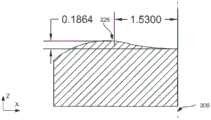
(a)在包含光学透明构件的纵轴的平面中定义的弯曲前表面的横截面剖面的凹陷为大约186微米;以及(a) the depression of the cross-sectional profile of the curved front surface defined in the plane containing the longitudinal axis of the optically transparent member is about 186 microns; and
(b)在包含纵轴的平面中定义的弯曲前表面的径向剖面的最大值与纵轴之间的径向距离大约为1.53mm。(b) The radial distance between the maximum value of the radial profile of the curved front surface defined in the plane containing the longitudinal axis and the longitudinal axis is approximately 1.53 mm.
在任何实施例中,弯曲的前表面的维度可以被设置为包括周缘部分,该周缘部分在周向环绕轴向部分,同时与该轴向部分相切地合并。在这种情况下,可以至少满足以下条件之一:(i)轴向部分的曲率的符号与外围部分的曲率的符号彼此相反,以及ii)轴向部分的曲率的符号与壳的部分的曲率的符号相同。在任何实施方式中,用于测量的方法可以附加地包括以下步骤:在挤压过程中并且由于存在流体层而在壳的该部分与弯曲的前表面之间形成毛细力,所述毛细力当在壳的该部分与弯曲的表面之间的所述流体层的弯月面取平均值时不超过约0.0024N。可替代地或附加地,在任何实施例中,该方法可以包括以下步骤:在挤压过程中并且由于存在所述流体层而在壳的该部分与弯曲的前表面之间形成第一毛细力,所述第一毛细力比在相同测量条件下由于层的存在而将另一个光学透明构件的平面前表面压靠在壳的相同部分上时由于在平面前表面和壳的该部分之间存在液体层而形成的第二力小大约百分之30至大约百分之45。在光学系统中,弯曲的前表面可以响应于纵轴而旋转对称。In any embodiment, the curved front surface may be dimensioned to include a peripheral portion circumferentially surrounding the axial portion while merging tangentially with the axial portion. In this case, at least one of the following conditions may be satisfied: (i) the sign of the curvature of the axial portion and the sign of the curvature of the peripheral portion are opposite to each other, and ii) the sign of the curvature of the axial portion and the curvature of the portion of the shell symbols are the same. In any embodiment, the method for measuring may additionally include the step of creating a capillary force between the portion of the shell and the curved front surface during extrusion and due to the presence of the fluid layer, the capillary force being The meniscus of the fluid layer between the portion of the shell and the curved surface, when averaged, does not exceed about 0.0024N. Alternatively or additionally, in any embodiment, the method may include the step of creating a first capillary force between the portion of the shell and the curved front surface during extrusion and due to the presence of the fluid layer , said first capillary force ratio is due to the presence of the flat front surface of the other optically transparent member against the same part of the case due to the presence of the layer between the flat front surface and this part of the case under the same measurement conditions. The second force formed by the liquid layer is about 30 percent to about 45 percent less. In an optical system, the curved front surface may be rotationally symmetric in response to the longitudinal axis.
实施例还提供了光学系统对用于减少在眼压测量期间角膜上存在泪膜而引起的误差的用途(其中包括具有纵轴和弯曲的前表面的眼压计尖端的光学系统被配置为在测量期间与眼睛的角膜接触;眼压计尖端被配置为向角膜和从角膜传输光。该测量包括:(i)将弯曲的前表面的轴向部分压在角膜上,以向角膜施加力并在弯曲的前表面和角膜之间定义接触的第一表面区域,其中轴向部分相对于纵轴居中;(ii)由于泪膜的存在,在相等的压力下在角膜和弯曲的前表面之间形成第一毛细力,其中第一毛细力比在同等的测量条件下在将另一种光学透明构件的平面前表面压向角膜期间由于在平面前表面和角膜之间存在泪膜而形成的第二毛细力小大约百分之30至大约百分之45;以及(iii)基于由两次穿过弯曲的前表面并且已经从角膜反射的光形成的光学图像来评估眼压的值。Embodiments also provide the use of an optical system for reducing errors caused by the presence of a tear film on the cornea during tonometry (wherein the optical system comprising a tonometer tip having a longitudinal axis and a curved anterior surface is configured to Contact with the cornea of the eye during the measurement; the tonometer tip is configured to transmit light to and from the cornea. The measurement includes: (i) pressing an axial portion of the curved anterior surface against the cornea to apply a force to the cornea and A first surface area of contact is defined between the curved anterior surface and the cornea, with the axial portion centered with respect to the longitudinal axis; (ii) between the cornea and the curved anterior surface under equal pressure due to the presence of the tear film A first capillary force is formed, wherein the first capillary force is greater than the first capillary force formed due to the presence of a tear film between the planar anterior surface and the cornea during pressing of the planar anterior surface of another optically transparent member against the cornea under equivalent measurement conditions. The capillary force is about 30 percent to about 45 percent less; and (iii) the value of the intraocular pressure is estimated based on an optical image formed by light that has passed through the curved anterior surface twice and has been reflected from the cornea.
挤压步骤可以包括形成第一接触表面区域,同时由于具有弯曲的前表面不是平面的,而使前表面与壳的该部分之间的平均接触角保持在大约20度至大约35度的范围内。在任何实施方式中,轴向部分可以关于纵轴旋转对称;和/或弯曲的前表面可以附加地包括外围部分,该外围部分在周向环绕轴向部分同时与轴向部分切向合并。在后一种情况下,可以满足以下条件中的至少一个:(i)轴向部分的曲率的符号与外围部分的曲率的符号彼此相反,以及(ii)轴向部分的曲率的符号与角膜部分的曲率的符号相同。在任何实施方式中,光学系统的维度可以被设计为满足至少以下条件之一:(a)在包含光学透明构件的纵轴的平面中定义的弯曲前表面的横截面剖面的凹陷为大约186微米;(b)在包含纵轴的平面中定义的弯曲前表面的径向剖面的最大值与纵轴之间的径向距离大约为1.53mm。The extruding step may include forming the first contact surface region while maintaining an average contact angle between the front surface and the portion of the shell in the range of about 20 degrees to about 35 degrees due to the curved front surface being non-planar . In any embodiment, the axial portion may be rotationally symmetric about the longitudinal axis; and/or the curved front surface may additionally include a peripheral portion that circumferentially surrounds the axial portion while merging tangentially with the axial portion. In the latter case, at least one of the following conditions may be satisfied: (i) the sign of the curvature of the axial portion and the sign of the curvature of the peripheral portion are opposite to each other, and (ii) the sign of the curvature of the axial portion is opposite to that of the corneal portion The sign of the curvature is the same. In any embodiment, the dimensions of the optical system may be designed to satisfy at least one of the following conditions: (a) the depression of the cross-sectional profile of the curved front surface defined in the plane containing the longitudinal axis of the optically transparent member is about 186 microns ; (b) The radial distance between the maximum value of the radial profile of the curved front surface defined in the plane containing the longitudinal axis and the longitudinal axis is approximately 1.53 mm.
实施例还提供了一种用于测量作为目标的主体的内部压力的方法,该主体具有封住作为目标的主体的内部体积的可弹性变形的壳。该方法包括以下步骤:i)将第一光学透明构件的前表面的轴向部分压靠在壳的一部分上,该部分在其上承载流体层,以将力施加到壳上并定义前表面与壳之间接触的第一表面区域。(在此,第一光学透明构件具有纵轴并且壳的该部分具有壳轴;并且轴向部分相对于纵向构件轴轴向居中,并且壳的该部分相对于壳轴纵向居中);ii)形成与透射通过第一光学透明构件两次并从壳反射的光接触的第一表面区域的第一光学图像,该第一光学图像包括彼此基本上不相连的第一和第二图像部分;(iii)调整力以达到第一和第二部分的相邻端部接触或重叠的状态。仅当纵向构件轴与壳轴基本重合时才达到这种条件。仅当壳轴和纵向构件轴基本重合时才达到所述条件的结果是由于前表面是弯曲而不是平面的。可替代地或附加地,在任何实施方式中,该方法可附加地包括以下步骤:如果没有达到条件,那么相对于壳的该部分重新对准前表面并重复调整。Embodiments also provide a method for measuring the internal pressure of a target body having an elastically deformable shell enclosing the internal volume of the target body. The method includes the steps of: i) pressing an axial portion of the front surface of the first optically transparent member against a portion of the shell that carries a fluid layer thereon to apply a force to the shell and define the front surface and the shell The first surface area of contact between the shells. (Here, the first optically transparent member has a longitudinal axis and the portion of the shell has a shell axis; and the axial portion is axially centered relative to the longitudinal member axis, and the portion of the shell is longitudinally centered relative to the shell axis); ii) forming a first optical image of the first surface area in contact with light transmitted through the first optically transparent member twice and reflected from the housing, the first optical image comprising first and second image portions substantially unconnected to each other; (iii) ) adjusts the force to achieve a state in which adjacent ends of the first and second portions contact or overlap. This condition is only achieved if the longitudinal member axis is substantially coincident with the shell axis. The consequence of this condition being reached only when the shell axis and the longitudinal member axis are substantially coincident is that the front surface is curved rather than planar. Alternatively or additionally, in any embodiment, the method may additionally include the step of realigning the front surface relative to the portion of the housing and repeating the adjustment if the condition is not met.
挤压步骤可以包括将前表面的轴向部分压靠在壳的该部分上,前表面的轴向部分以第一曲率弯曲,其中第一曲率具有曲率的第一符号。(在这种情况下,曲率的第一符号等于第二符号,第二符号等于壳的该部分的第二弯曲率的符号。)前表面的最大径向剖面与纵向构件轴之间的径向距离可以为大约1.53mm。在任何实施方式中,第一光学透明构件的前表面的轮廓被定义为在包含纵向构件轴的横截面中与纵向构件轴的距离的函数,该轴向部分的特征在于呈凸状,凹陷大约186微米;和/或该方法可以满足以下要求中的至少一个:(a)由于所述调整而可逆地改变接触的第一表面积,以及(b)壳的该部分是光学透明的。The pressing step may include pressing an axial portion of the front surface against the portion of the shell, the axial portion of the front surface being curved with a first curvature, wherein the first curvature has a first sign of curvature. (In this case, the first sign of curvature is equal to the second sign, and the second sign is equal to the sign of the second curvature of that part of the shell.) The radial between the maximum radial section of the front surface and the longitudinal member axis The distance may be approximately 1.53mm. In any embodiment, the profile of the front surface of the first optically transparent member is defined as a function of distance from the longitudinal member axis in a cross-section including the longitudinal member axis, the axial portion being characterized by a convex shape and a concave shape of approximately 186 microns; and/or the method may satisfy at least one of the following requirements: (a) the first surface area of contact is reversibly changed as a result of the adjustment, and (b) the portion of the shell is optically transparent.
在任何实施例中,该方法还可以包括以下步骤:在将前表面的维度设计为弯曲的前表面时,在相邻的端部接触或重叠时的时刻,利用第一光学图像确定内部压力的第一值。(在此,确定的第一值包含小于参考值的第一误差,并且参考值是当通过执行挤压、成形和用具有平面前表面的第二光学透明构件的调整的步骤来测量内部压力时由于壳刚度、壳厚度、壳曲率、纵向构件轴与壳轴之间的未对准以及弯曲前表面与壳的该部分之间存在流体层中的至少一项而造成的第二误差的值。)在后一种情况下,第一误差与参考值之间的差异可以至少部分地由于由流体层产生的毛细力减小多达百分之45而造成,这是由于在弯曲的前表面与壳的表面之间形成接触角,该接触角在大约20度与大约30度之间。(应该认识到的是,在一种情况下,一般可以在(一个或多个)接触点处绘制到构成表面的切线之间定义接触角。)In any embodiment, the method may further comprise the step of using the first optical image to determine the magnitude of the internal pressure at the moment when adjacent ends contact or overlap when the dimensions of the front surface are designed to be curved front surfaces first value. (Here, the determined first value contains a first error smaller than a reference value, and the reference value is when the internal pressure is measured by performing the steps of pressing, forming, and adjusting with the second optically transparent member having a flat front surface The value of the second error due to at least one of shell stiffness, shell thickness, shell curvature, misalignment between longitudinal member axis and shell axis, and presence of a fluid layer between the curved front surface and the portion of the shell. ) in the latter case, the difference between the first error and the reference value may be due at least in part to a reduction in the capillary force generated by the fluid layer by as much as 45 percent due to the difference between the curved front surface and the A contact angle is formed between the surfaces of the shell, the contact angle being between about 20 degrees and about 30 degrees. (It should be recognized that, in one case, the contact angle may generally be defined between tangents drawn at the contact point(s) to the constituent surface.)
可替代地或附加地,第一光学构件的前表面可以包括具有第三曲率的外围区域,该第三曲率具有与第一曲率的符号相反的曲率的第三符号。在这种情况下,外围区域可以被构造为沿着轴向区域周向环绕,同时沿着在前表面中定义的该闭合曲线与轴向区域切线合并。在这种情况的具体实施方式中,主体可以由眼睛表示,壳的一部分可以由角膜表示,并且第一光学透明构件可以通过具有弯曲而不是平面角膜接触表面的眼压计尖端表示。Alternatively or additionally, the front surface of the first optical member may include a peripheral region having a third curvature having a third sign of curvature opposite the sign of the first curvature. In this case, the peripheral region may be configured to encircle circumferentially along the axial region while tangentially merging with the axial region along this closed curve defined in the front surface. In particular embodiments of this case, the body may be represented by the eye, a portion of the shell may be represented by the cornea, and the first optically transparent member may be represented by a tonometer tip having a curved rather than planar corneal contact surface.
实施例附加地提供了一种方法,该方法通过使用具有弯曲前表面的光学透明构件来减小主体内部的内部压力的测量误差(其中,该误差是由存在于主体的表面处的流体层造成的,并且其中主体具有封住主体的内部体积的壳),测量包括:(i)将弯曲的前表面的轴向部分压在承载所述流体层的壳的一部分上以向壳施加力,并在弯曲的前表面与壳的该部分之间定义接触的第一表面区域,同时由于弯曲的前表面不是平面的而将前表面和壳的该部分之间的平均接触角保持在大约20度至大约35度的范围内(在此,轴向部分相对于光学透明构件的纵轴居中),以及(ii)基于在两次穿过弯曲的前表面并且已经从接触的第一表面区域反射的光形成的光学图像来评估内部压力的值。Embodiments additionally provide a method of reducing measurement errors of internal pressure inside the body (wherein the error is caused by a fluid layer present at the surface of the body) by using an optically transparent member having a curved front surface and wherein the body has a shell enclosing the interior volume of the body), the measurement includes: (i) pressing an axial portion of the curved front surface against a portion of the shell carrying the fluid layer to apply a force to the shell, and A first surface area of contact is defined between the curved front surface and the portion of the shell, while maintaining the average contact angle between the front surface and the portion of the shell at approximately 20 degrees to within a range of approximately 35 degrees (where the axial portion is centered relative to the longitudinal axis of the optically transparent member), and (ii) based on light that has passed through the curved front surface twice and has been reflected from the contacted first surface area The optical image is formed to evaluate the value of internal pressure.
作为测量的结果,内部压力的导出值的特征在于大致比使用另一种具有平坦前表面的光学透明构件执行的主体内内部压力测量的对应误差小两倍的误差。可替代地或附加地,该方法包括以下步骤:在挤压过程期间,由于存在所述流体层而在壳的该部分与弯曲的前表面之间形成毛细力,其中(在流体层的弯液面上求平均的)毛细力不超过大约0.0024N。可替代地或附加地,该方法包括以下步骤:在挤压过程期间,由于存在流体层而在壳的该部分与弯曲的前表面之间形成第一毛细力,该第一毛细力比在同等的测量条件下由于在平面前表面与壳的该部分之间存在液体层而在抵靠壳的相同部分挤压另一个光学透明构件的平面前表面期间形成的第二力小大约百分之30至大约百分之45。As a result of the measurement, the derived value of the internal pressure is characterized by an error approximately two times smaller than the corresponding error of the internal pressure measurement within the body performed using another optically transparent member having a flat front surface. Alternatively or additionally, the method includes the step of: during the extrusion process, capillary forces are formed between the portion of the shell and the curved front surface due to the presence of the fluid layer, wherein (in the meniscus of the fluid layer) (averaged on the surface) capillary force does not exceed about 0.0024N. Alternatively or additionally, the method includes the step of: during the extrusion process, a first capillary force is created between the portion of the shell and the curved front surface due to the presence of the fluid layer, the first capillary force being equal to The second force formed during pressing of the planar front surface of the other optically transparent member against the same portion of the housing due to the presence of a liquid layer between the planar front surface and the portion of the housing is approximately 30 percent less under the measurement conditions of to about 45 percent.
附图说明Description of drawings
通过参考以下具体实施方式并结合一般不按比例绘制的附图,将更全面地理解本发明,这些附图仅参考一个典型且选定的压平眼压计的示例来提供对本发明的实施例的配置和用途的图示说明。附图中:A more complete understanding of the invention will be obtained by reference to the following detailed description in conjunction with the accompanying drawings, which are generally not to scale, and which provide embodiments of the invention with reference only to a typical and selected example of an applanation tonometer. Illustrated description of the configuration and use of the . In the attached picture:
图1A呈现了用于测量人眼的IOP的常规形状(平坦表面)Goldmann压平眼压计尖端的两个视图(一个视图的横截面图示出了角度为60度的双棱镜);Figure 1A presents two views of a conventionally shaped (flat surface) Goldmann applanation tonometer tip used to measure the IOP of the human eye (one view in cross-section showing a biprism at an angle of 60 degrees);
图1B是图示采用图1A的尖端或基本上类似于图1A的尖端的尖端结构的相关技术的Goldmann压平眼压计的图;FIG. 1B is a diagram illustrating a related art Goldmann applanation tonometer employing the tip of FIG. 1A or a tip configuration substantially similar to the tip of FIG. 1A ;
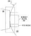
图2A是示意性地图示由于眼压计尖端施加的压力而使主体的壳的弹性表面(在这种情况下-角膜表面)展平的图;Figure 2A is a diagram schematically illustrating the flattening of the elastic surface of the shell of the subject (in this case - the corneal surface) due to the pressure exerted by the tonometer tip;
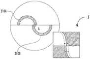
图2B是示出表示角膜表面的展平部分的图像的两个部分(在这个示例中被构造为半圆形)的压力依赖定位的图;2B is a graph showing the pressure-dependent positioning of two portions of an image representing a flattened portion of the corneal surface (configured as a semicircle in this example);
图3A和3B是示意性地图示根据本发明一个实施例的具有一定维度的前表面的光学透明构件的横截面图和俯视图(当在压平眼压计中使用时-眼压计尖端);Figures 3A and 3B are cross-sectional and top views (when used in an applanation tonometer - tonometer tip) schematically illustrating an optically transparent member having a dimensional front surface in accordance with one embodiment of the present invention;
图3C是在顶视图中图示本发明的相关实施例的图;3C is a diagram illustrating a related embodiment of the present invention in a top view;
图3D是图3A和/或3B和/或3C的实施例的附加图示;3D is an additional illustration of the embodiment of FIGS. 3A and/or 3B and/or 3C;
图4是图示将图3A、3B的光学构件用于测量目标主体的内部压力的具体示例的图。在此-眼压的测量;FIG. 4 is a diagram illustrating a specific example in which the optical member of FIGS. 3A, 3B is used to measure the internal pressure of the target body. Here - measurement of intraocular pressure;
图5A和5B是示意性地图示根据本发明另一个实施例的具有一定维度的前表面的光学构件的横截面图和顶视图;5A and 5B are cross-sectional and top views schematically illustrating an optical member having a dimensional front surface according to another embodiment of the present invention;
图6示意性地图示了被构造为眼压计尖端的图5A、5B的实施例的前表面的一部分的具体示例;Figure 6 schematically illustrates a specific example of a portion of the front surface of the embodiment of Figures 5A, 5B configured as a tonometer tip;
图7图示了由具有根据图6的实施例构造的尖端的IOP的测量造成的标准角膜中的von Misses应力;Figure 7 illustrates von Misses stress in a standard cornea resulting from measurement of IOP with a tip constructed in accordance with the embodiment of Figure 6;
图8提供了图示在根据图6的实施例构造的眼压计尖端执行压平规程之前和之后的角膜表面的表面剖面的图解;8 provides a diagram illustrating a surface profile of the corneal surface before and after performing an applanation procedure with a tonometer tip constructed in accordance with the embodiment of FIG. 6;
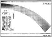
图9A提供了图示在使用平头(平坦表面)眼压计尖端、根据图3A、3B的实施例构造的眼压计尖端以及根据图6的实施例构造的眼压计尖端的IOP测量期间由角膜曲率造成的误差的曲线图;FIG. 9A provides diagrams illustrating IOP measurements made by a tonometer tip using a flat (flat surface) tonometer tip, a tonometer tip constructed in accordance with the embodiment of FIGS. 3A, 3B, and a tonometer tip constructed in accordance with the embodiment of FIG. 6 . A graph of errors due to corneal curvature;
图9B提供了图示由平头眼压计件和图6的实施例在IOP的测量期间由角膜刚度造成的误差的曲线图;Figure 9B provides a graph illustrating the error caused by corneal stiffness during measurement of IOP by the flat head tonometer piece and the embodiment of Figure 6;
图9C提供了图示用平坦表面的眼压计尖端和用根据图6的实施例确定维度的眼压计尖端执行IOP的测量期间由非零角膜厚度造成的误差的曲线图;9C provides a graph illustrating the error caused by non-zero corneal thickness during measurement of IOP with a flat-surfaced tonometer tip and with a tonometer tip dimensioned according to the embodiment of FIG. 6;
图9D呈现了图示在用平坦表面的眼压计尖端和根据图3A、3B、3C的实施例构造的眼压计尖端执行的IOP的测量期间由非零角膜厚度造成的误差的曲线图;9D presents a graph illustrating the error caused by non-zero corneal thickness during measurement of IOP performed with a flat-surfaced tonometer tip and a tonometer tip constructed in accordance with the embodiments of FIGS. 3A, 3B, 3C;
图9E包括图示在用平坦表面的眼压计尖端和根据图3A、3B、3C的实施例构造的眼压计尖端执行的IOP的测量期间由角膜刚性造成的误差的曲线图;9E includes a graph illustrating the error due to corneal stiffness during measurement of IOP performed with a flat-surfaced tonometer tip and a tonometer tip constructed in accordance with the embodiments of FIGS. 3A, 3B, 3C;
图10是示出标准角膜的等压曲线与角膜厚度的函数关系的等高线图;Figure 10 is a contour plot showing the isobaric curve of a standard cornea as a function of corneal thickness;
图11A和11B分别提供了用于图3A和5A的具体实施例的横截面剖面的具体示例;Figures 11A and 11B provide specific examples of cross-sectional profiles for the specific embodiments of Figures 3A and 5A, respectively;
图12是示出与用根据图3A、3B的实施例构造的眼压计尖端压平的角膜中的平均应力以及用根据图5A、5B的实施例构造的眼压计尖端压平的角膜中的平均应力相比用平坦表面眼压计尖端压平的角膜中的平均应力的曲线图;Figure 12 is a graph showing mean stress in the cornea flattened with the tonometer tip constructed in accordance with the embodiment of Figures 3A, 3B and in the cornea flattened with the tonometer tip constructed in accordance with the embodiment of Figures 5A, 5B Plot of the mean stress compared to the mean stress in the cornea flattened with the flat surface tonometer tip;
图13A提供了光学透明构件的实施例的示意性透视图,该光学透明构件具有根据图3A、3B中所示的本发明构思成形的前(压平)表面。在此,示出了被构造成与压平眼压计系统一起使用的光学透明构件;Figure 13A provides a schematic perspective view of an embodiment of an optically transparent member having a front (flattened) surface shaped in accordance with the inventive concepts shown in Figures 3A, 3B. Here, an optically transparent member configured for use with an applanation tonometer system is shown;
图13B示意性地图示了常规的平坦表面(GAT)眼压计尖端的实施例;Figure 13B schematically illustrates an embodiment of a conventional flat surface (GAT) tonometer tip;
图13C示出了表示与本发明的实施例相比而言常规的平坦表面眼压计尖端的横截面剖面的曲线图(明智地调整了本发明的眼压计尖端的尺寸以减小角膜的机械特性以及在测量期间眼部存在的泪膜的静水力影响造成的机械特性对眼压测量引起的误差的贡献);Figure 13C shows a graph representing a cross-sectional profile of a conventional flat surface tonometer tip compared to an embodiment of the invention (the size of the tonometer tip of the invention was judiciously adjusted to reduce the Mechanical properties and their contribution to the error caused by the tonometry measurement due to the hydrostatic influence of the tear film present in the eye during the measurement);
图13D、13E图示了用常规平坦表面的眼压计尖端压平的角膜中的von Mises应力的分布(图13B)以及用根据本发明实施例确定维度的弯曲眼压计尖端压平的角膜中的vonMises应力的分布(图13E),以在结构上支撑角膜组织的中心区段;Figures 13D, 13E illustrate the distribution of von Mises stress in a cornea flattened with a conventional flat-surfaced tonometer tip (Figure 13B) and a cornea flattened with a curved tonometer tip dimensioned according to an embodiment of the present invention The distribution of vonMises stress in (FIG. 13E) to structurally support the central segment of corneal tissue;
图14A、14B、14C提供了在主体的内部压力的测量期间由光学透明构件和主体的壳的一部分之间的流体膜的存在造成的粘附力(毛细力)的形成的示意图。在眼睛的IOP的测量的具体使用过程中,光学透明构件被构造为眼压计尖端,主体的壳是角膜,并且在眼压测量液体膜由眼中的泪膜表示。图14A:与主体的壳接触的光学透明构件(~在一个示例中,与角膜表面接触d眼压计尖端)以及由流体层(~泪膜)形成的粘附力的方向/向量的示意图。图14B:由流体层形成的弯液面以及在主体的壳与光学透明构件(在一个示例中为眼压计尖端)的平坦表面之间的夹角θ的图示。图14C:通过光学透明构件成像的压平部分(称为mires)的图示,证明mires厚度和测量压平端点;Figures 14A, 14B, 14C provide schematic illustrations of the formation of adhesive forces (capillary forces) caused by the presence of a fluid film between the optically transparent member and a portion of the body's shell during measurement of the body's internal pressure. In a specific use of the measurement of IOP of the eye, the optically transparent member is configured as a tonometer tip, the shell of the subject is the cornea, and the tonometric liquid film is represented by the tear film in the eye. Figure 14A: Schematic illustration of the optically transparent member in contact with the shell of the subject (~in one example, the corneal surface in contact with the d-tonometer tip) and the direction/vector of the adhesion force formed by the fluid layer (~tear film). Figure 14B: Illustration of the meniscus formed by the fluid layer and the angle Θ between the shell of the body and the flat surface of the optically transparent member (in one example, the tonometer tip). Figure 14C: Illustration of a flattened portion (referred to as mires) imaged through an optically transparent member, demonstrating mires thickness and measuring flattening endpoints;
图14D是图示由于由光学透明构件和主体的壳的表面之间存在流体层而形成的粘附力造成的测量误差的建模方面的示意图(并且为眼压测量的具体情况贴上标签);Figure 14D is a schematic diagram illustrating modeling aspects of measurement error due to adhesion forces formed by the presence of a fluid layer between the optically transparent member and the surface of the shell of the main body (and labels specific to the tonometry) ;
图14E是表示在压平眼压计的具体情况下光学透明构件的压平表面与被测主体的壳之间形成的粘附力/毛细力与平均接触角(即,跨流体层的弯月面求平均的接触角)的函数关系的曲线图,从而在与使用平坦表面构件和弯曲表面构件对应的力的值之间进行比较(0.003N=3mm Hg);Figure 14E is a graph showing the adhesion/capillary force and the average contact angle (ie, the meniscus across the fluid layer) developed between the applanation surface of the optically transparent member and the shell of the subject under test in the specific case of the applanation tonometer (0.003N=3mm Hg);
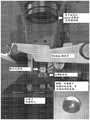
图15:使用Perkins眼压计、压克力半球作为模拟的眼睛的主体(和角膜表面)、标尺和mires成像显微镜进行压平眼压泪膜粘附力测量的设置的图像;Figure 15: Image of the setup for applanation intraocular pressure tear film adhesion measurements using a Perkins tonometer, acrylic hemisphere as a simulated eye's body (and corneal surface), ruler and mires imaging microscope;
图16图示了使用Perkins眼压计、尸体眼仪的压平眼压泪膜粘附力测量设置的图像;Figure 16 illustrates images of the applanation IOP tear film adhesion measurement setup using a Perkins tonometer, a cadaver eye;
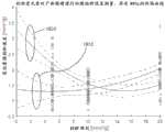
图17包括图示对于使用不同的眼压计棱镜或尖端(CATS,作为下面讨论的当前实施例之一)以及PMMA模拟角膜上的GAT(常规的平坦表面眼压计尖端)执行的眼压计测量的泪膜粘附力误差(mm Hg)的比较的曲线图。在使用人工泪液和荧光素作为泪膜模拟物的情况下,呈现箱须图(box-and-whisker plot),用于测得的膜粘附力。还图示了不同组的平均值之间的差异以及与两个样本均值差异t测试对应的p值;Figure 17 includes diagrams illustrating tonometers performed using different tonometer prisms or tips (CATS, as one of the current embodiments discussed below) and GAT (conventional flat surface tonometer tip) on the PMMA simulated cornea. Graph of comparison of measured tear film adhesion error (mm Hg). Box-and-whisker plots are presented for measured film adhesion using artificial tears and fluorescein as tear film mimics. Also plotted is the difference between the means of the different groups and the p-value corresponding to the t-test of the difference between the two sample means;
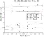
图18通过比较用CATS眼压计尖端获取的数据(曲线1810)与用GAT眼压计尖端获取的数据(曲线1820)来图示尸体眼球泪膜粘附力误差(mm Hg)的依赖性,并通过GLME(广义线性混合效应)以及95%的间隔分析证明曲线拟合;Figure 18 illustrates the dependence of cadaveric eyeball tear film adhesion error (mm Hg) by comparing data acquired with the CATS tonometer tip (curve 1810) to data acquired with the GAT tonometer tip (curve 1820), And the curve fit is proved by GLME (Generalized Linear Mixed Effects) and 95% interval analysis;
图19提供了表示泪膜粘附误差与平坦半月板厚度的关系的图,这些图是使用CATS和GAT眼压计棱镜以及GLME多变量分析来评估的;Figure 19 provides graphs representing tear film adhesion error versus flat meniscus thickness assessed using CATS and GAT tonometer prisms and GLME multivariate analysis;
图20是示出在使用Goldmann压平眼压计测得的IOP值、使用眼压计尖端的CATS实施例测得的IOP值以及使用平坦表面的常规GAT(或Goldmann眼压计)尖端测得的IOP值之间的差异的曲线图;Figure 20 is a graph showing IOP values measured using a Goldmann applanation tonometer, a CATS embodiment using a tonometer tip, and a conventional GAT (or Goldmann tonometer) tip using a flat surface A graph of the difference between the IOP values;
图21是图示在用本发明的实施例执行的IOP测量与使用常规的平坦表面眼压计尖端执行的IOP测量之间的相关性的曲线图;21 is a graph illustrating the correlation between IOP measurements performed with embodiments of the present invention and IOP measurements performed using a conventional flat surface tonometer tip;
一般而言,可以将附图中的元件的尺寸和相对比例设置为与实际尺寸不同,以适当地促进附图的简单、清楚和理解。出于相同的原因,不一定存在于一个附图中的所有元件都必需在另一个附图中示出。In general, the dimensions and relative proportions of elements in the figures may be varied from actual dimensions to appropriately facilitate simplicity, clarity, and understanding of the figures. For the same reason, not all elements that are not necessarily present in one drawing must be shown in another drawing.
具体实施方式Detailed ways
本发明构思的所讨论的实施方式解决了伴随眼内眼压测量的问题,该问题常规是通过使用具有带平坦平面角膜接触表面的尖端的Goldmann型压平眼压计(GAT)来执行的。所提出的实施例的使用通过消除而进一步促进IOP测量的准确性和/或精度,在一些情况下不需要为了角膜厚度和硬度(和/或其它讨论的眼睛的特点)的贡献而校正测量结果的需要,同时将由测量规程经常施加在眼球上的角膜曲率、角膜刚度和眼内应力所造成的IOP测量误差降至最低,但这些误差迄今为止在临床和相关领域都已被忽略。通过采用具有被构造为至少包括i)中心弯曲部分和ii)环绕中心部分并且具有与中心部分的曲率符号相反的符号的曲率的外周部分的角膜接触(一般为轴向对称)的眼压计尖端来获得这种有利效果。眼压计尖端表面的中心和外围部分被构造为沿着闭合平面曲线切向(切线平行)彼此融合。The discussed embodiments of the present inventive concept address the problems associated with intraocular tonometry conventionally performed by using a Goldmann-type applanation tonometer (GAT) with a tip with a flat, planar corneal contact surface. Use of the proposed embodiments further facilitates the accuracy and/or precision of IOP measurements by eliminating, in some cases, no need to correct the measurements for the contribution of corneal thickness and stiffness (and/or other characteristics of the eye in question) while minimizing errors in IOP measurement due to corneal curvature, corneal stiffness, and intraocular stress that measurement protocols often impose on the globe, but which have so far been ignored in the clinical and related fields. By employing a tonometer tip with corneal contact (generally axial symmetry) having a corneal contact (generally axial symmetry) that is configured to include at least i) a central curved portion and ii) a peripheral portion that surrounds the central portion and has a curvature of opposite sign to the curvature of the central portion to obtain this beneficial effect. The central and peripheral portions of the tonometer tip surface are configured to merge with each other tangentially (tangents parallel) along the closed plane curve.
与直觉相反,在本发明的一个实施例中,其中尖端的角膜接触表面的曲率的符号与角膜的曲率的符号具有相同的符号,从而使尖端的平坦表面在几何上匹配并与角膜的表面全等(congruent),这应当避免,因为这种基本完美的几何匹配会造成角膜的压平区域遇到眼压计尖端的零力(在IOP测量区号),从而否定测量本身并使得测量基本上没有用。术语“全等”在参考选定的第一和第二元件使用时指定这些元件在叠加时基本上在所有点上都重合。因而,本发明的方法的实施例可以包括以下步骤:作为调整由眼压计尖端的实施例施加在角膜上的力的结果,可逆地改变(眼压计尖端的实施例的)角膜接触弯曲表面与角膜之间的接触表面的表面积,而角膜接触弯曲表面基本上不与角膜的表面全等(处于其正常的静止状态)。(技术人员将容易地认识到,眼压计尖端的实施例的角膜接触表面之间缺乏完美的全等性和/或几何匹配在结构和功能上与例如基本上隐形眼镜的角膜接触表面与角膜之间完美全等的情况特别不同。确实,在后一种情况下,要求充分的全等性和几何匹配,并且精确地存在,因为在其它情况下,隐形眼镜的预期光学性能被定为光学上的矫正眼睛的不完美视觉是无法执行和/或实现的。顺便说一下,本领域技术人员将容易认识到的是,通过隐形眼镜施加到角膜上的力的改变(例如,由于按压安装在眼睛上的隐形眼镜)不会改变隐形眼镜和角膜的表面积重新物理接触:这保持基本恒定。此外,根据隐形眼镜的角膜接触表面的形状而确定维度的表面与角膜之间的操作接触根本不会在与这种表面对应的点上造成或导致角膜的压平。换句话说,无论隐形眼镜是处于静止状态还是被特定地压在角膜上,隐形眼镜下方的角膜形状都保持基本相同。Counterintuitively, in one embodiment of the present invention, the sign of the curvature of the cornea-contacting surface of the tip has the same sign as the sign of the curvature of the cornea, so that the flat surface of the tip is geometrically matched and fully aligned with the surface of the cornea. Congruent, this should be avoided, as this essentially perfect geometric match would cause the applanation region of the cornea to encounter zero force at the tonometer tip (at the IOP measurement zone number), negating the measurement itself and making the measurement essentially no use. The term "congruent" when used in reference to selected first and second elements specifies that these elements, when superimposed, coincide at substantially all points. Thus, embodiments of the method of the present invention may include the step of reversibly changing the corneal contact curved surface (of the embodiment of the tonometer tip) as a result of adjusting the force exerted on the cornea by the embodiment of the tonometer tip The surface area of the contact surface with the cornea, while the cornea-contacting curved surface is not substantially congruent with the surface of the cornea (in its normal resting state). (The skilled artisan will readily recognize that the lack of perfect congruence and/or geometric matching between the corneal contact surfaces of embodiments of the tonometer tip is structurally and functionally comparable to, for example, substantially contact lens corneal contact surfaces and the cornea The cases of perfect congruence are particularly different between . Indeed, in the latter case, sufficient congruence and geometric matching are required and exist precisely because in other cases the expected optical performance of a contact lens is specified as an optical Correction of imperfect vision on the eye cannot be performed and/or achieved. Incidentally, those skilled in the art will readily recognize that changes in the force applied to the cornea by the contact lens (e.g., due to pressure mounted on the cornea) The contact lens on the eye) does not change the surface area of the contact lens and the cornea to re-physically contact: this remains essentially constant. Furthermore, the operative contact between the surface and the cornea, which is dimensioned according to the shape of the corneal contact surface of the contact lens, does not at all Applanation of the cornea is caused or caused at a point corresponding to such a surface. In other words, the shape of the cornea beneath the contact lens remains substantially the same whether the contact lens is at rest or specifically pressed against the cornea.
反常识地,至少在IOP测量期间在角膜内应力最小化方面(相对于具有带平坦而不是弯曲表面的尖端的眼压计构件的常规设计而言)并且具有明显的实际优势,一个具体实施例的尖端的表面的中心部分的曲率优选地具有与角膜的曲率相反的符号。根据本发明的实施例,公开了用于眼科器械的方法和装置,所述眼科器械包括与GAT平台一起使用的根据本发明的构思构造的角膜接触构件。本发明的实施例包括眼压计尖端,该眼压计尖端包括含双棱镜的部分和角膜接触表面,该角膜接触表面的形状被构造为在眼压测量期间最小化角膜表面的变形和角膜内应力。Contrary to common sense, at least in terms of minimization of intracorneal stress during IOP measurements (relative to conventional designs of tonometer components with tips with flat rather than curved surfaces) and with clear practical advantages, a specific embodiment The curvature of the central portion of the surface of the tip preferably has an opposite sign to the curvature of the cornea. In accordance with embodiments of the present invention, methods and apparatus are disclosed for use with an ophthalmic instrument comprising a corneal contact member constructed in accordance with the concepts of the present invention for use with a GAT platform. Embodiments of the present invention include a tonometer tip that includes a biprism-containing portion and a corneal contact surface that is shaped to minimize deformation of the corneal surface and intracornea during tonometry. stress.
为了本公开和所附权利要求的目的,并且除非另有说明:For the purposes of this disclosure and the appended claims, and unless otherwise stated:
-平面曲线是在平面中定义的曲线。闭合平面曲线是没有端点并且完全封住区域的曲线。优选地,闭合平面曲线被定义在横穿轴的平面中,即,在相对于轴位于或横穿(或在横向方向上)延伸的平面中,并且在具体情况下是在垂直于轴延伸的平面中。当将角膜接触构件的角膜接触表面部分压靠在角膜上时,这增强了角膜变形的均匀性。- Planar curves are curves defined in a plane. A closed plane curve is one that has no endpoints and completely encloses the area. Preferably, a closed plane curve is defined in a plane transverse to the axis, ie in a plane extending in relation to the axis or extending transversely (or in a transverse direction), and in particular in a plane extending perpendicular to the axis in plane. This enhances the uniformity of corneal deformation when the corneal-contacting surface portion of the corneal-contacting member is pressed against the cornea.
-一般而言,光学透明构件的表面被构造为与被测物体的壳接触(在眼压计尖端的具体情况下为角膜接触构件)并且根据本发明的构思来确定维度,其表面不仅以明智的方式偏离平面以降低IOP测量对壳的机械参数(或在具体使用情况下角膜的生物力学参数)和液体层在壳上存在的敏感性(~泪膜,在眼睛是待测主体的具体情况下),而且还包括两个不同弯曲的表面部分,一个是凹表面部分,另一个是凸表面部分。为了本公开和所附权利要求的目的,诸如曲率半径、曲率、曲率的符号之类的术语和相关术语根据相关领域中公认的和常用的数学含义来识别。例如,给定曲线在该曲线的一点处的曲率半径一般被定义为最接近该点处的曲线的圆的半径。术语“曲率”是指曲率半径的倒数。可以扩展曲率的定义以允许曲率采用正值或负值(带有正号或负号的值)。这是通过选择沿着曲线的单位法向向量并为曲线的曲率指定正号(如果曲线向所选择的法线转向)或为负号(如果其远离曲线)而实现的。为了本公开和所附权利要求的目的,根据这样的约定来定义给定曲率的符号。对于这些和其它数学术语的定义,读者还可以参考数学上的标准参考教科书,诸如例如I.N.Bronstein,K.A.Semendyaev,Reference on Mathematics for Engineers andUniversity Students,Science,1981年(或任何其它版本)。在一个示例中,根据光学科学界公认的惯例,如果曲面的顶点位于其曲率中心的左侧,那么曲率半径和曲率本身具有正号;如果顶点位于曲率中心的右侧,那么曲率半径和曲率本身具有负号。- In general, the surface of the optically transparent member is configured to be in contact with the shell of the object to be measured (in the specific case of the tonometer tip, the corneal contact member) and dimensioned according to the concepts of the present invention, its surface is not only sensible off-plane in a manner to reduce the sensitivity of IOP measurements to the mechanical parameters of the shell (or biomechanical parameters of the cornea in specific use cases) and the presence of a liquid layer on the shell (~tear film, in the specific case where the eye is the subject to be measured) bottom), but also includes two differently curved surface portions, one concave surface portion and the other convex surface portion. For the purposes of this disclosure and the appended claims, terms such as radius of curvature, curvature, sign of curvature, and related terms are to be identified in accordance with accepted and commonly used mathematical meanings in the relevant art. For example, the radius of curvature of a given curve at a point of the curve is generally defined as the radius of the circle closest to the curve at that point. The term "curvature" refers to the inverse of the radius of curvature. The definition of curvature can be extended to allow curvature to take positive or negative values (values with a positive or negative sign). This is done by choosing a unit normal vector along the curve and assigning a positive sign (if the curve turns towards the chosen normal) or a negative sign (if it is farther away from the curve) for the curvature of the curve. For the purposes of this disclosure and the appended claims, the sign of a given curvature is defined according to such conventions. For definitions of these and other mathematical terms, the reader is also referred to standard reference texts in mathematics, such as, for example, I.N. Bronstein, K.A. Semendyaev, Reference on Mathematics for Engineers and University Students, Science, 1981 (or any other edition). In one example, according to accepted convention in optical science, the radius of curvature and the curvature itself have a positive sign if the vertex of the surface is to the left of its center of curvature; if the vertex is to the right of the center of curvature, then the radius of curvature and the curvature themselves Has a negative sign.
-根据其技术和科学含义,使用术语“表面”来表示两个介质或有形元件的边界或空间界限之间的边界;可以理解为具有长度和宽度但没有厚度的主体的皮肤(厚度为零)。- In accordance with its technical and scientific meaning, the term "surface" is used to denote the boundary between the boundaries or spatial boundaries of two media or tangible elements; it can be understood as the skin of a subject having length and width but no thickness (thickness zero) .
-术语“压平”、“平坦化”、“展平”和类似术语一般是指这样的过程或动作,其结果是减小受试体的表面曲率,即,该表面被弄平或压平(导致与曲率的初始值相比,其曲率至少减小的表面,和/或在具体情况下,导致表面基本上是平坦的或平面的)。- The terms "flattening", "flattening", "flattening" and similar terms generally refer to the process or action which results in reducing the curvature of the surface of the subject, ie the surface is flattened or flattened (resulting in a surface whose curvature is at least reduced compared to the initial value of the curvature, and/or, in particular cases, resulting in a surface that is substantially flat or planar).
一般考虑事项General Considerations
在此,使用压平眼压计的具体示例,呈现了适用于测量主体内部压力的一般情况的考虑事项。应该理解的是,基于对眼睛IOP测量的具体分析和实验得出的结果和结论随后被使用并归纳为眼压测量法,用于测量在使用光学透明构件时受具有一定弹性的壳限制的主体内部压力,光学透明构件的前表面与主体的壳接触并压在其上。Here, using a specific example of an applanation tonometer, considerations applicable to the general case of measuring pressure inside a subject are presented. It should be understood that the results and conclusions based on the specific analysis and experimentation of ocular IOP measurements were subsequently used and generalized as tonometry for the measurement of subjects confined by a shell with some elasticity when using optically transparent members. Internal pressure, the front surface of the optically transparent member is in contact with and pressed against the shell of the main body.
眼压测量法是眼保健专业人员执行的确定眼内压(即,眼内流体压力)的非侵入性规程。这是评估患有青光眼风险的患者的重要测试,青光眼是一种常常导致患者视力障碍的疾病。在压平眼压计中,根据Imbert-Fick假设,由展平(压平)恒定的预定义角膜区域所需的力来推断眼内压。这个假设认为,当在给定的内部压力下将平坦表面压在闭合的球体上时,施加在球体表面上的力被施加在平坦表面和(现已变形的)球体之间形成的接触区域上的球体内部压力所平衡,从而达到平衡。换句话说,在柔性、弹性(并且可能无限薄)的球体内的压力P大约等于使一部分球体展平所需的外力f并通过展平的面积A进行归一化,P=f/A。因而,将具有平面接触表面的透明压力元件(诸如图1A中所示的元件100,例如;GAT尖端元件)压在眼睛的角膜上,使后者在预定区域(实际上大约为7.3mm2)上展平。Tonometry is a non-invasive procedure performed by eye care professionals to determine intraocular pressure (ie, intraocular fluid pressure). This is an important test for evaluating patients at risk for glaucoma, a disease that often causes vision impairment in patients. In applanation tonometers, intraocular pressure is inferred from the force required to flatten (applanate) a constant, predefined corneal area according to the Imbert-Fick hypothesis. This hypothesis holds that when a flat surface is pressed against a closed sphere at a given internal pressure, the force exerted on the surface of the sphere is exerted on the area of contact formed between the flat surface and the (now deformed) sphere The internal pressure of the sphere is balanced to achieve equilibrium. In other words, the pressure P within a flexible, elastic (and potentially infinitely thin) sphere is approximately equal to the external force f required to flatten a portion of the sphere and normalized by the flattened area A, P=f/A. Thus, a transparent pressure element with a planar contact surface (such as
用等式(1)表示的Imbert-Fick原理指出,眼睛的反作用力F是IOP P的线性函数。(基于Imbert-Fick原理,所施加的力到压力的转换常规上假设IOP唯一且完全负责压平角膜所需的力。)反作用力还取决于使角膜组织T变形所需的力和眼压计表面的横截面接触面积A。在这项研究中,正常IOP P0被认为是16.0mm Hg。The Imbert-Fick principle expressed in equation (1) states that the reaction force F of the eye is a linear function of IOP P. (The conversion of applied force to pressure, based on the Imbert-Fick principle, conventionally assumes that the IOP is solely and entirely responsible for the force required to flatten the cornea.) The reaction force also depends on the force required to deform the corneal tissue T and the tonometer The cross-sectional contact area A of the surface. In this study, the normal IOP P0 was considered to be 16.0 mm Hg.
F(P)=T(δ)+PA(δ) (1)F(P)=T(δ)+PA(δ) (1)
接触面积是由于由眼压计尖端施加的压力而引起的沿着眼压计尖端的轴的角膜位移深度δ的函数。在这项研究中,建模的角膜的球形半径为7.800mm,眼压计尖端的圆柱半径为1.53mm。这导致最大位移为0.147mm,最大接触面积为7.354mm2。等式(2)中示出了作为角膜的球形半径R和深度位移δ的函数的接触面积A的计算。The contact area is a function of the depth of corneal displacement δ along the axis of the tonometer tip due to the pressure exerted by the tonometer tip. In this study, the spherical radius of the modeled cornea was 7.800 mm, and the cylindrical radius of the tonometer tip was 1.53 mm. This results in a maximum displacement of 0.147mm and a maximum contact area of 7.354mm2 . The calculation of the contact area A as a function of the spherical radius R of the cornea and the depth displacement δ is shown in equation (2).
A(δ)=π(2Rδ+δ2) (2)A(δ)=π(2Rδ+δ2 ) (2)
在Goldmann压平眼压计中,测得的IOP PGAT(使用平坦眼压计尖端测得的IOP)是反作用力的线性函数。测得的IOP值还取决于校准反作用力F(P),该值与正常角膜F550(P0)进行比较,其中550表示标称中心角膜厚度为550μm,而P0是标称IOP。这在等式(3)中示出。In the Goldmann applanation tonometer, the measured IOP PGAT (the IOP measured with a flat tonometer tip) is a linear function of the reaction force. The measured IOP value is also dependent on the calibrated reaction force F(P), which is compared to the normal cornea F550 (P0) , where550 represents a nominal central corneal thickness of 550 [mu]m andP0 is the nominal IOP. This is shown in equation (3).

为了评估使用曲面表面眼压计尖端执行的IOP测量的实质,在Autodesk InventorLT 2015中设计了虚拟模型,并在Autodesk Simulation Mechanical 2015(San Rafael,CA)对虚拟模型进行了模拟。执行几次模拟以确定IOP测量对角膜的各种特性的敏感性,其中IOP测量用配备有具有弯曲的角膜接触表面的尖端的实施例的眼压计执行。这些特性包括至少角膜刚度(杨氏模量)、中心角膜厚度(CCT)、中心角膜曲率(CCC)和泪膜的存在。所有这些都进行了模拟,以便与这个领域其它研究的结果相比较。To evaluate the nature of IOP measurements performed using a curved surface tonometer tip, a virtual model was designed in Autodesk InventorLT 2015 and simulated in Autodesk Simulation Mechanical 2015 (San Rafael, CA). Several simulations were performed to determine the sensitivity of IOP measurements to various properties of the cornea, performed with a tonometer equipped with an embodiment of a tip with a curved corneal contact surface. These properties include at least corneal stiffness (Young's modulus), central corneal thickness (CCT), central corneal curvature (CCC) and the presence of tear film. All of these were simulated for comparison with results from other studies in this area.
在实践中执行IOP测量之前,并且因为压力构件(~眼压计尖端)与角膜接触,所以通常将局部麻醉剂(诸如代美卡因)引入眼睛表面(例如,以眼药水的形式)。在测量期间,眼睛会被蓝光(例如,从配备有蓝光滤光镜的灯发出的光)照亮。作为接触的结果,在角膜表面和压力构件之间的接触区中,泪膜(含有荧光素,并且当被蓝光照射时具有绿黄色调)被移位,因此角膜的展平区域和弯曲区域之间的边界很容易识别。展平所需的接触压力用作眼压的测量。Prior to performing IOP measurements in practice, and because the pressure member (~tonometer tip) is in contact with the cornea, a local anesthetic such as demicaine is typically introduced to the surface of the eye (eg, in the form of eye drops). During the measurement, the eye is illuminated by blue light (eg, light from a lamp equipped with a blue light filter). As a result of the contact, in the contact area between the corneal surface and the pressure member, the tear film (which contains fluorescein and has a greenish-yellow tint when illuminated by blue light) is displaced so that the difference between the flattened and curved areas of the cornea is The boundaries between are easy to identify. The contact pressure required for flattening was used as a measure of intraocular pressure.
经典的Goldmann眼压计(参见图1B中的示例114)具有透明塑料压平式GAT尖端100,其形状为具有平坦表面的截头圆锥体,该平坦表面在眼压计的操作中与角膜接触。用裂隙灯显微镜通过塑料压平尖端观察角膜120的表面。设备114是在利用角膜120的压平的眼压测量的当前实践中最广泛使用的眼压计的版本。尖端100(也可互换地称为压力构件或角膜接触构件)通常包含双棱镜(两个棱镜在其顶点处接触的组合),参考图2A,该双棱镜产生角膜120、220的展平表面202的图像的光学倍增并且将两个半圆形图像分量(mires)210A、210B在视场上分开固定的距离或空间。这样的距离或间隔取决于棱镜的顶角。进一步参考图1B,Goldmann眼压计角膜接触构件或尖端100通过杠杆臂或杆连接到眼压计主体116。眼压计主体116包含可以变化的重量。A classic Goldmann tonometer (see example 114 in Figure IB) has a clear plastic
观察者-检查者使用光学滤光片(通常是钴蓝色滤光片)来观察在通过压平眼压计(在这种情况下是尖端100)传播的光中形成的两个mires(在图2B中示为半圆210A、210图像分量B)。然后使用连接到设备的可变张力弹簧的标尺盘(旋钮)来调整通过眼压计尖端100沿着尖端100的轴224施加到角膜120、220的表面220的力F,直到在取景器中观察到的半圆210A、210B的边缘相遇或重合(参见图2B的插图I)。当直径约为3.06mm的角膜区域已经展平并且当两个相反的反作用力(第一反作用力由刚性角膜的阻力产生,第二反作用力由泪膜的张力产生)变得基本相等且彼此抵消时,否则在空间上彼此不同并与图像的另一个部分(mires)分开的这种“边缘相遇”(即,接触和/或重叠)发生,从而允许由施加在角膜上的力来确定眼睛中的压力。值得注意的是,如熟练的技术人员将容易认识到的那样,由于通过平坦表面尖端100的平坦角膜接触表面形成图像的光学器件,形成mires并且mires的边缘的相遇(即,彼此相邻的端部的接触和/或重叠)不论眼压计尖端100的平坦表面是否相对于角膜的轴居中(共入射)都可以在图像中(如图2B的插图I中所示)实现。这种确定眼压的非侵入性方法本质上是不精确的。Observer - The examiner uses an optical filter (usually a cobalt blue filter) to observe the two mires (in the Shown in Figure 2B are semicircles 210A, 210 image component B). The force F applied by the
主体内部压力的测量误差源的示例。An example of a source of measurement error for the internal pressure of a body.
使用压平眼压计仅出于说明的目的,压平眼压计的理论假设角膜是无限薄的膜。角膜的刚度受角膜厚度和角膜曲率的几何特性的显著影响。角膜的可变材料特性(诸如杨氏模量和剪切弹性模量)均会显著影响角膜的压平力。Applanation tonometers are used for illustrative purposes only, the theory of applanation tonometers assumes that the cornea is an infinitely thin membrane. The stiffness of the cornea is significantly affected by the geometric properties of corneal thickness and corneal curvature. Variable material properties of the cornea, such as Young's modulus and shear modulus of elasticity, can significantly affect the applanation force of the cornea.
特别地,在使用GAT形(平坦表面)尖端执行的测量期间,由于角膜(与理想球体不同)具有非零厚度的事实而导致一些误差:比平均角膜更薄通常会造成IOP的低估,而比平均角膜更厚会造成实际IOP的高估。为了平衡角膜的非零刚度并压平角膜的一部分,需要附加的力,该力无法为确定IOP的实际值而简单地计算或考虑。研究表明,角膜厚度与角膜硬度之间存在相关性。显然,那么至少角膜的非零厚度和刚度会给IOP的测量带来误差。因而,为了减小-IOP-测量误差,在这种情况下,必须基于对角膜厚度的第二次测量来校正最初测量的施加到角膜上的力的值(后一测量是使用测厚仪执行的)。这种校正的准确性取决于角膜厚度与硬度特点之间的相关性的准确性,而这固有地也是不准确的(由于诸如人的年龄、角膜直径、角膜曲率、以及各种眼疾产生的影响之类的变化因素的影响)。In particular, during measurements performed with GAT-shaped (flat surface) tips, some errors are introduced due to the fact that the cornea (unlike an ideal sphere) has a non-zero thickness: thinner than average corneas generally result in an underestimation of IOP, whereas A thicker average cornea would result in an overestimation of actual IOP. To balance the non-zero stiffness of the cornea and flatten a portion of the cornea, additional forces are required that cannot be simply calculated or considered for determining the actual value of IOP. Studies have shown that there is a correlation between corneal thickness and corneal stiffness. Obviously, then at least the non-zero thickness and stiffness of the cornea will introduce errors in the measurement of IOP. Thus, in order to reduce the -IOP- measurement error, in this case the initially measured value of the force applied to the cornea must be corrected based on the second measurement of the corneal thickness (the latter measurement is performed using a pachymeter of). The accuracy of this correction depends on the accuracy of the correlation between corneal thickness and stiffness characteristics, which is inherently inaccurate (due to effects such as age, corneal diameter, corneal curvature, and various eye diseases) influences such as changing factors).
导致测量误差的附加原因(本领域迄今尚未解决)是非零角膜曲率的影响。从理论上讲,角膜曲率对眼压测量准确性的影响可以用角膜面积展平之后位移的眼液体积的差异和/或原始体积的差异来解释。(Liu和Roberts,Influence of corneal biomechanicalproperties on intraocular pressure measurement,J.Cataract Refract.Surg.,第31卷,第146-155页,2005年1月)。角膜曲率的影响与眼内压无关,但显示了从眼球传递到与其接触的眼压计尖端的力的重要分量。An additional cause of measurement error, hitherto unsolved in the art, is the effect of non-zero corneal curvature. Theoretically, the effect of corneal curvature on the accuracy of IOP measurements could be explained by differences in the displaced ocular fluid volume after flattening of the corneal area and/or by differences in the original volume. (Liu and Roberts, Influence of corneal biomechanical properties on intraocular pressure measurement, J. Cataract Refract. Surg., Vol. 31, pp. 146-155, January 2005). The effect of corneal curvature was independent of intraocular pressure, but showed a significant component of the force transmitted from the eyeball to the tonometer tip in contact with it.
此外,通过使否则不平坦的角膜的一部分展平的事实,使常规的平坦尖端眼压计(GAT)棱镜与之接触,测量IOP的常规“角膜压平”规程在角膜表面产生某种空间“扭结”。这种“扭结”表现出其中部分压平的角膜的曲率以非常高的速率改变的角膜区域。可以理解,这种“扭结”区域位于角膜的压平部分的周界附近并且定义这种压平部分和角膜的仍然弯曲的部分之间的空间过渡,该弯曲部分与眼压计的平坦尖端不接触。换句话说,在“扭结”区域,表示部分压平的角膜形状的函数的二阶导数的值非常高,并且角膜明显变形,这导致角膜内应力(造成力和压力的附加分量施加到眼压计尖端上;这种分量与IOP不相关并且会给其测量增加误差)。In addition, the conventional "applanation" protocol for measuring IOP creates some kind of space on the corneal surface by the fact that it flattens a portion of the otherwise uneven cornea with which a conventional flat-tip tonometer (GAT) prism is brought into contact." kink". This "kink" represents an area of the cornea in which the curvature of the partially flattened cornea changes at a very high rate. It will be appreciated that this "kink" region is located near the perimeter of the flattened portion of the cornea and defines the spatial transition between this flattened portion and the still curved portion of the cornea that is not in contact with the flat tip of the tonometer. touch. In other words, in the "kink" region, the value of the second derivative of the function representing the shape of the partially flattened cornea is very high, and the cornea is significantly deformed, which results in intracorneal stress (causing an additional component of force and pressure to be applied to the IOP) on the tip of the meter; this component is not correlated with the IOP and will add error to its measurement).
(类似地,如本领域技术人员将容易认识到的,受某种程度弹性的壳限制的眼压测量法的结果(如主体(无论是乳房植入物还是燃料囊或任何其它被测主体)中内部压力的测量)必然会因壳曲率、壳厚度、壳弹性或刚度而产生误差。)(Similarly, as those skilled in the art will readily recognize, the results of tonometry (such as subjects (whether breast implants or fuel sacs or any other subject being measured) that are limited by a somewhat elastic shell) (measurement of internal pressure in the middle) is bound to have errors due to shell curvature, shell thickness, shell elasticity or stiffness.)
而且,在测量期间由泪膜的静水表面张力在眼压计尖端和眼睛表面之间产生的粘附力也为测量结果增加了一些高度可变的误差,该误差是由角膜和与角膜接触的眼压计尖端之间的液桥施加的毛细管压力造成的。但是,迄今为止,由于本发明人的知识,尚未证明或考虑由这种有吸引力的毛细力引起的IOP误差的临床量化(容易理解,通过使用如果并且当存在于与光学测量系统的光学透明部件接触的主体的壳表面上时,利用类眼压测量方法的主体的内部压力的测量遭受并容易产生由水分/液体层造成的误差。)Also, the adhesion force between the tonometer tip and the eye surface caused by the hydrostatic surface tension of the tear film during the measurement also adds some highly variable error to the measurement, which is caused by the cornea and the eye contacting the cornea. Caused by capillary pressure exerted by the liquid bridge between the manometer tips. However, to date, due to the knowledge of the inventors, clinical quantification of IOP errors caused by such attractive capillary forces has not been demonstrated or considered (easy to understand, by using if and when there is an optical transparency with the optical measurement system The measurement of the internal pressure of the subject using the tonometry-like method suffers from and is prone to errors caused by the moisture/liquid layer when the parts are in contact with the shell surface of the subject.)
值得注意的是,到目前为止,就眼压测量而言,还没有关于角膜生物力学特性和相关因素的量值的结论性和一致的数据。错误的IOP读数-所需的校正的确切量始终不确定-会增加发生误诊的风险,从而导致眼科疾病的漏检或误诊。因此,需要增加IOP结果的精度和准确性的测量技术和系统。本发明的实施例的使用实现了这个目标-它提高了IOP的测量准确性(例如,通过使用通用的Goldmann压平眼压计系统来执行),从而减少或甚至消除了对例如以常规方式对角膜厚度执行辅助/补充测量的需求,从而降低了IOP测量的总体成本,并提高了护理质量。而且,本发明的实施例的使用最小化了角膜曲率对IOP测量规程的贡献以及由这种规程在眼睛上引起的眼内应力。It is worth noting that, to date, there are no conclusive and consistent data on the magnitude of corneal biomechanical properties and associated factors in terms of IOP measurements. Inaccurate IOP readings - the exact amount of correction needed is always uncertain - increases the risk of misdiagnosis, leading to missed or misdiagnosed eye diseases. Accordingly, there is a need for measurement techniques and systems that increase the precision and accuracy of IOP results. The use of embodiments of the present invention achieves this goal - it improves the measurement accuracy of IOP (eg, performed by using the common Goldmann applanation tonometer system), thereby reducing or even eliminating the need for The need to perform ancillary/supplemental measurements of corneal thickness, thereby reducing the overall cost of IOP measurements and improving the quality of care. Furthermore, the use of embodiments of the present invention minimizes the contribution of corneal curvature to the IOP measurement protocol and the intraocular stress on the eye caused by such a protocol.
参考本发明的具体实施例(被配置用于眼压测量的那些实施例),下面讨论的校正压平眼压测量表面(CATS)眼压计尖端棱镜的此类实施例被设计为并且意图在眼压计系统的操作中替代常规的平坦表面GAT眼压计棱镜。照此,包括力到压力转换的CATS棱镜的临床用途与GAT棱镜的临床用途相同。下面讨论的CATS棱镜和相关联的测量方法的实施例被配置为执行与对于“标称”角膜用GAT棱镜测得的压力相同的压力的测量。“标称”角膜被定义为具有平均角膜厚度、曲率、刚度和泪膜的角膜,并且一般以大约7.8mm的曲率半径(在轴向点处)为特征;中心角膜厚度大约为550微米;宽度大约为11mm;p值为0.82-这是椭圆偏心率的测量;并且平均角膜弹性模量从大约0.5MPa到大约1.5MPa。With reference to specific embodiments of the present invention (those configured for tonometry), such embodiments of the Corrected Applanation Tonometry Surface (CATS) tonometer tip prism discussed below are designed and intended to be used in The operation of the tonometer system replaces the conventional flat surface GAT tonometer prism. As such, the clinical use of CATS prisms including force to pressure conversion is the same as that of GAT prisms. Embodiments of the CATS prism and associated measurement method discussed below are configured to perform the same pressure measurements as measured with the GAT prism for the "nominal" cornea. A "nominal" cornea is defined as a cornea with mean corneal thickness, curvature, stiffness, and tear film, and is generally characterized by a radius of curvature (at the axial point) of approximately 7.8 mm; central corneal thickness is approximately 550 microns; width is about 11 mm; the p-value is 0.82 - this is a measure of elliptical eccentricity; and the mean corneal elastic modulus is from about 0.5 MPa to about 1.5 MPa.
但是,众所周知的是,约50%的患者人群没有“标称”角膜。下文讨论的CATS眼压计棱镜被设计为显著减少由于患者中与角膜相关的参数的变化性而在GAT IOP测量期间存在的所有已识别出的误差。CATS棱镜的实施例的所有物理特点、材料和特性与典型的GAT棱镜基本相似或甚至完全相同(除了与棱镜的压平表面的几何形状相关的例外之外),并且CATS眼压计棱镜的实践用途打算用于整个GAT或Perkins测量支架中并且由相同的从业者协议和测量技术控制,而无需计算或增加诊所时间。However, it is known that about 50% of the patient population does not have a "nominal" cornea. The CATS tonometer prisms discussed below are designed to significantly reduce all identified errors that exist during GAT IOP measurements due to variability in corneal-related parameters among patients. All physical features, materials and properties of embodiments of CATS prisms are substantially similar or even identical to typical GAT prisms (with exceptions related to the geometry of the applanation surfaces of the prisms), and the practice of CATS tonometer prisms Use is intended to be used throughout the GAT or Perkins measurement stand and is controlled by the same practitioner protocol and measurement technique, without the need to calculate or increase clinic time.
值得注意的是,在设计本发明实施例的角膜接触表面的形状时,使用有限元方法(FEM)建模。Notably, finite element method (FEM) modeling was used in designing the shape of the corneal contact surface of embodiments of the present invention.
下面,并参考图3A、3B、3C和5A、5B,讨论根据本发明的构思弯曲成形的眼压计尖端的非限制性具体示例。在这样的讨论期间,即使仅参考图3A、3B的实施例,也应理解,类似的考虑同样适用于图5A、5B的实施例。In the following, and with reference to Figures 3A, 3B, 3C and 5A, 5B, non-limiting specific examples of tonometer tips curved in accordance with the concepts of the present invention are discussed. During such discussion, even if only reference is made to the embodiment of Figures 3A, 3B, it should be understood that similar considerations apply equally to the embodiment of Figures 5A, 5B.
在实验核实期间,使用压平眼压计系统(例如,Goldmann压平眼压计系统)执行了实际的IOP测量,以通过压平角膜以获得指定的展平表面积来估计IOP的值。During experimental verification, actual IOP measurements were performed using an applanation tonometer system (eg, Goldmann applanation tonometer system) to estimate the value of IOP by applanating the cornea to obtain a specified flattened surface area.
在建模期间,对角膜组织的物理行为做出了适当的假设。角膜的组织是具有复杂解剖结构和结构特性的细胞的集合。在模拟中,组织被分析为具有不均匀材料特性的连续体。为了本研究的目的,假设模型具有三个可变的材料特性:(1)角膜基板弹性模量,(2)胶原弹性模量和(3)相对胶原厚度。这些材料被指派给虚拟组件中的特定物理实体,并进行优化以匹配实际行为。During modeling, appropriate assumptions were made about the physical behavior of the corneal tissue. The tissue of the cornea is a collection of cells with complex anatomical and structural properties. In the simulation, the tissue is analyzed as a continuum with inhomogeneous material properties. For the purposes of this study, the model was assumed to have three variable material properties: (1) corneal placode elastic modulus, (2) collagen elastic modulus and (3) relative collagen thickness. These materials are assigned to specific physical entities in virtual components and optimized to match actual behavior.
基于先前研究的结果选择几何形状和本构模型。材料特性是经由有限元模拟分析确定的。在先前的研究中测量并研究了角膜的各种几何形状的影响。由于公开的角膜材料特性差异很大,因此选择具体特性来近似对GAT诊断的已知反应。压平标称角膜所需的力设置在1.6g附近。在正常情况下,仅允许角膜贡献这个压平力的30%,其余部分来自眼内压。设置有限元网格密度,以使压平区域的周长精确到30μm以内,但测量公差不精细到0.1g。Geometry and constitutive models were chosen based on the results of previous studies. Material properties were determined via finite element simulation analysis. The effects of various geometries of the cornea were measured and investigated in previous studies. Because the published properties of corneal materials vary widely, specific properties were chosen to approximate known responses to a GAT diagnosis. The force required to flatten the nominal cornea was set around 1.6 g. Under normal conditions, only the cornea is allowed to contribute 30% of this applanation force, with the remainder from intraocular pressure. The finite element mesh density is set so that the perimeter of the flattened region is accurate to within 30 μm, but the measurement tolerance is not fine to 0.1 g.
示例I。Example I.
如图3A和3B中所示,表示例如设计为与眼睛的角膜接触的光学元件(称为角膜接触构件)的实施例的尖端的相关部分300分别以局部横截面图和正视图示出。这个实施例可互换地称为CATS眼压计尖端或CATS眼压计棱镜。角膜接触表面304包括中心凹入表面部分304A,在一个具体的实施方式中,该中心凹入表面部分304A适于典型眼睛的角膜的曲率(其半径大约在7.8mm+/-0.38mm的范围内;在本申请的其它地方讨论了典型眼睛角膜的典型弹性模量和角膜厚度范围)。As shown in Figures 3A and 3B, a
在角膜接触表面304的外围,中心凹入表面部分304A切向平行地进入并与具有相反符号(与中心表面部分304A的符号相比)的曲率的外围表面部分304B合并。如图3A的示例的横截面图中所示,表面部分304B可以表征为凸的。外围表面部分304B可以定义沿着轴306并且在横穿轴306的平面上的环形(并且在具体描绘的情况下为圆环形)投影,并且形成圆环,围绕中心部分304A的环。中心凹表面部分304A和外围圆环形部分304B相切地(以切线-平行的方式)并且沿着在与表面304相切并且横穿轴306延伸的平面中定义的闭合曲线310彼此无缝地合并。换句话说,第一平面(在表面部分304A、304B之间的边界310的一点处与中心表面部分304A相切)和第二平面(在由表面部分304A、304B共享的边界310的同一点上与外围表面部分304B相切)彼此重合并且不形成二面角。表面304在沿着曲线310的任何且每个点处的曲率基本上为零。表面304A和表面304B在这两个表面合并的任何和每个点处具有相等的空间斜率值。总体上,术语“切向”、“切向-平行”以及与给定表面和/或线的相互空间朝向相关的相似术语是根据这些术语常规的、众所周知的数学含义来定义的。At the periphery of the
在操作中,中心凹表面部分304A可以与角膜表面220接触,以在包含在由包含外围部分304B的最大值点的曲线所定义的边界内的区域上使角膜表面压平。如本领域技术人员将容易认识到的,在不形成角膜上的空间扭结的情况下,可被压平的角膜表面部分的最大延伸(由于在IOP的测量期间将表面304压靠在角膜上)由位于表面304中并且包括表面304的多个顶点(诸如顶点i和ii,如图3A中所示)的基本上轴向对称的曲线定义。(可以理解的是,通过使用常规的平坦表面的眼压计尖端100可将角膜表面压平的程度没有这种限制,并且角膜表面的可用平坦表面的眼压计尖端压平的部分基本上与这种尖端的平坦角膜接触表面一样大。)一般而言,不要求眼压计尖端沿着表面304的横向边界或周长320的满足任何特定的光学、机械或几何要求,因为这种边界在与角膜的接触区域之外。In operation, the
虽然设备300的前表面304的周界曲线320以及中心弯曲表面部分304A和外围弯曲表面部分304B沿着其合并的闭合曲线310均显示为圆形,但是应该认识到的是,可以将表面304配置为诸如将这些曲线310、320中的至少一条定义为普通椭圆(由从其到两个给定点的距离之和是恒定的点的轨迹定义)。但是,在具体情况下,表面304关于轴306旋转对称。图3A和3B的示例仅示出了这种旋转对称表面304。Although the
在一种实施方式中,并且进一步参考图3A、3B,凹表面部分304A包括具有例如大约-9.0mm的曲率半径R的球形表面(在包含轴306的平面中定义),以及沿着轴306的足迹或法线投影,其直径d为例如大约3.06mm(在与轴306垂直的平面中定义)。外围圆环形(即,具有环的形式)表面部分304B具有例如大约3.0mm的曲率半径(在包含轴306的平面中定义)。在这样的实施方式中,角膜接触表面304在垂直于轴306的平面上的覆盖区或投影定义了直径D为例如大约6.0mm的圆圈。角膜接触表面304可以由聚合材料(例如,聚碳酸酯,折射率为1.5的量级)或具有光学品质的抛光饰面的玻璃形成。In one embodiment, and with further reference to FIGS. 3A, 3B , the
分别由图13B和13A提供了常规平坦眼压计尖端(实施例100)与具有压平表面304的眼压计尖端的透视图的再现结果的比较。A comparison of the reproduced results of the perspective views of a conventional flat tonometer tip (Example 100 ) and a tonometer tip with a flattened
与在用常规平坦表面眼压计尖端进行的压平变形期间(在角膜的相同位置处)产生的角膜内应力相比,通过本发明的(一个或多个)实施例的实施方式实现的弯曲表面304、504的设计的目标是最小化或至少减小在压平变形期间在角膜位置处(在一种情况下-在角膜接触表面304、504之间的接触表面内的位置)形成的内腔内应力。在一个实施例中,这种位置基本上在尖端到角膜接触区域的周界。(与这种眼内应力的减小对应,由于使用表面304、504而在压平变形期间发生的角膜表面的剖面的变化率(二阶导数)低于由于使用常规的平坦表面眼压计尖端而在压平变形期间发生的角膜表面的剖面的变化率。)这转化为相对于产生误差的生物力学参数(诸如角膜厚度、角膜刚度、角膜曲率和泪膜粘附影响)使模拟的IOP的等压曲线展平的目的。在一种具体的实施方式中,CATS尖端表面300的所得剖面由图13C的曲线1310表示。图13D和13E中分别示出了常规平坦尖端表面剖面的Von Mises应力和由曲线1310表示的实施例的Von Mises应力,如下所示:顶部条表示外表面,底部条表示内表面,并且中心形状表示矢状横截面,表明当使用本发明的实施例时,应力系数显著降低-高达一个数量级或更大(包括压平区域的周界,用箭头P示意性地指示的位置)。具有横截面剖面1310的实施例300产生(测量)的对中心角膜厚度的大约5.0mm Hg/mm的敏感性,与当使用常规平坦表面尖端100、1320进行测量时存在的敏感性相比,存在的明显改进是大约88.2%。本发明的实施例300、1310的弯曲表面通过在结构上支撑组织的中心部分而降低了测量对角膜厚度的敏感性,从而使得角膜中的应力在测量期间更均匀地分布。(比较图13D、13E中呈现的结果)。Bending achieved by embodiments of the embodiment(s) of the present invention compared to intracorneal stresses generated during applanation deformation (at the same location of the cornea) with a conventional flat surface tonometer tip The goal of the design of the
下面讨论使用曲线形的角膜接触表面300进行眼压测量的附加优点。Additional advantages of using the curved
实施例II。Example II.
在与图3A、3B的实施例300相关的实施例中,与实施例300相比,对角膜接触表面304进行了修改,诸如,使其在不同方向上具有不同的程度,并且一般而言具有非轴对称覆盖区或法线投影。在这种情况下,角膜接触表面的中心凹表面部分在保持基本配合(曲率方向)到角膜表面的同时在两个(在具体情况下-相互垂直)方向上可以具有不相等的范围。因而,外围表面部分在以上述方式保持与中心凹表面部分邻接的同时还具有横向范围的比率,该比率与表征中心凹部分的比率相似或甚至相等。In an embodiment related to the
在图3C中的顶视图中所示的具体示例中,如此构造的角膜接触表面350在垂直于z轴的平面上具有由椭圆或卵形定义的足迹352。表面350包括基本上球形的中心表面部分354A和外围圆环形部分354B,每个部分在垂直于轴306(在图3C中平行于指示的本地坐标系的z轴)的平面上具有椭圆形的对应投影。如图所示,中心表面部分354A沿着对应覆盖区的短轴和长轴的尺寸分别是a和b。外围表面部分354B沿着其足迹的相应的短轴和长轴的最大尺寸分别是A和B并由周界320'指示。表面部分354A、354B以类似于参考图3A和3B描述的方式沿着椭圆形闭合平面曲线310'切向、无缝地彼此合并。在这个具体示例中,角膜接触表面是轴向对称的。在一个实施方式中,a约为2.13mm,b约为3.06mm。具有表面350的角膜接触构件内部的双棱柱元件(未示出)可以被定向为诸如以将图3C的覆盖区352的长范围B大致平分。In the particular example shown in the top view in Figure 3C, the corneal contacting surface 350 so constructed has a footprint 352 defined by an ellipse or oval in a plane perpendicular to the z-axis. Surface 350 includes a substantially spherical central surface portion 354A and a peripheral annular portion 354B, each portion having an elliptical shape in a plane perpendicular to axis 306 (parallel to the z-axis of the indicated local coordinate system in FIG. 3C ). corresponding projection. As shown, the dimensions of the central surface portion 354A along the short and long axes of the corresponding footprint are a and b, respectively. The largest dimensions of peripheral surface portion 354B along its respective minor and major axes of its footprint are A and B, respectively, and are indicated by perimeter 320'. Surface portions 354A, 354B merge tangentially and seamlessly into each other along elliptical closed plane curve 310' in a manner similar to that described with reference to Figures 3A and 3B. In this particular example, the corneal contact surface is axially symmetric. In one embodiment, a is about 2.13 mm and b is about 3.06 mm. A biprismal element (not shown) inside the corneal contact member with surface 350 may be oriented, such as to approximately bisect long extent B of footprint 352 of Figure 3C.
图3C中所示的实施方式适于促进对具有睑间特征的患者的IOP的测量,该睑间特征不一定使观察者-检查者能够适应图3A和3B的实施例的对称结构的角膜接触表面。应该认识到的是,当在实践中使用本发明的实施方式(其操作由图3C表示)时,经受压平的角膜的面积基本上与对应于图3B的实施方式相同。与足迹304A的直径相比,与适应狭窄的睑间裂(部分闭合的眼睑)的354A对应的椭圆形足迹的横向维度减小,而足迹(沿着眼睑)的正交维度增大。在一些情况下,可以减小压平所需的力。The embodiment shown in Figure 3C is adapted to facilitate the measurement of IOP in patients with interlid features that do not necessarily enable observer-examiner to accommodate the symmetrical configuration of the corneal contact of the embodiment of Figures 3A and 3B surface. It should be appreciated that when using an embodiment of the present invention in practice, the operation of which is represented by Figure 3C, the area of the cornea subjected to applanation is substantially the same as the embodiment corresponding to Figure 3B. Compared to the diameter of the
一般而言,角膜接触构件300的角膜接触表面被构造为包括方位角对称的双弯曲表面,该表面的横截面被定义为(在包含接触构件300的光轴的平面中)具有第一和第二局部最大值、与这种曲线的对称轴重合的一个最小值的轴向对称单调曲线。这种轴向对称的单调曲线具有在这个曲线的任何点处定义的二阶导数(因此,在该曲线的限制内是完全可微分的)。这种角膜接触表面包括中心凹部分和与中心凹部分外接的外围凸部分。在操作中,角膜接触表面的中心凹部分对与之接触的角膜中心部分产生基本上可忽略的压缩。沿着周界凸部分和中心接触部分彼此邻接的角膜接触表面区域产生轻微的角膜压缩以定义来自半环形式的周界环形图案,以反映来自角膜的光。In general, the corneal contact surface of the
图3D提供了附加说明,示出了CATS眼压计的实施例的表面304、350、352、354与GAT眼压计尖端的表面(由点划线360图示)的空间偏离,并且示出了CATS眼压计尖端的角膜接触表面的“凹陷”364,其以尖端的轴为中心。Figure 3D provides an additional illustration showing the spatial offset of the
实施例III。Example III.
图5A、5B分别以局部横截面图和正视图示意性地描绘了角膜接触构件的尖端的相关实施例500。角膜接触表面504包括中心表面部分504A,该中心表面部分的曲率具有与角膜曲率的符号相反的符号。在角膜接触表面504的外围,中心表面部分504A越过并与具有相反符号的曲率(与中心表面部分504A的曲率相比)的外围表面部分504B合并。如图5A的横截面图中所示,表面部分504A可以表征为凸的。外围凹表面部分504B沿着轴506并在垂直于轴506的平面上定义环形(并且在具体情况下为圆环形)投影。中心凸表面部分504A和外围凹圆环形部分504B沿着在与表面504相切并且横穿轴506的平面中定义的闭合曲线510相切、无缝地彼此融合。换句话说,第一平面(在表面部分504A、504B之间的边界510处与中心表面部分504A相切)和第二平面(在由表面部分504A、504B共享的边界510处与外围表面部分504B相切)彼此重合并且不形成二面角。表面504在沿着曲线510的任何点处的曲率基本上为零。Figures 5A, 5B schematically depict a
在操作中,中心凸表面部分504A与角膜表面220接触。一般而言,不要求沿着表面504的横向边界或周界520的眼压计尖端满足任何特定的光学、机械或几何要求,因为这个边界在与角膜的接触区域之外。In operation, the central
设备500的前表面504的周界曲线520和中心弯曲表面部分504A和外围弯曲表面部分504B沿着其合并的闭合曲线510均示为圆圈,但是应该认识到的是,可以将表面504配置为诸如将这些曲线510、520中的至少一个定义为大体椭圆。但是,在具体情况下,表面504关于轴506旋转对称。图3A和3B的示例仅仅示出了这种旋转对称表面504。The
在一个实施方式中,并且进一步参考图5A、5B的实施例,凸表面部分504A包括具有大约+9.0mm的曲率半径R的球形表面(在包含轴506的平面中限定),以及沿着轴506的足迹或法线投影,直径d大约为3.06mm(在垂直于轴506的平面中定义)。外围圆环形(即,具有环的形式)表面部分504B具有大约3.0mm的曲率半径(在包含轴506的平面中定义)。在这样的实施方式中,角膜接触表面504在垂直于轴506的平面上的覆盖区或投影定义了直径D大约为3.06mm的圆圈。角膜接触表面504可以由聚合材料(例如,聚碳酸酯,折射率为1.5的量级)或具有基本光学质量的抛光涂层的玻璃形成。可以不要求表面504的横向边界或周界520满足任何特定的光学、机械或几何要求,因为它在与角膜的接触区域之外。In one embodiment, and with further reference to the embodiments of FIGS. 5A, 5B , the
在图6的局部横截面图中示意性地示出了具有角膜接触表面504的眼压计尖端的相关实施方式600。如图所示,相对于506定义的半径(圆环形凹部分504B以其到达其最低点(极值)604)为1.15mm;尖端600的顶点608与外围边缘510之间的轴向间隔为29微米;部分504A的顶点608与部分504B的底部604之间的轴间隔为60微米;并且在垂直于轴506的平面中测得的尖端的总半径为1.505mm。A
通过优化用多项式表示的通用表面504(诸如为了最小化实施例600有力接触的角膜的剖面的二阶导数)来确定实施例600的表面504的剖面。通过最小化在给定半径上通过角膜的厚度平均的vonMises应力的模量来进行优化。The profile of the
实施例500的角膜接触表面504的多项式优化是通过使用有限元方法对平均的标称角膜执行的。图7以局部横截面图图示了平均角膜C,其指示在外部胶原层E中(在角膜的外表面处)和在内部胶原层I中(在角膜的内表面处)形成的应力的空间分布。术语“平均角膜”是指具有基于这样的角膜参数在人群中的已知统计分布而平均的几何和机械参数的角膜,即,由人类角膜的几何和材料特性的统计平均值表示的角膜。Polynomial optimization of the
图8中示出了平均标称角膜与实施例600的表面504接触时其剖面改变的程度,该改变通过多项式拟合示出,该图提供了比较,在相同的空间尺度上,独立式(不与任何外部工具接触)角膜表面的径向剖面P、器械实施例600的表面504的径向剖面R以及与角膜接触的实施例600的相同角膜后压平的径向剖面S。沿着y轴的零值(“圆柱高度”)与角膜曲率的中心对应。The extent to which the profile of the average nominal cornea in contact with the
示例IV。Example IV.
在实施例(未示出)中,可以修改角膜接触表面504,诸如,以使周界520和曲线510中的至少一个定义大致椭圆。圆环形部分504B还可以被成形为围绕中心凸表面部分504A定义对应的椭圆形环。In an embodiment (not shown), the corneal-contacting
为了说明根据本发明的构思配置的眼压计尖端的操作优势,本发明的设备的尖端的角膜接触表面的形状也可以在造成测量IOP时出错的几个参数的范围内评估。这些参数包括角膜曲率(6-9mm 95%;6mm是非常陡峭的角膜的曲率)、角膜弹性模量(0.1-0.9MPa,95%;0.9MPa是刚性角膜的弹性模量)、角膜的厚度(450-700微米95%)和泪膜的厚度(0-1mm 95%)。To illustrate the operational advantages of a tonometer tip configured in accordance with the concepts of the present invention, the shape of the corneal-contacting surface of the tip of the device of the present invention can also be assessed within a range of several parameters that contribute to errors in measuring IOP. These parameters include corneal curvature (6-9 mm 95%; 6 mm is the curvature of a very steep cornea), corneal elastic modulus (0.1-0.9 MPa, 95%; 0.9 MPa is the elastic modulus of a rigid cornea), thickness of the cornea ( 450-700 microns 95%) and the thickness of the tear film (0-1 mm 95%).
减少由角膜曲率造成的测量误差。Reduce measurement errors caused by corneal curvature.
在图9A中对于常规的平坦尖端的GAT角膜接触构件100(数据和线性拟合910)、本发明的实施例300(数据和线性拟合920)和本发明的实施例500(数据和线性拟合930)中的每一个呈现了由于存在角膜曲率而要求考虑使用眼内压校正的有限元方法(FEM)的校正值的计算。角膜曲率的半径从6.8到9.4mm变化,以适应经验上已知的角膜曲率与平均标准角膜曲率的偏差。本领域技术人员将认识到的是,与用平坦表面眼压计尖端执行的测量相比,根据本发明的实施例确定尺寸的眼压计尖端(诸如CATS尖端300或实施例500)执行的IOP的测量对角膜施加了较小的眼内应力,因此,对于实施例300、500,由角膜曲率造成的误差对测量结果的贡献较小。例如(并且考虑到具有9mm半径的特定角膜),当用实施例300执行测量时必须引入以考虑角膜曲率的IOP的校正比使用平坦尖端角膜接触构件100时所需的校正小δ≥1mm Hg或更小。实施例500的使用产生甚至更精确的测量:在此,由角膜曲率引入的误差比实施例100伴随测量的对应误差小Δ≈2mm Hg(或甚至更大)。显然,将可达到的IOP测定准确度提高约2mm Hg(超出标准的16mm Hg眼内压,或提高12%以上)在确定是否必须对特定的眼睛进行手术方面存在实际差异。虽然预期存在泪膜的影响会在一定程度上影响IOP测量的结果,但它并未包括在模型中。In Figure 9A for a conventional flat tipped GAT corneal contact member 100 (data and linear fit 910), an inventive example 300 (data and linear fit 920), and an inventive example 500 (data and linear fit 920) 930) each presents the calculation of correction values requiring consideration of the finite element method (FEM) of intraocular pressure correction due to the presence of corneal curvature. The radius of corneal curvature varied from 6.8 to 9.4 mm to accommodate the empirically known deviation of corneal curvature from the mean standard corneal curvature. Those skilled in the art will appreciate that the IOP performed by a tonometer tip sized according to embodiments of the present invention, such as the
测量用GAT和CATS眼压计尖端获得的结果的差异,并将其与角膜曲率相关联,参见图20,总体上确认了设计理论,并且使用GAT眼压计尖端100和CATS眼压计尖端300执行的测量结果之间的差异在平均角膜曲率下大致为零。平均角膜曲率(测量为群体的平均值并用角膜曲率半径所产生的屈光力表示,如熟练技术人员所理解的)为43.6屈光度+/-1.6,标准偏差。这些具体数据表明,在人类角膜曲率的极值处,使用CATS眼压计尖端比使用GAT尖端对应的误差值可以将由角膜曲率造成的IOP误差减小+/-2mm Hg(一般而言-至少为1mm Hg,模值)。这些发现表明,CATS尖端的使用在角膜曲率值的整个范围内基本上校正了(相关技术中已发表的)GAT上大约+/-2mm Hg的误差。与角膜曲率误差相关联的相关系数为0.20。The difference in results obtained with the GAT and CATS tonometer tips was measured and correlated to corneal curvature, see Figure 20, generally confirming the design theory, and using the
减少由角膜刚度造成的测量误差。Reduce measurement errors caused by corneal stiffness.
眼压计尖端的曲线实施例还被配置为减小IOP测量对受试者的角膜弹性模量的变化的敏感性。杨氏弹性模量或角膜刚度可以在个体中变化一个数量级,以前的研究表明,角膜的这种生物力学参数与年龄有关。The curved embodiment of the tonometer tip is also configured to reduce the sensitivity of the IOP measurement to changes in the subject's corneal elastic modulus. Young's modulus of elasticity, or corneal stiffness, can vary by an order of magnitude among individuals, and previous studies have shown that this biomechanical parameter of the cornea is age-related.
另一方面,在解决角膜的复合材料的弹性模量对IOP测量误差的影响时,必须考虑从大约0.1MPa到大约0.9MPa的这种模量的经验上已知的范围。图9B提供了图示当根据实施例500的构想构造眼压计尖端的角膜接触表面时对测得的IOP值的校正(补偿由角膜刚度造成的误差所需的)被大大减小的曲线图。使用FEM对厚度为545微米的角膜执行计算(对于典型的角膜,为从大约475微米到大约640微米的实用正常范围的角膜厚度提供中间值)。对于已知的角膜刚度个体变化,使用通过根据上述示例中的原理进行配置而优化的眼压计尖端(与平坦尖端的常规标准相比)可减少误差多达2mm Hg。On the other hand, the empirically known range of such moduli from about 0.1 MPa to about 0.9 MPa must be considered when addressing the effect of the elastic modulus of the composite material of the cornea on IOP measurement error. 9B provides a graph illustrating that the correction to the measured IOP value (needed to compensate for errors due to corneal stiffness) is greatly reduced when the corneal contact surface of the tonometer tip is constructed in accordance with the concepts of
对于实施例100和300,在图9E中示出了IOP测量的模拟敏感性与弹性模量的比较。表示使用实施例300的曲线的较浅斜率(与表示使用实施例100的曲线的斜率相比)表明,使用CATS眼压计尖端的测量对这个误差源较不敏感。这些具体数据证明,当使用实施例300时,最大IOP测量误差(由角膜杨氏模量或角膜刚度的变化造成)大约为+/-2mm Hg,而对于使用常规的平坦表面实施例100评估的误差为大约+/-8mm Hg。但是,一般而言,与使用常规的平坦表面眼压计尖端在IOP测量期间存在的类似测量误差相比,CATS眼压计尖端的使用有利于将测量误差(由角膜刚度引起)减小至少2mmHg(模值),优选地减小至少3mm Hg(模值),最优选地至少减小6mm Hg(模值)。For Examples 100 and 300, a comparison of simulated sensitivity to elastic modulus for IOP measurements is shown in Figure 9E. The shallower slope of the curve representing the use of Example 300 (compared to the slope of the curve representing the use of Example 100) indicates that the measurement using the CATS tonometer tip is less sensitive to this source of error. These specific data demonstrate that the maximum IOP measurement error (caused by changes in corneal Young's modulus or corneal stiffness) is approximately +/- 2 mm Hg when Example 300 is used, compared to approximately +/- 2 mm Hg for the measurement using the conventional flat surface Example 100. The error is about +/-8mm Hg. However, in general, the use of the CATS tonometer tip is beneficial in reducing measurement error (caused by corneal stiffness) by at least 2 mmHg compared to similar measurement errors present during IOP measurements using conventional flat surface tonometer tips (modulus), preferably by at least 3 mm Hg (modulus), most preferably by at least 6 mm Hg (modulus).
本领域技术人员将容易认识到的是,对杨氏模量(角膜刚度)的敏感性与中心角膜厚度(CCT)有些相关;图9E的给定曲线图的斜率基本上与CCT成比例。因此,随之而来的是角膜刚度(即,角膜的抗变形能力)既取决于弹性模量又取决于CCT。众所周知,在临床上校正眼压刚度对眼压测量的影响通常不会在临床上进行校正,但是与在IOP测量中由眼压厚度造成的误差相比,它会造成更显著的误差。Those skilled in the art will readily recognize that sensitivity to Young's modulus (corneal stiffness) is somewhat related to central corneal thickness (CCT); the slope of the given graph of Figure 9E is substantially proportional to CCT. It follows, therefore, that corneal stiffness (ie, the cornea's resistance to deformation) depends on both the elastic modulus and the CCT. It is well known that clinically correcting for the effect of IOP stiffness on IOP measurements is usually not clinically corrected, but it causes more significant errors than errors caused by IOP thickness in IOP measurements.
减少由角膜厚度造成的测量误差。Reduce measurement errors caused by corneal thickness.
图9C的曲线图图示了由眼压计尖端的实施例100和500引入IOP测量的误差的体内临床比较结果。当使用根据实施例500配置的眼压计尖端执行IOP的测量时,可以观察到朝向误差的显著减小的明显趋势。实际观察到的归因于非零角膜厚度的最大2mm Hg的误差减小-如通过使用根据本发明构思构造的眼压计尖端定义的,并与在使用常规平坦表面眼压计尖端执行的测量期间的误差相比-与数学模型(线性拟合)所做的预测一致。Figure 9C is a graph illustrating the results of an in vivo clinical comparison of errors introduced by
此外,图9D的曲线图图示了使用实施例100和(300、1310)执行的IOP测量的敏感性,其是在杨氏模量和角膜曲率恒定值的假设下计算的和恒定的IOP。在此,表示用实施例(300、1310)执行的测量的结果的经验数据的线性拟合的斜率越浅并且IOP的对应方差的值越低(与实施方式100的曲线相比)指示大大提高的测量准确性。这些具体数据证明,在使用实施例(300、1310)的情况下,由受试者的CCT的变化引起的最大IOP测量误差大约为+/-2mmHg,而在使用实施例100的情况下大约为+/-5mm Hg。一般而言,与使用平坦表面的常规眼压计棱镜执行测量时存在的误差相比,在使用CATS眼压计棱镜执行测量时存在的误差的减小(归因于受试者的CCT)至少为1mm Hg(模值),优选地为至少2mm Hg(模值),并且最优选地为至少3mm Hg(模值)。In addition, the graph of Figure 9D illustrates the sensitivity of IOP measurements performed using Examples 100 and (300, 1310), which is a calculated and constant IOP under the assumption of constant values of Young's modulus and corneal curvature. Here, a shallower slope of the linear fit of the empirical data representing the results of the measurements performed with the examples (300, 1310) and a lower value of the corresponding variance of the IOP (compared to the curve of the embodiment 100) indicates a large improvement measurement accuracy. These specific data demonstrate that the maximum IOP measurement error due to variations in the subject's CCT is approximately +/- 2 mmHg using the examples (300, 1310) and approximately +/- 2 mmHg using the example 100 +/-5mm Hg. In general, the reduction in error (attributable to the subject's CCT) when measurements are performed using a CATS tonometer prism compared to that present when measurements are performed using a conventional tonometer prism with a flat surface is at least is 1 mm Hg (modular value), preferably at least 2 mm Hg (modular value), and most preferably at least 3 mm Hg (modular value).
与实际IOP(示为方框1020中的值)相比,示出使用FEM为标准角膜设计的等压曲线的图10促进对标准角膜厚度对测得的IOP值的影响(等压曲线1010)的评估。例如,对于大约16mm Hg的典型IOP,由于大约1.5mm Hg至2.0mm Hg的误差,IOP的测得的值将超过实际IOP。Figure 10 illustrating the effect of an isobaric curve designed for a standard cornea using FEM facilitates the effect of standard corneal thickness on measured IOP values (isobaric curve 1010) compared to actual IOP (shown as the value in box 1020) evaluation of. For example, for a typical IOP of about 16 mm Hg, the measured value of the IOP will exceed the actual IOP due to an error of about 1.5 mm Hg to 2.0 mm Hg.
值得注意的是极端的眼睛特征的实际可能性,其极大地影响了Goldmann压平眼压计的测量误差。这些特点(通常与角膜异常相关联)可以包括半径为6mm的陡峭角膜、0.9MPa的硬性角膜、中心厚度为700微米的角膜和零泪膜。为此,图11A提供了针对这种极端情况而设计的表面304的旋转对称版本的具体设计的参数。如图所示,圆环形外围凸部分304B到达其顶点(极值,顶点)326的径向距离(相对于轴306)为大约1.53mm;并且外围部分304B的顶点与表面304的中心(表面304在轴306处的点)之间的轴向间隔(称为“凹陷”)为大约186微米。因而,被定义为与包含轴306的横截面的距离的函数的光学构件的这个具体实施例的前表面的径向剖面的特征在于具有大约186微米凹陷的凸的轴向中心部分。(类似地,图11B提供了针对这种极端情况而设计的表面504的具体设计的参数。)因此,眼压计尖端的角膜接触表面的弯曲/非平坦构造的如此明智的尺寸实施例允许以减少归因于眼睛生物力学特性的测量误差,不仅对于具有标准特点的典型眼睛,而且对于罕见、极端特点的眼睛也是如此。Of note is the actual possibility of extreme eye features that greatly influence the measurement error of the Goldmann applanation tonometer. These features, often associated with corneal abnormalities, can include a steep cornea with a radius of 6 mm, a hard cornea with a radius of 0.9 MPa, a cornea with a central thickness of 700 microns, and zero tear film. To this end, FIG. 11A provides parameters for a specific design of a rotationally symmetric version of
从以上讨论中可以认识到的是,设计优化的眼压计尖端的关键是在IOP测量期间发生压平变形期间将角膜内应力最小化。图12图示了与当前使用的GAT的平端标准相比由本发明的实施例300和500提供的优点的附加指南。示出的是距角膜顶点的给定压平径向距离处的平均角膜内应力(von Mises应力)。根据本发明的构思确定维度的眼压计尖端的使用减小了眼内应力,并且还减小了变形的角膜表面的二阶导数(或角膜曲率的变化率)。As can be appreciated from the above discussion, the key to designing an optimized tonometer tip is to minimize intracorneal stress during applanation deformation that occurs during IOP measurement. Figure 12 illustrates additional guidance on the advantages provided by
量化和减小由于光学构件和被测体的表面之间的粘附而引起的测量误差(“泪膜Quantify and reduce measurement errors due to adhesion between optical components and the surface of the subject ("tear film"误差”或TFE)。error" or TFE).
虽然一些角膜生物力学参数(包括角膜刚度)导致Goldmann压平眼压计测量的IOP被高估(或测得更高),但是通过有效低估压平眼压测量的结果,泪膜粘附的作用部分抵消了这些误差。本研究旨在检查Goldmann压平眼压计中孤立的泪膜粘附误差。Although some corneal biomechanical parameters, including corneal stiffness, lead to an overestimation of (or higher) IOP measured by Goldmann applanation tonometry, by effectively underestimating the results of applanation tonometry, the role of tear film adhesion partially offset these errors. This study aimed to examine isolated tear film adhesion errors in Goldmann applanation tonometers.
图14A、14B和14C示意性地图示了在IOP的眼压测量期间由泪膜的存在造成的粘附效果。先前在本研究中建模和验证的影响泪膜粘附的因素包括:i)眼压计尖端与泪膜弯液面上方角膜之间的接触角θ(θ),图14B;ii)泪膜弯月面的线性圆周,图14C,和iii)泪膜的表面张力。Figures 14A, 14B and 14C schematically illustrate the effect of adhesion caused by the presence of the tear film during intraocular pressure measurement of IOP. Factors affecting tear film adhesion previously modeled and validated in this study include: i) the contact angle theta (θ) between the tonometer tip and the cornea above the tear film meniscus, Figure 14B; ii) the tear film Linear circumference of the meniscus, Figure 14C, and iii) Surface tension of the tear film.
表示在完全压平期间眼压计棱镜表面和角膜的泪膜之间的关系的使用等式(4)执行的情况的数学建模在图14D中示意性地示出。Mathematical modeling representing the relationship between the tonometer prism surface and the tear film of the cornea performed using equation (4) during full applanation is shown schematically in Figure 14D.
F=π*ρ*σ*(2*SIN(α+θ)+SIN(α)*(R/r-R/l)) (4)F=π*ρ*σ*(2*SIN(α+θ)+SIN(α)*(R/r-R/l)) (4)
在此,F=泪膜粘附力/毛细力(N);ρ=接触圆柱半径(mm);σ=表面张力(N/mm);α=两个固体主体之间的平均(在液桥之上)角度(弧度);θ=接触角(弧度);R=有效角膜曲率(mm);r=液桥的曲率半径(mm);并且l=液桥半径(mm)。该模型预测,由于使用了CATS眼压计尖端而不是GAT眼压计尖端,由于标称角膜与眼压计尖端的平坦表面之间的接触角从大约5度(在GAT眼压计尖端的情况下)增加了几倍,至少两倍,更优选地至少三倍,并且如图所示大约25度(在对角膜接触器的曲线角膜接触表面进行明智成形的情况下)或者在相关实施例中甚至35度,甚至更高,因此TFE大致减小。为此,图14E图示了由于平均接触角(角膜和接触棱镜之间的平均接触角)增加而导致的眼压计棱镜和角膜之间由泪膜表面张力产生的吸引力/粘附力的降低,其中接触角是由用于测量的光学透明部件(~眼压计尖端)的前表面的弯曲而不是平坦的性质引起的。(如图所示,在从光学透明构件的前弯曲表面压向被测物体表面期间,由于流体层的存在而在壳的一部分和弯曲前表面之间产生了毛细力,当在壳的该部分与弯曲的前表面之前的所述流体层的弯月面上取平均时,毛细力通常不超过大约0.0024N。注意的是,为了本公开和所附权利要求的目的,应当理解,当光学透明构件被压靠在其上承载有流体层的壳部分上时,并且当据说在光学透明构件的前表面和壳的该部分之间建立了接触区域时,在壳的前表面和该表面的一部分表面之间可能会保留微量的流体,并且这种接触不一定是“干燥的”-技术人员将认识到的是,至少有可能在构件的前表面和壳的该部分之间保留存在分子流体层。同时,在这种挤压过程中,流体的绝大部分相对于光学透明构件的纵轴向外重新定位,以在前表面和壳部分表面之间形成弯液面。)Here, F=tear film adhesion/capillary force (N); ρ=contact cylinder radius (mm); σ=surface tension (N/mm); α=average between two solid bodies (at the liquid bridge above) angle (radians); θ = contact angle (radians); R = effective corneal curvature (mm); r = radius of curvature of the liquid bridge (mm); and / = radius of the liquid bridge (mm). The model predicts that due to the use of the CATS tonometer tip rather than the GAT tonometer tip, the contact angle between the nominal cornea and the flat surface of the tonometer tip will change from about 5 degrees (in the case of the GAT tonometer tip) lower) several times, at least two times, more preferably at least three times, and approximately 25 degrees as shown (with judicious shaping of the curved corneal contact surface of the contactor) or in related embodiments Even 35 degrees, or even higher, so the TFE is roughly reduced. To this end, Figure 14E illustrates the difference in the attractive force/adhesion force between the tonometer prism and the cornea due to the surface tension of the tear film due to the increase in the average contact angle (the average contact angle between the cornea and the contact prism). Decreases where the contact angle is caused by the curved rather than flat nature of the front surface of the optically transparent component (~tonometer tip) used for the measurement. (As shown, during pressing from the front curved surface of the optically transparent member to the surface of the object to be measured, a capillary force is generated between a portion of the shell and the curved front surface due to the presence of the fluid layer. When averaged with the meniscus of the fluid layer preceding the curved front surface, the capillary force typically does not exceed about 0.0024 N. Note that, for the purposes of this disclosure and the appended claims, it should be understood that when an optically transparent When the member is pressed against the portion of the shell on which the fluid layer is carried, and when an area of contact is said to be established between the front surface of the optically transparent member and the portion of the shell, the front surface of the shell and a portion of this surface Trace amounts of fluid may remain between the surfaces, and this contact need not be "dry" - the skilled person will recognize that it is at least possible that a layer of molecular fluid remains between the front surface of the component and this part of the shell At the same time, during this extrusion, the majority of the fluid is repositioned outwardly relative to the longitudinal axis of the optically transparent member to form a meniscus between the front surface and the surface of the shell portion.)
更具体地,基于研究的结果,估计TFE在Goldmann压平眼压计中在0.330克和0.415克的力之间,其被转化为测得的IOP值(与IOP的真实值相比)减小了大约3.30mm Hg至大约4.15mm Hg。More specifically, based on the results of the study, TFE was estimated to be between 0.330 grams and 0.415 grams of force in the Goldmann applanation tonometer, which was translated into a decrease in the measured IOP value (compared to the true value of the IOP) about 3.30mm Hg to about 4.15mm Hg.
由眼睛上流体层(眼泪)的存在造成的粘附力的经验量化是使用模拟角膜和尸体眼睛在眼压测量期间执行的。Empirical quantification of adhesion caused by the presence of a fluid layer (tears) on the eye was performed during tonometry using simulated corneas and cadaver eyes.
特别地,通过检查分离通过人工泪膜桥彼此粘附的两个主体(眼压计棱镜的主体和模拟角膜的主体)所需的力来经验性地测量由泪膜造成的粘附,如图14B在所示。分离力是在10次测量中的每一次之间以校准的标尺(WeighMax NJ-100,中国北京)测量的。眼压计的压平力以0.5克/分钟的速度减小,直到达到棱镜-角膜分离为止。在棱镜接触从角膜表面(模拟的丙烯酸角膜或尸体角膜)分离时,记录在标尺上测得的分离力。In particular, the adhesion caused by the tear film was empirically measured by examining the force required to separate two bodies (the body of the tonometer prism and the body of the simulated cornea) adhering to each other through the artificial tear film bridge, as shown in Fig. 14B is shown in. Separation force was measured with a calibrated scale (WeighMax NJ-100, Beijing, China) between each of 10 measurements. The applanation force of the tonometer was decreased at a rate of 0.5 g/min until prism-cornea separation was achieved. The separation force measured on the scale was recorded as the prism contact separated from the corneal surface (simulated acrylic cornea or cadaveric cornea).
使用CATS和GAT棱镜并且使用人工眼泪和荧光素两者进行测试。mire厚度的测量仅在荧光素测试中完成。对每个丙烯酸模拟角膜并且在对每个尸体眼睛的流体压力设置的压力下进行十次独立测量(总共140次测量)。将每组10次测量的结果取平均值,并显示标准偏差。使用一般线性混合效应(GLME)建模检查使用丙烯酸角膜获得的结果的统计意义,其包括变量(诸如CATS棱镜、GAT棱镜、泥浆厚度、人工泪液、荧光素),及其组合的相互作用。使用尸体眼睛获得的结果的统计意义还包括IOP(一阶和二阶IOP效应)和随机效应。检查组间平均值的差异,以及与两次样本均值差异t测试对应的p值。Testing was performed using CATS and GAT prisms and using both artificial tears and fluorescein. The measurement of mire thickness is only done in the fluorescein test. Ten independent measurements were made on each acrylic simulated cornea and at the pressure set for the fluid pressure of each cadaver eye (140 measurements in total). The results of each set of 10 measurements were averaged and the standard deviation is shown. Statistical significance of results obtained using acrylic corneas, including interactions of variables such as CATS prism, GAT prism, mud thickness, artificial tears, fluorescein, and combinations thereof, was examined using general linear mixed effects (GLME) modeling. Statistical significance of results obtained using cadaver eyes also includes IOP (first and second order IOP effects) and random effects. Differences in means between groups were examined, as well as p-values corresponding to t-tests for the difference between the two sample means.
测试1:参考涉及丙烯酸(PMMA)模拟的角膜的一个测量集合,图15呈现了用于确定PMMA丙烯酸半球粘附力测试的测试装置的图像。使用7.8mm半径的聚甲基丙烯酸甲酯(PMMA)丙烯酸半球1510模拟角膜完成了台式研究。将模拟的丙烯酸角膜在其顶表面上平整地加工成直径为3.06mm,以与GAT眼压计棱镜一起使用。模拟角膜也铺在平坦区域上方的CATS棱镜表面的反面,以进行CATS泪膜分离测量。完成此操作,以模拟GAT和CATS棱镜在完全压平时孤立的泪膜粘附力。当通过泪膜液桥连接的模拟丙烯酸角膜和眼压计棱镜被拉开时,记录了眼压计棱镜与模拟角膜表面分离所需的分离力。Test 1: Referring to a collection of measurements involving acrylic (PMMA) simulated corneas, Figure 15 presents images of the test setup used to determine the PMMA acrylic hemisphere adhesion test. The benchtop study was completed using a 7.8 mm radius polymethyl methacrylate (PMMA) acrylic hemisphere 1510 simulated cornea. The simulated acrylic cornea was machined flat on its top surface to a diameter of 3.06 mm for use with the GAT tonometer prism. A simulated cornea was also laid on the opposite side of the CATS prism surface above the flat area for CATS tear film separation measurements. This was done to simulate isolated tear film adhesion with GAT and CATS prisms when fully flattened. The separation force required to separate the tonometer prism from the simulated corneal surface was recorded as the simulated acrylic cornea and tonometer prism connected by the tear film fluid bridge were pulled apart.
图14B中所示的压平mire厚度是通过显微镜(Amscopel2-3,Irvine,CA)成像mires来测量的。显微镜图像还被用于确保角膜(丙烯酸角膜和尸体角膜两者)与眼压计棱镜之间完整而集中的压平,以进行准确的泪膜分离力测量。The flattened mire thickness shown in Figure 14B was measured by imaging mires with a microscope (Amscope 12-3, Irvine, CA). Microscopic images were also used to ensure complete and focused applanation between the cornea (both acrylic and cadaveric) and the tonometer prism for accurate tear film separation force measurements.
测试2:在两只新鲜的尸体眼睛上完成了独立的测试集合(Georgia眼库,Atlanta,GA)。为此,图16呈现了用于加压的尸体眼睛的装置的图像,其实现了在概念上与使用丙烯酸模拟角膜的测量过程相同的测量过程。(整个眼球在验尸后不到24小时就被运输,并在4℃下储存在Optisol室中直至使用。所有角膜均具有角膜移植质量,无需事先手术。尸体眼睛在到达之日起36个小时内使用。有病史或先前有眼前节段眼内手术(白内障除外)或角膜异常的证据的眼睛除外。)将眼球1610稳定在专门设计的装置中,通过角膜暴露对整个眼球进行测压和测量IOP。然后通过单独的巩膜入路将带有Y型适配器的22号针头(Dickinsonand Company,Franklin Lakes,NJ)插入前房。静脉注射针管连接到压力传感器(DwyerInstruments,Michigan市,IN),等渗氯化钠溶液输液瓶和露天参考管。通过压力传感器将IOP的压力设置为5、10和20mm Hg。对于所有测量,在角膜中心部分的眼球高程均保持相等,以确保恒定的前房内眼压。Test 2: An independent test set (Georgia Eye Bank, Atlanta, GA) was completed on two fresh cadaver eyes. To this end, Figure 16 presents an image of a device for a pressurized cadaver eye that implements a measurement procedure that is conceptually the same as that used to simulate the cornea using acrylic. (Whole eyeballs were shipped less than 24 hours after autopsy and stored in Optisol chambers at 4°C until use. All corneas were of corneal graft quality and did not require prior surgery. Cadaveric eyes within 36 hours of arrival Use. Eyes with a history or previous evidence of intraocular surgery in the anterior segment (other than cataracts) or corneal abnormalities are excluded.) Stabilize the eyeball 1610 in a specially designed device for manometry and IOP measurement of the entire eyeball through corneal exposure . A 22-gauge needle (Dickinson and Company, Franklin Lakes, NJ) with a Y-adapter was then inserted into the anterior chamber through a separate scleral approach. The IV needle was connected to a pressure transducer (Dwyer Instruments, Michigan, IN), an isotonic sodium chloride solution infusion bottle, and an open-air reference tube. The pressure of the IOP was set at 5, 10 and 20 mm Hg by the pressure sensor. The elevation of the globe in the central part of the cornea was kept equal for all measurements to ensure constant intracameral intraocular pressure.
当使用模拟的PMMA半球角膜1510执行测试时,在完全压平时,用GAT眼压计尖端进行的测量显示出明显的泪膜粘附误差为4.57+/-0.18mmHg。使用CATS眼压计棱镜进行的测量的TFE明显较小,为2.74+/-0.21mm Hg,p<0.001。使用这两种棱镜获得的TFA误差之间的差异在图17中示出。When testing was performed using a simulated PMMA hemispherical cornea 1510, measurements with the GAT tonometer tip showed a significant tear film adhesion error of 4.57 +/- 0.18 mmHg at full applanation. The TFE measured using the CATS tonometer prism was significantly smaller at 2.74 +/- 0.21 mm Hg, p<0.001. The difference between the TFA errors obtained using these two prisms is shown in Figure 17.
通过等式(5)表达在尸体眼睛数据上使用广义线性混合效应(GLME)统计模型(考虑到多个独立变量的t)获得的数据拟合。The data fit obtained using a generalized linear mixed effects (GLME) statistical model (t taking into account multiple independent variables) on cadaver eye data is expressed by equation (5).

在此,Z的值与在测量中使用的眼压计尖端的类型对应(对于GAT尖端,Z=0,对于CATS尖端,Z=1)。用人类尸体眼睛1610对结果进行独立验证,从20、10和5mm Hg前房内IOP分离测量(图18)推算出在零(0mm Hg)处测得的泪膜粘附力,指示在使用CATS眼压计尖端进行的测量期间存在的1.40+/-0.51mm Hg的泪膜粘附误差明显小于当使用GAT眼压计尖端进行测量时存在的3.30+/-0.58mm Hg误差,p=0.002。因而,使用弯曲的正面光学透明构件代替平面正面光学透明构件导致由于光学透明构件和主体的壳的表面之间存在流体层而引起的测量误差减小(正在测量其内部压力)至少2倍。Here, the value of Z corresponds to the type of tonometer tip used in the measurement (Z=0 for GAT tip, Z=1 for CATS tip). Results were independently validated with human cadaver eyes 1610, and tear film adhesion measured at zero (0 mm Hg) was extrapolated from 20, 10, and 5 mm Hg intracameral IOP separation measurements (Figure 18), indicating that when using CATS The tear film adhesion error of 1.40 +/- 0.51 mm Hg present during the measurement with the tonometer tip was significantly smaller than the 3.30 +/- 0.58 mm Hg error present when the GAT tonometer tip was used, p=0.002. Thus, using a curved front optically transparent member instead of a flat frontal optically transparent member results in at least a 2-fold reduction in measurement error due to the presence of a fluid layer between the optically transparent member and the surface of the body's shell (its internal pressure is being measured).
(在用光学透明构件对主体内部压力进行眼压测量的一般情况下,由于光学元件的前表面与主体的壳之间存在液体层而导致测量误差透明构件可以表示为(In the general case of tonometry of the internal pressure of the subject with an optically transparent member, the measurement error due to the presence of a liquid layer between the front surface of the optical element and the shell of the subject can be expressed as

其中A、B、C、D、E是根据经验确定的数值系数,而IP是内部压力的值。where A, B, C, D, E are numerical coefficients determined empirically, and IP is the value of the internal pressure.
再次参考在眼睛上的眼压测量,TFE似乎与压平mire厚度测量的结果没有显著相关(R2=0.09,p=0.04)。图19图示了泪膜粘附误差基本上独立于压平mire厚度(如使用PMMA模拟的角膜1510所测得的)。这种独立性与关于Goldmann压平技术的一般教导形成鲜明的对比,Goldmann压平技术的一般教导是,大多数相关技术都认为厚mires会造成GAT测得的IOP估计过高。经评估,与人工眼泪相比,荧光素产生更大的泪膜粘附误差(使用PMMA半球模拟角膜1510测量时大0.51+/-0.38mm Hg,p<0.001)。对于尸体眼睛(1610),在0.10+/-0.48mm Hg,p>0.05的情况下,使用荧光素和使用模拟的眼泪时出现的TFA误差值之间的差异没有显著变化。Referring again to intraocular pressure measurements on the eyes, TFE did not appear to correlate significantly with the results of applanation mire thickness measurements (R2=0.09,p =0.04). Figure 19 illustrates that tear film adhesion error is substantially independent of applanation mire thickness (as measured using a PMMA simulated cornea 1510). This independence is in stark contrast to the general teaching about the Goldmann applanation technique, which is generally taught by most relevant techniques that thick mires contribute to overestimation of IOP measured by GAT. Fluorescein was assessed to produce a larger error in tear film adhesion compared to artificial tears (0.51+/-0.38 mm Hg, p<0.001 when measured using a PMMA hemisphere simulated cornea 1510). For cadaveric eyes (1610), at 0.10+/-0.48 mm Hg, p>0.05, there was no significant change in the difference between the TFA error values that occurred with fluorescein and with simulated tears.
发生在基于压平眼压法的IOP测量中的TEF的理论和经验评估表明在IOP低估约4.57mm Hg的情况下Goldmann压平眼压法的泪膜粘附力和结果误差在临床上是重要的。通过测试凭经验显示,这个IOP低估值基本上接近4.1mm Hg和3.3mm Hg的理论IOP低估范围。虽然最初认为泪膜粘附可以抵消一些由角膜刚度造成的误差,但是与角膜刚度和泪膜误差相关的多个误差参数在各个患者中具有相当大的可变性,这导致IOP测量的临床上显著误差。Theoretical and empirical evaluations of TEF occurring in applanation-based IOP measurements suggest that Goldmann applanation tonometry tear film adhesion and result error are clinically important in the context of IOP underestimation by approximately 4.57 mm Hg of. Experience shows that this IOP underestimation is basically close to the theoretical IOP underestimation range of 4.1mm Hg and 3.3mm Hg. While it was initially thought that tear film adhesion could offset some of the errors due to corneal stiffness, multiple error parameters related to corneal stiffness and tear film errors had considerable variability across patients, which resulted in clinically significant IOP measurements error.
在上面讨论的一个非限制性实施例中,在压平眼压计的IOP的测量期间,使用CATS眼压计棱镜而不是GAT棱镜实质上将泪膜粘附造成的误差减小大约41%。在此,经验测试的结果证实数学建模的结果,从而预测由于增加压平棱镜表面和角膜之间的接触角而导致的泪膜引起的误差减小了45%(与由泪膜造成的毛细力的减小对应),在压平棱镜表面和角膜表面的泪膜弯月面上求平均(当使用CATS眼压计棱镜而不是GAT眼压计棱镜时)。In one non-limiting example discussed above, the use of a CATS tonometer prism instead of a GAT prism substantially reduced the error due to tear film adhesion by approximately 41% during the measurement of the IOP of the applanation tonometer. Here, the results of the empirical tests confirmed the results of the mathematical modeling, which predicted a 45% reduction in the error due to the tear film due to increasing the contact angle between the applanation prism surface and the cornea (compared to the capillary caused by the tear film). force reduction corresponding), averaged over the tear film meniscus of the applanation prism surface and the corneal surface (when using a CATS tonometer prism instead of a GAT tonometer prism).
但是,一般而言,在相关实施例中,归因于使用CATS眼压计尖端而不是使用GAT眼压计尖端的(由泪膜粘附造成的误差,使用GAT眼压计尖端执行的典型测量)减小为至少10%,优选地至少20%,更优选地至少30%,甚至更优选地至少40%,甚至更优选地至少50%。However, in general, in related embodiments, errors due to the use of a CATS tonometer tip rather than a GAT tonometer tip (caused by tear film adhesion, typical measurements performed with a GAT tonometer tip) ) is reduced by at least 10%, preferably at least 20%, more preferably at least 30%, even more preferably at least 40%, even more preferably at least 50%.
使用外推到零IOP的尸体眼睛进行的经验测试也验证了使用CATS眼压计棱镜和常规的平坦表面眼压计棱镜测得的泪膜粘附力的减小。眼压计棱镜与充满测压流体的尸体眼睛的分离同时测量了动态减小的压平面积上的泪膜粘附力和棱镜面上的眼内压(IOP)。因此,这种动态过程不适合直接测量完全压平的孤立泪膜分离,并且指示错误低的分离力。为此,使用带有PMMA半球1510的模拟角膜的静态泪膜分离条件可能更准确。0-mm Hg IOP尸体眼睛1610的平均眼泪膜粘附误差的95%区间与丙烯酸角膜数据的95%平均膜粘附误差的区间不重叠。但是,结果表明,当IOP接近0-mm Hg时,二阶曲线拟合确实会朝着丙烯酸角膜实验的膜粘附误差值方向发展。Empirical testing using cadaver eyes extrapolated to zero IOP also validated the reduction in tear film adhesion measured using CATS tonometer prisms and conventional flat surface tonometer prisms. Separation of the tonometer prism from a cadaveric eye filled with manometric fluid simultaneously measured tear film adhesion over a dynamically decreasing applanation area and intraocular pressure (IOP) on the prism face. Therefore, this dynamic process is not suitable for direct measurement of fully flattened isolated tear film separation and indicates falsely low separation forces. For this reason, it may be more accurate to use static tear film separation conditions using a simulated cornea with a PMMA hemisphere 1510. The 95% interval of the mean tear film adhesion error for the 0-mm Hg IOP cadaveric eye 1610 did not overlap with the interval of the 95% mean film adhesion error for the acrylic cornea data. However, the results show that the second-order curve fit does move in the direction of the membrane adhesion error value of the acrylic cornea experiment as the IOP approaches 0-mm Hg.
因此,本发明实施例的使用提供了一种通过使用压平眼压计系统来提高眼睛的IOP的测量准确性的方法。这种方法包括使用CATS眼压计尖端执行IOP测量的步骤,该CATS眼压计尖端的维度被确定为包括第一和第二区域。第一区域被构造为以眼压计尖端的轴为中心的凹部分,而第二区域的维度被设计为围绕第一区域并与第一区域相切合并的环圆形凸部分。该方法还包括以下步骤:获取表示IOP的第一数据,其中第一数据包含由CATS眼压计尖端与眼睛表面之间的泪膜粘附造成的误差,该误差关于第二数据中包含的相似误差至少减少了10%,第二数据表示使用GAT眼压计尖端获得的IOP。Thus, the use of embodiments of the present invention provides a method of improving the measurement accuracy of the IOP of the eye by using an applanation tonometer system. This method includes the step of performing an IOP measurement using a CATS tonometer tip, the CATS tonometer tip being dimensioned to include the first and second regions. The first region is configured as a concave portion centered on the axis of the tonometer tip, while the second region is dimensioned as an annular convex portion that surrounds and merges tangentially with the first region. The method further includes the step of acquiring first data representing the IOP, wherein the first data contains an error caused by tear film adhesion between the CATS tonometer tip and the surface of the eye, the error being similar to that contained in the second data The error was reduced by at least 10% and the second data represent the IOP obtained using the GAT tonometer tip.
光学测量系统的示意图。Schematic of the optical measurement system.
图4的示意图图示了在光学压平眼压计的具体示例上检查主体400的过程。在此,正在测量其内部压力的主体400由眼睛400表示,并且光学透明的主体压平构件300由根据图3A、3B的实施例300构造的眼压计尖端来表示。(将用实施例500或常规GAT眼压计尖端执行类似的检查过程)。在内部压力的测量期间(在这个具体情况下为IOP),使构件300(具有表面304或表面350)与主体的壳的表面220接触。构件300的前弯曲表面304(或表面350)根据本发明的对应实施例成形,并且维度被设计为在内部压力测量规程期间最小化壳表面220的变形(在此-为了最小化使用Goldmann眼压计系统测量眼压期间角膜表面的变形)。如本领域技术人员将理解的,在IOP的测量的具体情况下,角膜变形的最小化转化为响应于所施加的力的测量而角膜刚度对由眼睛定义的力的贡献的最小化(进而要求角膜表面的一部分的适当压平,其定义直径大约为3.06mm的圆形区域)。作为这种减小或最小化角膜贡献的实际结果,校正因子(考虑到角膜厚度,并且实际上如上面所讨论的被用于实际上不可靠地补偿未知的角膜硬度)几乎可以忽略不计。因此,实际上不需要IPO补偿的测量误差的计算补偿。类似地,基本上不需要执行昂贵且费时的测厚仪,以纠正与常规上使用Goldmann眼压计执行IPO测量时伴随的与角膜厚度相关的误差相关的问题,从而导致不包括测厚法的测量方法。Figure 4 is a schematic diagram illustrating the process of examining a subject 400 on a specific example of an optical applanation tonometer. Here, the
进一步参考图4,为了简化图示,省略了整个压平眼压计系统的一些部件。横穿包含双棱镜的角膜接触构件300从光源420传播到反射元件424、到角膜的表面220(以及反射到观察者430)的光路是用箭头440表示。施加到角膜表面220的可变压力用箭头450表示。With further reference to Figure 4, some components of the overall applanation tonometer system are omitted for simplicity of illustration. The path of light propagating from
对CATS与常规的平坦表面眼压计尖端之间的偏差的评估。Evaluation of the deviation between CATS and conventional flat-surface tonometer tips.
在所有IOP值上直接比较使用GAT眼压计尖端(常规Goldmann尖端,实施例100)与CATS眼压计尖端(实施例300)的IOP测量值,以检查两个尖端之间的偏差。图21中所示的结果确认平均测量与1:1IOP相关性并没有明显偏离,其系数大约为0.78。这个结果表明,至少在10至28mm Hg的压力范围内,GAT与CATS测量之间的平均偏差可以忽略不计。因而,仅角膜生物力学误差参数和/或由泪膜粘附造成的误差与GAT和CATS棱镜测量结果之间的差异显著相关/影响之。多元回归分析完成(Dof=3,95%CI)。具有三个角膜生物力学误差(厚度、刚度和曲率)的多元回归相关系数为R2=0.43,这与检查IOP相关性的其它研究相似。多元回归的ANOVA分析得出的F测试显著性值小于0.01。多元回归中每个独立变量的无显著性概率分别为:中心角膜厚度为p=0.02,角膜阻力因子为p=0.14,角膜曲率为p=0.19。如本领域技术人员将理解的,基于角膜滞后数据(在此-从Reichert公司制造的眼响应分析仪获取)计算的整体角膜硬度的测量的角膜抵抗因子。重复的IOP测量结果表明,对于使用CATS棱镜执行的测量,其最小方差为0.27(标准偏差=0.52mm Hg),对于使用传统的平坦表面GAT棱镜执行的测量,其方差为0.19(标准偏差=0.44mm Hg)。IOP measurements using a GAT tonometer tip (conventional Goldmann tip, Example 100) and a CATS tonometer tip (Example 300) were directly compared at all IOP values to check for deviations between the two tips. The results shown in Figure 21 confirm that the mean measurement does not deviate significantly from the 1:1 IOP correlation, with a coefficient of approximately 0.78. This result shows that, at least in the pressure range of 10 to 28 mm Hg, the mean deviation between GAT and CATS measurements is negligible. Thus, only corneal biomechanical error parameters and/or errors due to tear film adhesion were significantly correlated/influenced by differences between GAT and CATS prism measurements. Multiple regression analysis was completed (Dof=3, 95% CI). The multiple regression correlation coefficient withthree corneal biomechanical errors (thickness, stiffness and curvature) was R2 = 0.43, which is similar to other studies examining the correlation of IOP. ANOVA analysis of multiple regression yielded an F-test significance value of less than 0.01. The non-significant probabilities of each independent variable in the multiple regression were: p=0.02 for central corneal thickness, p=0.14 for corneal resistance factor, and p=0.19 for corneal curvature. As will be understood by those skilled in the art, the corneal resistance factor is a measure of global corneal stiffness calculated based on corneal hysteresis data (here - obtained from an ocular response analyzer manufactured by Reichert Corporation). Repeated IOP measurements showed a minimum variance of 0.27 (standard deviation = 0.52 mm Hg) for measurements performed using a CATS prism and 0.19 (standard deviation = 0.44) for measurements performed using a conventional flat surface GAT prism mm Hg).
迄今为止,临床医生几乎普遍具有使用压平眼压计测量眼压的能力,该压平眼压计配备有根据常规的GAT型实施例100构造的尖端(具有平面角膜接触表面),并且大多数临床医生认为它是最准确的眼压测量。带有GAT眼压计尖端的Goldmann压平眼压计系统被FDA用作参考眼压计,如果其它设备的IOP测量存在问题,那么这是公认的协议。但是,如此执行的眼压测量所产生的测量误差和不精确对大多数临床医生而言是众所周知的。例如,当前的临床实践不能校正由于角膜刚度、曲率和泪膜粘附引起的误差。但是,所公开的发明的实施例(即,配备有CATS眼压计尖端的压平眼压计系统)的使用令人信服地证明了避免或减小和/或校正误差的能力,并且可以提供单个经误差校正的测量,无需使用附加校正测量、计算或错误解释。To date, clinicians almost universally have the ability to measure intraocular pressure using an applanation tonometer equipped with a tip (with a planar corneal contact surface) constructed according to the conventional GAT-
上面讨论的研究从经验上表明,当压平眼压计配备有CATS尖端时,IOP压平眼压测量法对各种角膜生物力学误差的敏感性显著降低,并且泪膜粘附造成误差(与使用常规结构的Goldmann压平眼压计执行相同的测量时的测量结果相比)。结果证实,CATS棱镜功能(包括由GAT或Perkins电枢提供的力到压力转换)保持不变。在平均角膜生物力学条件下,CATS与GAT棱镜之间的IOP零测量差异支持这个结果。而且,在所有IOP上求平均的两个棱镜之间的IOP测量值的直接比较表明,1:1的相关性进一步支持两个眼压计棱镜之间没有偏差。先前发表的研究包括尸体的眼睛,与前房内换能器测量的IOP相比,其也指示两个棱镜之间的偏差可以忽略不计。The studies discussed above have shown empirically that IOP applanation tonometry is significantly less sensitive to various corneal biomechanical errors when the applanation tonometer is equipped with a CATS tip, and that tear film adhesion contributes to errors (compared to Comparison of measurements when the same measurements were performed using a Goldmann applanation tonometer of conventional configuration). The results confirmed that the CATS prism function, including the force-to-pressure conversion provided by the GAT or Perkins armature, remained unchanged. This result is supported by the zero-measured difference in IOP between CATS and GAT prisms under average corneal biomechanical conditions. Moreover, a direct comparison of IOP measurements between the two prisms averaged over all IOPs shows that a 1:1 correlation further supports that there is no bias between the two tonometer prisms. Previously published studies included cadaveric eyes, which also indicated negligible bias between the two prisms compared to IOP measured by transducers in the anterior chamber.
对于眼角膜厚度、刚度、曲率和泪膜的范围极值的患者,数据中IOP测量的组合误差总计为+/-15至19mm Hg。最常见的公认测量误差是由于中心角膜厚度(CCT)在+/-7mm Hg左右,这是总潜在误差的一部分并且可以仅在临床上使CCT校正不准确。但是,为简单起见,如果在有青光眼危险的一般人群中仅考虑变化的中心角膜厚度的标准分布中,仅考虑与CCT相关的+/-7mm Hg的误差校正(对于使用平坦表面眼压计尖端常规执行的测量),那么可以确定CCT误差大于+/-2mm Hg的人群百分比。使用这项研究的CCT人口分布,IOP误差大于+/-2mm Hg的人群百分比占所有患者的46%(从仅计入CCT相关的测量误差)。使用CATS眼压计棱镜并通过上述研究证明了CCT敏感性的预计下降,误差大于+/-2mm Hg的患者人数减少到大约3%。CATS眼压计棱镜的使用可能会消除通过CCT误差校正进行测厚法测量的需要,并且它同时也可以校正其它可能更显著的误差。Combined errors in IOP measurements in the data totaled +/- 15 to 19 mm Hg for patients with the extreme values of the range of corneal thickness, stiffness, curvature, and tear film. The most common accepted measurement error is due to central corneal thickness (CCT) around +/- 7 mm Hg, which is part of the total potential error and can make CCT corrections inaccurate only clinically. However, for simplicity, if only an error correction of +/- 7 mm Hg associated with CCT is considered in the standard distribution of varying central corneal thickness in the general population at risk for glaucoma (for use with a flat-surface tonometer tip routinely performed measurements), then the percentage of the population with a CCT error greater than +/- 2 mm Hg can be determined. Using the CCT population distribution from this study, the percentage of the population with an IOP error greater than +/- 2 mm Hg accounted for 46% of all patients (from only accounting for CCT-related measurement errors). Using the CATS tonometer prism and demonstrating the predicted decrease in CCT sensitivity by the aforementioned studies, the number of patients with an error greater than +/- 2 mm Hg was reduced to approximately 3%. The use of a CATS tonometer prism may eliminate the need for pachymetry measurements with CCT error correction, and it also corrects for other errors that may be more significant.
值得注意的是,本发明的光学系统的光学透明构件的前表面通常必须相对于在内部压力测量期间与之接触的主体的壳的部分居中而不倾斜(例如,压平眼压计装置中使用的眼压计尖端的前表面通常必须相对于与该尖端与之接触的角膜居中,以准确测量IOP)。使用压平眼压计的示例,虽然常规平坦表面的GAT尖端100的偏心不会阻止测量,但是本领域技术人员将容易认识到的是,这种偏心不容易被发现或识别(因为压平的mires(参见图2B中的210A、210B,也参见图14C)通过平坦尖端面上任何位置处的平坦尖端表面进行成像),同时大大降低了测量的质量和准确性。与常规的平坦表面眼压计尖端形成鲜明对比,例如,CATS眼压计尖端300在操作中是自对中的。立即发现CATS尖端相对于角膜可能偏心的原因是尖端的表面中心部分(304A、304B或354A、354B)的凹形,这根本不允许通过这种尖端产生的半圆形mires(压平的角膜的图像的半圆形部分)相交,除非CATS尖端在角膜的轴上居中:两个弯曲表面(角膜表面和CATS眼压计尖端的表面)的接触当且仅当接触区域的中心(两个表面的共同点)位于连接这两个弯曲表面的曲率中心的角膜轴上时才产生圆圈(和圆形的mires)。It is worth noting that the front surface of the optically transparent member of the optical system of the present invention must generally be centered and not inclined relative to the portion of the shell of the body that is in contact with it during internal pressure measurement (eg, as used in applanation tonometer devices). The anterior surface of the tonometer tip must generally be centered relative to the cornea with which the tip is in contact to accurately measure IOP). Using the example of an applanation tonometer, although the eccentricity of the
因而,可以使图像的两个部分的彼此相邻的端部(mires,在通过CATS尖端透射的光中形成为角膜的平坦部分的图像的一部分)仅当CATS眼压计尖端的轴和角膜的轴基本重合时才接触或重叠。为了本公开的目的,当第一轴和第二轴共享至少一个点(具有至少一个共同点)并且同时相对于彼此倾斜(即,形成一个角度)时,该角度小于15度、优选地小于10度、甚至更优选地小于5度并且最优选地小于2度,认为这两个轴基本上彼此重合(基本上一致)。在具体情况下,当两个轴具有至少两个公共的点并且彼此平行时(即,这两个轴彼此完全重叠),认为这两个轴基本上彼此重合。在实际实验中,在研究过程中,所有研究人员都很容易将CATS尖端居中,并且所有测量均可连续重复进行,这证明常规形状和CATS尖端均具有同样低的重复测量方差。(更一般地,这个条件转化为具有根据本发明的实施例确定尺寸的弯曲前表面的光学透明构件的第一纵轴与待测试的主体的第二轴基本重合的条件。)Thus, it is possible to make the ends of the two parts of the image adjacent to each other (mires, part of the image formed as a flat part of the cornea in the light transmitted through the CATS tip) only when the axis of the CATS tonometer tip and the Only when the axes are substantially coincident do they touch or overlap. For the purposes of this disclosure, when the first axis and the second axis share at least one point (have at least one point in common) and are simultaneously inclined (ie, form an angle) relative to each other, the angle is less than 15 degrees, preferably less than 10 degrees degrees, even more preferably less than 5 degrees and most preferably less than 2 degrees, the two axes are considered to be substantially coincident (substantially coincident) with each other. In particular, two axes are considered substantially coincident with each other when they have at least two points in common and are parallel to each other (ie, the two axes completely overlap each other). In actual experiments, it was easy for all researchers to center the CATS tip during the study, and all measurements were serially repeatable, demonstrating that both the conventional shape and the CATS tip had equally low repeated measures variance. (More generally, this condition translates to the condition that the first longitudinal axis of the optically transparent member having a curved front surface dimensioned in accordance with embodiments of the present invention substantially coincides with the second axis of the body to be tested.)
(类似地,并且现在参考用光学透明构件测量体内的内部压力的一般情况,取决于力的强度而更改光学透明构件压靠在主体的壳上的力以使光学构件变形并且进而更改壳的变形的部分的图像。这使得能够调整力,直到图像的第一部分和第二部分的相邻端彼此接触和/或重叠。当光学透明构件的纵轴(构件的弯曲的前表面的轴向部分相对于其居中)与壳轴(壳的部分相对于其居中并且构件抵靠在其上)基本上不重合时,不再满足当图像的第一部分和第二部分的相邻端彼此接触和/或重叠时的条件。(Similarly, and referring now to the general case of measuring internal pressure in a body with an optically transparent member, depending on the strength of the force, the force with which the optically transparent member is pressed against the shell of the body is modified to deform the optical member and in turn the deformation of the shell This enables the force to be adjusted until adjacent ends of the first and second portions of the image contact and/or overlap each other. When the longitudinal axis of the optically transparent member (the axial portion of the curved front surface of the member is opposite when the shell axis (with respect to which the shell part is centered and against which the member rests) does not substantially coincide with the shell axis, it is no longer satisfied when the adjacent ends of the first and second parts of the image are in contact with each other and/or Condition when overlapping.
由常规的平坦表面的眼压计尖端和CATS眼压计尖端的表面剖面之间的差异造成的上述操作的明显区别定义了适用于压平眼压测量的用于测量主体内部压力的方法的具体实施例。在压平眼压测量中,该方法是用压平眼压计测量IOP的方法,包括以下步骤:i)将第一眼压计尖端的与角膜接触的弯曲表面的轴向部分压靠在眼睛的角膜上以定义弯曲表面和角膜之间的接触表面,并在第一区域的周界造成第一角膜内应力。(在此,第一眼压计尖端具有第一轴,并且第一角膜内应力的值小于在接触的第二表面的周界处出现的第二角膜内应力的值。第二表面是由于将平面角膜接触表面压在角膜上而形成的第二眼压计尖端的平面角膜接触表面与角膜之间的接触表面。)该方法还包括以下步骤:ii)形成两次透射通过第一眼压计尖端并从角膜反射的光的第一接触区域的第一图像(该第一图像包括第一和第二半圆形部分)以及iii)改变由第一眼压计尖端向角膜施加的力以达到第一和第二半圆的相邻端部接触和/或重叠的条件,而仅当第一眼压计尖端的轴和角膜的轴基本重合时才能实现这种条件。此外,该方法还可以包括以下步骤:iv)如果没有实现该条件,那么重新调整弯曲表面相对于角膜的位置和朝向中的至少一个,并重复所述改变。The apparent difference in operation described above, caused by the difference between the surface profile of the conventional flat-surfaced tonometer tip and the CATS tonometer tip, defines the specificity of the method for measuring intra-subject pressure that is suitable for applanation tonometry. Example. In applanation tonometry, the method of measuring IOP with an applanation tonometer, comprising the steps of: i) pressing the axial portion of the curved surface of the first tonometer tip, which is in contact with the cornea, against the eye on the cornea to define the contact surface between the curved surface and the cornea and cause a first intracorneal stress at the perimeter of the first region. (Here, the first tonometer tip has a first axis, and the value of the first intracorneal stress is less than the value of the second intracorneal stress that occurs at the perimeter of the second surface in contact. The second surface is due to the The flat corneal contact surface is pressed against the cornea to form the contact surface between the flat corneal contact surface of the second tonometer tip and the cornea.) The method further comprises the steps of: ii) forming two transmissions through the first tonometer a first image of a first contact area of the tip and light reflected from the cornea (the first image comprising first and second semicircular portions) and iii) varying the force applied by the first tonometer tip to the cornea to achieve The condition that the adjacent ends of the first and second semicircles contact and/or overlap, which is only achieved when the axis of the first tonometer tip and the axis of the cornea are substantially coincident. Additionally, the method may further comprise the steps of: iv) if the condition is not fulfilled, readjusting at least one of the position and orientation of the curved surface relative to the cornea, and repeating the change.
应该认识到的是,为说明参考图3A、3B和4所描述的与眼压测量相关的实施例的示例而选择的具体数值一般可以在宽范围内变化以适应不同的应用。本领域普通技术人员将理解的是,可以在不脱离本文公开的发明构思的情况下,对所示实施例进行修改和变化。角膜接触表面的中心凹表面部分和相关联的外围表面部分都可以是不间断的并且在空间上连续(例如,诸如图3A、3B的部分304A、304B或图3C的部分354A、354B)。可替代地,中心凹部分和相关联的外周表面部分中的至少一个可以在空间上不连续(至少在垂直于角膜接触构件的光轴的一个方向上),诸如以在到垂直于角膜接触构件的光轴的平面的投影上定义角膜接触表面的分段的足迹。例如,中心凹表面部分和外围表面部分中的至少一个可以在空间上被打断,诸如以保持(一个或多个)这种打断的表面部分相对于至少一个空间轴的对称性。参考图3A、3B,并且作为具体示例,外围表面部分304B可以沿着y轴在空间上中断。在操作中,当压在角膜上时,这种分段的结构将限定多个压平区域,该压平区域关于存在表面中断的轴(在这种情况下,沿着y轴)基本对称地定位。It should be appreciated that the specific values chosen to illustrate the examples of the embodiments described with reference to Figures 3A, 3B and 4 related to tonometry may generally vary widely to suit different applications. It will be understood by those of ordinary skill in the art that modifications and variations of the illustrated embodiments may be made without departing from the inventive concepts disclosed herein. Both the foveal surface portion and the associated peripheral surface portion of the corneal contacting surface may be uninterrupted and spatially continuous (eg, such as
总体上,如上所述,已经证明了使用眼压计尖端(该眼压计尖端的角膜接触表面被格式化为偏离平坦的平面表面并且被配置为包括具有两个具有相反符号的曲率的弯曲表面)与采用带平坦表面的眼压计尖端的常规使用的GAT相比提高了IOP测量的准确性,并且至少减少了对测量结果进行校正的需要和校正值,以考虑至少以下因素之一:中心角膜厚度(或CCT)、角膜刚度或刚度、角膜曲率和/或角膜内应力。In general, as described above, it has been demonstrated to use a tonometer tip whose corneal contact surface is formatted away from a flat planar surface and configured to include a curved surface with two curvatures of opposite sign. ) improves the accuracy of IOP measurements compared to the conventionally used GAT with a tonometer tip with a flat surface, and at least reduces the need and value of corrections to the measurements to take into account at least one of the following: center Corneal thickness (or CCT), corneal stiffness or stiffness, corneal curvature and/or intracorneal stress.
-在整个说明书中对“一个实施例”、“实施例”、“相关实施例”或类似语言的引用意味着包括结合所提及的“实施例”描述的特定特征、结构或特点在本发明的至少一个实施例中。因此,贯穿本说明书的短语“在一个实施例中”、“在实施例中”和类似语言的出现可以但并非必须全部指同一个实施例。应该理解的是,本公开内容的任何部分(其自身以及与附图的可能联系)都不旨在提供对本发明所有特征的完整描述。在本说明书中,已经以使得能够写出清晰简洁的说明书的方式描述了实施例,但是旨在并且将认识到的是,在不脱离本发明的范围的情况下,可以对实施例进行各种组合或分离。特别地,将认识到的是,本文描述的所有特征适用于本发明的所有方面。- Reference throughout this specification to "one embodiment," "an embodiment," "a related embodiment," or similar language means that a particular feature, structure, or characteristic described in connection with the referenced "embodiment" is included in the present invention at least one embodiment of . Thus, appearances of the phrases "in one embodiment," "in an embodiment," and similar language throughout this specification may, but are not necessarily, all referring to the same embodiment. It should be understood that no part of this disclosure (by itself and possibly in connection with the accompanying drawings) is intended to provide a complete description of all features of the invention. In this specification, embodiments have been described in a manner that enables a clear and concise description to be written, but it is intended and will be appreciated that various modifications may be made to the embodiments without departing from the scope of the invention Combine or separate. In particular, it will be appreciated that all features described herein apply to all aspects of the invention.
-当本公开参考对应的附图描述本发明的特征时(在可能的情况下,其中相同的数字表示相同或相似的元件),所描绘的结构元件一般不按比例绘制,并且某些部件为了强调和理解的目的而相对于其它部件被放大。应该理解的是,没有单个附图旨在支持对本发明的所有特征的完整描述。换句话说,给定的附图一般仅描述本发明的一些并且一般不是全部特征。至少出于简化给定附图和讨论的目的,给定附图和包含引用该附图的描述的本公开的相关联部分一般不包含特定视图的所有元素或这个视图可以呈现的所有特征,并将讨论指向这个图中绘出的特定元素。本领域技术人员将认识到,可以在没有一个或多个具体特征、元件、部件、结构,细节或特性的情况下,或者在使用其它方法、部件、材料等的情况下实践本发明。因此,虽然不一定在描述这种实施例的每个附图中都示出了本发明的实施例的具体细节,但是除非说明书的上下文另外要求,否则可以暗示这个具体细节在附图中的存在。在其它情况下,众所周知的结构、细节、材料或操作可以在给定的附图中未示出或没有详细描述,以避免使正在讨论的本发明的实施例的各方面变得模糊。此外,所描述的本发明的单个特征、结构或特点可以在一个或多个另外的实施例中以任何合适的方式组合。- When this disclosure describes features of the invention with reference to the corresponding drawings (where possible, wherein like numerals refer to the same or similar elements), the depicted structural elements are generally not drawn to scale and some parts are Exaggerated relative to other components for emphasis and understanding. It should be understood that no single drawing is intended to support a complete description of all features of the invention. In other words, a given drawing generally depicts only some, and generally not all, features of the invention. At least for the purpose of simplifying a given drawing and discussion, a given drawing and associated portions of the disclosure containing the description referencing the drawing generally do not contain all elements of a particular view or all features that such a view may present, and Discussion will refer to specific elements depicted in this figure. Those skilled in the art will recognize that the present invention may be practiced without one or more of the specific features, elements, components, structures, details or characteristics, or with the use of other methods, components, materials, etc. Thus, although specific details of embodiments of the invention are not necessarily shown in each drawing describing such an embodiment, the presence of such specific details in a drawing may be implied unless the context of the specification requires otherwise. . In other instances, well-known structures, details, materials or operations may not be shown in the given figures or described in detail in order to avoid obscuring aspects of the embodiments of the invention in question. Furthermore, the described individual features, structures or characteristics of the invention may be combined in any suitable manner in one or more additional embodiments.
-而且,如果包括示意性流程图,那么逻辑流程的所描绘的次序和标记的步骤指示所提出的方法的一个实施例。可以设想在功能、逻辑或效果上与所示方法的一个或多个步骤或其部分等效的其它步骤和步骤次序。在不失一般性的情况下,处理步骤或特定方法的发生次序可以或可以不严格遵循所示的对应步骤的次序。- Furthermore, if a schematic flow diagram is included, the depicted order and labeled steps of the logic flow are indicative of one embodiment of the proposed method. Other steps and sequences of steps may be envisioned that are equivalent in function, logic, or effect to one or more steps, or portions thereof, of the illustrated method. The order in which process steps or particular methods occur may or may not strictly follow the order of the corresponding steps shown, without loss of generality.
-如本公开所附的权利要求中所阐述的本发明旨在根据本公开作为一个整体进行评估,包括进行参考的现有技术中公开的特征。- The invention as set forth in the claims attached to this disclosure is intended to be evaluated in light of this disclosure as a whole, including features disclosed in the prior art to which reference is made.
-为了本公开和所附权利要求的目的,术语“基本上”、“大致”、“大约”和类似术语参考手头的值、元素、特性或特点的描述符的使用旨在强调所指的值、元素、特性或特点不一定如确切所陈述的那样,但是出于实际目的,如本领域技术人员所陈述的那样被考虑。这些术语在应用于指定的特点或质量描述符时表示“大部分”、“主要”、“相当”、“大体上”、“基本上”、“在很大程度上”、“在很大程度上但不一定完全相同”,诸如为了合理地表示近似语言并描述指定的特点或描述符,从而使本领域普通技术人员可以理解其范围。在描述所选择的特点或概念时这个术语的使用既不暗示也不提供不确定性的基础,也不为指定的特点或描述符增加数值限制。如本领域技术人员所理解的,这种值、元素或特性的确切值或特点与所述值的实际偏差可以在由实验测量误差定义的范围内变化,该实验测量误差是使用本领域公认用于此目的的测量方法时典型的误差。例如,对基本平行于参考线或平面的向量或线或平面的引用应当被解释为沿着与参考线或平面相同或非常接近的方向或轴延伸的向量或线(与参考方向或轴的角度偏差在本领域中被认为是典型的,例如,在零到十五度之间,更优选地在零到十度之间,甚至更优选地在零到5度之间,并且最优选地在零到2度之间)。当参考为所讨论的器具提供机械支撑的壳体或结构元件而使用时,术语“基本上刚性的”一般识别其刚度高于该结构元件所支撑的器具的刚度的结构元件。作为另一个示例,术语“基本上平坦”参考指定表面的使用意味着这种表面可以具有一定程度的不平坦度和/或粗糙度,该不平坦度和/或粗糙度的尺寸和表达如具体情况下本领域技术人员通常理解的那样。例如,术语“大致”和“大约”在参考数值使用时表示相对于指定值的加或减20%的范围,更优选地加或减10%,甚至更优选地加或减5%,最优选地加或减2%。- For the purposes of this disclosure and the appended claims, the use of the terms "substantially," "approximately," "approximately," and similar terms with reference to a descriptor of a value, element, property, or characteristic at hand is intended to emphasize the value indicated , elements, characteristics or characteristics are not necessarily exactly stated, but are considered for practical purposes as stated by those skilled in the art. These terms mean "mostly", "predominantly", "substantially", "substantially", "substantially", "to a large extent", "to a large extent" when applied to a specified characteristic or quality descriptor but not necessarily identical", such as in order to reasonably express approximate language and describe the specified features or descriptors so that those of ordinary skill in the art can understand their scope. The use of this term in describing a selected feature or concept neither implies nor provides a basis for uncertainty, nor does it add numerical limitations to the specified feature or descriptor. As will be understood by those skilled in the art, the exact value of such a value, element or property, or the actual deviation of the property from the stated value, may vary within a range defined by experimental measurement error using art-recognized measures Typical errors when measuring methods for this purpose. For example, a reference to a vector or line or plane that is substantially parallel to a reference line or plane should be interpreted as a vector or line extending along the same or very close direction or axis as the reference line or plane (an angle to the reference direction or axis). The deviation is considered typical in the art, eg, between zero and fifteen degrees, more preferably between zero and ten degrees, even more preferably between zero and 5 degrees, and most preferably at between zero and 2 degrees). When used with reference to a housing or structural element that provides mechanical support for the appliance in question, the term "substantially rigid" generally identifies a structural element whose stiffness is higher than that of the appliance supported by the structural element. As another example, the use of the term "substantially flat" in reference to a designated surface means that such surface may have a degree of unevenness and/or roughness, the unevenness and/or roughness being sized and expressed as specified as generally understood by those skilled in the art. For example, the terms "approximately" and "approximately" when used with reference to a numerical value mean a range of plus or minus 20% relative to the specified value, more preferably plus or minus 10%, even more preferably plus or minus 5%, most preferably plus or minus 2%.
总体而言,本发明的实施方式解决了通过使用在接触眼压测量法中使用的方法来减小主体的内部压力的光学测量的误差的问题。具体而言,由于明智地重新构造了光学构件的前表面(其尺寸设计为在接触期间与主体接触),从常规平面到特定弯曲构件的测量期间,与表示主体的壳的机械特点的因素以及在主体的表面上存在流体膜的因素相关联的误差减小。虽然已讨论了有关一般内部压力测量的实施例,但是有关压平眼压测量法的使用的具体实施例包括但不限于:In general, embodiments of the present invention address the problem of reducing errors in optical measurements of a subject's internal pressure by using methods used in contact tonometry. In particular, due to the judicious reconfiguration of the front surface of the optical member (which is dimensioned to be in contact with the body during contact), during measurement from a conventional plane to a specific curved member, factors related to the mechanical characteristics of the shell representing the body and Errors associated with the presence of a fluid film on the surface of the body are reduced. While embodiments related to general internal pressure measurement have been discussed, specific examples related to the use of applanation tonometry include, but are not limited to:
-一种用于用接触眼压计测量眼内压(IOP)的方法。该方法包括以下步骤:将第一眼压计尖端的与角膜接触的弯曲表面的轴向部分压靠在眼睛的角膜上,以向角膜施加力并在弯曲表面之间定义第一接触表面。(在此,第一眼压计尖端具有第一轴并且角膜具有角膜轴)。该方法还包括在两次透射通过第一眼压计尖端并从角膜反射的光中形成第一接触表面的第一图像的步骤,该第一图像包括第一和第二基本半圆形部分;以及调整由第一眼压计尖端向角膜施加的力以达到第一和第二半圆形部分的相邻端部接触或重叠时的状态的步骤。在此,仅当第一眼压计尖端的轴与角膜的轴基本重合时才实现或才可以实现这种条件。如果没有实现该条件,那么该方法附加地包括以下步骤:相对于角膜重新对准弯曲表面,并重复调整力的步骤。挤压步骤可以包括挤压具有第一曲率的角膜接触弯曲表面的轴向部分,该第一曲率的曲率符号等于第二曲率符号(第二曲率符号等于角膜的曲率符号)。可替代地或附加地,该方法可以包括,作为调整力的结果,可逆地改变第一接触表面的表面积的步骤,和/或利用第一图像确定IOP的第一值的步骤(在相邻端部接触或重叠的时刻,使得第一误差小于第二误差的时刻)。在此,第一误差是由角膜刚度、角膜厚度、角膜曲率、第一轴与角膜轴之间的未对准以及在角膜接触弯曲表面之间存在流体膜而产生的影响对第一值有贡献的误差,而第二误差表示对由配备有第二眼压计尖端(第二眼压计尖端具有平面的角膜接触表面)的接触眼压计测得的IOP的第二值有贡献的误差。在具体情况下,挤压步骤可以包括作为第一接触表面处角膜的压平的结果而造成第一眼内应力,其中第一眼内应力的第一值小于第二眼内应力的第二值,该第二眼内应力的第二值作为角膜用第二眼压计尖端压平的结果而发生在该位置处,其中压平是通过用第二眼压计尖端的平面角膜接触表面向角膜施加相同的压力。在相关的实施例中,挤压步骤可以可替代地或附加地包括施加力以定义在接触角膜的弯曲表面和角膜之间的第一平均接触角,其中第一平均接触角至少比第二平均接触角大两倍。在此,第二平均接触角是通过使第二眼压计尖端的平面角膜接触表面与角膜接触而向角膜施加力而形成的接触角。- A method for measuring intraocular pressure (IOP) with a contact tonometer. The method includes the steps of pressing the axial portion of the first tonometer tip's curved surface in contact with the cornea against the cornea of the eye to apply a force to the cornea and define a first contact surface between the curved surfaces. (Here, the first tonometer tip has the first axis and the cornea has the corneal axis). The method further includes the step of forming a first image of the first contact surface in light twice transmitted through the first tonometer tip and reflected from the cornea, the first image including first and second substantially semicircular portions; and the step of adjusting the force applied by the first tonometer tip to the cornea to achieve a state where adjacent ends of the first and second semicircular portions are in contact or overlap. Here, this condition is only or can be achieved if the axis of the first tonometer tip is substantially coincident with the axis of the cornea. If this condition is not achieved, the method additionally includes the steps of realigning the curved surface relative to the cornea, and repeating the step of adjusting the force. The squeezing step may include squeezing an axial portion of the cornea having a first curvature in contact with the curved surface, the first curvature having a sign of curvature equal to a second sign of curvature (the second sign of curvature being equal to the sign of the curvature of the cornea). Alternatively or additionally, the method may include, as a result of adjusting the force, the step of reversibly changing the surface area of the first contact surface, and/or the step of using the first image to determine a first value of IOP (at the adjacent end). the moment when the parts touch or overlap so that the first error is smaller than the second error). Here, the first error is contributed to the first value by the effects of corneal stiffness, corneal thickness, corneal curvature, misalignment between the first axis and the corneal axis, and the presence of a fluid film between the cornea contacting the curved surface while the second error represents the error that contributes to the second value of IOP measured by a contact tonometer equipped with a second tonometer tip having a flat corneal contact surface. In particular cases, the squeezing step may include causing a first intraocular stress as a result of applanation of the cornea at the first contact surface, wherein a first value of the first intraocular stress is less than a second value of the second intraocular stress , the second value of this second intraocular stress occurs at this location as a result of the applanation of the cornea with the second tonometer tip, wherein the applanation is directed against the cornea with the flat corneal contact surface of the second tonometer tip Apply the same pressure. In related embodiments, the squeezing step may alternatively or additionally include applying a force to define a first average contact angle between the cornea-contacting curved surface and the cornea, wherein the first average contact angle is at least greater than the second average contact angle The contact angle is twice as large. Here, the second average contact angle is the contact angle formed by applying a force to the cornea by bringing the flat corneal contact surface of the second tonometer tip into contact with the cornea.
-一种被配置用于通过使用接触眼压计确定眼睛的眼内压(IOP)的设备。该设备包括具有第一轴的第一眼压计尖端;具有中心部分的前表面,该中心部分是非平面的并且具有非零曲率,并且被构造为在压向角膜时压平眼睛的角膜以形成角膜的压平部分,同时与第二误差相比减小第一误差。(在此,角膜有角膜轴;第一误差是由于前表面和角膜之间的粘附而对所确定的眼睛的IOP的第一值有贡献的误差,第二误差是对使用具有平面角膜接触表面的第二眼压计尖端测得的IOP的第二值有贡献的误差,并且粘附是由存在于前表面和角膜之间的流体膜引起的)。第一眼压计尖端附加地包含基本上横切于第一轴的后表面。该设备还可以包括在第一眼压计尖端的主体中的光学棱镜系统,该光学棱镜系统被部署为在透射通过前表面并通过光学棱镜系统的光中形成角膜的压平部分的图像(其中图像包含具有第一端的第一半圆和具有第二端的第二半圆,并且仅当第一轴和角膜轴基本重合时,第一和第二端才接触或重叠)。可替代地或附加地,当压向角膜时,前表面的维度设计为压平角膜,同时与第四误差相比减小第三误差。在此,第三误差是由角膜的曲率、角膜的厚度、角膜刚度以及第一轴与角膜轴之间的未对准中的任一个对确定的IOP的第一值有贡献的误差。第四误差是对IOP的第二值有贡献的误差。可替代地或附加地,非零曲率的符号可以等于角膜的曲率的符号;和/或对通过粘附而确定的眼睛IOP的第一值有贡献的第一误差的减小至少为10%。可替代地或附加地,前表面包括与中心部分外接的圆环形部分。(在此,圆环形部分沿着闭合的曲线与中心部分相切合并;该圆环形部分在包含该圆环形部分的多个顶点的该圆环形部分的表面中定义轴对称曲线。轴向不对称曲线的直径定义不需在角膜中形成空间扭结即可实现的角膜平坦部分的最大范围。在后一实施例的具体实施方式中,闭合平面曲线被定义在横穿第一轴的平面中。在相关的实施方式中,前表面包括与球形表面的一部分一致的表面部分,并且该表面部分没有穿过该表面的开口。在又一个相关实施例中,在包含第一轴的平面中,前表面制成具有横截面的方位对称双弯曲表面,该第一轴由轴向单调曲线定义,并且在轴向单调曲线的每个点处定义第二导数。在任何实施方式中,该设备可以被配置为压平眼压计并且包括光源,该光源定位成通过部署在第一眼压计尖端的主体中的光学棱镜朝着前表面透射光。- A device configured to determine the intraocular pressure (IOP) of the eye by using a contact tonometer. The device includes a first tonometer tip having a first axis; an anterior surface having a central portion that is non-planar and has a non-zero curvature and is configured to flatten the cornea of the eye when pressed against the cornea to form A flattened portion of the cornea while reducing the first error compared to the second error. (Here, the cornea has a corneal axis; the first error is the error that contributes to the first value of the IOP determined for the eye due to adhesion between the anterior surface and the cornea, and the second error is the The second value of IOP measured by the second tonometer tip on the surface contributed to the error and the adhesion was caused by the fluid film that existed between the anterior surface and the cornea). The first tonometer tip additionally includes a rear surface substantially transverse to the first axis. The device may also include an optical prism system in the body of the first tonometer tip, the optical prism system being deployed to form an image of the applanated portion of the cornea in light transmitted through the anterior surface and through the optical prism system (wherein The image contains a first semicircle having a first end and a second semicircle having a second end, and the first and second ends touch or overlap only if the first axis and the corneal axis substantially coincide). Alternatively or additionally, the dimensions of the anterior surface are designed to flatten the cornea when pressed against the cornea while reducing the third error compared to the fourth error. Here, the third error is an error that contributes to the determined first value of IOP by any of the curvature of the cornea, the thickness of the cornea, the stiffness of the cornea, and the misalignment between the first axis and the corneal axis. The fourth error is the error that contributes to the second value of IOP. Alternatively or additionally, the sign of the non-zero curvature may be equal to the sign of the curvature of the cornea; and/or a reduction of at least 10% in the first error contributing to the first value of IOP of the eye determined by adhesion. Alternatively or additionally, the front surface includes an annular portion circumscribing the central portion. (Here, the annular portion merges tangentially with the central portion along a closed curve; the annular portion defines an axisymmetric curve in the surface of the annular portion containing the vertices of the annular portion. The diameter of the axially asymmetric curve defines the maximum extent of the flat portion of the cornea that can be achieved without creating a spatial kink in the cornea. In a specific implementation of the latter embodiment, the closed plane curve is defined as a point transverse to the first axis in a plane. In a related embodiment, the front surface includes a surface portion that conforms to a portion of the spherical surface, and the surface portion does not pass through an opening of the surface. In yet another related embodiment, in a plane containing the first axis , the front surface is made as an azimuthally symmetrical doubly curved surface with a cross-section, the first axis is defined by an axial monotonic curve, and a second derivative is defined at each point of the axial monotonic curve. In any embodiment, the The device may be configured as an applanation tonometer and include a light source positioned to transmit light toward the front surface through an optical prism deployed in the body of the first tonometer tip.
-一种被配置用于通过使用接触眼压测量法来确定眼睛的眼内压(IOP)的设备。这种设备包括具有第一轴和前表面的第一眼压计尖端。前表面包含非平面的中心部分,该中心部分具有非零曲率,并且被配置为在压向角膜时压平眼睛的角膜(具有角膜轴的角膜)以形成角膜的压平部分,同时具有与第二误差相比减小的第一误差。第一误差是由于前表面和角膜之间的粘附而对所确定的眼睛的IOP的第一值有贡献的误差。第二误差是对使用具有平面角膜接触表面的第二眼压计尖端测得的IOP的第二值有贡献的误差。粘附是由存在于前表面和角膜之间的流体膜引起的。中心部分具有带第一符号的第一曲率,角膜具有带第二符号的角膜的曲率,并且第一符号等于第二符号。- A device configured to determine the intraocular pressure (IOP) of the eye by using contact tonometry. This device includes a first tonometer tip having a first shaft and an anterior surface. The anterior surface includes a non-planar central portion having a non-zero curvature and is configured to flatten the cornea of the eye (cornea with a corneal axis) when pressed against the cornea to form the flattened portion of the cornea, while having the same The second error is reduced compared to the first error. The first error is the error that contributes to the determined first value of the IOP of the eye due to adhesion between the anterior surface and the cornea. The second error is the error that contributes to the second value of IOP measured using a second tonometer tip with a flat corneal contact surface. Adhesion is caused by a fluid film that exists between the anterior surface and the cornea. The central portion has a first curvature with a first sign, the cornea has a curvature of the cornea with a second sign, and the first sign is equal to the second sign.
在一种实施方式中,当压向角膜时,将前表面的维度设计为压平眼睛的角膜,同时与第四误差相比减小第三误差。在此,第三误差是由角膜的曲率、角膜的厚度、角膜刚度以及第一轴与角膜轴之间的未对准中的任一个对确定的IOP的第一值有贡献的误差。第四个误差是对使用具有平面角膜接触表面的眼压计尖端导致的IOP的第二值有贡献的误差。可替代地或附加地,前表面还包括圆环形部分,该圆环形部分围绕中心部分并且沿着闭合平面曲线与中心部分相切合并,该圆环形部分具有带第三符号的曲率,第三符号与第一符号相反。在后一种情况下,圆环形部分可以在该圆环形部分的表面中包含轴向对称的曲线。(这样的轴向对称曲线由圆环形部分的多个顶点定义,并且轴向对称曲线的直径定义不需在角膜中形成空间扭结即可实现的角膜平坦部分的最大范围。)前表面可以是轴向对称的,并且在具体情况下可以是旋转对称的。可替代地或附加地,前表面可以是在包含由空间单调曲线定义的第一轴的平面中具有横截面的方位对称双弯曲表面,该第一轴由所述空间单调曲线的每个点处定义第二导数。可替代地或附加地,该设备可以包括在第一眼压计尖端的主体中的光学棱镜系统,该光学棱镜系统被部署为在透射通过前表面并通过光学棱镜系统的光中形成角膜的压平部分的图像。在这种情况下,图像包含具有第一端的第一半圆和具有第二端的第二半圆,并且仅当第一轴和角膜轴基本重合时,第一端和第二端才接触或重叠。该设备可以被配置为接触眼压计,并且在具体情况下,可以被配置为光学压平眼压计(例如,根据Goldmann压平眼压计的操作原理来操作的眼压计)。In one embodiment, the dimensions of the anterior surface are designed to flatten the cornea of the eye when pressing against the cornea, while reducing the third error compared to the fourth error. Here, the third error is an error that contributes to the determined first value of IOP by any of the curvature of the cornea, the thickness of the cornea, the stiffness of the cornea, and the misalignment between the first axis and the corneal axis. The fourth error is the error that contributes to the second value of IOP resulting from the use of a tonometer tip with a flat corneal contact surface. Alternatively or additionally, the front surface further comprises an annular portion surrounding the central portion and merging tangentially with the central portion along a closed plane curve, the annular portion having a curvature with a third sign, The third sign is the opposite of the first sign. In the latter case, the annular portion may contain axially symmetrical curves in the surface of the annular portion. (Such an axially symmetric curve is defined by the vertices of the annular portion, and the diameter of the axially symmetric curve defines the maximum extent of the corneal flat portion that can be achieved without creating a spatial kink in the cornea.) The anterior surface may be Axisymmetric, and in specific cases rotationally symmetric. Alternatively or additionally, the front surface may be an azimuthally symmetric doubly curved surface having a cross-section in a plane containing a first axis defined by a spatial monotonic curve defined by each point of the spatial monotonic curve. Define the second derivative. Alternatively or additionally, the device may include an optical prism system in the body of the first tonometer tip, the optical prism system being deployed to create pressure on the cornea in light transmitted through the anterior surface and through the optical prism system. Flat part of the image. In this case, the image contains a first semicircle with a first end and a second semicircle with a second end, and the first and second ends only touch or overlap if the first and corneal axes substantially coincide. The device may be configured as a contact tonometer, and in specific cases, as an optical applanation tonometer (eg, a tonometer operating according to the principle of operation of a Goldmann applanation tonometer).
-一种用于用接触眼压计测量眼压的方法(10)。该方法包括:-通过使第一眼压计尖端的角膜接触弯曲表面的轴向部分与角膜接触来向眼睛的角膜施加力,i)以定义弯曲表面与角膜的第一接触表面以及ii)由于在第一表面处角膜压平而在角膜的位置处造成第一角膜内应力。在此,第一眼压计尖端具有第一轴并且角膜具有角膜轴;-在两次透射通过第一眼压计尖端并从角膜反射的光中形成第一接触表面的第一图像;以及-使用第一图像眼压测量IOP的第一值(同时轴向部分具有第一曲率,该第一曲率的第一曲率符号等于角膜的曲率符号,并且第一角膜内应力的第一值小于第二角膜内应力的第二值,该第二角膜内应力由于通过用第二眼压计尖端的平坦角膜接触表面向角膜施加相同的力而用第二眼压计尖端压平角膜而在所述位置处发生)。- A method (10) for measuring intraocular pressure with a contact tonometer. The method comprises: - applying a force to the cornea of the eye by bringing the axial portion of the cornea of the first tonometer tip into contact with the cornea, i) to define the first contact surface of the curved surface with the cornea and ii) due to Applanation of the cornea at the first surface causes a first intracorneal stress at the location of the cornea. Here, the first tonometer tip has a first axis and the cornea has a corneal axis; - a first image of the first contact surface is formed in light that is twice transmitted through the first tonometer tip and reflected from the cornea; and - A first value of the IOP is measured using the first image IOP (while the axial portion has a first curvature with a first sign of curvature equal to that of the cornea and a first value of intracorneal stress less than the second the second value of the intracorneal stress at that location due to flattening the cornea with the second tonometer tip by applying the same force to the cornea with the flat corneal contact surface of the second tonometer tip occurs).
测量过程可以包括以小于第二误差的第一误差来测量第一值。在此,第一误差是由于以下因素中的任何一个而对第一值有贡献:角膜刚度、角膜厚度、角膜曲率、第一轴与角膜轴之间的未对准以及由于角膜接触弯曲表面和角膜之间存在流体膜而产生的影响。同时,第二误差表示在用第二眼压计尖端的平坦角膜接触表面向角膜施加相同的力的同时对由配备有第二眼压计尖端的压平眼压计测得的IOP的第二值有贡献的误差。可替代地或附加地,测量过程可以包括:测量具有第一误差的第一值,该第一误差表示仅由于角膜接触弯曲表面和角膜之间存在泪膜而对第一值有贡献的误差。(在后一种具体情况下,第二误差与第一误差之间的差表示,在通过使第二眼压计尖端的平坦角膜接触表面与角膜接触而向角膜施加相同的力的同时,与使用第二眼压计尖端测量IOP的第二值过程期间由泪膜形成的毛细力相比,由泪膜形成的毛细力至少减小了百分之10。)可替代地或附加地,形成施加在角膜上的力可以通过使第一眼压计尖端的与角膜接触的弯曲表面的轴向部分与角膜接触以定义在角膜接触弯曲表面与角膜之间的第一平均接触角来引起。(在此,第一平均接触角至少是第二平均接触角的两倍大;并且第二平均接触角是第二眼压计尖端的平坦角膜接触表面与第二眼压计尖端之间形成的平均接触角,该平均接触角由于通过使所述平坦角膜接触表面与角膜接触而向角膜施加相同的力而形成。)The measuring process may include measuring the first value with a first error that is less than the second error. Here, the first error is due to any one of the following factors contributing to the first value: corneal stiffness, corneal thickness, corneal curvature, misalignment between the first axis and the corneal axis, and due to corneal contact with curved surfaces and The effect of the presence of a fluid film between the corneas. Meanwhile, the second error represents the second difference in the IOP measured by the applanation tonometer equipped with the second tonometer tip while applying the same force to the cornea with the flat corneal contact surface of the second tonometer tip. The value contributes to the error. Alternatively or additionally, the measuring process may include measuring the first value with a first error representing an error that contributes to the first value only due to the presence of a tear film between the cornea contacting the curved surface and the cornea. (In the latter specific case, the difference between the second error and the first error means that while the same force is applied to the cornea by bringing the flat corneal contact surface of the second tonometer tip into contact with the cornea, The capillary force formed by the tear film is reduced by at least 10 percent compared to the capillary force formed by the tear film during the process of measuring the second value of IOP using the second tonometer tip.) Alternatively or additionally, forming The force exerted on the cornea may be induced by contacting the axial portion of the cornea-contacting curved surface of the first tonometer tip with the cornea to define a first average contact angle between the corneal-contacting curved surface and the cornea. (Here, the first average contact angle is at least twice as large as the second average contact angle; and the second average contact angle is formed between the flat corneal contact surface of the second tonometer tip and the second tonometer tip The average contact angle that results from applying the same force to the cornea by bringing the flat corneal contact surface into contact with the cornea.)
在具体实施例中,测量过程可以包括测量具有第一误差的第一值,第一误差表示由角膜曲率贡献的误差,其中第一误差的绝对值比第二误差的绝对值误差小至少1mm Hg,其中第二误差表示在通过使第二眼压计尖端的平坦角膜接触表面与角膜接触而向角膜施加相同的力的同时由角膜曲率对使用第二眼压计尖端测得的IOP的第二值贡献的误差。在相关的具体实施例中,测量过程可以包括测量具有第一误差的第一值,该第一误差表示由角膜厚度贡献的误差。在此,第一误差的绝对值比第二误差的绝对值小至少1mm Hg,并且第二误差表示在通过使第二眼压计尖端的平坦角膜接触表面与角膜接触向第二眼压计尖端施加相同的力的同时由角膜厚度对使用第二眼压计尖端测量的IOP的第二值贡献的误差。在又一个具体实施例中,测量过程包括测量具有第一误差的第一值,第一误差表示由角膜性刚度贡献的误差,其中第一误差的绝对值比第二误差的绝对值小至少1mm Hg,并且其中第二误差表示在通过使第二眼压计尖端的平坦角膜接触表面与角膜接触向第二眼压计尖端施加相同的力的同时由角膜刚度对使用第二眼压计尖端测得的IOP的第二值贡献的误差。In particular embodiments, the measuring process may include measuring a first value having a first error representing an error contributed by corneal curvature, wherein the absolute value of the first error is at least 1 mm Hg less than the absolute value error of the second error , where the second error represents the second difference between the curvature of the cornea and the IOP measured using the second tonometer tip while applying the same force to the cornea by bringing the flat corneal contact surface of the second tonometer tip into contact with the cornea The error contributed by the value. In a related embodiment, the measuring process may include measuring a first value having a first error representing an error contributed by corneal thickness. Here, the absolute value of the first error is less than the absolute value of the second error by at least 1 mm Hg, and the second error means that the second tonometer tip is moved toward the second tonometer tip by contacting the flat corneal contact surface of the second tonometer tip with the cornea. Error contributed by corneal thickness to the second value of IOP measured using the second tonometer tip while applying the same force. In yet another specific embodiment, the measuring process includes measuring a first value having a first error representing an error contributed by corneal stiffness, wherein the absolute value of the first error is at least 1 mm less than the absolute value of the second error Hg, and where the second error represents the difference between the corneal stiffness measured using the second tonometer tip while applying the same force to the second tonometer tip by bringing the flat corneal contact surface of the second tonometer tip into contact with the cornea The error contributed by the second value of the obtained IOP.
在该方法的基本上任何实施方式中,形成图像的过程都可以包括:形成包含第一和第二半圆形部分的第一图像;以及调整力以实现当第一和第二半圆形部分的相邻端部重合时的状态,其中仅当第一轴和角膜轴重合时才能实现这种条件。可替代地或附加地,该方法的任何实施方式都可以包括由于向角膜施加力而可逆地改变第一接触表面的表面积的步骤。In substantially any embodiment of the method, forming the image may include: forming a first image comprising first and second semicircular portions; and adjusting the force to achieve when the first and second semicircular portions The state when the adjacent ends of the s are coincident, wherein this condition is only achieved when the first axis and the corneal axis coincide. Alternatively or additionally, any embodiment of the method may include the step of reversibly changing the surface area of the first contact surface due to the application of force to the cornea.
以上公开内容参考对应附图描述了本发明的特征,其中相同的数字在可能的情况下表示相同或相似的元件。在附图中,所描绘的结构元件一般未按比例绘制,并且出于强调和理解的目的,某些部件相对于其它部件被放大。应该理解的是,没有单个附图旨在支持对本发明的所有特征的完整描述。换句话说,给定的附图一般仅描述本发明的一些且部件不是全部特征。为了简化给定的图和讨论,并且为了将讨论引导到该图中的特定元素,给定的图和包含参考该图的描述的本公开的相关部分一般不包含特定视图的所有元素或该视图可以呈现的所有特征。本领域技术人员将认识到的是,可以在没有一个或多个具体特征、元件、部件、结构、细节或特点的情况下,或者在使用其它方法、部件、材料等的情况下实践本发明。因此,虽然不一定在描述这种实施方式的每个附图中都示出了本发明的实施方式的具体细节,但是除非说明书的上下文另外要求,否则可以暗示这个细节在附图中的存在。在其它情况下,众所周知的结构、细节、材料或操作可以在给定的附图中未示出或没有详细描述,以避免使正在讨论的本发明的实施例的各方面变得模糊。此外,所描述的本发明的单个特征、结构或特点可以在一个或多个另外的实施例中以任何合适的方式组合。The foregoing disclosure describes features of the invention with reference to the corresponding drawings, wherein like numerals refer to the same or similar elements where possible. In the drawings, the depicted structural elements are generally not drawn to scale, and certain components are exaggerated relative to other components for emphasis and understanding. It should be understood that no single drawing is intended to support a complete description of all features of the invention. In other words, a given drawing generally depicts only some and not all features of the invention. In order to simplify a given figure and discussion, and in order to direct the discussion to specific elements in the figure, a given figure and relevant portions of the present disclosure containing descriptions that refer to the figure generally do not contain all elements of a particular view or that view All features that can be presented. Those skilled in the art will recognize that the present invention may be practiced without one or more of the specific features, elements, components, structures, details or characteristics, or with the use of other methods, components, materials, etc. Thus, although specific details of an embodiment of the invention are not necessarily shown in each drawing describing such an embodiment, the presence of such detail in a drawing may be implied unless the context of the specification requires otherwise. In other instances, well-known structures, details, materials or operations may not be shown in the given figures or described in detail in order to avoid obscuring aspects of the embodiments of the invention in question. Furthermore, the described individual features, structures or characteristics of the invention may be combined in any suitable manner in one or more additional embodiments.
如所附权利要求书所述的本发明旨在根据本公开作为一个整体进行评估,包括参考现有技术中公开的特征。因而,本发明不应被视为限于所公开的(一个或多个)实施例。The invention as set forth in the appended claims is intended to be evaluated in light of this disclosure as a whole, including reference to features disclosed in the prior art. Thus, the present invention should not be construed as limited to the disclosed embodiment(s).
Claims (34)
Applications Claiming Priority (7)
| Application Number | Priority Date | Filing Date | Title |
|---|---|---|---|
| US201762597714P | 2017-12-12 | 2017-12-12 | |
| US62/597,714 | 2017-12-12 | ||
| US201862658273P | 2018-04-16 | 2018-04-16 | |
| US62/658,273 | 2018-04-16 | ||
| US16/000,573 | 2018-06-05 | ||
| US16/000,573US11026576B2 (en) | 2015-04-15 | 2018-06-05 | Reducing errors of tonometric measurements by using a tonometer tip with a curved cornea-contacting surface |
| PCT/US2018/064878WO2019118410A1 (en) | 2017-12-12 | 2018-12-11 | Reducing errors of optical measurements of internal pressure of a body performed with an optical member in contact with the body |
Publications (1)
| Publication Number | Publication Date |
|---|---|
| CN111655120Atrue CN111655120A (en) | 2020-09-11 |
Family
ID=65237126
Family Applications (1)
| Application Number | Title | Priority Date | Filing Date |
|---|---|---|---|
| CN201880087966.XAPendingCN111655120A (en) | 2017-12-12 | 2018-12-11 | Reduces errors in optical measurements of pressure inside the body performed by optical members in contact with the body |
Country Status (6)
| Country | Link |
|---|---|
| EP (1) | EP3723582B1 (en) |
| JP (2) | JP6912675B2 (en) |
| CN (1) | CN111655120A (en) |
| AU (2) | AU2018385598B2 (en) |
| CA (1) | CA3086460C (en) |
| WO (1) | WO2019118410A1 (en) |
Cited By (1)
| Publication number | Priority date | Publication date | Assignee | Title |
|---|---|---|---|---|
| CN113189873A (en)* | 2021-04-21 | 2021-07-30 | 中国科学院光电技术研究所 | Rapid high-precision calculation method for rotating biprism based on symmetric error fitting |
Families Citing this family (1)
| Publication number | Priority date | Publication date | Assignee | Title |
|---|---|---|---|---|
| CN111380587B (en)* | 2020-03-05 | 2022-09-23 | 广州能源检测研究院 | Method for compensating result error of measuring vertical tank capacity by adopting Monte Carlo method |
Citations (12)
| Publication number | Priority date | Publication date | Assignee | Title |
|---|---|---|---|---|
| US5032020A (en)* | 1988-02-09 | 1991-07-16 | Yves Robert | Ophthalmological instrument |
| US20010051770A1 (en)* | 1997-12-22 | 2001-12-13 | Falck Francis Y. | Replaceable prism for applanation tonometer |
| US20020159031A1 (en)* | 2001-04-19 | 2002-10-31 | Hartmut Kanngiesser | Device for measuring intraocular pressure, in particular a tonometer |
| US20050041200A1 (en)* | 2001-11-07 | 2005-02-24 | Darren Rich | Gonioscopy assembly |
| CN1607924A (en)* | 2001-11-06 | 2005-04-20 | 李克特有限公司 | Hand-held non-contact tonometer |
| CN101305902A (en)* | 2002-07-01 | 2008-11-19 | 李克特有限公司 | Method for eliminating error in tonometric measurements |
| US20130144185A1 (en)* | 2011-12-02 | 2013-06-06 | Terry A.A. Fuller | Devices and Methods for Noninvasive Measurement of Intracranial Pressure |
| US20140073897A1 (en)* | 2012-09-10 | 2014-03-13 | Sean J. McCafferty | Ophthalmological instrument |
| US20150250585A1 (en)* | 2014-03-10 | 2015-09-10 | Amo Groningen B.V. | Enhanced toric lens that improves overall vision where there is a local loss of retinal function |
| WO2016167827A1 (en)* | 2015-04-15 | 2016-10-20 | Mccafferty Sean J | Optical instrument |
| US20170023486A1 (en)* | 2015-07-21 | 2017-01-26 | Crystalvue Medical Corporation | Measurement apparatus and operating method thereof |
| ITUA20164269A1 (en)* | 2016-06-10 | 2017-12-10 | Iromed Group S R L | HEAD OF APPLANATION FOR A GOLDMANN APPLIATION TONOMETER AND ITS TONOMETER, METHOD OF MEASURING INTRAOCULAR PRESSURE AND METHOD OF MEASURING THE CORNIS ELASTICITY MODULE |
Family Cites Families (5)
| Publication number | Priority date | Publication date | Assignee | Title |
|---|---|---|---|---|
| GB1360603A (en)* | 1970-07-01 | 1974-07-17 | Clarke Ltd Clement | Applanation tonometers |
| US5031622A (en)* | 1990-03-28 | 1991-07-16 | Lahaye Laboratories, Inc. | Disposable anticontamination tonometer tip cover or cap |
| US5830139A (en)* | 1996-09-04 | 1998-11-03 | Abreu; Marcio M. | Tonometer system for measuring intraocular pressure by applanation and/or indentation |
| IL166962A (en)* | 2005-02-17 | 2012-06-28 | Ait Advanced Medical Technologies Ltd | Device and method for intra-ocular pressure measurment |
| US9538917B2 (en)* | 2014-08-04 | 2017-01-10 | Lighttouch, Llc | Applanation tonometer having cornea alignment means |
- 2018
- 2018-12-11CNCN201880087966.XApatent/CN111655120A/enactivePending
- 2018-12-11JPJP2020551772Apatent/JP6912675B2/ennot_activeExpired - Fee Related
- 2018-12-11AUAU2018385598Apatent/AU2018385598B2/ennot_activeCeased
- 2018-12-11WOPCT/US2018/064878patent/WO2019118410A1/ennot_activeCeased
- 2018-12-11EPEP18840101.2Apatent/EP3723582B1/enactiveActive
- 2018-12-11CACA3086460Apatent/CA3086460C/enactiveActive
- 2021
- 2021-01-20AUAU2021200334Apatent/AU2021200334B2/ennot_activeCeased
- 2021-07-08JPJP2021113557Apatent/JP7003317B2/ennot_activeExpired - Fee Related
Patent Citations (14)
| Publication number | Priority date | Publication date | Assignee | Title |
|---|---|---|---|---|
| US5032020A (en)* | 1988-02-09 | 1991-07-16 | Yves Robert | Ophthalmological instrument |
| US20010051770A1 (en)* | 1997-12-22 | 2001-12-13 | Falck Francis Y. | Replaceable prism for applanation tonometer |
| US20020159031A1 (en)* | 2001-04-19 | 2002-10-31 | Hartmut Kanngiesser | Device for measuring intraocular pressure, in particular a tonometer |
| CN1607924A (en)* | 2001-11-06 | 2005-04-20 | 李克特有限公司 | Hand-held non-contact tonometer |
| US20050041200A1 (en)* | 2001-11-07 | 2005-02-24 | Darren Rich | Gonioscopy assembly |
| CN101305902A (en)* | 2002-07-01 | 2008-11-19 | 李克特有限公司 | Method for eliminating error in tonometric measurements |
| US20130144185A1 (en)* | 2011-12-02 | 2013-06-06 | Terry A.A. Fuller | Devices and Methods for Noninvasive Measurement of Intracranial Pressure |
| US20140073897A1 (en)* | 2012-09-10 | 2014-03-13 | Sean J. McCafferty | Ophthalmological instrument |
| US20150250585A1 (en)* | 2014-03-10 | 2015-09-10 | Amo Groningen B.V. | Enhanced toric lens that improves overall vision where there is a local loss of retinal function |
| US20170231485A1 (en)* | 2014-03-10 | 2017-08-17 | Amo Groningen B.V. | Dual-optic intraocular lens that improves overall vision where there is a local loss of retinal function |
| WO2016167827A1 (en)* | 2015-04-15 | 2016-10-20 | Mccafferty Sean J | Optical instrument |
| US20170023486A1 (en)* | 2015-07-21 | 2017-01-26 | Crystalvue Medical Corporation | Measurement apparatus and operating method thereof |
| CN106361265A (en)* | 2015-07-21 | 2017-02-01 | 明达医学科技股份有限公司 | Measuring device and operation method thereof |
| ITUA20164269A1 (en)* | 2016-06-10 | 2017-12-10 | Iromed Group S R L | HEAD OF APPLANATION FOR A GOLDMANN APPLIATION TONOMETER AND ITS TONOMETER, METHOD OF MEASURING INTRAOCULAR PRESSURE AND METHOD OF MEASURING THE CORNIS ELASTICITY MODULE |
Cited By (2)
| Publication number | Priority date | Publication date | Assignee | Title |
|---|---|---|---|---|
| CN113189873A (en)* | 2021-04-21 | 2021-07-30 | 中国科学院光电技术研究所 | Rapid high-precision calculation method for rotating biprism based on symmetric error fitting |
| CN113189873B (en)* | 2021-04-21 | 2022-08-02 | 中国科学院光电技术研究所 | Rapid high-precision calculation method for rotating biprism based on symmetric error fitting |
Also Published As
| Publication number | Publication date |
|---|---|
| CA3086460C (en) | 2022-08-16 |
| JP6912675B2 (en) | 2021-08-04 |
| EP3723582B1 (en) | 2021-10-06 |
| CA3086460A1 (en) | 2019-06-20 |
| JP7003317B2 (en) | 2022-01-20 |
| WO2019118410A1 (en) | 2019-06-20 |
| JP2021505357A (en) | 2021-02-18 |
| EP3723582A1 (en) | 2020-10-21 |
| JP2021164720A (en) | 2021-10-14 |
| AU2018385598B2 (en) | 2021-01-28 |
| AU2018385598A1 (en) | 2020-06-25 |
| AU2021200334A1 (en) | 2021-03-18 |
| AU2021200334B2 (en) | 2021-11-25 |
Similar Documents
| Publication | Publication Date | Title |
|---|---|---|
| US12082880B2 (en) | Reducing errors of tonometric measurements by using a tonometer tip with a curved cornea-contacting surface | |
| US20220125310A1 (en) | Implantable Micro-Fluidic Device for Monitoring of Intra-Ocular Pressure | |
| Araci et al. | An implantable microfluidic device for self-monitoring of intraocular pressure | |
| CN110785113B (en) | Method and related apparatus for determining the position of the center of eye rotation of a subject's eye | |
| JP2000517231A (en) | Method and apparatus for measuring intraocular pressure by flattening and / or depression | |
| Shih et al. | Estimation of the Corneal Young’s Modulus In Vivo Based on a Fluid‐Filled Spherical‐Shell Model with Scheimpflug Imaging | |
| McCafferty et al. | Goldmann tonometer prism with an optimized error correcting applanation surface | |
| Garcia-Feijoo et al. | New technologies for measuring intraocular pressure | |
| JP2003532473A (en) | Intraocular pressure test | |
| CN108595876A (en) | A kind of Portable tenonometer and intraocular pressure test method based on finite element analysis model | |
| JP7003317B2 (en) | Reduction of errors in optical measurement of internal pressure of the body performed by optical members in contact with the body | |
| US20240428461A1 (en) | Methods for Calibrating Retinal Imaging Systems | |
| KR20170139063A (en) | Optical instrument | |
| Hučko et al. | Glaucoma vs. biomechanical properties of cornea | |
| CN110292354A (en) | Contact lens visual intraocular pressure sensor based on strain sensing mechanism and its preparation | |
| RU83907U1 (en) | BINOCULAR VACUUM OPHTHALMODYNAMOMETER | |
| Shih et al. | Research Article Estimation of the Corneal Young’s Modulus In Vivo Based on a Fluid-Filled Spherical-Shell Model with Scheimpflug Imaging | |
| CN116564171A (en) | Disposable Whole Eye Calibration Standard Model Eye for Measurement of Ocular Biological Parameters | |
| Iskander | Optically inspired biomechanical model of the human eyeball. | |
| Conrad | The biomechanical principles of soft contact lens orthokeratology |
Legal Events
| Date | Code | Title | Description |
|---|---|---|---|
| PB01 | Publication | ||
| PB01 | Publication | ||
| SE01 | Entry into force of request for substantive examination | ||
| SE01 | Entry into force of request for substantive examination | ||
| WD01 | Invention patent application deemed withdrawn after publication | ||
| WD01 | Invention patent application deemed withdrawn after publication | Application publication date:20200911 |