CN111632206A - A self-anticoagulated and visualized small-caliber artificial blood vessel stent and preparation method thereof - Google Patents
A self-anticoagulated and visualized small-caliber artificial blood vessel stent and preparation method thereofDownload PDFInfo
- Publication number
- CN111632206A CN111632206ACN202010477269.4ACN202010477269ACN111632206ACN 111632206 ACN111632206 ACN 111632206ACN 202010477269 ACN202010477269 ACN 202010477269ACN 111632206 ACN111632206 ACN 111632206A
- Authority
- CN
- China
- Prior art keywords
- blood vessel
- artificial blood
- vessel stent
- small
- raw material
- Prior art date
- Legal status (The legal status is an assumption and is not a legal conclusion. Google has not performed a legal analysis and makes no representation as to the accuracy of the status listed.)
- Pending
Links
- 210000004204blood vesselAnatomy0.000titleclaimsabstractdescription74
- 239000002473artificial bloodSubstances0.000titleclaimsabstractdescription60
- 238000002360preparation methodMethods0.000titleclaimsabstractdescription16
- 229920001610polycaprolactonePolymers0.000claimsabstractdescription69
- 239000004632polycaprolactoneSubstances0.000claimsabstractdescription61
- 230000002792vascularEffects0.000claimsabstractdescription45
- 239000002994raw materialSubstances0.000claimsabstractdescription40
- 238000000034methodMethods0.000claimsabstractdescription31
- 239000012792core layerSubstances0.000claimsabstractdescription26
- 239000010410layerSubstances0.000claimsabstractdescription23
- 239000002872contrast mediaSubstances0.000claimsabstractdescription20
- 239000000463materialSubstances0.000claimsabstractdescription16
- 239000007924injectionSubstances0.000claimsabstractdescription13
- 238000002347injectionMethods0.000claimsabstractdescription13
- 239000003146anticoagulant agentSubstances0.000claimsabstractdescription12
- 229940127219anticoagulant drugDrugs0.000claimsabstractdescription10
- 239000011258core-shell materialSubstances0.000claimsabstractdescription4
- 239000000243solutionSubstances0.000claimsdescription37
- 238000009987spinningMethods0.000claimsdescription33
- 238000001523electrospinningMethods0.000claimsdescription31
- AMWRITDGCCNYAT-UHFFFAOYSA-Lhydroxy(oxo)manganese;manganeseChemical group[Mn].O[Mn]=O.O[Mn]=OAMWRITDGCCNYAT-UHFFFAOYSA-L0.000claimsdescription30
- OKKJLVBELUTLKV-UHFFFAOYSA-NMethanolChemical compoundOCOKKJLVBELUTLKV-UHFFFAOYSA-N0.000claimsdescription18
- 229920001661ChitosanPolymers0.000claimsdescription16
- 239000002105nanoparticleSubstances0.000claimsdescription15
- HEDRZPFGACZZDS-UHFFFAOYSA-NChloroformChemical compoundClC(Cl)ClHEDRZPFGACZZDS-UHFFFAOYSA-N0.000claimsdescription12
- 239000003960organic solventSubstances0.000claimsdescription12
- DTQVDTLACAAQTR-UHFFFAOYSA-NTrifluoroacetic acidChemical groupOC(=O)C(F)(F)FDTQVDTLACAAQTR-UHFFFAOYSA-N0.000claimsdescription7
- 239000011259mixed solutionSubstances0.000claimsdescription6
- WQZGKKKJIJFFOK-GASJEMHNSA-NGlucoseNatural productsOC[C@H]1OC(O)[C@H](O)[C@@H](O)[C@@H]1OWQZGKKKJIJFFOK-GASJEMHNSA-N0.000claimsdescription5
- 239000008103glucoseSubstances0.000claimsdescription5
- 229910052751metalInorganic materials0.000claimsdescription5
- 239000002184metalSubstances0.000claimsdescription5
- 239000012286potassium permanganateSubstances0.000claimsdescription5
- 239000007788liquidSubstances0.000claimsdescription4
- 239000011572manganeseSubstances0.000claimsdescription4
- 238000002156mixingMethods0.000claimsdescription4
- 239000000835fiberSubstances0.000claimsdescription3
- 108010039627AprotininProteins0.000claimsdescription2
- 229960004405aprotininDrugs0.000claimsdescription2
- ZPNFWUPYTFPOJU-LPYSRVMUSA-NiniprolChemical compoundC([C@H]1C(=O)NCC(=O)NCC(=O)N[C@H]2CSSC[C@H]3C(=O)N[C@@H](CCCCN)C(=O)N[C@@H](C)C(=O)N[C@@H](CCCNC(N)=N)C(=O)N[C@H](C(N[C@H](C(=O)N[C@@H](CCCNC(N)=N)C(=O)N[C@@H](CC=4C=CC(O)=CC=4)C(=O)N[C@@H](CC=4C=CC=CC=4)C(=O)N[C@@H](CC=4C=CC(O)=CC=4)C(=O)N[C@@H](CC(N)=O)C(=O)N[C@@H](C)C(=O)N[C@@H](CCCCN)C(=O)N[C@@H](C)C(=O)NCC(=O)N[C@@H](CC(C)C)C(=O)N[C@@H](CSSC[C@H](NC(=O)[C@H](CC(O)=O)NC(=O)[C@H](CCC(O)=O)NC(=O)[C@H](C)NC(=O)[C@H](CO)NC(=O)[C@H](CCCCN)NC(=O)[C@H](CC=4C=CC=CC=4)NC(=O)[C@H](CC(N)=O)NC(=O)[C@H](CC(N)=O)NC(=O)[C@H](CCCNC(N)=N)NC(=O)[C@H](CCCCN)NC(=O)[C@H](C)NC(=O)[C@H](CCCNC(N)=N)NC2=O)C(=O)N[C@@H](CCSC)C(=O)N[C@@H](CCCNC(N)=N)C(=O)N[C@@H]([C@@H](C)O)C(=O)N[C@@H](CSSC[C@H](NC(=O)[C@H](CC=2C=CC=CC=2)NC(=O)[C@H](CC(O)=O)NC(=O)[C@H]2N(CCC2)C(=O)[C@@H](N)CCCNC(N)=N)C(=O)N[C@@H](CC(C)C)C(=O)N[C@@H](CCC(O)=O)C(=O)N2[C@@H](CCC2)C(=O)N2[C@@H](CCC2)C(=O)N[C@@H](CC=2C=CC(O)=CC=2)C(=O)N[C@@H]([C@@H](C)O)C(=O)NCC(=O)N2[C@@H](CCC2)C(=O)N3)C(=O)NCC(=O)NCC(=O)N[C@@H](C)C(O)=O)C(=O)N[C@@H](CCC(N)=O)C(=O)N[C@H](C(=O)N[C@@H](CC=2C=CC=CC=2)C(=O)N[C@H](C(=O)N1)C(C)C)[C@@H](C)O)[C@@H](C)CC)=O)[C@@H](C)CC)C1=CC=C(O)C=C1ZPNFWUPYTFPOJU-LPYSRVMUSA-N0.000claimsdescription2
- 239000002405nuclear magnetic resonance imaging agentSubstances0.000claimsdescription2
- 229940094443oxytocics prostaglandinsDrugs0.000claimsdescription2
- 150000003180prostaglandinsChemical class0.000claimsdescription2
- 238000006479redox reactionMethods0.000claimsdescription2
- 208000007536ThrombosisDiseases0.000abstractdescription9
- 230000007774longtermEffects0.000abstractdescription9
- 238000003384imaging methodMethods0.000abstractdescription8
- 230000008569processEffects0.000abstractdescription8
- 238000012552reviewMethods0.000abstractdescription7
- 230000003872anastomosisEffects0.000abstractdescription5
- 230000015572biosynthetic processEffects0.000abstractdescription5
- 239000011148porous materialSubstances0.000abstractdescription5
- 230000002980postoperative effectEffects0.000abstractdescription4
- 238000012544monitoring processMethods0.000abstractdescription3
- 239000002121nanofiberSubstances0.000abstractdescription3
- 238000001356surgical procedureMethods0.000abstractdescription3
- 238000002054transplantationMethods0.000abstractdescription3
- 230000015556catabolic processEffects0.000abstractdescription2
- 238000006731degradation reactionMethods0.000abstractdescription2
- 238000012360testing methodMethods0.000description12
- 239000008280bloodSubstances0.000description10
- 210000004369bloodAnatomy0.000description10
- 230000000052comparative effectEffects0.000description10
- 239000000047productSubstances0.000description5
- 238000002560therapeutic procedureMethods0.000description5
- 230000017531blood circulationEffects0.000description3
- 239000003153chemical reaction reagentSubstances0.000description3
- 238000010586diagramMethods0.000description3
- 238000002474experimental methodMethods0.000description3
- 239000002504physiological saline solutionSubstances0.000description3
- 239000007921spraySubstances0.000description3
- 208000024172Cardiovascular diseaseDiseases0.000description2
- DPXJVFZANSGRMM-UHFFFAOYSA-Nacetic acid;2,3,4,5,6-pentahydroxyhexanal;sodiumChemical compound[Na].CC(O)=O.OCC(O)C(O)C(O)C(O)C=ODPXJVFZANSGRMM-UHFFFAOYSA-N0.000description2
- 230000002785anti-thrombosisEffects0.000description2
- 230000003143atherosclerotic effectEffects0.000description2
- 230000009286beneficial effectEffects0.000description2
- 239000012620biological materialSubstances0.000description2
- 238000006243chemical reactionMethods0.000description2
- 238000004140cleaningMethods0.000description2
- 230000015271coagulationEffects0.000description2
- 238000005345coagulationMethods0.000description2
- 239000002131composite materialSubstances0.000description2
- 208000029078coronary artery diseaseDiseases0.000description2
- 210000004351coronary vesselAnatomy0.000description2
- 201000010099diseaseDiseases0.000description2
- 208000037265diseases, disorders, signs and symptomsDiseases0.000description2
- 230000036541healthEffects0.000description2
- 239000000203mixtureSubstances0.000description2
- 230000014508negative regulation of coagulationEffects0.000description2
- 229920000642polymerPolymers0.000description2
- 239000002904solventSubstances0.000description2
- 208000010110spontaneous platelet aggregationDiseases0.000description2
- 238000012800visualizationMethods0.000description2
- PGOHTUIFYSHAQG-LJSDBVFPSA-N(2S)-6-amino-2-[[(2S)-5-amino-2-[[(2S)-2-[[(2S)-2-[[(2S)-2-[[(2S)-4-amino-2-[[(2S)-2-[[(2S)-2-[[(2S)-2-[[(2S)-2-[[(2S)-5-amino-2-[[(2S)-5-amino-2-[[(2S)-2-[[(2S)-2-[[(2S)-2-[[(2S,3R)-2-[[(2S)-5-amino-2-[[(2S)-2-[[(2S)-2-[[(2S,3R)-2-[[(2S)-2-[[(2S)-2-[[(2S)-2-[[(2S)-2-[[(2S)-5-amino-2-[[(2S)-1-[(2S,3R)-2-[[(2S)-2-[[(2S)-2-[[(2R)-2-[[(2S)-2-[[(2S)-2-[[2-[[(2S)-2-[[(2S)-2-[[(2S)-2-[[(2S)-1-[(2S)-2-[[(2S)-2-[[(2S)-2-[[(2S)-2-amino-4-methylsulfanylbutanoyl]amino]-3-(1H-indol-3-yl)propanoyl]amino]-5-carbamimidamidopentanoyl]amino]propanoyl]pyrrolidine-2-carbonyl]amino]-3-methylbutanoyl]amino]-4-methylpentanoyl]amino]-4-methylpentanoyl]amino]acetyl]amino]-3-hydroxypropanoyl]amino]-4-methylpentanoyl]amino]-3-sulfanylpropanoyl]amino]-4-methylsulfanylbutanoyl]amino]-5-carbamimidamidopentanoyl]amino]-3-hydroxybutanoyl]pyrrolidine-2-carbonyl]amino]-5-oxopentanoyl]amino]-3-hydroxypropanoyl]amino]-3-hydroxypropanoyl]amino]-3-(1H-imidazol-5-yl)propanoyl]amino]-4-methylpentanoyl]amino]-3-hydroxybutanoyl]amino]-3-(1H-indol-3-yl)propanoyl]amino]-5-carbamimidamidopentanoyl]amino]-5-oxopentanoyl]amino]-3-hydroxybutanoyl]amino]-3-hydroxypropanoyl]amino]-3-carboxypropanoyl]amino]-3-hydroxypropanoyl]amino]-5-oxopentanoyl]amino]-5-oxopentanoyl]amino]-3-phenylpropanoyl]amino]-5-carbamimidamidopentanoyl]amino]-3-methylbutanoyl]amino]-4-methylpentanoyl]amino]-4-oxobutanoyl]amino]-5-carbamimidamidopentanoyl]amino]-3-(1H-indol-3-yl)propanoyl]amino]-4-carboxybutanoyl]amino]-5-oxopentanoyl]amino]hexanoic acidChemical compoundCSCC[C@H](N)C(=O)N[C@@H](Cc1c[nH]c2ccccc12)C(=O)N[C@@H](CCCNC(N)=N)C(=O)N[C@@H](C)C(=O)N1CCC[C@H]1C(=O)N[C@@H](C(C)C)C(=O)N[C@@H](CC(C)C)C(=O)N[C@@H](CC(C)C)C(=O)NCC(=O)N[C@@H](CO)C(=O)N[C@@H](CC(C)C)C(=O)N[C@@H](CS)C(=O)N[C@@H](CCSC)C(=O)N[C@@H](CCCNC(N)=N)C(=O)N[C@@H]([C@@H](C)O)C(=O)N1CCC[C@H]1C(=O)N[C@@H](CCC(N)=O)C(=O)N[C@@H](CO)C(=O)N[C@@H](CO)C(=O)N[C@@H](Cc1cnc[nH]1)C(=O)N[C@@H](CC(C)C)C(=O)N[C@@H]([C@@H](C)O)C(=O)N[C@@H](Cc1c[nH]c2ccccc12)C(=O)N[C@@H](CCCNC(N)=N)C(=O)N[C@@H](CCC(N)=O)C(=O)N[C@@H]([C@@H](C)O)C(=O)N[C@@H](CO)C(=O)N[C@@H](CC(O)=O)C(=O)N[C@@H](CO)C(=O)N[C@@H](CCC(N)=O)C(=O)N[C@@H](CCC(N)=O)C(=O)N[C@@H](Cc1ccccc1)C(=O)N[C@@H](CCCNC(N)=N)C(=O)N[C@@H](C(C)C)C(=O)N[C@@H](CC(C)C)C(=O)N[C@@H](CC(N)=O)C(=O)N[C@@H](CCCNC(N)=N)C(=O)N[C@@H](Cc1c[nH]c2ccccc12)C(=O)N[C@@H](CCC(O)=O)C(=O)N[C@@H](CCC(N)=O)C(=O)N[C@@H](CCCCN)C(O)=OPGOHTUIFYSHAQG-LJSDBVFPSA-N0.000description1
- KIUKXJAPPMFGSW-DNGZLQJQSA-N(2S,3S,4S,5R,6R)-6-[(2S,3R,4R,5S,6R)-3-Acetamido-2-[(2S,3S,4R,5R,6R)-6-[(2R,3R,4R,5S,6R)-3-acetamido-2,5-dihydroxy-6-(hydroxymethyl)oxan-4-yl]oxy-2-carboxy-4,5-dihydroxyoxan-3-yl]oxy-5-hydroxy-6-(hydroxymethyl)oxan-4-yl]oxy-3,4,5-trihydroxyoxane-2-carboxylic acidChemical compoundCC(=O)N[C@H]1[C@H](O)O[C@H](CO)[C@@H](O)[C@@H]1O[C@H]1[C@H](O)[C@@H](O)[C@H](O[C@H]2[C@@H]([C@@H](O[C@H]3[C@@H]([C@@H](O)[C@H](O)[C@H](O3)C(O)=O)O)[C@H](O)[C@@H](CO)O2)NC(C)=O)[C@@H](C(O)=O)O1KIUKXJAPPMFGSW-DNGZLQJQSA-N0.000description1
- 102100028292AladinHuman genes0.000description1
- 101710065039AladinProteins0.000description1
- 206010003210ArteriosclerosisDiseases0.000description1
- 229920002749Bacterial cellulosePolymers0.000description1
- UXVMQQNJUSDDNG-UHFFFAOYSA-LCalcium chlorideChemical compound[Cl-].[Cl-].[Ca+2]UXVMQQNJUSDDNG-UHFFFAOYSA-L0.000description1
- 206010053567CoagulopathiesDiseases0.000description1
- 102000008186CollagenHuman genes0.000description1
- 108010035532CollagenProteins0.000description1
- 208000005189EmbolismDiseases0.000description1
- 108010022355FibroinsProteins0.000description1
- SXRSQZLOMIGNAQ-UHFFFAOYSA-NGlutaraldehydeChemical compoundO=CCCCC=OSXRSQZLOMIGNAQ-UHFFFAOYSA-N0.000description1
- HTTJABKRGRZYRN-UHFFFAOYSA-NHeparinChemical compoundOC1C(NC(=O)C)C(O)OC(COS(O)(=O)=O)C1OC1C(OS(O)(=O)=O)C(O)C(OC2C(C(OS(O)(=O)=O)C(OC3C(C(O)C(O)C(O3)C(O)=O)OS(O)(=O)=O)C(CO)O2)NS(O)(=O)=O)C(C(O)=O)O1HTTJABKRGRZYRN-UHFFFAOYSA-N0.000description1
- 206010021143HypoxiaDiseases0.000description1
- 241001465754MetazoaSpecies0.000description1
- 206010028851NecrosisDiseases0.000description1
- 208000031481Pathologic ConstrictionDiseases0.000description1
- 229920000954PolyglycolidePolymers0.000description1
- FAPWRFPIFSIZLT-UHFFFAOYSA-MSodium chlorideChemical compound[Na+].[Cl-]FAPWRFPIFSIZLT-UHFFFAOYSA-M0.000description1
- 108090000190ThrombinProteins0.000description1
- 108010000499ThromboplastinProteins0.000description1
- 102000002262ThromboplastinHuman genes0.000description1
- 230000002429anti-coagulating effectEffects0.000description1
- 208000011775arteriosclerosis diseaseDiseases0.000description1
- 239000005016bacterial celluloseSubstances0.000description1
- 230000023555blood coagulationEffects0.000description1
- 239000012503blood componentSubstances0.000description1
- 239000001110calcium chlorideSubstances0.000description1
- 229910001628calcium chlorideInorganic materials0.000description1
- 235000011148calcium chlorideNutrition0.000description1
- 230000008859changeEffects0.000description1
- 230000007012clinical effectEffects0.000description1
- 230000035602clottingEffects0.000description1
- 229920001436collagenPolymers0.000description1
- 238000013170computed tomography imagingMethods0.000description1
- 238000007887coronary angioplastyMethods0.000description1
- 229920006237degradable polymerPolymers0.000description1
- 239000008367deionised waterSubstances0.000description1
- 229910021641deionized waterInorganic materials0.000description1
- 238000009826distributionMethods0.000description1
- 238000002651drug therapyMethods0.000description1
- 230000005684electric fieldEffects0.000description1
- 238000005516engineering processMethods0.000description1
- 229920000295expanded polytetrafluoroethylenePolymers0.000description1
- 239000012065filter cakeSubstances0.000description1
- 239000000706filtrateSubstances0.000description1
- 239000010419fine particleSubstances0.000description1
- 208000019622heart diseaseDiseases0.000description1
- 229920000669heparinPolymers0.000description1
- 229960002897heparinDrugs0.000description1
- 229920002674hyaluronanPolymers0.000description1
- 229960003160hyaluronic acidDrugs0.000description1
- 230000007954hypoxiaEffects0.000description1
- 230000003100immobilizing effectEffects0.000description1
- 239000007943implantSubstances0.000description1
- 238000000338in vitroMethods0.000description1
- 208000014674injuryDiseases0.000description1
- 238000002386leachingMethods0.000description1
- 230000003902lesionEffects0.000description1
- 238000004519manufacturing processMethods0.000description1
- 238000012986modificationMethods0.000description1
- 230000004048modificationEffects0.000description1
- 208000031225myocardial ischemiaDiseases0.000description1
- 230000017074necrotic cell deathEffects0.000description1
- 229920000728polyesterPolymers0.000description1
- 229920000903polyhydroxyalkanoatePolymers0.000description1
- 239000002861polymer materialSubstances0.000description1
- 229920002635polyurethanePolymers0.000description1
- 239000004814polyurethaneSubstances0.000description1
- 239000001509sodium citrateSubstances0.000description1
- NLJMYIDDQXHKNR-UHFFFAOYSA-Ksodium citrateChemical compoundO.O.[Na+].[Na+].[Na+].[O-]C(=O)CC(O)(CC([O-])=O)C([O-])=ONLJMYIDDQXHKNR-UHFFFAOYSA-K0.000description1
- 210000004872soft tissueAnatomy0.000description1
- 238000001179sorption measurementMethods0.000description1
- 230000036262stenosisEffects0.000description1
- 208000037804stenosisDiseases0.000description1
- 230000002966stenotic effectEffects0.000description1
- 238000003756stirringMethods0.000description1
- 238000003786synthesis reactionMethods0.000description1
- 229920002994synthetic fiberPolymers0.000description1
- 238000010998test methodMethods0.000description1
- 229960004072thrombinDrugs0.000description1
- 210000001519tissueAnatomy0.000description1
- 230000008733traumaEffects0.000description1
- 230000003966vascular damageEffects0.000description1
- XLYOFNOQVPJJNP-UHFFFAOYSA-NwaterChemical compoundOXLYOFNOQVPJJNP-UHFFFAOYSA-N0.000description1
Images


Classifications
- A—HUMAN NECESSITIES
- A61—MEDICAL OR VETERINARY SCIENCE; HYGIENE
- A61L—METHODS OR APPARATUS FOR STERILISING MATERIALS OR OBJECTS IN GENERAL; DISINFECTION, STERILISATION OR DEODORISATION OF AIR; CHEMICAL ASPECTS OF BANDAGES, DRESSINGS, ABSORBENT PADS OR SURGICAL ARTICLES; MATERIALS FOR BANDAGES, DRESSINGS, ABSORBENT PADS OR SURGICAL ARTICLES
- A61L31/00—Materials for other surgical articles, e.g. stents, stent-grafts, shunts, surgical drapes, guide wires, materials for adhesion prevention, occluding devices, surgical gloves, tissue fixation devices
- A61L31/08—Materials for coatings
- A61L31/10—Macromolecular materials
- A—HUMAN NECESSITIES
- A61—MEDICAL OR VETERINARY SCIENCE; HYGIENE
- A61L—METHODS OR APPARATUS FOR STERILISING MATERIALS OR OBJECTS IN GENERAL; DISINFECTION, STERILISATION OR DEODORISATION OF AIR; CHEMICAL ASPECTS OF BANDAGES, DRESSINGS, ABSORBENT PADS OR SURGICAL ARTICLES; MATERIALS FOR BANDAGES, DRESSINGS, ABSORBENT PADS OR SURGICAL ARTICLES
- A61L31/00—Materials for other surgical articles, e.g. stents, stent-grafts, shunts, surgical drapes, guide wires, materials for adhesion prevention, occluding devices, surgical gloves, tissue fixation devices
- A61L31/04—Macromolecular materials
- A61L31/06—Macromolecular materials obtained otherwise than by reactions only involving carbon-to-carbon unsaturated bonds
- A—HUMAN NECESSITIES
- A61—MEDICAL OR VETERINARY SCIENCE; HYGIENE
- A61L—METHODS OR APPARATUS FOR STERILISING MATERIALS OR OBJECTS IN GENERAL; DISINFECTION, STERILISATION OR DEODORISATION OF AIR; CHEMICAL ASPECTS OF BANDAGES, DRESSINGS, ABSORBENT PADS OR SURGICAL ARTICLES; MATERIALS FOR BANDAGES, DRESSINGS, ABSORBENT PADS OR SURGICAL ARTICLES
- A61L31/00—Materials for other surgical articles, e.g. stents, stent-grafts, shunts, surgical drapes, guide wires, materials for adhesion prevention, occluding devices, surgical gloves, tissue fixation devices
- A61L31/14—Materials characterised by their function or physical properties, e.g. injectable or lubricating compositions, shape-memory materials, surface modified materials
- A61L31/148—Materials at least partially resorbable by the body
- A—HUMAN NECESSITIES
- A61—MEDICAL OR VETERINARY SCIENCE; HYGIENE
- A61L—METHODS OR APPARATUS FOR STERILISING MATERIALS OR OBJECTS IN GENERAL; DISINFECTION, STERILISATION OR DEODORISATION OF AIR; CHEMICAL ASPECTS OF BANDAGES, DRESSINGS, ABSORBENT PADS OR SURGICAL ARTICLES; MATERIALS FOR BANDAGES, DRESSINGS, ABSORBENT PADS OR SURGICAL ARTICLES
- A61L31/00—Materials for other surgical articles, e.g. stents, stent-grafts, shunts, surgical drapes, guide wires, materials for adhesion prevention, occluding devices, surgical gloves, tissue fixation devices
- A61L31/14—Materials characterised by their function or physical properties, e.g. injectable or lubricating compositions, shape-memory materials, surface modified materials
- A61L31/18—Materials at least partially X-ray or laser opaque
- A—HUMAN NECESSITIES
- A61—MEDICAL OR VETERINARY SCIENCE; HYGIENE
- A61L—METHODS OR APPARATUS FOR STERILISING MATERIALS OR OBJECTS IN GENERAL; DISINFECTION, STERILISATION OR DEODORISATION OF AIR; CHEMICAL ASPECTS OF BANDAGES, DRESSINGS, ABSORBENT PADS OR SURGICAL ARTICLES; MATERIALS FOR BANDAGES, DRESSINGS, ABSORBENT PADS OR SURGICAL ARTICLES
- A61L33/00—Antithrombogenic treatment of surgical articles, e.g. sutures, catheters, prostheses, or of articles for the manipulation or conditioning of blood; Materials for such treatment
- A61L33/0005—Use of materials characterised by their function or physical properties
- A61L33/0011—Anticoagulant, e.g. heparin, platelet aggregation inhibitor, fibrinolytic agent, other than enzymes, attached to the substrate
- A61L33/0041—Anticoagulant, e.g. heparin, platelet aggregation inhibitor, fibrinolytic agent, other than enzymes, attached to the substrate characterised by the choice of an antithrombatic agent other than heparin
- D—TEXTILES; PAPER
- D01—NATURAL OR MAN-MADE THREADS OR FIBRES; SPINNING
- D01D—MECHANICAL METHODS OR APPARATUS IN THE MANUFACTURE OF ARTIFICIAL FILAMENTS, THREADS, FIBRES, BRISTLES OR RIBBONS
- D01D5/00—Formation of filaments, threads, or the like
- D01D5/0007—Electro-spinning
- D01D5/0015—Electro-spinning characterised by the initial state of the material
- D01D5/003—Electro-spinning characterised by the initial state of the material the material being a polymer solution or dispersion
- D—TEXTILES; PAPER
- D01—NATURAL OR MAN-MADE THREADS OR FIBRES; SPINNING
- D01D—MECHANICAL METHODS OR APPARATUS IN THE MANUFACTURE OF ARTIFICIAL FILAMENTS, THREADS, FIBRES, BRISTLES OR RIBBONS
- D01D5/00—Formation of filaments, threads, or the like
- D01D5/0007—Electro-spinning
- D01D5/0061—Electro-spinning characterised by the electro-spinning apparatus
- D01D5/0069—Electro-spinning characterised by the electro-spinning apparatus characterised by the spinning section, e.g. capillary tube, protrusion or pin
- D—TEXTILES; PAPER
- D01—NATURAL OR MAN-MADE THREADS OR FIBRES; SPINNING
- D01D—MECHANICAL METHODS OR APPARATUS IN THE MANUFACTURE OF ARTIFICIAL FILAMENTS, THREADS, FIBRES, BRISTLES OR RIBBONS
- D01D5/00—Formation of filaments, threads, or the like
- D01D5/0007—Electro-spinning
- D01D5/0061—Electro-spinning characterised by the electro-spinning apparatus
- D01D5/0092—Electro-spinning characterised by the electro-spinning apparatus characterised by the electrical field, e.g. combined with a magnetic fields, using biased or alternating fields
- D—TEXTILES; PAPER
- D04—BRAIDING; LACE-MAKING; KNITTING; TRIMMINGS; NON-WOVEN FABRICS
- D04H—MAKING TEXTILE FABRICS, e.g. FROM FIBRES OR FILAMENTARY MATERIAL; FABRICS MADE BY SUCH PROCESSES OR APPARATUS, e.g. FELTS, NON-WOVEN FABRICS; COTTON-WOOL; WADDING ; NON-WOVEN FABRICS FROM STAPLE FIBRES, FILAMENTS OR YARNS, BONDED WITH AT LEAST ONE WEB-LIKE MATERIAL DURING THEIR CONSOLIDATION
- D04H1/00—Non-woven fabrics formed wholly or mainly of staple fibres or like relatively short fibres
- D04H1/40—Non-woven fabrics formed wholly or mainly of staple fibres or like relatively short fibres from fleeces or layers composed of fibres without existing or potential cohesive properties
- D04H1/42—Non-woven fabrics formed wholly or mainly of staple fibres or like relatively short fibres from fleeces or layers composed of fibres without existing or potential cohesive properties characterised by the use of certain kinds of fibres insofar as this use has no preponderant influence on the consolidation of the fleece
- D04H1/4382—Stretched reticular film fibres; Composite fibres; Mixed fibres; Ultrafine fibres; Fibres for artificial leather
- D—TEXTILES; PAPER
- D04—BRAIDING; LACE-MAKING; KNITTING; TRIMMINGS; NON-WOVEN FABRICS
- D04H—MAKING TEXTILE FABRICS, e.g. FROM FIBRES OR FILAMENTARY MATERIAL; FABRICS MADE BY SUCH PROCESSES OR APPARATUS, e.g. FELTS, NON-WOVEN FABRICS; COTTON-WOOL; WADDING ; NON-WOVEN FABRICS FROM STAPLE FIBRES, FILAMENTS OR YARNS, BONDED WITH AT LEAST ONE WEB-LIKE MATERIAL DURING THEIR CONSOLIDATION
- D04H1/00—Non-woven fabrics formed wholly or mainly of staple fibres or like relatively short fibres
- D04H1/70—Non-woven fabrics formed wholly or mainly of staple fibres or like relatively short fibres characterised by the method of forming fleeces or layers, e.g. reorientation of fibres
- D04H1/72—Non-woven fabrics formed wholly or mainly of staple fibres or like relatively short fibres characterised by the method of forming fleeces or layers, e.g. reorientation of fibres the fibres being randomly arranged
- D04H1/728—Non-woven fabrics formed wholly or mainly of staple fibres or like relatively short fibres characterised by the method of forming fleeces or layers, e.g. reorientation of fibres the fibres being randomly arranged by electro-spinning
- A—HUMAN NECESSITIES
- A61—MEDICAL OR VETERINARY SCIENCE; HYGIENE
- A61L—METHODS OR APPARATUS FOR STERILISING MATERIALS OR OBJECTS IN GENERAL; DISINFECTION, STERILISATION OR DEODORISATION OF AIR; CHEMICAL ASPECTS OF BANDAGES, DRESSINGS, ABSORBENT PADS OR SURGICAL ARTICLES; MATERIALS FOR BANDAGES, DRESSINGS, ABSORBENT PADS OR SURGICAL ARTICLES
- A61L2400/00—Materials characterised by their function or physical properties
- A61L2400/12—Nanosized materials, e.g. nanofibres, nanoparticles, nanowires, nanotubes; Nanostructured surfaces
Landscapes
- Health & Medical Sciences (AREA)
- Engineering & Computer Science (AREA)
- Veterinary Medicine (AREA)
- Textile Engineering (AREA)
- Public Health (AREA)
- General Health & Medical Sciences (AREA)
- Animal Behavior & Ethology (AREA)
- Life Sciences & Earth Sciences (AREA)
- Epidemiology (AREA)
- Surgery (AREA)
- Vascular Medicine (AREA)
- Heart & Thoracic Surgery (AREA)
- Chemical & Material Sciences (AREA)
- Mechanical Engineering (AREA)
- Optics & Photonics (AREA)
- Physics & Mathematics (AREA)
- Dispersion Chemistry (AREA)
- Materials Engineering (AREA)
- Hematology (AREA)
- Chemical Kinetics & Catalysis (AREA)
- Materials For Medical Uses (AREA)
Abstract
Translated fromChinese
Description
Translated fromChinese技术领域technical field
本发明属于血管支架领域,具体涉及一种自抗凝且可显影的小口径人工血管支架及其制备方法。The invention belongs to the field of vascular stents, and in particular relates to a self-anticoagulated and visualized small-caliber artificial vascular stent and a preparation method thereof.
背景技术Background technique
心血管疾病是危害人类健康的常见疾病之一,患者出现由于外伤、动脉硬化和血管栓塞等原因引起的血管损伤。其中,冠心病是目前危害人类生命健康的主要心血管疾病之一,其发病率越来越高,并且患者出现年轻化趋势。冠心病又称冠状动脉粥样硬化性心脏病,是由于人体内心脏冠状动脉血管发生粥样硬化病变而导致血管内部狭窄,严重者全部堵塞,造成心肌缺血、缺氧或坏死。目前,对于这类疾病的治疗方法主要采用药物治疗、手术治疗以及介入治疗,其中以介入治疗为主。介入治疗主要采用经皮冠状动脉腔内血管成形术(PTCA)介入治疗,将血管支架植入到狭窄部位,将支架撑开,用以支撑,使血液正常流通,以达到减少堵塞的目的。Cardiovascular disease is one of the common diseases that endanger human health. Patients suffer from vascular damage caused by trauma, arteriosclerosis and vascular embolism. Among them, coronary heart disease is one of the main cardiovascular diseases that endanger human life and health. Its incidence is getting higher and higher, and the patients tend to be younger. Coronary heart disease, also known as coronary atherosclerotic heart disease, is due to the occurrence of atherosclerotic lesions in the coronary vessels of the heart in the human body, which leads to the stenosis of the inside of the blood vessels, and the severe cases are all blocked, causing myocardial ischemia, hypoxia or necrosis. At present, the treatment methods for these diseases mainly adopt drug therapy, surgical therapy and interventional therapy, among which interventional therapy is the main method. Interventional therapy mainly adopts percutaneous transluminal coronary angioplasty (PTCA) interventional therapy, which implants a vascular stent into the stenotic site, stretches the stent to support, and allows normal blood flow to reduce blockage.
目前,已经报道过的制备人工血管支架的方法很多,包括去细胞组织复合法、浸渍-沥滤法、混凝法、静电纺丝法等。其中,静电纺丝法是用高压电源将一种极性的电荷引入聚合物溶液或熔体中,当电场强度足够强大时,毛细管尖端的液滴克服表面张力形成射流,聚合物射流在从毛细喷头尖端射出至接收装置的过程中被拉伸,溶剂挥发后形成纳米级纤维,然后在接收装置中形成一定尺寸的产品。由于静电纺丝法制得的纤维直径可以从几十纳米到几微米,而且具有孔隙率高、比表面积大、孔径分布较宽的特点,因此适合用来制备小口径人工血管支架。At present, many methods have been reported to prepare artificial vascular stents, including decellularized tissue composite method, dipping-leaching method, coagulation method, electrospinning method, etc. Among them, the electrospinning method uses a high-voltage power supply to introduce a polar charge into the polymer solution or melt. When the electric field strength is strong enough, the droplet at the tip of the capillary overcomes the surface tension to form a jet, and the polymer jet flows from the capillary. The tip of the nozzle is stretched during the process of being ejected to the receiving device, and the solvent is evaporated to form nano-scale fibers, and then a product of a certain size is formed in the receiving device. Because the diameter of fibers obtained by electrospinning can range from tens of nanometers to several micrometers, and has the characteristics of high porosity, large specific surface area, and wide pore size distribution, it is suitable for the preparation of small-caliber artificial blood vessel stents.
现在已经有多种可降解高分子材料被用于制备人工血管支架,主要分为两类:一类为可降解人工合成材料,如涤纶,膨化聚四氟乙烯,聚氨酯,PGA,PHA等;另一类为可降解天然生物材料,如胶原,透明质酸,丝素蛋白,壳聚糖,细菌纤维素等。这些材料合成的人工血管支架多为大口径(孔径大于6mm)人工血管支架,在针对大口径血管的手术治疗以及介入治疗中已取得了显著成效,但是,在针对小口径血管(孔径小于6mm,如四肢动脉血管、冠状动脉血管等)方面,还存在很多严重的问题,一直未获得满意的临床效果。At present, a variety of degradable polymer materials have been used to prepare artificial vascular stents, which are mainly divided into two categories: one is degradable artificial synthetic materials, such as polyester, expanded polytetrafluoroethylene, polyurethane, PGA, PHA, etc.; the other One is degradable natural biomaterials, such as collagen, hyaluronic acid, silk fibroin, chitosan, bacterial cellulose, etc. The artificial vascular stents synthesized from these materials are mostly large-diameter (pore size greater than 6mm) artificial blood vessel stents, which have achieved remarkable results in the surgical treatment and interventional treatment of large-diameter blood vessels. There are still many serious problems in terms of arterial vessels of limbs, coronary vessels, etc., and satisfactory clinical effects have not been obtained.
在临床应用中,小口径人工血管支架存在的主要问题是血液与管腔接触时易引发血小板聚集,血管吻合口易形成血栓,危及手术安全及患者生命安全,并降低血管的长期通畅率。此外,手术之后通常需要对患者进行术后复查,监测人工血管在体内的情况,而血管是软组织,一般位于人体较深的位置,普通的CT成像或核磁共振成像对小口径人工血管支架的显影能力较差。In clinical applications, the main problems of small-diameter artificial vascular stents are that platelet aggregation is easily caused when the blood contacts the lumen, and the vascular anastomosis is prone to thrombosis, which endangers the safety of surgery and the life of patients, and reduces the long-term patency rate of blood vessels. In addition, after the operation, it is usually necessary to carry out post-operative review of the patient to monitor the condition of the artificial blood vessel in the body. The blood vessel is a soft tissue and is generally located in a deep position in the human body. Ordinary CT imaging or MRI imaging of small-caliber artificial blood vessel stents. Poor ability.
因此,亟需制备出一种能够有效抑制血管吻合口血小板聚集、抑制血栓形成,同时具有优异的显影能力的小口径人工血管支架。Therefore, there is an urgent need to prepare a small-diameter artificial vascular stent that can effectively inhibit platelet aggregation at vascular anastomosis, inhibit thrombus formation, and at the same time have excellent imaging ability.
发明内容SUMMARY OF THE INVENTION
本发明的目的在于提供一种自抗凝性更佳、长期通畅率更高、显影能力更优异的小口径人工血管支架。The purpose of the present invention is to provide a small-diameter artificial blood vessel stent with better self-anticoagulation, higher long-term patency rate and better imaging ability.
本发明提供了一种人工血管支架材料,它是由以造影剂与聚己内酯为核层原料、以抗凝剂与聚己内酯为壳层原料制得的核壳结构的纳米纤维制备而成的。The invention provides an artificial blood vessel stent material, which is prepared from a nanofiber with a core-shell structure prepared by using a contrast agent and polycaprolactone as a core layer raw material, and using an anticoagulant and polycaprolactone as a shell layer raw material made of.
进一步地,所述造影剂为磁共振造影剂或CT造影剂,优选为Gd基造影剂、Fe基造影剂或Mn基造影剂;Further, the contrast agent is a magnetic resonance contrast agent or a CT contrast agent, preferably a Gd-based contrast agent, an Fe-based contrast agent or a Mn-based contrast agent;
和/或,所述抗凝剂选自壳聚糖或其衍生物、抑肽酶或前列腺素类物质。And/or, the anticoagulant is selected from chitosan or its derivatives, aprotinin or prostaglandins.
进一步地,所述Mn基造影剂为氧化锰,所述氧化锰优选为氧化锰纳米颗粒;Further, the Mn-based contrast agent is manganese oxide, and the manganese oxide is preferably manganese oxide nanoparticles;
和/或,所述壳聚糖衍生物为羧化壳聚糖,所述羧化壳聚糖的粘度优选为10~80mPa·s;And/or, the chitosan derivative is carboxylated chitosan, and the viscosity of the carboxylated chitosan is preferably 10-80 mPa·s;
和/或,所述聚己内酯的分子量为60000~10000,优选为80000。And/or, the molecular weight of the polycaprolactone is 60,000-10,000, preferably 80,000.
进一步地,所述氧化锰纳米颗粒是高锰酸钾和葡萄糖发生氧化还原反应后得到的,所述高锰酸钾和葡萄糖的质量比优选为376:28。Further, the manganese oxide nanoparticles are obtained after a redox reaction between potassium permanganate and glucose, and the mass ratio of the potassium permanganate and glucose is preferably 376:28.
进一步地,所述核层原料中,造影剂与聚己内酯的质量比为1:(5~25),优选为1:(10~20);Further, in the core layer raw material, the mass ratio of the contrast agent to the polycaprolactone is 1:(5-25), preferably 1:(10-20);
所述壳层原料中,所述抗凝剂与聚己内酯的质量比为4:(5~25),优选为4:(10~20);In the shell layer raw material, the mass ratio of the anticoagulant to polycaprolactone is 4:(5-25), preferably 4:(10-20);
所述核层原料中的聚己内酯与壳层原料中的聚己内酯质量比为1:(0.8~1.2),优选为1:1。The mass ratio of the polycaprolactone in the core layer raw material to the polycaprolactone in the shell layer raw material is 1:(0.8-1.2), preferably 1:1.
本发明还提供了一种小口径人工血管支架,所述小口径人工血管支架是由上述人工血管支架材料制得的;The present invention also provides a small-diameter artificial blood vessel stent, the small-diameter artificial blood vessel stent is prepared from the above-mentioned artificial blood vessel stent material;
优选的,所述小口径人工血管支架的孔径小于6mm,优选为2mm。Preferably, the aperture of the small-diameter artificial blood vessel stent is less than 6 mm, preferably 2 mm.
本发明还提供了上述小口径人工血管支架的制备方法,所述方法为:将上述核层原料与有机溶剂混合所得液体作为核层纺丝液,将壳层原料与有机溶剂混合所得液体作为壳层纺丝液,通过静电纺丝设备进行静电纺丝,即得。The present invention also provides a method for preparing the above-mentioned small-diameter artificial blood vessel stent, the method is as follows: a liquid obtained by mixing the above-mentioned core layer raw material with an organic solvent is used as a core layer spinning solution, and a liquid obtained by mixing the shell layer raw material with an organic solvent is used as a shell Layer spinning solution is obtained by electrospinning through electrospinning equipment.
进一步地,所述核层纺丝液中,有机溶剂为氯仿和甲醇的混合溶液,优选为氯仿和甲醇体积比为4:1的混合溶液;核层原料与有机溶剂的质量体积比为(10~12):100g/mL,优选为11:100g/mL;Further, in the core layer spinning solution, the organic solvent is a mixed solution of chloroform and methanol, preferably a mixed solution with a volume ratio of chloroform and methanol of 4:1; the mass volume ratio of the core layer raw material and the organic solvent is (10 ~12): 100 g/mL, preferably 11: 100 g/mL;
和/或,所述壳层纺丝液中,有机溶剂为三氟乙酸,壳层原料与有机溶剂的质量体积比为(12~16):100g/mL,优选为14:100g/mL。And/or, in the shell layer spinning solution, the organic solvent is trifluoroacetic acid, and the mass volume ratio of the shell layer raw material to the organic solvent is (12-16): 100 g/mL, preferably 14: 100 g/mL.
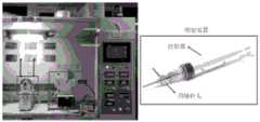
进一步地,所述静电纺丝设备为同轴静电纺丝设备,包括推注装置、喷射装置和接收装置,喷射装置包括针筒和同轴针头;Further, the electrospinning device is a coaxial electrospinning device, including a bolus device, a spray device and a receiving device, and the spray device includes a needle cylinder and a coaxial needle;
所述静电纺丝设备的工作温度为25~40℃,优选为35℃;工作湿度为30~50%,优选为40%;工作电压为负压-2.00V,正压+25.00V;所述静电纺丝时间为4~8小时,优选为6小时;The working temperature of the electrospinning equipment is 25-40°C, preferably 35°C; the working humidity is 30-50%, preferably 40%; the working voltage is negative pressure -2.00V, positive pressure +25.00V; the Electrospinning time is 4 to 8 hours, preferably 6 hours;
所述静电纺丝设备推注核层纺丝液的推注速度为0.008mL/min,采用的同轴针头型号为22号;The bolus injection speed of the electrospinning equipment for bolus injection of the core layer spinning solution is 0.008 mL/min, and the model of the coaxial needle used is No. 22;
所述静电纺丝设备推注壳层纺丝液的推注速度为0.010mL/min,采用的同轴针头型号为17号;The bolus injection speed of the electrospinning equipment for bolus injection of the shell spinning solution is 0.010 mL/min, and the model of the coaxial needle used is No. 17;
所述静电纺丝设备的接收装置为直径6mm的金属杆,优选为直径2mm的金属杆;所述接收装置与同轴针头的距离为15cm。The receiving device of the electrospinning equipment is a metal rod with a diameter of 6 mm, preferably a metal rod with a diameter of 2 mm; the distance between the receiving device and the coaxial needle is 15 cm.
本发明还提供了上述人工血管支架材料在制备小口径人工血管支架中的用途。The present invention also provides the use of the above artificial blood vessel stent material in preparing a small-diameter artificial blood vessel stent.
本发明以氧化锰纳米颗粒和PCL为核层原料、以羧化壳聚糖与PCL为壳层原料,通过同轴静电纺丝的方法制得了孔径小于6mm小口径人工血管支架,该小口径人工血管支架的自抗凝性显著改善,血管长期通畅率显著提高,能够有效抑制血管吻合口形成血栓;同时,该小口径人工血管支架还具有优异的显影能力,便于手术后续监测血管支架的降解情况,对患者术后复查进行血管定位,避免复查过程中注射造影剂给人体带来不适。本发明提供的小口径人工血管支架在血管移植手术中具有良好的应用前景。In the invention, manganese oxide nanoparticles and PCL are used as core layer raw materials, carboxylated chitosan and PCL are used as shell layer raw materials, and a small-caliber artificial blood vessel stent with a pore diameter of less than 6 mm is prepared by a coaxial electrospinning method. The self-anticoagulation of the vascular stent is significantly improved, the long-term patency rate of the blood vessel is significantly improved, and the thrombus formation at the vascular anastomosis can be effectively inhibited; at the same time, the small-caliber artificial vascular stent also has excellent visualization ability, which is convenient for the follow-up monitoring of the degradation of the vascular stent. , to locate the blood vessels in the postoperative review of the patient, so as to avoid the discomfort caused by the injection of contrast agent to the human body during the review process. The small-diameter artificial blood vessel stent provided by the invention has a good application prospect in the blood vessel transplantation operation.
与现有技术报道的抗凝血功能化修饰的聚己内酯/壳聚糖复合小口径人工血管相比(Biomaterials 30(2009)2276–2283),本发明提供的小口径人工血管支架原料中不含肝素,减少了原料种类,节约了成本。Compared with the polycaprolactone/chitosan composite small-diameter artificial blood vessel reported in the prior art (Biomaterials 30 (2009) 2276-2283), the small-diameter artificial blood vessel stent material provided by the present invention has It does not contain heparin, which reduces the types of raw materials and saves costs.
本发明采用的原料生物相容性好,制备方法简单,条件温和,适合扩大化生产。The raw material used in the invention has good biocompatibility, simple preparation method and mild conditions, and is suitable for enlarged production.
显然,根据本发明的上述内容,按照本领域的普通技术知识和惯用手段,在不脱离本发明上述基本技术思想前提下,还可以做出其它多种形式的修改、替换或变更。Obviously, according to the above-mentioned content of the present invention, according to the common technical knowledge and conventional means in the field, without departing from the above-mentioned basic technical idea of the present invention, other various forms of modification, replacement or change can also be made.
以下通过实施例形式的具体实施方式,对本发明的上述内容再作进一步的详细说明。但不应将此理解为本发明上述主题的范围仅限于以下的实例。凡基于本发明上述内容所实现的技术均属于本发明的范围。The above content of the present invention will be further described in detail below through the specific implementation in the form of examples. However, this should not be construed as limiting the scope of the above-mentioned subject matter of the present invention to the following examples. All technologies implemented based on the above content of the present invention belong to the scope of the present invention.
附图说明Description of drawings
图1为本发明小口径人工血管支架的制备工艺示意图。FIG. 1 is a schematic diagram of the preparation process of the small-diameter artificial blood vessel stent of the present invention.
图2为同轴静电纺丝设备的图片。Figure 2 is a picture of a coaxial electrospinning apparatus.
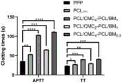
图3为APTT和TT测试结果,其中,*表示p<0.05,**表示p<0.01,***表示p<0.005,****表示p<0.001。Figure 3 shows the test results of APTT and TT, wherein * means p<0.05, ** means p<0.01, *** means p<0.005, and **** means p<0.001.
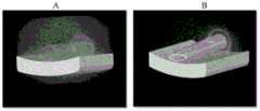
图4为血液长期通畅率测试操作方法示意图(A),测试结果:PCL100%(B)、实施例1制得的人工血管支架(C)。Figure 4 is a schematic diagram of the operation method for the long-term blood patency test (A), test results: PCL100% (B), artificial blood vessel stent (C) prepared in Example 1.
图5为Micro CT成像图:PCL100%(A)、实施例1制得的人工血管支架(B)。Figure 5 is a Micro CT image: PCL100% (A), artificial blood vessel stent (B) prepared in Example 1.
具体实施方式Detailed ways
本发明所用原料与设备均为已知产品,通过购买市售产品所得。The raw materials and equipment used in the present invention are all known products, obtained by purchasing commercially available products.
其中,聚己内酯(PCL)购买于Sigmae-Aldrich,分子量为80000。羧化壳聚糖购买于Aladin,粘度为10-80mPa·s。Among them, polycaprolactone (PCL) was purchased from Sigma-Aldrich with a molecular weight of 80,000. Carboxylated chitosan was purchased from Aladin with a viscosity of 10-80 mPa·s.
实施例1本发明小口径人工血管支架的制备Example 1 Preparation of the small-caliber artificial blood vessel stent of the present invention
1、合成氧化锰(BM)纳米颗粒1. Synthesis of manganese oxide (BM) nanoparticles
称取0.376g高锰酸钾、0.028g葡萄糖和30mL去离子水依次加入50mL烧杯中,快速搅拌20min后将烧杯置于70℃烘箱中保温6h。反应后自然冷却,洗涤沉淀至滤液为无色,所得滤饼自然干燥后研细,即得BM纳米颗粒。Weigh 0.376g potassium permanganate, 0.028g glucose and 30mL deionized water into a 50mL beaker in turn, stir quickly for 20min, and place the beaker in a 70°C oven for 6h. After the reaction, it is naturally cooled, washed and precipitated until the filtrate is colorless, and the obtained filter cake is naturally dried and then ground into fine particles to obtain BM nanoparticles.
2、制备小口径人工血管支架2. Preparation of small-caliber artificial vascular stent
采用同轴静电纺丝制备本发明的小口径人工血管支架,同轴静电纺丝设备如图2所示,设备包括推注装置、喷射装置和接收装置,喷射装置包括针筒和同轴针头。具体工艺如下(工艺示意图如图1所示):The small diameter artificial blood vessel stent of the present invention is prepared by coaxial electrospinning. The coaxial electrospinning equipment is shown in Figure 2. The equipment includes a bolus device, a spray device and a receiving device. The specific process is as follows (the schematic diagram of the process is shown in Figure 1):
将PCL(1g)溶于氯仿和甲醇(4:1,v/v)混合溶液(10mL)中,得到PCL溶液,将BM纳米颗粒(0.1g)作为分散相加入到PCL溶液中,混合均匀,得到的共混物作为核层纺丝溶液;将PCL(1g)和CMC(0.4g)以三氟乙酸(TFA,10mL)为溶剂溶解,混合均匀,配制成壳层纺丝溶液;核层和壳层纺丝溶液分别搅拌12h。Dissolve PCL (1 g) in a mixed solution (10 mL) of chloroform and methanol (4:1, v/v) to obtain a PCL solution, add BM nanoparticles (0.1 g) as a dispersed phase to the PCL solution, mix well, The obtained blend was used as a core layer spinning solution; PCL (1 g) and CMC (0.4 g) were dissolved in trifluoroacetic acid (TFA, 10 mL) as a solvent, mixed well, and prepared into a shell layer spinning solution; the core layer and The shell spinning solutions were stirred for 12 h respectively.
在同轴静电纺丝设备中分别推注核层纺丝溶液、壳层纺丝溶液,由同轴针头喷出具有核壳结构的纳米纤维,然后在距离同轴针头15cm的接收装置(直径2mm的金属杆)中得到本发明的小口径人工血管支架:PCL/CMC4-PCL/BM1。该小口径人工血管支架的内径为2mm。The core layer spinning solution and the shell layer spinning solution were injected into the coaxial electrospinning equipment respectively, and the nanofibers with the core-shell structure were ejected from the coaxial needle, and then placed in a receiving device (diameter 2 mm) 15 cm away from the coaxial needle. The small-caliber artificial blood vessel stent of the present invention is obtained from the metal rod: PCL/CMC4 -PCL/BM1 . The inner diameter of the small-diameter artificial blood vessel stent is 2 mm.
静电纺丝时,核层纺丝溶液推注速度为0.008mL/min,壳层纺丝溶液推注速度为0.010mL/mim;核层纺丝溶液采用的同轴针头型号为22号,壳层纺丝溶液采用的同轴针头型号为17号;纺丝环境为:温度35℃,湿度40%;纺丝设备的工作电压为负压-2.00V,正压+25.00V;接收装置的转速为2000r/min;纺丝时间为6h。During electrospinning, the bolus injection rate of the core layer spinning solution was 0.008mL/min, and the bolus injection rate of the shell layer spinning solution was 0.010mL/mim; The coaxial needle type used in the spinning solution is No. 17; the spinning environment is: temperature 35°C, humidity 40%; the working voltage of the spinning equipment is negative pressure -2.00V, positive pressure +25.00V; the rotating speed of the receiving device is 2000r/min; spinning time is 6h.
实施例2本发明小口径人工血管支架的制备Example 2 Preparation of the small-caliber artificial blood vessel stent of the present invention
参照实施例1的方法,将CMC的用量更改为0.2g,其余条件均与实施例1相同,得到本发明的小口径人工血管支架:PCL/CMC2-PCL/BM1。Referring to the method of Example 1, the amount of CMC was changed to 0.2 g, and other conditions were the same as those of Example 1, to obtain the small-diameter artificial blood vessel stent of the present invention: PCL/CMC2 -PCL/BM1 .
实施例3本发明小口径人工血管支架的制备Example 3 Preparation of the small-caliber artificial blood vessel stent of the present invention
参照实施例1的方法,将BM纳米颗粒的用量更改为0.05g,其余条件均与实施例1相同,得到本发明的小口径人工血管支架:PCL/CMC4-PCL/BM0.5。Referring to the method of Example 1, the dosage of BM nanoparticles was changed to 0.05 g, and the other conditions were the same as those of Example 1, to obtain the small-diameter artificial blood vessel stent of the present invention: PCL/CMC4 -PCL/BM0.5 .
以下制备对照样品。Control samples were prepared as follows.
对照例1对照人工血管支架的制备Control Example 1 Preparation of Control Artificial Vascular Stent
将PCL溶于氯仿和甲醇(4:1,v/v)混合溶液中,得到12%g/mL的PCL溶液。以该PCL溶液作为纺丝溶液,按照与实施例1相同的静电纺丝条件,得到纯PCL人工血管支架:PCL100%。PCL was dissolved in a mixed solution of chloroform and methanol (4:1, v/v) to obtain a PCL solution of 12% g/mL. Using the PCL solution as a spinning solution, according to the same electrospinning conditions as in Example 1, a pure PCL artificial blood vessel stent: PCL100% was obtained.
对照例2高CMC含量小口径人工血管支架的制备Comparative Example 2 Preparation of small-caliber artificial vascular stent with high CMC content
参照实施例1的方法,将CMC的用量更改为1g,其余条件均与实施例1相同,得到高CMC含量小口径人工血管支架:PCL/CMC10-PCL/BM1。Referring to the method of Example 1, the amount of CMC was changed to 1 g, and other conditions were the same as those of Example 1, to obtain a small-caliber artificial blood vessel stent with high CMC content: PCL/CMC10 -PCL/BM1 .
静电纺丝过程中发现,纺丝溶液粘度过大,易在针头处形成很大的液滴,需要反复清理,在高压下无法稳定连续出丝,使得纺丝时间由原来的6h增加至12h,时间成本和人工成本大大增加,故本对照例中的纺丝溶液的可纺性比实施例1降低。During the electrospinning process, it was found that the viscosity of the spinning solution was too large, and it was easy to form large droplets at the needle head, which required repeated cleaning, and the continuous spinning could not be stably produced under high pressure, so the spinning time increased from the original 6h to 12h. The time cost and labor cost are greatly increased, so the spinnability of the spinning solution in this comparative example is lower than that of Example 1.
对照例3高BM含量小口径人工血管支架的制备Comparative Example 3 Preparation of small-caliber artificial vascular stent with high BM content
参照实施例1的方法,将BM纳米颗粒的用量更改为0.5g,其余条件均与实施例1相同,得到高BM含量小口径人工血管支架:PCL/CMC4-PCL/BM5。Referring to the method of Example 1, the dosage of BM nanoparticles was changed to 0.5 g, and other conditions were the same as those of Example 1, to obtain a small-caliber artificial blood vessel stent with high BM content: PCL/CMC4 -PCL/BM5 .
静电纺丝过程中发现,由于BM纳米颗粒掺杂过多,针头易堵塞,需要反复清理疏通,在高压下核层溶液无法稳定连续出丝,使得纺丝时间由原来的6h增加至12h,时间成本和人工成本大大增加,故本对照例中的纺丝溶液的可纺性比实施例1降低。During the electrospinning process, it was found that due to the excessive doping of BM nanoparticles, the needle was easily blocked, which required repeated cleaning and dredging. Under high pressure, the core layer solution could not be stably and continuously produced, so that the spinning time increased from the original 6h to 12h. The cost and labor cost are greatly increased, so the spinnability of the spinning solution in this comparative example is lower than that of Example 1.
以下通过实验例证明本发明小口径人工血管支架的有益效果。The beneficial effects of the small-diameter artificial blood vessel stent of the present invention are demonstrated below through experimental examples.
实验例1本发明小口径人工血管支架的自抗凝性测试Experimental Example 1 Self-anticoagulation test of the small-caliber artificial vascular stent of the present invention
1、实验方法1. Experimental method
为了研究实施例1~3制得的小口径人工血管支架和对照例1制得的纯PCL人工血管支架的抗凝血性能,使用自动化血液凝固分析仪CA-50(Sysmex Corporation,Kobe,Japan)进行凝血时间测试。In order to study the anticoagulant properties of the small-diameter artificial vascular stents prepared in Examples 1 to 3 and the pure PCL artificial vascular stent prepared in Comparative Example 1, an automated blood coagulation analyzer CA-50 (Sysmex Corporation, Kobe, Japan) was used. Do a clotting time test.
(1)测试活化部分凝血活酶时间(APTT):首先将待测样品在生理盐水中浸泡过夜。之后将生理盐水吸出,换成新鲜的生理盐水在37℃下孵化1小时。之后将生理盐水换成贫血小板血浆(platelet-poor plasma,PPP),每个样品中加入200μL血浆,在37℃下孵化半小时。然后将50μL孵化过的PPP加入到样品杯中,随后加入50μL APTT试剂(西门子,使用前孵化10分钟),并在37℃下孵化3分钟。然后加入50μL的CaCl2溶液(0.025mol/L),之后测定APTT时间。(1) Test for activated partial thromboplastin time (APTT): first, soak the sample to be tested in physiological saline overnight. After that, the physiological saline was aspirated, replaced with fresh physiological saline, and incubated at 37°C for 1 hour. After that, the normal saline was changed to platelet-poor plasma (PPP), 200 μL of plasma was added to each sample, and the samples were incubated at 37° C. for half an hour. 50 μL of the incubated PPP was then added to the sample cup followed by 50 μL of APTT reagent (Siemens, incubated for 10 minutes prior to use) and incubated at 37°C for 3 minutes. Then, 50 μL of CaCl2 solution (0.025 mol/L) was added, and then the APTT time was measured.
(2)测试凝血酶时间(TT):其过程与APTT测试方法相似,区别仅在于将50μL APTT试剂替换为100μL TT试剂(西门子,使用前孵化15分钟)。(2) Test for thrombin time (TT): The procedure is similar to the APTT test method, except that 50 μL of APTT reagent is replaced with 100 μL of TT reagent (Siemens, incubated for 15 minutes before use).
对于每个样品的APTT和TT测试均重复三次取平均值,以减少误差。The APTT and TT tests for each sample were repeated three times and averaged to reduce errors.
2、实验结果2. Experimental results
根据APTT和TT来评价各人工血管支架的抗凝血活性。如图3所示,纯PCL人工血管支架(PCL100%)的TT和APTT与贫血小板血浆(PPP)几乎相同,说明PCL100%与血液成分之间基本没有吸附或反应。与PPP和PCL100%相比,本发明实施例制得的小口径人工血管支架的TT和APTT都提高了;特别是APTT,本发明实施例制得的小口径人工血管支架的APTT显著提高。The anticoagulant activity of each artificial vascular stent was evaluated according to APTT and TT. As shown in Figure 3, the TT and APTT of pure PCL artificial vascular stent (PCL100% ) are almost the same as those of platelet poor plasma (PPP), indicating that there is basically no adsorption or reaction between PCL100% and blood components. Compared with PPP and PCL100% , the TT and APTT of the small-diameter artificial blood vessel stent prepared in the embodiment of the present invention are both improved; especially APTT, the APTT of the small-diameter artificial blood vessel stent prepared by the embodiment of the present invention is significantly improved.
上述结果表明,本发明以氧化锰纳米颗粒和PCL为核层原料、以羧化壳聚糖与PCL为壳层原料制得的小口径人工血管支架能够显著延长凝血时间,有效防止血栓形成。The above results show that the small-diameter artificial vascular stent prepared by using manganese oxide nanoparticles and PCL as the core layer raw materials and carboxylated chitosan and PCL as the shell layer raw materials can significantly prolong the coagulation time and effectively prevent thrombosis.
此外,与PCL/CMC2-PCL/BM1相比,PCL/CMC4-PCL/BM1和PCL/CMC4-PCL/BM0.5的APTT明显提高,说明在原料中适当增加CMC的含量,有利于增加小口径人工血管支架的抗凝血活性。In addition, compared with PCL/CMC2 -PCL/BM1 , the APTT of PCL/CMC4 -PCL/BM1 and PCL/CMC4 -PCL/BM0.5 was significantly improved, indicating that the content of CMC in the raw materials was appropriately increased, and there were It is beneficial to increase the anticoagulant activity of small-caliber artificial vascular stents.
考虑到原料中CMC的含量太高,会影响纺丝溶液的可纺性(参见对照例2),所以,只有在本发明特定比例范围的原料配比下,才能同时保证材料制备过程的可纺性和目标产品的自抗凝性。Considering that the content of CMC in the raw material is too high, it will affect the spinnability of the spinning solution (refer to Comparative Example 2), so, only under the raw material ratio of the specific ratio of the present invention, can the spinnability of the material preparation process be guaranteed at the same time. properties and autoanticoagulant properties of the target product.
实验例2本发明小口径人工血管支架的血液长期通畅率测试Experimental Example 2 Long-term blood patency test of the small-caliber artificial blood vessel stent of the present invention
1、实验方法1. Experimental method
通过体外模拟血液循环,测试实施例1~3制得的小口径人工血管支架和对照例1制得的纯PCL人工血管支架的血液长期通畅率,评价其抗血栓形成的能力。具体操作如下:By simulating blood circulation in vitro, the long-term blood patency rates of the small-diameter artificial vascular stents prepared in Examples 1 to 3 and the pure PCL artificial vascular stent prepared in Comparative Example 1 were tested to evaluate their antithrombotic ability. The specific operations are as follows:
将待测人工血管支架在PBS中预浸过夜。The artificial vascular stent to be tested was pre-soaked in PBS overnight.
采用含有柠檬酸钠的真空管(5mL,Terumo Co.)收集健康人体新鲜血液(健康人,25岁),加入抗凝剂(血液:抗凝剂体积比=1:9),备用。A vacuum tube containing sodium citrate (5 mL, Terumo Co.) was used to collect fresh blood from healthy subjects (healthy subjects, 25 years old), and anticoagulant (blood:anticoagulant volume ratio=1:9) was added for use.
使用前,在全血中加入0.025mol/L CaCl2溶液,CaCl2溶液的添加量为全血的10%。将添加了CaCl2溶液的全血加入5ml注射器中,然后以1mL/min的速度泵入待测人工血管支架。用新鲜制备的戊二醛溶液(2.5wt.%)固定人工血管支架7天后,肉眼观察血管支架内腔。操作方法如图4A所示。Before use, 0.025mol/L CaCl2 solution was added to the whole blood, and the addition amount of the CaCl2 solution was 10% of the whole blood. The whole blood supplemented with CaCl2 solution was added into a 5ml syringe, and then pumped into the artificial blood vessel stent to be tested at a speed of 1mL/min. After immobilizing the artificial blood vessel stent with freshly prepared glutaraldehyde solution (2.5 wt. %) for 7 days, the lumen of the blood vessel stent was visually observed. The method of operation is shown in Figure 4A.
2、实验结果2. Experimental results
固定7天后,肉眼观察血管支架内腔,纯PCL人工血管支架(PCL100%)通过全血循环后内腔有大量血栓形成(图4B),而本发明实施例1~3制得的人工血管支架内腔均无明显血栓,其形成的血栓比PCL100%明显减少,其中,实施例1得的人工血管支架的测试结果如图4C所示。After 7 days of fixation, the lumen of the vascular stent was observed with the naked eye. After the pure PCL artificial vascular stent (PCL100% ) passed through the whole blood circulation, a large amount of thrombosis was formed in the lumen (Fig. 4B), while the artificial vascular stents prepared in Examples 1 to 3 of the present invention There was no obvious thrombus in the lumen, and the thrombus formed was significantly less than that of PCL by100% . The test results of the artificial blood vessel stent obtained in Example 1 are shown in Figure 4C.
上述结果表明,本发明以氧化锰纳米颗粒和PCL为核层原料、以羧化壳聚糖与PCL为壳层原料制得的小口径人工血管支架能够显著提高血液的长期通畅率,提高抗血栓形成能力。The above results show that the small-diameter artificial vascular stent prepared by using manganese oxide nanoparticles and PCL as the core layer raw materials and carboxylated chitosan and PCL as the shell layer raw materials can significantly improve the long-term blood patency rate and improve the antithrombotic rate. forming ability.
实验例3本发明小口径人工血管支架的显影能力测试Experimental Example 3 The imaging ability test of the small-diameter artificial blood vessel stent of the present invention
1、实验方法1. Experimental method
将实施例1~3制得的小口径人工血管支架和对照例1制得的纯PCL人工血管支架放置于小动物活体Micro CT成像仪(Quantum GX,美国PerkinElmer)中,扫描成像。The small-diameter artificial vascular stents prepared in Examples 1-3 and the pure PCL artificial vascular stent prepared in Comparative Example 1 were placed in a small animal living Micro CT imager (Quantum GX, PerkinElmer, USA), and scanned for imaging.
2、实验结果2. Experimental results
结果显示,加入BM后,本发明实施例1~3制得的小口径人工血管支架比对照例1制得的纯PCL人工血管支架(PCL100%)的成像更清晰,具有增强的显影能力。PCL100%的Micro CT成像图如图5A所示,实施例1得到的人工血管支架的测试结果如图5B所示。The results showed that after adding BM, the small-diameter artificial vascular stents prepared in Examples 1-3 of the present invention had clearer imaging than the pure PCL artificial vascular stent (PCL100% ) prepared in Comparative Example 1, and had enhanced developing ability. The Micro CT image of PCL100% is shown in FIG. 5A , and the test result of the artificial blood vessel stent obtained in Example 1 is shown in FIG. 5B .
上述结果表明,本发明以氧化锰纳米颗粒和PCL为核层原料、以羧化壳聚糖与PCL为壳层原料制得的小口径人工血管支架能够显著提高其显影能力。The above results show that the small-diameter artificial vascular stent prepared by using manganese oxide nanoparticles and PCL as the core layer raw materials and carboxylated chitosan and PCL as the shell layer raw materials can significantly improve its imaging ability.
此外,考虑到原料中BM的含量太高,会影响纺丝溶液的可纺性(参见对照例3),所以,只有在本发明特定比例范围的原料配比下,才能同时保证材料制备过程的可纺性和目标产品的显影能力。In addition, considering that the content of BM in the raw material is too high, it will affect the spinnability of the spinning solution (refer to Comparative Example 3), so, only under the raw material ratio of the specific ratio range of the present invention, can the material preparation process be guaranteed at the same time. Spinnability and developability of target products.
综上,本发明以氧化锰纳米颗粒和PCL为核层原料、以羧化壳聚糖与PCL为壳层原料,通过同轴静电纺丝的方法制得了孔径小于6mm小口径人工血管支架,该小口径人工血管支架的自抗凝性显著改善,血管长期通畅率显著提高,能够有效抑制血管吻合口形成血栓;同时,该小口径人工血管支架还具有优异的显影能力,便于手术后续监测血管支架的降解情况,对患者术后复查进行血管定位,避免复查过程中注射造影剂给人体带来不适。本发明提供的小口径人工血管支架在血管移植手术中具有良好的应用前景。To sum up, the present invention uses manganese oxide nanoparticles and PCL as the raw materials for the core layer, carboxylated chitosan and PCL as the raw materials for the shell layer, and obtains a small-diameter artificial blood vessel stent with a pore diameter of less than 6 mm by coaxial electrospinning. The self-anticoagulation of the small-caliber artificial vascular stent is significantly improved, the long-term patency rate of the blood vessel is significantly improved, and the thrombus formation at the vascular anastomosis can be effectively inhibited; at the same time, the small-caliber artificial vascular stent also has excellent visualization ability, which is convenient for the follow-up monitoring of the vascular stent. In order to avoid the discomfort caused by the injection of the contrast agent during the review process, the blood vessels can be positioned for the postoperative review of the patient. The small-diameter artificial blood vessel stent provided by the invention has a good application prospect in the blood vessel transplantation operation.
Claims (10)
Translated fromChinesePriority Applications (1)
| Application Number | Priority Date | Filing Date | Title |
|---|---|---|---|
| CN202010477269.4ACN111632206A (en) | 2020-05-29 | 2020-05-29 | A self-anticoagulated and visualized small-caliber artificial blood vessel stent and preparation method thereof |
Applications Claiming Priority (1)
| Application Number | Priority Date | Filing Date | Title |
|---|---|---|---|
| CN202010477269.4ACN111632206A (en) | 2020-05-29 | 2020-05-29 | A self-anticoagulated and visualized small-caliber artificial blood vessel stent and preparation method thereof |
Publications (1)
| Publication Number | Publication Date |
|---|---|
| CN111632206Atrue CN111632206A (en) | 2020-09-08 |
Family
ID=72322767
Family Applications (1)
| Application Number | Title | Priority Date | Filing Date |
|---|---|---|---|
| CN202010477269.4APendingCN111632206A (en) | 2020-05-29 | 2020-05-29 | A self-anticoagulated and visualized small-caliber artificial blood vessel stent and preparation method thereof |
Country Status (1)
| Country | Link |
|---|---|
| CN (1) | CN111632206A (en) |
Cited By (3)
| Publication number | Priority date | Publication date | Assignee | Title |
|---|---|---|---|---|
| CN115804873A (en)* | 2021-09-14 | 2023-03-17 | 中国科学院理化技术研究所 | A nanofibrous vascular scaffold, its preparation method and its application |
| CN116036387A (en)* | 2023-01-30 | 2023-05-02 | 博裕纤维科技(苏州)有限公司 | A preparation method of outsourcing drug-loaded visible nanofiber artificial vascular stent |
| CN118001462A (en)* | 2024-02-03 | 2024-05-10 | 武汉纺织大学 | Artificial blood vessel woven based on high molecular yarns and preparation method thereof |
Citations (7)
| Publication number | Priority date | Publication date | Assignee | Title |
|---|---|---|---|---|
| CN101156968A (en)* | 2007-10-26 | 2008-04-09 | 东华大学 | Preparation method of shell-core fiber-covered intravascular stent |
| US20110301697A1 (en)* | 2009-04-10 | 2011-12-08 | Hemoteq Ag | Manufacture, method and use of drug-eluting medical devices for permanently keeping blood vessels open |
| CN102755670A (en)* | 2011-04-29 | 2012-10-31 | 李文涛 | Preparation method of traceable biodegradable polymer bracket |
| CN104507509A (en)* | 2012-07-06 | 2015-04-08 | 埃克赛尔蒂斯有限公司 | Implants |
| US20150374519A1 (en)* | 2006-11-22 | 2015-12-31 | Inspiremd, Ltd | Optimized drug-eluting stent assembly |
| KR101585028B1 (en)* | 2015-06-01 | 2016-01-14 | (주)시지바이오 | Polymer coated stent for treatment of aneurysm and manufacturing method of the same |
| WO2019132238A1 (en)* | 2017-12-28 | 2019-07-04 | 오스템카디오텍 주식회사 | Stent and manufacturing method therefor |
- 2020
- 2020-05-29CNCN202010477269.4Apatent/CN111632206A/enactivePending
Patent Citations (7)
| Publication number | Priority date | Publication date | Assignee | Title |
|---|---|---|---|---|
| US20150374519A1 (en)* | 2006-11-22 | 2015-12-31 | Inspiremd, Ltd | Optimized drug-eluting stent assembly |
| CN101156968A (en)* | 2007-10-26 | 2008-04-09 | 东华大学 | Preparation method of shell-core fiber-covered intravascular stent |
| US20110301697A1 (en)* | 2009-04-10 | 2011-12-08 | Hemoteq Ag | Manufacture, method and use of drug-eluting medical devices for permanently keeping blood vessels open |
| CN102755670A (en)* | 2011-04-29 | 2012-10-31 | 李文涛 | Preparation method of traceable biodegradable polymer bracket |
| CN104507509A (en)* | 2012-07-06 | 2015-04-08 | 埃克赛尔蒂斯有限公司 | Implants |
| KR101585028B1 (en)* | 2015-06-01 | 2016-01-14 | (주)시지바이오 | Polymer coated stent for treatment of aneurysm and manufacturing method of the same |
| WO2019132238A1 (en)* | 2017-12-28 | 2019-07-04 | 오스템카디오텍 주식회사 | Stent and manufacturing method therefor |
Cited By (4)
| Publication number | Priority date | Publication date | Assignee | Title |
|---|---|---|---|---|
| CN115804873A (en)* | 2021-09-14 | 2023-03-17 | 中国科学院理化技术研究所 | A nanofibrous vascular scaffold, its preparation method and its application |
| CN116036387A (en)* | 2023-01-30 | 2023-05-02 | 博裕纤维科技(苏州)有限公司 | A preparation method of outsourcing drug-loaded visible nanofiber artificial vascular stent |
| CN116036387B (en)* | 2023-01-30 | 2024-10-29 | 博裕纤维科技(苏州)有限公司 | Preparation method of external-wrapping type drug-loaded visual nanofiber artificial vascular stent |
| CN118001462A (en)* | 2024-02-03 | 2024-05-10 | 武汉纺织大学 | Artificial blood vessel woven based on high molecular yarns and preparation method thereof |
Similar Documents
| Publication | Publication Date | Title |
|---|---|---|
| CN111632206A (en) | A self-anticoagulated and visualized small-caliber artificial blood vessel stent and preparation method thereof | |
| Wang et al. | The fabrication of a highly efficient self-healing hydrogel from natural biopolymers loaded with exosomes for the synergistic promotion of severe wound healing | |
| CN106029361B (en) | Multilayer matrix for tissue replacement and its use | |
| CN104921841B (en) | A kind of preparation method of double-decker artificial blood vessel | |
| CN106178121B (en) | An X-ray imaging blood vessel substitute and preparation method thereof | |
| Chen et al. | Synthesis and assessment of sodium alginate-modified silk fibroin microspheres as potential hepatic arterial embolization agent | |
| Zhai et al. | Coaxial electrospinning of P (LLA‐CL)/heparin biodegradable polymer nanofibers: Potential vascular graft for substitution of femoral artery | |
| JP2004321484A (en) | Medical high molecular nano-micro fiber | |
| Yin et al. | Performance of PEGylated chitosan and poly (L-lactic acid-co-ε-caprolactone) bilayer vascular grafts in a canine femoral artery model | |
| CN103876859A (en) | Artificial blood vessel composed of micrometer fiber and provided with large-hole structure and preparation method and application thereof | |
| CN101283025B (en) | Biodegradable particle and method for producing the same | |
| CN115137881B (en) | Three-layer biomimetic artificial blood vessel for antithrombotic and tissue regeneration promotion and preparation method thereof | |
| CN101653624A (en) | Preparation method of composite nanometer fiber small-diameter intravascular tissue engineering stent material | |
| CN111926462A (en) | Medical sponge and preparation method thereof | |
| CN105079874A (en) | Method for preparing small-diameter artificial blood vessels on basis of nanotechnologies | |
| CN109381732A (en) | Electrostatic spinning dressing, preparation method and the application of growth factor-loaded micromolecular inhibitor | |
| CN105536055A (en) | Shape memory type high-elasticity activity nano-fiber stent and application thereof | |
| CN104353128A (en) | Degradable intravascular stent and preparation method and application thereof | |
| CN110292652A (en) | Mercaptophenyl boronic acid activates gold nano grain, preparation method and application | |
| CN110520166A (en) | Blood platelet is inhibited to absorb | |
| Xu et al. | Electrostatic self-assemble modified electrospun poly-L-lactic acid/poly-vinylpyrrolidone composite polymer and its potential applications in small-diameter artificial blood vessels | |
| CN108187127A (en) | A kind of polyvinyl alcohol nano suppository and its preparation method and application | |
| TR201707216A2 (en) | BIOMIMETIC AN ARTIFICIAL BLOOD VESSEL AND ITS PRODUCTION METHOD | |
| CN102133434A (en) | Biodegradable extravascular stent and preparation method thereof | |
| HK1231356A1 (en) | A composite biological cannula and preparation method and application thereof |
Legal Events
| Date | Code | Title | Description |
|---|---|---|---|
| PB01 | Publication | ||
| PB01 | Publication | ||
| SE01 | Entry into force of request for substantive examination | ||
| SE01 | Entry into force of request for substantive examination | ||
| RJ01 | Rejection of invention patent application after publication | Application publication date:20200908 | |
| RJ01 | Rejection of invention patent application after publication |