CN111601626A - Medical device with integrated biosensor - Google Patents
Medical device with integrated biosensorDownload PDFInfo
- Publication number
- CN111601626A CN111601626ACN201880068334.9ACN201880068334ACN111601626ACN 111601626 ACN111601626 ACN 111601626ACN 201880068334 ACN201880068334 ACN 201880068334ACN 111601626 ACN111601626 ACN 111601626A
- Authority
- CN
- China
- Prior art keywords
- medical device
- nitric oxide
- biosensor
- oxidase
- lumen
- Prior art date
- Legal status (The legal status is an assumption and is not a legal conclusion. Google has not performed a legal analysis and makes no representation as to the accuracy of the status listed.)
- Granted
Links
Images


Classifications
- A—HUMAN NECESSITIES
- A61—MEDICAL OR VETERINARY SCIENCE; HYGIENE
- A61B—DIAGNOSIS; SURGERY; IDENTIFICATION
- A61B5/00—Measuring for diagnostic purposes; Identification of persons
- A61B5/145—Measuring characteristics of blood in vivo, e.g. gas concentration or pH-value ; Measuring characteristics of body fluids or tissues, e.g. interstitial fluid or cerebral tissue
- A61B5/14503—Measuring characteristics of blood in vivo, e.g. gas concentration or pH-value ; Measuring characteristics of body fluids or tissues, e.g. interstitial fluid or cerebral tissue invasive, e.g. introduced into the body by a catheter or needle or using implanted sensors
- A—HUMAN NECESSITIES
- A61—MEDICAL OR VETERINARY SCIENCE; HYGIENE
- A61B—DIAGNOSIS; SURGERY; IDENTIFICATION
- A61B5/00—Measuring for diagnostic purposes; Identification of persons
- A61B5/145—Measuring characteristics of blood in vivo, e.g. gas concentration or pH-value ; Measuring characteristics of body fluids or tissues, e.g. interstitial fluid or cerebral tissue
- A61B5/14532—Measuring characteristics of blood in vivo, e.g. gas concentration or pH-value ; Measuring characteristics of body fluids or tissues, e.g. interstitial fluid or cerebral tissue for measuring glucose, e.g. by tissue impedance measurement
- A—HUMAN NECESSITIES
- A61—MEDICAL OR VETERINARY SCIENCE; HYGIENE
- A61B—DIAGNOSIS; SURGERY; IDENTIFICATION
- A61B5/00—Measuring for diagnostic purposes; Identification of persons
- A61B5/68—Arrangements of detecting, measuring or recording means, e.g. sensors, in relation to patient
- A61B5/6846—Arrangements of detecting, measuring or recording means, e.g. sensors, in relation to patient specially adapted to be brought in contact with an internal body part, i.e. invasive
- A61B5/6847—Arrangements of detecting, measuring or recording means, e.g. sensors, in relation to patient specially adapted to be brought in contact with an internal body part, i.e. invasive mounted on an invasive device
- A61B5/6852—Catheters
- A—HUMAN NECESSITIES
- A61—MEDICAL OR VETERINARY SCIENCE; HYGIENE
- A61B—DIAGNOSIS; SURGERY; IDENTIFICATION
- A61B5/00—Measuring for diagnostic purposes; Identification of persons
- A61B5/145—Measuring characteristics of blood in vivo, e.g. gas concentration or pH-value ; Measuring characteristics of body fluids or tissues, e.g. interstitial fluid or cerebral tissue
- A61B5/14546—Measuring characteristics of blood in vivo, e.g. gas concentration or pH-value ; Measuring characteristics of body fluids or tissues, e.g. interstitial fluid or cerebral tissue for measuring analytes not otherwise provided for, e.g. ions, cytochromes
- A—HUMAN NECESSITIES
- A61—MEDICAL OR VETERINARY SCIENCE; HYGIENE
- A61B—DIAGNOSIS; SURGERY; IDENTIFICATION
- A61B5/00—Measuring for diagnostic purposes; Identification of persons
- A61B5/145—Measuring characteristics of blood in vivo, e.g. gas concentration or pH-value ; Measuring characteristics of body fluids or tissues, e.g. interstitial fluid or cerebral tissue
- A61B5/1468—Measuring characteristics of blood in vivo, e.g. gas concentration or pH-value ; Measuring characteristics of body fluids or tissues, e.g. interstitial fluid or cerebral tissue using chemical or electrochemical methods, e.g. by polarographic means
- A61B5/1486—Measuring characteristics of blood in vivo, e.g. gas concentration or pH-value ; Measuring characteristics of body fluids or tissues, e.g. interstitial fluid or cerebral tissue using chemical or electrochemical methods, e.g. by polarographic means using enzyme electrodes, e.g. with immobilised oxidase
- A61B5/14865—Measuring characteristics of blood in vivo, e.g. gas concentration or pH-value ; Measuring characteristics of body fluids or tissues, e.g. interstitial fluid or cerebral tissue using chemical or electrochemical methods, e.g. by polarographic means using enzyme electrodes, e.g. with immobilised oxidase invasive, e.g. introduced into the body by a catheter or needle or using implanted sensors
- A—HUMAN NECESSITIES
- A61—MEDICAL OR VETERINARY SCIENCE; HYGIENE
- A61L—METHODS OR APPARATUS FOR STERILISING MATERIALS OR OBJECTS IN GENERAL; DISINFECTION, STERILISATION OR DEODORISATION OF AIR; CHEMICAL ASPECTS OF BANDAGES, DRESSINGS, ABSORBENT PADS OR SURGICAL ARTICLES; MATERIALS FOR BANDAGES, DRESSINGS, ABSORBENT PADS OR SURGICAL ARTICLES
- A61L2300/00—Biologically active materials used in bandages, wound dressings, absorbent pads or medical devices
- A61L2300/10—Biologically active materials used in bandages, wound dressings, absorbent pads or medical devices containing or releasing inorganic materials
- A61L2300/114—Nitric oxide, i.e. NO
- A—HUMAN NECESSITIES
- A61—MEDICAL OR VETERINARY SCIENCE; HYGIENE
- A61M—DEVICES FOR INTRODUCING MEDIA INTO, OR ONTO, THE BODY; DEVICES FOR TRANSDUCING BODY MEDIA OR FOR TAKING MEDIA FROM THE BODY; DEVICES FOR PRODUCING OR ENDING SLEEP OR STUPOR
- A61M2202/00—Special media to be introduced, removed or treated
- A61M2202/02—Gases
- A61M2202/0266—Nitrogen (N)
- A61M2202/0275—Nitric oxide [NO]
- C—CHEMISTRY; METALLURGY
- C12—BIOCHEMISTRY; BEER; SPIRITS; WINE; VINEGAR; MICROBIOLOGY; ENZYMOLOGY; MUTATION OR GENETIC ENGINEERING
- C12Q—MEASURING OR TESTING PROCESSES INVOLVING ENZYMES, NUCLEIC ACIDS OR MICROORGANISMS; COMPOSITIONS OR TEST PAPERS THEREFOR; PROCESSES OF PREPARING SUCH COMPOSITIONS; CONDITION-RESPONSIVE CONTROL IN MICROBIOLOGICAL OR ENZYMOLOGICAL PROCESSES
- C12Q1/00—Measuring or testing processes involving enzymes, nucleic acids or microorganisms; Compositions therefor; Processes of preparing such compositions
- C12Q1/001—Enzyme electrodes
- C12Q1/005—Enzyme electrodes involving specific analytes or enzymes
Landscapes
- Health & Medical Sciences (AREA)
- Life Sciences & Earth Sciences (AREA)
- Physics & Mathematics (AREA)
- Molecular Biology (AREA)
- Animal Behavior & Ethology (AREA)
- Pathology (AREA)
- Engineering & Computer Science (AREA)
- Biomedical Technology (AREA)
- Heart & Thoracic Surgery (AREA)
- Medical Informatics (AREA)
- Veterinary Medicine (AREA)
- Surgery (AREA)
- Biophysics (AREA)
- General Health & Medical Sciences (AREA)
- Public Health (AREA)
- Optics & Photonics (AREA)
- Emergency Medicine (AREA)
- Measurement Of The Respiration, Hearing Ability, Form, And Blood Characteristics Of Living Organisms (AREA)
- Chemical & Material Sciences (AREA)
- Chemical Kinetics & Catalysis (AREA)
- General Chemical & Material Sciences (AREA)
Abstract
Description
Translated fromChinese技术领域technical field
本发明涉及医疗装置;并且更具体地,涉及导管和类似的医疗装置,所述导管和类似的医疗装置被构造为具有一个或多个集成的生物传感器以用于检测葡萄糖、乳酸、或感兴趣的任何其他分析物、或其组合。The present invention relates to medical devices; and more particularly, to catheters and similar medical devices configured with one or more integrated biosensors for detecting glucose, lactate, or of any other analytes, or combinations thereof.
背景技术Background technique
存在各种需要分析血液或皮下流体中存在的生物组分的浓度的诊断性和治疗性医学治疗。从患者抽取的随后通过定点照护型装置进行分析的血液或流体样品提供非常准确的浓度测量结果,但由于对可以从患者进行的抽取频率和抽取数量、或者两者上的限制,通常间隔很长的时间间隔,尤其是在儿科或新生儿重症监护病房(PICU和NICU)中。此类定点照护型装置维护成本高昂,并且可能由于有限的样品通量而受到阻碍,尤其是在可能有大量患者的重症监护病房(所有患者都需要频繁的分析物监测)中。将低型面医疗装置插入或接触到人体内或人组织上以能够进行准确且连续的测量是可行的替代方案,所述替代方案能够以较高频率提供准确的分析物读取。There are various diagnostic and therapeutic medical treatments that require analysis of the concentration of biological components present in blood or subcutaneous fluids. Blood or fluid samples drawn from a patient and subsequently analyzed by a point-of-care device provide very accurate concentration measurements, but often at long intervals due to limitations on the frequency and number of draws that can be performed from the patient, or both , especially in pediatric or neonatal intensive care units (PICU and NICU). Such point-of-care devices are expensive to maintain and can be hampered by limited sample throughput, especially in intensive care units where there may be a large number of patients (all requiring frequent analyte monitoring). Inserting or contacting low-profile medical devices into the human body or onto human tissue to enable accurate and continuous measurements is a viable alternative that provides accurate analyte readings at higher frequencies.
发明内容SUMMARY OF THE INVENTION
技术问题technical problem
具有集成的生物传感器的常规医疗装置在传感器元件部分处易于受凝结、生物膜形成或两者的影响,从而限制了此类生物传感器的感测品质和可用持续时间。Conventional medical devices with integrated biosensors are susceptible to condensation, biofilm formation, or both at the sensor element portion, limiting the sensing quality and usable duration of such biosensors.
问题的解决方案solution to the problem
医疗装置被构造成用于利用一氧化氮(NO)供体材料以在与水分接触时释放NO气体,以便减轻集成在所述医疗装置内的生物传感器的部位处的细菌增殖、凝结、或其组合。The medical device is configured to utilize a nitric oxide (NO) donor material to release NO gas upon contact with moisture in order to mitigate bacterial proliferation, condensation, or the like at the site of a biosensor integrated within the medical device combination.
描述了一种新颖的多内腔医疗装置,所述装置具有用于含有所述NO供体材料并产生NO气体的第一内腔,并且具有用于容纳生物传感器元件部分的第二内腔。所述第一内腔与所述第二内腔之间的通道用于将NO气体从所述第一内腔传递至生物传感器的感测体积。A novel multi-lumen medical device is described having a first lumen for containing the NO donor material and generating NO gas, and a second lumen for containing a portion of a biosensor element. A channel between the first lumen and the second lumen is used to transfer NO gas from the first lumen to the sensing volume of the biosensor.
描述了独特的组成,并且还描述了创新的结构特征和部件间的关系,诸如各种组成层的安排,所述组成、结构和关系用于实现本文所阐述的目的,即提供具有集成的一个或多个生物传感器的医疗装置以测量人和动物患者中的分析物,所述一个或多个生物传感器适于使用NO气体的连通减轻细菌增殖和/或凝结。Unique components are described, and also innovative structural features and relationships between components, such as the arrangement of layers of the various components, used to achieve the purposes set forth herein, which are to provide an integrated A medical device of biosensor(s) adapted to mitigate bacterial proliferation and/or coagulation using communication of NO gas to measure analytes in human and animal patients.
本文描述了针对这些以及其他问题的其他解决方案。This article describes other solutions to these and other problems.
本发明的有利效果Advantageous Effects of the Invention
所述的具有集成的生物传感器的医疗装置:(i)由于NO气体的抗细菌和抗凝结作用提供改善的使用持续使用;并且(ii)由于降低了干扰而提供改善的感测。The described medical device with integrated biosensor: (i) provides improved longevity of use due to the antibacterial and anti-condensation effects of NO gas; and (ii) provides improved sensing due to reduced interference.
附图简述Brief Description of Drawings
在全面审阅以下细节和描述后,特别是在结合附图审阅时,本发明的这些以及其他特征对于本领域普通技术人员将变得清楚,在附图中:These and other features of the present invention will become apparent to those of ordinary skill in the art after a comprehensive review of the following details and description, particularly in conjunction with the accompanying drawings, wherein:
图1示出了根据一个实施例的具有集成的生物传感器的医疗装置;1 illustrates a medical device with an integrated biosensor according to one embodiment;
图2进一步展示了图1的具有集成的生物传感器的医疗装置,所述医疗装置被构造成用于在与水分接触时产生一氧化氮气体,并且将一氧化氮气体通过通道且朝向与生物传感器相关联的感测体积传递;FIG. 2 further illustrates the medical device of FIG. 1 with an integrated biosensor configured to generate nitric oxide gas upon contact with moisture, and to pass the nitric oxide gas through the channel and toward the biosensor. associated sensed volume delivery;
图3A示出了根据另一个实施例的医疗装置的管状主体的截面,所述管状主体包括三个内腔;3A shows a cross-section of a tubular body of a medical device including three lumens, according to another embodiment;
图3B示出了根据又另一个实施例的医疗装置的管状主体的截面,所述管状主体包括四个内腔;3B shows a cross-section of a tubular body of a medical device including four lumens, according to yet another embodiment;
图4示出了根据另一个实施例的具有集成的生物传感器的医疗装置;并且Figure 4 illustrates a medical device with an integrated biosensor according to another embodiment; and
图5示出了根据又另一个实施例的具有集成的生物传感器的医疗装置。Figure 5 shows a medical device with an integrated biosensor according to yet another embodiment.
具体实施方式Detailed ways
为了解释而非限制的目的,下文中提供了某些优选实施例的细节和描述,以使得本领域普通技术人员可以能够制作和使用本发明。这些细节和描述仅代表某些优选实施例,然而,本领域技术人员在全面审阅本披露之后,将容易理解许多未明确描述的其他实施例。因此,本披露的任何审阅者应仅通过权利要求来解释本发明的范围,因为这种范围并不旨在由本文所描述和例示的实施例限制。For purposes of explanation and not limitation, the following details and descriptions of certain preferred embodiments are provided to enable any person of ordinary skill in the art to make and use the present invention. These details and descriptions represent only certain preferred embodiments, however, many other embodiments not expressly described will be readily apparent to those skilled in the art after a thorough review of this disclosure. Accordingly, any reviewer of the present disclosure should interpret the scope of the invention only by the claims, as such scope is not intended to be limited by the embodiments described and illustrated herein.
一般实施例General Example
根据一般实施例,披露了具有集成的生物传感器的医疗装置。所述医疗装置包括:管状主体,所述管状主体在其中包括第一内腔和第二内腔;所述第一内腔包括布置于其中的一氧化氮供体材料,其中所述一氧化氮供体材料被构造成用于在与水分接触时释放一氧化氮气体;并且所述第二内腔在纵向上布置为相对于所述第一内腔平行、被构造成用于容纳至少第一生物传感器的元件部分,所述第一传感器具有与其相关联的第一感测体积;其中第一一氧化氮通道布置在所述一氧化氮供体材料与所述第一感测体积之间,所述第一一氧化氮通道被构造成用于将所述一氧化氮气体的流引导到所述第一感测体积中,以防止细菌增殖、凝结、或其组合。According to general embodiments, medical devices with integrated biosensors are disclosed. The medical device includes: a tubular body including a first lumen and a second lumen therein; the first lumen including a nitric oxide donor material disposed therein, wherein the nitric oxide the donor material is configured to release nitric oxide gas upon contact with moisture; and the second lumen is longitudinally disposed parallel with respect to the first lumen and is configured to receive at least a first lumen an element portion of a biosensor having a first sensing volume associated therewith; wherein a first nitric oxide channel is disposed between the nitric oxide donor material and the first sensing volume, The first nitric oxide channel is configured to direct the flow of the nitric oxide gas into the first sensing volume to prevent bacterial proliferation, coagulation, or a combination thereof.
所述第一生物传感器可以包括:工作电极和参比电极。所述工作电极通常包括:隔离层,所述隔离层覆盖所述工作电极的至少一部分并且被构造成用于限制电活性干扰化合物扩散通过所述隔离层;分析物反应层,所述分析物反应层包含一种或多种固定化或化学连接的抗体、氧化还原酶、或螯合剂以用于与一种或多种相应的分析物反应,从而实现可检测信号;以及分析物扩散控制层,所述分析物扩散控制层用于调控感兴趣的分析物到所述第一生物传感器的所述第一感测体积中的扩散。所述参比电极通常接近所述工作电极定位;然而,所述参比电极通常与所述工作电极隔开在0.2um与10.0mm之间的距离。就这一点而言,所述参比电极的终端通常与孔的周边间隔开最多至3.0mm的距离。The first biosensor may include: a working electrode and a reference electrode. The working electrode generally includes: a spacer layer covering at least a portion of the working electrode and configured to limit diffusion of electroactive interfering compounds through the spacer layer; an analyte-reactive layer, the analyte-reactive layer a layer comprising one or more immobilized or chemically linked antibodies, oxidoreductases, or chelating agents for reaction with one or more corresponding analytes to achieve a detectable signal; and an analyte diffusion control layer, The analyte diffusion control layer is used to regulate the diffusion of an analyte of interest into the first sensing volume of the first biosensor. The reference electrode is typically positioned close to the working electrode; however, the reference electrode is typically separated from the working electrode by a distance between 0.2 um and 10.0 mm. In this regard, the terminal end of the reference electrode is typically spaced a distance of up to 3.0 mm from the perimeter of the hole.
所述医疗装置可以任选地包括膜,所述膜布置在一氧化氮通道处,从而将所述第一内腔和所述第二内腔隔开。所述膜可以包含硅氧烷。The medical device may optionally include a membrane disposed at the nitric oxide channel, thereby separating the first lumen and the second lumen. The membrane may contain silicone.
所述一氧化氮供体材料被选择为被构造成用于响应于与水分的接触而释放一氧化氮气体的一氧化氮供体材料。此类一氧化氮供体材料的实例包括但不限于二醇二氮烯鎓化的二胺、S-亚硝基-白蛋白、S-亚硝基-N-青霉胺(SNAP)、S-亚硝基半胱氨酸(CysNO)、S-亚硝基谷胱甘肽(GSNO)、二醇二氮烯鎓化的二丁基己二胺(DBHD N2O2)、二亚乙基三胺/一氧化氮加合物(DEAT/NO)、二乙胺NONO亲核复合体(DEA/NO)、二亚丙基三胺NONO亲核复合体(DPTA/NO)、6-(2-羟基-1-甲基-2-亚硝基肼基)-N-甲基-1-己胺(MAHMA/NO)或其任何组合。The nitric oxide donor material is selected as a nitric oxide donor material configured to release nitric oxide gas in response to contact with moisture. Examples of such nitric oxide donor materials include, but are not limited to, glycol diazeniumylated diamines, S-nitroso-albumin, S-nitroso-N-penicillamine (SNAP), S-nitroso-N-penicillamine (SNAP), - Nitrosocysteine (CysNO), S-nitrosoglutathione (GSNO), dibutyl hexamethylene diamine (DBHD N2O2), diethylenetriamine /Nitric oxide adduct (DEAT/NO), Diethylamine NONO nucleophile complex (DEA/NO), Dipropylene triamine NONO nucleophile complex (DPTA/NO), 6-(2-hydroxyl -1-Methyl-2-nitrosohydrazino)-N-methyl-1-hexylamine (MAHMA/NO) or any combination thereof.
在某些实施例中,所述一氧化氮供体材料可以包括按重量计在0.1%与4.0%之间的SNAP、和按重量计最多至99.9%的赋形剂。In certain embodiments, the nitric oxide donor material may include between 0.1% and 4.0% by weight SNAP, and up to 99.9% by weight excipients.
术语“赋形剂”在本文中定义为“充当活性物质的媒介物或介质的非活性物质”。适用于所披露的发明的此类赋形剂的实例包括但不限于纤维素、交联聚维酮、羟丙基纤维素、羟丙基甲基纤维素、山梨糖醇、木糖醇、聚维酮(PVP)、全氟弹性体、全氟磺酸异构体、乙烯、氟乙烯、偏二氟乙烯、三氟氯乙烯、丙烯、六氟丙烯、全氟丙基乙烯基醚、全氟甲基乙烯基醚、硅氧烷、聚乙烯、聚氨酯、或其任何组合。The term "excipient" is defined herein as "an inactive substance that acts as a vehicle or medium for the active substance". Examples of such excipients suitable for use in the disclosed invention include, but are not limited to, cellulose, crospovidone, hydroxypropyl cellulose, hydroxypropyl methylcellulose, sorbitol, xylitol, poly Vidone (PVP), perfluoroelastomers, perfluorosulfonic acid isomers, ethylene, vinyl fluoride, vinylidene fluoride, chlorotrifluoroethylene, propylene, hexafluoropropylene, perfluoropropyl vinyl ether, perfluoro Methyl vinyl ether, silicone, polyethylene, polyurethane, or any combination thereof.
为了利用检测到的信号,将所述工作电极和参比电极中的每一个耦接至用于接收和处理其信号的电极信号监测系统。To utilize the detected signal, each of the working and reference electrodes is coupled to an electrode signal monitoring system for receiving and processing its signal.
在优选的实施例中,所述工作电极包括金、铂、银、汞、不锈钢、或碳。In preferred embodiments, the working electrode comprises gold, platinum, silver, mercury, stainless steel, or carbon.
所述参比电极通常被定位在距所述分析物反应层0.2mm与10.0mm之间的位置。就这一点而言,如果放置得太近,则参比电极可能短路,而如果放置得太远,则不会实现信号。The reference electrode is typically positioned between 0.2 mm and 10.0 mm from the analyte reactive layer. In this regard, if placed too close, the reference electrode may be shorted, and if placed too far, no signal will be achieved.
在一个独特的方面,所述医疗装置的隔离层可以包括:一个或多个含全氟化磺酸树脂的子层,和多个电聚合子层,所述多个电聚合子层布置在所述含全氟化磺酸树脂的子层的外表面上。术语“全氟化磺酸树脂(PFSA)”是用于描述
在各种实施例中,所述含全氟化磺酸树脂的子层可以包含磺化四氟乙烯和以下中的一种或多种:全氟(烷基乙烯基醚)、磺酰酸氟化物、全氟环烯烃、乙烯、氟乙烯、偏二氟乙烯、三氟氯乙烯、丙烯、六氟丙烯、全氟丙基乙烯基醚、全氟甲基乙烯基醚、聚三氟氯乙烯、全氟烷氧基聚合物、氟化乙烯丙烯、聚乙烯四氟乙烯、聚乙烯三氟氯乙烯、全氟弹性体、四氟乙烯丙烯、聚氨酯、聚乙烯、硅氧烷、或其任何组合。In various embodiments, the perfluorinated sulfonic acid resin-containing sublayer may comprise sulfonated tetrafluoroethylene and one or more of the following: perfluoro(alkyl vinyl ether), sulfonic acid fluoride compound, perfluorocyclic olefin, ethylene, vinyl fluoride, vinylidene fluoride, chlorotrifluoroethylene, propylene, hexafluoropropylene, perfluoropropyl vinyl ether, perfluoromethyl vinyl ether, polychlorotrifluoroethylene, perfluoroalkoxy polymer, fluorinated ethylene propylene, polyethylene tetrafluoroethylene, polyethylene chlorotrifluoroethylene, perfluoroelastomer, tetrafluoroethylene propylene, polyurethane, polyethylene, silicone, or any combination thereof.
此外,在优选的实施例中,所述含全氟化磺酸树脂的子层包含经退火的磺化四氟乙烯。就这一点而言,当对含全氟化磺酸树脂的子层进行退火时,磺化四氟乙烯中的孔隙尺寸减小、或完全消除。Additionally, in preferred embodiments, the perfluorinated sulfonic acid resin-containing sublayer comprises annealed sulfonated tetrafluoroethylene. In this regard, the pore size in the sulfonated tetrafluoroethylene is reduced, or completely eliminated, when the perfluorinated sulfonic acid resin-containing sublayer is annealed.
在一些实施例中,所述电聚合子层可以包含:1,3二氨基苯、间苯二酚、或其组合。一般来讲,具有集成的一个或多个生物传感器的医疗装置在工作电极的一部分在孔处暴露的部位处涂覆有含全氟化磺酸树脂的子层。所述含全氟化磺酸树脂的子层是如上文所述退火的。接下来,将具有一个或多个生物传感器的医疗装置放置在聚合浴中,在其中施加电压和电流以引起在含全氟化磺酸树脂的子层上方的许多层的电聚合。In some embodiments, the electropolymeric sub-layer may comprise: 1,3 diaminobenzene, resorcinol, or a combination thereof. Generally, medical devices with integrated one or more biosensors are coated with a sub-layer containing a perfluorinated sulfonic acid resin where a portion of the working electrode is exposed at the aperture. The perfluorinated sulfonic acid resin-containing sublayer is annealed as described above. Next, the medical device with one or more biosensors is placed in a polymerization bath where voltage and current are applied to cause electropolymerization of the many layers above the perfluorinated sulfonic acid resin-containing sublayer.
在各种实施例中,所述分析物反应层可以包含选自由以下组成的酶:肌酸酶、肌酸酰胺水解酶、肌氨酸氧化酶、苹果酸氧化酶、醇脱氢酶、D-天冬氨酸氧化酶、精胺氧化酶、NAD(P)H氧化酶、尿酸氧化酶、乳酸氧化酶、醇氧化酶、丙酮酸氧化酶、葡萄糖氧化酶、谷氨酸氧化酶、胆碱氧化酶、谷胱甘肽巯基氧化酶、胆固醇氧化酶、组胺氧化酶、L-赖氨酸氧化酶、L-天冬氨酸氧化酶、甘氨酸氧化酶、和半乳糖氧化酶。通常使用键合中间体(诸如但不限于戊二醛或聚合的戊二醛结构)将分析物反应中的一种或多种酶附接至隔离层的外表面。In various embodiments, the analyte-reactive layer can comprise an enzyme selected from the group consisting of: creatinase, creatinamide hydrolase, sarcosine oxidase, malate oxidase, alcohol dehydrogenase, D- Aspartate oxidase, spermine oxidase, NAD(P)H oxidase, uricase, lactate oxidase, alcohol oxidase, pyruvate oxidase, glucose oxidase, glutamate oxidase, choline oxidase enzymes, glutathione sulfhydryl oxidase, cholesterol oxidase, histamine oxidase, L-lysine oxidase, L-aspartate oxidase, glycine oxidase, and galactose oxidase. One or more enzymes in the analyte reaction are typically attached to the outer surface of the isolation layer using a bonding intermediate such as, but not limited to, glutaraldehyde or polymeric glutaraldehyde structures.
所述分析物扩散控制层可以包含:按重量计30%-70%的聚氨酯、按重量计20%-60%的硅氧烷、和最多至50%的以下中的一种或多种:泊洛沙姆多元醇表面活性剂、聚乳酸、聚乙交酯、聚乳酸-共-羟基乙酸。The analyte diffusion control layer may comprise: 30%-70% by weight polyurethane, 20%-60% by weight siloxane, and up to 50% one or more of the following: poise Loxamer polyol surfactant, polylactic acid, polyglycolide, polylactic acid-co-glycolic acid.
在一些实施例中,所述隔离层、所述分析物反应层、和所述扩散控制层之一可以进一步包含不同于所述第一一氧化氮供体材料的第二一氧化氮供体材料。In some embodiments, one of the isolation layer, the analyte reactive layer, and the diffusion control layer may further comprise a second nitric oxide donor material different from the first nitric oxide donor material .
在一些实施例中,所述参比电极可以包括盘绕导线,所述盘绕导线围绕包围所述工作电极的套管的至少一部分缠绕以最大化其表面积。在其他实施例中,所述参比电极可以包括布置在套管上的导电迹线,其中所述套管布置在所述参比电极与所述工作电极的导线部件之间。在一些实施例中,所述参比电极包括印刷在施加到所述工作电极上的涂层上的迹线。In some embodiments, the reference electrode may comprise a coiled wire wrapped around at least a portion of a sleeve surrounding the working electrode to maximize its surface area. In other embodiments, the reference electrode may comprise a conductive trace disposed on a sleeve, wherein the sleeve is disposed between the reference electrode and the lead member of the working electrode. In some embodiments, the reference electrode includes traces printed on a coating applied to the working electrode.
在一些实施例中,所述第一生物传感器可以被构造成用于检测葡萄糖。在其他实施例中,所述第一生物传感器可以被构造成用于检测乳糖。在一些实施例中,所述第一传感器可以被构造成用于检测葡萄糖并且所述第二生物传感器被构造成用于检测乳酸,或反之亦然。In some embodiments, the first biosensor may be configured to detect glucose. In other embodiments, the first biosensor may be configured to detect lactose. In some embodiments, the first sensor may be configured to detect glucose and the second biosensor may be configured to detect lactate, or vice versa.
所述医疗装置可以包括第二生物传感器,所述第二生物传感器被布置成距所述第一生物传感器第一距离,其中所述第一距离包括在5.0mm与50.0mm之间。The medical device may include a second biosensor arranged at a first distance from the first biosensor, wherein the first distance is comprised between 5.0 mm and 50.0 mm.
在一些实施例中,所述医疗装置可以包括第三内腔,所述第三内腔被构造成用于接收以下之一:引导线或流体。在其他实施例中,所述医疗装置可以包括第四内腔,所述第四内腔被构造成用于独立地接收以下之一:引导线或流体、或一氧化氮供体材料。所述医疗装置可以可替代地包括四个或更多个内腔。In some embodiments, the medical device can include a third lumen configured to receive one of: a guidewire or a fluid. In other embodiments, the medical device may include a fourth lumen configured to independently receive one of: a guidewire or fluid, or a nitric oxide donor material. The medical device may alternatively include four or more lumens.
如权利要求所述的医疗装置通常包括至少一个孔(330),所述孔延伸穿过所述医疗装置的管状主体到所述第二内腔的一部分中,其中所述孔邻近所述生物传感器的所述第一感测体积布置。所述孔通常用于将生物传感器部件暴露于患者的循环内的周边血液以接收可检测的分析物。所述医疗装置可以包括覆盖所述孔和所述生物传感器的通过所述孔而暴露的部分的外层,其中所述外层包含硅氧烷。The medical device of claim generally includes at least one aperture (330) extending through the tubular body of the medical device into a portion of the second lumen, wherein the aperture is adjacent to the biosensor of said first sensing volume arrangement. The apertures are typically used to expose the biosensor components to peripheral blood in the patient's circulation to receive detectable analytes. The medical device may include an outer layer covering the aperture and the portion of the biosensor exposed through the aperture, wherein the outer layer comprises silicone.
例示的实施例Illustrated Embodiment
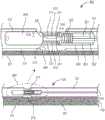
现在转向附图,根据例示的实施例,图1示出了根据第一实施例的具有集成的生物传感器的医疗装置,其中医疗装置100的管状主体被构造为具有第一内腔150和第二内腔250。第一内腔包括布置于其中的一氧化氮(NO)供体材料201。一氧化氮供体材料被构造成用于在与水分接触时提供NO气体。就这一点而言,当将装置放置在体内时,水分将渗透管状主体101到第一内腔中,在该处它使NO供体材料饱和,从而释放NO气体。NO气体通过通道275从第一内腔传递至第二内腔。所述通道紧邻与生物传感器300相关联的感测体积375定位。Turning now to the drawings, in accordance with an illustrative embodiment, FIG. 1 shows a medical device with an integrated biosensor according to a first embodiment, wherein the tubular body of the
生物传感器通常包括隔离层311、分析物反应层312和分析物扩散控制层313,将它们统称为“施加层”。将隔离层施加到工作电极301的导线部分上,在该处已去除了PFA套管的约1.0mm区段。如本文别处所述,隔离层包括多个子层,所述多个子层包括多个含全氟化磺酸树脂的子层和多个电聚合子层。将分析物反应层施加在隔离层的外表面上。最后,将分析物扩散控制层施加在分析物反应层的外表面上。A biosensor typically includes an
工作电极导线通常用
工作电极、隔离层、分析物反应层和分析物扩散控制层加上参比电极共同限定了功能性生物传感器300。The working electrode, spacer layer, analyte reactive layer, and analyte diffusion control layer plus the reference electrode collectively define a
将生物传感器放置在第二内腔250中,在邻近管状主体101的孔330的位置处。一般来讲,管状主体由激光修饰以分别蚀刻孔和通道,但可以类似地实施其他技术。在引入生物传感器之前,可以将任选的硅氧烷涂层施加到通道上,以在第二内腔与NO供体材料之间形成膜285。硅氧烷膜将可透过水分和NO气体。The biosensor is placed in the
为了清楚起见,生物传感器的相关部分在图1的放大部分中示出。For clarity, relevant parts of the biosensor are shown in an enlarged part of FIG. 1 .
第三距离(D3)在图1中表示出,并且涉及参比电极与工作电极之间的距离。参比电极必须足够靠近工作电极以获得信号,但不能太靠近以免短路。参比电极与工作电极之间的距离的径向分量(D3a)(通常为工作电极导线上的PTFE涂层的厚度)包括在约0.2mm与5.0mm之间。工作电极与参比电极之间的距离的纵向分量(D3b)在功能上限定了分析物反应层与参比电极之间的间隙,包括在约0.2mm与10.0mm之间。因此,可以说工作电极与参比电极之间的距离或第三距离(D3)包括在0.2mm与10.0mm之间。优选的是,参比电极(如果包括盘绕导线)应包括至少十个绕组,以使得线圈的表面积是工作电极的表面积的至少两倍。在任何实施例中,参比电极的表面积应为工作电极的表面积的约两倍。The third distance (D3) is represented in Figure 1 and relates to the distance between the reference electrode and the working electrode. The reference electrode must be close enough to the working electrode to obtain a signal, but not too close to avoid shorting. The radial component (D3a) of the distance between the reference electrode and the working electrode (typically the thickness of the PTFE coating on the working electrode wire) is comprised between about 0.2 mm and 5.0 mm. The longitudinal component (D3b) of the distance between the working electrode and the reference electrode functionally defines the gap between the analyte-reactive layer and the reference electrode, and is comprised between about 0.2 mm and 10.0 mm. Therefore, it can be said that the distance or third distance (D3) between the working electrode and the reference electrode is comprised between 0.2 mm and 10.0 mm. Preferably, the reference electrode (if comprising coiled wire) should comprise at least ten windings such that the surface area of the coil is at least twice the surface area of the working electrode. In any embodiment, the surface area of the reference electrode should be about twice the surface area of the working electrode.
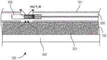
图2进一步展示了具有集成的生物传感器300的医疗装置100,所述医疗装置被构造成用于在NO供体材料201与水分接触时产生一氧化氮气体(“NO气体”),并且将一氧化氮气体通过通道且围绕与生物传感器相关联的感测体积传递。如图所示,一氧化氮供体材料包含在第一内腔150中,而生物传感器元件部分包含在第二内腔250中。进一步示出了第三内腔350,其中第三内腔可以用于平移引导线,从而将药物或流体传递至患者、或其组合。Figure 2 further illustrates a
管状主体101可以通过挤出制造以含有任何希望数量的内腔。观察管状主体101的截面,内壁199变得可见。The
图3A示出了根据三内腔实施例的医疗装置的管状主体的截面,所述管状主体包括布置在管状主体的中心处的第一内腔150;布置在管状主体的周边处的第二内腔250和布置在管状主体的周边处并且在与第二内腔相对的一侧处的第三内腔350。可以类似地实施其他构造。3A shows a cross-section of a tubular body of a medical device including a
图3B示出了根据四内腔实施例的医疗装置的管状主体的截面,所述管状主体包括第一内腔150、第二内腔250、第三内腔350、和第四内腔450。还示出了布置在内腔之间的内壁199。3B shows a cross-section of a tubular body of a medical device including a
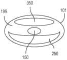
图4示出了根据另一个实施例的具有两个集成的生物传感器(分别为300a;300b)的医疗装置100。尽管示出了两个生物传感器,但是可以实施任何多个生物传感器到此类生物传感器可以装配在内腔空间内的程度。医疗装置包括管状主体101和内壁199。在管状主体周边与内壁之间是至少三个内腔(分别为150;250;350)。在第一内腔150中布置一氧化氮供体材料201。示出了第一生物传感器300a,所述第一生物传感器具有各自在第二内腔250内延伸的第一工作电极301a和第一参比电极321a。示出了第二生物传感器300b,所述第二生物传感器具有各自也在第二内腔250内延伸的第二工作电极301b和第二参比电极321b。就这一点而言,将两个生物传感器容纳在第二内腔中,所述第二内腔被取向成平行于第一内腔并且邻近其布置。在第一生物传感器300a与第二内腔之间是第一NO通道275a,所述第一NO通道被构造成用于将NO气体传递至第一生物传感器的第一感测体积。而且,在第二生物传感器200b与第二内腔之间是第二NO通道275b,所述第二NO通道被构造成用于将NO气体传递至第二生物传感器的第二感测体积。第二生物传感器被定位成距第一生物传感器距离D1,即在5.0mm与50.0mm之间,优选在10.0mm与20.0mm之间的距离。孔的直径(D2)在0.1mm与10.0mm之间,优选1.0mm。Figure 4 shows a
图5示出了根据又另一个实施例的具有三个集成的生物传感器(分别为300a;300b;300c)的医疗装置100。在此,第一生物传感器和第二生物传感器共享共同的工作电极导线,而第三且独立的生物传感器邻近第二生物传感器定位。第一生物传感器被构造成用于检测第一分析物,并且第二生物传感器和第三生物传感器中的每一个均被构造成用于检测第二分析物,这意味着将适当的“施加层”施加至相应的电极。将来自第三生物传感器300c的信号的值用于测量第二分析物;而从第一生物传感器300a和第二生物传感器300b的组合值中减去第三生物传感器300c的信号值以发现用于测量第一分析物的增量信号。Figure 5 shows a
医疗装置包括管状主体101和内壁199。在管状主体周边与内壁之间是至少三个内腔(分别为150;250;350)。在第一内腔150中布置一氧化氮供体材料201。示出了第一生物传感器300a,所述第一生物传感器具有各自在第二内腔250内延伸的第一工作电极301a和第一参比电极321a。示出了第二生物传感器300b,所述第二生物传感器共享工作电极301a,并且进一步包括第二参比电极321b,它们各自也在第二内腔250内延伸。第三生物传感器300c包括第二工作电极301b和第三参比电极321c。就这一点而言,将三个生物传感器各自容纳在第二内腔中,所述第二内腔被取向成平行于第一内腔并且邻近其布置。在第一生物传感器300a与第二内腔之间是第一NO通道275a,所述第一NO通道被构造成用于将NO气体传递至第一生物传感器的第一感测体积。而且,在第二生物传感器300b和第三生物传感器300c与第二内腔之间是第二NO通道275b,所述第二NO通道被构造成用于将NO气体传递至第二生物传感器的第二感测体积和第三生物传感器的第三感测体积。The medical device includes a
上文的每个实施例展示了具有集成的一个或多个生物传感器的医疗装置,所述医疗装置被构造成用于由于NO气体释放组分(诸如NO供体材料)和管状主体内壁内的用于传递NO气体的通道而改善功能。Each of the above examples demonstrates a medical device with integrated one or more biosensors that is configured for use with NO gas releasing components (such as NO donor materials) and within the inner wall of the tubular body. The channel for the delivery of NO gas improves the function.
实例1:具有集成的生物传感器的医疗装置的制造。Example 1: Fabrication of medical devices with integrated biosensors.
概述:Overview:
在通常包括以下的一系列步骤之后制造出释放一氧化氮(NO)的乳酸和葡萄糖安培电化学传感器(两个电极系统:工作/参比):(i)清洁接受者工作电极引线;(ii)放置/聚合隔离层(包括子层);(iii)沉积和交联分析物反应层;(iv)放置分析物扩散控制层;(v)任选地将传感器安装在医疗装置的内腔内,所述医疗装置诸如导管,更特别地是多内腔导管;(vi)使用恒电位仪装置测试传感器以评价分析准确性,或在一氧化氮分析仪(NOA)中测试传感器以评价NO释放量值、寿命、或两者。Nitric oxide (NO)-releasing lactate and glucose amperometric electrochemical sensors (two electrode systems: working/reference) are fabricated after a series of steps typically including: (i) cleaning of the receiver working electrode lead; (ii) ) placing/polymerizing a spacer layer (including sublayers); (iii) depositing and cross-linking an analyte reactive layer; (iv) placing an analyte diffusion control layer; (v) optionally mounting a sensor within the lumen of the medical device , the medical device such as a catheter, more particularly a multi-lumen catheter; (vi) testing the sensor using a potentiostat device to evaluate analytical accuracy, or a nitric oxide analyzer (NOA) to evaluate NO release magnitude, lifetime, or both.
材料:Material:
将铂-铱导线(或类似的工作电极导线)充当生物传感器以及工作电极的结构基础,对于此,约10%铱含量将是足够的,而纯铂导线本身太软。导线上的PFA涂层使所有地方的导线均绝缘,除了根据设计将其去除以防止测量期间的短路的地方。A platinum-iridium wire (or similar working electrode wire) serves as the structural basis for the biosensor as well as the working electrode, for which a content of about 10% iridium would be sufficient, whereas pure platinum wire is inherently too soft. The PFA coating on the wire insulates the wire everywhere except where it is removed by design to prevent short circuits during measurements.
一旦将经暴露的引线在氯化银(AgCl)中进行涂覆以在水性环境(PBS,含有Cl-)中起作用,银导线(或类似的参比电极导线)就充当参比电极。导线上的PFA涂层使所有地方的导线均绝缘,除了根据设计将其去除以防止传感器在测量期间短路的地方。Once the exposed leads were coated in silver chloride (AgCl) to function in an aqueous environment (PBS, containing Cl-), the silver wire (or similar reference electrode lead) served as the reference electrode. The PFA coating on the wire insulates the wire everywhere except where it is removed by design to prevent the sensor from shorting out during measurement.
将全氟化磺酸树脂(PFSA),诸如可商购获得的
包含1-3二氨基苯(间苯二胺)和间苯二酚的电聚合膜形成了生物传感器的隔离层的两个隔离子层中的第二子层。作为尺寸排阻限制层,它针对高分子量物种,即对乙酰氨基酚进行选择,所述对乙酰氨基酚可以通过氧化产生干扰电流响应。The electropolymerized film comprising 1-3 diaminobenzene (m-phenylenediamine) and resorcinol formed the second of the two spacer sublayers of the biosensor's spacer layer. As a size exclusion limiting layer, it is selected for the high molecular weight species, acetaminophen, which can interfere with the current response through oxidation.
传感器的功能性取决于酶(诸如本文提及葡萄糖氧化酶和乳酸氧化酶等)的交联/固定化,所述酶消耗感兴趣的分析物(局部的,对本体浓度无影响)并且产生过氧化氢,所述过氧化氢随后在工作电极界面处被氧化,以产生与本体溶液中分析物浓度成比例的响应电流信号。The functionality of the sensor depends on the cross-linking/immobilization of enzymes (such as glucose oxidase and lactate oxidase mentioned herein) that consume the analyte of interest (locally, with no effect on bulk concentration) and generate excess energy. Hydrogen oxide, which is then oxidized at the working electrode interface, to produce a response current signal proportional to the analyte concentration in the bulk solution.
向传感器添加外层限制了感兴趣的分析物向酶的扩散,从而使酶辅因子氧成为限量试剂。该层可以由单一聚合物构成,诸如例如E2A Elast-eon聚氨酯(2%-5%溶液),FGRTV硅橡胶(湿固化)或PLURONIC
在没有分析物扩散控制层的情况下,生物传感器响应可能不具有可操作的线性范围,并且可能变成非常敏感但为二元的分析物检测器。Without the analyte diffusion control layer, the biosensor response may not have an operational linear range and may become a very sensitive but binary analyte detector.
传感器壳体典型地是小的多内腔导管,其中一个内腔专门用于容纳生物传感器元件部分。可以在导管/壳体中切割出孔,以使得生物传感器与外部溶液具有接触。这些孔是使用CO2激光蚀刻机可再现地产生的。The sensor housing is typically a small multi-lumen catheter with one lumen dedicated to housing the biosensor element portion. A hole can be cut in the conduit/housing to allow the biosensor to have contact with the external solution. The holes were reproducibly produced using aCO2 laser etcher.
与溶液接触端相对的生物传感器的两个导线电极(工作电极和参比电极)的PFA涂层必须稍微剥离,以提供与恒电位仪装置的电接触界面。这是为了向传感器供应外部电压并测量所产生的电流信号。The PFA coating of the two wire electrodes (working and reference) of the biosensor opposite the solution contacting end had to be peeled off slightly to provide an electrical contact interface with the potentiostat device. This is to supply an external voltage to the sensor and measure the resulting current signal.
如果要实施导管壳体,则通常将这两个电极引线通过耳机接口连接器连接到无线移动式恒电位仪上。将长电极引线以远端-到-近端送入(以避免损坏酶端)并且焊接到耳机接口上。If a catheter housing is to be implemented, these two electrode leads are typically connected to a wireless mobile potentiostat through a headphone jack connector. The long electrode leads were fed distal-to-proximal (to avoid damaging the enzyme end) and soldered to the headphone jack.
一旦已经放置传感器,就可以使用UV固化RTV来固定传感器,否则生物传感器可能会移动离开孔并变得不起作用。Once the sensor has been placed, a UV-cured RTV can be used to secure the sensor, otherwise the biosensor may move out of the hole and become ineffective.
已将SNAP(RSNO)和UV固化RTV的混合物用于使其他导管内腔填充有NO释放聚合物,但是可以类似地实施其他NO释放材料。SNAP和RTV混合物可以储存在注射器中,经由冰箱冷藏,并且由于溶液粘度高,与钻床一起使用以施加足以填充内腔的外部压力。Mixtures of SNAP (RSNO) and UV-cured RTV have been used to fill other catheter lumens with NO-releasing polymers, but other NO-releasing materials can be implemented similarly. The SNAP and RTV mixture can be stored in a syringe, refrigerated via refrigerator, and used with a drill press to apply external pressure sufficient to fill the lumen due to the high viscosity of the solution.
组装步骤:Assembly steps:
第1天:Day 1:
将工作电极和参比电极从导线的源线轴上切下成所希望的长度,并且拉直至所希望的程度。将导线用PFA涂覆。The working and reference electrodes were cut to the desired length from the source spool of wire and pulled to the desired extent. Coat the wires with PFA.
在工作电极的端部上切出大约1mm的小内腔。这可以通过剃刀刀片并应用“圆形切割”技术来完成。重要的是切割PFA涂层,但切割的深度不能足以切下或切断导线。而且,从相对端部切掉大约5mm的PFA以作为用于连接电聚合导线的引线。A small lumen of about 1 mm was cut on the end of the working electrode. This can be done with a razor blade and applying the "circle cut" technique. It is important to cut the PFA coating, but not deep enough to cut or sever the wire. Also, about 5 mm of PFA was cut from the opposite end to serve as lead wires for connecting the electropolymerized wires.
使用戴有乳胶手套的手指用拇指和食指指甲提拉PFA涂层,以产生工作电极区域(暴露的Pt/Ir导线),至关重要的是该区域的表面积小于参比电极。用剃刀刀片切掉任何多余的PFA。Using a latex gloved finger, lift the PFA coating with the thumb and index fingernail to create the working electrode area (exposed Pt/Ir wire), which critically has a smaller surface area than the reference electrode. Cut off any excess PFA with a razor blade.
通过将工作电极在HCl中然后在乙醇中进行超声处理来清洁工作电极。The working electrode was cleaned by sonicating the working electrode in HCl and then in ethanol.
将导线垂直悬挂,其顶端在下端。使用导线套环(或小瓶)施加全氟化磺酸树脂(PFSA)的约五次浸涂,在每次施加之间进行干燥。Hang the wire vertically with the top end at the bottom. About five dip coats of perfluorinated sulfonic acid resin (PFSA) were applied using a wire collar (or vial), drying between each application.
准备烘箱至约165℃以用于退火PFSA子层。Prepare the oven to about 165°C for annealing the PFSA sublayer.
在充分的干燥时间之后,将导线放置在烘箱中以进行退火。使用玻璃支撑导线。注意不要妨碍空腔/工作电极区域。After a sufficient drying time, the wire is placed in an oven for annealing. Use glass to support the wires. Take care not to obstruct the cavity/working electrode area.
将传感器在165℃下静置约一小时,然后将烘箱上的标度盘调至最小/关闭。Let the sensor sit at 165°C for about an hour, then turn the dial on the oven to min/off.
在传感器和烘箱达到室温之前,请勿打开烘箱门。快速冷却会导致PFSA破裂或变得无效。Do not open the oven door until the sensor and oven have reached room temperature. Rapid cooling can cause the PFSA to crack or become ineffective.
在烘箱冷却的同时,准备一定量的PBS溶液以进行电聚合,并且在使用前置于氮气(N2)气体吹扫(针头)下以便去除溶解的氧。While the oven was cooling, an amount of PBS solution was prepared for electropolymerization and placed under a nitrogen (N2 ) gas purge (needle) to remove dissolved oxygen prior to use.
将第一量的经N2吹扫的PBS添加至一个含有1,3-二氨基苯的琥珀色小瓶中,将第二量的经N2吹扫的PBS添加至另一个含有间苯二酚的琥珀色小瓶中,然后将它们合并在反应池中。A first amount of N2-purged PBS was added to one amber vial containing 1,3-diaminobenzene and a second amount of N2-purged PBS was added to another amber containing resorcinol color vials and combine them in the reaction cell.
将所有工作电极引线连接至来自恒电位仪的一根工作电极缆线上。所有孔都应在溶液中。将来自恒电位仪的参比电极缆线连接至Ag/AgCl电极上。Ag/AgCl电极可以是商用的或者是浸泡在HCl/FeCl3溶液中的粗银导线。该Ag/AgCl电极的表面积必须大于Pt/Ir组合表面积的表面积。Connect all working electrode leads to one working electrode cable from the potentiostat. All wells should be in solution. Connect the reference electrode cable from the potentiostat to the Ag/AgCl electrode. Ag/AgCl electrodes can be commercial or thick silver wires soaked in HCl/FeCl3 solution. The surface area of the Ag/AgCl electrode must be larger than that of the combined Pt/Ir surface area.
执行循环伏安法过夜或持续至少6小时。Perform cyclic voltammetry overnight or for at least 6 hours.
第2天:Day 2:
在电聚合结束之前,准备一种或多种酶溶液和一种或多种戊二醛溶液。Before the end of the electropolymerization, one or more enzyme solutions and one or more glutaraldehyde solutions are prepared.
通过将牛血清白蛋白(BSA)、GOx酶和去离子水合并,然后充分混合来准备葡萄糖氧化酶(“GOx”)。Glucose oxidase ("GOx") was prepared by combining bovine serum albumin (BSA), GOx enzyme, and deionized water, followed by thorough mixing.
通过将戊二醛和去离子水或磷酸盐缓冲盐水(PBS)合并来准备谷氨酰胺-GOx(针对GOx)。Glutamine-GOx (for GOx) was prepared by combining glutaraldehyde and deionized water or phosphate buffered saline (PBS).
通过将BSA、PBS和聚乙烯亚胺(PEI)合并(所述组合形成“溶液1”)来准备乳酸氧化酶。将溶液1与乳酸氧化酶的酶组合。Lactate oxidase was prepared by combining BSA, PBS and polyethyleneimine (PEI) (the combination formed "Solution 1"). Solution 1 was combined with the enzyme lactate oxidase.
通过将戊二醛和PBS合并来准备戊二醛-LOx;需注意,对于LOx提供的戊二醛浓度较低。Prepare glutaraldehyde-LOx by combining glutaraldehyde and PBS; note that lower concentrations of glutaraldehyde are provided for LOx.
取出恒电位仪电极导线,用去离子水短暂冲洗电极,并且用一短阵的压缩空气干燥。Remove the potentiostat electrode leads, rinse the electrode briefly with deionized water, and dry with a short burst of compressed air.
固定电极以将酶溶液放置到孔/工作电极区域中。溶解酶溶液并确保小瓶中没有酶残留。The electrode is fixed to place the enzyme solution into the well/working electrode area. Dissolve the enzyme solution and ensure that no enzyme remains in the vial.
用去离子水清洗并准备好气密性注射器。Rinse with deionized water and prepare a gas-tight syringe.
为正准备的多个传感器拟定所希望的酶量,对于每个传感器使用约0.5-1.0uL。小心地沉积酶溶液,确保液滴跨越了PGA涂层的区段之间的间隙并且涂覆了整个工作电极区域。在沉积相同类型的所有酶后,充分冲洗注射器。Work out the desired amount of enzyme for the multiple sensors being prepared, using about 0.5-1.0 uL for each sensor. The enzyme solution was deposited carefully, ensuring that the droplet spanned the gap between the PGA-coated segments and coated the entire working electrode area. After depositing all enzymes of the same type, rinse the syringe thoroughly.
让酶溶液在传感器工作区域上滴干,例如约一小时,这取决于酶。Allow the enzyme solution to drip dry on the sensor work area, eg, about an hour, depending on the enzyme.
用注射器将戊二醛溶液添加到工作电极区域,在干燥的酶之上。戊二醛应重新水合酶。确保整个区域均涂覆有戊二醛并且冲洗注射器。The glutaraldehyde solution was added with a syringe to the working electrode area, on top of the dried enzyme. Glutaraldehyde should rehydrate the enzyme. Make sure the entire area is coated with glutaraldehyde and flush the syringe.
使酶-戊二醛发生交联。交联所需的时间取决于所涉及的酶,但可能需要一或两个小时。已经证明交联时间过长(例如,长于12小时)对酶功能有害。Crosslinks the enzyme-glutaraldehyde. The time required for crosslinking depends on the enzymes involved, but can take an hour or two. Excessive cross-linking times (eg, longer than 12 hours) have been shown to be detrimental to enzyme function.
在进行酶/戊二醛交联时,准备参比电极以在第3天使用。A reference electrode was prepared for use on day 3 when enzymatic/glutaraldehyde cross-linking was performed.
将涂有PFA的Ag导线的长度切割为比Pt/Ir工作电极长约5cm。使用带有圆形切割技术的剃刀刀片(或激光切割机)从导线的一端去除PFA涂层,从而留下暴露的参考电极区域。将这些导线放置到酸化的HCL/FeCl3溶液中持续约一小时,以生成参比电极操作所需的AgCl涂层。The length of the PFA-coated Ag wire was cut to be approximately 5 cm longer than the Pt/Ir working electrode. Use a razor blade (or laser cutter) with a circular cutting technique to remove the PFA coating from one end of the wire, leaving the exposed reference electrode area. These wires were placed into an acidified HCL/FeCl3 solution for about one hour to generate the AgCl coating required for the operation of the reference electrode.
在酶/戊二醛交联过程中准备分析物扩散控制层涂覆溶液。测量出干燥的组分,包括聚氨酯、硅氧烷RTV、PLURONIC
第3天:3rd day:
在去离子水或PBS的小瓶中冲洗工作电极(现在具有固定化酶)。这种冲洗将溶解并冲洗掉未交联的酶。Rinse the working electrode (now with immobilized enzyme) in a vial of deionized water or PBS. This rinse will dissolve and wash away uncrosslinked enzyme.
将Ag/AgCl参比电极以紧密的线圈缠绕在Pt/Ir/酶工作电极周围。确保线圈紧密接近(在传感器的酶区域的1厘米之内),但不位于酶之上。The Ag/AgCl reference electrode was wound in a tight coil around the Pt/Ir/enzyme working electrode. Make sure the coil is in close proximity (within 1 cm of the enzyme area of the sensor), but not over the enzyme.
用放置在工作(Pt/Ir)和参比(Ag/Cl)电极引线上方的聚对苯二甲酸乙二醇酯(PET)热缩管固定Ag/AgCl电极。这些应紧密接近经盘绕的AgCl和酶区域,以便通过不覆盖它们将它们固定在一起。该热缩管还为传感器组件增加了一些结构上的增强。The Ag/AgCl electrodes were secured with polyethylene terephthalate (PET) heat shrink tubing placed over the working (Pt/Ir) and reference (Ag/Cl) electrode leads. These should be in close proximity to the coiled AgCl and enzyme regions in order to hold them together by not covering them. The heat shrink also adds some structural enhancements to the sensor assembly.
使用热风枪或可调节温度的吹风机将热缩管和所有电极全部活化并将其锁定在恰当位置。未固定的电极可能会散开或受到损坏。Use a heat gun or adjustable temperature hair dryer to fully activate and lock the heat shrink tubing and all electrodes in place. Unsecured electrodes may come apart or be damaged.
将电极悬挂,以便通过导线套环浇铸或浸涂施加外层(分析物扩散控制层)。The electrodes were suspended so that the outer layer (analyte diffusion control layer) was applied by wire collar casting or dip coating.
导线套环浇铸:将分析物扩散控制层涂覆溶液沉积到导线套环上并且以向上和向下的模式使其穿过传感器组件的外表面(通常约2cm)。为了确保各个传感器之间的一致性,在向每个传感器施加外层涂覆之间,应将套环用THF溶液中的浸液冲洗,并且随后进行干燥。使每个传感器在涂覆之间进行干燥。向每个传感器应施加约五至八次外层涂覆。Wire ferrule casting: The analyte diffusion control layer coating solution was deposited onto the wire ferrule and passed through the outer surface of the sensor assembly (typically about 2 cm) in an upward and downward pattern. To ensure consistency from sensor to sensor, the collars should be rinsed with a dip in a THF solution and then dried between applications of the overcoat to each sensor. Allow each sensor to dry between coats. Approximately five to eight overcoats should be applied to each sensor.
浸涂:将一定量的分析物扩散控制层涂覆溶液分配到薄的小瓶中,所述小瓶优选具有可密封的顶部以最大程度地减少THF的蒸发。将每个传感器浸渍在小瓶中,以使得在每个传感器上涂覆相同的长度(约2cm),以增强各个传感器之间的一致性。使每个传感器在涂覆之间进行干燥。向每个传感器应施加约五次外层涂覆。Dip coating: Dispense an amount of the analyte diffusion control layer coating solution into a thin vial, preferably with a sealable top to minimize THF evaporation. Each sensor was dipped in a vial so that the same length (approximately 2 cm) was coated on each sensor to enhance uniformity between individual sensors. Allow each sensor to dry between coats. About five overcoats should be applied to each sensor.
如果传感器要立即使用,则在连接到恒电位仪之前将其放置在PBS溶液中。然而,如果将传感器保持储存且供随后使用,则放置在冰箱或室温储存容器中。不建议冷冻,这是由于会形成微裂纹,从而可能使传感器不起作用。If the sensor is to be used immediately, place it in the PBS solution before connecting to the potentiostat. However, if the sensor is to be kept in storage for later use, place in a refrigerator or room temperature storage container. Freezing is not recommended as microcracks can form which may render the sensor inoperative.
前述步骤说明了用于实践本发明的一个具体实施例;然而,本领域技术人员应了解本文所披露的特征的多种可能的替代组合和安排。这样,描述旨在仅是使能性的,而非限制性的。而是,本发明的精神和范围在所附权利要求中阐明。The foregoing steps illustrate one specific embodiment for practicing the invention; however, those skilled in the art will appreciate many possible alternative combinations and arrangements of the features disclosed herein. As such, the description is intended to be enabling only, and not restrictive. Rather, the spirit and scope of the invention are set forth in the appended claims.
工业适用性Industrial Applicability
具有集成的生物传感器的医疗装置适用于人和动物医疗诊所以及医院来进行医学治疗。具体地,本文描述的医疗装置可用于检测分析物,诸如但不限于患者血液中的葡萄糖和乳酸,以及可用于导管等的常规用途。Medical devices with integrated biosensors are suitable for human and animal medical clinics as well as hospitals for medical treatment. In particular, the medical devices described herein can be used to detect analytes, such as, but not limited to, glucose and lactate in a patient's blood, and can be used for routine use in catheters and the like.
附图标记清单List of reference numbers
医疗装置(100)Medical Devices(100)
管状主体(101)Tubular Body (101)
第一内腔(150)first lumen (150)
内壁(199)Inner Wall(199)
一氧化氮供体材料(201)Nitric Oxide Donor Materials (201)
第二内腔(250)Second lumen (250)
一氧化氮通道(275;275a;275b)Nitric oxide channels (275; 275a; 275b)
NO和H2O可透过膜(285)NO and H2 O permeable membrane (285)
生物传感器(300;300a;300b)Biosensors (300; 300a; 300b)
工作电极(301;301a;301b)Working electrode (301; 301a; 301b)
基于聚四氟乙烯的涂层(302)PTFE based coatings (302)
热缩管(303)Heat Shrink Tube(303)
聚氨酯涂层(304)Polyurethane coating(304)
隔离层(311)Isolation Layer(311)
分析物反应层(312)Analyte Response Layer (312)
分析物扩散控制层(313)Analyte Diffusion Control Layer (313)
参比电极(321;321a;321b;321c)Reference electrode (321; 321a; 321b; 321c)
盘绕导线(322)Coiled Wire(322)
孔(330)Hole(330)
第三内腔(350)Third lumen (350)
感测体积(375)Sensing Volume(375)
第四内腔(450)Fourth lumen (450)
第一距离(D1)First distance (D1)
第二距离(D2)Second distance (D2)
第三距离(D3;D3a;D3b)Third distance (D3; D3a; D3b)
Claims (22)
Translated fromChineseApplications Claiming Priority (3)
| Application Number | Priority Date | Filing Date | Title |
|---|---|---|---|
| US201762553832P | 2017-09-02 | 2017-09-02 | |
| US62/553,832 | 2017-09-02 | ||
| PCT/US2018/049377WO2019046853A1 (en) | 2017-09-02 | 2018-09-04 | Medical device with integrated biosensor |
Publications (2)
| Publication Number | Publication Date |
|---|---|
| CN111601626Atrue CN111601626A (en) | 2020-08-28 |
| CN111601626B CN111601626B (en) | 2022-05-06 |
Family
ID=65517578
Family Applications (1)
| Application Number | Title | Priority Date | Filing Date |
|---|---|---|---|
| CN201880068334.9AActiveCN111601626B (en) | 2017-09-02 | 2018-09-04 | Medical device with integrated biosensor |
Country Status (3)
| Country | Link |
|---|---|
| US (1) | US10709360B2 (en) |
| CN (1) | CN111601626B (en) |
| WO (1) | WO2019046853A1 (en) |
Cited By (1)
| Publication number | Priority date | Publication date | Assignee | Title |
|---|---|---|---|---|
| CN118203451A (en)* | 2024-05-16 | 2024-06-18 | 中国科学院苏州生物医学工程技术研究所 | Nitric Oxide Therapy Device for Animal Disease Prevention and Treatment |
Families Citing this family (2)
| Publication number | Priority date | Publication date | Assignee | Title |
|---|---|---|---|---|
| KR20220020347A (en)* | 2019-06-10 | 2022-02-18 | 더 리젠츠 오브 더 유니버시티 오브 미시건 | catheterization device |
| US11813059B2 (en) | 2021-11-30 | 2023-11-14 | Zense-Life Inc. | Sensor for a continuous biological monitor having nitric oxide releasing compound |
Citations (5)
| Publication number | Priority date | Publication date | Assignee | Title |
|---|---|---|---|---|
| US20030009127A1 (en)* | 2001-05-25 | 2003-01-09 | Trescony Paul V. | Implantable medical device with controllable gaseous agent release system |
| CN101175529A (en)* | 2005-05-19 | 2008-05-07 | 埃-皮尔制药公司 | Ingestible device for producing nitric oxide in tissues |
| CN102256635A (en)* | 2008-12-19 | 2011-11-23 | 麦德托尼克瓦斯科尔勒公司 | Dry diazeniumdiolation methods for producing nitric oxide releasing medical devices |
| US20150073331A1 (en)* | 2012-03-30 | 2015-03-12 | The Regents Of The University Of Michigan | Nitric oxide delivery devices |
| US20160339197A1 (en)* | 2012-03-30 | 2016-11-24 | The Regents Of The University Of Michigan | Nitric oxide delivery devices |
Family Cites Families (12)
| Publication number | Priority date | Publication date | Assignee | Title |
|---|---|---|---|---|
| US7769420B2 (en)* | 2000-05-15 | 2010-08-03 | Silver James H | Sensors for detecting substances indicative of stroke, ischemia, or myocardial infarction |
| US9295391B1 (en)* | 2000-11-10 | 2016-03-29 | The General Hospital Corporation | Spectrally encoded miniature endoscopic imaging probe |
| US7476210B2 (en)* | 2001-01-04 | 2009-01-13 | Transvivo Inc. | Apparatus and method for in-vivo plasmapheresis using periodic backflush containing anticoagulant |
| US7128904B2 (en)* | 2001-01-16 | 2006-10-31 | The Regents Of The University Of Michigan | Material containing metal ion ligand complex producing nitric oxide in contact with blood |
| US20060008529A1 (en)* | 2004-07-12 | 2006-01-12 | Meyerhoff Mark E | Use of additive sites to control nitric oxide release from nitric oxide donors contained within polymers |
| US20060039950A1 (en)* | 2004-08-23 | 2006-02-23 | Zhengrong Zhou | Multi-functional biocompatible coatings for intravascular devices |
| CA2613106A1 (en) | 2005-06-30 | 2007-01-11 | Mc3, Inc. | Analyte sensors and compositions for use therein |
| US20070093697A1 (en)* | 2005-10-21 | 2007-04-26 | Theranova, Llc | Method and apparatus for detection of right to left shunting in the cardiopulmonary vasculature |
| US20090069743A1 (en)* | 2007-09-11 | 2009-03-12 | Baxter International Inc. | Infusion therapy sensor system |
| EP2209472A1 (en)* | 2007-10-12 | 2010-07-28 | The University of North Carolina at Chapel Hill | Use of nitric oxide to enhance the efficacy of silver and other topical wound care agents |
| EP2448486B1 (en) | 2009-07-02 | 2021-08-25 | Dexcom, Inc. | Analyte sensors and methods of manufacturing same |
| EP2953660B1 (en)* | 2013-02-07 | 2020-04-01 | The Regents Of The University Of Michigan | Thromboresistant/bactericidal s-nitroso-n-acetylpenicillamine (snap)-doped nitric oxide release polymers with enhanced stability |
- 2018
- 2018-09-04USUS16/121,405patent/US10709360B2/enactiveActive
- 2018-09-04CNCN201880068334.9Apatent/CN111601626B/enactiveActive
- 2018-09-04WOPCT/US2018/049377patent/WO2019046853A1/ennot_activeCeased
Patent Citations (5)
| Publication number | Priority date | Publication date | Assignee | Title |
|---|---|---|---|---|
| US20030009127A1 (en)* | 2001-05-25 | 2003-01-09 | Trescony Paul V. | Implantable medical device with controllable gaseous agent release system |
| CN101175529A (en)* | 2005-05-19 | 2008-05-07 | 埃-皮尔制药公司 | Ingestible device for producing nitric oxide in tissues |
| CN102256635A (en)* | 2008-12-19 | 2011-11-23 | 麦德托尼克瓦斯科尔勒公司 | Dry diazeniumdiolation methods for producing nitric oxide releasing medical devices |
| US20150073331A1 (en)* | 2012-03-30 | 2015-03-12 | The Regents Of The University Of Michigan | Nitric oxide delivery devices |
| US20160339197A1 (en)* | 2012-03-30 | 2016-11-24 | The Regents Of The University Of Michigan | Nitric oxide delivery devices |
Cited By (1)
| Publication number | Priority date | Publication date | Assignee | Title |
|---|---|---|---|---|
| CN118203451A (en)* | 2024-05-16 | 2024-06-18 | 中国科学院苏州生物医学工程技术研究所 | Nitric Oxide Therapy Device for Animal Disease Prevention and Treatment |
Also Published As
| Publication number | Publication date |
|---|---|
| US10709360B2 (en) | 2020-07-14 |
| CN111601626B (en) | 2022-05-06 |
| WO2019046853A1 (en) | 2019-03-07 |
| US20190069820A1 (en) | 2019-03-07 |
Similar Documents
| Publication | Publication Date | Title |
|---|---|---|
| US20230060985A1 (en) | Glucose sensor electrode design | |
| US10772540B2 (en) | Microarray electrodes useful with analyte sensors and methods for making and using them | |
| US8696917B2 (en) | Analyte sensor and fabrication methods | |
| Weltin et al. | Microfabricated, amperometric, enzyme-based biosensors for in vivo applications | |
| US20190094169A1 (en) | Enzyme matrices for use with ethylene oxide sterilization | |
| JP5234967B2 (en) | Flux limiting membranes for intravenous amperometric biosensors | |
| US10426383B2 (en) | Muting glucose sensor oxygen response and reducing electrode edge growth with pulsed current plating | |
| US20130150689A1 (en) | Device for sensing a target chemical and method of its making | |
| CN111601626B (en) | Medical device with integrated biosensor | |
| US20150276651A1 (en) | Analyte sensor and fabrication methods | |
| AU2019217883B2 (en) | Methods for controlling physical vapor deposition metal film adhesion to substrates and surfaces | |
| US20110017594A1 (en) | Analyte sensor fabrication | |
| EP4003166B1 (en) | Sensor with improved oxygen delivery and corresponding method | |
| CN116223788A (en) | Ketone limiting membrane and bilayer membrane method for ketone sensing |
Legal Events
| Date | Code | Title | Description |
|---|---|---|---|
| PB01 | Publication | ||
| PB01 | Publication | ||
| SE01 | Entry into force of request for substantive examination | ||
| SE01 | Entry into force of request for substantive examination | ||
| GR01 | Patent grant | ||
| GR01 | Patent grant |