CN111526778A - Intraoral imaging device, medical device, and program - Google Patents
Intraoral imaging device, medical device, and programDownload PDFInfo
- Publication number
- CN111526778A CN111526778ACN201880083646.7ACN201880083646ACN111526778ACN 111526778 ACN111526778 ACN 111526778ACN 201880083646 ACN201880083646 ACN 201880083646ACN 111526778 ACN111526778 ACN 111526778A
- Authority
- CN
- China
- Prior art keywords
- intraoral
- imaging device
- image
- imaging
- determination
- Prior art date
- Legal status (The legal status is an assumption and is not a legal conclusion. Google has not performed a legal analysis and makes no representation as to the accuracy of the status listed.)
- Granted
Links
Images


Classifications
- A—HUMAN NECESSITIES
- A61—MEDICAL OR VETERINARY SCIENCE; HYGIENE
- A61B—DIAGNOSIS; SURGERY; IDENTIFICATION
- A61B1/00—Instruments for performing medical examinations of the interior of cavities or tubes of the body by visual or photographical inspection, e.g. endoscopes; Illuminating arrangements therefor
- A61B1/24—Instruments for performing medical examinations of the interior of cavities or tubes of the body by visual or photographical inspection, e.g. endoscopes; Illuminating arrangements therefor for the mouth, i.e. stomatoscopes, e.g. with tongue depressors; Instruments for opening or keeping open the mouth
- A—HUMAN NECESSITIES
- A61—MEDICAL OR VETERINARY SCIENCE; HYGIENE
- A61B—DIAGNOSIS; SURGERY; IDENTIFICATION
- A61B1/00—Instruments for performing medical examinations of the interior of cavities or tubes of the body by visual or photographical inspection, e.g. endoscopes; Illuminating arrangements therefor
- A61B1/32—Devices for opening or enlarging the visual field, e.g. of a tube of the body
- A—HUMAN NECESSITIES
- A61—MEDICAL OR VETERINARY SCIENCE; HYGIENE
- A61B—DIAGNOSIS; SURGERY; IDENTIFICATION
- A61B1/00—Instruments for performing medical examinations of the interior of cavities or tubes of the body by visual or photographical inspection, e.g. endoscopes; Illuminating arrangements therefor
- A61B1/00002—Operational features of endoscopes
- A61B1/00004—Operational features of endoscopes characterised by electronic signal processing
- A61B1/00009—Operational features of endoscopes characterised by electronic signal processing of image signals during a use of endoscope
- A61B1/000096—Operational features of endoscopes characterised by electronic signal processing of image signals during a use of endoscope using artificial intelligence
- A—HUMAN NECESSITIES
- A61—MEDICAL OR VETERINARY SCIENCE; HYGIENE
- A61B—DIAGNOSIS; SURGERY; IDENTIFICATION
- A61B1/00—Instruments for performing medical examinations of the interior of cavities or tubes of the body by visual or photographical inspection, e.g. endoscopes; Illuminating arrangements therefor
- A61B1/00002—Operational features of endoscopes
- A61B1/00043—Operational features of endoscopes provided with output arrangements
- A61B1/00045—Display arrangement
- A—HUMAN NECESSITIES
- A61—MEDICAL OR VETERINARY SCIENCE; HYGIENE
- A61B—DIAGNOSIS; SURGERY; IDENTIFICATION
- A61B1/00—Instruments for performing medical examinations of the interior of cavities or tubes of the body by visual or photographical inspection, e.g. endoscopes; Illuminating arrangements therefor
- A61B1/00131—Accessories for endoscopes
- A61B1/00135—Oversleeves mounted on the endoscope prior to insertion
- A—HUMAN NECESSITIES
- A61—MEDICAL OR VETERINARY SCIENCE; HYGIENE
- A61B—DIAGNOSIS; SURGERY; IDENTIFICATION
- A61B1/00—Instruments for performing medical examinations of the interior of cavities or tubes of the body by visual or photographical inspection, e.g. endoscopes; Illuminating arrangements therefor
- A61B1/00147—Holding or positioning arrangements
- A61B1/00154—Holding or positioning arrangements using guiding arrangements for insertion
- A—HUMAN NECESSITIES
- A61—MEDICAL OR VETERINARY SCIENCE; HYGIENE
- A61B—DIAGNOSIS; SURGERY; IDENTIFICATION
- A61B1/00—Instruments for performing medical examinations of the interior of cavities or tubes of the body by visual or photographical inspection, e.g. endoscopes; Illuminating arrangements therefor
- A61B1/04—Instruments for performing medical examinations of the interior of cavities or tubes of the body by visual or photographical inspection, e.g. endoscopes; Illuminating arrangements therefor combined with photographic or television appliances
- A61B1/045—Control thereof
- A—HUMAN NECESSITIES
- A61—MEDICAL OR VETERINARY SCIENCE; HYGIENE
- A61B—DIAGNOSIS; SURGERY; IDENTIFICATION
- A61B1/00—Instruments for performing medical examinations of the interior of cavities or tubes of the body by visual or photographical inspection, e.g. endoscopes; Illuminating arrangements therefor
- A61B1/06—Instruments for performing medical examinations of the interior of cavities or tubes of the body by visual or photographical inspection, e.g. endoscopes; Illuminating arrangements therefor with illuminating arrangements
- A61B1/0661—Endoscope light sources
- A—HUMAN NECESSITIES
- A61—MEDICAL OR VETERINARY SCIENCE; HYGIENE
- A61B—DIAGNOSIS; SURGERY; IDENTIFICATION
- A61B5/00—Measuring for diagnostic purposes; Identification of persons
- A61B5/0059—Measuring for diagnostic purposes; Identification of persons using light, e.g. diagnosis by transillumination, diascopy, fluorescence
- A61B5/0062—Arrangements for scanning
- A—HUMAN NECESSITIES
- A61—MEDICAL OR VETERINARY SCIENCE; HYGIENE
- A61B—DIAGNOSIS; SURGERY; IDENTIFICATION
- A61B5/00—Measuring for diagnostic purposes; Identification of persons
- A61B5/0059—Measuring for diagnostic purposes; Identification of persons using light, e.g. diagnosis by transillumination, diascopy, fluorescence
- A61B5/0082—Measuring for diagnostic purposes; Identification of persons using light, e.g. diagnosis by transillumination, diascopy, fluorescence adapted for particular medical purposes
- A61B5/0088—Measuring for diagnostic purposes; Identification of persons using light, e.g. diagnosis by transillumination, diascopy, fluorescence adapted for particular medical purposes for oral or dental tissue
- A—HUMAN NECESSITIES
- A61—MEDICAL OR VETERINARY SCIENCE; HYGIENE
- A61B—DIAGNOSIS; SURGERY; IDENTIFICATION
- A61B5/00—Measuring for diagnostic purposes; Identification of persons
- A61B5/74—Details of notification to user or communication with user or patient; User input means
- A61B5/742—Details of notification to user or communication with user or patient; User input means using visual displays
- G—PHYSICS
- G06—COMPUTING OR CALCULATING; COUNTING
- G06T—IMAGE DATA PROCESSING OR GENERATION, IN GENERAL
- G06T7/00—Image analysis
- G06T7/0002—Inspection of images, e.g. flaw detection
- G06T7/0012—Biomedical image inspection
- G—PHYSICS
- G16—INFORMATION AND COMMUNICATION TECHNOLOGY [ICT] SPECIALLY ADAPTED FOR SPECIFIC APPLICATION FIELDS
- G16H—HEALTHCARE INFORMATICS, i.e. INFORMATION AND COMMUNICATION TECHNOLOGY [ICT] SPECIALLY ADAPTED FOR THE HANDLING OR PROCESSING OF MEDICAL OR HEALTHCARE DATA
- G16H50/00—ICT specially adapted for medical diagnosis, medical simulation or medical data mining; ICT specially adapted for detecting, monitoring or modelling epidemics or pandemics
- G16H50/20—ICT specially adapted for medical diagnosis, medical simulation or medical data mining; ICT specially adapted for detecting, monitoring or modelling epidemics or pandemics for computer-aided diagnosis, e.g. based on medical expert systems
- A—HUMAN NECESSITIES
- A61—MEDICAL OR VETERINARY SCIENCE; HYGIENE
- A61B—DIAGNOSIS; SURGERY; IDENTIFICATION
- A61B1/00—Instruments for performing medical examinations of the interior of cavities or tubes of the body by visual or photographical inspection, e.g. endoscopes; Illuminating arrangements therefor
- A61B1/00002—Operational features of endoscopes
- A61B1/00059—Operational features of endoscopes provided with identification means for the endoscope
- A—HUMAN NECESSITIES
- A61—MEDICAL OR VETERINARY SCIENCE; HYGIENE
- A61B—DIAGNOSIS; SURGERY; IDENTIFICATION
- A61B1/00—Instruments for performing medical examinations of the interior of cavities or tubes of the body by visual or photographical inspection, e.g. endoscopes; Illuminating arrangements therefor
- A61B1/06—Instruments for performing medical examinations of the interior of cavities or tubes of the body by visual or photographical inspection, e.g. endoscopes; Illuminating arrangements therefor with illuminating arrangements
- A61B1/0638—Instruments for performing medical examinations of the interior of cavities or tubes of the body by visual or photographical inspection, e.g. endoscopes; Illuminating arrangements therefor with illuminating arrangements providing two or more wavelengths
- A—HUMAN NECESSITIES
- A61—MEDICAL OR VETERINARY SCIENCE; HYGIENE
- A61B—DIAGNOSIS; SURGERY; IDENTIFICATION
- A61B1/00—Instruments for performing medical examinations of the interior of cavities or tubes of the body by visual or photographical inspection, e.g. endoscopes; Illuminating arrangements therefor
- A61B1/267—Instruments for performing medical examinations of the interior of cavities or tubes of the body by visual or photographical inspection, e.g. endoscopes; Illuminating arrangements therefor for the respiratory tract, e.g. laryngoscopes, bronchoscopes
- G—PHYSICS
- G06—COMPUTING OR CALCULATING; COUNTING
- G06T—IMAGE DATA PROCESSING OR GENERATION, IN GENERAL
- G06T2200/00—Indexing scheme for image data processing or generation, in general
- G06T2200/24—Indexing scheme for image data processing or generation, in general involving graphical user interfaces [GUIs]
- G—PHYSICS
- G06—COMPUTING OR CALCULATING; COUNTING
- G06T—IMAGE DATA PROCESSING OR GENERATION, IN GENERAL
- G06T2207/00—Indexing scheme for image analysis or image enhancement
- G06T2207/10—Image acquisition modality
- G06T2207/10068—Endoscopic image
- G—PHYSICS
- G06—COMPUTING OR CALCULATING; COUNTING
- G06T—IMAGE DATA PROCESSING OR GENERATION, IN GENERAL
- G06T2207/00—Indexing scheme for image analysis or image enhancement
- G06T2207/20—Special algorithmic details
- G06T2207/20081—Training; Learning
- G—PHYSICS
- G06—COMPUTING OR CALCULATING; COUNTING
- G06T—IMAGE DATA PROCESSING OR GENERATION, IN GENERAL
- G06T2207/00—Indexing scheme for image analysis or image enhancement
- G06T2207/30—Subject of image; Context of image processing
- G06T2207/30004—Biomedical image processing
- G06T2207/30092—Stomach; Gastric
- G—PHYSICS
- G06—COMPUTING OR CALCULATING; COUNTING
- G06T—IMAGE DATA PROCESSING OR GENERATION, IN GENERAL
- G06T2207/00—Indexing scheme for image analysis or image enhancement
- G06T2207/30—Subject of image; Context of image processing
- G06T2207/30168—Image quality inspection
Landscapes
- Health & Medical Sciences (AREA)
- Life Sciences & Earth Sciences (AREA)
- Engineering & Computer Science (AREA)
- Surgery (AREA)
- Medical Informatics (AREA)
- General Health & Medical Sciences (AREA)
- Public Health (AREA)
- Biomedical Technology (AREA)
- Physics & Mathematics (AREA)
- Pathology (AREA)
- Biophysics (AREA)
- Heart & Thoracic Surgery (AREA)
- Animal Behavior & Ethology (AREA)
- Molecular Biology (AREA)
- Veterinary Medicine (AREA)
- Nuclear Medicine, Radiotherapy & Molecular Imaging (AREA)
- Radiology & Medical Imaging (AREA)
- Optics & Photonics (AREA)
- Dentistry (AREA)
- Oral & Maxillofacial Surgery (AREA)
- Signal Processing (AREA)
- Evolutionary Computation (AREA)
- Artificial Intelligence (AREA)
- Audiology, Speech & Language Pathology (AREA)
- General Physics & Mathematics (AREA)
- Computer Vision & Pattern Recognition (AREA)
- Quality & Reliability (AREA)
- Theoretical Computer Science (AREA)
- Data Mining & Analysis (AREA)
- Physiology (AREA)
- Pulmonology (AREA)
- Otolaryngology (AREA)
- Databases & Information Systems (AREA)
- Epidemiology (AREA)
- Primary Health Care (AREA)
- Endoscopes (AREA)
- Dental Tools And Instruments Or Auxiliary Dental Instruments (AREA)
- Measuring And Recording Apparatus For Diagnosis (AREA)
Abstract
Description
Translated fromChinese技术领域technical field
本发明涉及用于拍摄口内的口内摄影装置、包括该口内摄影装置和口内摄影辅助器械的医疗装置、以及用于根据所拍摄的口内图像判定规定的疾病的程序。The present invention relates to an intraoral imaging device for imaging the inside of the mouth, a medical device including the intraoral imaging device and an intraoral imaging auxiliary device, and a program for determining a predetermined disease based on the captured intraoral image.
背景技术Background technique
非专利文献1报告了出现在咽喉的最深部的淋巴滤泡具有流感特有的模式(pattern)。将具有该模式的淋巴滤泡称为流感滤泡。流感滤泡是流感特征性的标志,发病后2小时左右出现。Non-Patent
现有技术文献prior art literature
非专利文献Non-patent literature
非专利文献1:宮本·渡辺著「咽頭の診察所見(インフルエンザ濾胞)の意味と価値の考察」日大医誌72(1):11-18(2013)Non-Patent Document 1: Miyamoto Watanabe, "The Implications of the Diagnosis of the Pharyngeal Head (インフルエンザ Filtration Cell) and the Examination of the Surveillance" Nippon Medical Journal 72(1):11-18(2013)
发明内容SUMMARY OF THE INVENTION
发明要解决的问题Invention to solve problem
可期待流感滤泡的准确判别与诊断的准确性的显著提高相关。但是,流感滤泡的适当估计需要通过较多病例的集中训练,对于一般医生来说决不容易。遗憾的是,上述研究成果停留于在有限范围的医生之间被有效利用。Accurate identification of influenza follicles can be expected to be associated with a significant improvement in diagnostic accuracy. However, proper estimation of influenza follicles requires intensive training with a large number of cases, which is by no means easy for the general physician. Unfortunately, the above-mentioned research results have been effectively utilized among a limited range of physicians.
这里,除了流感以外,还存在许多导致咽喉的观察结果出现异常的疾病。关于该疾病的诊断,也能够适用与上述同样的指摘。Here, in addition to influenza, there are many diseases that cause abnormal throat observations. Regarding the diagnosis of this disease, the same indications as above can be applied.
并且,图像诊断对于导致口腔的观察结果出现异常的疾病也是有用的。In addition, image diagnosis is also useful for diseases that cause abnormality in the observation results of the oral cavity.
因此,本发明的目的在于提供一种能够提供辅助材料的口内摄影装置、医疗装置和程序,所述辅助材料是与导致口内的观察结果出现异常的疾病相关的判断用的辅助材料。Therefore, an object of the present invention is to provide an intraoral imaging apparatus, a medical apparatus, and a procedure capable of providing an auxiliary material for judging a disease that causes abnormal intraoral observation results.
用于解决问题的手段means to solve the problem
为了解决上述课题,本发明提供一种口内摄影装置,其具有:摄像设备,其取得口内的图像;光源,其对所述摄像设备的被摄体照射光;存储装置,其存储用于判定规定的疾病的算法;以及运算装置,其特征在于,所述运算装置执行以下处理:判定处理,根据所述图像和所述算法,判定所述规定的疾病的可能性;以及输出处理,输出所述判定处理的结果。In order to solve the above-mentioned problems, the present invention provides an intraoral imaging apparatus including: an imaging device that acquires an intraoral image; a light source that irradiates light to a subject of the imaging device; and a storage device that stores a rule for determining an algorithm for the disease; and an arithmetic device, wherein the arithmetic device performs the following processing: determination processing for determining the possibility of the predetermined disease based on the image and the algorithm; and output processing for outputting the The result of judgment processing.
这里,“口内”不仅包括人,还包括所有动物的口腔内和咽喉。此外,“咽喉”还包含“韦氏环(waldeyer’s ring)”,该“韦氏环”包含咽喉后壁的淋巴滤泡。因此,本发明中的“患者”不仅包括人,还包括所有动物。Here, "in the mouth" includes not only the human but also the mouth and throat of all animals. In addition, "throat" also contains "Waldeyer's ring", which contains the lymphoid follicles in the back wall of the throat. Therefore, the "patient" in the present invention includes not only humans but also all animals.
此外,光源广泛包含能够达到摄影的目的的光源,可以照射可见光,例如也可以照射近红外线、红外线这样的非可见光。In addition, the light source includes a wide range of light sources that can achieve the purpose of photography, and may irradiate visible light, for example, non-visible light such as near-infrared rays and infrared rays.
也可以在具有如上所述的结构的本发明的口内摄影装置中,所述摄像设备取得运动图像作为所述图像,所述运算装置还执行提取处理,在该提取处理中,从所述运动图像所包含的多张静态图像中提取满足规定条件的至少一张静态图像,所述运算装置根据所述提取处理的结果执行所述判定处理。这里,运动图像的取得意味着由摄像设备进行的摄影,可以伴随所拍摄的图像在记录介质上的记录,也可以不伴随。此外,规定条件可以是与图像的内容相关的条件,也可以是与图像提取的定时相关的条件。In the intraoral imaging apparatus of the present invention having the above-described configuration, the imaging device may acquire a moving image as the image, and the arithmetic unit may further execute extraction processing in which the moving image is obtained from the moving image. At least one still image that satisfies a predetermined condition is extracted from the plurality of still images included, and the arithmetic device executes the determination process based on the result of the extraction process. Here, acquisition of a moving image means photographing by an imaging device, which may or may not be accompanied by recording of the photographed image on a recording medium. In addition, the predetermined condition may be a condition related to the content of an image or a condition related to the timing of image extraction.
此外,也可以是,具有如上所述的结构的本发明的口内摄影装置还具有输入装置,该输入装置受理患者信息的输入,所述运算装置还根据所述患者信息执行所述判定处理。Further, the intraoral imaging apparatus of the present invention having the above-described configuration may further include an input device that accepts input of patient information, and the arithmetic device may further execute the determination process based on the patient information.
此外,也可以是,具有如上所述的结构的本发明的口内摄影装置还具有显示装置,该显示装置显示由所述摄像设备取得的图像和所述提取处理的结果中的至少一方。Further, the intraoral imaging apparatus of the present invention having the above-described configuration may further include a display device that displays at least one of the image acquired by the imaging device and the result of the extraction process.
此外,也可以是,在具有如上所述的结构的本发明的口内摄影装置中,所述摄像设备在安装于所述口内的筒状的口内摄影辅助器械内取得所述口内的图像,所述存储装置记录使用过的口内摄影辅助器械的识别信息,所述运算装置还执行以下处理:取得处理,取得在新的判定中使用的口内摄影辅助器械的识别信息;检索处理,调查是否存在与在所述新的判定中使用的口内摄影辅助器械的识别信息一致的所述存储装置的记录;以及限制处理,根据所述检索处理的结果限制所述新的判定。In addition, in the intraoral imaging apparatus of the present invention having the above-described configuration, the imaging device may acquire an intraoral image in a cylindrical intraoral imaging assisting device mounted in the mouth, and the The storage device records the identification information of the used intraoral photographic assisting device, and the computing device further executes the following processing: acquisition processing to obtain the identification information of the intraoral photographic assisting device used in the new determination; retrieval processing to check whether there is a A record of the storage device in which the identification information of the intraoral imaging assisting device used in the new determination is matched; and a restriction process for restricting the new determination based on the result of the retrieval process.
本发明还提供一种医疗装置,该医疗装置的特征在于,包含:上述的口内摄影装置;筒状的口内摄影辅助器械,其以能够装卸的方式安装于口内,所述口内摄影辅助器械包含:第1端部,其具有开口;以及第2端部,其位于与所述第1端部相反的一侧,具有提供从所述口内摄影辅助器械的内侧朝向所述口内摄影辅助器械的外侧的视野的窗部。The present invention also provides a medical device, which is characterized by comprising: the above-mentioned intraoral photographing device; A first end portion having an opening; and a second end portion located on the opposite side of the first end portion, and having a portion that provides a direction from the inner side of the intraoral photographing assisting device to the outer side of the intraoral photographing assisting device Window of the field of view.
此外,本发明还提供一种程序,该程序用于使计算机执行判定处理和输出处理,该计算机具有:存储装置,其存储有用于判定规定的疾病的算法;以及运算装置,在所述判定处理中,根据由摄像设备取得的口内图像和所述算法,判定所述规定的疾病的可能性,在所述输出处理中,输出所述判定处理的结果。The present invention also provides a program for causing a computer to execute determination processing and output processing, the computer having: a storage device that stores an algorithm for determining a predetermined disease; and an arithmetic device that performs the determination processing in the determination processing. In the process, the possibility of the predetermined disease is determined based on the intraoral image acquired by the imaging device and the algorithm, and in the output process, the result of the determination process is output.
发明效果Invention effect
根据本发明,可以提供与口内的观察结果出现异常的疾病相关的判断的辅助材料。According to the present invention, it is possible to provide an aid for determination of a disease in which an intraoral observation result is abnormal.
附图说明Description of drawings
图1是包含本发明的代表性实施方式的口内摄影装置5和口内摄影辅助器械3的医疗装置1的概要图。FIG. 1 is a schematic diagram of a
图2是示出图1的口内摄影辅助器械3的立体图。FIG. 2 is a perspective view showing the intraoral
图3是图2的口内摄影辅助器械3的纵剖视图。FIG. 3 is a vertical cross-sectional view of the intraoral
图4是示出将图2的口内摄影辅助器械3插入口腔71内的状态的一例的图。FIG. 4 is a diagram showing an example of a state in which the intraoral
图5是示出变形例1的口内摄影辅助器械13的立体图。FIG. 5 is a perspective view showing the intraoral
图6是示出图5的口内摄影辅助器械13的纵剖视图。FIG. 6 is a longitudinal cross-sectional view showing the intraoral
图7是示出将图5的口内摄影辅助器械13插入口腔71内的状态的一例的图。FIG. 7 is a diagram showing an example of a state in which the intraoral
图8是示出变形例2的口内摄影辅助器械23的立体图。FIG. 8 is a perspective view showing an intraoral
图9是变形例3的口内摄影辅助器械43的立体图。FIG. 9 is a perspective view of the intraoral
图10是图9的口内摄影辅助器械43的横剖视图。FIG. 10 is a transverse cross-sectional view of the intraoral
图11是示出被包装的口内摄影辅助器械3的概要图。FIG. 11 is a schematic view showing the packaged intraoral
图12是示出图1的口内摄影装置5的外观的一例的图。FIG. 12 is a diagram showing an example of the external appearance of the
图13是示出图9的口内摄影装置5的硬件结构的一例的框图。FIG. 13 is a block diagram showing an example of the hardware configuration of the
图14是示出图9的口内摄影装置5的功能结构的一例的框图。FIG. 14 is a block diagram showing an example of the functional configuration of the
图15是图9的口内摄影装置5的动作例1的流程图。FIG. 15 is a flowchart of an operation example 1 of the
图16是图9的口内摄影装置5的动作例2的流程图。FIG. 16 is a flowchart of an operation example 2 of the
图17是示出判定算法的生成过程的流程图。FIG. 17 is a flowchart showing the generation process of the determination algorithm.
图18是示出图像提取过程的一例的流程图。FIG. 18 is a flowchart showing an example of an image extraction process.
图19是图9的口内摄影装置5的动作例3的流程图。FIG. 19 is a flowchart of an operation example 3 of the
图20是示出口内摄影装置5的摄影动作的例子的流程图。FIG. 20 is a flowchart showing an example of the imaging operation of the
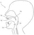
图21是人7的头部的截面形状的概要图。FIG. 21 is a schematic view of the cross-sectional shape of the head of the
图22是口腔71内的概要图。FIG. 22 is a schematic view of the inside of the
具体实施方式Detailed ways
以下,参照附图,详细地说明包含本发明的代表性实施方式的口内摄影装置和口内摄影辅助器械的医疗装置。但是,本发明不限定于该实施方式和附图。此外,附图是用于示意性地说明本发明的图,所以为了容易理解,有时根据需要夸张或简化地表示尺寸、比例或数量。Hereinafter, a medical apparatus including an intraoral imaging apparatus and an intraoral imaging auxiliary device according to a representative embodiment of the present invention will be described in detail with reference to the accompanying drawings. However, the present invention is not limited to this embodiment and the drawings. In addition, since the drawings are for schematically explaining the present invention, the dimensions, ratios, or numbers may be exaggerated or simplified as necessary for easy understanding.
此外,以下的公开设想了为了进行流感的判定而使用医疗装置,但本发明不限于此。例如,存在许多如链球菌感染、腺病毒感染症、EB病毒感染症、支原体感染症等那样在咽喉的观察结果中出现异常的疾病。此外,在这些疾病中,有时也会在口腔内出现重要的观察结果。并且,在除了这些疾病以外的疾病中,有时也会在口腔内出现观察结果。这样,可以使用医疗装置,以判定在咽喉和口腔内出现观察结果的任意的疾病。此外,口内摄影辅助器械也可以与例如智能手机和平板终端这样的其他装置组合起来使用。In addition, the following disclosure assumes the use of a medical device for the determination of influenza, but the present invention is not limited to this. For example, there are many diseases in which abnormality occurs in the observation of the throat, such as streptococcal infection, adenovirus infection, Epstein-Barr virus infection, and mycoplasma infection. In addition, in these diseases, important observations are sometimes made in the oral cavity. Furthermore, in diseases other than these diseases, observations may be made in the oral cavity. In this way, a medical device can be used to determine any disease for which observations occur in the throat and oral cavity. In addition, the intraoral photographic aid can also be used in combination with other devices such as smartphones and tablet terminals.
1医疗装置的概要1 Overview of Medical Devices
参照图1,说明本实施方式的医疗装置1的概要。如图所示,医疗装置1包含口内摄影装置5和口内摄影辅助器械3。口内摄影装置5优选通过与口内摄影辅助器械3组合使用,但是,也可以单独使用或与其他辅助器械组合使用。1, the outline of the
口内摄影装置5包含取得被摄体的图像的摄像设备57,并且,在该装置5中预先安装有专用的软件。用户(例如医生)使怀疑得了流感的判定对象者7(例如患者)咬住口内摄影辅助器械3,确保用于摄影的视野。然后,用户将摄像设备57插入到口内摄影辅助器械3中,拍摄该对象者7的咽喉72。此外,用户通过调节口内摄影辅助器械3的插入深度和插入角度,还能够拍摄对象者7的口腔内71。或者,用户可以将在内部收纳有摄像设备57的口内摄影辅助部3插入到判定对象者7的口内,也可以使判定对象者7将口内摄影辅助部3插入到口内。The
通过事先生成的判定算法来处理所拍摄的图像。设想了通过口内摄影装置5进行该处理,但是,也可以通过其他计算机进行。例如,在判定是关于流感的可能性的情况下,判定算法检测流感滤泡等流感固有的咽喉症状(模式),显示流感可能度。由此,例如,即使是经验不足的医生或培训医生,也能够准确且早期地进行流感诊断。此外,对于经验丰富的医生来说,也能够获得有用的判断资料。并且,流感的正确诊断率提高,由此,患者能够通过一次就诊就结束就医,并且能够从更早期起接受适当的治疗。The captured images are processed by a pre-generated decision algorithm. It is assumed that this process is performed by the
2-1口内摄影辅助器械2-1 Intraoral photography aids
参照图2-图4,详细地说明本实施方式的口内摄影辅助器械3。2 to 4 , the intraoral
口内摄影辅助器械3是为了拍摄人7的口内(口腔内71和咽喉72)而被使用的辅助工具。更具体而言,口内摄影辅助器械3在拍摄口内时被插入口腔71内,用于获得包含口腔内71或咽喉72在内的摄影区域的良好视野。基于获得更良好视野的观点,口内摄影辅助器械3优选具有透光性。在本实施方式中,设想了吹嘴(Mouthpiece)作为口内摄影辅助器械3的一例,但是,不限于此。The intraoral
如图2所示,口内摄影辅助器械3具有主体31、沿部34和限制部35。但是,口内摄影辅助器械3只要具有主体31即可,可以不具有沿部34和限制部35双方或一方。在本实施方式中,设想了一体成型的树脂产品作为口内摄影辅助器械3,但是,口内摄影辅助器械3可以利用纸、布、金属等其他材料制作,也可以利用多个材料制作。此外,设想了口内摄影辅助器械3为一次性类型,但也可以是可重新利用的类型。As shown in FIG. 2 , the intraoral photographing
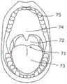
主体31整体上呈筒状。当主体31较深地插入到口腔71内时,例如,根据图1与图21的比较可知,将舌73推向下方,并且将软腭74推向上方。由此,例如,根据图4与图22的比较可知,能够从口内摄影辅助器械3(主体31)的内侧确保咽喉72的良好视野。The
在本实施方式中,主体31呈实质上大致直线状地延伸。即,主体31的内径和外径在整个长度方向地实质上恒定。但是,主体31只要不妨碍摄像设备57在该主体31的内周面上的滑动即可,可以一部分或全体弯曲,主体31的内径或外径也可以发生变化。In the present embodiment, the
作为主体31的截面形状,这里,设想了正圆形。但是,截面形状可以为椭圆形,也可以为如四边形那样的多边形状的截面,还可以为例如大致D字形那样的非对称形状。在主体31具有椭圆形、多边形或非对称的截面形状的情况下,当与主体31的形状对应的摄像设备57在主体31内滑动时,以主体31的轴向为中心的圆周方向上的移动(即,旋转)被限制或抑制,因此,能够获得适合于判定的、朝向一致的口内图像。As the cross-sectional shape of the
在主体31为正圆形的截面形状的情况下,如使用图6所后述的那样,也可以通过设置与摄像设备57的外表面和主体31的内周面相互卡合的导轨和突起部,限制摄像设备57在主体31内的旋转。When the
如图3所示,主体31包含相互位于相反侧的端部32、33。在将口内摄影辅助器械3安装于口腔71中时,端部32向外部露出,端部33位于口腔71内。因此,端部32相当于第1端部,端部33相当于第2端部。As shown in FIG. 3 , the
主体31的外周面平滑,主体31和端部33也经由连接面36平滑地连接而连接成为一体。即,主体31的外表面被加工成平滑,以不伤害口腔71。The outer peripheral surface of the
主体31具有刻度37。刻度37沿着主体31的长度方向配置,作为将主体31以何种程度较深地插入到口腔71内的基准发挥作用。刻度37设置于主体31的外周面和内周面中的任意一方即可。在刻度37设置于外周面的情况下,如图2所示,可以设置于与上唇以及上前牙75接触的一侧、即上侧。刻度37以规定的间隔(例如每隔1cm)配置即可。此外,图2所示的刻度37在主体31的圆周方向上具有接近半周的长度,但是不限于此,可以短,也可以长。The
在本实施方式中,作为刻度37的一例,设想了沿着主体31的长度方向排列的多个隆起部。该隆起部的表面可以被加工成平滑,以不损伤判定对象者7的口腔。可是,刻度37例如可以为在主体31的外周面的长度方向上延伸的大致直线状的凸部,并且,也可以打印或显示在主体31的外周面或内周面上。In the present embodiment, as an example of the
如图3所示,端部32具有开口32A,能够将摄像设备57插入到开口32A中。因此,摄像设备57从开口32A插入到主体31内,从开口32A取出到主体31的外部。As shown in FIG. 3 , the
在端部32设置有沿部34。沿部34从端部32朝向主体31的径向的外侧延伸。沿部34在主体31进入到口腔71的深处时,与判定对象者7的嘴唇或前牙接触,防止该判定对象者7误吞口内摄影辅助器械3。即,沿部34发挥作为止动件的功能。在本实施方式中,沿部34设置在端部32的整周上,也可以例如在端部32中局部地形成于与上唇以及下唇对应的部位。此外,沿部34的外缘可以如图2那样为正圆形,也可以为椭圆形,还可以为如四边形那样的多边形。A
例如,在判定对象者7咳嗽时,来自判定对象者7的嘴的飞沫溅到沿部34。即,沿部34有助于医生避免溅到来自判定对象者7的飞沫。为了具有该飞沫避免功能,例如,可以根据使用年龄或体格等适当地选择沿部34的形状和尺寸。For example, when the determination
端部33也开口,形成了窗部33A。该窗部33A用于从主体31的内侧朝向主体31的外侧提供视野,这里,使主体31内的摄像设备57的镜头露出到外部。但是,窗部33A例如也可以被透明部件覆盖。The
端部33朝向主体31的内侧突出,形成了限制部35。限制部35与主体31内的摄像设备57接触,为了限制其在端部33中通过而设置。可是,限制部35也可以不设置于端部33。例如,之后在与图6的关系中叙述的引导部141(凹部或槽)在其末端部处与摄像设备57的卡合突起57A接触,限制摄像设备57向深侧的移动,因此,还发挥作为限制部35的作用。The
在使用上述的口内摄影辅助器械3拍摄咽喉72的情况下,如图1所示,用户将口内摄影辅助器械3插入到判定对象者7的口腔71内。这时,主体31将舌73推向下方,将软腭74推向上方。然后或同时地,用户将摄像设备57插入到主体31内。这时,如图4所示,舌73和软腭74都不进入摄像设备57的视野,或者即使进入也仅占据微小的范围。因此,能够获得咽喉72的良好视野。When imaging the
此外,能够利用沿部34,防止判定对象者7误吞口内摄影辅助器械3。同时,沿部34能够抑制飞沫从判定对象者7的嘴向用户的飞散,减少流感等传染病对用户的二次感染的风险。In addition, the
并且,刻度37能够将口内摄影辅助器械3根据判定对象者7的体格、摄影部位而配置于适当的口腔深度。这能够有助于所希望的部位的清晰图像的摄影,并且抑制由于不必要地将主体31插入得过深而引起的判定对象者7的不适感和吞入。In addition, the
2-2口内摄影辅助器械的变形例12-2 Modification Example 1 of Intraoral Photography Aid
参照图5-图7,说明本实施方式的变形例1的口内摄影辅助器械13。5 to 7 , the intraoral
口内摄影辅助器械13具有与上述的口内摄影辅助器械3相同的结构要素,还包含大致直线状的引导部141。另一方面,摄像设备57包含卡合突起57A,该卡合突起57A与引导部141卡合。因此,引导部141能够在摄像设备57不相对于主体131旋转的情况下,在主体131内滑动。The intraoral
在变形例1中,如图6所示,引导部141的一例是以与摄像设备57的外周面上的卡合突起57A对应的方式从端部132朝向端部133呈大致直线状地延伸的槽。或者,引导部141也可以是从主体的内周面突出的一对导轨。In
可选地,在主体131的截面形状为椭圆形、多边形或者例如大致D字形那样的非对称形状并且摄像设备57是与主体131的截面形状对应的外形的情况下,摄像设备57能够在不相对于主体131旋转的情况下在主体131内滑动,所以该情况下的主体131的内周面还作为引导部发挥作用。Alternatively, in the case where the cross-sectional shape of the
如图5所示,在主体131和沿部134的外表面上设置有指示显示142,该指示显示142用于使主体131相对于判定对象者7的嘴唇的定位变得容易。如图7所示,指示显示142例如可以配置在与判定对象者7的上唇的中央部或上前牙75对应的位置、即上部的中央。用户例如在使指示显示142与判定对象者7的上唇的中央对齐的状态下将口内摄影辅助器械3安装到口腔71中,由此使由摄像设备57拍摄的图像的方向在几乎相同的方向上对齐。由此,使用了图像的机械学习和特定的疾病(例如流感)的判定处理变得容易。As shown in FIG. 5 , an
2-3口内摄影辅助器械的变形例22-3 Modification Example 2 of Intraoral Photography Aids
参照图8,说明本实施方式的变形例2的口内摄影辅助器械23。Referring to FIG. 8 , the intraoral
口内摄影辅助器械23具有与上述的口内摄影辅助器械3相同的结构要素,并且,在主体231上具有至少1个孔243。孔243贯穿主体231的内侧与外侧之间。通过该孔243,咬住了口内摄影辅助器械23的判定对象者7容易呼吸,能够给该判定对象者7带来安心感。另外,孔243的大小、数量和配置可以适当地设定成判定对象者7的唾液难以进入。The intraoral
上述的变形例2的结构要素、例如引导部还能够应用于上述实施方式、变形例1。The constituent elements of the above-described second modification, such as the guide portion, can also be applied to the above-described embodiment and
2-4口内摄影辅助器械的变形例32-4 Modification Example 3 of Intraoral Photography Aid
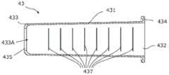
参照图9和图10,说明本实施方式的变形例3的口内摄影辅助器械43。口内摄影辅助器械43具有与上述的口内摄影辅助器械3相同的结构要素。Referring to FIGS. 9 and 10 , the intraoral
口内摄影辅助器械43的主体431为透明或半透明的树脂成型品。因此,口内摄影装置5的摄影区域比从窗部433A观察到的范围更大,能够获得更大的视野。此外,主体431的内径从端部432朝向端部433稍微减小,由此口内摄影辅助器械43的树脂成型变得容易。The
如图10所示,刻度437形成于主体431的内周面,由此能够使主体431的外周面平滑。刻度437可以为从主体431的内周面突出的突起,也可以为显示或涂敷在内周面上的标记。As shown in FIG. 10 , the
如图9所示,沿部434的外缘呈在上下方向上较长的椭圆形,由此能够容易地将口内摄影辅助器械43相对于判定对象者7的口唇定位。此外,沿部434的外缘形成为朝向端部433突出或比其他部分更厚。由此,减少了沿部434的材料的量和成本并确保了必要的强度。As shown in FIG. 9 , the outer edge of the
2-5口内摄影辅助器械的其他变形例2-5 Other Modifications of Intraoral Photography Aids
主体31无需在从端部32到端部33的整个范围内具有实质上相同的内径。例如,主体31可以构成为随着朝向端部32而内径扩大。通过该喇叭状的主体31,能够将多个口内摄影辅助器械3重叠,能够削减用于输送或保管的空间。The
此外,可以采用搭载于智能手机或平板终端上的照相机作为摄像设备57。可以在主体31或沿部34设置框、夹子,以固定这样的照相机与口内摄影辅助器械3(开口32A)的位置关系。例如,口内摄影辅助器械3可以具有用于从上缘或横缘夹住智能手机的夹子、用于抵靠于智能手机的角部的L字状的框。In addition, a camera mounted on a smartphone or a tablet terminal may be employed as the
2-6口内摄影辅助器械的包装例2-6 Packaging Examples of Intraoral Photography Assisting Devices
口内摄影辅助器械3也可以被灭菌,如图11所示那样利用显示有标识符91的袋9单独地包装,由此能够进行口内摄影辅助器械3的卫生管理。另外,标识符91可以显示在口内摄影辅助器械3上。The intraoral
标识符91例如包含识别信息,该识别信息包含口内摄影辅助器械3的产品ID。作为标识符91,例如可以举出条形码和RF标签,但如果考虑口内摄影装置5的读取和医疗装置1在医疗机构中的使用,则优选条形码。可以使用一维条形码或二维条形码中的任意一方作为条形码。The identifier 91 includes, for example, identification information including the product ID of the intraoral
例如,在口内的摄影之前,由摄影设备57拍摄标识符91,将袋9内的口内摄影辅助器械3的识别信息读入到口内摄影装置5中,由此能够确认对应的口内摄影辅助器械3的批次信息。由此,即使在因接触口腔而对患者产生了有害事件的情况下,也能够确保可追溯性,并且,由于还能够检测口内摄影辅助器械3的重新利用,所以能够防止由此引起的污染和二次感染,在卫生方面确保安全性。For example, before the intraoral imaging, the identifier 91 is photographed by the
3-1口内摄影装置3-1 Intraoral photography device
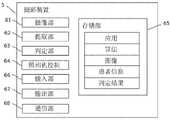
参照图12-图20,详细地说明口内摄影装置5。12 to 20, the
口内摄影装置5是包含摄像设备、运算装置和存储装置的计算机。这里,设想了口内摄影装置5由1台计算机构成,但是,也可以由多台计算机构成。例如,在远程图像诊断中,摄影功能和判定功能可以通过单独的计算机来执行。如图13所示,口内摄影装置5具有摄像设备57、作为运算装置的一例的中央处理装置(CPU)51、作为存储装置的一例的随机存取存储器(RAM)52和读出专用存储器(ROM)53、通信接口54、输入设备55、输出设备56和光源58。The
通信接口54是有线或无线的通信模块,例如用于应用程序(applicationprogram)和判定算法的取得和更新、以及所拍摄的图像的回收(发送)。作为无线的通信接口,例如设想了满足如Wi-Fi那样的无线LAN标准、如BLUETOOTH(商标)那样的近距离无线通信标准、第三代/第四代移动通信标准中的任意一方的设备,但不限于此。The
输入设备55是受理用户输入的输入装置的一例,例如是如图12所示的各种操作按钮和操作键。或者,输入设备55也可以是触摸面板、麦克风、操作拨盘、记录针等其他输入单元。输入设备55可以配置于口内摄影装置5的主体(壳体),例如也可以配置于摄像设备57。The
输出设备56是输出判定结果的输出装置的一例,例如是显示器、扬声器、打印机。The
摄像设备57取得图像。在该实施方式中,设想了运动图像,作为在摄像设备57中取得的图像。所取得的图像尺寸例如为640X480像素,帧率例如为30fps即可。但是,图像尺寸和帧率不限定于此。The
可选地或附加地,可以设为摄像设备57能够拍摄静态图像。在拍摄静态图像的情况下,摄像设备57优选具有连拍功能,但不限于此。Alternatively or additionally, the
在本实施方式中,如图12所示,设想了摄像设备57与口内摄影装置5的主体(壳体)分离,单独地被插入到口内摄影辅助器械3内。摄像设备57还优选具有与口内摄影辅助器械3的主体31的内周面对应的外形(例如圆柱形状)。In the present embodiment, as shown in FIG. 12 , it is assumed that the
但是,摄像设备57也可以如例如智能手机或平板终端那样组装到口内摄影装置5的主体中。However, the
摄像设备57具有未图示的自动对焦功能,例如,设定成在镜头的正面焦点对准特定部位。摄像设备57可以还具有自动地识别特定部位(例如咽喉后壁)并对焦于该特定部位的功能。摄像设备57也可以设定为还具有变焦(zoom)功能,例如根据咽喉后壁或滤泡的尺寸以适当的倍率进行拍摄。The
在摄像设备57的动作中,也可以从光源58向被摄体照射光,由此实现优质图像的确保。这里,光源58照射的光可以为可见光,也可以为例如近红外线、红外线那样的非可见光。此外,也可以是例如一个光源照射可见光、其他光源照射近红外线或红外线那样,由多个光源58分别负责特定范围的波长的光的照射。During the operation of the
与此相对,摄像设备57具有能够适当地接收光源58发出的光的种类和数量的受光元件即可。例如,在光源58发出可见光的情况下,摄像设备57具有适合于接收可见光的受光元件,在光源58发出非可见光的情况下,摄像设备57具有适合于接收非可见光的受光元件即可。光源58例如为发光二极管(LED)或有机电致发光(OEL)即可。On the other hand, the
也可以是可选地,编程成在摄像设备57的动作中,在作为输出设备56的显示器上重叠显示摄影中的图像和半透明的插图。当用户使摄像设备57移动使得插图与对应于该插图的部位(例如,悬雍垂或腭扁桃体)的图像重叠时,能够得到适当的图像以进行判定。Alternatively, it may be programmed to superimpose the image being photographed and the translucent illustration on the display serving as the
ROM 53存储有用于执行口内摄影装置5中的各种处理的应用程序和用于判定特定的疾病的算法。ROM 53也可以还存储在摄像设备57中取得的图像和口内摄影辅助器械3的识别信息、从输入设备55输入的各种信息(例如,判定对象者7的信息)、判定结果。ROM 53可以是内置型,也可以是例如USB存储器或MicroSD卡那样的可装卸型,还可以进一步包含外部服务器等外部计算机的存储装置的存储区域。The
CPU 51能够将ROM 53中存储的应用程序读出到RAM 52中,执行包括以下的处理(a)~(d)的各种处理。另外,在摄像设备57所取得的图像为一张静态图像的情况下,也可以省略提取处理。The
3-2CPU所执行的主要处理3-2 Main processing performed by the CPU
(a)提取处理(a) Extraction processing
CPU 51从由摄像设备57取得的图像中提取满足规定条件的至少一张静态图像。例如,在摄像设备57拍摄运动图像的情况下,CPU 51从运动图像所包含的多张静态图像中提取至少一张静态图像。在运动图像的摄影时以规定的帧率记录静态图像。其中应该包含摄影条件好的情况和摄影条件不好的情况。选出摄影条件好的几张作为用于在下一个判定处理中使用的图像。The
例如,预先根据许多个相同的图像数据,找出当满足特定的拍摄角度、特定的灯的照射程度、特定的视野宽度、特定部位(例如咽喉等)的描绘位置、对特定部位的对焦的程度的全部或一部分时、特定疾病(例如流感)的诊断判定的精度高这样的条件,将其作为摄影条件的好坏的判断基准。在采用这样的基准的情况下,例如,如图18所示,提取处理经过如下过程:根据摄影对象的描绘位置对图像进行排序(步骤S31),根据对特定部位的对焦程度对图像进行排序(步骤S32),根据光的照射状态对图像进行排序(步骤S33),最后选出在综合顺序中位于上位的几张图像(步骤S34)。追加地,也可以在计算综合顺序之前,根据视野的大小或摄影角度的基准对图像进行排序。或者,也可以在提取处理中使用深度学习(深层学习)功能。For example, based on many pieces of the same image data in advance, find out when a specific shooting angle, a specific light irradiation degree, a specific field of view width, a drawing position of a specific part (such as the throat, etc.), and the degree of focus on a specific part are satisfied. When all or part of the conditions are high, the accuracy of the diagnosis and determination of a specific disease (eg, influenza) is high, and this condition is used as a criterion for determining whether the imaging conditions are good or bad. In the case of using such a reference, for example, as shown in FIG. 18 , the extraction process goes through the following procedures: images are sorted according to the drawing position of the photographic subject (step S31 ), and images are sorted according to the degree of focus on a specific part (step S31 ). In step S32), the images are sorted according to the irradiation state of the light (step S33), and finally several images located in the upper position in the comprehensive order are selected (step S34). Additionally, the images may be sorted according to the size of the field of view or the reference of the shooting angle before calculating the comprehensive order. Alternatively, deep learning (deep learning) functions can also be used in the extraction process.
(b)判定处理(b) Judgment processing
CPU 51根据从摄像设备57取得的图像(例如通过提取处理而获得的至少一张静态图像)和ROM 53中存储的判定算法,判定特定的疾病的可能性。在本实施方式中,特定的疾病为流感。为了进行判定,可以同时使用用户经由输入设备55而输入的判定对象者7的信息(以下,称作患者信息)。患者信息例如包括年龄、性别、从发病起的经过时间、有无流感的各种症状(咳嗽、流鼻涕、发冷等)、和与其他流感患者的接触史。患者信息可以包含人种或基因信息、日期或季节、位置信息(就诊地的纬度经度)和气象信息(例如天气、气温等)的全部或一部分。The
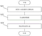
这里,判定算法例如可以通过图17所示的过程来生成。Here, the determination algorithm can be generated by, for example, the procedure shown in FIG. 17 .
例如,在流感的情况下,首先,在医疗机构中收集患者的咽喉图像和附属于该咽喉图像的、表示是否为流感的正解标签(步骤S21)。例如,根据由患者接受到的、基于拭子的流感快速检查、PCR检查、病毒分离培养检查的结果来赋予正确标签。PCR检查和病毒分离培养检查虽然在判明结果之前需要数周的时间,但由于表示非常高的准确性,所以将其结果作为正解标签是合适的。此外,正解标签不仅包含图像数据,还包含上述的患者信息。For example, in the case of influenza, first, a throat image of a patient and a positive solution label indicating whether or not it is influenza attached to the throat image are collected in a medical institution (step S21 ). For example, correct labeling based on the results of swab-based rapid influenza tests, PCR tests, virus isolation and culture tests received by the patient. Although several weeks are required for PCR tests and virus isolation and culture tests to be determined, it is appropriate to use the results as positive-solution labels because they show very high accuracy. In addition, the positive solution label contains not only the image data, but also the above-mentioned patient information.
接着,生成将上述的PCR检查或病毒分离培养检查的判定结果作为正解附加了标签的教师数据(图像数据)(步骤S22)。根据该教师数据进行机械学习,生成判定算法(步骤S23)。该判定算法是当提供了图像时用于判定该图像是否有流感可能性的算法。由此,能够将是流感的可能性数值化,例如表示为“98.5%”。Next, teacher data (image data) to which the above-mentioned determination result of the PCR test or virus isolation and culture test is identified as positive solutions are generated (step S22). Machine learning is performed based on the teacher data, and a determination algorithm is generated (step S23). The determination algorithm is an algorithm for determining whether the image has the possibility of influenza when the image is provided. Thereby, the possibility of influenza can be quantified, for example, expressed as "98.5%".
可选地,例如可以如“判定:流感阳性”和“判定:流感阴性”那样判定阳性或阴性。在该情况下,除了阳性或阴性的判定以外,还可以将判定的可靠性数值化,或以高中低等的等级评价来表示。或者,也可以通过高中低等的等级评价来表示是流感的确信度。Alternatively, for example, it can be determined as positive or negative as in "determination: influenza positive" and "determination: influenza negative". In this case, in addition to the determination of positive or negative, the reliability of the determination may be quantified, or it may be expressed as a rank evaluation such as high, medium and low. Alternatively, the degree of certainty that it is influenza can also be expressed by a graded evaluation of high, medium and low.
(c)输出处理(c) Output processing
CPU 51输出判定处理的结果。输出方法的一例为显示器上的显示、向其他计算机的发送。The
(d)口内摄影辅助器械的识别信息的取得(d) Acquisition of identification information of intraoral photographic aids
可选地,CPU 51可以在新的判定之前,请求用户输入用于在该判定中使用的口内摄影辅助器械3的识别信息。例如,当用户通过摄像设备57拍摄标识符91时,CPU 51从标识符91的图像中取得对应的口内摄影辅助器械3的识别信息。Alternatively, the
接着,CPU 51检索ROM 53内的记录,调查是否存在与对应的口内摄影辅助器械3一致的记录。或者,CPU 51也可以调查外部服务器上的口内摄影辅助器械3的使用记录。Next, the
在不存在一致的记录的情况下,CPU 51容许新的判定。在存在一致的记录的情况下,CPU 51判定为对应的口内摄影辅助器械3已使用过。然后,CPU 51例如可以使显示器显示警报,或结束一系列的处理。即,CPU 51能够根据检索结果限制新的判定。In the case where there is no consistent record, the
3-3口内摄影装置的功能结构3-3 Functional structure of the intraoral photographic device
图14示出口内摄影装置5的功能结构。口内摄影装置5包含摄像部61、提取部62、判定部63、照相机控制64、存储部65、输入部66、输出部67和通信部68。如果表示与口内摄影装置5的硬件结构的对应关系,则摄像部61对应于摄像设备57,提取部62、判定部63和照相机控制64对应于CPU 51,存储部65对应于ROM 53,输入部66对应于输入设备55,输出部67对应于输出设备56,通信部68对应于通信接口54。这里,照相机控制64控制摄像设备57,从而实现对焦功能和变焦功能等各种功能。FIG. 14 shows the functional configuration of the
作为其他功能,口内摄影装置5也可以具有取得进行摄影的时刻的计时功能、取得进行摄影的位置信息的位置信息取得功能、取得该位置的气象信息的气象信息取得功能。计时功能可以作为内置的时钟来实现,也可以是访问外部的时间服务器的通信接口的一个功能。位置信息取得功能例如可以作为全球定位系统(GPS)来实现。气象信息取得功能也可以是访问外部的气象服务器的通信接口的一个功能。As other functions, the
3-4口内摄影装置的动作例13-4 Operation example 1 of the intraoral imaging device
参照图15,说明口内摄影装置5的动作例1。这里,列举口内摄影装置5拍摄运动图像的情况的动作为例。但是,口内摄影装置5可以拍摄静态图像,在该情况下,口内摄影装置5也同样地进行动作。Referring to FIG. 15 , an operation example 1 of the
当接通电源时,口内摄影装置5开始利用摄像设备57进行的摄影并显示在显示器上,并且在步骤S1中,判定录制按钮(输入设备55之一)是否被按下。在未确认到录制按钮的按下时,口内摄影装置5反复执行步骤S1。When the power is turned on, the
当确认了录制按钮的按下时,口内摄影装置5在步骤S2中开始录制。同时,口内摄影装置5点亮光源58。用户将口内摄影辅助器械3安装到判定对象者7的口腔71中,然后,将摄像设备57插入到口内摄影辅助器械3中(参照图1)。摄像设备57在口内摄影辅助器械3内滑动的过程中,也拍摄判定对象者7的口内(口腔内71或咽喉72)。When the pressing of the recording button is confirmed, the
在步骤S3中,口内摄影装置5从所取得的运动图像所包含的多个静态图像中提取适当的静态图像,以进行判定。这时,所提取的静态图像也可以作为提取结果显示在显示器上。In step S3, the
口内摄影装置5在步骤S4中根据所提取的静态图像和判定算法来判定特定的疾病可能性,在步骤S5中将判定结果输出到例如显示器。由此,一系列的动作结束。The
这样,由于使用通过机械学习而生成的判定算法来评价特定的疾病的可能性,所以能够期待高精度的判定结果。In this way, since the possibility of a specific disease is evaluated using a determination algorithm generated by machine learning, a highly accurate determination result can be expected.
由于可选择摄影条件好的图像以进行判定,所以判定的精度提高。Since images with good photographing conditions can be selected for determination, the accuracy of determination is improved.
通过与口内摄影辅助器械3组合起来使用口内摄影装置5,能够获得视野大、清晰的图像。这有助于提高判定精度。By using the
3-5口内摄影装置的动作例23-5 Operation example 2 of the intraoral imaging device
参照图16,说明口内摄影装置5的动作例2。该动作例2包含与之前所叙述的动作例1相同的步骤,还包含由用户进行的提取图像的确认步骤(S14)、患者信息的输入步骤(S16)和综合判定(S17)。Referring to FIG. 16 , an operation example 2 of the
具体而言,口内摄影装置5在接通电源时,确认录制按钮的按下(步骤S11),开始录制(步骤S12),提取用于判定的图像(步骤S13)。然后,口内摄影装置5将所提取的图像例如显示在显示器上,要求用户确认是否使用该图像进行判定(步骤S14)。如果用户选择了“否”,则再次执行步骤S12、S13,向用户提示新的判定用的图像。Specifically, the
当在步骤S14中用户选择了“是”时,口内摄影装置5根据所提取的图像来判定特定的疾病可能性(步骤S15)。When the user selects "Yes" in step S14, the
接着,口内摄影装置5要求用户输入患者信息(步骤S16)。这里,患者信息例如是就诊时的体温、有无疫苗接种这样的定型化的简单信息。如图9所示,以软件能够统一处理的形式输入该患者信息。例如,也可以利用下拉菜单、拒绝半角数字以外的输入格式设定。或者,口内摄影装置5也可以通过有线或无线线路从外部计算机(例如,电子病历系统)取得患者信息。另外,步骤S16也可以在步骤S11之前执行。Next, the
然后,综合上述患者信息和基于图像的特定疾病可能性的评价双方,整体上计算该疾病可能性(步骤S17)。计算结果例如显示在显示器上(步骤S18),一系列的过程结束。Then, the above-mentioned patient information and the evaluation of the probability of a specific disease based on the image are integrated, and the probability of the disease as a whole is calculated (step S17). The calculation result is displayed, for example, on the display (step S18), and the series of processes ends.
这样,口内摄影装置5通过根据用户已确认的图像来评价特定的疾病的可能性,提高判断的精度和可靠性。In this way, the
如果还根据患者信息进行判定,则可以期待进一步提高判定的精度和可靠性。Further improvement in the accuracy and reliability of the determination can be expected if the determination is also made based on patient information.
3-6口内摄影装置的动作例33-6 Operation example 3 of the intraoral imaging device
参照图19,说明口内摄影装置5的动作例3。该动作例3包含取得口内摄影辅助器械3的识别信息的过程(步骤S41)。Referring to FIG. 19 , an operation example 3 of the
具体而言,当接通电源时,口内摄影装置5在步骤S41中通过例如向显示器上的显示而要求用户输入口内摄影辅助器械3的识别信息。例如,当用户利用摄像装置57拍摄袋9上的标识符91或通过输入设备55输入识别信息时,口内摄影装置5取得口内摄影辅助器械3的识别信息。Specifically, when the power is turned on, the
口内摄影装置5也可以使用识别信息,调查该口内摄影辅助器械3的批次信息。例如,口内摄影装置5可以检索在ROM 53内的记录中是否包含该口内摄影辅助器械3的产品ID,也可以让外部计算机调查是否存在该产品ID。The
在对应的产品ID存在于内部或外部的记录中的情况下,口内摄影装置5也可以输出警报或使一系列的处理结束。作为警报,例如,可举出在显示器上显示该口内摄影辅助器械3已使用这一消息。由此,限制了口内摄影辅助器械3的重新利用,提高了包含二次感染的预防在内的口内摄影辅助器械3的安全性。此外,能够使口内摄影辅助器械3与使用该口内摄影辅助器械3的判定对象者7一对一对应,因此,将来的追踪调查变得容易。When the corresponding product ID exists in the internal or external records, the
然后,口内摄影装置5在步骤S42中进行口内的摄影。这里,口内的摄影处理可以与上述的动作例1、2相同,也可以按照后面叙述的过程进行。而且,口内摄影装置5在步骤S43中判定特定的疾病可能性,在步骤S44中输出判定结果,结束一系列的处理。Then, the
3-7口内摄影的其他例3-7 Other examples of intraoral photography
参照图20,说明口内摄影的其他过程。这里所述的过程可以替代上述的动作例1~3中的图像取得过程。此外,该过程能够用于收集机械学习用的训练数据。20, another procedure of intraoral photography will be described. The process described here can be substituted for the image acquisition process in the above-described operation examples 1 to 3. Furthermore, this process can be used to collect training data for machine learning.
首先,在步骤S51中,口内摄影装置5进行患者登记。患者登记例如通过由用户输入到口内摄影装置5或记录所拍摄的患者ID来进行。First, in step S51, the
接着,在步骤S52中,口内摄影装置5取得图像。这里所说的图像为静态图像,例如,可以由从摄像设备57中拍摄的图像中以规定的时间间隔(例如每1秒)提取出的一组静态图像构成。Next, in step S52, the
而且,在步骤S53中,口内摄影装置5将所取得的图像例如显示在显示器上,要求用户确认。当在步骤S54中从用户得到认可时,口内摄影装置5在步骤S55中保存被认可的图像,结束一系列的摄影处理。在未得到用户的认可的情况下,返回步骤S52,再次取得图像。And in step S53, the
以上,对本发明的代表性实施方式进行了说明,但本发明并不限定于这些代表性实施方式,能够进行各种设计变更,这些设计变更也包含在本发明中。如上所述,还可以应用于例如链球菌感染、腺病毒感染症、EB病毒感染症、支原体感染症等咽喉或口腔内的观察中出现问题的疾病的判定。The representative embodiments of the present invention have been described above, but the present invention is not limited to these representative embodiments, and various design changes are possible, and these design changes are also included in the present invention. As described above, it is also applicable to the determination of diseases that cause problems in observation of the throat or oral cavity, such as streptococcal infection, adenovirus infection, Epstein-Barr virus infection, and mycoplasma infection.
标号说明Label description
1:医疗装置;1: medical device;
3、13、23、43:口内摄影辅助器械;3, 13, 23, 43: intraoral photographic aids;
5:口内摄影装置;5: Intraoral photography device;
31、131、231、431:主体;31, 131, 231, 431: main body;
34、134、234、434:沿部;34, 134, 234, 434: along the part;
35、135、235、435:限制部;35, 135, 235, 435: Restriction Department;
37、137、237、437:刻度;37, 137, 237, 437: scale;
55:输入设备;55: input device;
56:输出设备;56: output device;
57:摄像设备;57: camera equipment;
58:光源。58: Light source.
Claims (7)
Translated fromChinesePriority Applications (1)
| Application Number | Priority Date | Filing Date | Title |
|---|---|---|---|
| CN202510116767.9ACN119632485A (en) | 2017-12-28 | 2018-12-18 | Intraoral photography aids |
Applications Claiming Priority (3)
| Application Number | Priority Date | Filing Date | Title |
|---|---|---|---|
| JP2017254164 | 2017-12-28 | ||
| JP2017-254164 | 2017-12-28 | ||
| PCT/JP2018/046559WO2019131327A1 (en) | 2017-12-28 | 2018-12-18 | Oral photographing apparatus, medical apparatus, and program |
Related Child Applications (1)
| Application Number | Title | Priority Date | Filing Date |
|---|---|---|---|
| CN202510116767.9ADivisionCN119632485A (en) | 2017-12-28 | 2018-12-18 | Intraoral photography aids |
Publications (2)
| Publication Number | Publication Date |
|---|---|
| CN111526778Atrue CN111526778A (en) | 2020-08-11 |
| CN111526778B CN111526778B (en) | 2025-02-18 |
Family
ID=67063606
Family Applications (2)
| Application Number | Title | Priority Date | Filing Date |
|---|---|---|---|
| CN201880083646.7AActiveCN111526778B (en) | 2017-12-28 | 2018-12-18 | Intraoral photography devices, medical devices and procedure products |
| CN202510116767.9APendingCN119632485A (en) | 2017-12-28 | 2018-12-18 | Intraoral photography aids |
Family Applications After (1)
| Application Number | Title | Priority Date | Filing Date |
|---|---|---|---|
| CN202510116767.9APendingCN119632485A (en) | 2017-12-28 | 2018-12-18 | Intraoral photography aids |
Country Status (7)
| Country | Link |
|---|---|
| US (2) | US11523741B2 (en) |
| EP (1) | EP3714764A4 (en) |
| JP (4) | JP7267608B2 (en) |
| KR (1) | KR20200103676A (en) |
| CN (2) | CN111526778B (en) |
| SG (1) | SG11202006040YA (en) |
| WO (1) | WO2019131327A1 (en) |
Cited By (2)
| Publication number | Priority date | Publication date | Assignee | Title |
|---|---|---|---|---|
| CN113077446A (en)* | 2021-04-02 | 2021-07-06 | 重庆金山医疗器械有限公司 | Interaction method, device, equipment and medium |
| CN114340637A (en)* | 2019-07-03 | 2022-04-12 | 阿伊里斯株式会社 | Pharmaceutical composition for treating influenza virus infection |
Families Citing this family (19)
| Publication number | Priority date | Publication date | Assignee | Title |
|---|---|---|---|---|
| CN111526778B (en)* | 2017-12-28 | 2025-02-18 | 阿伊里斯株式会社 | Intraoral photography devices, medical devices and procedure products |
| JP7170032B2 (en)* | 2018-04-13 | 2022-11-11 | 富士フイルム株式会社 | Image processing device, endoscope system, and image processing method |
| JPWO2021020187A1 (en)* | 2019-07-26 | 2021-02-04 | ||
| CN114650765A (en)* | 2019-10-31 | 2022-06-21 | 阿伊里斯株式会社 | Image pickup apparatus and image pickup system |
| CN115776863A (en)* | 2020-07-06 | 2023-03-10 | 阿伊里斯株式会社 | Processing device, processing program, processing method and processing system |
| JP2022087967A (en)* | 2020-12-02 | 2022-06-14 | 正男 山本 | Camera, generation method of learned model about respiratory infection diseases, learned model about respiratory infection diseases, automatic diagnostic method about respiratory infection diseases and computer program |
| FR3119755B1 (en)* | 2021-02-17 | 2023-10-13 | Dental Monitoring | Acquisition KIT |
| FR3119762B1 (en) | 2021-02-17 | 2023-06-16 | Dental Monitoring | Acquisition KIT |
| FR3119757B1 (en)* | 2021-02-17 | 2023-10-06 | Dental Monitoring | Acquisition KIT |
| FR3119761B1 (en) | 2021-02-17 | 2023-06-30 | Dental Monitoring | Acquisition KIT |
| FR3119756B1 (en) | 2021-02-17 | 2023-05-19 | Dental Monitoring | Acquisition KIT |
| JPWO2022230084A1 (en)* | 2021-04-28 | 2022-11-03 | ||
| US11382727B1 (en)* | 2021-05-19 | 2022-07-12 | Thamer Marghalani | Three-dimensional oral imaging system and method |
| WO2023286199A1 (en)* | 2021-07-14 | 2023-01-19 | アイリス株式会社 | Processing device, processing program, processing method, and processing system |
| CN118785843A (en)* | 2021-10-27 | 2024-10-15 | 阿伊里斯株式会社 | Processing device, processing program and processing method |
| USD1082892S1 (en)* | 2022-03-08 | 2025-07-08 | Aillis Inc. | Camera |
| WO2023170788A1 (en)* | 2022-03-08 | 2023-09-14 | アイリス株式会社 | Assistance tool, imaging device, program, and method |
| KR102668823B1 (en)* | 2022-08-25 | 2024-05-22 | 조정환 | Oral imaging apparatus for oral care of pet |
| KR102674263B1 (en)* | 2022-09-05 | 2024-06-12 | 조정환 | System of providing oral image of pet |
Citations (14)
| Publication number | Priority date | Publication date | Assignee | Title |
|---|---|---|---|---|
| JPH09189659A (en)* | 1995-11-08 | 1997-07-22 | Kaltenbach & Voigt Gmbh & Co | Recognizing device for tooth state |
| KR200171384Y1 (en)* | 1999-06-05 | 2000-03-15 | 박운식 | Endoscopic mouth-piece |
| JP2005000631A (en)* | 2002-07-03 | 2005-01-06 | Shiyoufuu:Kk | Dental oral cavity colorimetry photographic system |
| JP2005103048A (en)* | 2003-09-30 | 2005-04-21 | Hidehiro Fujie | Dental unit |
| KR200395743Y1 (en)* | 2005-06-22 | 2005-09-13 | 김동선 | Mouthpiece |
| CN101902966A (en)* | 2007-12-17 | 2010-12-01 | 画像诊断株式会社 | Medical imaging marker and program for utilizing same |
| JP2011021893A (en)* | 2009-07-13 | 2011-02-03 | Medeia Kk | Support system for creating intraoral bacteria map |
| WO2012002564A1 (en)* | 2010-06-29 | 2012-01-05 | 株式会社アドバンス | Intraoral imaging system |
| CN103119158A (en)* | 2010-04-27 | 2013-05-22 | 莱桑多公司 | Method of reducing biofilms |
| CN103445877A (en)* | 2012-06-01 | 2013-12-18 | 索尼公司 | Dental apparatus, medical apparatus and calculation method |
| US20160287063A1 (en)* | 2013-11-22 | 2016-10-06 | Duke University | Colposcopes having light emitters and image capture devices and associated methods |
| CN106491068A (en)* | 2015-09-06 | 2017-03-15 | 孟立 | A kind of oral photographing system and method based on mobile terminal |
| US20170172418A1 (en)* | 2015-12-01 | 2017-06-22 | University Of South Florida | Standardized oral health assessment and scoring using digital imaging |
| CN107249425A (en)* | 2015-05-14 | 2017-10-13 | 奥林巴斯株式会社 | Endoscope apparatus |
Family Cites Families (19)
| Publication number | Priority date | Publication date | Assignee | Title |
|---|---|---|---|---|
| US4905082A (en)* | 1987-05-06 | 1990-02-27 | Olympus Optical Co., Ltd. | Rigid video endoscope having a detachable imaging unit |
| US10595710B2 (en)* | 2001-10-19 | 2020-03-24 | Visionscope Technologies Llc | Portable imaging system employing a miniature endoscope |
| JP2005013708A (en)* | 2003-05-30 | 2005-01-20 | Olympus Corp | Endoscope and method for assembling it |
| US7303528B2 (en)* | 2004-05-18 | 2007-12-04 | Scimed Life Systems, Inc. | Serialization of single use endoscopes |
| JP4526601B2 (en)* | 2008-10-16 | 2010-08-18 | オリンパスメディカルシステムズ株式会社 | Medical equipment |
| US8973573B2 (en)* | 2009-06-29 | 2015-03-10 | Creighton University | Bite block with airway mount |
| EP2538841A2 (en)* | 2010-02-26 | 2013-01-02 | Myskin, Inc. | Analytic methods of tissue evaluation |
| US8998609B2 (en)* | 2012-02-11 | 2015-04-07 | The Board Of Trustees Of The Leland Stanford Jr. University | Techniques for standardized imaging of oral cavity |
| CA2902592C (en)* | 2013-03-15 | 2023-03-07 | Synaptive Medical (Barbados) Inc. | Insert imaging device for surgical procedures |
| WO2015053313A1 (en) | 2013-10-10 | 2015-04-16 | 住友ベークライト株式会社 | Mouthpiece for endoscopic examination and de-aeration prevention valve device |
| WO2015145424A1 (en)* | 2014-03-23 | 2015-10-01 | Doc@Home Ltd | A system for conducting a remote physical examination |
| US11147442B2 (en)* | 2014-08-08 | 2021-10-19 | Wm & Dg, Inc. | Medical devices and methods of placement |
| ES2865298T3 (en)* | 2015-05-19 | 2021-10-15 | Tyto Care Ltd | Throat Imaging Systems and Methods |
| US11623058B2 (en)* | 2017-04-27 | 2023-04-11 | Wedge Therapeutics Llc | Lower jaw thrusting, mandibular protracting, tongue holding, universal oropharyngeal airway device |
| IT201700046337A1 (en)* | 2017-04-28 | 2018-10-28 | Francesco Antoniomaria Garbagnati | INSTRUMENT FOR VISUAL ACCESS TO CABLES |
| WO2019092723A1 (en)* | 2017-11-12 | 2019-05-16 | Ofek Eshkolot Research And Development Ltd. | System and method for determining pathological status of a laryngopharyngeal area in a patient |
| CN111526778B (en)* | 2017-12-28 | 2025-02-18 | 阿伊里斯株式会社 | Intraoral photography devices, medical devices and procedure products |
| CN114650765A (en)* | 2019-10-31 | 2022-06-21 | 阿伊里斯株式会社 | Image pickup apparatus and image pickup system |
| JPWO2022230084A1 (en)* | 2021-04-28 | 2022-11-03 |
- 2018
- 2018-12-18CNCN201880083646.7Apatent/CN111526778B/enactiveActive
- 2018-12-18KRKR1020207018105Apatent/KR20200103676A/ennot_activeCeased
- 2018-12-18JPJP2019561542Apatent/JP7267608B2/enactiveActive
- 2018-12-18WOPCT/JP2018/046559patent/WO2019131327A1/ennot_activeCeased
- 2018-12-18SGSG11202006040YApatent/SG11202006040YA/enunknown
- 2018-12-18EPEP18896683.2Apatent/EP3714764A4/enactivePending
- 2018-12-18USUS16/958,085patent/US11523741B2/enactiveActive
- 2018-12-18CNCN202510116767.9Apatent/CN119632485A/enactivePending
- 2022
- 2022-11-03USUS17/979,987patent/US20230047709A1/enactivePending
- 2023
- 2023-04-13JPJP2023065907Apatent/JP7498521B2/enactiveActive
- 2024
- 2024-05-24JPJP2024085185Apatent/JP7689772B2/enactiveActive
- 2025
- 2025-05-21JPJP2025084634Apatent/JP2025122092A/enactivePending
Patent Citations (14)
| Publication number | Priority date | Publication date | Assignee | Title |
|---|---|---|---|---|
| JPH09189659A (en)* | 1995-11-08 | 1997-07-22 | Kaltenbach & Voigt Gmbh & Co | Recognizing device for tooth state |
| KR200171384Y1 (en)* | 1999-06-05 | 2000-03-15 | 박운식 | Endoscopic mouth-piece |
| JP2005000631A (en)* | 2002-07-03 | 2005-01-06 | Shiyoufuu:Kk | Dental oral cavity colorimetry photographic system |
| JP2005103048A (en)* | 2003-09-30 | 2005-04-21 | Hidehiro Fujie | Dental unit |
| KR200395743Y1 (en)* | 2005-06-22 | 2005-09-13 | 김동선 | Mouthpiece |
| CN101902966A (en)* | 2007-12-17 | 2010-12-01 | 画像诊断株式会社 | Medical imaging marker and program for utilizing same |
| JP2011021893A (en)* | 2009-07-13 | 2011-02-03 | Medeia Kk | Support system for creating intraoral bacteria map |
| CN103119158A (en)* | 2010-04-27 | 2013-05-22 | 莱桑多公司 | Method of reducing biofilms |
| WO2012002564A1 (en)* | 2010-06-29 | 2012-01-05 | 株式会社アドバンス | Intraoral imaging system |
| CN103445877A (en)* | 2012-06-01 | 2013-12-18 | 索尼公司 | Dental apparatus, medical apparatus and calculation method |
| US20160287063A1 (en)* | 2013-11-22 | 2016-10-06 | Duke University | Colposcopes having light emitters and image capture devices and associated methods |
| CN107249425A (en)* | 2015-05-14 | 2017-10-13 | 奥林巴斯株式会社 | Endoscope apparatus |
| CN106491068A (en)* | 2015-09-06 | 2017-03-15 | 孟立 | A kind of oral photographing system and method based on mobile terminal |
| US20170172418A1 (en)* | 2015-12-01 | 2017-06-22 | University Of South Florida | Standardized oral health assessment and scoring using digital imaging |
Cited By (2)
| Publication number | Priority date | Publication date | Assignee | Title |
|---|---|---|---|---|
| CN114340637A (en)* | 2019-07-03 | 2022-04-12 | 阿伊里斯株式会社 | Pharmaceutical composition for treating influenza virus infection |
| CN113077446A (en)* | 2021-04-02 | 2021-07-06 | 重庆金山医疗器械有限公司 | Interaction method, device, equipment and medium |
Also Published As
| Publication number | Publication date |
|---|---|
| CN119632485A (en) | 2025-03-18 |
| JP2024109854A (en) | 2024-08-14 |
| JP7689772B2 (en) | 2025-06-09 |
| KR20200103676A (en) | 2020-09-02 |
| EP3714764A1 (en) | 2020-09-30 |
| JP7267608B2 (en) | 2023-05-02 |
| JP7498521B2 (en) | 2024-06-12 |
| US20210059534A1 (en) | 2021-03-04 |
| JPWO2019131327A1 (en) | 2021-03-04 |
| US20230047709A1 (en) | 2023-02-16 |
| CN111526778B (en) | 2025-02-18 |
| EP3714764A4 (en) | 2021-06-30 |
| JP2023089157A (en) | 2023-06-27 |
| US11523741B2 (en) | 2022-12-13 |
| SG11202006040YA (en) | 2020-07-29 |
| WO2019131327A1 (en) | 2019-07-04 |
| JP2025122092A (en) | 2025-08-20 |
Similar Documents
| Publication | Publication Date | Title |
|---|---|---|
| JP7498521B2 (en) | Intraoral photography device, medical device and program | |
| US20200126227A1 (en) | Systems and methods for urianlysis using a personal communications device | |
| JP5401986B2 (en) | Diagnostic system | |
| EP2976014A1 (en) | Pre-intubation examination apparatus and probe | |
| WO2019131328A1 (en) | Oral photographing auxiliary tool, and medical apparatus including oral photographing auxiliary tool | |
| US20140354830A1 (en) | System and method for adding scale to photographic images | |
| US20070127781A1 (en) | Animal eye biometrics | |
| KR100874186B1 (en) | Method and apparatus for photographing snow-collected images of subjects by themselves | |
| WO2023073844A1 (en) | Processing device, processing program, and processing method | |
| KR102263572B1 (en) | De-identification device for healthcare image | |
| KR20200059535A (en) | Method, Apparatus and Recording For Computerizing Of Electro-Magnetic Resonance | |
| US20220392629A1 (en) | Portable healthcare system installed at a remote location having limited internet connectivity | |
| JP2023004843A (en) | Mouth corner retractor | |
| JP2007130227A (en) | Capsule endoscope system and method for processing information of capsule endoscope | |
| US20250261859A1 (en) | Medical imaging device | |
| EP4371466A1 (en) | Processing device, processing program, processing method, and processing system | |
| US20250014697A1 (en) | Imaging Device, Program, And Method | |
| US11423534B2 (en) | System and method for diagnosing potential diseases from photo and video data and informing the user | |
| CN114340637A (en) | Pharmaceutical composition for treating influenza virus infection | |
| Mohammed-Ali et al. | P64 Facial frame for maintaining exact head positioning for serial photographs in facial reanimation patients | |
| Bhandari et al. | P63 Out of hours tool kit for maxillofacial use in emergencey departments | |
| Brown et al. | P62 CRABEL Score; an audit of inpatient medical records and operative notes of patients treated within the oral and maxillofacial surgery department at Addenbrookes Hospital | |
| Santhanam | P65 Nasogastric tube placement–strategies for success! |
Legal Events
| Date | Code | Title | Description |
|---|---|---|---|
| PB01 | Publication | ||
| PB01 | Publication | ||
| SE01 | Entry into force of request for substantive examination | ||
| SE01 | Entry into force of request for substantive examination | ||
| GR01 | Patent grant | ||
| GR01 | Patent grant |