CN111494035A - Trabecular bone porous tantalum dental implant and preparation method thereof - Google Patents
Trabecular bone porous tantalum dental implant and preparation method thereofDownload PDFInfo
- Publication number
- CN111494035A CN111494035ACN202010331329.1ACN202010331329ACN111494035ACN 111494035 ACN111494035 ACN 111494035ACN 202010331329 ACN202010331329 ACN 202010331329ACN 111494035 ACN111494035 ACN 111494035A
- Authority
- CN
- China
- Prior art keywords
- tantalum
- dental implant
- functional area
- trabecular
- porous tantalum
- Prior art date
- Legal status (The legal status is an assumption and is not a legal conclusion. Google has not performed a legal analysis and makes no representation as to the accuracy of the status listed.)
- Pending
Links
- 210000000988bone and boneAnatomy0.000titleclaimsabstractdescription118
- GUVRBAGPIYLISA-UHFFFAOYSA-Ntantalum atomChemical compound[Ta]GUVRBAGPIYLISA-UHFFFAOYSA-N0.000titleclaimsabstractdescription110
- 229910052715tantalumInorganic materials0.000titleclaimsabstractdescription100
- 239000004053dental implantSubstances0.000titleclaimsabstractdescription92
- 238000002360preparation methodMethods0.000titleabstractdescription7
- 238000004519manufacturing processMethods0.000claimsabstractdescription34
- 239000000843powderSubstances0.000claimsabstractdescription32
- 238000005516engineering processMethods0.000claimsabstractdescription19
- 229910001362Ta alloysInorganic materials0.000claimsabstractdescription18
- 239000002994raw materialSubstances0.000claimsabstractdescription12
- 239000011664nicotinic acidSubstances0.000claimsabstractdescription4
- 239000000654additiveSubstances0.000claimsdescription30
- 230000000996additive effectEffects0.000claimsdescription30
- 239000011148porous materialSubstances0.000claimsdescription19
- 230000008018meltingEffects0.000claimsdescription17
- 238000002844meltingMethods0.000claimsdescription17
- 238000000034methodMethods0.000claimsdescription12
- 239000002245particleSubstances0.000claimsdescription10
- 230000003014reinforcing effectEffects0.000claimsdescription10
- 238000010894electron beam technologyMethods0.000claimsdescription5
- 229910001339C alloyInorganic materials0.000claimsdescription3
- 239000007943implantSubstances0.000abstractdescription21
- 238000002513implantationMethods0.000abstractdescription14
- 230000000694effectsEffects0.000abstractdescription6
- 230000032798delaminationEffects0.000abstractdescription4
- 239000007769metal materialSubstances0.000abstractdescription2
- 238000004140cleaningMethods0.000description25
- 238000007639printingMethods0.000description16
- 238000005520cutting processMethods0.000description15
- 229910001069Ti alloyInorganic materials0.000description12
- 230000003592biomimetic effectEffects0.000description12
- 238000004506ultrasonic cleaningMethods0.000description12
- RTAQQCXQSZGOHL-UHFFFAOYSA-NTitaniumChemical compound[Ti]RTAQQCXQSZGOHL-UHFFFAOYSA-N0.000description9
- 238000010438heat treatmentMethods0.000description9
- 239000010936titaniumSubstances0.000description9
- 229910052719titaniumInorganic materials0.000description9
- 238000000137annealingMethods0.000description8
- 238000005488sandblastingMethods0.000description8
- 239000000758substrateSubstances0.000description8
- 238000010883osseointegrationMethods0.000description7
- 239000000463materialSubstances0.000description6
- LFQSCWFLJHTTHZ-UHFFFAOYSA-NEthanolChemical compoundCCOLFQSCWFLJHTTHZ-UHFFFAOYSA-N0.000description5
- ZOKXTWBITQBERF-UHFFFAOYSA-NMolybdenumChemical compound[Mo]ZOKXTWBITQBERF-UHFFFAOYSA-N0.000description5
- 230000006835compressionEffects0.000description5
- 238000007906compressionMethods0.000description5
- 239000012153distilled waterSubstances0.000description5
- 239000004576sandSubstances0.000description5
- XLYOFNOQVPJJNP-UHFFFAOYSA-NwaterChemical compoundOXLYOFNOQVPJJNP-UHFFFAOYSA-N0.000description5
- 241001465754MetazoaSpecies0.000description4
- 238000007664blowingMethods0.000description4
- 230000001054cortical effectEffects0.000description4
- 238000013461designMethods0.000description4
- 238000002474experimental methodMethods0.000description4
- 239000012528membraneSubstances0.000description4
- 239000011812mixed powderSubstances0.000description4
- 238000012669compression testMethods0.000description3
- 125000004122cyclic groupChemical group0.000description3
- 230000007547defectEffects0.000description3
- 238000010586diagramMethods0.000description3
- 238000011068loading methodMethods0.000description3
- 230000008569processEffects0.000description3
- 238000001356surgical procedureMethods0.000description3
- VSSLEOGOUUKTNN-UHFFFAOYSA-Ntantalum titaniumChemical group[Ti].[Ta]VSSLEOGOUUKTNN-UHFFFAOYSA-N0.000description3
- OKTJSMMVPCPJKN-UHFFFAOYSA-NCarbonChemical group[C]OKTJSMMVPCPJKN-UHFFFAOYSA-N0.000description2
- 208000003076OsteolysisDiseases0.000description2
- 208000001132OsteoporosisDiseases0.000description2
- UGJMVKGHAJNSHF-UHFFFAOYSA-N[Ta].[Zr].[Nb].[Ti]Chemical compound[Ta].[Zr].[Nb].[Ti]UGJMVKGHAJNSHF-UHFFFAOYSA-N0.000description2
- 229910045601alloyInorganic materials0.000description2
- 239000000956alloySubstances0.000description2
- 239000003519biomedical and dental materialSubstances0.000description2
- 238000000576coating methodMethods0.000description2
- 238000002591computed tomographyMethods0.000description2
- 238000000151depositionMethods0.000description2
- 230000001066destructive effectEffects0.000description2
- 239000003814drugSubstances0.000description2
- 229910021397glassy carbonInorganic materials0.000description2
- 208000029791lytic metastatic bone lesionDiseases0.000description2
- 238000005245sinteringMethods0.000description2
- 210000001519tissueAnatomy0.000description2
- 241000283707CapraSpecies0.000description1
- 208000025157Oral diseaseDiseases0.000description1
- FAPWRFPIFSIZLT-UHFFFAOYSA-MSodium chlorideChemical compound[Na+].[Cl-]FAPWRFPIFSIZLT-UHFFFAOYSA-M0.000description1
- 229910000883Ti6Al4VInorganic materials0.000description1
- 208000008312Tooth LossDiseases0.000description1
- 206010052428WoundDiseases0.000description1
- 208000027418Wounds and injuryDiseases0.000description1
- 239000002253acidSubstances0.000description1
- 230000009471actionEffects0.000description1
- 238000010171animal modelMethods0.000description1
- 238000013459approachMethods0.000description1
- 230000009286beneficial effectEffects0.000description1
- 230000000975bioactive effectEffects0.000description1
- 230000004071biological effectEffects0.000description1
- 230000008468bone growthEffects0.000description1
- 230000010478bone regenerationEffects0.000description1
- 239000000919ceramicSubstances0.000description1
- 238000005229chemical vapour depositionMethods0.000description1
- 239000011248coating agentSubstances0.000description1
- 230000007797corrosionEffects0.000description1
- 238000005260corrosionMethods0.000description1
- 230000006378damageEffects0.000description1
- 230000007423decreaseEffects0.000description1
- 208000002925dental cariesDiseases0.000description1
- 238000011161developmentMethods0.000description1
- 230000018109developmental processEffects0.000description1
- 206010012601diabetes mellitusDiseases0.000description1
- 208000031748disorder of facial skeletonDiseases0.000description1
- 238000010892electric sparkMethods0.000description1
- 238000005530etchingMethods0.000description1
- 210000003414extremityAnatomy0.000description1
- 210000003195fasciaAnatomy0.000description1
- 239000000945fillerSubstances0.000description1
- 230000004927fusionEffects0.000description1
- 239000011521glassSubstances0.000description1
- 230000036541healthEffects0.000description1
- 235000008085high protein dietNutrition0.000description1
- 230000008676importEffects0.000description1
- 208000014674injuryDiseases0.000description1
- 238000011031large-scale manufacturing processMethods0.000description1
- 230000007774longtermEffects0.000description1
- 210000003141lower extremityAnatomy0.000description1
- 238000003754machiningMethods0.000description1
- 239000000155meltSubstances0.000description1
- 229910052751metalInorganic materials0.000description1
- 239000002184metalSubstances0.000description1
- 238000010603microCTMethods0.000description1
- 238000001000micrographMethods0.000description1
- 238000002324minimally invasive surgeryMethods0.000description1
- 238000012986modificationMethods0.000description1
- 230000004048modificationEffects0.000description1
- 238000000465mouldingMethods0.000description1
- 208000030194mouth diseaseDiseases0.000description1
- 239000002159nanocrystalSubstances0.000description1
- 230000000399orthopedic effectEffects0.000description1
- 230000004820osteoconductionEffects0.000description1
- 230000004819osteoinductionEffects0.000description1
- 238000012805post-processingMethods0.000description1
- 208000034133primary 1 avascular necrosis of femoral headDiseases0.000description1
- 238000003672processing methodMethods0.000description1
- 230000008439repair processEffects0.000description1
- 238000007788rougheningMethods0.000description1
- 238000001878scanning electron micrographMethods0.000description1
- 238000004626scanning electron microscopyMethods0.000description1
- 210000003625skullAnatomy0.000description1
- 238000005507sprayingMethods0.000description1
- 210000002435tendonAnatomy0.000description1
- 238000012360testing methodMethods0.000description1
- 230000017423tissue regenerationEffects0.000description1
- 230000008733traumaEffects0.000description1
- 210000000689upper legAnatomy0.000description1
- 210000001835visceraAnatomy0.000description1
Images


Classifications
- A—HUMAN NECESSITIES
- A61—MEDICAL OR VETERINARY SCIENCE; HYGIENE
- A61C—DENTISTRY; APPARATUS OR METHODS FOR ORAL OR DENTAL HYGIENE
- A61C8/00—Means to be fixed to the jaw-bone for consolidating natural teeth or for fixing dental prostheses thereon; Dental implants; Implanting tools
- A61C8/0048—Connecting the upper structure to the implant, e.g. bridging bars
- A61C8/005—Connecting devices for joining an upper structure with an implant member, e.g. spacers
- A61C8/0051—Abutment monobloc with restoration
- A—HUMAN NECESSITIES
- A61—MEDICAL OR VETERINARY SCIENCE; HYGIENE
- A61C—DENTISTRY; APPARATUS OR METHODS FOR ORAL OR DENTAL HYGIENE
- A61C8/00—Means to be fixed to the jaw-bone for consolidating natural teeth or for fixing dental prostheses thereon; Dental implants; Implanting tools
- A61C8/0012—Means to be fixed to the jaw-bone for consolidating natural teeth or for fixing dental prostheses thereon; Dental implants; Implanting tools characterised by the material or composition, e.g. ceramics, surface layer, metal alloy
- A—HUMAN NECESSITIES
- A61—MEDICAL OR VETERINARY SCIENCE; HYGIENE
- A61C—DENTISTRY; APPARATUS OR METHODS FOR ORAL OR DENTAL HYGIENE
- A61C13/00—Dental prostheses; Making same
- A61C13/0003—Making bridge-work, inlays, implants or the like
- A61C13/0006—Production methods
- A61C13/0019—Production methods using three dimensional printing
- A—HUMAN NECESSITIES
- A61—MEDICAL OR VETERINARY SCIENCE; HYGIENE
- A61C—DENTISTRY; APPARATUS OR METHODS FOR ORAL OR DENTAL HYGIENE
- A61C8/00—Means to be fixed to the jaw-bone for consolidating natural teeth or for fixing dental prostheses thereon; Dental implants; Implanting tools
- A61C8/0018—Means to be fixed to the jaw-bone for consolidating natural teeth or for fixing dental prostheses thereon; Dental implants; Implanting tools characterised by the shape
- A—HUMAN NECESSITIES
- A61—MEDICAL OR VETERINARY SCIENCE; HYGIENE
- A61C—DENTISTRY; APPARATUS OR METHODS FOR ORAL OR DENTAL HYGIENE
- A61C8/00—Means to be fixed to the jaw-bone for consolidating natural teeth or for fixing dental prostheses thereon; Dental implants; Implanting tools
- A61C8/0048—Connecting the upper structure to the implant, e.g. bridging bars
- A61C8/005—Connecting devices for joining an upper structure with an implant member, e.g. spacers
- A61C8/0059—Connecting devices for joining an upper structure with an implant member, e.g. spacers with additional friction enhancing means
- A—HUMAN NECESSITIES
- A61—MEDICAL OR VETERINARY SCIENCE; HYGIENE
- A61C—DENTISTRY; APPARATUS OR METHODS FOR ORAL OR DENTAL HYGIENE
- A61C8/00—Means to be fixed to the jaw-bone for consolidating natural teeth or for fixing dental prostheses thereon; Dental implants; Implanting tools
- A61C8/0048—Connecting the upper structure to the implant, e.g. bridging bars
- A61C8/005—Connecting devices for joining an upper structure with an implant member, e.g. spacers
- A61C8/006—Connecting devices for joining an upper structure with an implant member, e.g. spacers with polygonal positional means, e.g. hexagonal or octagonal
- A—HUMAN NECESSITIES
- A61—MEDICAL OR VETERINARY SCIENCE; HYGIENE
- A61C—DENTISTRY; APPARATUS OR METHODS FOR ORAL OR DENTAL HYGIENE
- A61C8/00—Means to be fixed to the jaw-bone for consolidating natural teeth or for fixing dental prostheses thereon; Dental implants; Implanting tools
- A61C8/0048—Connecting the upper structure to the implant, e.g. bridging bars
- A61C8/005—Connecting devices for joining an upper structure with an implant member, e.g. spacers
- A61C8/0068—Connecting devices for joining an upper structure with an implant member, e.g. spacers with an additional screw
- B—PERFORMING OPERATIONS; TRANSPORTING
- B33—ADDITIVE MANUFACTURING TECHNOLOGY
- B33Y—ADDITIVE MANUFACTURING, i.e. MANUFACTURING OF THREE-DIMENSIONAL [3-D] OBJECTS BY ADDITIVE DEPOSITION, ADDITIVE AGGLOMERATION OR ADDITIVE LAYERING, e.g. BY 3-D PRINTING, STEREOLITHOGRAPHY OR SELECTIVE LASER SINTERING
- B33Y80/00—Products made by additive manufacturing
Landscapes
- Health & Medical Sciences (AREA)
- Public Health (AREA)
- Dentistry (AREA)
- Epidemiology (AREA)
- Life Sciences & Earth Sciences (AREA)
- Animal Behavior & Ethology (AREA)
- General Health & Medical Sciences (AREA)
- Oral & Maxillofacial Surgery (AREA)
- Veterinary Medicine (AREA)
- Orthopedic Medicine & Surgery (AREA)
- Engineering & Computer Science (AREA)
- Ceramic Engineering (AREA)
- Manufacturing & Machinery (AREA)
- Plastic & Reconstructive Surgery (AREA)
- Chemical & Material Sciences (AREA)
- Materials Engineering (AREA)
- Dental Prosthetics (AREA)
- Materials For Medical Uses (AREA)
- Prostheses (AREA)
Abstract
Translated fromChinese
Description
Translated fromChinese技术领域technical field
本发明涉及生物医用金属材料和口腔植入医疗器械技术领域,特别涉及一种骨小梁多孔钽牙种植体及其制备方法。The invention relates to the technical field of biomedical metal materials and oral implantation medical devices, in particular to a trabecular bone porous tantalum dental implant and a preparation method thereof.
背景技术Background technique
口腔颌面疾病和创伤以及重度龋齿造成的牙齿缺失数量巨大,严重影响人类健康和生活质量。随着生物医学材料、口腔医学、外科导航、微创手术、先进制造等领域科学与技术的发展,20世纪50年代出现了现代牙种植技术。随后,人们发现生物金属植入骨骼中能够与周围的骨组织结合在一起,基于这种原理,纯钛和钛合金牙种植体逐步商业化。在口腔临床医学中选择种植牙来治疗牙齿缺失越来越普遍。The number of missing teeth caused by oral and maxillofacial diseases and trauma, as well as severe dental caries, seriously affects human health and quality of life. With the development of science and technology in the fields of biomedical materials, stomatology, surgical navigation, minimally invasive surgery, and advanced manufacturing, modern dental implant technology emerged in the 1950s. Subsequently, it was found that the biometal implanted in the bone could combine with the surrounding bone tissue. Based on this principle, pure titanium and titanium alloy dental implants were gradually commercialized. The choice of dental implants to treat missing teeth is becoming more and more common in oral clinical medicine.
目前,牙种植体主要通过传统机械加工将钛和钛合金制备为致密螺钉而成。由于钛和钛合金的力学性能与人骨差异较大,尤其是其弹性模量远高于人骨,在临床使用中存在由应力屏蔽引起骨溶解造成约5~10%牙种植体无菌松动失效的问题。并且,钛和钛合金牙种植体只能缓慢形成表面骨结合,不能形成快速骨长入,使得种植周期较长,难以实现即刻种植。此外,对于存在牙槽骨质量较差,骨质疏松等问题的中老年人,钛和钛合金牙种植体的初期植入稳定性较差,导致种植难度大,治疗周期长。种植体表面喷涂生物活性陶瓷/玻璃涂层、表面酸蚀粗糙化处理等仍然无法完全解决上述问题。因此,亟待采用新的口腔生物医学材料体系和更先进的加工方法开发下一代牙种植体。Currently, dental implants are mainly fabricated from titanium and titanium alloys into dense screws by conventional machining. Since the mechanical properties of titanium and titanium alloys are quite different from those of human bone, especially their elastic modulus is much higher than that of human bone, in clinical use, there are aseptic loosening failures of about 5-10% of dental implants caused by osteolysis caused by stress shielding. question. In addition, titanium and titanium alloy dental implants can only form surface osseointegration slowly, and cannot form rapid bone ingrowth, which makes the implantation cycle longer and it is difficult to achieve immediate implantation. In addition, for middle-aged and elderly people with problems such as poor alveolar bone quality and osteoporosis, the initial implantation stability of titanium and titanium alloy dental implants is poor, resulting in difficult implantation and long treatment periods. Spraying bioactive ceramic/glass coatings on implant surfaces, surface acid etching and roughening treatment, etc., still cannot completely solve the above problems. Therefore, there is an urgent need to develop next-generation dental implants using new oral biomedical material systems and more advanced processing methods.
钽金属作为生物医学材料,延展性和韧性很好,可靠性高,抗疲劳性好,耐腐蚀,且与人体组织具有良好的生物相容性。1940年,纯钽开始用于骨科医疗领域。钽丝作为手术缝合线,用于缝合骨、肌腱、筋膜、内脏器官等;钽片作为骨骼补片,用于修复颅骨、四肢骨折固定等;多孔钽棒、块作为人工骨填充物,用于治疗早中期股骨头缺血坏死和修复人体各部位骨缺损、椎体间融合等。研究证实钽金属具有比钛和钛合金更好的生物相容性、骨传导性、骨结合性和诱导骨再生的能力,是下一代牙种植体的理想材料。但是,由于钽金属的熔点高达2996℃,容易氧化造成力学性能下降,因而不易制备成牙种植体;本领域中目前有一些将钽金属应用于牙种植体的报道,其中牙种植体的主体仍然是钛合金,中部则利用化学气相沉积涂层技术,通过在玻璃碳骨架表面沉积钽金属得到多孔结构,然后采用分体机械组合的方式将钛合金主体和中部多孔钽薄层组合到一起,这样的牙种植体植入稳定性仍然较差,植入时在扭转力作用下存在分层脱离的风险。As a biomedical material, tantalum metal has good ductility and toughness, high reliability, good fatigue resistance, corrosion resistance, and good biocompatibility with human tissues. In 1940, pure tantalum began to be used in the field of orthopedic medicine. Tantalum wire is used as surgical suture for suturing bones, tendons, fascia, internal organs, etc.; tantalum sheet is used as bone patch for repairing skull and limb fracture fixation, etc.; porous tantalum rods and blocks are used as artificial bone fillers. It is used to treat avascular necrosis of the femoral head in the early and middle stages, repair bone defects in various parts of the human body, and intervertebral fusion. Studies have confirmed that tantalum metal has better biocompatibility, osteoconductivity, osseointegration and bone regeneration than titanium and titanium alloys, and is an ideal material for the next generation of dental implants. However, since the melting point of tantalum metal is as high as 2996°C, it is easy to oxidize and cause mechanical properties to decline, so it is not easy to prepare dental implants; there are some reports in the field of applying tantalum metal to dental implants, and the main body of dental implants is still It is a titanium alloy, and the middle part uses chemical vapor deposition coating technology to obtain a porous structure by depositing tantalum metal on the surface of the glassy carbon skeleton, and then uses a split mechanical combination to combine the titanium alloy body and the middle porous tantalum thin layer together, so that The implantation stability of the dental implants is still poor, and there is a risk of delamination under the action of torsional force during implantation.
发明内容SUMMARY OF THE INVENTION
有鉴于此,本发明目的在于提供一种骨小梁多孔钽牙种植体及其制备方法。本发明提供的骨小梁多孔钽牙种植体为整体结构,力学性能优异,摩擦力高,强度和模量与人骨接近,骨长入效果优异,与骨组织结合力高,植入稳定性高,抗疲劳性能优异,使用寿命长。In view of this, the present invention aims to provide a trabecular bone porous tantalum dental implant and a preparation method thereof. The trabecular bone porous tantalum dental implant provided by the invention has an integral structure, excellent mechanical properties, high friction, strength and modulus close to human bone, excellent bone ingrowth effect, high binding force with bone tissue, and high implant stability , Excellent fatigue resistance and long service life.
为了实现上述发明目的,本发明提供以下技术方案:In order to achieve the above-mentioned purpose of the invention, the present invention provides the following technical solutions:
一种骨小梁多孔钽牙种植体,所述骨小梁多孔钽牙种植体为圆柱体结构,自上而下依次包括顶部功能区、中部功能区和底部功能区;所述顶部功能区为致密结构,中部功能区为仿生骨小梁多孔结构,底部功能区为致密结构;所述骨小梁多孔钽牙种植体以纯钽或医用钽合金粉体为原料通过增材制造技术一体化制备得到。A trabecular bone porous tantalum dental implant, the trabecular bone porous tantalum dental implant has a cylindrical structure, and includes a top functional area, a middle functional area and a bottom functional area in sequence from top to bottom; the top functional area is: Dense structure, the middle functional area is a biomimetic trabecular bone porous structure, and the bottom functional area is a dense structure; the trabecular bone porous tantalum dental implant is prepared from pure tantalum or medical tantalum alloy powder as a raw material through an integrated additive manufacturing technology .
优选的,所述骨小梁多孔钽牙种植体的总长度为5~25mm,直径为3~10mm,顶部功能区长度为1~10mm,中部功能区长度为1~15mm,底部功能区长度为1~10mm。Preferably, the total length of the trabecular bone porous tantalum dental implant is 5-25 mm, the diameter is 3-10 mm, the length of the top functional area is 1-10 mm, the length of the middle functional area is 1-15 mm, and the length of the bottom functional area is 1-15 mm. 1~10mm.
优选的,所述顶部功能区外侧设置有机械固定用螺纹或沟槽,所述螺纹或沟槽的深度为0.1~3mm;所述顶部功能区上表面设置有连接基台用凹槽,所述凹槽的深度为0.1~10mm,直径为1~8mm。Preferably, the outer side of the top functional area is provided with threads or grooves for mechanical fixing, and the depth of the threads or grooves is 0.1-3 mm; the upper surface of the top functional area is provided with grooves for connecting bases, the The depth of the groove is 0.1-10mm, and the diameter is 1-8mm.
优选的,所述仿生骨小梁多孔结构的平均孔径为100~1500μm,平均丝径为200~1000μm,孔隙率为10~90%,孔道连通性大于90%。Preferably, the bionic bone trabecular porous structure has an average pore diameter of 100-1500 μm, an average wire diameter of 200-1000 μm, a porosity of 10-90%, and a pore connectivity greater than 90%.
优选的,所述中部功能区的中心设置有加强柱,所述加强柱的长度为1~15mm,直径为1~9.5mm。Preferably, a reinforcing column is arranged in the center of the middle functional area, and the length of the reinforcing column is 1-15 mm and the diameter is 1-9.5 mm.
优选的,所述底部功能区外侧设置有机械固定用螺纹或沟槽,所述螺纹或沟槽的深度为0.1~3mm。Preferably, the outer side of the bottom functional area is provided with threads or grooves for mechanical fixing, and the depth of the threads or grooves is 0.1-3 mm.
优选的,所述纯钽的纯度>99%,所述医用钽合金中的钽元素含量>1wt%,所述纯钽或医用碳合金粉体的粒径独立地为10~150μm。Preferably, the purity of the pure tantalum is >99%, the content of tantalum element in the medical tantalum alloy is >1wt%, and the particle size of the pure tantalum or medical carbon alloy powder is independently 10-150 μm.
优选的,所述增材制造技术包括激光熔融增材制造技术或电子束熔融增材制造技术。Preferably, the additive manufacturing technology includes a laser melting additive manufacturing technology or an electron beam melting additive manufacturing technology.
本发明提供了上述方案所述骨小梁多孔钽牙种植体的制备方法,,包括以下步骤:The present invention provides a preparation method of the trabecular bone porous tantalum dental implant described in the above scheme, comprising the following steps:
利用建模软件构建骨小梁多孔钽牙种植体的三维模型,按照构建的模型,以纯钽或者医用钽合金粉体为原料进行增材制造。The three-dimensional model of the trabecular bone porous tantalum dental implant is constructed by modeling software, and pure tantalum or medical tantalum alloy powder is used as raw material for additive manufacturing according to the constructed model.
有益效果:Beneficial effects:
(1)本发明提供的骨小梁多孔钽牙种植体中部功能区为骨小梁多孔结构,具有致密钛和钛合金种植体所不具备的高孔隙率,且连通性好,除表面骨结合以外还能够实现内部骨长入,能够形成高强度的生物固定,与骨结合强度高,初始植入稳定性和长期稳定性好,可应用于即刻种植,缩短种植周期。本发明提供的种植体中部功能区的耐压强度可达到30MPa以上,抗弯强度可达到50MPa以上,弹性模量为0.5~10GPa,综合力学性能优异;在50~500N循环压应力、加载频率15Hz条件下,疲劳性能大于500万次;与带膜皮质骨组成摩擦副,经过测试显示摩擦系数大于1.3;经工业CT和扫描电子显微镜(SEM)检测,孔道连通性在90%以上。(1) The middle functional area of the trabecular bone porous tantalum dental implant provided by the present invention is a trabecular bone porous structure, which has a high porosity that dense titanium and titanium alloy implants do not have, and has good connectivity, except for surface osseointegration. In addition, it can also achieve internal bone ingrowth, can form high-strength biological fixation, has high osseointegration strength, good initial implantation stability and long-term stability, and can be used for immediate implantation and shorten the implantation period. The compressive strength of the functional area in the middle of the implant provided by the invention can reach more than 30 MPa, the flexural strength can reach more than 50 MPa, the elastic modulus is 0.5-10 GPa, and the comprehensive mechanical properties are excellent; Under these conditions, the fatigue performance is greater than 5 million times; it forms a friction pair with the cortical bone with membrane, and the friction coefficient is greater than 1.3 after testing; the pore connectivity is more than 90% by industrial CT and scanning electron microscopy (SEM).
(2)本发明提供的牙种植物材质为纯钽或医用钽合金,钽金属本身的生物学特性和独特的仿生骨小梁多孔结构赋予本发明牙种植体优异的骨传导和骨诱导性能,能够促进骨组织再生,不仅适用于健康人群牙齿缺失后进行牙种植,也可用于存在牙槽骨质量较差、骨质疏松、糖尿病等问题的中老年人。(2) The dental implant material provided by the present invention is pure tantalum or medical tantalum alloy, and the biological properties of tantalum metal itself and the unique biomimetic bone trabecular porous structure endow the dental implant of the present invention with excellent osteoconduction and osteoinduction properties, It can promote bone tissue regeneration, and is not only suitable for dental implants after tooth loss in healthy people, but also for middle-aged and elderly people with problems such as poor alveolar bone quality, osteoporosis, and diabetes.
(3)本发明提供的骨小梁多孔钽牙种植体中部功能区的厚度容易调节,且多孔部分能够贯穿整个种植体直径,和现有技术中通过在玻璃碳骨架表面沉积钽金属制备多孔结构的方案相比,本发明提供的骨小梁多孔钽牙种植体能够更好地提高骨长入体积,形成更牢固的生物结合。(3) The thickness of the central functional area of the trabecular bone porous tantalum dental implant provided by the present invention is easy to adjust, and the porous part can penetrate the entire diameter of the implant, and the porous structure is prepared by depositing tantalum metal on the surface of the glassy carbon skeleton in the prior art. Compared with the conventional solution, the porous tantalum dental implant for trabecular bone provided by the present invention can better increase the volume of bone ingrowth and form a stronger biological bond.
(4)本发明提供的骨小梁多孔钽牙种植体为整体结构,顶部功能区和底部功能区的材质也是纯钽或钽合金,比钛合金的骨整合和骨结合能力更好,植入时不会发生分层脱离的风险,整体的植入稳定性更好,成功率更高,使用寿命更长。(4) The trabecular bone porous tantalum dental implant provided by the present invention is an integral structure, and the material of the top functional area and the bottom functional area is also pure tantalum or tantalum alloy, which has better osseointegration and osseointegration ability than titanium alloy. There is no risk of delamination and detachment, the overall implantation stability is better, the success rate is higher, and the service life is longer.
(5)在应用时,本发明骨小梁多孔钽牙种植体顶部功能区和底部功能区通过螺纹机械结合形成物理固定,同时通过表面骨结合形成生物固定;中部功能区通过骨长入形成内部生物固定,大大增强种植体与周围骨组织的结合强度,提高种植体的咬合耐受力。(5) During application, the top functional area and the bottom functional area of the trabecular bone porous tantalum dental implant of the present invention are mechanically combined to form physical fixation, and at the same time, biological fixation is formed by surface osseointegration; the middle functional area is formed by bone ingrowth. Biological fixation greatly enhances the bonding strength of the implant and the surrounding bone tissue, and improves the occlusal tolerance of the implant.
(6)本发明利用增材制造技术进行一体化制备,所得骨小梁多孔钽牙种植体孔筋烧结致密,并且具有纳米晶粒显微结构,赋予其优异的力学性能、可靠性和抗疲劳性能。压缩实验显示,中部功能区形变20%仍然具有较好的强度,压缩变形70%仍未发生破坏性断裂,表明具有优异的塑性和韧性,很好的力学可靠性和安全性。(6) The present invention utilizes additive manufacturing technology for integrated preparation, and the obtained trabecular bone porous tantalum dental implant is sintered densely and has nano-grain microstructure, giving it excellent mechanical properties, reliability and fatigue resistance. performance. Compression experiments show that the central functional area still has good strength after 20% deformation, and 70% compression deformation still has no destructive fracture, indicating that it has excellent plasticity and toughness, good mechanical reliability and safety.
(7)本发明利用增材制造技术特有的多孔结构设计自有性和可控性,制备的骨小梁多孔钽牙种植体具有跟人骨相似的力学特性,可有效减小力学屏蔽效应,避免发生骨溶解造成牙种植体无菌松动失效,提高种植成功率和种植体使用寿命。(7) The present invention utilizes the uniqueness and controllability of the porous structure design of the additive manufacturing technology, and the prepared trabecular bone porous tantalum dental implant has mechanical properties similar to those of human bone, which can effectively reduce the mechanical shielding effect and avoid Osteolysis causes aseptic loosening of dental implants, which improves the success rate of implants and the service life of implants.
(8)本发明利用增材制造制备骨小梁多孔钽牙种植体,增材制造技术是智能制造技术,具有数字化、绿色化、无模具、高效率、高精度等优点,可实现个性化定制、规模化生产。(8) The present invention uses additive manufacturing to prepare trabecular bone porous tantalum dental implants. The additive manufacturing technology is an intelligent manufacturing technology, which has the advantages of digitization, greening, no mold, high efficiency, high precision, etc., and can realize personalized customization , large-scale production.
附图说明Description of drawings
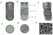
图1为本发明骨小梁多孔钽牙种植体的结构图,其中:a为种植体结构三维展示图;b为种植体纵剖面图,其中:1-顶部功能区,2-中部功能区,3-底部功能区;c为顶部俯视图;d为中部截面图;e为种植体实物照片;f为中部多孔功能区骨小梁结构显微照片;Fig. 1 is the structural drawing of the porous tantalum dental implant of the trabecular bone of the present invention, wherein: a is a three-dimensional display diagram of the implant structure; b is a longitudinal section view of the implant, wherein: 1-top functional area, 2-middle functional area, 3- Bottom functional area; c is the top plan view; d is the middle section view; e is the actual photo of the implant; f is the microphotograph of the trabecular bone structure in the middle porous functional area;
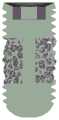
图2为实施例1中骨小梁多孔钽牙种植体的纵剖面图;Fig. 2 is the longitudinal sectional view of trabecular bone porous tantalum dental implant in Example 1;
图3为实施例1制备的骨小梁多孔钽牙种植体中部功能区的显微结构照片,其中a为标尺为500μm时观察到的骨小梁多孔结构;b为标尺为200μm时观察到的骨小梁多孔结构;c为熔融烧结形成的致密孔筋;d为观察到的纳米晶粒;Figure 3 is a photo of the microstructure of the middle functional area of the trabecular bone porous tantalum dental implant prepared in Example 1, wherein a is the trabecular bone porous structure observed when the scale is 500 μm; b is the observed trabecular bone structure when the scale is 200 μm. Porous structure of trabecular bone; c is the dense pore bars formed by melting and sintering; d is the observed nanocrystals;
图4为实施例1制备的骨小梁多孔钽牙种植体中部功能区的开口孔隙工业CT扫描图;4 is an industrial CT scan of the open pores in the middle functional area of the trabecular bone porous tantalum dental implant prepared in Example 1;
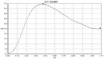
图5为实施例1制备的骨小梁多孔钽牙种植体中部功能区的压缩实验应力-应变曲线;5 is a compression test stress-strain curve of the middle functional area of the trabecular bone porous tantalum dental implant prepared in Example 1;
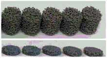
图6为实施例1制备的骨小梁多孔钽牙种植体中部功能区压缩实验前(上)后(下)的试样照片;Figure 6 is a sample photo of the trabecular bone porous tantalum dental implant prepared in Example 1 before (top) and after (bottom) the compression test of the middle functional area;
图7为实施例2中骨小梁多孔钽牙种植体纵剖面图;Fig. 7 is the longitudinal sectional view of trabecular bone porous tantalum dental implant in Example 2;
图8为实施例3中骨小梁多孔钽牙种植体纵剖面图。8 is a longitudinal cross-sectional view of the trabecular bone porous tantalum dental implant in Example 3. FIG.
具体实施方式Detailed ways
本发明提供了一种骨小梁多孔钽牙种植体,结构示意图如图1所示,图1中a为种植体的三维展示图;b为种植体的纵剖面图,其中:1-顶部功能区,2-中部功能区,3-底部功能区;c为顶部俯视图;d为中部截面图。The present invention provides a trabecular bone porous tantalum dental implant. The schematic structural diagram is shown in Figure 1. In Figure 1, a is a three-dimensional display diagram of the implant; b is a longitudinal section of the implant, wherein: 1-top function Zone, 2-middle functional zone, 3-bottom functional zone; c is the top plan view; d is the middle cross-sectional view.
在本发明中,所述骨小梁多孔钽牙种植体为圆柱体结构,自上而下依次包括顶部功能区、中部功能区和底部功能区;所述顶部功能区为致密结构,中部功能区为仿生骨小梁多孔结构,底部功能区为致密结构;所述骨小梁多孔钽牙种植体以纯钽或医用钽合金粉体为原料通过增材制造技术一体化制备得到。In the present invention, the trabecular bone porous tantalum dental implant has a cylindrical structure, including a top functional area, a middle functional area and a bottom functional area in order from top to bottom; the top functional area is a dense structure, and the middle functional area is It is a biomimetic trabecular bone porous structure, and the bottom functional area is a dense structure; the trabecular bone porous tantalum dental implant is prepared by using pure tantalum or medical tantalum alloy powder as a raw material through an integrated additive manufacturing technology.
在本发明中,所述骨小梁多孔钽牙种植体的总长度优选为5~25mm,更优选为8~22mm,直径优选为3~10mm,更优选为5~8mm。In the present invention, the total length of the trabecular bone porous tantalum dental implant is preferably 5-25 mm, more preferably 8-22 mm, and the diameter is preferably 3-10 mm, more preferably 5-8 mm.
本发明提供的骨小梁多孔钽牙种植体包括顶部功能区。在本发明中,所述顶部功能区为致密结构,所述顶部功能区长度优选为1~10mm,更优选为3~8mm;所述顶部功能区外侧优选设置有机械固定用螺纹或沟槽,所述螺纹或沟槽的深度优选为0.1~3mm,更优选为0.5~2mm;所述顶部功能区上表面优选设置有连接基台用凹槽,所述凹槽的深度优选为0.1~10mm,更优选为0.3~8mm,直径优选为1~8mm,更优选为3~5mm;所述凹槽优选为内六角凹槽。The trabecular bone porous tantalum dental implant provided by the present invention includes a top functional area. In the present invention, the top functional area is a dense structure, and the length of the top functional area is preferably 1-10 mm, more preferably 3-8 mm; the outer side of the top functional area is preferably provided with threads or grooves for mechanical fixing, The depth of the thread or groove is preferably 0.1-3mm, more preferably 0.5-2mm; the upper surface of the top functional area is preferably provided with a groove for connecting the base, and the depth of the groove is preferably 0.1-10mm, More preferably, the diameter is 0.3-8 mm, and the diameter is preferably 1-8 mm, more preferably 3-5 mm; the groove is preferably an inner hexagonal groove.
本发明提供的骨小梁多孔钽牙种植体包括中部功能区。在本发明中,所述中部功能区的长度优选为1~15mm,更优选为2~15mm,进一步优选为3~12mm;所述仿生骨小梁多孔结构的平均孔径为100~1500μm,更优选为300~1200μm,平均丝径优选为200~1000μm,更优选为300~800μm,孔隙率优选为10~90%,更优选为20~80%,孔道连通性优选大于90%。本发明提供的骨小梁多孔钽牙种植体的中部功能区为仿生骨小梁多孔结构,且长度容易调节,多孔部分能够贯穿整个种植体直径,能够更好地提高骨长入体积,形成更牢固的生物结合。The trabecular bone porous tantalum dental implant provided by the present invention includes a middle functional area. In the present invention, the length of the middle functional zone is preferably 1-15 mm, more preferably 2-15 mm, and further preferably 3-12 mm; the average pore size of the biomimetic bone trabecular porous structure is 100-1500 μm, more preferably It is 300-1200 μm, the average wire diameter is preferably 200-1000 μm, more preferably 300-800 μm, the porosity is preferably 10-90%, more preferably 20-80%, and the pore connectivity is preferably greater than 90%. The central functional area of the trabecular bone porous tantalum dental implant provided by the present invention is a biomimetic bone trabecular porous structure, and the length is easy to adjust, the porous part can penetrate the entire diameter of the implant, can better increase the volume of bone ingrowth, and form a more Strong biological bond.
在本发明中,所述中部功能区的中心优选设置有加强柱,所述加强柱的长度为优选1~15mm,更优选为2~15mm,进一步优选为3~12mm,直径优选为1~9.5mm,更优选为2~8mm;所述加强柱的材质也为纯钽或医用钽合金,在增材制造过程中一体化制备即可。In the present invention, the center of the middle functional area is preferably provided with a reinforcing column, the length of the reinforcing column is preferably 1-15 mm, more preferably 2-15 mm, further preferably 3-12 mm, and the diameter is preferably 1-9.5 mm mm, more preferably 2-8 mm; the material of the reinforcing column is also pure tantalum or medical tantalum alloy, which can be prepared integrally in the additive manufacturing process.
本发明提供的骨小梁多孔钽牙种植体包括底部功能区。在本发明中,所述底部功能区为致密结构;所述底部功能区外侧优选设置有机械固定用螺纹或沟槽,所述螺纹或沟槽的深度优选为0.1~3mm,更优选为0.5~2.5mm;所述底部功能区的底面优选为球形。The trabecular bone porous tantalum dental implant provided by the present invention includes a bottom functional area. In the present invention, the bottom functional area is a dense structure; the outer side of the bottom functional area is preferably provided with threads or grooves for mechanical fixing, and the depth of the threads or grooves is preferably 0.1 to 3 mm, more preferably 0.5 to 0.5 mm. 2.5mm; the bottom surface of the bottom functional area is preferably spherical.
在本发明中,所述骨小梁多孔钽牙种植体以纯钽或医用钽合金粉体为原料通过增材制造技术一体化制备得到;所述纯钽的纯度优选>99%,所述医用钽合金中的钽元素含量优选>1wt%,更优选为5~95wt%;所述纯钽或医用碳合金粉体的粒径独立地优选为10~150μm,更优选为20~130μm;所述纯钽或医用钽合金粉体优选为球形粉体,所述球形粉体的球形度优选大于80%,更优选为85~99%;在本发明中,所述医用钽合金粉体优选为钽钛混合粉体或Ti35Nb3Zr2Ta预合金粉体;所述钽钛混合粉体中优选包括70vol%钽和30vol%钛。本发明利用增材制造技术制备本发明的牙种植体,所得牙种植体的顶部、中部、底部三个功能区为整体结构,而不是机械拼接而成,和机械拼接的牙种植体相比,本发明提供的牙种植体种植稳定性更好,没有分层脱离的风险。In the present invention, the trabecular bone porous tantalum dental implant is prepared by using pure tantalum or medical tantalum alloy powder as raw materials through an integrated additive manufacturing technology; the purity of the pure tantalum is preferably >99%, and the medical tantalum The content of tantalum element in the alloy is preferably >1wt%, more preferably 5-95wt%; the particle size of the pure tantalum or medical carbon alloy powder is independently preferably 10-150μm, more preferably 20-130μm; The tantalum or medical tantalum alloy powder is preferably spherical powder, and the sphericity of the spherical powder is preferably greater than 80%, more preferably 85-99%; in the present invention, the medical tantalum alloy powder is preferably tantalum titanium Mixed powder or Ti35Nb3Zr2Ta pre-alloyed powder; the tantalum-titanium mixed powder preferably includes 70vol% tantalum and 30vol% titanium. The present invention uses additive manufacturing technology to prepare the dental implant of the present invention, and the three functional areas of the top, middle and bottom of the obtained dental implant are integral structures instead of mechanically spliced. Compared with the mechanically spliced dental implant, The dental implant provided by the invention has better planting stability and no risk of delamination.
在本发明中,所述增材制造技术优选包括激光熔融增材制造技术或电子束熔融增材制造技术。In the present invention, the additive manufacturing technology preferably includes a laser melting additive manufacturing technology or an electron beam melting additive manufacturing technology.
本发明还提供了上述方案所述骨小梁多孔钽牙种植体的制备方法,包括以下步骤:The present invention also provides a preparation method of the trabecular bone porous tantalum dental implant described in the above scheme, comprising the following steps:
利用建模软件构建骨小梁多孔钽牙种植体的三维模型,按照构建的模型,以纯钽或者医用钽合金粉体为原料进行增材制造。The three-dimensional model of the trabecular bone porous tantalum dental implant is constructed by modeling software, and pure tantalum or medical tantalum alloy powder is used as raw material for additive manufacturing according to the constructed model.
本发明对所述建模软件没有特殊要求,使用本领域技术人员熟知的建模软件即可。The present invention has no special requirements on the modeling software, and modeling software well known to those skilled in the art can be used.
得到三维模型后,本发明优选将三维模型转换为打印设备所需格式并导入到增材制造设备中,然后以纯钽或者含钽合金医用钽合金粉体为原料进行打印;在本发明中,当所述增材制造为激光熔融增材制造时,所述打印的参数优选为:激光功率为150~300W,优选为180~260W,扫描速度为100~350mm/s,优选为120~330mm/s,扫描线间距为0.05~0.1mm,优选为0.06~0.09mm,切片层厚为20~50μm,优选为30~40μm,基板温度为100~200℃,优选为130~180℃;当所述增材制造为电子束熔融增材制造时,所述打印的参数优选为:粉体预热温度为900~1500℃,更优选为1000~1400℃,扫描电流为5~15mA,更优选为6~12mA,扫描速度为1.0×104~8.0×105mm/s,优选为2×104~7×105mm/s;切片层厚优选20~50μm;优选为30~40μm。After obtaining the three-dimensional model, the present invention preferably converts the three-dimensional model into the format required by the printing equipment and imports it into the additive manufacturing equipment, and then uses pure tantalum or tantalum-containing alloy medical tantalum alloy powder as the raw material for printing; in the present invention, when When the additive manufacturing is laser melting additive manufacturing, the printing parameters are preferably: laser power is 150-300W, preferably 180-260W, scanning speed is 100-350mm/s, preferably 120-330mm/s , the scanning line spacing is 0.05-0.1 mm, preferably 0.06-0.09 mm, the slice layer thickness is 20-50 μm, preferably 30-40 μm, and the substrate temperature is 100-200 ℃, preferably 130-180 ℃; When the material manufacturing is electron beam melting additive manufacturing, the printing parameters are preferably: the powder preheating temperature is 900-1500 ℃, more preferably 1000-1400 ℃, the scanning current is 5-15mA, more preferably 6- 12mA, the scanning speed is 1.0×10⁴ -8.0×105 mm/s, preferably 2×10⁴ -7×105 mm/s; the slice thickness is preferably 20-50 μm; preferably 30-40 μm.
打印完成后,本发明优选将打印成型件进行后处理,得到本发明的骨小梁多孔钽牙种植体。在本发明中,所述后处理优选包括依次进行的清粉、线切割、去支撑、喷砂、超声清洗和热处理。After the printing is completed, the present invention preferably performs post-processing on the printed molded part to obtain the trabecular bone porous tantalum dental implant of the present invention. In the present invention, the post-treatment preferably includes cleaning, wire cutting, de-supporting, sandblasting, ultrasonic cleaning and heat treatment in sequence.
在本发明中,所述清粉优选为物理方式清粉,具体的清粉方式优选为湿式防爆吸尘器吸除、压缩空气喷吹以及超声清洗中的一种或几种;本发明通过清粉将打印成型件表面的粉末清理干净。In the present invention, the cleaning powder is preferably a physical cleaning powder, and the specific cleaning powder method is preferably one or more of wet-type explosion-proof vacuum cleaner suction, compressed air blowing and ultrasonic cleaning; The powder on the surface of the printed part is cleaned up.
在本发明中,所述线切割优选使用放电钼丝线切割机进行,钼丝电火花将金属微熔化形成切割,对切割面机械损伤小,精度高。本发明通过线切割将打印成型件从基板上面切割分离下来。In the present invention, the wire cutting is preferably performed by a discharge molybdenum wire wire cutting machine, and the molybdenum wire electric spark melts the metal slightly to form the cutting, which has little mechanical damage to the cutting surface and high precision. The invention cuts and separates the printed molding from the substrate by wire cutting.
本发明对所述去支撑没有特殊要求,使用本领域技术人员熟知的方式,将打印成型件下部与基板连接的支撑物拆除即可,在本发明的具体实施例中,可以使用老虎钳等工具轻微敲击或夹断。The present invention has no special requirements for the de-support, and the support connecting the lower part of the printed part and the substrate can be removed by using a method well known to those skilled in the art. Knock or pinch off.
在本发明中,所述喷砂优选使用非金属砂,所述非金属砂的粒径优选为100~500目,更优选为200~400目。In the present invention, non-metallic sand is preferably used for the sand blasting, and the particle size of the non-metallic sand is preferably 100-500 mesh, more preferably 200-400 mesh.
在本发明中,所述超声清洗优选为依次用无水乙醇和蒸馏水进行清洗,在无水乙醇和蒸馏水中的总清洗次数优选大于10次,更优选为12~20次,总清洗时间优选大于5h,更优选为7~10h。In the present invention, the ultrasonic cleaning is preferably performed sequentially with absolute ethanol and distilled water, and the total cleaning times in absolute ethanol and distilled water are preferably more than 10 times, more preferably 12 to 20 times, and the total cleaning time is preferably greater than 5h, more preferably 7-10h.
在本发明中,所述热处理优选为真空去应力退火处理;所述热处理的退火温度优选为850~1050℃,更优选为900~1000℃,真空度优选<10×10-3Pa,保温时间优选为1~5h,更优选为2~4h。In the present invention, the heat treatment is preferably vacuum stress relief annealing treatment; the annealing temperature of the heat treatment is preferably 850-1050°C, more preferably 900-1000°C, the degree of vacuum is preferably <10×10-3 Pa, and the holding time is It is preferably 1 to 5 hours, and more preferably 2 to 4 hours.
下面结合实施例对本发明提供的方案进行详细的说明,但是不能把它们理解为对本发明保护范围的限定。The solutions provided by the present invention will be described in detail below in conjunction with the examples, but they should not be construed as limiting the protection scope of the present invention.
实施例1Example 1
采用专业建模软件设计三个功能区的结构,构建骨小梁多孔钽牙种植体三维模型,其中,顶部功能区为致密结构;中部功能区为仿生骨小梁多孔结构;底部功能区为致密结构。总长度16mm,直径6mm,顶部功能区长度5mm,中部功能区长度7mm,底部功能区长度4mm。顶部功能区外侧有机械固定用螺纹,深度1mm;上表面有连接基台用内六角凹槽,深度3mm,直径3mm。中部功能区为仿生骨小梁多孔结构,平均孔径500μm,平均丝径300μm,孔隙率70%,孔道连通性99%;多孔结构中心无内部加强柱。底部功能区外侧有机械固定用螺纹,深度1mm。种植体底面为球形。实施例1的骨小梁多孔钽牙种植体的纵剖面图如图2所示。Professional modeling software was used to design the structure of three functional areas, and a three-dimensional model of trabecular bone porous tantalum dental implant was constructed. Among them, the top functional area was a dense structure; the middle functional area was a biomimetic trabecular bone porous structure; structure. The total length is 16mm, the diameter is 6mm, the length of the top functional area is 5mm, the length of the middle functional area is 7mm, and the length of the bottom functional area is 4mm. There is a mechanical fixing thread on the outside of the top functional area, with a depth of 1mm; the upper surface has a hexagonal socket for connecting the abutment, with a depth of 3mm and a diameter of 3mm. The middle functional area is a biomimetic bone trabecular porous structure with an average pore diameter of 500 μm, an average wire diameter of 300 μm, a porosity of 70%, and a pore connectivity of 99%; there is no internal reinforcing column in the center of the porous structure. There are mechanical fixing threads on the outside of the bottom functional area, with a depth of 1mm. The underside of the implant is spherical. The longitudinal sectional view of the trabecular bone porous tantalum dental implant of Example 1 is shown in FIG. 2 .
将所述三维模型转换为打印设备所需格式并导入到设备中。以纯钽粉体为原料,采用激光熔融增材制造工艺进行打印。纯钽粉体,纯度>99.9%,粒径15~45μm,球形度95。激光熔融主要打印工艺参数为:激光功率150W,扫描速度200mm/s,扫描线间距0.05mm,切片层厚50μm,基板温度200℃。The three-dimensional model is converted into a format required by the printing device and imported into the device. Using pure tantalum powder as raw material, it is printed by laser melting additive manufacturing process. Pure tantalum powder, purity >99.9%, particle size 15-45μm, sphericity 95. The main printing process parameters of laser melting are: laser power 150W, scanning speed 200mm/s, scanning line spacing 0.05mm, slice layer thickness 50μm, and substrate temperature 200℃.
打印完成后,对打印成型件依次进行清粉、线切割、去支撑、喷砂、超声清洗和热处理。清粉操作为物理方式清粉,具体包括使用湿式防爆吸尘器吸除、压缩空气喷吹和超声波清洗进行清粉;线切割采用放电钼丝线切割机进行;去支撑为采用老虎钳将打印成型件下部与基板连接的支撑物拆除;喷砂步骤使用的是非金属砂,粒径500目;超声波清洗为依次采用无水乙醇、蒸馏水进行清洗,总清洗次数为20次,总清洗时间为8h;热处理为真空去应力退火处理;退火温度为900℃,真空度<5×10⁻³Pa,保温时间3h。After the printing is completed, the printed parts are subjected to powder cleaning, wire cutting, de-supporting, sandblasting, ultrasonic cleaning and heat treatment in sequence. The cleaning operation is physical cleaning, which includes the use of wet explosion-proof vacuum cleaner for suction removal, compressed air blowing and ultrasonic cleaning for cleaning powder; wire cutting is carried out with a discharge molybdenum wire cutting machine; The support connected to the substrate was removed; the sandblasting step used non-metallic sand with a particle size of 500 mesh; the ultrasonic cleaning was performed with absolute ethanol and distilled water in sequence, and the total cleaning times were 20 times and the total cleaning time was 8h; the heat treatment was vacuum Stress relief annealing treatment; the annealing temperature is 900℃, the vacuum degree is less than 5×10⁻³ Pa, and the holding time is 3h.
对实施例1制备的骨小梁多孔钽牙种植体的中部多孔功能区的力学性能进行测试,结果显示中部功能区的耐压强度52±5.8MPa,抗弯强度77±6.3MPa以上,弹性模量为2.3±0.2GPa,综合力学性能优异;50~500N循环压应力、加载频率15Hz条件下,疲劳性能930±60万次;与带膜皮质骨组成摩擦副进行摩擦系数测试,摩擦系数为1.6±0.3。The mechanical properties of the central porous functional area of the trabecular bone porous tantalum dental implant prepared in Example 1 were tested. The amount is 2.3±0.2GPa, and the comprehensive mechanical properties are excellent; under the condition of 50-500N cyclic compressive stress and loading frequency of 15Hz, the fatigue performance is 9.30±600,000 times; the friction coefficient of the friction pair formed with the membrane cortical bone is tested, and the friction coefficient is 1.6 ±0.3.
图3为所得骨小梁多孔钽牙种植体的中部功能区的SEM图,其中a为标尺为500μm时观察到的骨小梁多孔结构;b为标尺为200μm时观察到的骨小梁多孔结构;c为熔融烧结形成的致密孔筋;d为观察到的纳米晶粒。根据图3可以看出,中部功能区的仿生骨小梁多孔结构具有较高的高孔隙率,且由激光熔融增材制造形成孔筋非常致密,孔筋表面具有纳米晶粒显微结构。Figure 3 is the SEM image of the middle functional area of the obtained trabecular bone porous tantalum dental implant, in which a is the trabecular bone porous structure observed when the scale is 500 μm; b is the trabecular bone porous structure observed when the scale is 200 μm ; c is the dense pores formed by melt sintering; d is the observed nanograin. It can be seen from Figure 3 that the biomimetic bone trabecular porous structure in the central functional area has a high porosity, and the pore bars formed by laser melting additive manufacturing are very dense, and the surface of the pore bars has a nano-grain microstructure.
图4为所得骨小梁多孔钽牙种植体中部功能区开口孔隙工业CT扫描图;根据图4可以看出,中部功能区的多孔结构连通性大于90%。Figure 4 is an industrial CT scan of the open pores in the middle functional area of the obtained trabecular bone porous tantalum dental implant; it can be seen from Figure 4 that the connectivity of the porous structure in the middle functional area is greater than 90%.
图5为所得骨小梁多孔钽牙种植体中部功能区压缩实验应力-应变曲线;根据图5可以看出,中部功能区压缩形变为20%时仍然具有较好的强度。Figure 5 shows the stress-strain curve of the obtained trabecular bone porous tantalum dental implant in the middle functional area of the compression experiment; according to Figure 5, it can be seen that the middle functional area still has good strength when the compression deformation is 20%.
图6为所得骨小梁多孔钽牙种植体中部功能区压缩实验前(上)后(下)的试样照片;根据图6可以看出,中部功能区压缩变形70%时仍未发生破坏性断裂,表明具有优异的塑性和韧性,很好的力学可靠性和安全性。Figure 6 is the sample photos of the obtained trabecular bone porous tantalum dental implant middle functional area before (top) and after (bottom) compression test; according to Figure 6, it can be seen that the middle functional area is not destructive when the compression deformation is 70% Fracture, showing excellent plasticity and toughness, good mechanical reliability and safety.
实施例2Example 2
采用专业建模软件设计三个功能区的结构,构建骨小梁多孔钽牙种植体三维模型,其中,顶部功能区为致密结构;中部功能区为仿生骨小梁多孔结构;底部功能区为致密结构。总长度13mm,直径5mm,顶部长度4mm,中部长度5mm,底部长度4mm。顶部功能区外侧有机械固定用螺纹,深度0.8mm;上表面有连接基台用内六角凹槽,深度2mm,直径3mm。中部功能区具有仿生骨小梁多孔结构,平均孔径470μm,平均丝径330μm,孔隙率75%,孔道连通性99%;多孔结构中心有内部加强柱,直径2.5mm。底部功能区外侧有机械固定用螺纹,深度0.8mm。种植体底面为球形。实施例2的骨小梁多孔钽牙种植体的纵剖面图如图7所示。Professional modeling software was used to design the structure of three functional areas, and a three-dimensional model of trabecular bone porous tantalum dental implant was constructed. Among them, the top functional area was a dense structure; the middle functional area was a biomimetic trabecular bone porous structure; structure. Overall length 13mm, diameter 5mm, top length 4mm, middle length 5mm, bottom length 4mm. There are mechanical fixing threads on the outside of the top functional area, with a depth of 0.8mm; the upper surface has a hexagonal socket for connecting the abutment, with a depth of 2mm and a diameter of 3mm. The middle functional area has a biomimetic bone trabecular porous structure with an average pore diameter of 470 μm, an average wire diameter of 330 μm, a porosity of 75%, and a pore connectivity of 99%; there is an internal reinforcing column in the center of the porous structure, with a diameter of 2.5 mm. There are mechanical fixing threads on the outside of the bottom functional area, with a depth of 0.8mm. The underside of the implant is spherical. The longitudinal sectional view of the trabecular bone porous tantalum dental implant of Example 2 is shown in FIG. 7 .
将所述三维模型转换为打印设备所需格式并导入到设备中,以钽钛混合粉体为原料,采用激光熔融增材制造工艺进行打印。混合粉体包括70vol%钽和30vol%钛,粒径15~45μm,球形度97。激光熔融主要打印工艺参数为:激光功率200w,扫描速度150mm/s,扫描线间距0.1mm,切片层厚30μm,基板温度150℃。The three-dimensional model is converted into the format required by the printing equipment and imported into the equipment, and the tantalum-titanium mixed powder is used as the raw material, and the laser melting additive manufacturing process is used for printing. The mixed powder includes 70vol% of tantalum and 30vol% of titanium, with a particle size of 15-45 μm and a sphericity of 97. The main printing process parameters of laser melting are: laser power 200w, scanning speed 150mm/s, scanning line spacing 0.1mm, slice layer thickness 30μm, and substrate temperature 150℃.
打印完成后,对打印成型件依次进行清粉、线切割、去支撑、喷砂、超声清洗和热处理。清粉操作为物理方式清粉,具体包括使用湿式防爆吸尘器吸除、压缩空气喷吹和超声波清洗进行清粉;线切割采用放电钼丝线切割机进行;去支撑为采用老虎钳将打印成型件下部与基板连接的支撑物拆除;超声波清洗为依次采用无水乙醇、蒸馏水进行清洗,总清洗次数为18次,总清洗时间10h;喷砂步骤使用的是非金属砂,粒径500目;热处理为真空去应力退火处理;退火温度为950℃,真空度<5×10⁻³Pa,保温时间2h。After the printing is completed, the printed parts are subjected to powder cleaning, wire cutting, de-supporting, sandblasting, ultrasonic cleaning and heat treatment in sequence. The cleaning operation is physical cleaning, which includes the use of wet explosion-proof vacuum cleaner for suction removal, compressed air blowing and ultrasonic cleaning for cleaning powder; wire cutting is carried out with a discharge molybdenum wire cutting machine; The supports connected to the substrate are removed; the ultrasonic cleaning is to use anhydrous ethanol and distilled water for cleaning in sequence, the total cleaning times is 18 times, and the total cleaning time is 10h; the sandblasting step uses non-metallic sand with a particle size of 500 mesh; heat treatment is vacuum removal Stress annealing treatment; the annealing temperature is 950℃, the degree of vacuum is less than 5×10⁻³ Pa, and the holding time is 2h.
对实施例2制备的骨小梁多孔钽牙种植体的中部多孔功能区的力学性能进行测试,结果显示中部功能区的耐压强度47±3.6MPa,抗弯强度65±4.3MPa以上,弹性模量为2.0±0.3GPa,综合力学性能优异;50~500N循环压应力、加载频率15Hz条件下,疲劳性能770±50万次;与带膜皮质骨组成摩擦副进行摩擦系数测试,摩擦系数为1.4±0.3。The mechanical properties of the middle porous functional area of the trabecular bone porous tantalum dental implant prepared in Example 2 were tested. The amount is 2.0±0.3GPa, and the comprehensive mechanical properties are excellent; under the condition of 50-500N cyclic compressive stress and loading frequency of 15Hz, the fatigue performance is 7.70±500,000 times; the friction coefficient of the friction pair formed with the membrane cortical bone is tested, and the friction coefficient is 1.4 ±0.3.
实施例3Example 3
采用专业建模软件设计三个功能区的结构,构建骨小梁多孔钽牙种植体三维模型,其中,顶部功能区为致密结构;中部功能区为仿生骨小梁多孔结构;底部功能区为致密结构。总长度12mm,直径4.1mm,顶部长度4mm,中部长度5mm,底部长度3mm。顶部功能区外侧有机械固定用螺纹,深度0.7mm;上表面有连接基台用内六角凹槽,深度2mm,直径3mm。中部功能区具有仿生骨小梁多孔结构,平均孔径600μm,平均丝径350μm,孔隙率68%,孔道连通性99%;多孔结构中心有内部加强柱,直径1mm。底部功能区外侧有机械固定用螺纹,深度0.7mm。种植体底面为球形。实施例3的骨小梁多孔钽牙种植体的纵剖面图如图8所示。Professional modeling software was used to design the structure of three functional areas, and a three-dimensional model of trabecular bone porous tantalum dental implant was constructed. Among them, the top functional area was a dense structure; the middle functional area was a biomimetic trabecular bone porous structure; structure. The overall length is 12mm, the diameter is 4.1mm, the top length is 4mm, the middle length is 5mm, and the bottom length is 3mm. There are mechanical fixing threads on the outside of the top functional area, with a depth of 0.7mm; the upper surface has a hexagonal socket for connecting the abutment, with a depth of 2mm and a diameter of 3mm. The middle functional area has a biomimetic bone trabecular porous structure with an average pore diameter of 600 μm, an average wire diameter of 350 μm, a porosity of 68%, and a pore connectivity of 99%; there is an internal reinforcing column in the center of the porous structure, with a diameter of 1 mm. There are mechanical fixing threads on the outside of the bottom functional area, with a depth of 0.7mm. The underside of the implant is spherical. The longitudinal sectional view of the trabecular bone porous tantalum dental implant of Example 3 is shown in FIG. 8 .
将所述三维模型转换为打印设备所需格式并导入到设备中,以钛铌锆钽合金Ti35Nb3Zr2Ta预合金粉体为原料,采用粉末床激光熔融增材制造工艺进行打印。钛铌锆钽合金预合金粉体的粒径为53~105μm,球形度为93。电子束熔融主要打印工艺参数为:粉末预热温度1000℃,扫描电流15mA,扫描速度为5.0×105mm/s,切片层厚20μm。The three-dimensional model is converted into the format required by the printing equipment and imported into the equipment, and the pre-alloyed powder of titanium niobium zirconium tantalum alloy Ti35Nb3Zr2Ta is used as the raw material, and the powder bed laser melting additive manufacturing process is used for printing. The particle size of the titanium-niobium-zirconium-tantalum alloy pre-alloyed powder is 53-105 μm, and the sphericity is 93. The main printing process parameters of electron beam melting are: powder preheating temperature 1000℃, scanning current 15mA, scanning speed 5.0×105 mm/s, slice layer thickness 20μm.
打印完成后,对打印成型件依次进行清粉、线切割、去支撑、喷砂、超声清洗和热处理。清粉操作为物理方式清粉,具体包括使用湿式防爆吸尘器吸除、压缩空气喷吹和超声波清洗进行清粉;线切割采用放电钼丝线切割机进行;去支撑为采用老虎钳将打印成型件下部与基板连接的支撑物拆除;喷砂步骤使用的是非金属砂,粒径300目;超声波清洗为依次采用无水乙醇、蒸馏水进行超声清洗,总清洗次数为15次,总清洗时间7h;热处理为真空去应力退火处理;退火温度为1000℃,真空度<5×10⁻³Pa,保温时间1.5h。After the printing is completed, the printed parts are subjected to powder cleaning, wire cutting, de-supporting, sandblasting, ultrasonic cleaning and heat treatment in sequence. The cleaning operation is physical cleaning, which includes the use of wet explosion-proof vacuum cleaner for suction removal, compressed air blowing and ultrasonic cleaning for cleaning powder; wire cutting is carried out with a discharge molybdenum wire cutting machine; The supports connected to the substrates were removed; the sandblasting step used non-metallic sand with a particle size of 300 mesh; the ultrasonic cleaning was carried out with absolute ethanol and distilled water in sequence, with a total cleaning frequency of 15 times and a total cleaning time of 7h; heat treatment was vacuum Stress relief annealing treatment; the annealing temperature is 1000℃, the vacuum degree is less than 5×10⁻³ Pa, and the holding time is 1.5h.
对实施例3制备的骨小梁多孔钽牙种植体的中部多孔功能区的力学性能进行测试,结果显示中部功能区的耐压强度37±4.3MPa,抗弯强度55±3.7MPa以上,弹性模量为2.1±0.3GPa,综合力学性能优异;50~500N循环压应力、加载频率15Hz条件下,疲劳性能750±36万次;与带膜皮质骨组成摩擦副进行摩擦系数测试,摩擦系数为1.5±0.2。The mechanical properties of the central porous functional area of the trabecular bone porous tantalum dental implant prepared in Example 3 were tested. The amount is 2.1±0.3GPa, and the comprehensive mechanical properties are excellent; under the condition of 50-500N cyclic compressive stress and loading frequency of 15Hz, the fatigue performance is 7.50±360,000 times; the friction coefficient of the friction pair formed with the cortical bone with membrane is tested, and the friction coefficient is 1.5 ±0.2.
实施例4Example 4
采用大动物实验测定骨长入效果。以健康成年山羊作为实验动物模型,在每只动物的左右后肢股骨头髓腔建缺损模型,分别植入本发明的骨小梁多孔钽牙种植体和对照试样-多孔钛合金Ti6Al4V,试样规格与实施例2相同。植入手术:采用股骨远端外侧入路,在股骨外侧采用1cm切口,用环钻低速在股骨外侧髁制造直径为5mm,深度为10mm的圆柱状骨和骨膜缺损,并用0.9%的生理盐水进行冲洗,根据分组植入术前准备好的试样料,逐层关闭伤口,术后常规高蛋白饲料圈养。植入后1个月和三个月时间点将动物处死。取出植入试样局部一段,采用轴向应力推出方法检测植入试样与周围骨组织结合强度,进行Micro-CT扫描,定性、定量评价骨长入效果,包括新生骨体积等。硬组织切片进行组织染色,评价新生骨长入效果。The effect of bone ingrowth was determined by large animal experiments. Taking healthy adult goats as experimental animal models, a defect model was built in the femoral head pulp cavity of each animal's left and right hind limbs, and the trabecular bone porous tantalum dental implant of the present invention and a control sample-porous titanium alloy Ti6Al4V were implanted respectively. The specifications are the same as in Example 2. Implantation surgery: A 5mm diameter, 10mm depth cylindrical bone and periosteal defect was created in the lateral femoral condyle with trephine at low speed using a 1cm incision on the lateral side of the femur using a distal distal femoral approach and performed with 0.9% saline. After rinsing, the samples prepared before surgery were implanted in groups, the wounds were closed layer by layer, and they were kept in high-protein diets after surgery. Animals were sacrificed at 1 and 3 months post-implantation time points. A partial section of the implanted sample was taken out, and the axial stress pushing method was used to detect the bonding strength between the implanted sample and the surrounding bone tissue, and Micro-CT scanning was performed to evaluate the effect of bone ingrowth qualitatively and quantitatively, including the volume of new bone. Hard tissue sections were stained to evaluate the effect of new bone ingrowth.
大动物实验结果表明,本发明的种植体术后一个月与周围骨组织结合强度大于18MPa;术后一个月新生骨长入体积比高于25%,术后三个月新生骨长入体积比高于60%,术后三个月新生骨长入体积比高于85%,远优于钛合金牙种植体。The results of the large animal experiments show that the bonding strength of the implant of the present invention to the surrounding bone tissue is greater than 18MPa one month after the operation; It is higher than 60%, and the volume ratio of new bone growth in three months after operation is higher than 85%, which is much better than titanium alloy dental implants.
以上所述仅是本发明的优选实施方式,应当指出,对于本技术领域的普通技术人员来说,在不脱离本发明原理的前提下,还可以做出若干改进和润饰,这些改进和润饰也应视为本发明的保护范围。The above are only the preferred embodiments of the present invention. It should be pointed out that for those skilled in the art, without departing from the principles of the present invention, several improvements and modifications can be made. It should be regarded as the protection scope of the present invention.
Claims (9)
Priority Applications (2)
| Application Number | Priority Date | Filing Date | Title |
|---|---|---|---|
| CN202010331329.1ACN111494035A (en) | 2020-04-24 | 2020-04-24 | Trabecular bone porous tantalum dental implant and preparation method thereof |
| US17/238,419US12097089B2 (en) | 2020-04-24 | 2021-04-23 | Trabecular porous tantalum dental implant and preparation method thereof |
Applications Claiming Priority (1)
| Application Number | Priority Date | Filing Date | Title |
|---|---|---|---|
| CN202010331329.1ACN111494035A (en) | 2020-04-24 | 2020-04-24 | Trabecular bone porous tantalum dental implant and preparation method thereof |
Publications (1)
| Publication Number | Publication Date |
|---|---|
| CN111494035Atrue CN111494035A (en) | 2020-08-07 |
Family
ID=71874654
Family Applications (1)
| Application Number | Title | Priority Date | Filing Date |
|---|---|---|---|
| CN202010331329.1APendingCN111494035A (en) | 2020-04-24 | 2020-04-24 | Trabecular bone porous tantalum dental implant and preparation method thereof |
Country Status (2)
| Country | Link |
|---|---|
| US (1) | US12097089B2 (en) |
| CN (1) | CN111494035A (en) |
Cited By (10)
| Publication number | Priority date | Publication date | Assignee | Title |
|---|---|---|---|---|
| CN112336477A (en)* | 2020-11-06 | 2021-02-09 | 北京市春立正达医疗器械股份有限公司 | Dental implant with bone induction structure |
| CN113974875A (en)* | 2021-11-02 | 2022-01-28 | 广西医科大学 | Porous implant for dentistry |
| CN114010349A (en)* | 2021-11-19 | 2022-02-08 | 北京大学口腔医学院 | Dental implant with porous surface zirconia printed in 3D mode and design and processing method |
| CN114867498A (en)* | 2020-09-25 | 2022-08-05 | 北京华宇创新钽铌科技有限公司 | Bone implant with porous membrane and method for producing same |
| CN115054388A (en)* | 2022-07-05 | 2022-09-16 | 北京大学口腔医学院 | Function-guided individual bionic porous pure tantalum dental implant and preparation method thereof |
| CN115252189A (en)* | 2022-07-26 | 2022-11-01 | 宁夏东方智造科技有限公司 | Tantalum metal dental implant and preparation method thereof |
| WO2023015722A1 (en)* | 2021-08-12 | 2023-02-16 | 广州市健齿生物科技有限公司 | Modular layered dental implant |
| CN115740495A (en)* | 2022-11-02 | 2023-03-07 | 大博医疗科技股份有限公司 | Method for 3D printing of trabecular oral implant |
| CN115814151A (en)* | 2022-12-21 | 2023-03-21 | 北京市春立正达医疗器械股份有限公司 | Preparation method of 3D printing bone implant surface multilevel micron structure |
| CN116275097A (en)* | 2021-12-07 | 2023-06-23 | 广东汉邦激光科技有限公司 | Preparation method of porous implant graft printing |
Families Citing this family (7)
| Publication number | Priority date | Publication date | Assignee | Title |
|---|---|---|---|---|
| EP3287150B1 (en)* | 2016-08-22 | 2022-12-14 | WALDEMAR LINK GmbH & Co. KG | Coating for an implant |
| ES2971693T3 (en)* | 2019-11-08 | 2024-06-06 | Politechnika Warszawska | Bioactive intraosseous dental implant |
| US20220087788A1 (en)* | 2020-09-24 | 2022-03-24 | Biorl S.R.L. | Dental screw |
| US11935771B2 (en) | 2021-02-17 | 2024-03-19 | Applied Materials, Inc. | Modular mainframe layout for supporting multiple semiconductor process modules or chambers |
| US11935770B2 (en) | 2021-02-17 | 2024-03-19 | Applied Materials, Inc. | Modular mainframe layout for supporting multiple semiconductor process modules or chambers |
| CN115089328B (en)* | 2022-06-17 | 2023-05-12 | 汪晓晖 | Dental implant system with surface gradient microporous structure and preparation method thereof |
| US11771528B1 (en)* | 2022-09-12 | 2023-10-03 | Spectrum Spine Ip Holdings, Llc | Implant with enhanced osteoinductivity |
Citations (5)
| Publication number | Priority date | Publication date | Assignee | Title |
|---|---|---|---|---|
| US5989027A (en)* | 1995-12-08 | 1999-11-23 | Sulzer Calcitek Inc. | Dental implant having multiple textured surfaces |
| CN101193664A (en)* | 2005-05-04 | 2008-06-04 | 维塔尔植入物公司 | Dental implant comprising a porous trabecular structure |
| US20100003638A1 (en)* | 2008-07-02 | 2010-01-07 | Michael Collins | Modular implant with secured porous portion |
| CN204484368U (en)* | 2015-03-18 | 2015-07-22 | 浙江科惠医疗器械有限公司 | A kind of rotary squeezing with porous self-bone grafting structure is with regard to bit-type tooth implant |
| CN109175376A (en)* | 2018-11-07 | 2019-01-11 | 成都先进金属材料产业技术研究院有限公司 | The post-processing approach of increasing material manufacturing titanium or titanium alloy part |
Family Cites Families (12)
| Publication number | Priority date | Publication date | Assignee | Title |
|---|---|---|---|---|
| US8684734B1 (en)* | 2003-02-27 | 2014-04-01 | Philip Scott Lyren | Dental implant with porous body |
| US9095396B2 (en) | 2008-07-02 | 2015-08-04 | Zimmer Dental, Inc. | Porous implant with non-porous threads |
| US9707058B2 (en)* | 2009-07-10 | 2017-07-18 | Zimmer Dental, Inc. | Patient-specific implants with improved osseointegration |
| US8602782B2 (en) | 2009-11-24 | 2013-12-10 | Zimmer Dental, Inc. | Porous implant device with improved core |
| US9433480B2 (en) | 2010-12-21 | 2016-09-06 | Zimmer Dental, Inc. | Implant with porous sleeve including anti-rotation features |
| EP2877117B1 (en)* | 2012-07-25 | 2019-02-20 | Zimmer Dental Inc. | Porous dental implant |
| WO2018100250A1 (en)* | 2016-11-30 | 2018-06-07 | Abdelmadjid Djemai | Dental implant and self-locking fastening element with heterogeneous porous structures, and method for its production |
| EP3417828B1 (en)* | 2017-06-20 | 2021-03-03 | Ruetschi Technology AG | Method of manufacturing an implant with titanium-based three-dimensional printing material |
| TWI877173B (en)* | 2019-07-19 | 2025-03-21 | 美商環球高級金屬美國公司 | Spherical tantalum-titanium alloy powder, products containing the same, and methods of making the same |
| ES2971693T3 (en)* | 2019-11-08 | 2024-06-06 | Politechnika Warszawska | Bioactive intraosseous dental implant |
| US12144706B2 (en)* | 2019-11-26 | 2024-11-19 | Biomet 3I, Llc | Additive manufactured dental implants and methods thereof |
| US20220087788A1 (en)* | 2020-09-24 | 2022-03-24 | Biorl S.R.L. | Dental screw |
- 2020
- 2020-04-24CNCN202010331329.1Apatent/CN111494035A/enactivePending
- 2021
- 2021-04-23USUS17/238,419patent/US12097089B2/enactiveActive
Patent Citations (5)
| Publication number | Priority date | Publication date | Assignee | Title |
|---|---|---|---|---|
| US5989027A (en)* | 1995-12-08 | 1999-11-23 | Sulzer Calcitek Inc. | Dental implant having multiple textured surfaces |
| CN101193664A (en)* | 2005-05-04 | 2008-06-04 | 维塔尔植入物公司 | Dental implant comprising a porous trabecular structure |
| US20100003638A1 (en)* | 2008-07-02 | 2010-01-07 | Michael Collins | Modular implant with secured porous portion |
| CN204484368U (en)* | 2015-03-18 | 2015-07-22 | 浙江科惠医疗器械有限公司 | A kind of rotary squeezing with porous self-bone grafting structure is with regard to bit-type tooth implant |
| CN109175376A (en)* | 2018-11-07 | 2019-01-11 | 成都先进金属材料产业技术研究院有限公司 | The post-processing approach of increasing material manufacturing titanium or titanium alloy part |
Non-Patent Citations (1)
| Title |
|---|
| 叶宁: ""骨小梁"金属:一种新型的牙种植体", 《中国医疗设备》* |
Cited By (11)
| Publication number | Priority date | Publication date | Assignee | Title |
|---|---|---|---|---|
| CN114867498A (en)* | 2020-09-25 | 2022-08-05 | 北京华宇创新钽铌科技有限公司 | Bone implant with porous membrane and method for producing same |
| CN112336477A (en)* | 2020-11-06 | 2021-02-09 | 北京市春立正达医疗器械股份有限公司 | Dental implant with bone induction structure |
| WO2023015722A1 (en)* | 2021-08-12 | 2023-02-16 | 广州市健齿生物科技有限公司 | Modular layered dental implant |
| CN113974875A (en)* | 2021-11-02 | 2022-01-28 | 广西医科大学 | Porous implant for dentistry |
| CN114010349A (en)* | 2021-11-19 | 2022-02-08 | 北京大学口腔医学院 | Dental implant with porous surface zirconia printed in 3D mode and design and processing method |
| CN116275097A (en)* | 2021-12-07 | 2023-06-23 | 广东汉邦激光科技有限公司 | Preparation method of porous implant graft printing |
| CN115054388A (en)* | 2022-07-05 | 2022-09-16 | 北京大学口腔医学院 | Function-guided individual bionic porous pure tantalum dental implant and preparation method thereof |
| CN115054388B (en)* | 2022-07-05 | 2024-04-02 | 北京大学口腔医学院 | Functional guide individual bionic porous pure tantalum dental implant and preparation method thereof |
| CN115252189A (en)* | 2022-07-26 | 2022-11-01 | 宁夏东方智造科技有限公司 | Tantalum metal dental implant and preparation method thereof |
| CN115740495A (en)* | 2022-11-02 | 2023-03-07 | 大博医疗科技股份有限公司 | Method for 3D printing of trabecular oral implant |
| CN115814151A (en)* | 2022-12-21 | 2023-03-21 | 北京市春立正达医疗器械股份有限公司 | Preparation method of 3D printing bone implant surface multilevel micron structure |
Also Published As
| Publication number | Publication date |
|---|---|
| US12097089B2 (en) | 2024-09-24 |
| US20210330431A1 (en) | 2021-10-28 |
Similar Documents
| Publication | Publication Date | Title |
|---|---|---|
| CN111494035A (en) | Trabecular bone porous tantalum dental implant and preparation method thereof | |
| CN110935069B (en) | Composite material, raw material composition, bone restoration body, preparation method and application | |
| EP2121053B1 (en) | Metal oxide scaffolds | |
| CN111449808A (en) | Material increase manufactured porous tantalum metal acetabulum outer cup and preparation method thereof | |
| CN103483768B (en) | Bio-vitric/polyether-ether-ketone composite material, bone prosthesis and its preparation method and application | |
| Duan et al. | Selective laser melted titanium implants play a positive role in early osseointegration in type 2 diabetes mellitus rats | |
| US20130150227A1 (en) | Composite Bio-Ceramic Dental Implant and Fabricating Method Thereof | |
| CN102643514B (en) | Polyether ether ketone composite material, bone prosthesis, preparation method and application thereof | |
| CN114748686B (en) | Personalized zinc alloy bone implant and preparation method and application thereof | |
| CN113583406B (en) | Composite polyether-ether-ketone material and preparation method and application thereof | |
| CN110200727A (en) | A kind of 3D printing tubular porous titanium prosthesis and preparation method thereof for four limbs large segmental bone defect | |
| CN111281616B (en) | Total hip metal cup prosthesis and manufacturing method thereof | |
| CN102028971A (en) | Biological fixation type bionic joint and preparation method thereof | |
| Mocanu et al. | BIOMATERIALS (METAL/CERAMIC), ADVANTAGES AND DISADVANTAGES IN MEDICAL PROSTHETICS | |
| CN106073916A (en) | Controllable type intumescent biological active glass ceramic Dental Implant and preparation method thereof | |
| CN106510904B (en) | A kind of femoral stem component of artificial hip joint and preparation method thereof | |
| CN113599027A (en) | Material increase manufacturing porous tantalum artificial tibia cushion block | |
| CN110279497B (en) | A porous tantalum support rod and a preparation method thereof | |
| CN220025313U (en) | Knee joint tibia platform support prosthesis | |
| CN210644251U (en) | A 3D printed tubular porous titanium alloy prosthesis for large segmental bone defects of the extremities | |
| CN215606595U (en) | Material increase manufacturing porous tantalum artificial tibia cushion block | |
| CN111511314A (en) | Bone Implant | |
| CN211325893U (en) | Porous tantalum support rod | |
| Babu et al. | Review on Emerging Applications of Nanobiomaterials in Dentistry and Orthopaedics. | |
| CN109847103B (en) | Artificial blood vessel and preparation method thereof |
Legal Events
| Date | Code | Title | Description |
|---|---|---|---|
| PB01 | Publication | ||
| PB01 | Publication | ||
| SE01 | Entry into force of request for substantive examination | ||
| SE01 | Entry into force of request for substantive examination | ||
| RJ01 | Rejection of invention patent application after publication | Application publication date:20200807 | |
| RJ01 | Rejection of invention patent application after publication |