CN111493855B - System and method for non-invasive measurement of individualized cardiac output - Google Patents
System and method for non-invasive measurement of individualized cardiac outputDownload PDFInfo
- Publication number
- CN111493855B CN111493855BCN202010317299.9ACN202010317299ACN111493855BCN 111493855 BCN111493855 BCN 111493855BCN 202010317299 ACN202010317299 ACN 202010317299ACN 111493855 BCN111493855 BCN 111493855B
- Authority
- CN
- China
- Prior art keywords
- pulse wave
- artery
- module
- vector
- blood pressure
- Prior art date
- Legal status (The legal status is an assumption and is not a legal conclusion. Google has not performed a legal analysis and makes no representation as to the accuracy of the status listed.)
- Active
Links
- 230000000747cardiac effectEffects0.000titleclaimsabstractdescription65
- 238000005259measurementMethods0.000titleclaimsabstractdescription62
- 238000000034methodMethods0.000titleclaimsabstractdescription38
- 239000013598vectorSubstances0.000claimsabstractdescription97
- 230000002093peripheral effectEffects0.000claimsabstractdescription75
- 210000001367arteryAnatomy0.000claimsabstractdescription63
- 210000002302brachial arteryAnatomy0.000claimsabstractdescription60
- 230000004872arterial blood pressureEffects0.000claimsabstractdescription44
- 238000013528artificial neural networkMethods0.000claimsabstractdescription43
- 238000004364calculation methodMethods0.000claimsabstractdescription17
- 238000009530blood pressure measurementMethods0.000claimsabstractdescription15
- 238000000605extractionMethods0.000claimsabstractdescription12
- 230000036772blood pressureEffects0.000claimsdescription29
- 210000002321radial arteryAnatomy0.000claimsdescription22
- 230000035487diastolic blood pressureEffects0.000claimsdescription17
- 230000035488systolic blood pressureEffects0.000claimsdescription17
- 238000012549trainingMethods0.000claimsdescription16
- 230000003321amplificationEffects0.000claimsdescription14
- 238000012937correctionMethods0.000claimsdescription14
- 238000003199nucleic acid amplification methodMethods0.000claimsdescription14
- 238000012545processingMethods0.000claimsdescription14
- 238000012360testing methodMethods0.000claimsdescription9
- 230000008569processEffects0.000claimsdescription8
- 238000005070samplingMethods0.000claimsdescription8
- 239000011159matrix materialSubstances0.000claimsdescription7
- 230000008859changeEffects0.000claimsdescription5
- 210000003141lower extremityAnatomy0.000claimsdescription5
- 230000010355oscillationEffects0.000claimsdescription4
- 239000000284extractSubstances0.000claimsdescription3
- 238000010606normalizationMethods0.000claimsdescription3
- 230000000630rising effectEffects0.000claimsdescription2
- 230000001174ascending effectEffects0.000claims1
- 238000007599dischargingMethods0.000claims1
- 210000003414extremityAnatomy0.000claims1
- PCHJSUWPFVWCPO-UHFFFAOYSA-NgoldChemical compound[Au]PCHJSUWPFVWCPO-UHFFFAOYSA-N0.000abstractdescription6
- 238000000691measurement methodMethods0.000abstractdescription5
- 238000010561standard procedureMethods0.000abstractdescription3
- 230000017531blood circulationEffects0.000description6
- 238000005516engineering processMethods0.000description6
- 238000004458analytical methodMethods0.000description4
- 210000000748cardiovascular systemAnatomy0.000description3
- 238000010586diagramMethods0.000description3
- 238000012417linear regressionMethods0.000description3
- 238000013178mathematical modelMethods0.000description3
- 238000002604ultrasonographyMethods0.000description3
- 208000024172Cardiovascular diseaseDiseases0.000description2
- 239000008280bloodSubstances0.000description2
- 210000004369bloodAnatomy0.000description2
- 238000001914filtrationMethods0.000description2
- 230000006870functionEffects0.000description2
- 208000015181infectious diseaseDiseases0.000description2
- 210000000115thoracic cavityAnatomy0.000description2
- 206010003210ArteriosclerosisDiseases0.000description1
- 208000028399Critical IllnessDiseases0.000description1
- FAPWRFPIFSIZLT-UHFFFAOYSA-MSodium chlorideChemical compound[Na+].[Cl-]FAPWRFPIFSIZLT-UHFFFAOYSA-M0.000description1
- 210000000709aortaAnatomy0.000description1
- 230000008321arterial blood flowEffects0.000description1
- 208000011775arteriosclerosis diseaseDiseases0.000description1
- 210000004191axillary arteryAnatomy0.000description1
- 230000005540biological transmissionEffects0.000description1
- 210000000038chestAnatomy0.000description1
- 230000007547defectEffects0.000description1
- 238000001514detection methodMethods0.000description1
- 238000009792diffusion processMethods0.000description1
- 230000002526effect on cardiovascular systemEffects0.000description1
- 230000000694effectsEffects0.000description1
- 210000001105femoral arteryAnatomy0.000description1
- 230000004927fusionEffects0.000description1
- 238000011478gradient descent methodMethods0.000description1
- 230000036541healthEffects0.000description1
- 230000004217heart functionEffects0.000description1
- 208000014674injuryDiseases0.000description1
- 210000005240left ventricleAnatomy0.000description1
- 230000007774longtermEffects0.000description1
- 239000000203mixtureSubstances0.000description1
- 230000003287optical effectEffects0.000description1
- 238000013186photoplethysmographyMethods0.000description1
- 210000001147pulmonary arteryAnatomy0.000description1
- 230000010349pulsationEffects0.000description1
- 238000000611regression analysisMethods0.000description1
- 210000005245right atriumAnatomy0.000description1
- 238000013179statistical modelMethods0.000description1
- 238000001356surgical procedureMethods0.000description1
- 230000009466transformationEffects0.000description1
- 230000008733traumaEffects0.000description1
- 230000002792vascularEffects0.000description1
Images


Classifications
- A—HUMAN NECESSITIES
- A61—MEDICAL OR VETERINARY SCIENCE; HYGIENE
- A61B—DIAGNOSIS; SURGERY; IDENTIFICATION
- A61B5/00—Measuring for diagnostic purposes; Identification of persons
- A61B5/02—Detecting, measuring or recording for evaluating the cardiovascular system, e.g. pulse, heart rate, blood pressure or blood flow
- A61B5/026—Measuring blood flow
- A61B5/029—Measuring blood output from the heart, e.g. minute volume
- A—HUMAN NECESSITIES
- A61—MEDICAL OR VETERINARY SCIENCE; HYGIENE
- A61B—DIAGNOSIS; SURGERY; IDENTIFICATION
- A61B5/00—Measuring for diagnostic purposes; Identification of persons
- A61B5/02—Detecting, measuring or recording for evaluating the cardiovascular system, e.g. pulse, heart rate, blood pressure or blood flow
- A61B5/021—Measuring pressure in heart or blood vessels
- A61B5/02108—Measuring pressure in heart or blood vessels from analysis of pulse wave characteristics
- A—HUMAN NECESSITIES
- A61—MEDICAL OR VETERINARY SCIENCE; HYGIENE
- A61B—DIAGNOSIS; SURGERY; IDENTIFICATION
- A61B5/00—Measuring for diagnostic purposes; Identification of persons
- A61B5/72—Signal processing specially adapted for physiological signals or for diagnostic purposes
- A61B5/7235—Details of waveform analysis
- A61B5/7264—Classification of physiological signals or data, e.g. using neural networks, statistical classifiers, expert systems or fuzzy systems
Landscapes
- Health & Medical Sciences (AREA)
- Life Sciences & Earth Sciences (AREA)
- Engineering & Computer Science (AREA)
- Cardiology (AREA)
- Physics & Mathematics (AREA)
- Biophysics (AREA)
- Public Health (AREA)
- Veterinary Medicine (AREA)
- Physiology (AREA)
- General Health & Medical Sciences (AREA)
- Animal Behavior & Ethology (AREA)
- Surgery (AREA)
- Molecular Biology (AREA)
- Pathology (AREA)
- Biomedical Technology (AREA)
- Heart & Thoracic Surgery (AREA)
- Medical Informatics (AREA)
- Artificial Intelligence (AREA)
- Hematology (AREA)
- Evolutionary Computation (AREA)
- Fuzzy Systems (AREA)
- Signal Processing (AREA)
- Psychiatry (AREA)
- Mathematical Physics (AREA)
- Computer Vision & Pattern Recognition (AREA)
- Vascular Medicine (AREA)
- Measuring Pulse, Heart Rate, Blood Pressure Or Blood Flow (AREA)
Abstract
Translated fromChinese
Description
Translated fromChinese技术领域technical field
本发明属于心输出量测量技术领域,尤其涉及无创心输出量测量技术领域。The invention belongs to the technical field of cardiac output measurement, in particular to the technical field of non-invasive cardiac output measurement.
背景技术Background technique
心输出量(CO)是指左心室每分钟的射血量,是表征心血管系统健康状态最重要的参数,是心脏功能及心血管疾病的重要诊断依据。另外,基于心输出量可以辅助计算出许多其他心血管系统参数,因此,心输出量的准确测量在心血管疾病检测和治疗等方面十分关键,具有重要临床意义。Cardiac output (CO) refers to the amount of blood ejected by the left ventricle per minute. It is the most important parameter to characterize the health of the cardiovascular system and an important diagnostic basis for cardiac function and cardiovascular diseases. In addition, many other parameters of the cardiovascular system can be calculated based on cardiac output. Therefore, accurate measurement of cardiac output is critical in the detection and treatment of cardiovascular diseases and has important clinical significance.
目前,测量心输出量的装置及方法分为有创、微创和无创三大类。At present, devices and methods for measuring cardiac output fall into three categories: invasive, minimally invasive, and noninvasive.
有创的装置及方法是利用介入设备将导管从外周动脉插入,通过导管在右心房上部注入一定量生理盐水,与血液混合,同时记录温度变化曲线,通过温度曲线的计算得到CO。该方法称为热稀释法,是测量心输出量的“金标准”。有创法主要用于急救、心血管介入手术和重症监护等领域,具有创伤性大、操作复杂等特点,并且长时间测量容易引起感染或并发症。所以,该方法临床上具有一定的局限性。The invasive device and method is to use interventional equipment to insert a catheter from the peripheral artery, inject a certain amount of normal saline into the upper part of the right atrium through the catheter, mix it with blood, record the temperature curve at the same time, and obtain CO through the calculation of the temperature curve. This method, called thermodilution, is the "gold standard" for measuring cardiac output. Invasive methods are mainly used in the fields of first aid, cardiovascular interventional surgery, and intensive care. They are characterized by large trauma and complicated operations, and long-term measurement is prone to infection or complications. Therefore, this method has certain limitations clinically.
微创的装置及方法结合单次热稀释法和脉搏波进行CO测量,该技术称为PICCO技术。FloTrac/Vigileo系统是典型的微创装置,它利用FloTrac传感器采集患者股动脉或腋动脉压力波形,通过对动脉压力波形曲线下的面积进行分析,结合患者年龄、性别、身高、体重、体表面积等参数,计算相对每搏量,并且利用单次热稀释法定标,实现心输出量连续测量。虽然微创法的有创性大幅减小,并且大幅降低了感染和并发症的风险,但是,仍然具有创性,无法满足非危重病人和健康人对心输出量测量的需求,因此,一些无创心输出量测量方法被相继提出。A minimally invasive device and method for CO measurement combined with single-shot thermodilution and pulse waves is called the PICCO technique. The FloTrac/Vigileo system is a typical minimally invasive device. It uses the FloTrac sensor to collect the pressure waveform of the patient's femoral or axillary artery. By analyzing the area under the arterial pressure waveform curve, it combines the patient's age, gender, height, weight, body surface area, etc. Parameters, calculate relative stroke volume, and use single thermodilution calibration to realize continuous measurement of cardiac output. Although the invasiveness of the minimally invasive method has been greatly reduced, and the risk of infection and complications has been greatly reduced, it is still invasive and cannot meet the needs of non-critically ill patients and healthy people for cardiac output measurement. Therefore, some non-invasive methods Cardiac output measurement methods have been proposed.
无创的装置及方法主要包括超声法、胸阻抗法、脉搏波波形分析法等。Non-invasive devices and methods mainly include ultrasound, thoracic impedance, and pulse waveform analysis.
超声法通过连续多普勒超声波技术,测量主动脉或者肺动脉的血流速度,得到血流速度的时间积分,再乘其管腔截面面积计算出每搏量等指标。代表性仪器有澳大利亚USCOM仪器。基于超声法的CO测量仪器对操作者要求较高,传感器的力度和方向都会影响血流速度波形,从而影响计算结果,超声法与热稀释法的测量结果误差可达40%,所以,其准确性和稳定性有待改进。The ultrasound method measures the blood flow velocity of the aorta or pulmonary artery through continuous Doppler ultrasound technology, obtains the time integral of the blood flow velocity, and then multiplies the cross-sectional area of the lumen to calculate the stroke volume and other indicators. Representative instruments include Australian USCOM instruments. The CO measuring instrument based on the ultrasonic method has high requirements on the operator. The force and direction of the sensor will affect the blood flow velocity waveform, thereby affecting the calculation results. The error of the measurement results of the ultrasonic method and the thermodilution method can reach 40%, so it is accurate. Performance and stability could be improved.
胸阻抗法通过测量人体体表电极的电位变化,建立电位与心排量之间的关系,从而实现心排量的测量。该方法的典型代表包括美国的NICOM系统和德国的ICON系统等。阻抗法因其无创、连续测量、设备简单、安全已普遍应用与临床,但是该技术较依赖于振荡电流通过胸腔的扩散,抗干扰能力较差。The chest impedance method measures the potential change of the electrode on the human body surface, and establishes the relationship between the potential and the cardiac output, thereby realizing the measurement of the cardiac output. Typical representatives of this method include the NICOM system in the United States and the ICON system in Germany. Impedance method has been widely used and clinical because of its non-invasive, continuous measurement, simple equipment, and safety. However, this technology relies more on the diffusion of oscillating current through the chest cavity, and its anti-interference ability is poor.
基于脉搏波波形分析法可以通过光学技术获得动脉内血流容量变化引起的透射或反射光的变化。当动脉中血流量随心脏搏动变化时,光源发出的光经过动脉后被探测器接收,其接收光强与血流量同步发生脉动变化,进而利用动脉血流量与CO之间的关系模型计算出CO,从而实现CO测量。光电容积脉搏波测量心输出量时成本较低、信号稳定,但波形形状易受传感器位置和背景光的影响,准确性有待提高。Based on the pulse wave waveform analysis method, the change of transmitted or reflected light caused by the change of blood flow volume in the artery can be obtained by optical technology. When the blood flow in the artery changes with the heartbeat, the light emitted by the light source passes through the artery and is received by the detector, and the intensity of the received light pulsates synchronously with the blood flow, and then the relationship model between the arterial blood flow and CO is used to calculate CO, thereby realizing CO measurement. The cost of photoplethysmography is low and the signal is stable when measuring cardiac output, but the waveform shape is easily affected by the sensor position and background light, and the accuracy needs to be improved.
基于脉搏波波形分析法也可以通过压力传感器获得的外周脉搏波形,然后建立血管数学模型和统计模型,如Windkessel模型、传输线模型、回归模型等,通过模型计算CO。由于外周脉搏波形获取十分容易,且设备廉价,能很好地监测心输出量的相对变化,所以,该技术具有宽广的应用前景,但是,该技术还存在一些不足,如数学模型不够准确,尤其是缺乏对个体差异性的考虑,往往导致心输出量绝对值偏差较大。Based on the pulse wave waveform analysis method, the peripheral pulse waveform obtained by the pressure sensor can also be used, and then the vascular mathematical model and statistical model, such as Windkessel model, transmission line model, regression model, etc., can be used to calculate CO. Because the peripheral pulse waveform is very easy to obtain, and the equipment is cheap, it can monitor the relative changes of cardiac output well, so this technology has broad application prospects, but there are still some shortcomings in this technology, such as the mathematical model is not accurate enough, especially It is the lack of consideration of individual differences, which often leads to large deviations in the absolute value of cardiac output.
综上所述,心输出量的测量正从有创和微创向无创方向发展,基于脉搏波的波形分析法的优势较为突出,但是存在心输出量测量不够准确的问题。To sum up, the measurement of cardiac output is developing from invasive and minimally invasive to non-invasive. The advantages of the waveform analysis method based on pulse wave are more prominent, but there is a problem that the measurement of cardiac output is not accurate enough.
发明内容Contents of the invention
针对上述技术的不足,本发明提供了一种个体化心输出量的无创测量方法,解决现有技术中的无创测量方法针对个体的心输出量测量的准确性不高的技术问题。In view of the shortcomings of the above technologies, the present invention provides a non-invasive measurement method of individualized cardiac output, which solves the technical problem that the non-invasive measurement method in the prior art is not accurate in measuring individual cardiac output.
为解决上述技术问题,本发明的技术方案如下:一种个体化心输出量的无创测量系统,包括肱动脉血压测量模块与外周浅层动脉脉搏波测量模块,肱动脉测量血压测量模块用于采集肱动脉的血压参数,包括肱动脉收缩压与肱动脉舒张压;外周浅层动脉脉搏波测量模块用于采集外周浅层动脉脉搏波;In order to solve the above technical problems, the technical solution of the present invention is as follows: a non-invasive measurement system for individualized cardiac output, including a brachial artery blood pressure measurement module and a peripheral superficial artery pulse wave measurement module, and the brachial artery blood pressure measurement module is used to collect Blood pressure parameters of brachial artery, including brachial artery systolic pressure and brachial artery diastolic pressure; peripheral superficial artery pulse wave measurement module is used to collect peripheral superficial artery pulse wave;
脉搏波校正模块用于输入肱动脉的血压参数与外周浅层动脉脉搏波,利用肱动脉的血压参数对外周浅层动脉脉搏波进行校正,并将校正后的外周浅层动脉脉搏波输出给脉搏波特征提取模块;The pulse wave correction module is used to input the blood pressure parameters of the brachial artery and the peripheral superficial artery pulse wave, use the blood pressure parameters of the brachial artery to correct the peripheral superficial artery pulse wave, and output the corrected peripheral superficial artery pulse wave to the pulse wave Wave feature extraction module;
脉搏波特征提取模块用于从校正后的外周浅层动脉脉搏波中提取脉搏波特征,并包括以下时域特征:血压特征、时间特征、面积特征与比例特征,并将脉搏波特征输出给个体化脉搏波特征向量生成模块;The pulse wave feature extraction module is used to extract pulse wave features from the corrected peripheral superficial arterial pulse wave, including the following time domain features: blood pressure feature, time feature, area feature and proportional feature, and output the pulse wave feature to the individual A pulse wave feature vector generation module;
个体化脉搏波特征向量生成模块用于根据脉搏波特征与受测者的个体生理参数生成个体化脉搏波特征向量,并输出给已训练完成的人工神经网络;The individualized pulse wave feature vector generation module is used to generate individualized pulse wave feature vectors according to the pulse wave features and the individual physiological parameters of the subject, and output them to the trained artificial neural network;
人工神经网络用于根据输入的个体化脉搏波特征向量,计算出对应的每博量,并输出给心输出量计算模块;The artificial neural network is used to calculate the corresponding stroke volume according to the input individualized pulse wave feature vector, and output it to the cardiac output calculation module;
心输出量计算模块用于根据每博量计算出心输出量,计算公式如下:The cardiac output calculation module is used to calculate the cardiac output according to the stroke volume, and the calculation formula is as follows:
CO=SV*N/Fs*60CO=SV*N/Fs*60
其中,CO表示心输出量,SV表示每博量,N表示一个心动周期内脉搏波的采样点数,Fs表示系统的信号采样频率。Among them, CO represents the cardiac output, SV represents the stroke volume, N represents the number of sampling points of the pulse wave in one cardiac cycle, and Fs represents the signal sampling frequency of the system.
进一步的,还包括用于测量从肱动脉到外周浅层动脉的脉搏波传导速度的脉搏波传导速度测量模块;外周浅层动脉脉搏波为桡动脉血压波、手指动脉血压波或下肢动脉血压波,脉搏波校正模块根据如下公式对外周浅层动脉脉搏波进行校正:Further, it also includes a pulse wave velocity measurement module for measuring the pulse wave velocity from the brachial artery to the peripheral superficial artery; the peripheral superficial artery pulse wave is radial artery blood pressure wave, finger artery blood pressure wave or lower extremity arterial blood pressure wave , the pulse wave correction module corrects the peripheral superficial arterial pulse wave according to the following formula:

其中,

进一步的,放大因子α的取值范围限制在[1,1.5]。Further, the value range of the amplification factor α is limited to [1,1.5].
进一步的,人工神经网络的输入层采用如下公式对个体化脉搏波特征向量进行归一化:Further, the input layer of the artificial neural network uses the following formula to normalize the individualized pulse wave feature vector:

其中,


归一化后的个体化脉搏波特征向量


其中,f表示人工神经网络中的结合函数,


输出层将准输出向量

其中,SV表示每博量,

进一步的,用于训练人工神经网络的样本数据来源于热稀释法的心输出量、每博量和桡动脉血压波形,每个样本均以饶动脉血压波形的时频特征和个体生理参数组成的特征向量作为输入量,并以与桡动脉血压波形对应的每博量作为标准输出量;样本数据被分成训练样本集与测试样本集,采用训练样本集对人工神经网络进行训练;采用测试样本集对训练后的人工神经网络进行测试,若输出量与标准输出量的误差满足阈值,则表示训练完成,训练完成后的人工神经网络获得能够使输出量与标准输出量的误差满足阈值的参数组合,参数组合中的参数包括归一化增益向量


本发明还提供一种个体化心输出量的无创测量方法,包括以下步骤:The present invention also provides a non-invasive measurement method of individualized cardiac output, comprising the following steps:
步骤1:获取受测者的肱动脉的血压参数,包括肱动脉收缩压与肱动脉舒张压;获取受测者的外周浅层动脉脉搏波,外周浅层动脉脉搏波为桡动脉血压波、手指动脉血压波或下肢动脉血压波;获取受测者从肱动脉到外周浅层动脉的脉搏波传导速度;Step 1: Obtain blood pressure parameters of the subject's brachial artery, including brachial artery systolic pressure and brachial artery diastolic pressure; obtain the subject's peripheral superficial artery pulse wave, the peripheral superficial artery pulse wave is radial artery blood pressure wave, finger Arterial blood pressure wave or lower extremity arterial blood pressure wave; obtain the pulse wave velocity of the subject from the brachial artery to the peripheral superficial artery;
步骤2:利用肱动脉的血压参数对外周浅层动脉脉搏波进行校正,按如下公式:Step 2: Use the blood pressure parameters of the brachial artery to correct the peripheral superficial artery pulse wave, according to the following formula:

其中,

步骤3:从校正后的外周浅层动脉脉搏波中提取脉搏波特征,包括时域特征与频域特征;所述时域特征包括血压特征、时间特征、面积特征与比例特征,所述频域特征包括多倍频的谐波幅值;时域特征与频域特征合并组成脉搏波特征;Step 3: Extract pulse wave features from the corrected peripheral superficial artery pulse wave, including time domain features and frequency domain features; the time domain features include blood pressure features, time features, area features and proportional features, and the frequency domain features Features include multi-octave harmonic amplitude; time domain features and frequency domain features are combined to form pulse wave features;
步骤4:根据脉搏波特征与受测者的个体生理参数生成个体化脉搏波特征向量;Step 4: Generate an individualized pulse wave feature vector according to the pulse wave features and the individual physiological parameters of the subject;
步骤5:将个体化脉搏波特征向量输出给已训练完成的人工神经网络,人工神经网络根据个体化脉搏波特征向量计算出对应的每搏量;Step 5: Output the individualized pulse wave feature vector to the trained artificial neural network, and the artificial neural network calculates the corresponding stroke volume according to the individualized pulse wave feature vector;
步骤6:根据每博量计算出心输出量,按如下公式:Step 6: Calculate the cardiac output according to the stroke volume, according to the following formula:
CO=SV*N/Fs*60CO=SV*N/
其中,CO表示心输出量,SV表示每博量,N表示一个心动周期内脉搏波的采样点数,Fs表示系统的信号采样频率。Among them, CO represents the cardiac output, SV represents the stroke volume, N represents the number of sampling points of the pulse wave in one cardiac cycle, and Fs represents the signal sampling frequency of the system.
与现有技术相比,本发明具有的优点包括:Compared with the prior art, the advantages of the present invention include:
1、脉搏波较微弱且容易受到干扰,本发明通过肱动脉血压参数对外周浅层动脉脉搏波进行校正,以降低失真度,是准确提取脉搏波特征的基础。与现有技术采用固定比例因子进行校正所不同的是,本发明在对脉搏波进行校正时,基于动脉硬化与脉搏波放大存在正比例关系,先利用
2、本发明的放大因子α基于个体的脉搏波传导速度进行计算,使得校正后的脉搏波体现出个体差异性。放大因子α的取值范围限制在[1,1.5],防止因测量脉搏波传导速度不准确导致α过大的情况发生。同时考虑到个体生理差异对脉搏波传导过程影响的程度不同,在建立脉搏波特征向量时引入个体生理参数(如身高、年龄、体重、性别、病史等),避免片面的关注波形特征,从而形成多参数融合的个体化脉搏波特征向量。2. The amplification factor α of the present invention is calculated based on the pulse wave conduction velocity of the individual, so that the corrected pulse wave reflects individual differences. The value range of the amplification factor α is limited to [1,1.5] to prevent the occurrence of excessive α due to inaccurate measurement of pulse wave velocity. At the same time, taking into account the different degrees of influence of individual physiological differences on the pulse wave conduction process, individual physiological parameters (such as height, age, weight, gender, medical history, etc.) Individualized pulse wave feature vectors from multi-parameter fusion.
3、本发明结合人工神经网络对个体化脉搏波特征向量进行每博量识别,避免复杂的数学建模,同时也克服了数学模型表达能力不足的缺陷。训练样本源于热稀释法的测量数据,保证了人工神经网络的准确性,使训练后的人工神经网络能够输出更加接近于热稀释法的测量结果。3. The present invention combines the artificial neural network to identify individualized pulse wave feature vectors per stroke, avoiding complicated mathematical modeling, and also overcomes the defect of insufficient expressive ability of mathematical models. The training samples come from the measurement data of the thermodilution method, which ensures the accuracy of the artificial neural network and enables the trained artificial neural network to output measurement results closer to the thermodilution method.
4、通过对输入向量进行归一化将向量中的各种参数的数量级调整为一致,便于人工神经网络的训练。反归一化将准输出向量调整至实际范围。4. By normalizing the input vector, the magnitudes of various parameters in the vector are adjusted to be consistent, which facilitates the training of the artificial neural network. Denormalization scales the quasi-output vectors to the actual range.
附图说明Description of drawings
图1为个体化心输出量的无创测量系统的结构框图;Fig. 1 is a structural block diagram of a non-invasive measurement system for individualized cardiac output;
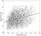
图2为采用现有技术中的测量模型测量的心输出量与“金标准”方法测量结果的对比图;Fig. 2 is the comparison diagram of the cardiac output measured by the measurement model in the prior art and the measurement result of the "gold standard" method;
图3为采用本具体实施方式中的个体化心输出量的无创测量系统测量的心输出量与“金标准”方法测量结果的对比图;Fig. 3 is a comparison chart between the cardiac output measured by the non-invasive measurement system of individualized cardiac output in this specific embodiment and the measurement results of the "gold standard" method;
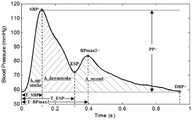
图4为校正前的桡动脉脉搏波的波形图。Fig. 4 is a waveform diagram of the radial artery pulse wave before correction.
具体实施方式detailed description
一)系统结构1) System structure
参考图1所示,一种个体化心输出量的无创测量系统,包括肱动脉血压测量模块、外周浅层动脉脉搏波测量模块与脉搏波传导速度测量模块,肱动脉测量血压测量模块用于采集肱动脉的血压参数,包括肱动脉收缩压与肱动脉舒张压;外周浅层动脉脉搏波测量模块用于采集外周浅层动脉脉搏波;脉搏波传导速度测量模块用于从肱动脉到外周浅层动脉的脉搏波传导速度;As shown in Figure 1, a non-invasive measurement system for individualized cardiac output includes a brachial artery blood pressure measurement module, a peripheral superficial artery pulse wave measurement module, and a pulse wave velocity measurement module. The brachial artery blood pressure measurement module is used to collect Blood pressure parameters of brachial artery, including brachial artery systolic pressure and brachial artery diastolic pressure; peripheral superficial artery pulse wave measurement module is used to collect peripheral superficial artery pulse wave; pulse wave conduction velocity measurement module is used to measure from brachial artery to peripheral superficial Arterial pulse wave velocity;
脉搏波校正模块用于输入肱动脉的血压参数与外周浅层动脉脉搏波,利用肱动脉的血压参数对外周浅层动脉脉搏波进行校正,并将校正后的外周浅层动脉脉搏波输出给脉搏波特征提取模块;The pulse wave correction module is used to input the blood pressure parameters of the brachial artery and the peripheral superficial artery pulse wave, use the blood pressure parameters of the brachial artery to correct the peripheral superficial artery pulse wave, and output the corrected peripheral superficial artery pulse wave to the pulse wave Wave feature extraction module;
脉搏波特征提取模块用于从校正后的外周浅层动脉脉搏波中提取脉搏波特征,并包括以下时域特征:血压特征、时间特征、面积特征与比例特征,并将脉搏波特征输出给个体化脉搏波特征向量生成模块;The pulse wave feature extraction module is used to extract pulse wave features from the corrected peripheral superficial arterial pulse wave, including the following time domain features: blood pressure feature, time feature, area feature and proportional feature, and output the pulse wave feature to the individual A pulse wave feature vector generation module;
个体化脉搏波特征向量生成模块用于根据脉搏波特征与受测者的个体生理参数生成个体化脉搏波特征向量,并输出给已训练完成的人工神经网络;The individualized pulse wave feature vector generation module is used to generate individualized pulse wave feature vectors according to the pulse wave features and the individual physiological parameters of the subject, and output them to the trained artificial neural network;
人工神经网络用于根据输入的个体化脉搏波特征向量,计算出对应的每博量,并输出给心输出量计算模块;The artificial neural network is used to calculate the corresponding stroke volume according to the input individualized pulse wave feature vector, and output it to the cardiac output calculation module;
心输出量计算模块用于根据每博量计算出心输出量,计算公式如下:The cardiac output calculation module is used to calculate the cardiac output according to the stroke volume, and the calculation formula is as follows:
CO=SV*N/Fs*60CO=SV*N/
其中,CO表示心输出量,SV表示每博量,N表示一个心动周期内脉搏波的采样点数,Fs表示系统的信号采样频率。Among them, CO represents the cardiac output, SV represents the stroke volume, N represents the number of sampling points of the pulse wave in one cardiac cycle, and Fs represents the signal sampling frequency of the system.
外周浅层动脉脉搏波为桡动脉血压波、手指动脉血压波或下肢动脉血压波,脉搏波校正模块根据如下公式对外周浅层动脉脉搏波进行校正:The peripheral superficial arterial pulse wave is radial artery blood pressure wave, finger arterial blood pressure wave or lower extremity arterial blood pressure wave, and the pulse wave correction module corrects the peripheral superficial arterial pulse wave according to the following formula:

其中,

放大因子α的取值范围限制在[1,1.5]。The value range of the amplification factor α is limited to [1,1.5].
为了建立能更加准确反映脉搏波的特征向量,脉搏波特征提取模块还从校正后的外周浅层动脉脉搏波中提取包含多倍频的谐波幅值在内的频域特征,时域特征与频域特征合并组成脉搏波特征后输出给个体化脉搏波特征向量生成模块。In order to establish a feature vector that can more accurately reflect the pulse wave, the pulse wave feature extraction module also extracts frequency domain features including multi-octave harmonic amplitudes from the corrected peripheral superficial arterial pulse wave, time domain features and The frequency domain features are combined to form pulse wave features and then output to the individualized pulse wave feature vector generation module.
人工神经网络的输入层采用如下公式对个体化脉搏波特征向量进行归一化:The input layer of the artificial neural network uses the following formula to normalize the individualized pulse wave feature vector:

其中,


归一化后的个体化脉搏波特征向量


其中,f表示人工神经网络中的结合函数,


输出层将准输出向量

其中,SV表示每博量,

还包括数据显示模块与数据采集控制模块,所述数据采集控制模块用于控制肱动脉血压测量模块、外周浅层动脉脉搏波测量模块与脉搏波传导速度测量模块的数据采集过程;数据显示模块用于显示心输出量测量过程中的相关数据,包括肱动脉血压参数、外周浅层动脉脉搏波、校正后的外周浅层动脉脉搏波、每博量、心输出量与受测者的个体生理参数。It also includes a data display module and a data acquisition control module, the data acquisition control module is used to control the data acquisition process of the brachial artery blood pressure measurement module, the peripheral superficial artery pulse wave measurement module and the pulse wave velocity measurement module; the data display module is used for It is used to display relevant data during cardiac output measurement, including brachial artery blood pressure parameters, peripheral superficial arterial pulse wave, corrected peripheral superficial arterial pulse wave, stroke volume, cardiac output and individual physiological parameters of the subject .
肱动脉血压测量模块包括袖带、气路导管、气压传感器、充放气电机与血压数据处理模块;电机通过气路导管对袖带进行充放气,气压传感器监测袖带或气路中的气压变化,并将测量的气压信号传递给血压数据处理模块;血压数据处理模块按如下程序计算肱动脉血压参数:对气压信号进行分解,得到振荡信号和线性上升或下降信号;检测出振荡信号的峰值,在峰值左右按峰值幅度的一定比例(如60%或80%)获得对应两个时间点,左右两个时间点对应的上升或下降信号的压力,即为收缩压和舒张压;The brachial artery blood pressure measurement module includes a cuff, an air circuit catheter, an air pressure sensor, an inflation and deflation motor, and a blood pressure data processing module; the motor inflates and deflates the cuff through the air circuit catheter, and the air pressure sensor monitors the air pressure in the cuff or air circuit change, and transmit the measured air pressure signal to the blood pressure data processing module; the blood pressure data processing module calculates the brachial artery blood pressure parameters according to the following procedures: decompose the air pressure signal to obtain an oscillation signal and a linear rise or fall signal; detect the peak value of the oscillation signal , according to a certain proportion (such as 60% or 80%) of the peak amplitude at the left and right of the peak value, corresponding to two time points, the pressure of the rising or falling signal corresponding to the left and right two time points is the systolic blood pressure and the diastolic blood pressure;
外周浅层动脉脉搏波测量模块为桡动脉测量装置、手指动脉测量装置或下肢动脉测量装置。本具体实施方式采用桡动脉血压测量模块:由压电传感器、一路导线和血压信号处理电路组成,其中,压电传感器获得桡动脉的脉动信号,并将信号通过导线传输给血压信号处理电路,该电路完成血压信号的滤波和放大等处理,并将处理后的信号通过数据采集控制模块传递给血压数据处理模块。The peripheral superficial artery pulse wave measurement module is a radial artery measurement device, a finger artery measurement device or a lower extremity artery measurement device. This specific embodiment adopts the radial artery blood pressure measurement module: it is composed of a piezoelectric sensor, a wire and a blood pressure signal processing circuit, wherein the piezoelectric sensor obtains the pulsation signal of the radial artery, and transmits the signal to the blood pressure signal processing circuit through the wire. The circuit completes processing such as filtering and amplification of the blood pressure signal, and transmits the processed signal to the blood pressure data processing module through the data acquisition control module.
脉搏波传导速度测量模块采用肱动脉血压测量模块和外周浅层动脉脉搏波测量模块同步测量肱动脉脉搏波和外周浅层动脉脉搏波,并计算两路波形的波足点时间差PTT(脉搏波传导时间),然后采用卷尺测量两测量点之间的人体体表距离L,最后利用L除以PTT即为脉搏波传导速度。一般将袖带压升至肱动脉平均压,不能升得过高,这会导致远端外周动脉脉搏波消失,从而使测量失败。The pulse wave velocity measurement module uses the brachial artery blood pressure measurement module and the peripheral superficial artery pulse wave measurement module to simultaneously measure the brachial artery pulse wave and the peripheral superficial artery pulse wave, and calculate the foot point time difference PTT (pulse wave conduction time difference) of the two waveforms. Time), then use a tape measure to measure the distance L between the two measurement points on the human body surface, and finally divide L by PTT to get the pulse wave velocity. Generally, the cuff pressure is raised to the mean pressure of the brachial artery, and it cannot be raised too high, which will cause the pulse wave of the distal peripheral artery to disappear, thus making the measurement fail.
二)、训练人工神经网络2) Training artificial neural network
采用误差反向传播法结合梯度下降法对人工神经网络进行训练。用于训练人工神经网络的样本数据来源于热稀释法的心输出量、每博量和桡动脉血压波形,这些数据通过公用数据就可以查询到。每个样本均以饶动脉血压波形的时频特征和个体生理参数组成的特征向量作为输入量,并以与桡动脉血压波形对应的每博量作为标准输出量;样本数据被分成训练样本集与测试样本集,采用训练样本集对人工神经网络进行训练;采用测试样本集对训练后的人工神经网络进行测试,若输出量与标准输出量的误差满足阈值,则表示训练完成,训练完成后的人工神经网络获得能够使输出量与标准输出量的误差满足阈值的参数组合,参数组合中的参数包括归一化增益向量


受测者的部分波形特征参数统计如下表1:The statistics of some waveform characteristic parameters of the subjects are shown in Table 1:
表1病人的部分波形特征参数的统计值Table 1 Statistical values of some waveform characteristic parameters of patients

图2现有模型ModelRcdecay与“金标准”法的测量结果对比图,横坐标是金标准测量的SV,纵坐标是现有模型ModelRCdecay计算的SV,图中直线表示线性回归直线。Figure 2. Comparison of measurement results between the existing model ModelRCdecay and the "gold standard" method. The abscissa is the SV measured by the gold standard, and the ordinate is the SV calculated by the existing model ModelRCdecay . The straight line in the figure represents the linear regression line.
图3为本发明的人工神经网络(ANN)与“金标准”法的测量结果对比图,横坐标是金标准测量的SV,本发明的人工神经网络计算的SV,图中直线表示线性回归直线。Fig. 3 is artificial neural network (ANN) of the present invention (ANN) and the measuring result contrast figure of " gold standard " method, abscissa is the SV that gold standard measures, the SV that artificial neural network of the present invention calculates, and straight line represents linear regression straight line among the figure .
本发明提出人工神经网络(ANN)与其他现有模型的结果比较,如下表2所示,斜率和截距为模型与实测SV的线性回归方程参数,相关系数采用皮尔森相关系数。The present invention proposes artificial neural network (ANN) and the result comparison of other existing models, as shown in table 2 below, slope and intercept are the linear regression equation parameters of model and measured SV, and correlation coefficient adopts Pearson correlation coefficient.
表2Table 2

从上表可以看出,本发明与金标准法的测量结果的标准差远远小于现有模型与金标准法的测量结果之间的标准差,本发明的人工神经网络模型大大提高了无创测量的准确性。As can be seen from the above table, the standard deviation of the measurement results of the present invention and the gold standard method is far less than the standard deviation between the measurement results of the existing model and the gold standard method, and the artificial neural network model of the present invention greatly improves the non-invasive measurement. accuracy.
三)、测量过程3) Measurement process
采用本具体实施方式中的个体化心输出量的无创测量系统对受测者进行检查心输出量测量,具体过程如下。The individualized cardiac output non-invasive measurement system in this specific embodiment is used to measure the cardiac output of the subject, and the specific process is as follows.
受测者平躺休息3-5分钟后,通过数据采集控制模块启动和控制肱动脉血压测量模块中气泵的充放气速度和袖带中的气压,完成肱动脉血压的测量。After the subject lies down and rests for 3-5 minutes, the data acquisition control module starts and controls the inflation and deflation speed of the air pump in the brachial artery blood pressure measurement module and the air pressure in the cuff to complete the measurement of the brachial artery blood pressure.
通过数据采集控制模块启动和控制桡动脉血压测量模块,完成桡动脉血压信号的测量,并通过模拟电路滤波和放大等处理,并将处理后的信号通过数据采集控制模块传递给数据处理模块。Start and control the radial artery blood pressure measurement module through the data acquisition control module, complete the measurement of the radial artery blood pressure signal, and process it through analog circuit filtering and amplification, and pass the processed signal to the data processing module through the data acquisition control module.
通过数据采集控制模块启动数据处理模块,对其输入的肱动脉气压信号进行处理得到收缩压、平均压和舒张压;对桡动脉血压信号数据进行去噪、去漂移、归一化和校准等处理;然后利用这三个血压对桡动脉血压波形进行校正和一定增益的放大,将校正并放大后的桡动脉脉搏波信号分别发送至脉搏波特征提取模块和显示模块。Start the data processing module through the data acquisition control module, process the input brachial artery air pressure signal to obtain systolic pressure, mean pressure and diastolic pressure; perform denoising, de-drifting, normalization and calibration processing on the radial artery blood pressure signal data ; Then use these three blood pressures to correct and amplify the radial artery blood pressure waveform with a certain gain, and send the corrected and amplified radial artery pulse wave signal to the pulse wave feature extraction module and display module respectively.
在脉搏波特征提取模块中,首先,通过脉搏波的二阶或以上高阶数值差分,检测差分数列的极小值和极大值,从而获得脉搏波下降期的二重波的降中峡和波峰,其特征点参考图4所示。基于这两个特征点和收缩期峰值特征点,计算其他相关的时域特征。In the pulse wave feature extraction module, firstly, through the second-order or higher-order numerical difference of the pulse wave, the minimum and maximum values of the difference series are detected, so as to obtain the descending gorge and The wave peak and its characteristic points are shown in Figure 4. Based on these two feature points and the systolic peak feature point, other relevant time-domain features are calculated.
频域特征通过脉搏波的傅里叶变换后获得多倍频的谐波幅值,然后,合并时域和频域特征构建脉搏波特征向量。启动脉搏波特征计算模块,完成脉搏波时域和频域特征的处理和计算,构建脉搏波特征向量,将脉搏波特征向量与受测者的个体生理参数输入个体化脉搏波特征向量生成模块,生成个体化的脉搏波特征向量,并输入至人工神经网络,部分参数发送给数据显示模块显示。The frequency-domain features are obtained through the Fourier transform of the pulse wave to obtain multi-octave harmonic amplitudes, and then the time-domain and frequency-domain features are combined to construct the pulse wave feature vector. Start the pulse wave feature calculation module, complete the processing and calculation of the pulse wave time domain and frequency domain features, construct the pulse wave feature vector, input the pulse wave feature vector and the individual physiological parameters of the subject into the individualized pulse wave feature vector generation module, An individualized pulse wave feature vector is generated and input to the artificial neural network, and some parameters are sent to the data display module for display.
人工神经网络对输入的个体化脉搏波特性向量进行输入归一化、变换和输出反归一化等一系列操作,计算出每搏量并输出给心输出量计算模块,心输出量计算模块根据每博量计算出心输出量。The artificial neural network performs a series of operations such as input normalization, transformation and output denormalization on the input individualized pulse wave characteristic vector, calculates the stroke volume and outputs it to the cardiac output calculation module, and the cardiac output calculation module Cardiac output was calculated from stroke volume.
数据显示模块脉搏波波形、每搏量、心输出量和相关心血管系统参数显示在显示屏上,同时生成测量报告。The data display module pulse waveform, stroke volume, cardiac output and related cardiovascular system parameters are displayed on the display screen, and a measurement report is generated at the same time.
Claims (9)


Priority Applications (1)
| Application Number | Priority Date | Filing Date | Title |
|---|---|---|---|
| CN202010317299.9ACN111493855B (en) | 2020-04-21 | 2020-04-21 | System and method for non-invasive measurement of individualized cardiac output |
Applications Claiming Priority (1)
| Application Number | Priority Date | Filing Date | Title |
|---|---|---|---|
| CN202010317299.9ACN111493855B (en) | 2020-04-21 | 2020-04-21 | System and method for non-invasive measurement of individualized cardiac output |
Publications (2)
| Publication Number | Publication Date |
|---|---|
| CN111493855A CN111493855A (en) | 2020-08-07 |
| CN111493855Btrue CN111493855B (en) | 2023-01-06 |
Family
ID=71865898
Family Applications (1)
| Application Number | Title | Priority Date | Filing Date |
|---|---|---|---|
| CN202010317299.9AActiveCN111493855B (en) | 2020-04-21 | 2020-04-21 | System and method for non-invasive measurement of individualized cardiac output |
Country Status (1)
| Country | Link |
|---|---|
| CN (1) | CN111493855B (en) |
Families Citing this family (11)
| Publication number | Priority date | Publication date | Assignee | Title |
|---|---|---|---|---|
| EP3964124A1 (en)* | 2020-09-02 | 2022-03-09 | Koninklijke Philips N.V. | Method and apparatus for estimating the reliability of cardiac output measurements |
| CN112790748A (en)* | 2020-12-30 | 2021-05-14 | 重庆理工大学 | A central arterial pressure waveform reconstruction system and method |
| CN113080907B (en)* | 2021-04-14 | 2022-10-25 | 贵州省人民医院 | Pulse wave signal processing method and device |
| CN113057617B (en)* | 2021-04-30 | 2022-08-26 | 重庆理工大学 | Non-invasive monitoring system for cardiac output |
| CN113143230B (en)* | 2021-05-11 | 2022-05-20 | 重庆理工大学 | A peripheral arterial blood pressure waveform reconstruction system |
| CN113499048B (en)* | 2021-07-22 | 2022-07-08 | 重庆理工大学 | A system and method for central arterial pressure waveform reconstruction based on CNN-BiLSTM |
| CN114027804A (en)* | 2021-12-03 | 2022-02-11 | 科思技术(温州)研究院 | Method, device and readable storage medium for pulse diagnosis |
| EP4480400A1 (en)* | 2023-06-23 | 2024-12-25 | Koninklijke Philips N.V. | System and method for estimating cardiac output |
| WO2025060099A1 (en)* | 2023-09-22 | 2025-03-27 | Oppo广东移动通信有限公司 | Blood pressure measurement method and apparatus, blood pressure measurement device, and storage medium |
| CN119700065B (en)* | 2025-02-25 | 2025-07-11 | 北京航空航天大学 | Dynamic noninvasive cardiac output monitoring system and method |
| CN120381255B (en)* | 2025-06-27 | 2025-09-19 | 北京麦邦光电仪器有限公司 | Noninvasive cardiac output function detection method and noninvasive cardiac output function detection device |
Citations (4)
| Publication number | Priority date | Publication date | Assignee | Title |
|---|---|---|---|---|
| US5400793A (en)* | 1991-01-29 | 1995-03-28 | Nederlandse Organisatie Voor Toegepast-Natuurwetenschappelijk Onderzoek Tno | Method of determining the stroke volume and the cardiac output of the human heart |
| CN103892818A (en)* | 2012-12-28 | 2014-07-02 | 吴健康 | Non-invasive central aortic blood pressure measuring method and device |
| CN104244814A (en)* | 2012-05-15 | 2014-12-24 | 皇家飞利浦有限公司 | Monitoring of cardiac output |
| CN104323768A (en)* | 2012-11-20 | 2015-02-04 | 深圳市理邦精密仪器股份有限公司 | Parameter calibration method for continuously monitoring cardiac output |
Family Cites Families (14)
| Publication number | Priority date | Publication date | Assignee | Title |
|---|---|---|---|---|
| US5913826A (en)* | 1996-06-12 | 1999-06-22 | K-One Technologies | Wideband external pulse cardiac monitor |
| US7822470B2 (en)* | 2001-10-11 | 2010-10-26 | Osypka Medical Gmbh | Method for determining the left-ventricular ejection time TLVE of a heart of a subject |
| AU2003275033A1 (en)* | 2003-02-10 | 2004-09-06 | Massachusetts Institute Of Technology | Methods and apparatus for determining cardiac output |
| US20070197924A1 (en)* | 2004-03-05 | 2007-08-23 | O'rourke Michael F | Method and apparatus for determination of cardiac output from the arterial pressure pulse waveform |
| CN101006919A (en)* | 2007-01-26 | 2007-08-01 | 北京工业大学 | Detection method of cardiac output under the high differential pressure and device thereof |
| CN101176663B (en)* | 2007-12-06 | 2010-10-27 | 山东大学 | Non-invasive cardiac output detection method and device based on pulse wave |
| US10405762B2 (en)* | 2009-04-22 | 2019-09-10 | Vital Metrix, Inc. | System and method for noninvasively measuring ventricular stroke volume and cardiac output |
| CN102499669B (en)* | 2011-10-26 | 2014-12-24 | 中国科学院深圳先进技术研究院 | Heart parameter measuring method and device |
| JP5985355B2 (en)* | 2012-10-30 | 2016-09-06 | 日本光電工業株式会社 | Blood volume measuring method and measuring apparatus |
| US9949696B2 (en)* | 2013-03-14 | 2018-04-24 | Tensys Medical, Inc. | Apparatus and methods for computing cardiac output of a living subject via applanation tonometry |
| US10542961B2 (en)* | 2015-06-15 | 2020-01-28 | The Research Foundation For The State University Of New York | System and method for infrasonic cardiac monitoring |
| CN105726000B (en)* | 2016-01-29 | 2019-03-22 | 北京工业大学 | A kind of device of the cardiovascular functional parameter based on blood pressure of four limbs pulse |
| US10918292B2 (en)* | 2016-07-19 | 2021-02-16 | Mayo Foundation For Medical Education And Research | Non-invasive cardiac output assessment |
| CN109512412B (en)* | 2018-11-21 | 2021-10-08 | 重庆理工大学 | A central arterial blood pressure measuring device |
- 2020
- 2020-04-21CNCN202010317299.9Apatent/CN111493855B/enactiveActive
Patent Citations (4)
| Publication number | Priority date | Publication date | Assignee | Title |
|---|---|---|---|---|
| US5400793A (en)* | 1991-01-29 | 1995-03-28 | Nederlandse Organisatie Voor Toegepast-Natuurwetenschappelijk Onderzoek Tno | Method of determining the stroke volume and the cardiac output of the human heart |
| CN104244814A (en)* | 2012-05-15 | 2014-12-24 | 皇家飞利浦有限公司 | Monitoring of cardiac output |
| CN104323768A (en)* | 2012-11-20 | 2015-02-04 | 深圳市理邦精密仪器股份有限公司 | Parameter calibration method for continuously monitoring cardiac output |
| CN103892818A (en)* | 2012-12-28 | 2014-07-02 | 吴健康 | Non-invasive central aortic blood pressure measuring method and device |
Also Published As
| Publication number | Publication date |
|---|---|
| CN111493855A (en) | 2020-08-07 |
Similar Documents
| Publication | Publication Date | Title |
|---|---|---|
| CN111493855B (en) | System and method for non-invasive measurement of individualized cardiac output | |
| US5101828A (en) | Methods and apparatus for nonivasive monitoring of dynamic cardiac performance | |
| CN109512412B (en) | A central arterial blood pressure measuring device | |
| US7220230B2 (en) | Pressure-based system and method for determining cardiac stroke volume | |
| US5265615A (en) | Method and apparatus for continuous measurement of cardiac output and SVR | |
| US4873987A (en) | Noninvasive continuous monitor of arterial blood pressure waveform | |
| CN107233087A (en) | A kind of Woundless blood pressure measuring device based on photoplethysmographic feature | |
| CN102499669B (en) | Heart parameter measuring method and device | |
| CN109730663B (en) | Blood pressure assessment method based on nonlinear analysis of pulse wave velocity | |
| CN101229058A (en) | Initial calibration device for measuring arterial blood pressure by pulse wave transmission time method | |
| CN104000573B (en) | Central artery pulse wave monitoring system and method based on body surface two-point pulse wave | |
| EP1150604B1 (en) | Method and device for continuous analysis of cardiovascular activity of a subject | |
| Xu et al. | Online continuous measurement of arterial pulse pressure and pressure waveform using ultrasound | |
| CN114587307B (en) | A non-contact blood pressure detector and method based on capacitive coupling electrode | |
| Stork et al. | Cuff pressure pulse waveforms: their current and prospective applications in biomedical instrumentation | |
| CN115316968B (en) | Systolic blood pressure measurement device and method based on real-time pulse wave signal | |
| Nabeel et al. | Deep learning for blood pressure estimation: an approach using local measure of arterial dual diameter waveforms | |
| Liu et al. | A new oscillometry-based method for estimating the brachial arterial compliance under loaded conditions | |
| RU2327414C1 (en) | Method of blood pressure measurement based on three-dimensional compression oscillogram | |
| Wang et al. | An improved algorithm for noninvasive blood pressure measurement | |
| Sidhu et al. | Comparison of artificial intelligence based oscillometric blood pressure estimation techniques: a review paper | |
| Seabra et al. | Blood Pressure Models for Wearable Sensors | |
| EA008756B1 (en) | Method for diagnosis of functional state of blood circulatory system by volumetric compression oscillometry | |
| Park et al. | Cuffless and noninvasive tonometry mean arterial pressure measurement by physiological characteristics and applied pressure | |
| CN118252481B (en) | A blood pressure monitoring algorithm independent of dicrotic notch |
Legal Events
| Date | Code | Title | Description |
|---|---|---|---|
| PB01 | Publication | ||
| PB01 | Publication | ||
| SE01 | Entry into force of request for substantive examination | ||
| SE01 | Entry into force of request for substantive examination | ||
| GR01 | Patent grant | ||
| GR01 | Patent grant | ||
| OL01 | Intention to license declared | ||
| OL01 | Intention to license declared |