CN111491579A - Endoscopic surgical device and method of use - Google Patents
Endoscopic surgical device and method of useDownload PDFInfo
- Publication number
- CN111491579A CN111491579ACN201880082131.5ACN201880082131ACN111491579ACN 111491579 ACN111491579 ACN 111491579ACN 201880082131 ACN201880082131 ACN 201880082131ACN 111491579 ACN111491579 ACN 111491579A
- Authority
- CN
- China
- Prior art keywords
- jaws
- end effector
- surgical device
- longitudinally extending
- effector
- Prior art date
- Legal status (The legal status is an assumption and is not a legal conclusion. Google has not performed a legal analysis and makes no representation as to the accuracy of the status listed.)
- Pending
Links
Images


Classifications
- A—HUMAN NECESSITIES
- A61—MEDICAL OR VETERINARY SCIENCE; HYGIENE
- A61B—DIAGNOSIS; SURGERY; IDENTIFICATION
- A61B17/00—Surgical instruments, devices or methods
- A61B17/28—Surgical forceps
- A61B17/29—Forceps for use in minimally invasive surgery
- A—HUMAN NECESSITIES
- A61—MEDICAL OR VETERINARY SCIENCE; HYGIENE
- A61B—DIAGNOSIS; SURGERY; IDENTIFICATION
- A61B18/00—Surgical instruments, devices or methods for transferring non-mechanical forms of energy to or from the body
- A61B18/04—Surgical instruments, devices or methods for transferring non-mechanical forms of energy to or from the body by heating
- A61B18/12—Surgical instruments, devices or methods for transferring non-mechanical forms of energy to or from the body by heating by passing a current through the tissue to be heated, e.g. high-frequency current
- A61B18/14—Probes or electrodes therefor
- A61B18/1442—Probes having pivoting end effectors, e.g. forceps
- A61B18/1445—Probes having pivoting end effectors, e.g. forceps at the distal end of a shaft, e.g. forceps or scissors at the end of a rigid rod
- A61B18/1447—Probes having pivoting end effectors, e.g. forceps at the distal end of a shaft, e.g. forceps or scissors at the end of a rigid rod wherein sliding surfaces cause opening/closing of the end effectors
- A—HUMAN NECESSITIES
- A61—MEDICAL OR VETERINARY SCIENCE; HYGIENE
- A61B—DIAGNOSIS; SURGERY; IDENTIFICATION
- A61B10/00—Instruments for taking body samples for diagnostic purposes; Other methods or instruments for diagnosis, e.g. for vaccination diagnosis, sex determination or ovulation-period determination; Throat striking implements
- A61B10/02—Instruments for taking cell samples or for biopsy
- A61B10/0233—Pointed or sharp biopsy instruments
- A61B10/0283—Pointed or sharp biopsy instruments with vacuum aspiration, e.g. caused by retractable plunger or by connected syringe
- A—HUMAN NECESSITIES
- A61—MEDICAL OR VETERINARY SCIENCE; HYGIENE
- A61B—DIAGNOSIS; SURGERY; IDENTIFICATION
- A61B10/00—Instruments for taking body samples for diagnostic purposes; Other methods or instruments for diagnosis, e.g. for vaccination diagnosis, sex determination or ovulation-period determination; Throat striking implements
- A61B10/02—Instruments for taking cell samples or for biopsy
- A61B10/06—Biopsy forceps, e.g. with cup-shaped jaws
- A—HUMAN NECESSITIES
- A61—MEDICAL OR VETERINARY SCIENCE; HYGIENE
- A61B—DIAGNOSIS; SURGERY; IDENTIFICATION
- A61B17/00—Surgical instruments, devices or methods
- A61B17/32—Surgical cutting instruments
- A61B17/320016—Endoscopic cutting instruments, e.g. arthroscopes, resectoscopes
- A—HUMAN NECESSITIES
- A61—MEDICAL OR VETERINARY SCIENCE; HYGIENE
- A61B—DIAGNOSIS; SURGERY; IDENTIFICATION
- A61B17/00—Surgical instruments, devices or methods
- A61B17/34—Trocars; Puncturing needles
- A61B17/3478—Endoscopic needles, e.g. for infusion
- A—HUMAN NECESSITIES
- A61—MEDICAL OR VETERINARY SCIENCE; HYGIENE
- A61B—DIAGNOSIS; SURGERY; IDENTIFICATION
- A61B18/00—Surgical instruments, devices or methods for transferring non-mechanical forms of energy to or from the body
- A61B18/04—Surgical instruments, devices or methods for transferring non-mechanical forms of energy to or from the body by heating
- A61B18/12—Surgical instruments, devices or methods for transferring non-mechanical forms of energy to or from the body by heating by passing a current through the tissue to be heated, e.g. high-frequency current
- A61B18/14—Probes or electrodes therefor
- A61B18/1442—Probes having pivoting end effectors, e.g. forceps
- A61B18/1445—Probes having pivoting end effectors, e.g. forceps at the distal end of a shaft, e.g. forceps or scissors at the end of a rigid rod
- A—HUMAN NECESSITIES
- A61—MEDICAL OR VETERINARY SCIENCE; HYGIENE
- A61B—DIAGNOSIS; SURGERY; IDENTIFICATION
- A61B18/00—Surgical instruments, devices or methods for transferring non-mechanical forms of energy to or from the body
- A61B18/04—Surgical instruments, devices or methods for transferring non-mechanical forms of energy to or from the body by heating
- A61B18/12—Surgical instruments, devices or methods for transferring non-mechanical forms of energy to or from the body by heating by passing a current through the tissue to be heated, e.g. high-frequency current
- A61B18/14—Probes or electrodes therefor
- A61B18/1482—Probes or electrodes therefor having a long rigid shaft for accessing the inner body transcutaneously in minimal invasive surgery, e.g. laparoscopy
- A—HUMAN NECESSITIES
- A61—MEDICAL OR VETERINARY SCIENCE; HYGIENE
- A61B—DIAGNOSIS; SURGERY; IDENTIFICATION
- A61B18/00—Surgical instruments, devices or methods for transferring non-mechanical forms of energy to or from the body
- A61B2018/00053—Mechanical features of the instrument of device
- A61B2018/00059—Material properties
- A61B2018/00071—Electrical conductivity
- A61B2018/00083—Electrical conductivity low, i.e. electrically insulating
- A—HUMAN NECESSITIES
- A61—MEDICAL OR VETERINARY SCIENCE; HYGIENE
- A61B—DIAGNOSIS; SURGERY; IDENTIFICATION
- A61B18/00—Surgical instruments, devices or methods for transferring non-mechanical forms of energy to or from the body
- A61B2018/00053—Mechanical features of the instrument of device
- A61B2018/00184—Moving parts
- A61B2018/00196—Moving parts reciprocating lengthwise
- A—HUMAN NECESSITIES
- A61—MEDICAL OR VETERINARY SCIENCE; HYGIENE
- A61B—DIAGNOSIS; SURGERY; IDENTIFICATION
- A61B18/00—Surgical instruments, devices or methods for transferring non-mechanical forms of energy to or from the body
- A61B2018/00315—Surgical instruments, devices or methods for transferring non-mechanical forms of energy to or from the body for treatment of particular body parts
- A61B2018/00505—Urinary tract
- A61B2018/00517—Urinary bladder or urethra
- A—HUMAN NECESSITIES
- A61—MEDICAL OR VETERINARY SCIENCE; HYGIENE
- A61B—DIAGNOSIS; SURGERY; IDENTIFICATION
- A61B18/00—Surgical instruments, devices or methods for transferring non-mechanical forms of energy to or from the body
- A61B2018/00571—Surgical instruments, devices or methods for transferring non-mechanical forms of energy to or from the body for achieving a particular surgical effect
- A61B2018/00577—Ablation
- A—HUMAN NECESSITIES
- A61—MEDICAL OR VETERINARY SCIENCE; HYGIENE
- A61B—DIAGNOSIS; SURGERY; IDENTIFICATION
- A61B18/00—Surgical instruments, devices or methods for transferring non-mechanical forms of energy to or from the body
- A61B2018/00571—Surgical instruments, devices or methods for transferring non-mechanical forms of energy to or from the body for achieving a particular surgical effect
- A61B2018/00595—Cauterization
- A—HUMAN NECESSITIES
- A61—MEDICAL OR VETERINARY SCIENCE; HYGIENE
- A61B—DIAGNOSIS; SURGERY; IDENTIFICATION
- A61B18/00—Surgical instruments, devices or methods for transferring non-mechanical forms of energy to or from the body
- A61B2018/00982—Surgical instruments, devices or methods for transferring non-mechanical forms of energy to or from the body combined with or comprising means for visual or photographic inspections inside the body, e.g. endoscopes
- A—HUMAN NECESSITIES
- A61—MEDICAL OR VETERINARY SCIENCE; HYGIENE
- A61B—DIAGNOSIS; SURGERY; IDENTIFICATION
- A61B18/00—Surgical instruments, devices or methods for transferring non-mechanical forms of energy to or from the body
- A61B18/04—Surgical instruments, devices or methods for transferring non-mechanical forms of energy to or from the body by heating
- A61B18/12—Surgical instruments, devices or methods for transferring non-mechanical forms of energy to or from the body by heating by passing a current through the tissue to be heated, e.g. high-frequency current
- A61B18/14—Probes or electrodes therefor
- A61B18/1442—Probes having pivoting end effectors, e.g. forceps
- A61B2018/1452—Probes having pivoting end effectors, e.g. forceps including means for cutting
Landscapes
- Health & Medical Sciences (AREA)
- Life Sciences & Earth Sciences (AREA)
- Surgery (AREA)
- Engineering & Computer Science (AREA)
- General Health & Medical Sciences (AREA)
- Medical Informatics (AREA)
- Molecular Biology (AREA)
- Biomedical Technology (AREA)
- Animal Behavior & Ethology (AREA)
- Public Health (AREA)
- Veterinary Medicine (AREA)
- Heart & Thoracic Surgery (AREA)
- Nuclear Medicine, Radiotherapy & Molecular Imaging (AREA)
- Pathology (AREA)
- Otolaryngology (AREA)
- Physics & Mathematics (AREA)
- Plasma & Fusion (AREA)
- Biodiversity & Conservation Biology (AREA)
- Orthopedic Medicine & Surgery (AREA)
- Ophthalmology & Optometry (AREA)
- Endoscopes (AREA)
- Surgical Instruments (AREA)
- Radiology & Medical Imaging (AREA)
Abstract
Description
Translated fromChinese本发明涉及一种内窥镜外科手术(endosurgical)装置,该装置包括:The present invention relates to an endoscopic surgical (endosurgical) device comprising:
-柔性管,该柔性管具有远侧管端部、近侧管端部和至少两个纵长向延伸通道,- a flexible tube having a distal tube end, a proximal tube end and at least two longitudinally extending channels,
-端部执行器,该端部执行器包括具有相对的切削刃的两个相对的钳夹部,该端部执行器被设置在远侧管端部,- an end effector comprising two opposing jaws with opposing cutting edges, the end effector being provided at the distal tube end,
-执行器套筒,该执行器套筒至少在远侧管端部围绕管,以及- an actuator sleeve surrounding the tube at least at the distal tube end, and
-用于使端部执行器相对于执行器套筒或使执行器套筒相对于端部执行器轴向地往复运动以使钳夹部打开和闭合的装置。- Means for axially reciprocating the end effector relative to the effector sleeve or the effector sleeve relative to the end effector to open and close the jaws.
膀胱癌是瑞典和国际上最花钱的癌症形式之一。在美国,每个新诊断出的人的估计费用约为140,000美元。在瑞典每年大约有2,400个新的诊断出患有膀胱癌的人。在过去的20年中,病例数总共增加了35%,并且泌尿系统癌症对男性的影响是女性的三倍。在瑞典的男性中,膀胱和尿路中的癌症是第三最常见的癌症形式。Bladder cancer is one of the most expensive forms of cancer in Sweden and internationally. In the United States, the estimated cost per newly diagnosed person is about $140,000. About 2,400 people are newly diagnosed with bladder cancer each year in Sweden. In the past 20 years, the number of cases has increased by a total of 35%, and urinary system cancer affects men three times more than women. Cancer in the bladder and urinary tract is the third most common form of cancer among men in Sweden.
约70%的患者患有局限于粘膜的浅表性膀胱癌形式。这意味着这些患者可以被治愈,但不幸地是存在复发和残留肿瘤的风险。大约三分之二的患者需要进行新的外科手术。同一个患者经受几次手术并不罕见,有时在第一次手术之后仅几个月。将到泌尿科医生处进行多次定期复查,以早日发现复发,使得癌症不会扩散。About 70% of patients have a superficial form of bladder cancer confined to the mucosa. This means that these patients can be cured, but unfortunately there is a risk of recurrence and residual tumor. About two-thirds of patients require new surgical procedures. It is not uncommon for the same patient to undergo several surgeries, sometimes only a few months after the first. There will be multiple regular check-ups with a urologist to catch the recurrence early so the cancer doesn't spread.
膀胱镜检查是诊断膀胱癌的主要诊断方式。常规的膀胱镜通常具有两个端口,允许看到膀胱内部的光学端口和用于插入各种装置的端口。Cystoscopy is the main diagnostic modality for diagnosing bladder cancer. Conventional cystoscopes typically have two ports, optical ports that allow viewing inside the bladder and ports for inserting various devices.
如果在膀胱镜检查期间癌症区域或可疑区域被看到,则患者通常会被安排在手术室进行外科手术,然后在泌尿科病房停留一夜。在瑞典,患者需要等待约两周以进行该外科手术,并且在可以在外科手术部进行程序之前,患者通常接受脊髓麻醉或全身麻醉。在外科手术期间,使用刚性金属仪器取活检标本,或者也使用该刚性金属仪器进行经尿道的切除术。在一些医院,活检可以被执行为使用柔性膀胱镜进行的门诊程序,但是由于使用柔性活检仪器得到的组织标本的质量不够好,因此需要使用具有较大直径的刚性仪器来接收较好的组织标本,以及以便能够切除癌。If a cancerous or suspicious area is seen during a cystoscopy, the patient is usually scheduled in the operating room for surgery, followed by an overnight stay in the urology ward. In Sweden, patients need to wait about two weeks for this surgery, and before the procedure can be performed in the surgical department, patients are usually given spinal or general anesthesia. During surgical procedures, rigid metal instruments are used to take biopsy specimens or also for transurethral resections. In some hospitals, biopsies can be performed as an outpatient procedure using flexible cystoscopes, but the quality of tissue samples obtained with flexible biopsy instruments is not good enough, requiring the use of rigid instruments with larger diameters to receive better tissue samples , as well as in order to be able to remove the cancer.
在膀胱镜检查期间,当怀疑膀胱中有癌时,泌尿科医生通常——通常使用穿刺活检钳——从膀胱中的多个位置获得多个膀胱组织标本,以帮助建立诊断并确定潜在的肿瘤的范围。通常,组织标本取自看起来异常的尿路上皮的任何区域以及取自疑似的肿瘤区域,但典型地取自膀胱三角区、膀胱顶部,以及右膀胱壁、左膀胱壁、前膀胱壁和后膀胱壁。在程序时,用流体灌洗膀胱。在使用柔性膀胱镜进行经尿道膀胱活检期间的组织获取之前,在样本部位进行局部麻醉,或者通过将麻醉剂滴注到膀胱中局部地获得麻醉。组织获取之后,烧灼活检部位以止血,并重复该程序。因为常规的膀胱镜仅具有一个工作通道用于外科手术程序期间所需的各种装置,所以每次从膀胱中取组织标本时,这些各种装置中的每个装置都被移进和移出工作通道。现今不可能仅使用一种仪器来给予麻醉、进行一次或多次活检、止血、以及破坏膀胱中的小癌。并且,由于尺寸小,活检组织的质量不够好,因为膀胱壁中的需要在显微镜中看到的重要层没有被包括。由于现今是通过当钳夹部挂在组织中时拉出仪器来取活检组织,因此组织样本的边缘被破坏,并且组织样本的质量下降且无法用于准确地诊断癌症。During cystoscopy, when bladder cancer is suspected, urologists typically—usually using biopsy forceps—obtain multiple specimens of bladder tissue from multiple locations in the bladder to help establish a diagnosis and identify the underlying tumor range. Typically, tissue samples are taken from any area of the urothelium that appears abnormal and from areas of suspected tumor, but typically from the trigone, roof of the bladder, and right, left, anterior, and posterior bladder walls bladder wall. During the procedure, the bladder is lavaged with fluid. Local anesthesia is administered at the sample site or locally by instilling an anesthetic into the bladder prior to tissue harvesting during a transurethral bladder biopsy using a flexible cystoscope. After tissue harvesting, the biopsy site is cauterized to stop bleeding, and the procedure is repeated. Because conventional cystoscopes have only one working channel for the various devices required during a surgical procedure, each of these various devices is moved in and out of work each time a tissue sample is taken from the bladder aisle. It is not currently possible to administer anesthesia, perform one or more biopsies, stop bleeding, and destroy small cancers in the bladder using only one instrument. Also, due to the small size, the quality of the biopsy tissue is not good enough because important layers in the bladder wall that need to be seen in the microscope are not included. Since biopsies are currently taken by pulling out the instrument while the jaws are hanging in the tissue, the edges of the tissue sample are damaged, and the quality of the tissue sample is degraded and cannot be used to accurately diagnose cancer.
患有浅表性膀胱癌的患者的治疗涉及许多不同类别的工作人员并且费用较高。如今,在外科手术部的操作室中从膀胱癌取组织标本每位患者在24,000SEK至32,000SEK之间。患者被安排进行外科手术、被固定、被麻醉、以及被使用刚性金属装置进行操作。导尿管被插入以排出血液,并且患者在病房被24小时监测。Treatment of patients with superficial bladder cancer involves many different categories of staff and is expensive. Today, tissue samples taken from bladder cancer in the operating room of the surgical department range from 24,000 SEK to 32,000 SEK per patient. Patients are scheduled for surgery, immobilized, anesthetized, and manipulated using rigid metal devices. A urinary catheter was inserted to drain the blood, and the patient was monitored on the ward for 24 hours.
如果可以在诊室以温和的活检程序取组织标本作为门诊程序的替代,而无需全身麻醉且无需随后的导尿管,那么每位患者的外科手术费用仅为3,600SEK加上装置费用。因此如果能够优化活检程序,可以节省大量成本。If tissue samples could be taken in the office as an alternative to outpatient procedures with a mild biopsy procedure, without general anesthesia and without a subsequent urinary catheter, the cost of surgery per patient would be only 3,600 SEK plus the cost of the device. Therefore, if the biopsy procedure can be optimized, significant cost savings can be achieved.
ThomasV.Taylor在Curr.Surg.2004年11月-12月;61(6):第594-596页上的文章“一种多活检钳系统(A system of multiple biopsy forceps)”描述了一种柔性内窥镜活检钳的新系统,与现有的单活检钳相比,该系统应该可以取较大的且较少创伤的活检组织。该装置使用一端带有有倒钩的尖状物的中心线,滑动弹簧钢钳夹部附接到该有倒钩的尖状物。外部塑料套筒围绕中心线,并且传统类型的手柄使钳夹部活动。Thomas V. Taylor, Curr. Surg. 2004 Nov-Dec; 61(6): pp. 594-596, "A system of multiple biopsy forceps" describes a flexible A new system of endoscopic biopsy forceps that should allow larger and less invasive biopsies than existing single biopsy forceps. The device uses a centerline with a barbed spike at one end to which a sliding spring steel jaw is attached. An outer plastic sleeve surrounds the centerline, and a conventional type of handle activates the jaws.
该已知装置沿着光纤内窥镜的活检通道通过,并且使要进行活检的组织可视化。当外部套筒在内部线上滑动从而允许弹簧钢钳夹部打开和闭合时,发生钳子的打开和闭合。在钳夹部打开的情况下,倒钩被推入组织中以进行活检。鱼钩状的倒钩将组织拉回到钳夹部中,该钳夹部以最佳角度咬住组织,因此可以获得较干净且较大的组织标本。对于第二次活检,简单地再次打开钳夹部,并且当有倒钩的尖状物再次刺穿组织时,第一个标本沿着中心线被推动。以这种方式,通过活检钳单次通过内窥镜,最多6个活检组织被存储。当装置从光纤内窥镜撤回时,所有组织标本都与所述装置一起撤回。然而,鱼钩状倒钩损坏组织标本,并且必须用另一对钳子将组织标本从鱼钩状倒钩取下,或者将倒钩的尖端推入一块软木塞中,并且将标本切下。两种方式都会缩小每个组织标本的尺寸,从而影响标本完整性,并导致对来源精确性的怀疑。因此,由于需要操纵组织标本离开倒钩,所以难以保持标本的来源处于控制之下。此外,这种倒钩类型活检装置对患者和组织标本都相当残忍、无法进行局部麻醉、无法在采样部位停止局部出血,并且由于沿着倒钩同时存储了6个组织标本,因此前端必须相当大。The known device is passed along the biopsy channel of the fiberoptic endoscope and visualizes the tissue to be biopsyed. Opening and closing of the pliers occurs when the outer sleeve slides over the inner wire allowing the spring steel jaws to open and close. With the jaws open, the barbs are pushed into the tissue for biopsy. The fishhook-like barbs pull the tissue back into the jaws, which bite the tissue at an optimal angle, so a cleaner and larger tissue specimen can be obtained. For the second biopsy, the jaws are simply opened again, and the first specimen is pushed along the centerline as the barbed tip pierces the tissue again. In this way, up to 6 biopsies are stored with a single pass of the biopsy forceps through the endoscope. When the device is withdrawn from the fiberoptic endoscope, all tissue specimens are withdrawn with the device. However, the fishhook barb damages the tissue specimen and the tissue specimen must be removed from the fishhook barb with another pair of forceps, or the tip of the barb is pushed into a piece of cork and the specimen is cut. Both approaches reduce the size of each tissue specimen, thereby affecting specimen integrity and leading to doubts about the accuracy of the source. Therefore, it is difficult to keep the source of the specimen under control due to the need to manipulate the tissue specimen away from the barbs. In addition, this barb-type biopsy device is rather cruel to both the patient and the tissue specimen, cannot administer local anesthesia, cannot stop local bleeding at the sampling site, and since 6 tissue specimens are stored simultaneously along the barb, the front end must be quite large .
Marc Beaghler和Michael Grasso在Urology.1994年11月;44(5):756-9中的文章“柔性膀胱镜检查的膀胱活检:用于下尿路膀胱上皮的门诊评估的技术(Flexiblecystoscopic bladder biopsies:a technique for outpatient evaluation of thelower urinary tract urothelium)”描述了一种用于获取活检组织的单极技术,该技术需要适于活检钳的活动电线和标准电灼装置。接地垫被放置在患者的大腿上。为了获得活检组织,钳子通过膀胱镜的工作通道前进。目标区域在直视下被钳子的齿状钳夹部接合,并且在撤回的同时凝固电流被施加以引起周围膀胱上皮的变白。膀胱镜和组织标本作为一个单元被移除,并且膀胱镜被重新插入以检查活检部位、出血情况以及取另外的组织标本。Marc Beaghler and Michael Grasso in Urology. 1994 Nov;44(5):756-9 "Flexible cystoscopic bladder biopsy: a technique for outpatient assessment of the lower urothelium (Flexiblecystoscopic bladder biopsies: a technique for outpatient evaluation of the lower urinary tract urothelium)" describes a monopolar technique for obtaining biopsy tissue that requires a live wire adapted for biopsy forceps and a standard electrocautery device. A grounding pad is placed on the patient's thigh. To obtain biopsy tissue, forceps are advanced through the working channel of the cystoscope. The target area is engaged by the toothed jaws of the forceps under direct vision, and a coagulation current is applied while withdrawing to cause blanching of the surrounding bladder epithelium. The cystoscope and tissue specimen are removed as a unit, and the cystoscope is reinserted to examine the biopsy site, bleeding, and to take additional tissue specimens.
欧洲专利号EP 1838229B1提出了一种包括护套和刚性内部不锈钢轴的活检钳,该刚性内部不锈钢轴可滑动地设置在护套内,并且具有一配置有多个抓持构件的端部。当轴和护套移动时,抓持构件能够在打开配置和闭合配置之间移动。抓持构件具有曲线轮廓,并且在处于打开配置时从纵向轴线被向外偏置。抓持构件在处于打开配置时不受护套的限制,并且在处于闭合配置时受到护套的限制。具有适于剪切、抓持、撕扯或切割组织的近侧边缘的多个抓持构件可以通过对轴的端部进行激光切割来形成,从而当多个抓持构件处于闭合配置时,它们形成用于保持一个或多个组织标本的容器。轴可以连接到电灼装置,以使轴通电以电外科学地切割组织。该已知活检钳装置也可以可操作地连接到输注源或抽吸源。建议将以真空泵或注射器的形式的抽吸源连接到轴,以辅助在活检部位周围进行的组织移除或一般的液体移除,或将组织标本拉回轴中以进行移除或用于取多个活检标本。除了必须交替使用的单腔轴外,简要建议使用替代性腔。EP 1838229 B1没有提到抓持构件如何设法一次保持多个组织标本,因此没有提到如何可以在闭合的抓持构件内的样本不会再次掉落的情况下打开抓持构件。European Patent No. EP 1838229 B1 proposes a biopsy forceps comprising a sheath and a rigid inner stainless steel shaft slidably disposed within the sheath and having an end provided with a plurality of gripping members. When the shaft and sheath are moved, the gripping member can move between an open configuration and a closed configuration. The gripping member has a curvilinear profile and is biased outwardly from the longitudinal axis when in the open configuration. The gripping member is not restrained by the sheath when in the open configuration, and is restrained by the sheath when in the closed configuration. Grip members having proximal edges suitable for shearing, grasping, tearing or cutting tissue can be formed by laser cutting the end of the shaft so that when the grip members are in a closed configuration, they form A container for holding one or more tissue specimens. The shaft can be connected to an electrocautery device to energize the shaft to electrosurgically cut tissue. The known biopsy forceps device may also be operably connected to an infusion or suction source. It is recommended to connect a suction source in the form of a vacuum pump or syringe to the shaft to assist in tissue removal or general fluid removal around the biopsy site, or to pull tissue specimens back into the shaft for removal or for retrieval Multiple biopsy specimens. In addition to single-lumen shafts, which must be used interchangeably, alternative lumens are briefly recommended. EP 1838229 B1 does not mention how the gripping member manages to hold multiple tissue specimens at once, and therefore how the gripping member can be opened without the samples within the closed gripping member falling out again.
因此,已知可以使用各种执行器工具来割除或切去组织标本,最常见的是钳子。在膀胱镜检查中已经引入了单极电灼系统,以尝试减少并发症的风险并为病理学医生提供较好的组织标本,然而,迄今为止,这些单极电灼系统倾向于向组织标本施加过多的热,这会破坏癌细胞并使组织标本无法用于癌症诊断。Therefore, it is known that a variety of implement tools can be used to excise or cut away tissue specimens, the most common being forceps. Monopolar cautery systems have been introduced in cystoscopy in an attempt to reduce the risk of complications and provide pathologists with better tissue specimens, however, to date, these monopolar cautery systems have tended to impose Excessive heat, which destroys cancer cells and makes tissue samples unusable for cancer diagnosis.
在本领域中需要一种用于通过接入通道从中空器官、体腔或组织表面取组织标本的改良的内窥镜外科手术装置。There is a need in the art for an improved endoscopic surgical device for taking tissue samples from hollow organs, body cavities or tissue surfaces through access channels.
在本发明的主要方面中,提供了一种在开头段落中提及类型的内窥镜外科手术装置,该装置具有多种可以在该装置仍插入内窥镜中时被启用的功能,使得不必将该装置多次移进和移出内窥镜来以非破坏且安全的方式完成所需数量的活检组织。In a main aspect of the present invention, there is provided an endoscopic surgical device of the type mentioned in the opening paragraph, which device has various functions that can be activated while the device is still inserted in the endoscope, so that it is not necessary to The device is moved in and out of the endoscope multiple times to complete the desired number of biopsies in a non-destructive and safe manner.
在本发明的又一方面中,提供了一种内窥镜活检钳形式的内窥镜外科手术装置,该内窥镜活检钳简单、生产便宜且是一次性的。In yet another aspect of the present invention, there is provided an endoscopic surgical device in the form of an endoscopic biopsy forceps that is simple, inexpensive to manufacture, and disposable.
在本发明的又一方面中,提供了一种用于取多个组织标本特别是用于从膀胱取组织标本的多功能内窥镜外科手术装置。In yet another aspect of the present invention, there is provided a multifunctional endoscopic surgical device for taking a plurality of tissue samples, particularly for taking tissue samples from the bladder.
根据本发明实现的这些和其他方面的新颖和独特之处在于:What is novel and unique about these and other aspects achieved in accordance with the present invention is that:
-内窥镜外科手术装置的端部执行器包括主管状体,该主管状体具有:连接到远侧管端部以提供与至少两个纵长向延伸通道的流体连通的第一端,以及具有相对的钳夹部的相反的第二端,- an end effector of an endoscopic surgical device comprising a main body having a first end connected to the distal tube end to provide fluid communication with at least two longitudinally extending channels, and having opposite second ends of opposing jaws,
-当端部执行器至少部分地位于执行器套筒外时,钳夹部中的至少一个钳夹部被布置成在松弛状态下从端部执行器的纵向轴线偏离,- at least one of the jaws is arranged to be offset from the longitudinal axis of the end effector in the relaxed state when the end effector is at least partially outside the effector sleeve,
-端部执行器和钳夹部被布置成使得当端部执行器至少部分地位于执行器套筒内时,钳夹部经受执行器套筒的压缩力,以保持钳夹部之间的间隙闭合,以限定闭合的活检杯,- the end effector and jaws are arranged such that when the end effector is at least partially within the effector sleeve, the jaws experience the compressive force of the actuator sleeve to maintain the gap between the jaws closed to define a closed biopsy cup,
-相对的钳夹部的外部面是电绝缘的,并且- the outer faces of the opposing jaws are electrically insulating, and
-用于向端部执行器提供电流的电线在纵长向延伸通道之一内延伸。- A wire for supplying current to the end effector extends in one of the longitudinally extending channels.
内窥镜外科手术装置可以在无需对接待病房处的门诊患者或患者进行全身麻醉的情况下有利地使用。内窥镜外科手术装置允许外科医生直接从可疑目标区域取多个组织标本,而不必为每次取组织标本而重复将一系列不同的工具插入内窥镜的工作通道中。当内窥镜外科手术装置仍在患者体内时,癌细胞可以在相同的外科手术程序中被破坏。患者可以立即回家,之后无需导管,也无需在外科手术活检程序前禁食。The endoscopic surgical device can be advantageously used without the need for general anesthesia for outpatients or patients at the reception ward. The endoscopic surgical device allows the surgeon to take multiple tissue samples directly from the suspected target area without having to repeatedly insert a series of different tools into the working channel of the endoscope for each tissue sample. Cancer cells can be destroyed during the same surgical procedure while the endoscopic surgical device is still in the patient. The patient can go home immediately without the need for a catheter or fasting before the surgical biopsy procedure.
相对的钳夹部的外部面在外部是电绝缘的,以构成允许组织标本从器官释放的电热疗法装置。The outer faces of the opposing jaw portions are electrically insulated on the outside to constitute an electrothermotherapy device that allows the tissue specimen to be released from the organ.
在本发明的上下文中通过使用术语“电热疗法”是指内窥镜外科手术装置的钳夹部适于通过高频电磁电流在器官组织中产生热。高频电磁电流穿过组织并像手术刀片一样进行精确的外科手术切割,借此,外科手术电热疗法提供精细、精确的切割和止血的组织标本。By using the term "electrothermotherapy" in the context of the present invention it is meant that the jaw portion of the endoscopic surgical device is adapted to generate heat in the organ tissue by means of high frequency electromagnetic currents. Surgical electrothermotherapy provides fine, precise cuts and hemostasis of tissue specimens by passing high-frequency electromagnetic current through tissue and making precise surgical cuts like a surgical blade.
钳夹部捏取组织标本,并且在电热疗法步骤中,钳夹部轻轻地使组织标本与器官分开,烧灼和/或灼烧留在活检部位的伤口以止血。以这种方式,组织标本从器官释放,并且不会粘在钳夹部上。当端部执行器位于执行器套筒内时,组织标本被自由地容纳和保护在钳夹部之间位于由闭合的钳夹部限定的活检杯内。执行器套筒将钳夹部紧紧地按压并保持在一起,以便以基本上防漏的方式闭合组织标本周围的活检杯。The jaws pinch the tissue specimen, and during the electrothermotherapy step, the jaws gently separate the tissue specimen from the organ, cauterizing and/or cauterizing the wound left at the biopsy site to stop bleeding. In this way, the tissue specimen is released from the organ and does not stick to the jaws. When the end effector is positioned within the effector sleeve, the tissue specimen is freely contained and protected between the jaws within the biopsy cup defined by the closed jaws. The actuator sleeve tightly presses and holds the jaws together to close the biopsy cup around the tissue specimen in a substantially leak-proof manner.
钳夹部的外部面的电绝缘可以优选地通过向所述外部面提供涂层来获得。这样的涂层可以优选地是可以容易地沉积在钳夹部的导电表面诸如金属表面——该金属是例如镍钛合金、铝或不锈钢——上的低摩擦涂层,例如

管可以是不导电的柔性管,诸如塑料管,并且所述细柔性管可以被加强构件围绕,以向内窥镜外科手术装置提供足够的结构以沿着内窥镜的工作通道被引导。The tube may be a non-conductive flexible tube, such as a plastic tube, and the thin flexible tube may be surrounded by a stiffening member to provide the endoscopic surgical device with sufficient structure to be guided along the working channel of the endoscope.
执行器套筒可以往复运动以打开和闭合钳夹部。在替代方案中,端部执行器可以往复运动进和出执行器套筒,以打开和闭合钳夹部。在钳夹部的闭合位置,其中,端部执行器完全位于执行器套筒内,执行器套筒尽可能地有效地密封了相对的钳夹部之间的任何间隙,密封了钳夹部和铰接构件摆脱主管状体的间隙,以及钳夹部的相对的切削刃之间的任何间隙。执行器套筒可以是具有弹性的钢套筒,以提高密封能力。围绕管的牢固适配件可以足以获得足够的密封。The actuator sleeve can be reciprocated to open and close the jaws. In the alternative, the end effector can be reciprocated into and out of the effector sleeve to open and close the jaws. In the closed position of the jaws, in which the end effector is fully seated within the actuator sleeve, the actuator sleeve seals any gap between opposing jaws as effectively as possible, sealing the jaws and The hinged member escapes the clearance of the main tubular body, and any clearance between the opposing cutting edges of the jaws. The actuator sleeve may be a resilient steel sleeve for improved sealing capability. A firm fitting around the tube may be sufficient to obtain an adequate seal.
在有利的实施方式中,当端部执行器至少部分地位于执行器套筒外时,两个钳夹部被布置成从端部执行器的纵向轴线偏离,处于松弛状态,使得钳夹部可以跨越大的组织标本。两个相对的偏离钳夹部可以例如打开约5mm。In an advantageous embodiment, when the end effector is at least partially outside the effector sleeve, the two jaws are arranged offset from the longitudinal axis of the end effector, in a relaxed state, so that the jaws can be Across large tissue specimens. The two opposing offset jaws may for example be open by about 5mm.
在优选实施方式中,管可以至少具有用于执行器线的第一纵长向延伸通道,该执行器线用于当执行器套筒往复运动时将钳夹部保持在适当的位置。在替代方案中,执行器线可以用于将端部执行器拉到执行器套筒内。执行器线可以是用于向端部执行器供应电流的电线。替代性地,电线可以设置在管的单独的第二纵长向延伸通道中。在替代方案中,执行器套筒可通过围绕管并端到端地连接到执行器套筒的加强构件进行往复运动。In a preferred embodiment, the tube may have at least a first longitudinally extending passage for the actuator wire for holding the jaws in place as the actuator sleeve reciprocates. In the alternative, an effector wire may be used to pull the end effector into the effector sleeve. The actuator wire may be a wire used to supply current to the end effector. Alternatively, the wires may be provided in a separate second longitudinally extending channel of the tube. In the alternative, the actuator sleeve may be reciprocated by a reinforcement member surrounding the tube and connected end-to-end to the actuator sleeve.
第三纵长向延伸通道可以被设置用于冲洗闭合的活检杯,以通过冲洗将组织标本转移回位于第三纵长向延伸通道的近侧端部处的出口,该近侧端部位于近侧管端部处,例如直接将组织标本收集在标有患者身份证明和膀胱图上的位置的组织小瓶中。因此,在无需进一步的人互动、无需人接触组织标本或无需其他种类的样本操纵的情况下行组织标本的收集。活检期间的进一步的人互动在本发明的范围内不被排除,不过在外科手术程序中优选地被排除,以使该外科手术程序不仅在组织标本的污染和保存方面较安全,而且易于执行且对患者的侵入性较小并且减轻患者的痛苦。组织标本在边缘处不会磨损,无需被取出内窥镜外科手术装置、从倒钩释放、被切成较小的块、或以任何其他方式与周围环境接触。一旦端部执行器在患者体内,内窥镜外科手术装置就能够实现完全封闭的程序。小瓶可以例如通过合适配置的适配器或歧管可拆卸地耦接成与管的第三纵长向延伸通道流体连通,或者管的近侧端部可以分成单独的管。在管分成单独的管的情况下,外科医生可以从患者外部例如在手柄部分处够到这些单独管。因此,对于患者外部的单独的管,无需遵守执行器套筒和/或围绕管的加强构件的最大外直径不得超过内窥镜工作通道的内直径的标准。The third longitudinally extending channel may be configured for flushing the closed biopsy cup to transfer the tissue specimen by flushing back to the outlet located at the proximal end of the third longitudinally extending channel, the proximal end being located near the At the end of the side tube, for example, the tissue specimen is collected directly in a tissue vial marked with the location on the patient identification and bladder map. Thus, the collection of tissue specimens is performed without further human interaction, without human contact with the tissue specimen, or without other kinds of specimen manipulation. Further human interaction during a biopsy is not excluded within the scope of the present invention, but is preferably excluded in a surgical procedure so that the surgical procedure is not only safer in terms of contamination and preservation of tissue specimens, but also easy to perform and Less invasive and less painful for the patient. The tissue specimen does not wear at the edges and does not need to be removed from the endoscopic surgical device, released from the barbs, cut into smaller pieces, or contacted with the surrounding environment in any other way. Once the end effector is inside the patient, the endoscopic surgical device is capable of a fully closed procedure. The vial may be removably coupled in fluid communication with the third longitudinally extending channel of the tube, eg, by a suitably configured adapter or manifold, or the proximal end of the tube may be split into a separate tube. Where the tubes are divided into separate tubes, the surgeon can reach these separate tubes from outside the patient, eg at the handle portion. Therefore, for a separate tube external to the patient, there is no need to adhere to the criterion that the maximum outer diameter of the actuator sleeve and/or the reinforcing member surrounding the tube must not exceed the inner diameter of the working channel of the endoscope.
市场上已知的使用电热疗法的钳子装置可能导致组织标本被热破坏,以及组织标本会粘在相对的钳夹部的相对的切削刃之间,所以必须手动地从钳子移除组织标本。当使用本发明的内窥镜外科手术装置时,电热疗法不会损坏组织标本,因为组织标本很容易地从器官和从外部绝缘的钳夹部释放,并且可选地由于冲洗流体引起的钳夹部的即时冷却。The forceps devices known on the market that use electrothermal therapy may result in thermal destruction of the tissue specimen, as well as the tissue specimen sticking between the opposing cutting edges of the opposite jaw portions, so the tissue specimen must be manually removed from the forceps. When using the endoscopic surgical device of the present invention, the electrothermal therapy does not damage the tissue specimen because the tissue specimen is easily released from the organ and from the externally insulated jaws, and optionally due to irrigation fluid induced jaws Instant cooling of the part.
相对的钳夹部之一可以具有被配置成使得用于在一个或多个目标部位或局部区域施加局部麻醉的针或喷嘴暴露的开口,组织标本待从该一个或多个目标部位或局部区域被切除。当活检杯闭合时,针或喷嘴可以在管的第四纵长向延伸通道内往复运动,以允许针或喷嘴撤回开口中以密封地闭合开口。One of the opposing jaws may have an opening configured such that a needle or nozzle for applying local anesthesia is exposed at one or more target sites or localized areas from which the tissue specimen is to be was cut off. When the biopsy cup is closed, the needle or nozzle can be reciprocated within the fourth longitudinally extending channel of the tube to allow the needle or nozzle to be withdrawn into the opening to sealingly close the opening.
有利地,针或喷嘴被布置成沿着偏离柔性管的中心轴线的轴线暴露。Advantageously, the needle or nozzle is arranged to be exposed along an axis offset from the central axis of the flexible tube.
在优选实施方式中,被配置成使针或喷嘴暴露的开口可以设置有当钳夹部闭合时在活检杯内部伸出的管状引导构件。管状引导构件可以使针的尖端或喷嘴的端部不与组织标本接触,并且可以防止针或喷嘴弯曲,使得可以始终实现并确保针或喷嘴的暴露。In a preferred embodiment, the opening configured to expose the needle or nozzle may be provided with a tubular guide member extending inside the biopsy cup when the jaws are closed. The tubular guide member can keep the tip of the needle or the end of the nozzle out of contact with the tissue specimen and can prevent bending of the needle or nozzle so that exposure of the needle or nozzle can be achieved and ensured at all times.
如上所述,第三纵长向延伸通道可以方便地用于冲洗闭合的活检杯。在该方面,第三纵长向延伸通道可以通过由闭合的相对的钳夹部限定的活检杯与纵长向延伸通道中的另一纵长向延伸通道流体连通。为了供应冲洗流体诸如水、盐水或非导电性液体诸如甘氨酸,另一纵长向延伸通道可以被配置成连接到冲洗流体的源,并且第三纵长向通道被配置成允许容纳在活检杯内的组织标本通过从另一纵长向延伸通道到达活检杯并进入第三纵长向通道的冲洗流体被冲洗出装置。另一纵长向延伸通道可以方便地是空的第二纵长向延伸通道。如果保持电线的第一纵长向延伸通道也用作冲洗流体的递送通道,则必须使电线绝缘。可以使用抽吸代替冲洗。As mentioned above, the third longitudinally extending channel may conveniently be used to flush the closed biopsy cup. In this aspect, the third longitudinally extending channel may be in fluid communication with another of the longitudinally extending channels through a biopsy cup defined by closed opposing jaw portions. For supplying irrigation fluid such as water, saline, or a non-conductive liquid such as glycine, another longitudinally extending channel may be configured to connect to a source of irrigation fluid, and a third longitudinally extending channel configured to allow accommodation within the biopsy cup The tissue specimen is flushed out of the device by flushing fluid from the other longitudinally extending channel to the biopsy cup and into the third longitudinally extending channel. The other longitudinally extending channel may conveniently be an empty second longitudinally extending channel. If the first longitudinally extending channel holding the wire also serves as a delivery channel for irrigation fluid, the wire must be insulated. Suction can be used instead of irrigation.
第三纵长向延伸通道可以具有比第一纵长向延伸通道、第二纵长向延伸通道和/或第四纵长向延伸通道中的任一纵长向延伸通道大的截面,以促进通过冲洗逐出组织标本。鉴于其他纵长向延伸通道的尺寸和管的总直径,优选地,第三纵长向延伸通道可以具有最大的截面。The third longitudinally extending channel may have a larger cross-section than any of the first, second and/or fourth longitudinally extending channels to facilitate Tissue specimens are expelled by rinsing. In view of the dimensions of the other longitudinally extending channels and the overall diameter of the tube, preferably, the third longitudinally extending channel may have the largest cross-section.
组织标本应足够深,以包括固有层(lamina propria)和粘膜肌层两者,即3-5mm深。组织标本将被冲洗流体的压力自动推向第三纵长向延伸通道的大截面,并被压入并沿着所述第三纵长向延伸通道,因为第三纵长向延伸通道是唯一允许组织标本在内滑动并与冲洗流体一起流出第三纵长向延伸通道的出口的通道。当组织标本经受冲洗流体的压力或抽吸时,其形状可能会稍微符合管腔,以容易地通过第三纵长向延伸通道。The tissue specimen should be deep enough to include both the lamina propria and the muscularis mucosae, ie 3-5 mm deep. The tissue specimen is automatically pushed against the large cross-section of the third longitudinally extending channel by the pressure of the flushing fluid, and is pressed into and along the third longitudinally extending channel, since the third longitudinally extending channel is the only allowed The channel of the outlet of the third longitudinally extending channel in which the tissue specimen slides in and out with the flushing fluid. When the tissue specimen is subjected to pressure or suction of the irrigation fluid, its shape may slightly conform to the lumen to easily pass through the third longitudinally extending channel.
在优选实施方式中,端部执行器的钳夹部可以由在第二端具有闭合的鼻部的传导性管型件获得。然后钳夹部可以通过切割例如激光切割管型件并强制地使钳夹部远离管型件的中心轴线偏转来获得。相对的钳夹部然后可以构成与管型件的主管状体成一体地铰接的相对的带弹性构件,并且当钳夹部被执行器套筒闭合时,相对的自由端部成为一起限定呈用于组织标本的临时容器的形式的活检杯的活检杯端部。In a preferred embodiment, the jaw portion of the end effector may be obtained from a conductive cast with a closed nose at the second end. The jaws can then be obtained by cutting, for example, laser cutting the tubular and forcibly deflecting the jaws away from the central axis of the tubular. The opposing jaws may then constitute opposing resilient members hinged integrally with the main body of the tubular, and when the jaws are closed by the actuator sleeve, the opposing free ends become together to define a function. The biopsy cup end of the biopsy cup in the form of a temporary container for the tissue specimen.
端部执行器的每个钳夹部可以从相应的传导性管型件获得,该传导性管型件具有闭合的鼻部,以制成两个被组装到端部执行器中的端部执行器半部。Each jaw of the end effector can be obtained from a corresponding conductive cast with a closed nose to make two end effectors assembled into the end effector device half.
例如,两个端部执行器半部可以通过以下制成:切割两个具有铰接构件和杯形钳夹部的单独的管型件,并将主管状体焊接在一起。主管状体还可以设置有阳耦接装置和/或阴耦接装置,该阳耦接装置和/或阴耦接装置可以在切割适当的管型件部分后紧密地配合在一起,即使没有焊接主管状体也是如此,使得活检杯可以通过执行器套筒密封地闭合,以防止在冲洗出组织标本时冲洗流体通过裂缝和/或间隙注入器官。For example, the two end effector halves can be made by cutting two separate tubular pieces with hinge members and cup jaws, and welding the main tubular bodies together. The main body can also be provided with male and/or female couplings that can fit snugly together after cutting the appropriate tubular sections, even without welding The same is true for the main body so that the biopsy cup can be sealingly closed by the actuator sleeve to prevent irrigation fluid from being injected into the organ through the crevices and/or gaps when the tissue specimen is flushed out.
管的外直径可以小至小于或等于2mm,以使内窥镜外科手术装置可移动地适配在膀胱镜内部,但是其他类型的内窥镜可以具有较大的工作通道,在该情况下,管的外直径可以宽达约2.5mm、3mm、3.5mm、4mm、4.5mm、或者甚至约5mm或以上。任何纵长向延伸通道的直径小于管的直径。The outer diameter of the tube can be as small as 2mm or less to allow the endoscopic surgical device to fit removably inside the cystoscope, but other types of endoscopes can have larger working channels, in which case, The outer diameter of the tube may be as wide as about 2.5mm, 3mm, 3.5mm, 4mm, 4.5mm, or even about 5mm or more. The diameter of any longitudinally extending channel is smaller than the diameter of the tube.
可以在管周围使用加强构件或管诸如盘绕构件或螺旋构件,以使管具有足够刚性以被移进和移出内窥镜的工作通道。当使用例如加强螺旋构件或盘绕构件来增加对管的支撑时,该构件可以有利地被外部热收缩管或护套封装。热收缩管有利地沿着内窥镜外科手术装置的长度密封内窥镜外科手术装置和使其绝缘,以防止其放出冲洗流体,而且由于低摩擦而使内窥镜外科手术装置易于通过工作通道。加强构件的远侧端部可以连接到执行器套筒的近侧端部,以将加强构件结合到执行器套筒,以允许加强构件使执行器套筒沿着管移动以打开和闭合钳夹部。外部热收缩管或护套可以用于将执行器套筒和加强构件端到端结合。A stiffening member or tube such as a coiled member or a helical member can be used around the tube to make the tube rigid enough to be moved into and out of the working channel of the endoscope. When using, for example, a reinforcing helical or coiled member to increase support for the tube, the member may advantageously be encapsulated by an external heat shrink tube or jacket. The heat shrink tubing advantageously seals and insulates the endoscopic surgical device along the length of the endoscopic surgical device to prevent it from escaping irrigation fluids and allows the endoscopic surgical device to easily pass through the working channel due to low friction . The distal end of the reinforcement member may be connected to the proximal end of the actuator sleeve to bond the reinforcement member to the actuator sleeve to allow the reinforcement member to move the actuator sleeve along the tube to open and close the jaws department. External heat shrink tubing or sheathing can be used to join the actuator sleeve and stiffening member end-to-end.
鉴于管的冲洗通道的直径和组织标本的尺寸,冲洗流体的冲洗压力或所施加的抽吸可以设定为使得组织标本不会截留在第三纵长向通道内,从而避免并防止所述第三纵长向通道堵塞。对于针对所有功能实施例如2mm外部总直径并意在与膀胱镜一起使用的管,可能需要通过递送通道优选地空的第二纵长向延伸的第二通道的至少5巴的相当高的冲洗流体递送压力。但是,流体递送压力可以高达10巴或20巴或甚至以上。例如26巴的流体递送压力已被证明是令人满意和有效的。Given the diameter of the irrigation channel of the tube and the size of the tissue specimen, the irrigation pressure of the irrigation fluid or the suction applied can be set such that the tissue specimen does not become trapped in the third longitudinal channel, thereby avoiding and preventing said first. Three longitudinal channels are blocked. For tubes intended for use with cystoscopes for all functional embodiments such as 2mm outer overall diameter, a relatively high flushing fluid of at least 5 bar may be required through the second, preferably empty, second longitudinally extending second channel of the delivery channel Delivery pressure. However, the fluid delivery pressure may be as high as 10 bar or 20 bar or even more. Fluid delivery pressures such as 26 bar have proven satisfactory and effective.
当通过拉动执行器线或移动执行器套筒使钳夹部朝向彼此移动使得端部执行器被布置在执行器套筒内时,钳夹部的杯形第二端的自由相对的边缘形成导电的捏取表面,以电热疗地接触组织。电热疗法改进了外科医生在端部执行器处大幅拉动以切除组织标本的需求。外科医生只需要等待数秒以待所施加的热释放组织标本,而患者的不适感极小,并且外科医生只需最少的步骤。一旦组织标本被释放,来自执行器套筒的力就使活检杯的钳夹部完全闭合,使得冲洗流体可以通过而活检杯不会明显泄漏。When the end effector is disposed within the actuator sleeve by moving the jaws toward each other by pulling the actuator wire or moving the actuator sleeve, the free opposing edges of the cup-shaped second ends of the jaws form an electrically conductive Pinch the surface to contact the tissue electrothermally. Electrothermal therapy improves upon the surgeon's need for a large pull at the end effector to remove tissue specimens. The surgeon only needs to wait a few seconds for the applied heat to release the tissue specimen with minimal patient discomfort and minimal steps for the surgeon. Once the tissue specimen is released, the force from the actuator sleeve fully closes the jaw portion of the biopsy cup, allowing irrigation fluid to pass through without significant leakage of the biopsy cup.
使用本发明的内窥镜外科手术装置取组织标本的外科手术程序的步骤可以包括以下步骤:The steps of the surgical procedure for taking tissue samples using the endoscopic surgical device of the present invention may include the following steps:
a)插入内窥镜优选地具有在内部使器官可视化的装置诸如光纤装置的内窥镜,a) inserting an endoscope preferably an endoscope with a device for visualizing the organ inside, such as a fiber optic device,
b)可选地用通过内窥镜的工作通道或其他通道供应的液体使待从其切除组织标本的器官扩展,b) optionally expanding the organ from which the tissue specimen is to be resected with a fluid supplied through the working channel or other channel of the endoscope,
c)将本发明的内窥镜外科手术装置插入内窥镜的工作通道中,可选地,如果在步骤b中未执行,则用通过内窥镜外科手术装置的纵长向延伸通道供应的液体使待从其切除组织标本的器官扩展,c) inserting the endoscopic surgical device of the present invention into the working channel of the endoscope, optionally, if not performed in step b, with the supply of the endoscopic surgical device through the longitudinally extending channel of the endoscopic surgical device The fluid expands the organ from which the tissue specimen is to be resected,
d)使用内窥镜外科手术装置的针或喷嘴对所有怀疑有癌症的区域进行麻醉,或在采样部位局部地进行麻醉,可选地引起肿胀以便于取组织标本,d) anaesthetize all areas suspected of having cancer using the needle or nozzle of an endoscopic surgical device, or locally at the sampling site, optionally causing swelling to facilitate tissue sampling,
e)通过使端部执行器移位到执行器套筒内来闭合所述钳夹部,以捏取组织标本,e) closing the jaws by displacing the end effector into the effector sleeve to grasp the tissue specimen,
f)通过经由电线向端部执行器提供电流来启用电热疗法,以释放组织标本容纳在活检杯内,f) enabling electrothermal therapy by supplying electrical current to the end effector via a wire to release the tissue specimen contained within the biopsy cup,
g)在高流体压力下将组织标本冲洗出活检杯,并在近侧管端部处收集组织标本,优选地将组织标本收集在含福尔马林的小瓶中,g) flushing the tissue specimen out of the biopsy cup under high fluid pressure and collecting the tissue specimen at the proximal tube end, preferably in a formalin-containing vial,
h)使端部执行器移出执行器套筒以打开钳夹部,h) moving the end effector out of the effector sleeve to open the jaws,
i)在对所有怀疑有癌症的区域进行麻醉的情况下重复步骤e)至h),或在局部麻醉的情况下重复步骤d)至h),直到已经取得相关数量的组织标本,i) Repeat steps e) to h) with anaesthesia for all areas suspected of having cancer, or d) to h) with local anaesthesia, until the relevant number of tissue samples have been obtained,
j)可选地通过使钳夹部移动到被破坏的组织通过烧灼功能破坏任何剩余的癌症区域,j) optionally destroying any remaining cancer areas by a cautery function by moving the jaws to the destroyed tissue,
k)撤回内窥镜外科手术装置,以及k) withdrawing the endoscopic surgical device, and
l)撤回内窥镜。l) Withdraw the endoscope.
优选地,器官是膀胱,但其他器官可以经受相同的原理和活检程序,例如,器官可以是:消化道,诸如肠、胃或食道;或者呼吸道,诸如肺。Preferably, the organ is the bladder, but other organs may be subjected to the same principles and biopsy procedures, for example, the organ may be: the digestive tract, such as the intestines, stomach or esophagus; or the respiratory tract, such as the lungs.
如果发生过多的出血,则可以局部地重复进行电热疗法,并且可以在外科手术程序的任何阶段执行步骤j)。电热疗法可以有利地是单极的。If excessive bleeding occurs, the electrothermal therapy can be repeated locally and step j) can be performed at any stage of the surgical procedure. Electrothermal therapy may advantageously be monopolar.
与第四纵长向延伸通道相关联的针或喷嘴可以用于除麻醉以外的其他目的,诸如用于在步骤c)中将扩展的流体递送到器官;注射药物或流体,诸如局部麻醉剂或肾上腺素以止血。其他选项包括但不限于通过第四纵长向延伸通道使用外科手术激光器或夹钳工具。The needle or nozzle associated with the fourth longitudinally extending channel may be used for purposes other than anesthesia, such as for delivering expanded fluid to an organ in step c); injecting a drug or fluid, such as a local anesthetic or adrenal gland To stop bleeding. Other options include, but are not limited to, the use of a surgical laser or clamp tool through the fourth longitudinally extending channel.
附图说明Description of drawings
现在将参考附图通过本发明的内窥镜外科手术装置的示例性实施方式来描述本发明。The present invention will now be described by way of exemplary embodiments of the endoscopic surgical device of the present invention with reference to the accompanying drawings.
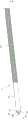
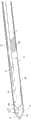
图1是用于本发明的内窥镜外科手术装置中的柔性4通道管的立体图,Figure 1 is a perspective view of a flexible 4-channel tube used in the endoscopic surgical device of the present invention,
图2是执行器套筒的立体图,Figure 2 is a perspective view of the actuator sleeve,
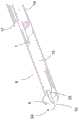
图3是从侧面看的内窥镜外科手术装置的第一实施方式的前端部分的立体图,Fig. 3 is a perspective view of the front end portion of the first embodiment of the endoscopic surgical apparatus as viewed from the side,
图4是沿着图3中的线III-III截取的纵长向放大比例剖视图,Fig. 4 is a longitudinally enlarged scale sectional view taken along the line III-III in Fig. 3,
图5以放大比例视图示出了具有打开的钳夹部的内窥镜外科手术装置的第一实施方式,Figure 5 shows a first embodiment of an endoscopic surgical device with an open jaw in an enlarged scale view,
图6以放大比例视图示出了具有闭合的钳夹部和暴露的针的内窥镜外科手术装置的第一实施方式,Figure 6 shows a first embodiment of an endoscopic surgical device with a closed jaw and an exposed needle in an enlarged scale view,
图7示出了端部执行器的第一实施方式,该端部执行器由具有锥形鼻部的两个管型件坯件制成,并且其中钳夹部是闭合的,Figure 7 shows a first embodiment of an end effector made from two tubular blanks with tapered noses and wherein the jaws are closed,
图8是具有打开的钳夹部的端部执行器的第一实施方式的分解放大比例视图,Figure 8 is an exploded enlarged scale view of the first embodiment of the end effector having an open jaw,
图9示出了处于组装状态的端部执行器的第一实施方式,其中钳夹部处于打开位置,Figure 9 shows the first embodiment of the end effector in an assembled state with the jaws in the open position,
图10是从连接到双腔管的端部执行器的第二实施方式的前面看的立体图,Figure 10 is a perspective view from the front of the second embodiment of the end effector connected to the dual lumen tube,
图11是实施图10所示的端部执行器的内窥镜外科手术装置的立体成角度侧视图,其中看到钳夹部处于闭合状态并且针被暴露,Figure 11 is a perspective angled side view of an endoscopic surgical device embodying the end effector shown in Figure 10 with the jaws seen in a closed state and the needle exposed,
图12是具有用于喷嘴的管状引导构件的端部执行器的第三实施方式的剖视图,Figure 12 is a cross-sectional view of a third embodiment of an end effector having a tubular guide member for a nozzle,
图13是从安装到管的替代性实施方式并且具有打开的钳夹部和暴露的针的端部执行器的第四实施方式的前面看的立体图,以及13 is a perspective view from the front of the fourth embodiment of the end effector mounted to the alternative embodiment of the tube and having an open jaw and an exposed needle, and
图14以前端视图示出了端部执行器的第四实施方式。Figure 14 shows a fourth embodiment of an end effector in a front end view.
图1中所示的柔性多通道管1具有四个纵长向延伸通道2、3、4、5:第一纵长向延伸通道2容纳执行器线(未示出);第二纵长向延伸通道3用作第一冲洗通道,用于在高压下向活检杯供应冲洗液体诸如水;第三纵长向延伸通道4用作第二冲洗通道,用于借助从第二纵长向延伸通道3到达的冲洗液体逐出组织标本;并且第四纵长向延伸通道5容纳纵长向可移位的针或喷嘴(未示出)。The flexible
图2所示的执行器套筒6具有下述内直径,所述内直径被选择用于使端部执行器紧紧地移进和移出执行器套筒,以打开和闭合钳夹部。执行器套筒6可以是金属管。The
图3是从侧面看的内窥镜外科手术装置的第一实施方式7的前端部分的立体片段图,并且图4是该前端部分的沿着图3中的线III-III截取的纵长向放大比例剖视图,3 is a perspective fragmentary view of the front end portion of the
内窥镜外科手术装置7具有端部执行器8的第一实施方式,该端部执行器容纳在执行器套筒6内,其中相对的钳夹部9、10由于通过所述执行器套筒6施加到钳夹部9、10的力而闭合。端部执行器8具有与多腔管1流体连通的第一端11以及包括相对的第一钳夹部9和第二钳夹部10的相反的第二端12,相对的第一钳夹部9和第二钳夹部10在所述相对的钳夹部9、10的闭合状态下一起界定活检杯13。The endoscopic
电线14通过管1的第一纵长向延伸通道2延伸,并且在端部执行器8的第一端11和第二端12之间的固定孔29处固定到主管状体15,以在电流被施加到端部执行器以取组织标本时加热相对的钳夹部9、10。The
如图4最佳所示,柔性管1被盘绕构件16围绕,以便以足够的柔性刚度加强柔性管,使得柔性管可以在内窥镜(未示出)的工作通道内移动。外部塑料管17围绕内窥镜外科手术装置的长度热收缩,以密封接头并使内窥镜外科手术装置与工作通道绝缘,并且使盘绕加强构件16结合到执行器套筒6,例如热熔合,以使执行器套筒能够在需要时纵长向地移动以打开和闭合钳夹部9、10。As best shown in Figure 4, the
第一钳夹部9具有用于使针或喷嘴19暴露的开口18,如图6最佳所示,该针或喷嘴紧贴地在管1的第四纵长向延伸通道5内延伸,使得当针19被撤回时,其密封地塞住开口18。The
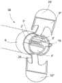
图7示出了由两个管型件26制成的端部执行器8的第一实施方式,管型件每个都具有锥形的鼻部24。管型件26中的每个管型件都已经沿着第一切割线22被切割成两个伸长管道半部19a、19b,例如通过激光切割被切割成下述型式,所述型式提供相应的具有相对的第一耦接装置20a'、20b'和第二耦接装置21a'、21b”的管道半部19a、19b,使得当相对的管道半部19a、19b随后被连接以获得端部执行器8的主管状体15时,所述耦接装置紧密地配合在一起,使得基本上防止了沿着第一组装线22的泄漏,并且可以在甚至没有焊接的情况下完成组装。不排除对主管状体的适当部件进行焊接或其他种类的熔合。FIG. 7 shows a first embodiment of an
如图7、图8和图9所示,每个钳夹部9、10也例如通过沿着第二切割线28a、28b的激光切割从相对的管道半部19a、19b的弯曲管道壁23a、23b中被切出。每个钳夹部9、10由此成形为具有发自相应的管道壁23a、23b的伸长铰接构件25和由于管型件26的最初闭合的锥形鼻部24而固有地获得的相对的较宽杯形端部27a、27b。As shown in Figures 7, 8 and 9, each
在切割之后,相应铰接构件25被偏转以远离彼此弯曲,如图8和图9所示。如图9所示,相对的第一耦接装置20a'、20b'和第二耦接装置21a'、21b”已经配合在一起,其中端部执行器8处于其松弛状态,准备好捏取组织标本。当端部执行器8撤回到执行器套筒6内部时,或者通过使加强构件移位使得执行器套筒位于加强构件前面而使执行器套筒向远侧移位时,相对的钳夹部9、10被迫使在一起,并且端部执行器8到达与如图3、图4和图7所示的配置类似的张紧配置。执行器套筒6沿着切割线22;28a、28b提供进一步的密封效果。用于电线14的固定孔29设置在主管状体15处。After cutting, the
图10是从通过执行器套筒6连接到双腔管31的端部执行器的第二实施方式30的前面看的立体图。图11示出了实施图10所示的端部执行器的内窥镜外科手术装置的前端部分的片段,其中看到钳夹部处于闭合状态并且麻醉针暴露,但是其中在图11中省略了执行器套筒6,以较好地例示端部执行器。为了示出双腔管31的两个腔,该管被示出为透明的,并且也没有示出加强构件。FIG. 10 is a perspective view from the front of the
端部执行器30的第二实施方式与端部执行器8的第一实施方式的主要不同之处在于,鼻部24'是平坦的而不是锥形的,如图11所示。平坦鼻部在制造期间需要较少的处理,因为管型件仅通过模制期间的平坦端板封闭或通过焊接封闭。端部执行器30类似于针对端部执行器的第一实施方式所描述的那样被切割,并且通过使切割的铰接构件远离彼此偏转而制成的钳夹部9'、10'也如针对端部执行器的第一实施方式所描述的那样。平头的端部执行器可以由两个单独的被纵长向地切割并随后连接的端部封闭的管道制成,或者由一个端部封闭的管道制成。The main difference between the second embodiment of the
使用用于端部执行器和管的第一实施方式的术语,双腔管31具有用于针19的第四纵长向延伸通道5和用于通过冲洗或抽吸将组织标本运出管31的第三纵长向延伸通道4。绝缘电线14在双腔管31的所述第三纵长向延伸通道4内在第三纵长向延伸通道4的壁旁边延伸,然后该第三纵长向延伸通道4也可以用作第一纵长向延伸通道2。可选地,所述管的外壁中的外部纵长向延伸凹槽可以用作第一纵长向延伸通道2。第四纵长向延伸通道5也可以用作第二纵长向延伸通道3,以在相对的钳夹部9'、10'的闭合状态下——其中针19塞住开口18'——将冲洗流体递送到活检杯13',以使所述针19暴露。Using the terminology used for the first embodiment of the end effector and tube, the
对于内窥镜外科手术装置的第一实施方式和第二实施方式两者,执行器线33可以是电线14或者是单独的部件。For both the first and second embodiments of the endoscopic surgical device, the
图12是具有用于控制针19的移位的管状引导构件34的端部执行器的第三实施方式34的剖视图。该管状引导构件35将针19引导出开口18并且具有足以在注射冲程中将针尖端保持在该管状引导构件35内的长度。FIG. 12 is a cross-sectional view of a
图13是从第四端部执行器36的前面看的立体图,其中打开的钳夹部9”、10'和暴露的针19以及安装在管1'的另一替代性实施方式上。图14以前端视图示出了该第四端部执行器。端部执行器36的第四实施方式基本上对应于图10和图11所示的第二实施方式,并且对于相似的部件,使用相同的附图标记。Figure 13 is a perspective view from the front of the
端部执行器36的第四实施方式与端部执行器的第二实施方式的不同之处在于,平坦鼻部24'不具有开口18,当钳夹部9'、10”闭合时针19可以通过该开口暴露。取而代之的是,针19被限制在由端部执行器36的闭合钳夹部9'、10”限定的闭合活检杯内。一旦端部执行器36被执行器套筒6释放,要么通过使执行器套筒6撤回要么通过将端部执行器36向前推出执行器套筒6的远侧开口,针可以被释放以采用一暴露活跃的位置,其中,针可以被操纵以施加局部麻醉。在该方面,活跃位置可以通过被弹簧偏置的针触发。为了实际进行活检,可以使针19撤回,将相对的钳夹部闭合在选定的组织周围,施加电热疗法,并且将切下的组织标本通过管1'朝向与具有端部执行器36的内窥镜外科手术装置一起使用的内窥镜的手柄冲洗。The fourth embodiment of the
替代性管1'基本上对应于管1的第一实施方式,并且对于相似的部件,使用相同的附图标记。替代性管1'与管1的第一实施方式仅略有不同在于,纵长向延伸通道2'、3'、4'、5'的直径和截面不同。特别地,用作用于在高压下将冲洗液体诸如水供应到闭合的活检杯的第一冲洗通道的第二纵长向延伸通道3'和例如用作用于通过从第二纵长向延伸通道3到达的冲洗液体逐出组织标本的第二冲洗通道的第三纵长向延伸通道4较大或用于施加抽吸。The
至于内窥镜外科手术装置7的第一实施方式,实施端部执行器30;34;36的任何实施方式的内窥镜外科手术装置的第二实施方式以及任何种类的多腔管1、1'都可以通过盘绕加强构件进行加强,执行器套筒端到端地连接到盘绕加强构件,并且热收缩管17应用于外部,如上所述。As for the first embodiment of the endoscopic
本发明的内窥镜外科手术装置提出了一种从中空器官进行活检的全新方式。为这些患者的治疗创造了全新的条件。可以大幅度降低费用,并且患者受益极高。将可以在定期复查的几分钟内诊断并指出患有癌症的门诊患者。患者在手术后不再需要任何导管治疗,这意味着它们消除了导管引起的尿路感染的高风险。不再需要涉及所有类别的工作人员;有医生和护士就足够了,这意味着大幅减少费用,并且患者可以更快地得到癌症治疗,并且释放了用于其他目的的外科手术能力。The endoscopic surgical device of the present invention presents an entirely new way of biopsying hollow organs. New conditions have been created for the treatment of these patients. Costs can be greatly reduced, and patient benefits are extremely high. Outpatients with cancer will be diagnosed and indicated within minutes of regular review. Patients no longer require any catheter therapy after surgery, which means they eliminate the high risk of catheter-induced urinary tract infections. There is no longer a need to involve all categories of staff; doctors and nurses are sufficient, which means significantly reduced costs and faster access to cancer treatment for patients, freeing up surgical capacity for other purposes.
Claims (15)
Applications Claiming Priority (3)
| Application Number | Priority Date | Filing Date | Title |
|---|---|---|---|
| SE1751639ASE541805C2 (en) | 2017-12-22 | 2017-12-22 | An endosurgical device |
| SE1751639-4 | 2017-12-22 | ||
| PCT/SE2018/051315WO2019125289A1 (en) | 2017-12-22 | 2018-12-14 | An endosurgical device and method of use |
Publications (1)
| Publication Number | Publication Date |
|---|---|
| CN111491579Atrue CN111491579A (en) | 2020-08-04 |
Family
ID=66993753
Family Applications (1)
| Application Number | Title | Priority Date | Filing Date |
|---|---|---|---|
| CN201880082131.5APendingCN111491579A (en) | 2017-12-22 | 2018-12-14 | Endoscopic surgical device and method of use |
Country Status (10)
| Country | Link |
|---|---|
| US (2) | US11547470B2 (en) |
| EP (1) | EP3709910A4 (en) |
| JP (1) | JP7422677B2 (en) |
| KR (2) | KR20250034184A (en) |
| CN (1) | CN111491579A (en) |
| AU (1) | AU2018388488B2 (en) |
| CA (1) | CA3086278A1 (en) |
| IL (1) | IL275494A (en) |
| SE (1) | SE541805C2 (en) |
| WO (1) | WO2019125289A1 (en) |
Cited By (4)
| Publication number | Priority date | Publication date | Assignee | Title |
|---|---|---|---|---|
| CN112022340A (en)* | 2020-09-25 | 2020-12-04 | 成都安捷畅医疗科技有限公司 | Electric coagulation forceps |
| CN113598901A (en)* | 2021-08-05 | 2021-11-05 | 江苏省人民医院(南京医科大学第一附属医院) | A medical multifunctional vocal cord filling and puncturing device |
| CN114983491A (en)* | 2022-06-10 | 2022-09-02 | 苏州市美新迪斯医疗科技有限公司 | Biopsy needle, biopsy needle set, and bone marrow biopsy device |
| CN115624372A (en)* | 2022-12-15 | 2023-01-20 | 北京新云医疗科技有限公司 | Surgical instrument |
Families Citing this family (12)
| Publication number | Priority date | Publication date | Assignee | Title |
|---|---|---|---|---|
| SE542926C2 (en) | 2018-12-14 | 2020-09-15 | Multi4 Ab | A tissue specimen collector comprising a rack arranged to move translatory within a collector housing |
| SE542910C2 (en) | 2018-12-14 | 2020-09-15 | Multi4 Ab | An endosurgical device |
| US12096915B2 (en)* | 2019-03-11 | 2024-09-24 | Gyrus Acmi, Inc. | Sheath location indicator and overextension preventer |
| CN110447968B (en)* | 2019-08-13 | 2024-11-19 | 江门摩尔科技有限公司 | Electronic atomization device, atomizer and liquid injection structure thereof |
| KR102294046B1 (en)* | 2020-02-18 | 2021-08-25 | 가톨릭대학교 산학협력단 | Variable cap device for Endoscopic Submucosal Dissection |
| KR102374893B1 (en)* | 2020-03-26 | 2022-03-17 | 한양대학교 산학협력단 | Apparatus for imaging and treating of cervix |
| JP2022102146A (en)* | 2020-12-25 | 2022-07-07 | 東京都 | Tumor sampler |
| JP7619880B2 (en)* | 2021-04-26 | 2025-01-22 | 朝日インテック株式会社 | Lancing Device |
| WO2023248072A1 (en)* | 2022-06-22 | 2023-12-28 | Covidien Lp | Systems and methods for providing and operating dissecting catheters |
| CN116327087B (en)* | 2023-03-31 | 2025-06-06 | 湖南省华芯医疗器械有限公司 | A flow guide element, a suction valve assembly, a handle and an endoscope of an endoscope |
| CN117462065B (en)* | 2023-12-26 | 2024-04-12 | 深圳市宏济医疗技术开发有限公司 | Endoscope for urology department |
| CN118986421B (en)* | 2024-08-12 | 2025-08-01 | 宁夏医科大学总医院 | Colorectal polyp biopsy mechanism capable of being controlled accurately |
Citations (14)
| Publication number | Priority date | Publication date | Assignee | Title |
|---|---|---|---|---|
| US5573546A (en)* | 1995-03-07 | 1996-11-12 | Nakao; Naomi L. | Biopsy ejecting forceps |
| JPH1119086A (en)* | 1997-07-03 | 1999-01-26 | Nippon Zeon Co Ltd | Forceps type electric treatment instrument |
| US5922002A (en)* | 1989-12-05 | 1999-07-13 | Yoon; Inbae | Surgical instrument with jaws and movable internal biopsy device and method for use thereof |
| US20070055172A1 (en)* | 2005-09-04 | 2007-03-08 | Nitesh Ratnakar | Multi Forceps Biopsy Catheter |
| JP2008528109A (en)* | 2005-01-20 | 2008-07-31 | ウィルソン−クック・メディカル・インコーポレーテッド | Biopsy forceps |
| US20080255424A1 (en)* | 2007-04-10 | 2008-10-16 | Boston Scientific Scimed, Inc. | Endoscopes including distal chamber and related methods of use |
| CN101365388A (en)* | 2006-01-12 | 2009-02-11 | 多种活检标本有限责任公司 | A sampling apparatus for taking a number of samples |
| WO2009111717A1 (en)* | 2008-03-06 | 2009-09-11 | Trustees Of Boston University | Low cost disposable medical forceps to enable a hollow central channel for various functionalities |
| US20100121153A1 (en)* | 2008-08-27 | 2010-05-13 | To John T | Retractor cannula system for accessing and visualizing spine and related methods |
| CN102421385A (en)* | 2009-05-13 | 2012-04-18 | 住友电木株式会社 | High-frequency hemostatic forceps for endoscope |
| CN102641150A (en)* | 2010-10-06 | 2012-08-22 | 女子慈善机构叙利亚黎巴嫩医院 | Surgical endoscope and process for exchanging surgical tools in a surgical endoscope |
| US20140323910A1 (en)* | 2008-05-16 | 2014-10-30 | Conquest Medical Technologies | Biopsy device |
| US20150141866A1 (en)* | 2013-11-18 | 2015-05-21 | Martin L. Mayse | System and method for evaluation of the pleural space |
| CN106572874A (en)* | 2014-07-28 | 2017-04-19 | 泰利福医疗公司 | Needlescopic scissor end effector and method of use |
Family Cites Families (14)
| Publication number | Priority date | Publication date | Assignee | Title |
|---|---|---|---|---|
| US5919202A (en)* | 1989-12-05 | 1999-07-06 | Yoon; Inbae | Surgical instrument with jaws and movable internal needle and method for use thereof |
| US5482054A (en)* | 1990-05-10 | 1996-01-09 | Symbiosis Corporation | Edoscopic biopsy forceps devices with selective bipolar cautery |
| US5217460A (en)* | 1991-03-22 | 1993-06-08 | Knoepfler Dennis J | Multiple purpose forceps |
| EP0526115B1 (en)* | 1991-07-29 | 1997-04-02 | Smith & Nephew Richards Inc | Forceps |
| CA2106039A1 (en)* | 1992-09-23 | 1994-03-24 | David A. Nicholas | Surgical biopsy forceps apparatus |
| US5820630A (en)* | 1996-10-22 | 1998-10-13 | Annex Medical, Inc. | Medical forceps jaw assembly |
| US5993466A (en)* | 1997-06-17 | 1999-11-30 | Yoon; Inbae | Suturing instrument with multiple rotatably mounted spreadable needle holders |
| FR2767051B1 (en)* | 1997-08-07 | 1999-10-29 | Jean Marie Hugueny | IMPROVED PINCHING DEVICE, ESPECIALLY OF THE BIOPSY FORCEPS |
| US7083618B2 (en)* | 2001-04-06 | 2006-08-01 | Sherwood Services Ag | Vessel sealer and divider |
| US8758342B2 (en) | 2007-11-28 | 2014-06-24 | Covidien Ag | Cordless power-assisted medical cauterization and cutting device |
| JP2013521994A (en)* | 2010-03-24 | 2013-06-13 | ユナイテッド ステイツ エンドスコピー グループ,インコーポレイテッド | Multiple biopsy device |
| JP5226830B2 (en) | 2011-06-09 | 2013-07-03 | オリンパス株式会社 | Endoscope |
| US9636168B2 (en)* | 2012-08-09 | 2017-05-02 | Covidien Lp | Electrosurgical instrument including nested knife assembly |
| US10595889B2 (en)* | 2016-04-11 | 2020-03-24 | RELIGN Corporation | Arthroscopic devices and methods |
- 2017
- 2017-12-22SESE1751639Apatent/SE541805C2/enunknown
- 2018
- 2018-12-14CNCN201880082131.5Apatent/CN111491579A/enactivePending
- 2018-12-14CACA3086278Apatent/CA3086278A1/enactivePending
- 2018-12-14KRKR1020257006099Apatent/KR20250034184A/enactivePending
- 2018-12-14AUAU2018388488Apatent/AU2018388488B2/enactiveActive
- 2018-12-14EPEP18892325.4Apatent/EP3709910A4/enactivePending
- 2018-12-14KRKR1020207020984Apatent/KR102775137B1/enactiveActive
- 2018-12-14USUS16/955,725patent/US11547470B2/enactiveActive
- 2018-12-14WOPCT/SE2018/051315patent/WO2019125289A1/ennot_activeCeased
- 2018-12-14JPJP2020555009Apatent/JP7422677B2/enactiveActive
- 2020
- 2020-06-18ILIL275494Apatent/IL275494A/enunknown
- 2023
- 2023-01-06USUS18/094,048patent/US12433666B2/enactiveActive
Patent Citations (14)
| Publication number | Priority date | Publication date | Assignee | Title |
|---|---|---|---|---|
| US5922002A (en)* | 1989-12-05 | 1999-07-13 | Yoon; Inbae | Surgical instrument with jaws and movable internal biopsy device and method for use thereof |
| US5573546A (en)* | 1995-03-07 | 1996-11-12 | Nakao; Naomi L. | Biopsy ejecting forceps |
| JPH1119086A (en)* | 1997-07-03 | 1999-01-26 | Nippon Zeon Co Ltd | Forceps type electric treatment instrument |
| JP2008528109A (en)* | 2005-01-20 | 2008-07-31 | ウィルソン−クック・メディカル・インコーポレーテッド | Biopsy forceps |
| US20070055172A1 (en)* | 2005-09-04 | 2007-03-08 | Nitesh Ratnakar | Multi Forceps Biopsy Catheter |
| CN101365388A (en)* | 2006-01-12 | 2009-02-11 | 多种活检标本有限责任公司 | A sampling apparatus for taking a number of samples |
| US20080255424A1 (en)* | 2007-04-10 | 2008-10-16 | Boston Scientific Scimed, Inc. | Endoscopes including distal chamber and related methods of use |
| WO2009111717A1 (en)* | 2008-03-06 | 2009-09-11 | Trustees Of Boston University | Low cost disposable medical forceps to enable a hollow central channel for various functionalities |
| US20140323910A1 (en)* | 2008-05-16 | 2014-10-30 | Conquest Medical Technologies | Biopsy device |
| US20100121153A1 (en)* | 2008-08-27 | 2010-05-13 | To John T | Retractor cannula system for accessing and visualizing spine and related methods |
| CN102421385A (en)* | 2009-05-13 | 2012-04-18 | 住友电木株式会社 | High-frequency hemostatic forceps for endoscope |
| CN102641150A (en)* | 2010-10-06 | 2012-08-22 | 女子慈善机构叙利亚黎巴嫩医院 | Surgical endoscope and process for exchanging surgical tools in a surgical endoscope |
| US20150141866A1 (en)* | 2013-11-18 | 2015-05-21 | Martin L. Mayse | System and method for evaluation of the pleural space |
| CN106572874A (en)* | 2014-07-28 | 2017-04-19 | 泰利福医疗公司 | Needlescopic scissor end effector and method of use |
Cited By (5)
| Publication number | Priority date | Publication date | Assignee | Title |
|---|---|---|---|---|
| CN112022340A (en)* | 2020-09-25 | 2020-12-04 | 成都安捷畅医疗科技有限公司 | Electric coagulation forceps |
| CN113598901A (en)* | 2021-08-05 | 2021-11-05 | 江苏省人民医院(南京医科大学第一附属医院) | A medical multifunctional vocal cord filling and puncturing device |
| CN114983491A (en)* | 2022-06-10 | 2022-09-02 | 苏州市美新迪斯医疗科技有限公司 | Biopsy needle, biopsy needle set, and bone marrow biopsy device |
| CN114983491B (en)* | 2022-06-10 | 2024-09-13 | 苏州市美新迪斯医疗科技有限公司 | Biopsy needle, biopsy needle set and bone marrow biopsy device |
| CN115624372A (en)* | 2022-12-15 | 2023-01-20 | 北京新云医疗科技有限公司 | Surgical instrument |
Also Published As
| Publication number | Publication date |
|---|---|
| US20230181238A1 (en) | 2023-06-15 |
| EP3709910A1 (en) | 2020-09-23 |
| IL275494A (en) | 2020-08-31 |
| US20200375655A1 (en) | 2020-12-03 |
| JP2024038384A (en) | 2024-03-19 |
| KR20200103746A (en) | 2020-09-02 |
| SE1751639A1 (en) | 2019-06-23 |
| JP7422677B2 (en) | 2024-01-26 |
| KR20250034184A (en) | 2025-03-10 |
| US12433666B2 (en) | 2025-10-07 |
| AU2018388488B2 (en) | 2024-05-30 |
| JP2021508580A (en) | 2021-03-11 |
| CA3086278A1 (en) | 2019-06-27 |
| AU2018388488A1 (en) | 2020-08-06 |
| US11547470B2 (en) | 2023-01-10 |
| EP3709910A4 (en) | 2021-08-04 |
| BR112020012203A2 (en) | 2020-11-24 |
| WO2019125289A1 (en) | 2019-06-27 |
| SE541805C2 (en) | 2019-12-17 |
| KR102775137B1 (en) | 2025-03-04 |
Similar Documents
| Publication | Publication Date | Title |
|---|---|---|
| US12433666B2 (en) | Endosurgical device and method of use | |
| US6066102A (en) | Optical biopsy forceps system and method of diagnosing tissue | |
| US20250186031A1 (en) | Endosurgical device | |
| US20250255589A1 (en) | Tissue specimen collector for use in biopsy | |
| CN108523985A (en) | A kind of multifunctional high frequency cutting is operated on | |
| JP7753412B2 (en) | Endosurgical devices and methods of use | |
| BR112020012203B1 (en) | ENDOSURGICAL DEVICE | |
| CN110584852A (en) | Hot puncture support imbedding device | |
| CN209107567U (en) | A kind of multifunctional high frequency cutting is operated on |
Legal Events
| Date | Code | Title | Description |
|---|---|---|---|
| PB01 | Publication | ||
| PB01 | Publication | ||
| SE01 | Entry into force of request for substantive examination | ||
| SE01 | Entry into force of request for substantive examination | ||
| CB02 | Change of applicant information | ||
| CB02 | Change of applicant information | Country or region after:Sweden Address after:Swedish Yan Xueping Applicant after:Maldivian Medical Co. Address before:Swedish Yan Xueping Applicant before:Martti multifunctional medical device Co. Country or region before:Sweden |