CN111481221A - Medical X-ray measurement device and program - Google Patents
Medical X-ray measurement device and programDownload PDFInfo
- Publication number
- CN111481221A CN111481221ACN201911378735.7ACN201911378735ACN111481221ACN 111481221 ACN111481221 ACN 111481221ACN 201911378735 ACN201911378735 ACN 201911378735ACN 111481221 ACN111481221 ACN 111481221A
- Authority
- CN
- China
- Prior art keywords
- image
- ray
- irradiation
- subject
- irradiation step
- Prior art date
- Legal status (The legal status is an assumption and is not a legal conclusion. Google has not performed a legal analysis and makes no representation as to the accuracy of the status listed.)
- Granted
Links
Images


Classifications
- A—HUMAN NECESSITIES
- A61—MEDICAL OR VETERINARY SCIENCE; HYGIENE
- A61B—DIAGNOSIS; SURGERY; IDENTIFICATION
- A61B6/00—Apparatus or devices for radiation diagnosis; Apparatus or devices for radiation diagnosis combined with radiation therapy equipment
- A61B6/40—Arrangements for generating radiation specially adapted for radiation diagnosis
- A61B6/4007—Arrangements for generating radiation specially adapted for radiation diagnosis characterised by using a plurality of source units
- A—HUMAN NECESSITIES
- A61—MEDICAL OR VETERINARY SCIENCE; HYGIENE
- A61B—DIAGNOSIS; SURGERY; IDENTIFICATION
- A61B6/00—Apparatus or devices for radiation diagnosis; Apparatus or devices for radiation diagnosis combined with radiation therapy equipment
- A61B6/50—Apparatus or devices for radiation diagnosis; Apparatus or devices for radiation diagnosis combined with radiation therapy equipment specially adapted for specific body parts; specially adapted for specific clinical applications
- A61B6/505—Apparatus or devices for radiation diagnosis; Apparatus or devices for radiation diagnosis combined with radiation therapy equipment specially adapted for specific body parts; specially adapted for specific clinical applications for diagnosis of bone
- A—HUMAN NECESSITIES
- A61—MEDICAL OR VETERINARY SCIENCE; HYGIENE
- A61B—DIAGNOSIS; SURGERY; IDENTIFICATION
- A61B6/00—Apparatus or devices for radiation diagnosis; Apparatus or devices for radiation diagnosis combined with radiation therapy equipment
- A61B6/52—Devices using data or image processing specially adapted for radiation diagnosis
Landscapes
- Health & Medical Sciences (AREA)
- Life Sciences & Earth Sciences (AREA)
- Engineering & Computer Science (AREA)
- Medical Informatics (AREA)
- Heart & Thoracic Surgery (AREA)
- Molecular Biology (AREA)
- Biophysics (AREA)
- Nuclear Medicine, Radiotherapy & Molecular Imaging (AREA)
- Optics & Photonics (AREA)
- Pathology (AREA)
- Radiology & Medical Imaging (AREA)
- Biomedical Technology (AREA)
- Physics & Mathematics (AREA)
- High Energy & Nuclear Physics (AREA)
- Surgery (AREA)
- Animal Behavior & Ethology (AREA)
- General Health & Medical Sciences (AREA)
- Public Health (AREA)
- Veterinary Medicine (AREA)
- Orthopedic Medicine & Surgery (AREA)
- Dentistry (AREA)
- Oral & Maxillofacial Surgery (AREA)
- Computer Vision & Pattern Recognition (AREA)
- Apparatus For Radiation Diagnosis (AREA)
Abstract
Translated fromChinese
Description
Translated fromChinese技术领域technical field
本发明涉及医疗用X射线测定装置以及程序,具体涉及表示被检查者所包含的特定成分的二维分布的图像的生成。The present invention relates to a medical X-ray measurement device and a program, and more particularly, to the generation of an image representing a two-dimensional distribution of a specific component contained in a subject.
背景技术Background technique
DEXA(dual-energy X-ray absorptiometry:双能X射线吸收法)一般是以下的方法,即根据通过依次向被检查者照射具有不同的能量的2种X射线而得到的2种检测数据,生成表示被检查者所包含的特定成分的图像。具体地说,生成表示被检查者所包含的特定成分的含有量或含有比例的二维分布的图像。作为这样的图像,已知骨密度(bone density)图像。骨密度图像也被称为骨矿物含量(bone mineral content)图像。也能够通过DEXA法生成表示脂肪量、脂肪率、无脂量、无脂率等的二维分布的图像。无脂量是软组织中的脂肪以外的组织的量,无脂率是软组织中的脂肪以外的组织的含有量。DEXA法也被称为DXA法。DEXA (dual-energy X-ray absorptiometry: dual-energy X-ray absorptiometry) is generally a method of generating two types of detection data obtained by sequentially irradiating a subject with two types of X-rays having different energies An image representing a specific component contained in the examinee. Specifically, an image representing a two-dimensional distribution of the content or content ratio of the specific component contained in the subject is generated. As such an image, a bone density image is known. Bone density images are also known as bone mineral content images. It is also possible to generate an image representing a two-dimensional distribution of fat mass, fat percentage, fat-free mass, fat-free percentage, and the like by the DEXA method. The fat-free amount is the amount of tissues other than fat in soft tissue, and the fat-free rate is the content of tissue other than fat in soft tissue. The DEXA method is also known as the DXA method.
在应用DEXA法时,在使面状的X射线束进行扫描而形成二维照射区域的情况下,测定时间变长。对此,如果利用具有金字塔形状的X射线束形成二维照射区域,则能够缩短测定时间。When the DEXA method is applied, when a planar X-ray beam is scanned to form a two-dimensional irradiation area, the measurement time becomes long. On the other hand, if the two-dimensional irradiation area is formed by the X-ray beam having a pyramid shape, the measurement time can be shortened.
还已知以下的方法:即通过在照射具有大范围的能量的X射线的基础上,在X射线检测阶段使能量灵敏度不同,由此得到2种检测数据。它也是DEXA法的一种方式。There is also known a method of obtaining two types of detection data by irradiating X-rays having a wide range of energy and varying the energy sensitivities in the X-ray detection stage. It is also a way of the DEXA method.
在专利文献1记载的医疗用X射线测定装置中,在被检查者的上方设置产生具有立体形状的X射线束的X射线产生装置,在被检查者的下方设置X射线检测装置(放射线图像摄像装置)。X射线检测装置具备在垂直方向上层叠的第一检测器、放射线限制构件以及第二检测器。通过第一检测器和第二检测器同时得到2种检测数据。In the medical X-ray measuring apparatus described in Patent Document 1, an X-ray generating apparatus for generating an X-ray beam having a three-dimensional shape is provided above the subject, and an X-ray detecting apparatus (radiographic image pickup) is provided below the subject. device). The X-ray detection apparatus includes a first detector, a radiation limiting member, and a second detector stacked in the vertical direction. Two kinds of detection data are simultaneously obtained by the first detector and the second detector.
现有技术文献prior art literature
专利文献Patent Literature
专利文献1:日本特开2018-23768号公报Patent Document 1: Japanese Patent Laid-Open No. 2018-23768
发明要解决的问题Invention to solve problem
在通过一次的X射线照射同时得到2种检测数据的情况下,一般向被检查者照射大能量范围的X射线。在这样的方法中,无法在测定中途判断测定中止,作为结果可以认为对被检查者产生了必要以上的辐射。另外,在这样的方法中,在采用具备第一检测器和第二检测器的层叠构造的情况下,随着从射束中心轴偏离,在第一检测器和第二检测器之间产生的位置误差会增大。When two types of detection data are simultaneously obtained by a single X-ray irradiation, the subject is generally irradiated with X-rays in a wide energy range. In such a method, it is not possible to determine that the measurement has been terminated in the middle of the measurement, and as a result, it is considered that more than necessary radiation is generated to the subject. In addition, in such a method, when a laminated structure including a first detector and a second detector is employed, as the deviation from the beam center axis occurs, a difference occurs between the first detector and the second detector. The position error will increase.
发明内容SUMMARY OF THE INVENTION
本发明的目的在于:使得能够避免对被检查者的无用的X射线照射。An object of the present invention is to make it possible to avoid useless X-ray irradiation of the subject.
或者,本发明的目的在于:避免在2个检测数据之间产生位置误差的问题。Alternatively, an object of the present invention is to avoid the problem that a positional error occurs between two pieces of detection data.
解决方案solution
本发明的医疗用X射线测定装置的特征在于,具备:照射部,其在第一照射工序中向被检查者照射具有第一能量且具有立体形状的第一X射线束,在其后的第二照射工序中向上述被检查者照射具有与上述第一能量不同的第二能量且具有上述立体形状的第二X射线束;检测部,其检测在上述第一照射工序中穿过了上述被检查者的X射线而生成第一二维检测数据,并检测在上述第二照射工序中穿过了上述被检查者的X射线而生成第二二维检测数据;主图像生成部,其根据上述第一二维检测数据和上述第二二维检测数据,生成表示上述被检查者所包含的特定成分的二维分布的主图像;以及控制部,其在上述第一照射工序和上述第二照射工序之间的中间工序中,允许或中止上述第二照射工序的执行。The medical X-ray measuring apparatus of the present invention is characterized by including an irradiation unit that irradiates the subject with a first X-ray beam having a first energy and a three-dimensional shape in a first irradiation step, and a second X-ray beam thereafter. In the second irradiation step, the subject is irradiated with a second X-ray beam having a second energy different from the first energy and having the three-dimensional shape; The X-rays of the examiner are generated to generate first two-dimensional detection data, and the X-rays that have passed through the examinee in the second irradiation step are detected to generate second two-dimensional detection data; the first two-dimensional detection data and the second two-dimensional detection data to generate a master image representing the two-dimensional distribution of the specific components included in the subject; and a control unit for performing the first irradiation step and the second irradiation In an intermediate process between the processes, the execution of the above-mentioned second irradiation process is permitted or suspended.
本发明的程序的特征在于,包括:控制第一照射工序的执行的功能;控制上述第一照射工序之后的中间工序的执行的功能;控制上述中间工序之后的第二照射工序的执行的功能;以及控制上述第二照射工序之后的显示工序的执行的功能,在上述第一照射工序中,向被检查者照射具有第一能量且具有立体形状的第一X射线束,并且通过检测穿过了上述被检查者的X射线而生成第一二维检测数据,在上述中间工序中,根据上述第一二维检测数据生成和显示骨折诊断用图像,并且接受继续指示或中止指示,在接受到上述继续指示的情况下允许上述第二照射工序的执行,在接受到上述中止指示的情况下中止上述第二照射工序的执行,在上述第二照射工序中,向上述被检查者照射具有与上述第一能量不同的第二能量且具有上述立体形状的第二X射线束,并且通过检测穿过了上述被检查者的X射线而生成第二二维检测数据,在上述显示工序中,根据上述第一二维检测数据和上述第二二维检测数据来生成和显示骨密度图像。The program of the present invention is characterized by comprising: a function of controlling the execution of the first irradiation step; a function of controlling the execution of an intermediate step after the first irradiation step; and a function of controlling the execution of the second irradiation step after the intermediate step; and a function of controlling the execution of the display process following the second irradiation process, in which the subject is irradiated with a first X-ray beam having a first energy and a three-dimensional shape, and the The X-ray of the subject is used to generate first two-dimensional detection data, and in the intermediate step, an image for fracture diagnosis is generated and displayed based on the first two-dimensional detection data, and an instruction to continue or abort is received. The execution of the second irradiation step is permitted in the case of continuing the instruction, and execution of the second irradiation step is aborted when the discontinuation instruction is received. A second X-ray beam having a second energy of different energy and having the above-mentioned three-dimensional shape, and the second two-dimensional detection data is generated by detecting X-rays passing through the above-mentioned subject, and in the above-mentioned displaying step, according to the above-mentioned first A two-dimensional detection data and the above-mentioned second two-dimensional detection data are used to generate and display a bone density image.
发明效果Invention effect
根据本发明,能够避免向被检查者的无用的X射线照射。或者,根据本发明,能够避免在多个检测数据之间产生位置误差。According to the present invention, useless X-ray irradiation to the subject can be avoided. Alternatively, according to the present invention, it is possible to avoid the occurrence of positional errors between a plurality of detection data.
附图说明Description of drawings
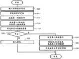
图1是表示实施方式的医疗用X射线测定装置的图。FIG. 1 is a diagram showing a medical X-ray measuring apparatus according to an embodiment.
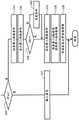
图2是表示数据处理的图。FIG. 2 is a diagram showing data processing.
图3是表示第一动作例的流程图。FIG. 3 is a flowchart showing a first operation example.
图4是表示第二动作例的流程图。FIG. 4 is a flowchart showing a second operation example.
图5是表示第三动作例的流程图。FIG. 5 is a flowchart showing a third operation example.
图6是表示第四动作例的流程图。FIG. 6 is a flowchart showing a fourth operation example.
图7是表示X射线图像的显示例的图。FIG. 7 is a diagram showing a display example of an X-ray image.
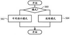
图8是表示第五动作例的流程图。FIG. 8 is a flowchart showing a fifth operation example.
图9是表示投影器的作用的图。FIG. 9 is a diagram showing the operation of the projector.
图10是表示投影器的构造的图。FIG. 10 is a diagram showing the structure of a projector.
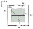
图11是表示第一投影像的图。FIG. 11 is a diagram showing a first projected image.
图12是表示第二投影像的图。FIG. 12 is a diagram showing a second projected image.
图13是表示第三投影像的图。FIG. 13 is a diagram showing a third projected image.
图14是表示第四投影像的图。FIG. 14 is a diagram showing a fourth projected image.
图15是表示投影方法的变形例子的图。FIG. 15 is a diagram showing a modified example of the projection method.
图16是表示假体(fantom)上表面的第一例的图。FIG. 16 is a diagram showing a first example of the upper surface of a prosthesis (fantom).
图17是表示假体上表面的第二例的图。Fig. 17 is a diagram showing a second example of the upper surface of the prosthesis.
图18是表示假体上表面的第三例的图。Fig. 18 is a view showing a third example of the upper surface of the prosthesis.
图19是表示X射线图像和骨密度图像的显示例的图。FIG. 19 is a diagram showing a display example of an X-ray image and a bone density image.
附图标记说明:Description of reference numbers:
10:测定部;12:信息处理部;18:拍摄台;20:被检查者;22:照射部;28:检测部;32:运算部;34:控制部;40:X射线图像生成部;42:X射线图像解析部;44:照射条件运算部;46:骨密度图像生成部;48:骨密度图像解析部;50:第一二维检测数据;52:X射线图像;54:第二二维检测数据;56:骨密度图像。10: measurement unit; 12: information processing unit; 18: imaging table; 20: subject; 22: irradiation unit; 28: detection unit; 32: calculation unit; 34: control unit; 40: X-ray image generation unit; 42: X-ray image analysis unit; 44: irradiation condition calculation unit; 46: bone density image generation unit; 48: bone density image analysis unit; 50: first two-dimensional detection data; 52: X-ray image; 54: second Two-dimensional detection data; 56: bone density image.
具体实施方式Detailed ways
以下,根据附图说明实施方式。Hereinafter, embodiments will be described with reference to the drawings.
(1)实施方式的概要(1) Outline of Embodiment
实施方式的医疗用X射线测定装置具备照射部、检测部、主图像生成部以及控制部。照射部在第一照射工序中,向被检查者照射具有第一能量并且具有立体形状的第一X射线束,在其后的第二照射工序中,向被检查者照射具有与上述第一能量不同的第二能量并且具有上述立体形状的第二X射线束。检测部检测在第一照射工序中穿过了被检查者的X射线而生成第一二维检测数据,检测在第二照射工序中穿过了被检查者的X射线而生成第二二维检测数据。主图像生成部根据第一二维检测数据和第二二维检测数据,生成表示被检查者所包含的特定成分的二维分布的图像。控制部在第一照射工序和第二照射工序之间的中间工序中,允许或中止第二照射工序的执行。The medical X-ray measurement apparatus according to the embodiment includes an irradiation unit, a detection unit, a main image generation unit, and a control unit. In the first irradiation step, the irradiation unit irradiates the subject with a first X-ray beam having a first energy and a three-dimensional shape, and in a subsequent second irradiation step, irradiates the subject with the first energy having the same energy as the first X-ray beam. A second X-ray beam of a different second energy and having the above-mentioned three-dimensional shape. The detection unit detects X-rays that have passed through the subject in the first irradiation process to generate first two-dimensional detection data, and detects X-rays that have passed through the subject in the second irradiation process to generate second two-dimensional detection data data. The main image generation unit generates an image representing a two-dimensional distribution of specific components included in the subject based on the first two-dimensional detection data and the second two-dimensional detection data. The control unit allows or suspends execution of the second irradiation step in an intermediate step between the first irradiation step and the second irradiation step.
根据上述结构,在第一照射工序和第二照射工序之间设置中间工序,在该中间工序中,判断可否执行第二照射工序。在中间工序中判明了不应该执行第二照射工序或不需要执行第二照射工序的状况的情况下,不执行第二照射工序,因此能够与之相应地减少被检查者的被辐射量。阶段性地执行第一照射工序和第二照射工序,因此能够在这些工序之间共用检测部。换言之,即使不利用具有特殊构造的部件作为检测部也可以。在不执行第二照射工序的情况下,能够尽早解除被检查者的捆绑状态,因此能够减轻被检查者的负担。According to the above configuration, an intermediate step is provided between the first irradiation step and the second irradiation step, and in this intermediate step, it is determined whether or not the second irradiation step can be executed. When it is determined in an intermediate step that the second irradiation step should not be performed or that the second irradiation step is not required, the second irradiation step is not performed, and accordingly, the exposure amount of the examinee can be reduced. Since the first irradiation step and the second irradiation step are performed in stages, the detection unit can be shared among these steps. In other words, it is not necessary to use a member having a special structure as the detection portion. When the second irradiation step is not performed, the bound state of the subject can be released as soon as possible, so that the burden on the subject can be reduced.
通常由检查者判断可否执行第二照射工序。在该情况下,理想的是,向检查者提供用于支援检查者的判断的信息。控制部通常依照来自检查者的指示,允许或中止第二照射工序的执行。但是,控制部也可以自发自动地判断可否执行第二照射工序。Usually, the inspector judges whether or not the second irradiation step can be performed. In this case, it is desirable to provide the examiner with information for supporting the examiner's judgment. The control unit usually permits or suspends the execution of the second irradiation step in accordance with an instruction from the inspector. However, the control unit may automatically and automatically determine whether or not the second irradiation step can be executed.
主图像的典型例子是表示骨密度(骨矿物含量)的二维分布的图像。主图像的概念包括表示脂肪量或者脂肪率的二维分布的图像、以及表示无脂量或无脂率的二维分布的图像。也可以一边使X射线的能量不同,一边阶段性地执行3个以上的照射工序。在该情况下,理想的是,在第一照射工序和第二照射工序之间设置中间工序。不过,也可以针对相邻的2个照射工序的每一个,在它们之间设置中间工序或与之相当的工序。A typical example of the main image is an image representing a two-dimensional distribution of bone density (bone mineral content). The concept of the main image includes an image representing the two-dimensional distribution of the fat mass or fat percentage, and an image representing the two-dimensional distribution of the fat-free mass or fat-free percentage. Three or more irradiation steps may be performed in stages while varying the energy of the X-rays. In this case, it is desirable to provide an intermediate step between the first irradiation step and the second irradiation step. However, for each of two adjacent irradiation steps, an intermediate step or a step corresponding thereto may be provided between them.
实施方式的医疗用X射线测定装置具备:副图像生成部,其在中间工序中,根据第一二维检测数据,生成被检查者的X射线像作为副图像;显示器,其在中间工序中显示副图像;输入器,其在中间工序中,接受继续指示或中止指示。控制部在接受到继续指示的情况下允许第二照射工序的执行,在接受到中止指示的情况下中止上述第二照射工序的执行。The medical X-ray measurement apparatus according to the embodiment includes: a sub-image generation unit that generates an X-ray image of the subject as a sub-image based on the first two-dimensional detection data in an intermediate process; and a display that displays in the intermediate process A secondary image; an input device that accepts a continuation instruction or an abort instruction in an intermediate process. The control unit permits the execution of the second irradiation step when receiving a continuation instruction, and aborts the execution of the second irradiation step when receiving a stop instruction.
上述结构在向检查者提供副图像的基础上,让检查者判断可否执行第二照射工序,并接受作为其判断结果的继续指示或中止指示。例如,在即使没有执行第二照射工序也只能够根据副图像进行疾病的诊断的情况、由于在第一照射工序中被检查者有移动等在副图像内不包含诊断对象骨等理由而无法采用第一照射工序的结果的情况下,输入中止指示。The above configuration allows the examiner to judge whether or not the second irradiation step can be executed after providing the sub-image to the examiner, and to accept a continuation instruction or a stop instruction as a result of the judgement. For example, when a disease diagnosis can only be performed from the sub-image even if the second irradiation step is not performed, the sub-image cannot be used for reasons such as movement of the examinee during the first irradiation step, and other reasons such as the bone to be diagnosed is not included in the sub-image. In the case of the result of the first irradiation step, a stop instruction is input.
在实施方式中,主图像是骨密度图像,在副图像中的诊断对象骨像中没有识别出骨折部位的情况下向输入器输入继续指示,在副图像中的诊断对象骨像中识别出骨折部位的情况下向输入器输入中止指示。例如,在诊断对象骨像中识别出脆弱性骨折的情况下,能够诊断骨质疏松症,在该情况下,并不一定必须执行第二照射工序。反倒要优先降低辐射,而省略第二照射工序的执行。上述结构能够与这样的需求对应。不过也可以根据检查者的判断,与副图像的内容无关地,指示第二照射工序的执行。In an embodiment, the main image is a bone density image, and if the fracture site is not recognized in the bone image of the diagnosis target in the sub image, a continuation instruction is input to the input device, and the fracture is recognized in the bone image of the diagnosis target in the sub image. In the case of the part, a stop instruction is input to the input device. For example, when a fragile fracture is recognized in the bone image to be diagnosed, osteoporosis can be diagnosed, and in this case, it is not always necessary to perform the second irradiation step. Instead, priority should be given to reducing radiation, and the execution of the second irradiation process is omitted. The above-described structure can respond to such needs. However, according to the judgment of the inspector, the execution of the second irradiation step may be instructed regardless of the content of the sub-image.
在实施方式中,控制部进行控制使得在中止了第二照射工序的执行的情况下执行意见输入工序。它记录基于副图像的判断的结果,与副图像一起记录意见。也可以执行意见输入工序以外的其他工序。In the embodiment, the control unit performs control such that the opinion input step is executed when the execution of the second irradiation step is suspended. It records the result of the judgment based on the sub-image, and records the opinion together with the sub-image. It is also possible to perform operations other than the opinion input operation.
实施方式的医疗用X射线测定装置具备运算部,该运算部根据第一二维检测检测数据来运算第二照射工序中的照射条件。根据该结构,能够针对被检查者而优化第二照射工序中的照射条件。在实施方式中,运算部具备根据第一二维检测数据来运算与被检查者有关的代表厚度的单元、根据代表厚度修正第二照射工序中的照射条件的单元。例如,在被检查者的厚度的平均值大的情况下,提高第二能量,另一方面,在被检查者的厚度的平均值小的情况下,降低第二能量。由此,能够兼顾测定精度和降低辐射。The medical X-ray measurement apparatus according to the embodiment includes a computing unit that computes irradiation conditions in the second irradiation step based on the first two-dimensional detection data. According to this configuration, the irradiation conditions in the second irradiation step can be optimized for the subject. In the embodiment, the calculation unit includes means for calculating a representative thickness of the subject based on the first two-dimensional detection data, and means for correcting the irradiation conditions in the second irradiation step based on the representative thickness. For example, when the average value of the thickness of the examinee is large, the second energy is increased, and on the other hand, when the average value of the thickness of the examinee is small, the second energy is decreased. As a result, both measurement accuracy and radiation reduction can be achieved.
实施方式的医疗用X射线测定装置具备支援信息生成部,该支援信息生成部根据副图像,生成支援第二照射工序的执行可否的判断的支援信息,其中,将支援信息与副图像一起显示到显示器。根据该结构,能够支援检查者的判断。The medical X-ray measurement apparatus according to the embodiment includes a support information generation unit that generates support information for supporting the determination of whether or not the execution of the second irradiation step can be performed based on the sub-image, wherein the support information is displayed together with the sub-image on the sub-image. monitor. According to this structure, the judgment of the examiner can be supported.
实施方式的医疗用X射线测定装置具备:拍摄台,其承载被检查者;以及投影器,其向拍摄台投影在定位被检查者时利用的投影像,其中,投影像是表示照射第一X射线束和第二X射线束的区域的光学像。通过光圈调整等,照射X射线束的区域的大小变动。根据投影像,能够识别当前的照射区域,能够与之对应地正确地定位被检查者。The medical X-ray measurement apparatus according to the embodiment includes: an imaging table on which a subject is placed; and a projector for projecting a projection image used for positioning the subject on the imaging table, wherein the projection image indicates that the first X is irradiated Optical image of the region of the beam and the second X-ray beam. The size of the area to which the X-ray beam is irradiated varies by aperture adjustment or the like. From the projection image, the current irradiation area can be recognized, and the subject can be accurately positioned in accordance with the projection image.
在实施方式中,投影像包括在定位校正用基准物质时利用的要素。该要素例如作为表示设置包含校正用基准物质的假体的位置或区域的标记而发挥功能。根据该结构,容易地将校正用基准物质配置到正确的位置。例如,能够防止校正用基准物质从具有立体形状的X射线束脱离。In an embodiment, the projected image includes elements used in positioning the reference material for calibration. This element functions, for example, as a marker indicating the position or region where the prosthesis containing the calibration reference substance is installed. According to this configuration, it is easy to arrange the reference material for calibration at the correct position. For example, it is possible to prevent the reference material for calibration from escaping from the X-ray beam having a three-dimensional shape.
在实施方式的医疗用X射线测定装置的动作方法(动作控制方法)中,包括第一照射工序、中间工序、第二照射工序以及显示工序。在第一照射工序中,向被检查者照射具有第一能量并且具有立体形状的第一X射线束,并且通过检测穿过了被检查者的X射线而生成第一二维检测数据。在中间工序中,根据第一二维检测检测数据而生成和显示骨折诊断图像,并且接受继续指示或中止指示。在接受到继续指示的情况下允许第二照射工序的执行,在接受到中止指示的情况下中止第二照射工序的执行。在第二照射工序中,向被检查者照射具有第二能量并且具有立体形状的第二X射线束,并且通过检测穿过了被检查者的X射线而生成第二二维检测数据。在显示工序中,根据第一二维检测数据和第二二维检测数据而生成和显示骨密度图像。The operation method (operation control method) of the medical X-ray measuring apparatus according to the embodiment includes a first irradiation step, an intermediate step, a second irradiation step, and a display step. In the first irradiation step, the subject is irradiated with a first X-ray beam having first energy and a three-dimensional shape, and first two-dimensional detection data is generated by detecting the X-rays that have passed through the subject. In the intermediate process, a fracture diagnosis image is generated and displayed based on the first two-dimensional detection data, and a continuation instruction or a stop instruction is accepted. The execution of the second irradiation step is permitted when a continuation instruction is received, and the execution of the second irradiation step is suspended when a stop instruction is received. In the second irradiation step, the subject is irradiated with a second X-ray beam having a second energy and a three-dimensional shape, and second two-dimensional detection data is generated by detecting the X-rays that have passed through the subject. In the display step, a bone density image is generated and displayed based on the first two-dimensional detection data and the second two-dimensional detection data.
在实施方式中,依照程序控制上述动作方法。经由可移动性存储介质或网络,将该程序安装到医疗用X射线测定装置或其具备的信息处理装置。In an embodiment, the above-described operation method is controlled in accordance with a program. This program is installed in the medical X-ray measurement apparatus or an information processing apparatus provided with it via a portable storage medium or a network.
(2)实施方式的详情(2) Details of the embodiment
在图1中,表示出实施方式的医疗用X射线测定装置。医疗用X射线测定装置具有伦琴射线拍摄功能、骨密度测定功能等。根据医疗用X射线测定装置,不利用骨密度测定专用的装置,就能够进行骨密度测定。在进行骨密度测定时,利用DEXA法。此外,利用医疗用X射线测定装置,还能够测定脂肪的含有量和含有率、以及无脂肪(脂肪以外的软组织)的含有量和含有率。FIG. 1 shows a medical X-ray measurement apparatus according to an embodiment. The medical X-ray measuring apparatus has a Roentgen ray imaging function, a bone density measuring function, and the like. According to the medical X-ray measuring apparatus, the bone densitometry can be performed without using a special apparatus for bone densitometry. When the bone mineral density was measured, the DEXA method was used. In addition, the content and content rate of fat and the content and content rate of non-fat (soft tissue other than fat) can also be measured by the medical X-ray measuring apparatus.
在图1中,医疗用X射线测定装置大致由测定部10和信息处理部12构成。测定部10被设置在检查室14内,信息处理部12被设置在控制室16内。测定部10和信息处理部12相互通过电缆连接。它们也可以通过无线通信连接。信息处理部12例如由作为信息处理装置的计算机构成。信息处理部12也可以由多个计算机构成。在该情况下,它们经由网络相互连接。此外,x方向是第一水平方向,z方向是垂直方向(铅垂方向)。与x方向和z方向正交的方向是作为第二水平方向的y方向。In FIG. 1 , the medical X-ray measuring apparatus is roughly composed of a measuring
将拍摄台18设置在检查室14内。在测定时,将被检查者(人体)20承载在拍摄台18上。在该情况下,例如被检查者20在拍摄台18上以仰卧的姿势躺下。在该情况下,体轴(体干部的中心轴)与x方向平行。作为成为骨密度测定的对象的部位,可以列举腰椎、大腿骨等。The imaging table 18 is installed in the
在拍摄台18的上方即在被检查者20的上方设置有照射部22。照射部22被机器臂机构24支援。照射部22具备X射线产生管、光圈机构、投光器等。在进行X射线测定时,通过照射部22生成具有立体形状的X射线束26。具体地说,X射线束26具有金字塔状的形式。也将它称为光锥射束。既可以使X射线束26在xz平面内的传播角度θ与X射线束在yz平面内的传播角度φ相同,也可以使其不同。不过在进行骨密度测定时,通常将照射部22定位到规定的高度,角度θ与角度φ分别成为相同的规定角度。也可以使用具有四角锥形状以外的形状(例如圆锥形状)的X射线束。The
通过后述的控制部34,控制向X射线产生管供给的驱动信号的电压和电流、以及该驱动信号的供给时间。典型的是,通过使驱动信号的电压可变而变更生成的X射线的能量。如后述那样,在实施方式中,在第一照射工序中生成高能量X射线,在其后的第二照射工序中,生成比高能量低的低能量X射线。与成为测定对象的组织对应地,确定相互不同的第二能量。在实施方式中,选择适合于骨密度测定的2个能量。The voltage and current of the drive signal supplied to the X-ray generating tube and the supply time of the drive signal are controlled by the
在拍摄台18具备的顶板的下侧即被检查者20的下方配置有检测部28。检测部28具备平板检测器(FPD)30,通过该FPD检测X射线。FPD30由在x方向和y方向上排列的许多检测元件构成。各个检测元件例如具备将X射线变换为光的闪烁器、以及将光变换为电信号的电路。通过FPD30检测穿过了被检查者的X射线,由此得到二维检测数据。The
在进行骨密度测定时,在第一照射工序中生成第一二维检测数据,在第二照射工序中生成第二二维检测数据。这些二维检测数据被转送信息处理部12。在2个照射工序之间共通地利用实施方式的FPD30。在检测部28中,不采用由多个检测器构成的重叠构造。在实施方式中,在2个二维检测数据之间,不产生位置误差,也不产生放大率的差。When the bone density measurement is performed, the first two-dimensional detection data is generated in the first irradiation process, and the second two-dimensional detection data is generated in the second irradiation process. These two-dimensional detection data are transferred to the
接着,说明信息处理部12。信息处理部12具备运算部32、控制部34、存储部36、输入部37以及显示部38。运算部32和控制部34由依照程序动作的CPU构成。它们既可以由多个处理器构成,它们也可以由其他设备构成。存储部36由半导体存储器、硬盘等构成。在存储部36上存储有动作控制程序、运算处理程序。还能够将包括图像的数据存储到存储部36中。输入部37由键盘、指示设备等构成。通过输入部37接受后述的继续指示和中止指示的输入。显示部38由液晶显示器、有机EL显示器等构成。将后述的X射线图像和骨密度图像显示到显示部38的画面上。Next, the
控制部34依照动作控制程序,控制X射线的生成和检测、以及数据处理。在动作控制程序负责的骨密度测定过程中,如后述那样,包括第一照射工序、中间工序、第二照射工序、显示工序、意见输入工序等。具体地说,控制部34针对X射线束的形成和照射、X射线的检测、基于指示的工序转移、图像生成等执行控制。The
运算部32具有多个功能。在这些功能中,在图1中用多个模块表现出与骨密度测定关联的多个功能。具体地说,运算部32具备X射线图像生成部40、X射线图像解析部42、照射条件运算部44、骨密度图像生成部46以及骨密度图像解析部48。The
X射线图像生成部40在中间工序中,根据在第一照射工序中获取的第一二维检测数据,生成作为伦琴射线图像的X射线图像。在X射线图像中,包括测定对象部位的内部的诊断对象骨像。通过其观察,在执行第二照射工序之前,能够掌握测定对象骨的状态。从与后述的骨密度图像的关系来看,将X射线图像称为副图像或辅助图像,从其功能来看,将X射线图像称为骨折诊断用图像。In an intermediate step, the X-ray
X射线图像解析部42对X射线图像进行解析而确定异常部位。在解析X射线图像时,能够利用学习型图像识别技术等。在确定了异常部位的情况下,例如将表示它的涂色像或标记重叠显示到X射线图像上。The X-ray
照射条件运算部44在执行第二照射工序之前,根据第一二维检测数据,针对第二X射线束来运算照射条件。实施方式的照射条件运算部44根据第一二维检测数据来运算被检查者的厚度的平均值或最大值,根据这样的代表厚度,对与第二X射线束有关的第二能量进行优化。The irradiation
例如,在代表厚度是标准值的情况下,设定规定值作为第二能量,在代表厚度比标准值大的情况下,设定比规定值高的值作为第二能量,在代表厚度比标准值小的情况下,设定比规定值低的值作为第二能量。由此,避免测定粗壮的被检查者的情况下的测定精度下降、以及测定单薄的被检查者的情况下的过剩辐射。将运算出的照射条件从照射条件运算部44发送到控制部34。For example, when the representative thickness is a standard value, a predetermined value is set as the second energy, and when the representative thickness is larger than the standard value, a value higher than the predetermined value is set as the second energy. When the value is small, a value lower than the predetermined value is set as the second energy. As a result, it is possible to avoid a decrease in measurement accuracy in the case of measuring a thick subject, and excess radiation in the case of measuring a thin subject. The calculated irradiation conditions are sent from the irradiation
骨密度图像生成部46依照DEXA法,根据从被检查者得到的第一二维检测数据和第二二维检测数据,来生成骨密度图像。骨密度图像表示被检测体内的骨矿物质量的二维分布。The bone density
在运算骨密度图像时,为了进行灵敏度的修正,在将被检查者放置到拍摄台18上之前,执行第一照射工序和第二照射工序。由此,获取第一二维检测数据(第一二维空气系数数据)和第二二维检测数据(第二二维空气系数数据)。在生成骨密度图像的阶段,根据需要参照这些数据。此外,为了进行灵敏度的修正,还根据需要利用后述的假体。When calculating the bone density image, in order to correct the sensitivity, the first irradiation step and the second irradiation step are performed before the subject is placed on the imaging table 18 . Thereby, first two-dimensional detection data (first two-dimensional air coefficient data) and second two-dimensional detection data (second two-dimensional air coefficient data) are acquired. These data are referred to as necessary at the stage of generating the bone density image. In addition, in order to correct the sensitivity, a later-described prosthesis is also used as necessary.
也可以将衰减量图像显示为X射线图像。衰减量图像是根据第一二维检测数据和事先获取的第一空气系数数据生成的图像。具体地说,以像素为单位,作为对数值而运算空气系数与检测数据之比。对数值相当于衰减量。此外,基于空气系数的修正、利用了假体的修正是在骨密度测定装置中以前所利用的技术。The attenuation image can also be displayed as an X-ray image. The attenuation image is an image generated based on the first two-dimensional detection data and the first air coefficient data acquired in advance. Specifically, the ratio of the air coefficient to the detection data is calculated as a logarithmic value in units of pixels. The logarithmic value corresponds to the amount of attenuation. In addition, correction based on an air coefficient and correction using a prosthesis are techniques that have been conventionally used in bone densitometry devices.
骨密度图像解析部48通过进行骨密度图像的解析,确定异常部位。在该情况下,能够利用学习型图像识别技术。在确定了异常部位的情况下,将表示它的涂色像或标记重叠地显示到骨密度图像上。The bone density
在图2中,表示出数据处理的流程。通过执行第一照射工序,生成第一二维检测数据50。第一二维检测数据50由在x方向和y方向上排列的许多检测值50a构成。各个检测值50a与各个像素对应。在实施方式中,根据第一二维检测数据50生成X射线图像52,并显示它。In FIG. 2, the flow of data processing is shown. By executing the first irradiation process, the first two-
通过观察X射线图像52,检查者判断可否执行第二照射工序。例如,在针对测定对象骨识别出脆弱性骨折并能够在该阶段诊断骨质疏松症的情况下,判断为不需要执行第二照射工序,由检查者输入中止指示。除此以外,在通过观察X射线图像52而判明了没有正确地执行第一照射工序的情况下,也可以输入中止指示。为了支援X射线图像52的观察,也可以显示X射线图像52的解析结果。By observing the
另一方面,在通过观察X射线图像52而检查者判断为需要生成骨密度图像的情况下,由检查者输入继续指示。在虽然识别出脆弱性骨折但判断为需要获取骨密度图像的情况下,也可以输入继续指示。利用输入器输入中止指示和继续指示。On the other hand, when the examiner determines that it is necessary to generate a bone density image by observing the
在输入了中止指示的情况下,禁止执行第二照射工序,不获取第二二维检测数据54。另一方面,在输入了继续指示的情况下,执行第二照射工序,由此获取第二二维检测数据54。第二二维检测数据54与第一二维检测数据50同样地,由与多个像素对应的多个检测值54a构成。也可以在第二照射工序之前,根据第一二维检测数据50或基于它生成的X射线图像,计算第二照射工序中的第二照射条件。用附图标记53表示它。When a stop instruction is input, execution of the second irradiation step is prohibited, and the second two-
在执行第二照射工序之后,根据第一二维检测数据50和第二二维检测数据54,生成骨密度图像56。从作为副图像的X射线图像来看,将其称为主图像。显示该骨密度图像,并根据需要解析骨密度图像。After the second irradiation process is performed, a
接着,使用图3~图6,说明与图1所示的医疗用X射线测定装置有关的第一动作例~第四动作例。在上述控制部的控制下,执行构成各个动作例的各工序。Next, the first to fourth operation examples related to the medical X-ray measuring apparatus shown in FIG. 1 will be described with reference to FIGS. 3 to 6 . Each process constituting each operation example is executed under the control of the above-described control unit.
在图3中表示出第一动作例。在S10中,向医疗用X射线测定装置输入被检查者信息。被检查者信息例如是包含检查编号、姓名等的信息。使根据它获取的X射线图像和骨密度图像与被检查者信息关联起来。在S12中,在拍摄台上将被检查者定位。对被检查者进行定位以使得测定对象部位进入到照射区域内。在实施方式中,设定高能量作为第一X射线束的能量。这时也可以设定低能量,但根据被检查者的体格,能量会不足,因此设定了高能量。也可以显示第一照射条件,由检查者确认其内容。S16是第一照射工序。在S16中,向被检查者照射第一X射线束,检测穿过了被检查者的X射线。由此,获取第一二维检测数据。A first operation example is shown in FIG. 3 . In S10, the subject information is input to the medical X-ray measuring apparatus. The examinee information is, for example, information including an examination number, a name, and the like. The X-ray image and bone density image obtained therefrom are associated with the subject information. In S12, the examinee is positioned on the imaging table. The subject is positioned so that the measurement target portion enters the irradiation area. In an embodiment, high energy is set as the energy of the first X-ray beam. In this case, low energy can also be set, but depending on the physique of the examinee, the energy will be insufficient, so high energy is set. The first irradiation condition may be displayed, and the inspector may check the content. S16 is the first irradiation step. In S16, the subject is irradiated with the first X-ray beam, and the X-rays that have passed through the subject are detected. Thereby, the first two-dimensional detection data is acquired.
S18和S20构成中间工序。中间工序也可以包含后述的S24。在S18中,根据第一二维检测数据生成X射线图像,并显示它。由观察X射线图像的检查者判断测定的继续或中止。在判断为中止测定的情况下,在S20中,由检查者输入中止指示。另一方面,在判断为继续测定的情况下,在S20中,由检查者输入继续指示。S18 and S20 constitute an intermediate process. The intermediate step may include S24 described later. In S18, an X-ray image is generated from the first two-dimensional detection data, and it is displayed. The continuation or suspension of the measurement is determined by the examiner who observes the X-ray image. When it is determined that the measurement is to be stopped, in S20, a stop instruction is input by the examiner. On the other hand, when it is determined that the measurement is to be continued, in S20, a continuation instruction is input by the examiner.
在S20中输入了中止指示的情况下,执行S22。其构成意见输入工序,在S22中,由检查者利用输入器输入意见。与X射线图像对应地管理所输入的意见信息。例如,输入骨折部位位置、骨折程度作为意见。也可以输入诊断名。When a stop instruction is input in S20, S22 is performed. This constitutes an opinion input step, and in S22, an examiner inputs an opinion using an input device. The input opinion information is managed according to the X-ray image. For example, enter fracture site location, fracture degree as comments. You can also enter a diagnostic name.
另一方面,在S20中输入了继续指示的情况下,在S24中,通过控制部设定第二照射条件。也可以显示第二照射条件,由检查者确认其内容。接着S24而执行S26。S26是第二照射工序。即,在S26中,向被检查者照射第二X射线束,检测穿过了被检查者的X射线。由此,获取第二二维检测数据。S28是显示工序。在S28中,依照DEXA法,根据第一二维检测数据和第二二维检测数据生成骨密度图像,并显示它。On the other hand, when a continuation instruction is input in S20, in S24, the second irradiation condition is set by the control unit. The second irradiation conditions may be displayed, and the inspector may check the contents. Next to S24, S26 is executed. S26 is the second irradiation step. That is, in S26, the subject is irradiated with the second X-ray beam, and the X-rays that have passed through the subject are detected. Thereby, the second two-dimensional detection data is acquired. S28 is a display step. In S28, according to the DEXA method, a bone density image is generated from the first two-dimensional detection data and the second two-dimensional detection data, and displayed.
根据上述动作例,在S20中输入了中止指示的情况下,禁止执行第二照射工序,因此能够避免对被检查者的无用的辐射。另外,在该情况下,能够尽早将被检查者从捆绑状态中释放出来。According to the above-described operation example, when the stop instruction is input in S20, the execution of the second irradiation step is prohibited, so that unnecessary radiation to the subject can be avoided. In addition, in this case, the examinee can be released from the bound state as soon as possible.
此外,也可以阶段性地照射具有3个以上能量的3个X射线,生成多个图像(例如骨密度图像、脂肪量图像、无脂量图像)。在该情况下,理想的是,还在最初的照射工序之间设置中间工序。也可以根据需要,进而在第二个以后的照射工序之间设置相当于中间工序的工序。In addition, three X-rays having three or more energies may be irradiated in stages to generate a plurality of images (for example, a bone density image, a fat mass image, and a fat-free mass image). In this case, it is desirable to further provide an intermediate step between the first irradiation steps. If necessary, a process corresponding to an intermediate process may be further provided between the second and subsequent irradiation processes.
在图4中表示出第二动作例。此外,在图4~图6中表示出一连串动作中的一部分。省略的部分与图3所示的部分相同。在图4~图6中,对已经说明了的工序附加相同的工序编号,省略其说明。A second operation example is shown in FIG. 4 . In addition, a part of a series of operations are shown in FIGS. 4 to 6 . The omitted parts are the same as those shown in FIG. 3 . In FIGS. 4 to 6 , the same step numbers are attached to the steps already described, and the description thereof is omitted.
在图4所示的第二动作例中,在S28的后面设置有S30和S32。在S30中解析骨密度图像,在S32中显示该解析结果。例如,在骨密度值存在异常高的部分、异常低的部分的情况下,将它们确定为异常部位,将表示它们的符号等重叠地显示到骨密度图像上。根据第二动作例,能够支援基于骨密度图像的诊断。In the second operation example shown in FIG. 4, S30 and S32 are provided after S28. The bone density image is analyzed in S30, and the analysis result is displayed in S32. For example, when there are abnormally high parts and abnormally low parts in the bone density value, these are identified as abnormal parts, and symbols or the like indicating these are displayed in a superimposed manner on the bone density image. According to the second operation example, the diagnosis based on the bone density image can be supported.
在图5所示的第三动作例中,在S24的前面设置有S34~S40。具体地说,在S20中输入了继续指示的情况下,在S34中根据第一二维检测数据或X射线图像计算第二照射条件。例如,如上述那样计算代表厚度,根据它适应性地设定第二能量。在S36中,将第二照射条件显示到画面上。由此,由检查者确认第二照射条件。在S38中,在检查者进行了承认第二照射条件的输入的情况下,执行S24。另一方面,在S38中,在检查者进行了要求变更第二照射条件的输入的情况下,执行S40。在S40中,由检查者变更第二照射条件。然后,执行S24。根据第三动作例,能够与状况对应地对第二照射条件进行优化。In the third operation example shown in FIG. 5, S34 to S40 are provided before S24. Specifically, when a continuation instruction is input in S20, the second irradiation condition is calculated in S34 based on the first two-dimensional detection data or the X-ray image. For example, the representative thickness is calculated as described above, and the second energy is adaptively set according to it. In S36, the second irradiation condition is displayed on the screen. Thereby, the examiner confirms the second irradiation condition. In S38, when the examiner has inputted the approval of the second irradiation condition, S24 is executed. On the other hand, in S38, when the examiner has inputted a request to change the second irradiation condition, S40 is executed. In S40, the examiner changes the second irradiation conditions. Then, S24 is performed. According to the third operation example, the second irradiation condition can be optimized according to the situation.
在图6所示的第四动作例中,在S16的后面设置有S50~S54。具体地说,在S50中,根据第一二维检测数据,生成X射线图像。接着在S52中,解析X射线图像。例如确定有可能骨折的部位。在S54中,显示X射线图像。这时,也显示X射线图像解析结果。例如在X射线图像上标记有可能骨折的部位。根据该第四动作例,能够支援检查者在中间工序中的判断。In the fourth operation example shown in FIG. 6 , S50 to S54 are provided after S16 . Specifically, in S50, an X-ray image is generated based on the first two-dimensional detection data. Next, in S52, the X-ray image is analyzed. For example, determine the site of a possible fracture. In S54, the X-ray image is displayed. At this time, the X-ray image analysis results are also displayed. For example, X-ray images are marked with possible fractures. According to this fourth operation example, the judgment of the inspector in the intermediate process can be supported.
在图7中示例了X射线图像。图示的X射线图像包含多个椎骨像。其中如用附图标记60A所示那样,涂色地表现出有可能骨折的特定的椎骨。An X-ray image is illustrated in FIG. 7 . The illustrated X-ray image contains multiple vertebral images. Among them, as indicated by
在图8中示出第五动作例。在S60中,判断用户所选择的模式是中间指示模式还是连续模式。在选择了中间指示模式的情况下,执行S62,执行图3~图6所示的处理即包含中间工序的处理。另一方面,在选择了连续模式的情况下,执行S64。在S64中,接着第一照射工序,连续地执行第二照射工序。在该情况下,不进行中止指示或继续指示的输入的接受。此外,在选择了连续模式的情况下,也可以在执行第一照射工序后,显示X射线图像。另外,也可以根据第一二维检测数据来运算第二照射条件。在第五动作例中,在始终执行第二照射工序的情况下,可以选择连续模式,在该情况下,不需要检查者的指示行为,因此能够减轻检查者的负担。A fifth operation example is shown in FIG. 8 . In S60, it is determined whether the mode selected by the user is the intermediate instruction mode or the continuous mode. When the intermediate instruction mode is selected, S62 is executed, and the processing shown in FIGS. 3 to 6 , that is, the processing including the intermediate step is executed. On the other hand, when the continuous mode is selected, S64 is executed. In S64, following the first irradiation step, the second irradiation step is continuously performed. In this case, the input of the stop instruction or the continuation instruction is not accepted. In addition, when the continuous mode is selected, the X-ray image may be displayed after the first irradiation step is performed. In addition, the second irradiation condition may be calculated based on the first two-dimensional detection data. In the fifth operation example, when the second irradiation step is always performed, the continuous mode can be selected. In this case, the inspector's instruction behavior is not required, so the burden on the inspector can be reduced.
接着,使用图9~图15说明投影器及其动作。Next, the projector and its operation will be described with reference to FIGS. 9 to 15 .
在图9中,将被检查者20承载到拍摄台18上。将照射部22设置到其上方,将检测部28设置到其下方。照射部22具备X射线产生器61和投影器62。投影器62生成具有与四角锥状的X射线束相同的形状的光束64,作为投光器或瞄准器而发挥作用。In FIG. 9 , the subject 20 is placed on the imaging table 18 . The
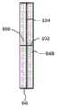
在图9中,表示出被支援机构68支援的假体(模拟体)66。假体66例如包含模拟骨的第一基准物质和模拟软组织的第二基准物质。在倾斜状态下支援假体66使得假体66的中心轴与X射线发射方向一致,而在X射线束空间内包含假体66。假体66用于修正第一二维检测数据和第二二维检测数据,以前就利用了其自身。此外,既可以在与拍摄台18的上表面接触的状态下,通过支援机构68支援假体66,也可以在从拍摄台18的顶板上表面浮起的状态下,通过支援机构68支援假体66。也可以不使用支援机构68,而在拍摄台18的顶板上表面设置独立式假体。In FIG. 9, the prosthesis (simulation body) 66 supported by the
在图10中示出投影器62的结构例。在投影器62的出射口设置有光圈机构62A。通过光圈机构62A规定X射线束26的立体形状。特别规定角度θ和角度φ(在图10中只示出角度φ)。顺便地说,在测定骨密度时,将角度θ和角度φ分别规定为规定角度。由此,不需要使投影器62的作用动态地可变。A configuration example of the
具体地说,投影器62除了具备光圈机构62A以外,还具备光源70、第一反射镜72、以及第二反射镜74。从光源70发出的光通过第一反射镜72反射,该反射的光进而通过第二反射镜74进行反射。该反射的光构成光束64。光束64具有角锥状的形式,该形式与X射线束26的形式一致。Specifically, the
光圈机构62A具有出射口,在此配置有透光构件76。透光构件76具有的图案被投影到拍摄台的顶板(以及被检查者)上。透光构件76具有遮光性或包含黑色的2条线。这些线十字交叉为“+”状。因此,在投影像上包含十字交叉的2条线。此外,理想的是,反射镜74和透光构件76由几乎不减弱X射线的材料构成。The
接着,作为通过投影器62形成的投影像的变形,使用图11~图15说明第一投影像~第四投影像。Next, the first to fourth projected images will be described with reference to FIGS. 11 to 15 as modifications of the projected images formed by the
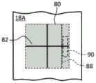
在图11中示出第一投影像。投影像80表示出顶板上表面18A上的X射线照射区域,两者的外形一致。通过对上述透光构件76进行着色,而对投影像80实施着色。投影像80包含标记82,其由在射束中心十字交叉的2条线84、86构成。根据投影像80,能够在视觉上作为投影区域而确认X射线照射区域。在此基础上,能够利用标记82,将测定部位正确地定位到X射线照射区域内。The first projection image is shown in FIG. 11 . The projected
在图12中示出第二投影像。此外,在图12~图15中,对与图11所示的要素相同的要素附加相同的附图标记,省略其说明。The second projection image is shown in FIG. 12 . In addition, in FIGS. 12-15, the same code|symbol is attached|subjected to the same element as the element shown in FIG. 11, and the description is abbreviate|omitted.
在图12中,在投影像80中包含在配置假体90时作为基准发挥功能的线88。通过向上述透光构件附加施加了与周围不同的着色的线,来生成线88。具体地说,在线88和投影像80的端部边缘(在图12中为右侧端部边缘)之间的带状的区域设置假体90。线88是作为定位假体用的要素发挥功能的标记。根据包含线88的投影像80,能够确实并且容易地将假体90设置在角锥状的X射线束中、并且不对被检查者产生障碍的位置。此外,既可以预先确定假体90的设置角度,也可以通过利用了投影像的后述的方法来优化假体90的设置角度。In FIG. 12 , the
在图13中示出第三投影像。在投影像80中包含在配置假体90时作为基准发挥功能的标记92、94。通过向上述透光构件附加施加了与周围不同的着色的2个标记,来生成2个标记92、94。例如使存在于假体90的下部的2个角(在图13中为左侧的2个角)与2个标记92、94对准,而自然地优化设置假体90的位置。2个标记92、94是作为定位假体用的要素发挥功能的标记。The third projection image is shown in FIG. 13 . The
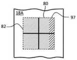
在图14中示出第四投影像。在投影像80中包含表示假体90的配置位置的像96。像96具有规定的色调。通过在上述透光构件的一部分上形成具有规定的色调的透光部分,来生成像96。在图示的第四投影像中,作为其要素的像96例如构成为具有与假体的底面相同的形状、或比该底面稍大的矩形的图形。如果将假体设置在像96上,则作为结果优化了设置假体的位置。也可以在设置假体后,向假体的上表面投影像96的一部分或全部,事后确认将假体设置到了正确的位置。将在后面使用图16~图18进一步说明它。如以上那样,像96是作为定位假体用的要素发挥功能的标记。A fourth projection image is shown in FIG. 14 . The projected
在图15中示出变形例。也可以形成投影像80,并且对顶板上表面18A施加着色或粘贴胶带,可识别地表现出假体配置区域97。一般在伦琴拍摄中也使用投影台,因此在希望避免对拍摄台的特别加工的情况下,理想的是,采用上述第二投影像~第四投影像。A modification example is shown in FIG. 15 . The
在图16~图18中示出与假体66的上表面有关的几个实施例。在图16所示的第一例中,对假体66的上表面66A施加粗面加工。由此,抑制光的反射。由此,能够提高投影像的识别性。为了进一步提高投影像的识别性,也可以对上表面66A施加着色等。Several embodiments related to the upper surface of the
在图17所示的第二例中,在假体66的上表面66B上描绘出作为标志的标记100。该标记100由在上表面66B的中心点十字交叉的2条线102、104构成。通过参照该标记100,容易地根据与投影像的关系来确认假体66的位置、姿势。In the second example shown in FIG. 17 , on the
在图18所示的第三例中,向假体66的上表面66B附加2个标记106、108。另一方面,在投影像中包含与它们对应的2个标记像。通过使2个标记像与2个标记106、108一致,能够优化假体的位置和姿势。In the third example shown in FIG. 18 , two
根据以上说明的各结构,正确地定位被检查者变得容易,另外,正确地定位假体变得容易。能够防止产生假体从具有立体形状的X射线束脱离的问题。进而,能够减轻定位假体时的检查者的负担。如果被检查者和假体的定位被优化,则能够减轻因定位错误造成的测定失败的风险。According to each of the structures described above, it becomes easy to accurately position the subject, and it becomes easy to accurately position the prosthesis. It is possible to prevent the occurrence of a problem that the prosthesis is detached from the X-ray beam having a three-dimensional shape. Furthermore, the burden on the examiner when positioning the prosthesis can be reduced. If the positioning of the subject and the prosthesis is optimized, the risk of assay failure due to positioning errors can be mitigated.
在不执行中间工序的医疗用X射线测定装置或骨密度测定装置中,也可以采用图9~图18所示的结构。例如,也可以针对能够通过一次照射获取与多个能量对应的多个二维检测数据的医疗用X射线测定装置、将校正用物质与被检查者一起设置在X射线束内的医疗用X射线测定装置,采用图9~图18的实施例。The configurations shown in FIGS. 9 to 18 may also be employed in a medical X-ray measuring apparatus or a bone density measuring apparatus that does not perform intermediate steps. For example, a medical X-ray measuring apparatus capable of acquiring a plurality of two-dimensional detection data corresponding to a plurality of energies by one irradiation, and a medical X-ray in which a calibration substance is placed in the X-ray beam together with the subject may be used. As the measuring apparatus, the examples shown in FIGS. 9 to 18 were used.
从这样的观点看,本发明的医疗用X射线测定装置具备:照射部,其向拍摄台上的被检查者照射具有立体形状的X射线束;检测部,其检测穿过了被检测体的X射线而输出二维检测数据;图像形成部,其根据X射线检测数据,形成表示被检测体的图像;投影器,其向投影台投影在定位被检查者和校正用基准物质中的至少一方时利用的引导像。理想的是,引导像是表示照射X射线束的区域的像。理想的是,在引导像中包含在定位校正用基准物质时利用的要素。也可以对校正用基准物质的上表面或包含校正用基准物质的构件的上表面,施加与其他面不同的加工、着色。也可以在该上表面设置在照射引导像时成为定位用的基准的图形。From such a viewpoint, the medical X-ray measuring apparatus of the present invention includes: an irradiation unit that irradiates an X-ray beam having a three-dimensional shape to a subject on an imaging table; and a detection unit that detects an X-ray beam passing through the subject. X-rays to output two-dimensional detection data; an image forming unit that forms an image representing a subject based on the X-ray detection data; a projector that projects on a projection table at least one of positioning the subject and a reference material for calibration The guide image used when using. Ideally, the guide image represents an image of an area where the X-ray beam is irradiated. Ideally, the guide image includes elements used for positioning the reference material for calibration. The upper surface of the reference material for calibration or the upper surface of the member containing the reference material for calibration may be processed and colored differently from other surfaces. A figure that serves as a reference for positioning when the guide image is irradiated may be provided on the upper surface.
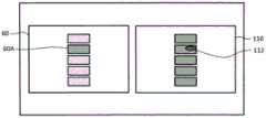
在图19中示出通过图1所示的医疗用X射线测定装置显示的图像的一个例子。在第二照射工序后的显示工序中,同时显示X射线图像60和骨密度图像110。在X射线图像60中包含表示异常部位的标记像60A,在骨密度图像110中也包含表示异常部位的标记像112。通过同时观察这些图像60、110,能够综合地诊断测定对象骨。An example of an image displayed by the medical X-ray measurement apparatus shown in FIG. 1 is shown in FIG. 19 . In the display step after the second irradiation step, the
为了参考而说明了基于骨密度图像的异常判定方法。例如在测定对象骨是腰椎的情况下,也可以对每节锥骨计算平均骨密度,在其从适当范围偏离的情况下,判定为异常。也可以对多个椎骨计算平均骨密度,在其从适当范围偏离的情况下,判定为异常。The abnormality determination method based on a bone density image is demonstrated for reference. For example, when the bone to be measured is the lumbar spine, the average bone density may be calculated for each conical bone, and if it deviates from an appropriate range, it may be determined to be abnormal. The average bone density of a plurality of vertebrae may be calculated, and if it deviates from the appropriate range, it may be determined as abnormal.
例如,由于压迫性骨折、血管的钙化、造影剂、异物等的原因,产生高于适当范围的异常骨密度。也可以对每个像素判定异常,并通过亮度、色调等表现它。例如在由于骨质疏松症而骨矿物量低的情况下,产生低于适当范围的异常骨密度。在该情况下,也可以对每个像素判定异常,并通过亮度、色调等表现它。For example, abnormal bone density higher than an appropriate range occurs due to compression fractures, calcification of blood vessels, contrast agents, foreign bodies, and the like. It is also possible to determine abnormality for each pixel and express it by brightness, hue, and the like. Abnormal bone densities below the appropriate range are produced, for example, in the case of low bone mineral content due to osteoporosis. In this case, it is also possible to determine abnormality for each pixel and express it by brightness, hue, or the like.
Claims (10)
Translated fromChineseApplications Claiming Priority (2)
| Application Number | Priority Date | Filing Date | Title |
|---|---|---|---|
| JP2019011812AJP7112343B2 (en) | 2019-01-28 | 2019-01-28 | Medical X-ray measuring device and program |
| JP2019-011812 | 2019-01-28 |
Publications (2)
| Publication Number | Publication Date |
|---|---|
| CN111481221Atrue CN111481221A (en) | 2020-08-04 |
| CN111481221B CN111481221B (en) | 2023-11-03 |
Family
ID=71788553
Family Applications (1)
| Application Number | Title | Priority Date | Filing Date |
|---|---|---|---|
| CN201911378735.7AActiveCN111481221B (en) | 2019-01-28 | 2019-12-27 | Medical X-ray measuring device and storage medium |
Country Status (2)
| Country | Link |
|---|---|
| JP (1) | JP7112343B2 (en) |
| CN (1) | CN111481221B (en) |
Families Citing this family (1)
| Publication number | Priority date | Publication date | Assignee | Title |
|---|---|---|---|---|
| JP2024066977A (en)* | 2022-10-28 | 2024-05-16 | 京セラ株式会社 | Image generating device, image generating method, image generating program, and recording medium |
Citations (9)
| Publication number | Priority date | Publication date | Assignee | Title |
|---|---|---|---|---|
| US5204888A (en)* | 1989-12-14 | 1993-04-20 | Aloka Co., Ltd. | Bone mineral content measuring apparatus |
| EP0761166A2 (en)* | 1995-09-08 | 1997-03-12 | Hologic, Inc. | X-ray bone densitometry |
| JP2009240435A (en)* | 2008-03-28 | 2009-10-22 | Fujifilm Corp | Radiographic imaging apparatus |
| JP2011104103A (en)* | 2009-11-17 | 2011-06-02 | Canon Inc | Energy subtraction image photographing apparatus |
| US20120288062A1 (en)* | 2010-01-27 | 2012-11-15 | Canon Kabushiki Kaisha | Radiation imaging apparatus, and control method and program of the apparatus |
| WO2013103048A1 (en)* | 2012-01-05 | 2013-07-11 | 日立アロカメディカル株式会社 | Medical device |
| CN103442638A (en)* | 2011-03-18 | 2013-12-11 | 日立阿洛卡医疗株式会社 | Bone density measurement device |
| KR101573918B1 (en)* | 2015-06-22 | 2015-12-02 | 한승무 | Scanning optimal adaptive scanning method and bone mineral density measuring device |
| CN108937982A (en)* | 2017-05-18 | 2018-12-07 | 富士胶片株式会社 | Radiation image picking-up system, radiation image photography method, program storage medium and body thickness apparatus for predicting |
Family Cites Families (5)
| Publication number | Priority date | Publication date | Assignee | Title |
|---|---|---|---|---|
| US6385283B1 (en)* | 1999-11-24 | 2002-05-07 | Hologic, Inc. | Device and method for determining future fracture risk |
| JP2005034539A (en)* | 2003-07-18 | 2005-02-10 | Ibaraki Prefecture | X-ray diagnostic imaging device with bone density distribution measurement function |
| JP6047347B2 (en)* | 2012-09-11 | 2016-12-21 | 株式会社日立製作所 | Medical X-ray measuring device |
| JP2015093000A (en)* | 2013-11-11 | 2015-05-18 | 日立アロカメディカル株式会社 | Medical x-ray measuring device |
| JP2017217227A (en)* | 2016-06-08 | 2017-12-14 | 株式会社日立製作所 | Radiographic diagnosis apparatus and bone density measurement method |
- 2019
- 2019-01-28JPJP2019011812Apatent/JP7112343B2/enactiveActive
- 2019-12-27CNCN201911378735.7Apatent/CN111481221B/enactiveActive
Patent Citations (9)
| Publication number | Priority date | Publication date | Assignee | Title |
|---|---|---|---|---|
| US5204888A (en)* | 1989-12-14 | 1993-04-20 | Aloka Co., Ltd. | Bone mineral content measuring apparatus |
| EP0761166A2 (en)* | 1995-09-08 | 1997-03-12 | Hologic, Inc. | X-ray bone densitometry |
| JP2009240435A (en)* | 2008-03-28 | 2009-10-22 | Fujifilm Corp | Radiographic imaging apparatus |
| JP2011104103A (en)* | 2009-11-17 | 2011-06-02 | Canon Inc | Energy subtraction image photographing apparatus |
| US20120288062A1 (en)* | 2010-01-27 | 2012-11-15 | Canon Kabushiki Kaisha | Radiation imaging apparatus, and control method and program of the apparatus |
| CN103442638A (en)* | 2011-03-18 | 2013-12-11 | 日立阿洛卡医疗株式会社 | Bone density measurement device |
| WO2013103048A1 (en)* | 2012-01-05 | 2013-07-11 | 日立アロカメディカル株式会社 | Medical device |
| KR101573918B1 (en)* | 2015-06-22 | 2015-12-02 | 한승무 | Scanning optimal adaptive scanning method and bone mineral density measuring device |
| CN108937982A (en)* | 2017-05-18 | 2018-12-07 | 富士胶片株式会社 | Radiation image picking-up system, radiation image photography method, program storage medium and body thickness apparatus for predicting |
Also Published As
| Publication number | Publication date |
|---|---|
| CN111481221B (en) | 2023-11-03 |
| JP2020116295A (en) | 2020-08-06 |
| JP7112343B2 (en) | 2022-08-03 |
Similar Documents
| Publication | Publication Date | Title |
|---|---|---|
| CN109452947B (en) | Method for generating a positioning image and for imaging a patient, X-ray imaging system | |
| US11273326B2 (en) | Radiotherapy system and treatment support apparatus | |
| TW450799B (en) | X-ray computed tomography method and apparatus arranged to contain image data pixel value ranges for several slices within the same preferred | |
| US8139712B2 (en) | Radiation imaging apparatus and method for breast | |
| US7873198B2 (en) | Methods and apparatus for determining proportions of body materials | |
| US20140303522A1 (en) | Scoliosis evaluation system and evaluation apparatus applied to the same system | |
| EP2162067B1 (en) | Method for correcting an acquired medical image and medical imager | |
| JP6296553B2 (en) | Radiographic imaging apparatus and method of operating radiographic imaging apparatus | |
| US7699523B2 (en) | Radiation image photographing apparatus | |
| CN102652004A (en) | Method and apparatus for measuring characteristics of a patient's spine | |
| US20190021682A1 (en) | System and Method for Low X-Ray Dose Breast Density Evaluation | |
| WO2022008397A1 (en) | User interface for x-ray tube-detector alignment | |
| CN111481221B (en) | Medical X-ray measuring device and storage medium | |
| JP2004500173A (en) | Method for improving radiological examination and apparatus for performing the method | |
| JP2020127600A (en) | Medical image processing device, x-ray diagnostic device, and medical information processing system | |
| WO2015190102A1 (en) | Beam irradiation target confirmation device | |
| EP4186431A1 (en) | X-ray diagnostic apparatus, medical image processing apparatus, and medical image processing method | |
| JP6907962B2 (en) | Radiation image processing equipment, scattered radiation correction method and program | |
| JP7710955B2 (en) | Machine learning model evaluation device, machine learning model evaluation method, and program | |
| JP7392478B2 (en) | Magnification calculation device, long-length photographing system, program, and magnification calculation method | |
| JP2008119080A (en) | Radiographic apparatus | |
| JP2023125605A (en) | Radiographic image analysis device, its operation method, and radiographic image analysis program | |
| JP2025109089A (en) | Radiation image capturing system, control method thereof, and program | |
| KR200311099Y1 (en) | Qality Inspection Method And Equipment Of Antiscatter Grid For Digital Radiography System Use X-ray Film And Film Digitizer | |
| JP2023116868A (en) | Radiation imaging apparatus, imaging support method and program |
Legal Events
| Date | Code | Title | Description |
|---|---|---|---|
| PB01 | Publication | ||
| PB01 | Publication | ||
| SE01 | Entry into force of request for substantive examination | ||
| SE01 | Entry into force of request for substantive examination | ||
| TA01 | Transfer of patent application right | Effective date of registration:20220121 Address after:Chiba County, Japan Applicant after:Fujifilm medical health Co.,Ltd. Address before:Tokyo, Japan Applicant before:Hitachi, Ltd. | |
| TA01 | Transfer of patent application right | ||
| GR01 | Patent grant | ||
| GR01 | Patent grant | ||
| TR01 | Transfer of patent right | Effective date of registration:20241029 Address after:Japan Patentee after:FUJIFILM Corp. Country or region after:Japan Address before:Chiba County, Japan Patentee before:Fujifilm medical health Co.,Ltd. Country or region before:Japan | |
| TR01 | Transfer of patent right |