CN111481174A - Anesthesia and consciousness depth monitoring system and method - Google Patents
Anesthesia and consciousness depth monitoring system and methodDownload PDFInfo
- Publication number
- CN111481174A CN111481174ACN202010296599.3ACN202010296599ACN111481174ACN 111481174 ACN111481174 ACN 111481174ACN 202010296599 ACN202010296599 ACN 202010296599ACN 111481174 ACN111481174 ACN 111481174A
- Authority
- CN
- China
- Prior art keywords
- blood oxygen
- cerebral blood
- data
- oxygen saturation
- anesthesia
- Prior art date
- Legal status (The legal status is an assumption and is not a legal conclusion. Google has not performed a legal analysis and makes no representation as to the accuracy of the status listed.)
- Granted
Links
- 206010002091AnaesthesiaDiseases0.000titleclaimsabstractdescription92
- 230000037005anaesthesiaEffects0.000titleclaimsabstractdescription92
- 238000012544monitoring processMethods0.000titleclaimsabstractdescription47
- 238000000034methodMethods0.000titleclaimsabstractdescription40
- QVGXLLKOCUKJST-UHFFFAOYSA-Natomic oxygenChemical compound[O]QVGXLLKOCUKJST-UHFFFAOYSA-N0.000claimsabstractdescription194
- 229910052760oxygenInorganic materials0.000claimsabstractdescription194
- 239000001301oxygenSubstances0.000claimsabstractdescription194
- 239000008280bloodSubstances0.000claimsabstractdescription184
- 210000004369bloodAnatomy0.000claimsabstractdescription184
- 230000002490cerebral effectEffects0.000claimsabstractdescription175
- 238000012545processingMethods0.000claimsabstractdescription59
- 230000001360synchronised effectEffects0.000claimsabstractdescription26
- 238000004458analytical methodMethods0.000claimsabstractdescription17
- 210000001061foreheadAnatomy0.000claimsdescription43
- 210000004556brainAnatomy0.000claimsdescription31
- 238000007405data analysisMethods0.000claimsdescription21
- 238000001514detection methodMethods0.000claimsdescription18
- 239000000758substrateSubstances0.000claimsdescription12
- 238000004364calculation methodMethods0.000claimsdescription8
- 238000013500data storageMethods0.000claimsdescription8
- 210000000988bone and boneAnatomy0.000claimsdescription7
- 230000003702neurovascular coupling effectEffects0.000claimsdescription7
- 238000007637random forest analysisMethods0.000claimsdescription7
- 238000004422calculation algorithmMethods0.000claimsdescription5
- 238000013473artificial intelligenceMethods0.000claimsdescription4
- 210000004709eyebrowAnatomy0.000claims4
- 238000000605extractionMethods0.000abstractdescription5
- 238000005259measurementMethods0.000description27
- 239000000523sampleSubstances0.000description13
- 210000005013brain tissueAnatomy0.000description12
- 238000005516engineering processMethods0.000description11
- 230000006870functionEffects0.000description8
- 210000001519tissueAnatomy0.000description6
- 208000003443UnconsciousnessDiseases0.000description5
- 230000005540biological transmissionEffects0.000description4
- 210000004720cerebrumAnatomy0.000description4
- 238000006243chemical reactionMethods0.000description4
- 239000003814drugSubstances0.000description4
- 210000004761scalpAnatomy0.000description4
- 230000006378damageEffects0.000description3
- 238000003066decision treeMethods0.000description3
- 238000010586diagramMethods0.000description3
- 229940079593drugDrugs0.000description3
- 238000001914filtrationMethods0.000description3
- 238000002695general anesthesiaMethods0.000description3
- 230000007954hypoxiaEffects0.000description3
- 230000003287optical effectEffects0.000description3
- 238000011160researchMethods0.000description3
- 210000003625skullAnatomy0.000description3
- 206010021143HypoxiaDiseases0.000description2
- 208000032358Intraoperative AwarenessDiseases0.000description2
- 238000004497NIR spectroscopyMethods0.000description2
- 208000012902Nervous system diseaseDiseases0.000description2
- 208000031649Postoperative Nausea and VomitingDiseases0.000description2
- 206010039897SedationDiseases0.000description2
- 230000003321amplificationEffects0.000description2
- 230000003444anaesthetic effectEffects0.000description2
- 230000036770blood supplyEffects0.000description2
- 210000001175cerebrospinal fluidAnatomy0.000description2
- 230000000694effectsEffects0.000description2
- 238000010801machine learningMethods0.000description2
- 238000007726management methodMethods0.000description2
- 238000003199nucleic acid amplification methodMethods0.000description2
- 230000005693optoelectronicsEffects0.000description2
- 230000002980postoperative effectEffects0.000description2
- 230000002360prefrontal effectEffects0.000description2
- 230000008569processEffects0.000description2
- 230000036280sedationEffects0.000description2
- 238000012549trainingMethods0.000description2
- INGWEZCOABYORO-UHFFFAOYSA-N2-(furan-2-yl)-7-methyl-1h-1,8-naphthyridin-4-oneChemical compoundN=1C2=NC(C)=CC=C2C(O)=CC=1C1=CC=CO1INGWEZCOABYORO-UHFFFAOYSA-N0.000description1
- 208000002381Brain HypoxiaDiseases0.000description1
- 201000006474Brain IschemiaDiseases0.000description1
- 206010008120Cerebral ischaemiaDiseases0.000description1
- 208000028698Cognitive impairmentDiseases0.000description1
- 206010012218DeliriumDiseases0.000description1
- 208000010496Heart ArrestDiseases0.000description1
- 108010054147HemoglobinsProteins0.000description1
- 102000001554HemoglobinsHuman genes0.000description1
- 208000025966Neurological diseaseDiseases0.000description1
- 108010064719OxyhemoglobinsProteins0.000description1
- 206010066962Procedural nauseaDiseases0.000description1
- 206010066963Procedural vomitingDiseases0.000description1
- 208000006011StrokeDiseases0.000description1
- 238000010521absorption reactionMethods0.000description1
- 230000036592analgesiaEffects0.000description1
- 230000000202analgesic effectEffects0.000description1
- 230000003042antagnostic effectEffects0.000description1
- 238000013528artificial neural networkMethods0.000description1
- 230000009286beneficial effectEffects0.000description1
- 230000036772blood pressureEffects0.000description1
- 230000037396body weightEffects0.000description1
- 230000036995brain healthEffects0.000description1
- 238000007675cardiac surgeryMethods0.000description1
- 230000002612cardiopulmonary effectEffects0.000description1
- 238000002680cardiopulmonary resuscitationMethods0.000description1
- 238000013130cardiovascular surgeryMethods0.000description1
- 206010008118cerebral infarctionDiseases0.000description1
- 230000003788cerebral perfusionEffects0.000description1
- 230000003930cognitive abilityEffects0.000description1
- 208000010877cognitive diseaseDiseases0.000description1
- 230000003750conditioning effectEffects0.000description1
- 238000010219correlation analysisMethods0.000description1
- 108010002255deoxyhemoglobinProteins0.000description1
- 238000013461designMethods0.000description1
- 238000011161developmentMethods0.000description1
- 238000011156evaluationMethods0.000description1
- 230000000763evoking effectEffects0.000description1
- 230000004424eye movementEffects0.000description1
- 230000008921facial expressionEffects0.000description1
- 210000005153frontal cortexAnatomy0.000description1
- 210000003128headAnatomy0.000description1
- 238000007917intracranial administrationMethods0.000description1
- 208000028867ischemiaDiseases0.000description1
- 230000007774longtermEffects0.000description1
- 238000012986modificationMethods0.000description1
- 230000004048modificationEffects0.000description1
- 229940035363muscle relaxantsDrugs0.000description1
- 239000003158myorelaxant agentSubstances0.000description1
- 210000000653nervous systemAnatomy0.000description1
- 230000007658neurological functionEffects0.000description1
- 210000002569neuronAnatomy0.000description1
- 230000007604neuronal communicationEffects0.000description1
- 210000000056organAnatomy0.000description1
- 230000008557oxygen metabolismEffects0.000description1
- 238000006213oxygenation reactionMethods0.000description1
- 230000010412perfusionEffects0.000description1
- 238000004393prognosisMethods0.000description1
- 229940001470psychoactive drugDrugs0.000description1
- 239000004089psychotropic agentSubstances0.000description1
- 238000002106pulse oximetryMethods0.000description1
- 230000001179pupillary effectEffects0.000description1
- 238000011084recoveryMethods0.000description1
- 230000011514reflexEffects0.000description1
- 230000029058respiratory gaseous exchangeEffects0.000description1
- 230000002441reversible effectEffects0.000description1
- 230000001020rhythmical effectEffects0.000description1
- 239000000932sedative agentSubstances0.000description1
- 230000001624sedative effectEffects0.000description1
- 230000035945sensitivityEffects0.000description1
- 230000002269spontaneous effectEffects0.000description1
- 230000000638stimulationEffects0.000description1
- 230000001629suppressionEffects0.000description1
- 238000001356surgical procedureMethods0.000description1
- 230000035900sweatingEffects0.000description1
- 208000024891symptomDiseases0.000description1
- 230000009897systematic effectEffects0.000description1
- 230000000451tissue damageEffects0.000description1
- 231100000827tissue damageToxicity0.000description1
- 230000001960triggered effectEffects0.000description1
- 229940124549vasodilatorDrugs0.000description1
- 239000003071vasodilator agentSubstances0.000description1
- 230000008673vomitingEffects0.000description1
Images


Classifications
- A—HUMAN NECESSITIES
- A61—MEDICAL OR VETERINARY SCIENCE; HYGIENE
- A61B—DIAGNOSIS; SURGERY; IDENTIFICATION
- A61B5/00—Measuring for diagnostic purposes; Identification of persons
- A61B5/48—Other medical applications
- A61B5/4821—Determining level or depth of anaesthesia
- A—HUMAN NECESSITIES
- A61—MEDICAL OR VETERINARY SCIENCE; HYGIENE
- A61B—DIAGNOSIS; SURGERY; IDENTIFICATION
- A61B5/00—Measuring for diagnostic purposes; Identification of persons
- A61B5/145—Measuring characteristics of blood in vivo, e.g. gas concentration or pH-value ; Measuring characteristics of body fluids or tissues, e.g. interstitial fluid or cerebral tissue
- A—HUMAN NECESSITIES
- A61—MEDICAL OR VETERINARY SCIENCE; HYGIENE
- A61B—DIAGNOSIS; SURGERY; IDENTIFICATION
- A61B5/00—Measuring for diagnostic purposes; Identification of persons
- A61B5/24—Detecting, measuring or recording bioelectric or biomagnetic signals of the body or parts thereof
- A61B5/316—Modalities, i.e. specific diagnostic methods
- A61B5/369—Electroencephalography [EEG]
- A—HUMAN NECESSITIES
- A61—MEDICAL OR VETERINARY SCIENCE; HYGIENE
- A61B—DIAGNOSIS; SURGERY; IDENTIFICATION
- A61B5/00—Measuring for diagnostic purposes; Identification of persons
- A61B5/72—Signal processing specially adapted for physiological signals or for diagnostic purposes
- A61B5/7235—Details of waveform analysis
- A61B5/7264—Classification of physiological signals or data, e.g. using neural networks, statistical classifiers, expert systems or fuzzy systems
- A—HUMAN NECESSITIES
- A61—MEDICAL OR VETERINARY SCIENCE; HYGIENE
- A61B—DIAGNOSIS; SURGERY; IDENTIFICATION
- A61B5/00—Measuring for diagnostic purposes; Identification of persons
- A61B5/74—Details of notification to user or communication with user or patient; User input means
- A61B5/746—Alarms related to a physiological condition, e.g. details of setting alarm thresholds or avoiding false alarms
Landscapes
- Health & Medical Sciences (AREA)
- Life Sciences & Earth Sciences (AREA)
- Engineering & Computer Science (AREA)
- Physics & Mathematics (AREA)
- Surgery (AREA)
- Public Health (AREA)
- Pathology (AREA)
- Veterinary Medicine (AREA)
- Biomedical Technology (AREA)
- Heart & Thoracic Surgery (AREA)
- Medical Informatics (AREA)
- Molecular Biology (AREA)
- Biophysics (AREA)
- Animal Behavior & Ethology (AREA)
- General Health & Medical Sciences (AREA)
- Psychiatry (AREA)
- Artificial Intelligence (AREA)
- Physiology (AREA)
- Psychology (AREA)
- Evolutionary Computation (AREA)
- Fuzzy Systems (AREA)
- Mathematical Physics (AREA)
- Computer Vision & Pattern Recognition (AREA)
- Signal Processing (AREA)
- Optics & Photonics (AREA)
- Anesthesiology (AREA)
- Measurement Of The Respiration, Hearing Ability, Form, And Blood Characteristics Of Living Organisms (AREA)
Abstract
Translated fromChinese
Description
Translated fromChinese技术领域technical field
本发明涉及麻醉深度监测技术领域,具体涉及一种麻醉与意识深度监 护系统及方法。The invention relates to the technical field of anesthesia depth monitoring, in particular to an anesthesia and consciousness depth monitoring system and method.
背景技术Background technique
麻醉深度监测一直是麻醉医生所关注的问题。麻醉深度取决于麻醉药 剂量和手术刺激这两种拮抗因素之间的平衡。最佳麻醉深度需要足够量的 麻醉药来维持无意识状态而不影响重要器官的功能。全身麻醉深度水平需 要适合接受手术的个体患者。如果麻醉比保持患者无意识所需的深度更深, 那么麻醉相关的并发症可能会增加,例如术后恶心、呕吐和认知功能障碍 等。如果麻醉深度太浅,患者可能没有完全失去意识,则有发生术中知晓 的风险。因此,最佳给药剂量、精准施药对于实现理想有效的镇痛、无意 识和不动性以减少剂量不足或过量给药的潜在负面影响至关重要。Anesthesia depth monitoring has always been a concern of anesthesiologists. The depth of anesthesia depends on the balance between two antagonistic factors, anesthetic dose and surgical stimulation. The optimal depth of anesthesia requires an adequate amount of anesthetic to maintain unconsciousness without affecting vital organ function. The level of depth of general anesthesia needs to be appropriate for the individual patient undergoing surgery. Anesthesia-related complications, such as postoperative nausea, vomiting, and cognitive impairment, may increase if anesthesia is deeper than necessary to keep the patient unconscious. If the depth of anesthesia is too shallow, the patient may not be completely unconscious and there is a risk of intraoperative awareness. Therefore, optimal dosing and precise administration are critical to achieve ideal and effective analgesia, unconsciousness, and immobility to reduce the potential negative effects of under- or over-dosing.
以往,麻醉医生主要通过观察临床体征如血压、心率、呼吸、出汗、 瞳孔反射、脉搏血氧饱和度、流泪、眼球运动、面部表情等判断麻醉深度 和调整麻醉用药。然而,肌肉松弛剂和血管扩张剂等药物的使用,使对这 些体征的分析变得困难和不可靠,通过简单的临床观察并不能完全掌握麻 醉深度水平。In the past, anesthesiologists mainly judged the depth of anesthesia and adjusted the anesthesia drugs by observing clinical signs such as blood pressure, heart rate, respiration, sweating, pupillary reflex, pulse oximetry, tearing, eye movements, and facial expressions. However, the use of drugs such as muscle relaxants and vasodilators makes the analysis of these signs difficult and unreliable, and the level of anesthesia depth cannot be fully grasped by simple clinical observation.
准确监测麻醉深度将有助于提高麻醉的安全性和质量,保护患者的生 命和康复,并为患者提供更好的体验。随着近年来物理、生物以及计算机 技术的进步,麻醉深度的系统监测经历了广泛的研究和尝试,目前根据脑 电图产生的衍化指标,例如脑电双频指数、熵、听觉诱发电位等,在临床 上逐渐得到广泛应用。Accurate monitoring of the depth of anesthesia will help improve the safety and quality of anesthesia, protect the life and recovery of patients, and provide a better experience for patients. With the advancement of physics, biology and computer technology in recent years, the systematic monitoring of the depth of anesthesia has undergone extensive research and attempts. It has gradually been widely used in clinical practice.
脑电图是反映神经通信和状态的最普遍的非侵入性信号,其通过放置 在患者前额上的传感器,测量和描述额叶皮层自发或诱发的节律性生物电 活动以监测无意识状态深度。清醒状态下的EEG波形呈低幅、高频信号, 全麻药可引起EEG频率、波形的改变及出现爆发性抑制,变为高幅、低频 信号,这些与麻醉药物种类有关,并且在麻醉不同阶段有不同的特点,EEG is the most ubiquitous non-invasive signal reflecting neural communication and state, which measures and characterizes spontaneous or evoked rhythmic bioelectrical activity in the frontal cortex through sensors placed on the patient's forehead to monitor the depth of unconsciousness. The EEG waveform in the awake state is a low-amplitude, high-frequency signal. General anesthesia can cause changes in EEG frequency and waveform, and burst suppression into high-amplitude, low-frequency signals. have different characteristics,
基于脑电信号的麻醉深度监护技术主要监测麻醉中的镇静成分变化, 对麻醉中的镇痛成分监测不敏感。因此,将脑电信号中的参数用于麻醉深 度监测的临床价值与麻醉方法和麻醉用药密切相关。其他缺点包括监测意 识水平存在滞后现象,敏感性相对较低,不适用于新生儿、神经系统疾病 患者和服用精神活性药物的患者。基于脑电信号进行麻醉深度监护的技术, 例如BIS、熵、AEP等指标已经得到临床的广泛认可。其中,尤其是BIS技术已经在临床使用非常普遍。但是由于BIS等脑电指标主要是分析患者的镇 静指标,数据变化范围较大,而且临床为了避免术中知晓,会尽可能让患 者进入较深的镇静状态。这容易导致麻醉过量,使得患者颅内发生缺血缺 氧,进而导致术后患者出现谵妄等症状。然而占人体体重仅2%的脑组织消 耗着人体吸入的20%的氧,且对缺氧异常敏感,一旦发生脑缺血缺氧,很容 易造成围术期神经系统并发症。因此临床也迫切需要进行脑组织供血供氧的实时监测。而脑血氧监护技术是无创的脑组织血氧饱和度检测方式,能 够实时评价患者脑组织血氧状态,一旦发生血氧下降就会报警,从而避免 脑缺血缺氧的发生。因此如果能够将麻醉深度监护与脑血氧检测技术结合 则能很好的实现麻醉过程的安全、有效,避免围术期神经系统受损的可能。Anesthesia depth monitoring technology based on EEG mainly monitors the changes of sedative components in anesthesia, and is not sensitive to the monitoring of analgesic components in anesthesia. Therefore, the clinical value of using EEG parameters for anesthesia depth monitoring is closely related to anesthesia methods and anesthesia drugs. Other disadvantages include lag in monitoring levels of consciousness, relatively low sensitivity, and unsuitability for neonates, patients with neurological disorders, and patients taking psychoactive drugs. Techniques for deep monitoring of anesthesia based on EEG signals, such as BIS, entropy, AEP and other indicators have been widely recognized in clinical practice. Among them, BIS technology in particular has been very common in clinical use. However, since BIS and other EEG indicators are mainly used to analyze the sedation indicators of patients, the data changes in a wide range, and in order to avoid intraoperative awareness, the patient will be put into a deep sedation state as much as possible. This can easily lead to overdose of anesthesia, resulting in intracranial ischemia and hypoxia, which in turn leads to postoperative symptoms such as delirium. However, only 2% of the body weight of the brain consumes 20% of the oxygen inhaled by the human body, and is extremely sensitive to hypoxia. Once cerebral ischemia and hypoxia occur, it is easy to cause perioperative neurological complications. Therefore, there is an urgent need for real-time monitoring of blood supply and oxygen supply to brain tissue. The cerebral blood oxygen monitoring technology is a non-invasive method of detecting the blood oxygen saturation of the brain tissue, which can evaluate the blood oxygen status of the patient's brain tissue in real time. Therefore, if the anesthesia depth monitoring can be combined with the cerebral blood oxygen detection technology, the safety and effectiveness of the anesthesia process can be well realized, and the possibility of perioperative nervous system damage can be avoided.
随着光学技术发展,光以及光电技术由于生物效度优势在各领域已经 有效地得到广泛应用。利用近红外光谱法(Near Infrared Spectroscopy, NIRS),根据血红蛋白在特定波段下的吸收特性可以无创检测脑部供血供 氧。该技术在日本、美国、英国已经开展起来并取得了不少成绩。这是将 先进光电技术运用于医学研究的一个极有意义的尝试。采用近红外光透过 头部浅层组织,可以进行脑组织血氧饱和度检测,进而能够实时进行脑组织血氧灌注的无创监测。目前主要是有美国CASMED公司研发的Fore-Sight 设备,日本滨松公司的NIR500和美国Medtronic公司生产的INVOS7100。 这些设备都是通过在前额排布两个光极探头实现脑组织血氧饱和度监测。 国内有代表性的是清华大学研制的近红外组织血氧参数监护仪,主要应用 于脑氧研究、组织血氧监测以及运动医学检测等。来自于中国科学院的中 科搏锐团队研制了脑血氧监护仪和脑血氧头带等,可以实现脑血氧、组织 血氧以及指脉血氧等的实时监测。针对脑血氧检测,上述技术与系统都是 通过前额左右侧各排布一个探头的方式进行检测。With the development of optical technology, optical and optoelectronic technologies have been effectively and widely used in various fields due to the advantages of bioavailability. Using Near Infrared Spectroscopy (NIRS), the blood supply and oxygen supply to the brain can be detected non-invasively according to the absorption characteristics of hemoglobin in a specific wavelength band. The technology has been developed in Japan, the United States, and the United Kingdom and has achieved many achievements. This is a very meaningful attempt to apply advanced optoelectronic technology to medical research. By using near-infrared light to pass through the superficial tissue of the head, the blood oxygen saturation of the brain tissue can be detected, and then the non-invasive monitoring of the blood oxygen perfusion of the brain tissue can be performed in real time. At present, there are mainly Fore-Sight equipment developed by CASMED Company of the United States, NIR500 of Hamamatsu Company of Japan and INVOS7100 of Medtronic Company of the United States. These devices all realize the monitoring of blood oxygen saturation in brain tissue by arranging two optode probes on the forehead. The representative one in China is the near-infrared tissue blood oxygen parameter monitor developed by Tsinghua University, which is mainly used in cerebral oxygen research, tissue blood oxygen monitoring and sports medicine detection. The Zhongke Burui team from the Chinese Academy of Sciences has developed a cerebral blood oxygen monitor and a cerebral blood oxygen headband, which can realize real-time monitoring of cerebral blood oxygen, tissue blood oxygen, and finger blood oxygen. For the detection of cerebral blood oxygen, the above technologies and systems are all detected by arranging a probe on the left and right sides of the forehead.
随着这些产品的推广,相关技术也逐渐应用于临床一线,为脑卒中检 测与床旁监护带来了光明。脑血氧饱和度是反应脑灌注和脑氧合情况的客 观评价指标。目前近红外脑氧检测已在神经外科手术、心血管手术、急危 重症抢救、体外循环、心脏骤停后的心肺复苏等治疗中得到有效应用。在 全身麻醉中,如果缺乏对脑组织供氧的监护手段,就有可能造成脑组织神 经功能的损害甚至丧失,因此临床特别是术中脑血氧的检测是非常必要的。实时、连续、无创伤的检测大脑供氧状况,可有效减少病患脑组织的损伤, 优化围术期管理、改善术后认知能力,改善患者预后。这些临床价值已经 在《临床麻醉监测指南2017版》、《中国老年患者围术期脑健康多学科专 家共识》、《心脏外科围手术期脑保护中国专家共识》等专业指南中明确 体现。With the promotion of these products, related technologies have been gradually applied to the front line of clinical practice, bringing light to stroke detection and bedside monitoring. Cerebral blood oxygen saturation is an objective evaluation index reflecting cerebral perfusion and cerebral oxygenation. At present, near-infrared cerebral oxygen detection has been effectively used in neurosurgery, cardiovascular surgery, emergency and critical care, cardiopulmonary bypass, and cardiopulmonary resuscitation after cardiac arrest. In general anesthesia, if there is a lack of monitoring means for brain tissue oxygen supply, it may cause damage or even loss of brain tissue neurological function. Therefore, clinical, especially intraoperative, cerebral blood oxygen detection is very necessary. Real-time, continuous and non-invasive detection of brain oxygen supply can effectively reduce brain tissue damage, optimize perioperative management, improve postoperative cognitive ability, and improve patient prognosis. These clinical values have been clearly reflected in professional guidelines such as "Clinical Anesthesia Monitoring Guidelines 2017 Edition", "Multidisciplinary Expert Consensus on Perioperative Brain Health in Chinese Elderly Patients", and "Chinese Expert Consensus on Perioperative Brain Protection in Cardiac Surgery".
这两种方式的结合,不能仅仅是通过两套系统各自独立的进行信号采 集,再有麻醉医生去判别。这一方面由于是两个不同的系统,各自的时间 是相互独立的,会造成监测的数值不一定是患者同一个时间点的状态。另 外,由于是各自独立的系统,也不方便医务工作者联合两种系统的数据进 行长时程的相关性分析等。除此之外,还有一个不方便的地方在于难以结 合这两个指标进行实时联合分析。The combination of these two methods cannot just be performed independently by the two systems for signal acquisition, and then an anesthesiologist can judge. On the one hand, since they are two different systems and their respective times are independent of each other, the monitored values may not necessarily be the state of the patient at the same time point. In addition, since they are independent systems, it is inconvenient for medical workers to combine the data of the two systems for long-term correlation analysis. In addition, another inconvenience is that it is difficult to combine these two indicators for real-time joint analysis.
中国专利文献CN 108113668中公开了一体型麻醉深度及脑血氧饱和度 检测传感器,其分别将麻醉深度传感器和脑血氧饱和度传感器固定在束带 上,其虽然解决了两套系统先后检测的问题,实现了能用一个检测传感器 同时测量麻醉深度和脑血氧饱和度,由于是简单得将两种传感器整合在了 一起用来检测,仅仅提高了检测的便利性而已,未实现在数据处理方面的 多参数同步、联合分析与指标的实时提取。Chinese patent document CN 108113668 discloses an integrated anesthesia depth sensor and cerebral blood oxygen saturation detection sensor, which respectively fix the anesthesia depth sensor and the cerebral blood oxygen saturation sensor on the belt, although it solves the problem that the two systems detect successively The problem is that a detection sensor can be used to measure the depth of anesthesia and cerebral blood oxygen saturation at the same time. Because it is simple to integrate the two sensors for detection, it only improves the convenience of detection, and does not realize the data processing. Multi-parameter synchronization, joint analysis and real-time extraction of indicators.
发明内容SUMMARY OF THE INVENTION
本发明目的是解决现有技术中对脑电信号和脑组织血氧饱和度检测的 多参数联合分析,真正实现同步采集、分析与相关指标的实时提取,为此, 本发明提供了一种麻醉与意识深度监护系统及方法。The purpose of the present invention is to solve the multi-parameter joint analysis of EEG signal and cerebral tissue blood oxygen saturation detection in the prior art, and truly realize synchronous acquisition, analysis and real-time extraction of relevant indicators. Therefore, the present invention provides an anesthesia A system and method for deep monitoring of consciousness.
本发明采用如下技术方案:The present invention adopts following technical scheme:
一方面,本发明提供了一种麻醉与意识深度监护系统,包括柔性基板 和固定于所述柔性基板上的麻醉深度传感器和脑血氧饱和度传感器,所述 系统还包括与所述麻醉深度传感器和脑血氧饱和度传感器通过线缆连接的 主控制器,所述主控制器包括:In one aspect, the present invention provides an anesthesia and consciousness depth monitoring system, including a flexible substrate, an anesthesia depth sensor and a cerebral blood oxygen saturation sensor fixed on the flexible substrate, and the system further includes an anesthesia depth sensor and the anesthesia depth sensor. A main controller connected to the cerebral blood oxygen saturation sensor through a cable, the main controller includes:
脑电信号采集模块,与所述麻醉深度传感器连接,用于向所述麻醉深 度传感器传输指令采集信号及接收所述麻醉深度传感器所采集到的脑电信 号;an EEG signal acquisition module, connected to the anesthesia depth sensor, for transmitting an instruction acquisition signal to the anesthesia depth sensor and receiving the EEG signal collected by the anesthesia depth sensor;
红外光收发模块,与所述脑血氧饱和度传感器连接,用于向所述脑血 氧饱和度传感器传输指令采集信号及接收所述脑血氧饱和度传感器所采集 到的脑血氧信号;an infrared light transceiver module, connected to the cerebral blood oxygen saturation sensor, for transmitting an instruction acquisition signal to the cerebral blood oxygen saturation sensor and receiving the cerebral blood oxygen signal collected by the cerebral blood oxygen saturation sensor;
中央处理单元,用于向所述脑电信号采集模块和红外光收发模块发送 同步指令采集信号,接收经所述脑电信号采集模块处理的脑电采集数据及 经所述红外光收发模块处理的脑血氧采集数据,所述中央处理单元对所述 脑电采集数据和脑血氧采集数据进行同步数据处理,得到当前采集的脑电 和脑血氧饱和度同步数据;The central processing unit is used to send a synchronous command collection signal to the EEG signal collection module and the infrared light transceiver module, and to receive the EEG collection data processed by the EEG signal collection module and the data processed by the infrared light transceiver module. Cerebral blood oxygen collection data, the central processing unit performs synchronous data processing on the EEG collection data and cerebral blood oxygen collection data to obtain currently collected EEG and cerebral blood oxygen saturation synchronization data;
显示输出模块,用于显示脑电数据和脑血氧饱和度数据的数值和/或趋 势曲线;Display output module for displaying the value and/or trend curve of EEG data and cerebral blood oxygen saturation data;
用户输入模块,用于向中央处理单元输入用于脑电、脑血氧的监测信 息。The user input module is used to input monitoring information for EEG and cerebral blood oxygen to the central processing unit.
所述中央处理单元中包括:The central processing unit includes:
脑电数据处理模块,其对所接收到的脑电采集数据进行数据分析,得 到当前采集的脑电数据;an EEG data processing module, which performs data analysis on the received EEG acquisition data to obtain the currently collected EEG data;
脑血氧饱和度数据处理模块,其对所接收到的脑血氧采集数据进行数 据分析,得到当前采集的脑血氧饱和度数据。The cerebral blood oxygen saturation data processing module performs data analysis on the received cerebral blood oxygen acquisition data to obtain the currently collected cerebral blood oxygen saturation data.
进一步地,所述中央处理单元中还包括联合数据分析模块,结合同一 时间点所得到的脑电数据和脑血氧饱和度数据进行联合数据分析,得到神 经血管耦合指标(Neuro-VascularCouplingIndex,NVCI),其计算公式如下:Further, the central processing unit also includes a joint data analysis module, which combines the EEG data and cerebral blood oxygen saturation data obtained at the same time point for joint data analysis to obtain a neurovascular coupling index (Neuro-Vascular Coupling Index, NVCI) , and its calculation formula is as follows:
NVCI=f(α*BIS+β*rSo2)NVCI=f(α*BIS+β*rSo2 )
式中:BIS为脑电双频指数(Bispectral Index,BIS);In the formula: BIS is the Bispectral Index (BIS);
rSo2为脑血氧饱和度值;rSo2 is the cerebral blood oxygen saturation value;
α、β为权重系数。α and β are weight coefficients.
所述麻醉深度传感器包括用于检测前额左右侧眉骨上方外侧脑电信号 的第一测量电极和第二测量电极、用于检测前额左右侧眉骨上方内侧脑电 信号的第三测量电极和第四测量电极、位于所述第三测量电极和第四测量 电极之间的接地电极,及用于检测前额左右侧太阳穴位置的第一参考电极 和第二参考电极;The anesthesia depth sensor includes a first measurement electrode and a second measurement electrode for detecting the EEG signal above the brow bone on the left and right sides of the forehead, a third measurement electrode and a second measurement electrode for detecting the EEG signal above the brow bone on the left and right sides of the forehead. four measurement electrodes, a ground electrode located between the third measurement electrode and the fourth measurement electrode, and a first reference electrode and a second reference electrode for detecting the positions of the temples on the left and right sides of the forehead;
所述脑血氧饱和度传感器包括用于检测前额左侧的左侧脑血氧饱和度 采集组和用于检测前额右侧的右侧脑血氧饱和度采集组;The cerebral blood oxygen saturation sensor includes a left cerebral blood oxygen saturation acquisition group for detecting the left side of the forehead and a right cerebral blood oxygen saturation acquisition group for detecting the right side of the forehead;
所述左侧脑血氧饱和度采集组包括第一光源、第一光电管和第二光电 管,所述右侧脑血氧饱和度采集组包括第二光源、第三光电管和第四光电 管;所述第一光源设置于所述第三测量电极和接地电极之间,所述第二光 源设置于所述第四测量电极和接地电极之间,所述第一光电管和第二光电 管呈间隔设置于所述第一测量电极和第三测量电极之间,所述第三光电管 和第四光电管呈间隔设置于所述第二测量电极和第四测量电极之间。The left cerebral blood oxygen saturation collection group includes a first light source, a first photocell, and a second photocell, and the right cerebral blood oxygen saturation collection group includes a second light source, a third photocell, and a fourth photocell tube; the first light source is arranged between the third measurement electrode and the ground electrode, the second light source is arranged between the fourth measurement electrode and the ground electrode, the first photocell and the second photocell The tubes are arranged at intervals between the first measurement electrode and the third measurement electrode, and the third photocell and the fourth photocell are arranged at intervals between the second measurement electrode and the fourth measurement electrode.
更进一步地,所述中央处理单元经所述联合数据分析模块,结合同一 时间点所得到的前额左右两侧的脑电数据和脑血氧饱和度数据进行联合数 据分析,得到前额的NVCI,其计算公式如下:Further, through the joint data analysis module, the central processing unit performs joint data analysis in conjunction with the EEG data and cerebral blood oxygen saturation data obtained at the same time point on the left and right sides of the forehead to obtain the NVCI of the forehead, which Calculated as follows:
NVCI=f(α1*lBIS+α2*rBIS+β1*lrSO2+β2*rrSO2)NVCI=f(α1 *lBIS+α2 *rBIS+β1 *lrSO2 +β2 *rrSO2 )
式中:lBIS——左侧脑电双频指数(Bispectral Index,BIS);In the formula: lBIS——left EEG Bispectral Index (BIS);
rBIS——右侧脑电双频指数(Bispectral Index,BIS);rBIS——Right EEG Bispectral Index (BIS);
lrSO2——左侧脑血氧饱和度;lrSO2 - left cerebral blood oxygen saturation;
rrSO2——右侧脑血氧饱和度;rrSO2 - right cerebral blood oxygen saturation;
上式中的权重系数α1、β1、α2、β2通过加权法或投票法或人工智能 算法中的随机森林法获得。The weight coefficients α1 , β1 , α2 , and β2 in the above formula are obtained by the weighting method or the voting method or the random forest method in the artificial intelligence algorithm.
优选地,所述主控制器中设有报警单元,所述中央处理单元中设有判 别模块,在所述判别模块中设定脑电数据阈值、脑血氧阈值及NVCI阈值, 依据所设定的阈值对所述脑电数据处理模块、脑血氧饱和度数据处理模块 和联合数据分析模块中的计算数据是否超出所设定的对应参数阈值作出判 断,若超出所设定的对应参数阈值,所述报警单元对外发出警示信号。Preferably, the main controller is provided with an alarm unit, and the central processing unit is provided with a discrimination module, in which the EEG data threshold, cerebral blood oxygen threshold and NVCI threshold are set, according to the set The threshold value of the EEG data processing module, the cerebral blood oxygen saturation data processing module and the joint data analysis module is to judge whether the calculated data exceeds the set corresponding parameter threshold value, if it exceeds the set corresponding parameter threshold value, The alarm unit sends out a warning signal to the outside.
所述主控制器中还设有数据存储模块和数据输出模块,所述数据存储 模块用于实时接收所述脑电数据处理模块、脑血氧饱和度数据处理模块和 联合数据分析模块中的处理数据;所述数据输出模块用于输出脑电数据和 脑血氧饱和度数据。The main controller is also provided with a data storage module and a data output module, and the data storage module is used to receive the processing in the EEG data processing module, the cerebral blood oxygen saturation data processing module and the joint data analysis module in real time. data; the data output module is used for outputting EEG data and cerebral blood oxygen saturation data.
另一方面,本发明还提供了一种麻醉与意识深度监护方法,所述方法 包括如下步骤:On the other hand, the present invention also provides a kind of anesthesia and consciousness depth monitoring method, described method comprises the steps:
步骤1,将带有麻醉深度传感器和脑血氧饱和度传感器的柔性基板贴附 于患者的前额位置;Step 1, attach the flexible substrate with anesthesia depth sensor and cerebral blood oxygen saturation sensor to the patient's forehead position;
步骤2,通过线缆将主控制器分别与麻醉深度传感器和脑血氧饱和度传 感器连接;
步骤3,主控制器向麻醉深度传感器和脑血氧饱和度传感器发送同步指 令采集信号,麻醉深度传感器和脑血氧饱和度传感器对前额脑区位置进行 脑电信号和脑血氧信号的同步采集,采集到前额脑区的脑电信号和脑血氧 信号,并传输给主控制器;Step 3, the main controller sends a synchronous command acquisition signal to the anesthesia depth sensor and the cerebral blood oxygen saturation sensor, and the anesthesia depth sensor and the cerebral blood oxygen saturation sensor perform synchronous acquisition of the EEG signal and the cerebral blood oxygen signal for the position of the prefrontal brain area , collect the EEG signal and cerebral blood oxygen signal of the prefrontal brain area, and transmit it to the main controller;
步骤4,经主控制器进行同步数据处理,得到当前的脑电数据和脑血氧 饱和度数据,并显示脑电数据和脑血氧饱和度实时同步数据和/或同步曲线。In
所述步骤1中的麻醉深度传感器分别对患者前额两侧的左侧太阳穴区 域、右侧太阳穴区域及左侧眉骨上方区域和右侧眉骨上方区域进行脑电信 号和眼电信号的同步检测;脑血氧饱和度传感器分别对患者前额两侧区域 的脑血氧信号进行同步检测。The anesthesia depth sensor in the step 1 respectively performs simultaneous detection of the EEG signal and the OMG signal on the left temple area, the right temple area, the area above the left brow bone, and the area above the right brow bone on both sides of the patient's forehead. ; The cerebral blood oxygen saturation sensor synchronously detects the cerebral blood oxygen signals in the areas on both sides of the patient's forehead.
所述步骤3中使得脑电采集信号和脑血氧信号达到同步采集的方法是, 在主控制器中控制脑血氧采集频率与脑电采集频率保持一致,且主控制器 将指令采集信号同步发送给麻醉深度传感器和脑血氧饱和度传感器。The method for synchronizing acquisition of the EEG acquisition signal and the cerebral blood oxygen signal in the step 3 is to control the cerebral blood oxygen acquisition frequency in the main controller to be consistent with the EEG acquisition frequency, and the main controller synchronizes the command acquisition signal. Sent to anesthesia depth sensor and cerebral oxygen saturation sensor.
所述步骤3中使得脑电采集信号和脑血氧信号达到同步采集的方法是, 主控制器分别向麻醉深度传感器和脑血氧饱和度传感器发送指令采集信号, 且控制麻醉深度传感器的脑电信号采集时间点与脑血氧饱和度传感器的脑 血氧信号采集时间点同步。The method for synchronizing acquisition of the EEG acquisition signal and the cerebral blood oxygen signal in the step 3 is that the main controller sends an instruction acquisition signal to the anesthesia depth sensor and the cerebral blood oxygen saturation sensor respectively, and controls the EEG of the anesthesia depth sensor. The signal acquisition time point is synchronized with the cerebral blood oxygen signal acquisition time point of the cerebral blood oxygen saturation sensor.
所述方法还包括前额部位脑部数据的联合分析步骤5,对步骤4中所得 到的前额左右两侧的脑电、眼电数据及脑血氧饱和度数据进行联合分析, 得到前额部位的NVCI,并显示数值和/或趋势曲线。The method further includes a joint analysis step 5 of the brain data in the forehead, and joint analysis is performed on the EEG, OOG data and cerebral blood oxygen saturation data on the left and right sides of the forehead obtained in
所述方法还包括预警步骤6,在主控制器中对前额左右两侧的脑电数据、 脑血氧饱和度数据及NVCI设定阈值,当步骤5中所得到的各位置的脑电数 据、脑血氧饱和度数据及NVCI大于所设定的对应阈值时,主控制器对外发 出警示信号。。The method also includes an early warning step 6, in the main controller, the EEG data on the left and right sides of the forehead, the cerebral blood oxygen saturation data and the NVCI are set thresholds, when the EEG data of each position obtained in the step 5, When the cerebral blood oxygen saturation data and the NVCI are greater than the set corresponding thresholds, the main controller sends a warning signal to the outside world. .
本发明技术方案,具有如下优点:The technical scheme of the present invention has the following advantages:
A.本发明在检测时,将带有麻醉深度传感器和脑血氧饱和度传感器的柔 性基板贴附在患者的前额上,主控制器中的中央处理单元通过向脑电信号 采集模块和红外光收发模块发送同步指令采集信号,同步控制麻醉深度传 感器和脑血氧饱和度传感器的检测时间,能够实现脑电、脑血氧等脑组织 关键信息的同步、实时采集。可以通过控制脑血氧采集频率与脑电采集频 率保持一致或者使脑电和脑血氧采集信号的时间点同步,在同一个时间控 制下能够精确完成,且可以实现同一系统时间下这两种脑部关键信息的同 步读取,在应用时可以使医护这准确判断患者的麻醉与意识深度状态,避 免了过度麻醉给患者带来的伤害,解决了脑电信号的时间尺度与脑血氧变 化的时间尺度不一致所带来的检测不同步问题。A. The present invention attaches a flexible substrate with an anesthesia depth sensor and a cerebral blood oxygen saturation sensor to the patient's forehead during detection, and the central processing unit in the main controller passes the EEG signal acquisition module and infrared light The transceiver module sends a synchronous command to collect signals, synchronously controls the detection time of the anesthesia depth sensor and the cerebral blood oxygen saturation sensor, and can realize the synchronization and real-time acquisition of key information of brain tissue such as EEG and cerebral blood oxygen. By controlling the cerebral blood oxygen acquisition frequency to be consistent with the EEG acquisition frequency, or by synchronizing the time points of the EEG and cerebral blood oxygen acquisition signals, it can be accurately completed under the same time control, and can achieve the same system time. Simultaneous reading of key information in the brain can enable doctors and nurses to accurately judge the patient's state of anesthesia and depth of consciousness, avoid the harm caused by excessive anesthesia, and solve the time scale of EEG signals and changes in cerebral blood oxygen. The problem of detection asynchrony caused by inconsistent time scales.
B.本发明在同步采集到前额左右侧的脑电和眼电信号后,可以联合此部 位的脑血氧信号进行分析。脑电信号与脑血氧信号分别对应的是神经元放 电产生的信号和脑部局部血氧代谢产生的信号,对应于麻醉临床应用或者 意识水平判别时就需要发挥不同的作用。本发明将这两种或者多种脑部信 号进行联合分析,联合分析方法采用简单的加权法、投票法或者是人工智 能算法中的随机森林法等,将不同脑部的脑电数据和脑血氧饱和度数据给 予不同的权重,让它们在不同的临床应用中发挥不同的作用,以达到临床 精准救治的需求。本发明通过将来自于多种脑部信号的参数进行联合分析, 可以产生新的脑部指标进行综合判别,例如神经血管耦合指标 (Neuro-VascularCouplingIndex,NVCI),可对处于麻醉状态的患者进行综 合分析与监护。B. After synchronously collecting the EEG and OMG signals on the left and right sides of the forehead, the present invention can be combined with the cerebral blood oxygen signal of this part for analysis. The EEG signal and the cerebral blood oxygen signal respectively correspond to the signal generated by the neuron discharge and the signal generated by the local blood oxygen metabolism in the brain, and they need to play different roles when they correspond to the clinical application of anesthesia or the discrimination of the level of consciousness. The present invention performs joint analysis on these two or more kinds of brain signals, and the joint analysis method adopts simple weighting method, voting method or random forest method in artificial intelligence algorithm, etc. Oxygen saturation data are given different weights, allowing them to play different roles in different clinical applications to meet the needs of clinical precision treatment. The present invention can generate a new brain index for comprehensive discrimination by combining parameters from a variety of brain signals, such as neurovascular coupling index (Neuro-Vascular Coupling Index, NVCI), which can be comprehensively used for patients under anesthesia. Analysis and monitoring.
C.本发明在对多种脑部参数联合分析基础上,可以实现多参数联合得到 的数据指标的实时输出。结合脑电得到的BIS信号、脑血氧采集得到的rSO2 信号,以及联合分析多种参数后得到的NVCI信号,可以实现多种参数和联 合指标的实时输出与显示,在同一个界面上将上述参数显示出来,非常方 便医务工作者去判读。同时通过在主控制器中设置报警单元,当某一参数 超出设定阈值时,可以通过声音或发光等警示信号提醒医务工作者提高警 惕,显示数据更具参考价值,判读准确性更高。C. The present invention can realize the real-time output of the data index obtained by the combination of multiple parameters on the basis of the joint analysis of various brain parameters. Combined with the BIS signal obtained by EEG, the rSO2 signal obtained by cerebral blood oxygen acquisition, and the NVCI signal obtained after joint analysis of various parameters, the real-time output and display of various parameters and joint indicators can be realized. The parameters are displayed, which is very convenient for medical workers to interpret. At the same time, by setting an alarm unit in the main controller, when a certain parameter exceeds the set threshold, the medical workers can be reminded to increase their vigilance through warning signals such as sound or light, and the displayed data has more reference value and higher interpretation accuracy.
附图说明Description of drawings
为了更清楚地说明本发明具体实施方式,下面将对具体实施方式中所 需要使用的附图作简单地介绍,显而易见地,下面描述中的附图是本发明 的一些实施方式,对于本领域普通技术人员来讲,在不付出创造性劳动的 前提下,还可以根据这些附图获得其他的附图。In order to illustrate the specific embodiments of the present invention more clearly, the following will briefly introduce the accompanying drawings used in the specific embodiments. As far as technical personnel are concerned, other drawings can also be obtained based on these drawings without any creative effort.
图1是本发明所提供的系统采集示意图;Fig. 1 is the system acquisition schematic diagram provided by the present invention;
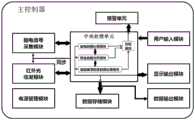
图2是图1中主控制器内部结构框图;Fig. 2 is a block diagram of the internal structure of the main controller in Fig. 1;
图3是图1中传感器排布示意图;Fig. 3 is a schematic diagram of sensor arrangement in Fig. 1;
图4是图1中主控制器的显示输出模块对外显示界面。FIG. 4 is an external display interface of the display output module of the main controller in FIG. 1 .
图中标识如下:The figure is marked as follows:
1-第一测量电极;2-第二测量电极;3-电三测量电极;4-第四测量电极; 5-接地电极;6-第一光源;7-第一光电管;8-第二光电管;9-第二光源;10- 第三光电管;20-第四光电管;30-主控制器;40-线缆;50-插头;60-柔性基 板;70-探头;80-第一参考电极;90-第二参考电极;a-左侧脑血氧饱和度采 集组;b-右侧脑血氧饱和度采集组。1-first measuring electrode; 2-second measuring electrode; 3-electric three-measuring electrode; 4-fourth measuring electrode; 5-ground electrode; 6-first light source; 7-first photocell; 8-second photocell; 9-second light source; 10-third photocell; 20-fourth photocell; 30-main controller; 40-cable; 50-plug; 60-flexible substrate; 70-probe; 80-th A reference electrode; 90 - the second reference electrode; a - the left cerebral blood oxygen saturation collection group; b - the right cerebral blood oxygen saturation collection group.
具体实施方式Detailed ways
下面将结合附图对本发明的技术方案进行清楚、完整地描述,显然, 所描述的实施例是本发明一部分实施例,而不是全部的实施例。基于本发 明中的实施例,本领域普通技术人员在没有做出创造性劳动前提下所获得 的所有其他实施例,都属于本发明保护的范围。The technical solutions of the present invention will be clearly and completely described below with reference to the accompanying drawings. Obviously, the described embodiments are part of the embodiments of the present invention, but not all of the embodiments. Based on the embodiments of the present invention, all other embodiments obtained by those of ordinary skill in the art without creative work fall within the protection scope of the present invention.
如图1、图2和图3所示,本发明提供了一种麻醉与意识深度监护系统, 包括柔性基板60和固定于柔性基板60上的麻醉深度传感器和脑血氧饱和 度传感器,系统还包括与麻醉深度传感器和脑血氧饱和度传感器通过线缆 连接的主控制器30,主控制器30如图2所示,包括:As shown in FIG. 1 , FIG. 2 and FIG. 3 , the present invention provides an anesthesia and consciousness depth monitoring system, including a
脑电信号采集模块,与麻醉深度传感器连接,用于向麻醉深度传感器 传输指令采集信号及接收麻醉深度传感器所采集到的脑电信号;The EEG signal acquisition module is connected with the anesthesia depth sensor, and is used for transmitting the instruction acquisition signal to the anesthesia depth sensor and receiving the EEG signal collected by the anesthesia depth sensor;
红外光收发模块,与脑血氧饱和度传感器连接,用于向脑血氧饱和度 传感器传输指令采集信号及接收脑血氧饱和度传感器所采集到的脑血氧信 号;The infrared light transceiver module is connected with the cerebral blood oxygen saturation sensor, and is used to transmit the command acquisition signal to the cerebral blood oxygen saturation sensor and receive the cerebral blood oxygen signal collected by the cerebral blood oxygen saturation sensor;
中央处理单元,用于向脑电信号采集模块和红外光收发模块发送同步 指令采集信号,接收经脑电信号采集模块处理的脑电采集数据及经红外光 收发模块处理的脑血氧采集数据,中央处理单元对脑电采集数据和脑血氧 采集数据进行同步数据处理,得到当前采集的脑电和脑血氧饱和度同步数 据;The central processing unit is used for sending synchronous command acquisition signals to the EEG signal acquisition module and the infrared light transceiver module, and for receiving the EEG acquisition data processed by the EEG signal acquisition module and the cerebral blood oxygen acquisition data processed by the infrared light transceiver module, The central processing unit performs synchronous data processing on the EEG collection data and the cerebral blood oxygen collection data to obtain the currently collected EEG and cerebral blood oxygen saturation synchronization data;
显示输出模块,用于显示脑电数据和脑血氧饱和度数据的数值和/或趋 势曲线;Display output module for displaying the value and/or trend curve of EEG data and cerebral blood oxygen saturation data;
用户输入模块,用于向中央处理单元输入用于脑电、脑血氧的监测信 息;The user input module is used to input monitoring information for EEG and cerebral blood oxygen to the central processing unit;
电源管理模块,用于给各个模块进行供电管理。The power management module is used to manage the power supply for each module.
中央处理单元中包括:The central processing unit includes:
脑电数据处理模块,其对所接收到的脑电采集数据进行数据分析,得 到当前采集的脑电数据;an EEG data processing module, which performs data analysis on the received EEG acquisition data to obtain the currently collected EEG data;
脑血氧饱和度数据处理模块,其对所接收到的脑血氧采集数据进行数 据分析,得到当前采集的脑血氧饱和度数据;a cerebral blood oxygen saturation data processing module, which performs data analysis on the received cerebral blood oxygen acquisition data to obtain the currently collected cerebral blood oxygen saturation data;
联合数据分析模块,结合同一时间点所得到的脑电数据和脑血氧饱和 度数据进行联合数据分析,得到神经血管耦合指标 (Neuro-VascularCouplingIndex,NVCI),其计算公式如下:The joint data analysis module, combined with the obtained EEG data and cerebral blood oxygen saturation data at the same time point, carries out joint data analysis to obtain the neurovascular coupling index (Neuro-Vascular Coupling Index, NVCI), and its calculation formula is as follows:
NVCI=f(α*BIS+β*rSo2)NVCI=f(α*BIS+β*rSo2 )
式中:BIS为脑电双频指数(Bispectral Index,BIS);In the formula: BIS is the Bispectral Index (BIS);
rSo2为脑血氧饱和度值;rSo2 is the cerebral blood oxygen saturation value;
α、β为权重系数。α and β are weight coefficients.
结合图3对贴附在患者额头部位的探头70结构进行具体说明。在柔性 基板60上设置的麻醉深度传感器包括用于检测前额左右侧眉骨上方外侧脑 电信号的第一测量电极1和第二测量电极2、用于检测前额左右侧眉骨上方 内侧脑电信号的第三测量电极3和第四测量电极4、位于第三测量电极3和 第四测量电极4之间的接地电极5,及用于检测前额左右侧太阳穴位置脑电 信号的第一参考电极80和第二参考电极90;柔性基板60上的脑血氧饱和 度传感器包括用于检测前额左侧的左侧脑血氧饱和度采集组a和用于检测 前额右侧的右侧脑血氧饱和度采集组b;其中的左侧脑血氧饱和度采集组a 包括第一光源6、第一光电管7和第二光电管8,右侧脑血氧饱和度采集组 b包括第二光源9、第三光电管10和第四光电管20;第一光源6设置于第 三测量电极3和参考电极5之间,第二光源9设置于第四测量电极4和参 考电极5之间,第一光电管7和第二光电管8呈间隔设置于第一测量电极1 和第三测量电极3之间,第三光电管10和第四光电管20呈间隔设置于第 二测量电极2和第四测量电极4之间。上述的各电极所形成的麻醉深度传 感器及各光源及光电管所形成的脑血氧饱和度采集组均可以采用现有技术, 这里不再对其结构原理进行赘述。The structure of the
为了便于采集脑电信号和脑血氧信号,在探头中,本发明设计了两组 传感器,分别可以采集左右侧脑半球的脑电信号和脑血氧信号。In order to facilitate the collection of EEG signals and cerebral blood oxygen signals, in the probe, the present invention designs two sets of sensors, which can respectively collect the EEG signals and cerebral blood oxygen signals of the left and right cerebral hemispheres.
第一测量电极1、第三测量电极3和参考电极5可以组成一个组合,采 集左侧脑半球BIS信号。第二测量电极2、第四测量电极4和参考电极5组 成另外一个组合,用来采集右侧脑半球的BIS信号。因此,共计五个电极 构成了探头中采集脑电的传感器。The first measuring electrode 1, the third measuring electrode 3 and the reference electrode 5 can form a combination to collect the BIS signal of the left cerebral hemisphere. The
脑血氧信号的采集由探头中光源和光电管组成,同样针对左右侧脑血 氧,本发明排布了两组脑血氧采集传感器。光源的作用是发射红光和红外 光进入颅内,光电管的作用是接收从头皮出射的红光和红外光并转换为电 信号。无论是脑电的电信号,还是脑血氧的电信号,都通过电缆再传输到 主控制器。The acquisition of the cerebral blood oxygen signal consists of a light source and a photocell in the probe, and also for the left and right cerebral blood oxygen, two sets of cerebral blood oxygen acquisition sensors are arranged in the present invention. The function of the light source is to emit red and infrared light into the skull, and the function of the photocell is to receive the red and infrared light emitted from the scalp and convert them into electrical signals. Whether it is the electrical signal of EEG or the electrical signal of cerebral blood oxygen, it is transmitted to the main controller through the cable.
本发明的主控制器30中还设有数据存储模块和数据输出模块,数据存 储模块用于实时接收脑电数据处理模块、脑血氧饱和度数据处理模块和联 合数据分析模块中的处理数据;数据输出模块用于输出脑电数据和脑血氧 饱和度数据。The
如图1所示,在具体使用时,首先是将探头70贴附在患者额头部位, 探头70跟主控制器30通过线缆40、插头50相连接。这里的主控制器具备 了输入、显示等功能,将监护得到的数据显示并存储下来。As shown in FIG. 1 , in specific use, the
主控制器30的主要功能模块如图2所示。The main functional modules of the
探头中的麻醉深度传感器传回脑电信号后由主控制器中的脑电信号采 集模块进行处理。脑电信号采集模块的主要功能包括信号放大、信号调理、 模数转换和数据发送。处理好的脑电信号发送到中央处理单元,经脑电数 据处理模块中的设定算法可以进行滤波、特征提取、参数计算等。脑电信 号处理得到BIS数据会进入缓存,同时脑电信号相关的计算数据会显示输 出模块中显示和在数据存储模块中存储,也可以通过数据输出模块进行发送,比如,经无线或有线传输给移动终端等。The anesthesia depth sensor in the probe sends back the EEG signal, which is processed by the EEG signal acquisition module in the main controller. The main functions of the EEG signal acquisition module include signal amplification, signal conditioning, analog-to-digital conversion and data transmission. The processed EEG signal is sent to the central processing unit, and filtering, feature extraction, parameter calculation, etc. can be performed through the setting algorithm in the EEG data processing module. The BIS data obtained from the EEG signal processing will enter the cache, and the calculation data related to the EEG signal will be displayed in the output module and stored in the data storage module, and can also be sent through the data output module, for example, via wireless or wired transmission. mobile terminal, etc.
对于脑血氧信号的采集,首先是主控制器中的红外光收发模块会发送 电信号到达探头,驱动探头中的光源发射红外光和红光进入头皮,穿过头 皮、颅骨、脑脊液等组织后,光信号进入脑组织。在脑组织中,含氧血红 蛋白和脱氧血红蛋白对光有不同程度的吸收。剩下的光再逆序通过脑脊液、 颅骨和头皮后出射出来。出射的光会被光电管接收。光电管的主要功能就 是完成光电转换,转化后的电信号由探头再发送到主控制器的红外光收发 模块,经红外光收发模块进行放大、模数转换和数据发送。处理好的信号 发送到中央处理单元。在中央处理单元中经脑血氧饱和度数据处理模块进 行信号滤波、脑血氧饱和度计算等,得到脑血氧饱和度数据,脑血氧饱和 度数据会进入缓存,同时脑血氧的数据会在显示输出模块中进行显示,在 数据存储模块中存储,也可以通过数据输出模块进行发送,比如,经无线 或有线传输给移动终端等。For the collection of cerebral blood oxygen signal, firstly, the infrared light transceiver module in the main controller will send electrical signals to the probe, drive the light source in the probe to emit infrared light and red light into the scalp, after passing through the scalp, skull, cerebrospinal fluid and other tissues , the light signal enters the brain tissue. In brain tissue, oxyhemoglobin and deoxyhemoglobin absorb light to varying degrees. The remaining light then exits through the cerebrospinal fluid, skull and scalp in reverse order. The emitted light is received by the photocell. The main function of the photocell is to complete the photoelectric conversion. The converted electrical signal is sent by the probe to the infrared light transceiver module of the main controller, and the infrared light transceiver module is used for amplification, analog-to-digital conversion and data transmission. The processed signal is sent to the central processing unit. In the central processing unit, the cerebral blood oxygen saturation data processing module performs signal filtering, cerebral blood oxygen saturation calculation, etc., to obtain the cerebral blood oxygen saturation data. The cerebral blood oxygen saturation data will enter the cache, and the cerebral blood oxygen data It will be displayed in the display output module, stored in the data storage module, and can also be sent through the data output module, such as wireless or wired transmission to the mobile terminal.
在脑电和脑血氧数据采集过程中,由于脑电信号的时间尺度是毫秒级, 而脑血氧的时间尺度是秒级,本发明为了保证这两种信号能够同步采集。 中央处理单元会发送同步采集信号到脑电信号采集模块和红外光收发模块, 通过这个同步信号实现这两种脑部信号的同步采集。具体地,在采集时可 以通过提高脑血氧采集频率与脑电采集频率一致,实现信号同步。也可以 是由中央处理单元向各传感器发送各自的采集信号,只在脑血氧采集的时 间点进行同步,此种方法可以减少脑血氧冗余数据的采集。参考图2所示, 中央处理单元根据时钟信号和系统设置,确定同步时间点。当同步时间点 信号到达时触发同步信号的输出,同步信号发送到脑电信号采集模块和红 外光收发模块,控制这两个模块中的模数转换或者同步输出,使得脑电信 号采集模块和红外光收发模块同时得到该时间点的数字信号,最终实现了 同一系统下两个模块中多路信号的同步。During the acquisition of EEG and cerebral blood oxygen data, since the time scale of EEG signals is milliseconds, and the time scale of cerebral blood oxygen is seconds, the present invention ensures that these two signals can be collected synchronously. The central processing unit will send a synchronous acquisition signal to the EEG signal acquisition module and the infrared light transceiver module, and the synchronous acquisition of these two brain signals is achieved through this synchronous signal. Specifically, signal synchronization can be achieved by increasing the cerebral blood oxygen acquisition frequency consistent with the EEG acquisition frequency during acquisition. Alternatively, the central processing unit can send its own acquisition signal to each sensor, and only synchronize at the time point of cerebral blood oxygen acquisition. This method can reduce the collection of cerebral blood oxygen redundant data. Referring to FIG. 2 , the central processing unit determines the synchronization time point according to the clock signal and the system setting. When the synchronization time point signal arrives, the output of the synchronization signal is triggered, and the synchronization signal is sent to the EEG signal acquisition module and the infrared light transceiver module to control the analog-to-digital conversion or synchronous output in these two modules, so that the EEG signal acquisition module and the infrared The optical transceiver module obtains the digital signal at the time point at the same time, and finally realizes the synchronization of multi-channel signals in the two modules under the same system.
在得到脑电的BIS数据、脑血氧的血氧饱和度数据之后,在中央处理 单元内可以启动数据联合分析的方法。所采用的计算公式如下:After obtaining the BIS data of the EEG and the blood oxygen saturation data of the cerebral blood oxygen, the method of data joint analysis can be started in the central processing unit. The calculation formula used is as follows:
NVCI=f(α1*lBIS+α2*rBIS+β1*lrSO2+β2*rrSO2)NVCI=f(α1 *lBIS+α2 *rBIS+β1 *lrSO2 +β2 *rrSO2 )
通过上述方程可以得到新的NVCI。具体实施过程具体如下:当中央处 理单元收到脑电信号采集模块采集到的脑电信号后,进行数据滤波、特征 提取等,并计算得到左侧BIS(lBIS)、右侧BIS(rBIS)。同时会得到来自 于红外光收发模块的信号,通过算法得到左侧脑血氧饱和度(lrSO2)和右侧 脑血氧饱和度(rrSO2),各个权重值则可以通过训练集数据进行确定,即 可通过上式得到新的参数NVCI数值。上述函数中综合考虑了左侧BIS(lBIS)、 右侧BIS(rBIS)、左侧脑血氧饱和度(lrSO2)和右侧脑血氧饱和度(rrSO2)。 综合多个参数在得到新的参数的方法有很多种,即f可以有很多种函数形式, 最简单的就是采用加权求和的方式进行。也可以采用人工智能的方法进行f 的判定,优选随机森林方法。The new NVCI can be obtained by the above equation. The specific implementation process is as follows: when the central processing unit receives the EEG signal collected by the EEG signal acquisition module, it performs data filtering, feature extraction, etc., and calculates the left BIS (lBIS) and the right BIS (rBIS). At the same time, the signal from the infrared light transceiver module will be obtained, and the left cerebral blood oxygen saturation (lrSO2 ) and the right cerebral blood oxygen saturation (rrSO2 ) will be obtained through the algorithm, and each weight value can be determined by the training set data. , the new parameter NVCI value can be obtained through the above formula. The left BIS (lBIS), the right BIS (rBIS), the left cerebral blood oxygen saturation (lrSO2 ) and the right cerebral blood oxygen saturation (rrSO2 ) are comprehensively considered in the above functions. There are many ways to synthesize multiple parameters to obtain new parameters, that is, f can have many functional forms, and the simplest method is to use weighted summation. Artificial intelligence method can also be used to determine f, preferably random forest method.
随机森林方法是一种比较新的机器学习模型。经典的机器学习模型是 神经网络,有半个多世纪的历史了,这里不再详细描述。The random forest method is a relatively new machine learning model. The classic machine learning model is the neural network, which has a history of more than half a century and will not be described in detail here.
在本发明中,采用随机森林方法时,通过建立左侧BIS(lBIS)、右侧 BIS(rBIS)、左侧脑血氧饱和度(lrSO2)和右侧脑血氧饱和度(rrSO2)的决 策树,各个权重值通过训练集数据进行确定,一旦决策树和各个决策树的 权重确定后,函数f也就确定下来了。等到新的数据输入后,根据这个随机 森林就可以确定NVCI结果。In the present invention, when the random forest method is adopted, the left BIS (lBIS), the right BIS (rBIS), the left cerebral blood oxygen saturation (lrSO2 ) and the right cerebral blood oxygen saturation (rrSO2 ) are established by The decision tree of , each weight value is determined by the training set data, once the decision tree and the weight of each decision tree are determined, the function f is also determined. After the new data is input, the NVCI result can be determined according to this random forest.
为了提醒医务人员注意患者的情况,本发明还在主控制器中设有报警 单元,中央处理单元中设有判别模块,在判别模块中设定脑电数据阈值、 脑血氧阈值及NVCI阈值,在得到脑电、脑血氧饱和度数值及NVCI数值之 后,中央处理单元会根据设定的阈值进行判别。如果数值超过阈值,就会 启动主控制器内的报警单元进行报警。警示信号会通过声音、光闪烁等多 种方式发出。In order to remind the medical staff to pay attention to the situation of the patient, the present invention is also provided with an alarm unit in the main controller, and a discrimination module is arranged in the central processing unit, in which the EEG data threshold, cerebral blood oxygen threshold and NVCI threshold are set, After obtaining the EEG, cerebral blood oxygen saturation value and NVCI value, the central processing unit will judge according to the set threshold. If the value exceeds the threshold, the alarm unit in the main controller will be activated to give an alarm. Warning signals will be issued in various ways such as sound, light flashing, etc.
如图4所示,在得到全部的数据之后,会将数据通过主控制器上的显 示界面显示出来。显示界面内可以对数据呈现不同的显示方式。最典型的 显示方式如图4所示,分别显示前额左侧脑区的BIS和脑血氧饱和度rSO2, 前额右侧脑区的BIS和脑血氧饱和度rSO2,以及NVCI值。在显示界面内, 左侧曲线对应的就是各个参数的趋势曲线,右侧显示的是各个参数的当前 数值,通过趋势曲线可以清楚看出各个脑参数的变化情况,准确获知患者 的麻醉与意识深度情况,避免过量麻醉所产生的负面作用,当显示数值超 出所设定的对应阈值时,通过发出警示提醒监护人员,非常有利于对患者 的术中监护。As shown in Figure 4, after getting all the data, the data will be displayed through the display interface on the main controller. The data can be displayed in different ways in the display interface. The most typical display method is shown in Figure 4, showing the BIS and cerebral oxygen saturation rSO2 of the left brain region of the forehead, the BIS and cerebral oxygen saturation rSO2 of the right brain region of the forehead, and the NVCI value respectively. In the display interface, the curve on the left corresponds to the trend curve of each parameter, and the curve on the right shows the current value of each parameter. Through the trend curve, the changes of each brain parameter can be clearly seen, and the depth of anesthesia and consciousness of the patient can be accurately known. When the displayed value exceeds the set corresponding threshold, it will alert the monitoring personnel by issuing a warning, which is very beneficial to the intraoperative monitoring of the patient.
显然,上述实施例仅仅是为清楚地说明所作的举例,而并非对实施方 式的限定。对于所属领域的普通技术人员来说,在上述说明的基础上还可 以做出其它不同形式的变化或变动。这里无需也无法对所有的实施方式予 以穷举。而由此所引伸出的显而易见的变化或变动仍处于本发明的保护范 围之中。Obviously, the above-mentioned embodiments are only examples for clear description, rather than limiting the implementation manner. For those of ordinary skill in the art, on the basis of the above description, other different forms of changes or modifications can also be made. There is no need and cannot be exhaustive of all implementations here. And the obvious changes or changes derived from this are still within the protection scope of the present invention.
Claims (13)
Priority Applications (1)
| Application Number | Priority Date | Filing Date | Title |
|---|---|---|---|
| CN202010296599.3ACN111481174B (en) | 2020-04-15 | 2020-04-15 | Anesthesia and consciousness depth monitoring system and method |
Applications Claiming Priority (1)
| Application Number | Priority Date | Filing Date | Title |
|---|---|---|---|
| CN202010296599.3ACN111481174B (en) | 2020-04-15 | 2020-04-15 | Anesthesia and consciousness depth monitoring system and method |
Publications (2)
| Publication Number | Publication Date |
|---|---|
| CN111481174Atrue CN111481174A (en) | 2020-08-04 |
| CN111481174B CN111481174B (en) | 2025-01-21 |
Family
ID=71796739
Family Applications (1)
| Application Number | Title | Priority Date | Filing Date |
|---|---|---|---|
| CN202010296599.3AActiveCN111481174B (en) | 2020-04-15 | 2020-04-15 | Anesthesia and consciousness depth monitoring system and method |
Country Status (1)
| Country | Link |
|---|---|
| CN (1) | CN111481174B (en) |
Cited By (9)
| Publication number | Priority date | Publication date | Assignee | Title |
|---|---|---|---|---|
| CN112137616A (en)* | 2020-09-22 | 2020-12-29 | 天津大学 | Consciousness detection device for multi-sense brain-body combined stimulation |
| CN113274035A (en)* | 2021-05-21 | 2021-08-20 | 李宏明 | Anesthesia depth monitoring system with wireless transmission function |
| CN114366030A (en)* | 2021-12-31 | 2022-04-19 | 中国科学院苏州生物医学工程技术研究所 | An intelligent auxiliary system and method for anesthesia surgery |
| CN114521871A (en)* | 2022-03-01 | 2022-05-24 | 上海市浦东医院(复旦大学附属浦东医院) | Medical system |
| CN114974566A (en)* | 2022-05-23 | 2022-08-30 | 国家康复辅具研究中心 | Cognitive function assessment method and system |
| CN115553765A (en)* | 2022-09-29 | 2023-01-03 | 中国人民解放军陆军军医大学 | Noninvasive real-time continuous cerebral blood oxygen monitoring device |
| CN116636817A (en)* | 2023-07-26 | 2023-08-25 | 四川新源生物电子科技有限公司 | Anesthesia depth evaluation method, anesthesia depth evaluation system, anesthesia depth evaluation device and storage medium |
| CN116763260A (en)* | 2023-08-21 | 2023-09-19 | 北京中医药大学 | Portable biological signal synchronous processing equipment and method |
| CN117814760A (en)* | 2024-03-04 | 2024-04-05 | 江西杰联医疗设备有限公司 | Anesthesia depth detection device based on multiple indexes and electronic equipment |
Citations (2)
| Publication number | Priority date | Publication date | Assignee | Title |
|---|---|---|---|---|
| EP0926984A1 (en)* | 1996-09-11 | 1999-07-07 | The University Court Of The University Of Glasgow | Anaesthesia control system |
| CN212438586U (en)* | 2020-04-15 | 2021-02-02 | 中科搏锐(北京)科技有限公司 | Anesthesia and consciousness depth monitoring system |
- 2020
- 2020-04-15CNCN202010296599.3Apatent/CN111481174B/enactiveActive
Patent Citations (2)
| Publication number | Priority date | Publication date | Assignee | Title |
|---|---|---|---|---|
| EP0926984A1 (en)* | 1996-09-11 | 1999-07-07 | The University Court Of The University Of Glasgow | Anaesthesia control system |
| CN212438586U (en)* | 2020-04-15 | 2021-02-02 | 中科搏锐(北京)科技有限公司 | Anesthesia and consciousness depth monitoring system |
Non-Patent Citations (1)
| Title |
|---|
| 胡凤艳;高明;李涛;李凯;: "脑电双频指数在临床麻醉中的应用现状", 中国实验诊断学, no. 04, 25 April 2019 (2019-04-25)* |
Cited By (15)
| Publication number | Priority date | Publication date | Assignee | Title |
|---|---|---|---|---|
| CN112137616B (en)* | 2020-09-22 | 2022-09-02 | 天津大学 | Consciousness detection device for multi-sense brain-body combined stimulation |
| CN112137616A (en)* | 2020-09-22 | 2020-12-29 | 天津大学 | Consciousness detection device for multi-sense brain-body combined stimulation |
| CN113274035A (en)* | 2021-05-21 | 2021-08-20 | 李宏明 | Anesthesia depth monitoring system with wireless transmission function |
| CN114366030B (en)* | 2021-12-31 | 2024-04-09 | 中国科学院苏州生物医学工程技术研究所 | Intelligent auxiliary system and method for anesthesia operation |
| CN114366030A (en)* | 2021-12-31 | 2022-04-19 | 中国科学院苏州生物医学工程技术研究所 | An intelligent auxiliary system and method for anesthesia surgery |
| CN114521871A (en)* | 2022-03-01 | 2022-05-24 | 上海市浦东医院(复旦大学附属浦东医院) | Medical system |
| CN114974566A (en)* | 2022-05-23 | 2022-08-30 | 国家康复辅具研究中心 | Cognitive function assessment method and system |
| CN114974566B (en)* | 2022-05-23 | 2023-03-28 | 国家康复辅具研究中心 | Cognitive function assessment method and system |
| CN115553765A (en)* | 2022-09-29 | 2023-01-03 | 中国人民解放军陆军军医大学 | Noninvasive real-time continuous cerebral blood oxygen monitoring device |
| CN116636817B (en)* | 2023-07-26 | 2023-11-03 | 四川新源生物电子科技有限公司 | Anesthesia depth assessment method, system, device and storage medium |
| CN116636817A (en)* | 2023-07-26 | 2023-08-25 | 四川新源生物电子科技有限公司 | Anesthesia depth evaluation method, anesthesia depth evaluation system, anesthesia depth evaluation device and storage medium |
| CN116763260A (en)* | 2023-08-21 | 2023-09-19 | 北京中医药大学 | Portable biological signal synchronous processing equipment and method |
| CN116763260B (en)* | 2023-08-21 | 2023-12-19 | 北京中医药大学 | Portable biological signal synchronization processing equipment and method |
| CN117814760A (en)* | 2024-03-04 | 2024-04-05 | 江西杰联医疗设备有限公司 | Anesthesia depth detection device based on multiple indexes and electronic equipment |
| CN117814760B (en)* | 2024-03-04 | 2024-05-17 | 江西杰联医疗设备有限公司 | Anesthesia depth detection device based on multiple indexes and electronic equipment |
Also Published As
| Publication number | Publication date |
|---|---|
| CN111481174B (en) | 2025-01-21 |
Similar Documents
| Publication | Publication Date | Title |
|---|---|---|
| CN111481174B (en) | Anesthesia and consciousness depth monitoring system and method | |
| US12383194B2 (en) | Depth of consciousness monitor | |
| CN112043287B (en) | Cerebral blood oxygen non-invasive monitoring method and monitoring device | |
| CN212438586U (en) | Anesthesia and consciousness depth monitoring system | |
| US8277385B2 (en) | Method and apparatus for non-invasive assessment of hemodynamic and functional state of the brain | |
| AU2017235723B2 (en) | Sleep apnea monitoring system | |
| US20050131288A1 (en) | Flexible, patient-worn, integrated, self-contained sensor systems for the acquisition and monitoring of physiologic data | |
| US20090024044A1 (en) | Data recording for patient status analysis | |
| US20070167694A1 (en) | Integrated Portable Anesthesia and Sedation Monitoring Apparatus | |
| CN201542615U (en) | Portable network vital sign monitoring instrument | |
| Jegan et al. | On the development of low power wearable devices for assessment of physiological vital parameters: a systematic review | |
| EP4356822A2 (en) | Sleep staging using an in-ear photoplethysmography (ppg) | |
| Li et al. | An IoT-Enabled Wearable Health Monitor for Synchronized ExG, PPG, and BioZ Measurement | |
| Zhang et al. | Twenty-four-hour ambulatory recording of cerebral hemodynamics, systemic hemodynamics, electrocardiography, and actigraphy during people’s daily activities | |
| Lee et al. | Measurement of motion activity during ambulatory using pulse oximeter and triaxial accelerometer | |
| CN118000676A (en) | Anesthesia management method based on brain function monitoring | |
| CN107822618A (en) | Non-intrusion type brain death check and evaluation instrument | |
| TWI494082B (en) | Multi anesthesia depth signal monitoring method | |
| CN208659382U (en) | A kind of monitor for muscle relaxation with oxygen saturation monitor function | |
| TWM521449U (en) | An anesthesia consciousness depth signal measuring device | |
| CN207837532U (en) | The recognition of face detecting system of sleep-apnea | |
| Jayadevappa et al. | Wireless monitoring and analysis of PPG signal for assessment of cardiovascular system in real time | |
| Kumar et al. | Wireless transmission of bio-medical signals: Wireless doc | |
| US12414725B2 (en) | Method and apparatus for detecting changes in blood flow in the head of a subject | |
| TWM451978U (en) | Multi anesthesia depth signal measuring device |
Legal Events
| Date | Code | Title | Description |
|---|---|---|---|
| PB01 | Publication | ||
| PB01 | Publication | ||
| SE01 | Entry into force of request for substantive examination | ||
| SE01 | Entry into force of request for substantive examination | ||
| GR01 | Patent grant | ||
| GR01 | Patent grant |