CN111465882A - Optical device, apparatus and system for assays - Google Patents
Optical device, apparatus and system for assaysDownload PDFInfo
- Publication number
- CN111465882A CN111465882ACN201880020973.8ACN201880020973ACN111465882ACN 111465882 ACN111465882 ACN 111465882ACN 201880020973 ACN201880020973 ACN 201880020973ACN 111465882 ACN111465882 ACN 111465882A
- Authority
- CN
- China
- Prior art keywords
- sample
- optical
- optical assembly
- light
- camera
- Prior art date
- Legal status (The legal status is an assumption and is not a legal conclusion. Google has not performed a legal analysis and makes no representation as to the accuracy of the status listed.)
- Granted
Links
Images


Classifications
- G—PHYSICS
- G01—MEASURING; TESTING
- G01N—INVESTIGATING OR ANALYSING MATERIALS BY DETERMINING THEIR CHEMICAL OR PHYSICAL PROPERTIES
- G01N21/00—Investigating or analysing materials by the use of optical means, i.e. using sub-millimetre waves, infrared, visible or ultraviolet light
- G01N21/84—Systems specially adapted for particular applications
- G01N21/8483—Investigating reagent band
- B—PERFORMING OPERATIONS; TRANSPORTING
- B01—PHYSICAL OR CHEMICAL PROCESSES OR APPARATUS IN GENERAL
- B01L—CHEMICAL OR PHYSICAL LABORATORY APPARATUS FOR GENERAL USE
- B01L3/00—Containers or dishes for laboratory use, e.g. laboratory glassware; Droppers
- B01L3/50—Containers for the purpose of retaining a material to be analysed, e.g. test tubes
- B01L3/502—Containers for the purpose of retaining a material to be analysed, e.g. test tubes with fluid transport, e.g. in multi-compartment structures
- B01L3/5027—Containers for the purpose of retaining a material to be analysed, e.g. test tubes with fluid transport, e.g. in multi-compartment structures by integrated microfluidic structures, i.e. dimensions of channels and chambers are such that surface tension forces are important, e.g. lab-on-a-chip
- B01L3/502715—Containers for the purpose of retaining a material to be analysed, e.g. test tubes with fluid transport, e.g. in multi-compartment structures by integrated microfluidic structures, i.e. dimensions of channels and chambers are such that surface tension forces are important, e.g. lab-on-a-chip characterised by interfacing components, e.g. fluidic, electrical, optical or mechanical interfaces
- B—PERFORMING OPERATIONS; TRANSPORTING
- B01—PHYSICAL OR CHEMICAL PROCESSES OR APPARATUS IN GENERAL
- B01L—CHEMICAL OR PHYSICAL LABORATORY APPARATUS FOR GENERAL USE
- B01L9/00—Supporting devices; Holding devices
- B01L9/52—Supports specially adapted for flat sample carriers, e.g. for plates, slides, chips
- G—PHYSICS
- G01—MEASURING; TESTING
- G01N—INVESTIGATING OR ANALYSING MATERIALS BY DETERMINING THEIR CHEMICAL OR PHYSICAL PROPERTIES
- G01N21/00—Investigating or analysing materials by the use of optical means, i.e. using sub-millimetre waves, infrared, visible or ultraviolet light
- G01N21/62—Systems in which the material investigated is excited whereby it emits light or causes a change in wavelength of the incident light
- G01N21/63—Systems in which the material investigated is excited whereby it emits light or causes a change in wavelength of the incident light optically excited
- G01N21/64—Fluorescence; Phosphorescence
- G01N21/645—Specially adapted constructive features of fluorimeters
- G01N21/6456—Spatial resolved fluorescence measurements; Imaging
- G01N21/6458—Fluorescence microscopy
- G—PHYSICS
- G01—MEASURING; TESTING
- G01N—INVESTIGATING OR ANALYSING MATERIALS BY DETERMINING THEIR CHEMICAL OR PHYSICAL PROPERTIES
- G01N21/00—Investigating or analysing materials by the use of optical means, i.e. using sub-millimetre waves, infrared, visible or ultraviolet light
- G01N21/75—Systems in which material is subjected to a chemical reaction, the progress or the result of the reaction being investigated
- G01N21/77—Systems in which material is subjected to a chemical reaction, the progress or the result of the reaction being investigated by observing the effect on a chemical indicator
- G01N21/78—Systems in which material is subjected to a chemical reaction, the progress or the result of the reaction being investigated by observing the effect on a chemical indicator producing a change of colour
- G—PHYSICS
- G01—MEASURING; TESTING
- G01N—INVESTIGATING OR ANALYSING MATERIALS BY DETERMINING THEIR CHEMICAL OR PHYSICAL PROPERTIES
- G01N21/00—Investigating or analysing materials by the use of optical means, i.e. using sub-millimetre waves, infrared, visible or ultraviolet light
- G01N21/84—Systems specially adapted for particular applications
- G—PHYSICS
- G01—MEASURING; TESTING
- G01N—INVESTIGATING OR ANALYSING MATERIALS BY DETERMINING THEIR CHEMICAL OR PHYSICAL PROPERTIES
- G01N33/00—Investigating or analysing materials by specific methods not covered by groups G01N1/00 - G01N31/00
- G01N33/48—Biological material, e.g. blood, urine; Haemocytometers
- G01N33/483—Physical analysis of biological material
- G—PHYSICS
- G02—OPTICS
- G02B—OPTICAL ELEMENTS, SYSTEMS OR APPARATUS
- G02B21/00—Microscopes
- G02B21/0004—Microscopes specially adapted for specific applications
- G02B21/0008—Microscopes having a simple construction, e.g. portable microscopes
- G—PHYSICS
- G02—OPTICS
- G02B—OPTICAL ELEMENTS, SYSTEMS OR APPARATUS
- G02B21/00—Microscopes
- G02B21/0004—Microscopes specially adapted for specific applications
- G02B21/002—Scanning microscopes
- G02B21/0024—Confocal scanning microscopes (CSOMs) or confocal "macroscopes"; Accessories which are not restricted to use with CSOMs, e.g. sample holders
- G02B21/0052—Optical details of the image generation
- G02B21/0076—Optical details of the image generation arrangements using fluorescence or luminescence
- G—PHYSICS
- G02—OPTICS
- G02B—OPTICAL ELEMENTS, SYSTEMS OR APPARATUS
- G02B21/00—Microscopes
- G02B21/06—Means for illuminating specimens
- G02B21/08—Condensers
- G02B21/12—Condensers affording bright-field illumination
- G—PHYSICS
- G02—OPTICS
- G02B—OPTICAL ELEMENTS, SYSTEMS OR APPARATUS
- G02B21/00—Microscopes
- G02B21/16—Microscopes adapted for ultraviolet illumination ; Fluorescence microscopes
- G—PHYSICS
- G02—OPTICS
- G02B—OPTICAL ELEMENTS, SYSTEMS OR APPARATUS
- G02B21/00—Microscopes
- G02B21/24—Base structure
- G—PHYSICS
- G02—OPTICS
- G02B—OPTICAL ELEMENTS, SYSTEMS OR APPARATUS
- G02B7/00—Mountings, adjusting means, or light-tight connections, for optical elements
- G02B7/02—Mountings, adjusting means, or light-tight connections, for optical elements for lenses
- G02B7/023—Mountings, adjusting means, or light-tight connections, for optical elements for lenses permitting adjustment
- H—ELECTRICITY
- H04—ELECTRIC COMMUNICATION TECHNIQUE
- H04N—PICTORIAL COMMUNICATION, e.g. TELEVISION
- H04N23/00—Cameras or camera modules comprising electronic image sensors; Control thereof
- H04N23/50—Constructional details
- H04N23/51—Housings
- H—ELECTRICITY
- H04—ELECTRIC COMMUNICATION TECHNIQUE
- H04N—PICTORIAL COMMUNICATION, e.g. TELEVISION
- H04N23/00—Cameras or camera modules comprising electronic image sensors; Control thereof
- H04N23/50—Constructional details
- H04N23/55—Optical parts specially adapted for electronic image sensors; Mounting thereof
- H—ELECTRICITY
- H04—ELECTRIC COMMUNICATION TECHNIQUE
- H04N—PICTORIAL COMMUNICATION, e.g. TELEVISION
- H04N23/00—Cameras or camera modules comprising electronic image sensors; Control thereof
- H04N23/56—Cameras or camera modules comprising electronic image sensors; Control thereof provided with illuminating means
- H—ELECTRICITY
- H04—ELECTRIC COMMUNICATION TECHNIQUE
- H04N—PICTORIAL COMMUNICATION, e.g. TELEVISION
- H04N23/00—Cameras or camera modules comprising electronic image sensors; Control thereof
- H04N23/57—Mechanical or electrical details of cameras or camera modules specially adapted for being embedded in other devices
- H—ELECTRICITY
- H04—ELECTRIC COMMUNICATION TECHNIQUE
- H04N—PICTORIAL COMMUNICATION, e.g. TELEVISION
- H04N23/00—Cameras or camera modules comprising electronic image sensors; Control thereof
- H04N23/60—Control of cameras or camera modules
- H04N23/67—Focus control based on electronic image sensor signals
- B—PERFORMING OPERATIONS; TRANSPORTING
- B01—PHYSICAL OR CHEMICAL PROCESSES OR APPARATUS IN GENERAL
- B01L—CHEMICAL OR PHYSICAL LABORATORY APPARATUS FOR GENERAL USE
- B01L2200/00—Solutions for specific problems relating to chemical or physical laboratory apparatus
- B01L2200/02—Adapting objects or devices to another
- B01L2200/025—Align devices or objects to ensure defined positions relative to each other
Landscapes
- Physics & Mathematics (AREA)
- Chemical & Material Sciences (AREA)
- Health & Medical Sciences (AREA)
- Analytical Chemistry (AREA)
- General Physics & Mathematics (AREA)
- Engineering & Computer Science (AREA)
- Life Sciences & Earth Sciences (AREA)
- Multimedia (AREA)
- Signal Processing (AREA)
- Optics & Photonics (AREA)
- General Health & Medical Sciences (AREA)
- Biochemistry (AREA)
- Pathology (AREA)
- Immunology (AREA)
- Chemical Kinetics & Catalysis (AREA)
- Hematology (AREA)
- Biomedical Technology (AREA)
- Clinical Laboratory Science (AREA)
- Molecular Biology (AREA)
- Biophysics (AREA)
- Plasma & Fusion (AREA)
- Nuclear Medicine, Radiotherapy & Molecular Imaging (AREA)
- Dispersion Chemistry (AREA)
- Urology & Nephrology (AREA)
- Food Science & Technology (AREA)
- Medicinal Chemistry (AREA)
- Investigating, Analyzing Materials By Fluorescence Or Luminescence (AREA)
- Microscoopes, Condenser (AREA)
- Investigating Or Analysing Materials By Optical Means (AREA)
Abstract
Translated fromChineseDescription
Translated fromChinese相关申请案的交叉引用CROSS-REFERENCE TO RELATED APPLICATIONS
本申请要求于2017年2月8日提交序列号为62/456,590、于2017年2月15日提交的序列号为62/459,554和于2017年2月16日提交的序列号为62/460,075、于2017年2月8日提交的序列号为62/456,504(ESX045PRV)、于2017年2月16日提交的序列号为62/460,062(ESX045PRV2)、于2017年2月9日提交美国的序列号为62/457,133(ESX046PRV)的美国临时专利申请的优先权,其全部内容通过引用并入本文用于所有目的。This application claims Serial Nos. 62/456,590, filed on February 8, 2017, 62/459,554, February 15, 2017, and 62/460,075, February 16, 2017, Serial No. 62/456,504 (ESX045PRV) filed on February 8, 2017, Serial No. 62/460,062 (ESX045PRV2), filed February 16, 2017, US Serial No. filed on February 9, 2017 Priority to US Provisional Patent Application 62/457,133 (ESX046PRV), the entire contents of which are incorporated herein by reference for all purposes.
技术领域technical field
其中本发明涉及进行生物和化学测定以及计算成像的装置和方法。Among other things, the present invention relates to apparatus and methods for performing biological and chemical assays and computational imaging.
背景技术Background technique
在生物和化学测定(例如诊断测试)中,通常需要简单、快速和灵敏的测定(包括成像)。本发明尤其提供了用于简单、快速和灵敏的测定(包括成像)的装置和方法。In biological and chemical assays, such as diagnostic tests, simple, rapid and sensitive assays (including imaging) are often desired. The present invention provides, inter alia, devices and methods for simple, rapid and sensitive assays, including imaging.
附图说明Description of drawings
本领域技术人员将理解,下面描述的附图仅用于说明的目的。附图不旨在以任何方式限制本发明的范围。附图不是完全按比例绘制的。在给出实验数据点的图中,连接数据点的线仅用于引导观察数据,而没有其他意义。Those skilled in the art will understand that the drawings described below are for illustration purposes only. The drawings are not intended to limit the scope of the invention in any way. The drawings are not drawn entirely to scale. In graphs showing experimental data points, the lines connecting the data points are only used to guide the observations and have no other meaning.
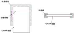
图1-A、1-B和1-C是根据本发明的一些实施方案所述的荧光照明模式下的系统测试样品的示意图。1-A, 1-B, and 1-C are schematic diagrams of system test samples in a fluorescent illumination mode according to some embodiments of the present invention.
图2-A、2-B和2-C是根据本发明的一些实施方案所述的明场照明模式下的系统测试样品的示意图。2-A, 2-B, and 2-C are schematic diagrams of system test samples in brightfield illumination mode according to some embodiments of the present invention.
图3是根据本发明的一些实施方案所述的系统和系统20中的光学适配器装置的示意性分解图。3 is a schematic exploded view of a system and an optical adapter arrangement in
图4是示出了根据本发明的一些实施方案所述的明场照明模式中的系统测试样品,特别是装置的细节的示意性截面图。Figure 4 is a schematic cross-sectional view showing details of a system test sample, particularly a device, in brightfield illumination mode according to some embodiments of the present invention.
图5是示出了根据本发明的一些实施方案所述的荧光照明模式下的系统测试样品,特别是装置的细节的示意性截面图。Figure 5 is a schematic cross-sectional view showing details of a system test sample, particularly a device, in a fluorescent illumination mode according to some embodiments of the present invention.
图6-A和图6-B是示出了根据本发明的一些实施方案在从装置向外拉动时使控制杆停止在预定位置的设计的示意性剖视图。6-A and 6-B are schematic cross-sectional views illustrating designs for stopping the lever in a predetermined position when pulled outward from the device, according to some embodiments of the present invention.
图7是根据本发明的一些实施方案所述的保持QMAX装置的样品滑块的结构的示意图。7 is a schematic illustration of the structure of a sample slide holding a QMAX device according to some embodiments of the present invention.
图8是根据本发明的一些实施方案在两个预定停止位置之间切换的活动臂的示意图。8 is a schematic diagram of a movable arm switching between two predetermined stop positions in accordance with some embodiments of the present invention.
图9是根据本发明的一些实施方案所述的滑块如何指示QMAX装置是否沿正确方向插入的示意图。9 is a schematic diagram of how a slider according to some embodiments of the present invention indicates whether a QMAX device is inserted in the correct orientation.
图10-A、10-B和10-C是根据本发明的一些实施方案所述的用于智能手机比色读取器的系统的示意图。Figures 10-A, 10-B and 10-C are schematic diagrams of a system for a smartphone colorimetric reader according to some embodiments of the present invention.
图11是根据本发明的一些实施方案所述的系统中的光学适配器装置的示意性分解图。11 is a schematic exploded view of an optical adapter device in a system according to some embodiments of the present invention.
图12是示出了根据本发明的一些实施方案所述的读取比色卡的系统的细节,特别是装置的细节的示意性截面图。Figure 12 is a schematic cross-sectional view showing details of a system for reading a colorimetric card, particularly a device, according to some embodiments of the present invention.
图13-A、13-B和13-C是根据本发明的一些实施方案所述的用于智能手机比色读取器的系统的示意图。Figures 13-A, 13-B and 13-C are schematic diagrams of a system for a smartphone colorimetric reader according to some embodiments of the present invention.
图14是根据本发明的一些实施方案所述的系统中的光学适配器装置的示意性分解图。14 is a schematic exploded view of an optical adapter device in a system according to some embodiments of the present invention.
图15-A、15-B和15-C是示出了根据本发明的一些实施方案所述的读取比色卡,特别是装置的系统的细节的示意图。Figures 15-A, 15-B and 15-C are schematic diagrams showing details of a system for reading a colorimetric card, particularly a device, according to some embodiments of the present invention.
图16-A示出了根据本发明的一些实施方案所述的由成像传感器、透镜和QMAX结构组成的断层摄影装置。Figure 16-A shows a tomography apparatus consisting of an imaging sensor, a lens, and a QMAX structure according to some embodiments of the present invention.
图16-B示出了字母E的柱阵列图案的实施例。Figure 16-B shows an example of a letter E column array pattern.
图16-C示出了薄透镜模型,其解释了焦距对捕获图像的影响。Figure 16-C shows a thin lens model explaining the effect of focal length on the captured image.
图16-D示出了由成像传感器拍摄的图16-B中的示例性柱阵列的图像。Figure 16-D shows an image of the exemplary column array in Figure 16-B taken by an imaging sensor.
图16-E示出了基于相位图像检索的方案的图。Figure 16-E shows a diagram of a phase image retrieval based scheme.
图17-A示出了根据本发明的一些实施方案所述的包括两个阶段、训练和预测的分析物检测和定位工作流程。Figure 17-A illustrates an analyte detection and localization workflow including two stages, training and prediction, according to some embodiments of the present invention.
图17-B示出了根据本发明的一些实施方案从排序列表中移除一个项目的过程。17-B illustrates the process of removing an item from a sorted list in accordance with some embodiments of the present invention.
图18-A示出了用于细胞成像的QMAX装置的实施方案。Figure 18-A shows an embodiment of a QMAX device for cellular imaging.
示例性实施方案所述的详细描述DETAILED DESCRIPTION OF EXEMPLARY EMBODIMENTS
以下详细描述通过示例而非限制的方式示出了本发明的一些实施方案。本文使用的章节标题和任何副标题仅用于组织目的,而不应被解释为以任何方式限制所描述的主题。章节标题和/或副标题下的内容不限于章节标题和/或副标题,而是适用于本发明的整个描述。The following detailed description shows, by way of example and not limitation, some embodiments of the invention. Section headings and any subheadings used herein are for organizational purposes only and should not be construed as limiting in any way the subject matter described. The content under the section headings and/or subheadings is not limited to the section headings and/or subheadings, but applies to the entire description of the invention.
任何出版物的引用是为了在申请日之前公开,并且不应被解释为承认本权利要求无权凭借在先发明而先于此类出版物。此外,所提供的公开日期可以不同于可能需要被独立地确认的实际的公开日期。The citation of any publication is for disclosure prior to the filing date and should not be construed as an admission that the claims are not entitled to antedate such publication by virtue of prior invention. Furthermore, the publication date provided may differ from the actual publication date that may need to be independently confirmed.
以下示出了七个示例性实施方案:附接到智能手机的用于明场和荧光显微成像的光学适配器的一个实施方案;使用倾斜的光纤端面作为光源附接到智能电话的用于色度测量的光学适配器的一个实施方案;附接到使用环形光纤的侧面照明作为光源的智能电话的用于色度测量的光学适配器的一个实施方案;断层摄像装置和方法的一个实施方案;机器学习辅助分析和成像的一个实施方案;组织染色和细胞成像的装置和方法的一个实施方案;双透镜成像系统的一个实施方案。Seven exemplary embodiments are shown below: one embodiment of an optical adapter for brightfield and fluorescence microscopy attached to a smartphone; an optical adapter for color-coding attached to a smartphone using an angled fiber end face as a light source An embodiment of an optical adapter for colorimetry; an embodiment of an optical adapter for colorimetry attached to a smartphone using side-illumination of a ring fiber as a light source; an embodiment of a tomography apparatus and method; machine learning One embodiment of an aided analysis and imaging; one embodiment of a device and method for tissue staining and cell imaging; one embodiment of a dual lens imaging system.
A.用于附接到智能电话上的明场和荧光显微镜的光学适配器A. Optical adapters for brightfield and fluorescence microscopes attached to smartphones
明场和荧光显微术是检验样品某些性质的非常有力的技术,在健康监测、疾病诊断、科学教育等方面有着广泛的应用。然而,传统上,拍摄显微图像需要昂贵的显微镜和有经验的人员,普通人无法做到。尽管有一些最近发明的能够将智能手机变成明场显微镜的配件,但这样的明场显微镜图像仅给出非常有限的样品信息。Brightfield and fluorescence microscopy are very powerful techniques for examining certain properties of samples and have a wide range of applications in health monitoring, disease diagnosis, science education, and more. Traditionally, however, taking microscopic images requires expensive microscopes and experienced personnel, which are beyond the reach of ordinary people. Although there are some recently invented accessories that can turn a smartphone into a brightfield microscope, such brightfield microscope images give only very limited information about the sample.
本文描述的本发明通过提供一种包括光学适配器和智能手机的系统来解决这个问题。所述光学适配器装置安装在智能手机上,将其转换为显微镜,该显微镜可以拍摄样品的荧光和明场图像。该系统可由普通人员在任何地点方便可靠地操作。光学适配器利用智能电话的现有资源,包括相机、光源、处理器和显示屏,这位用户提供了低成本进行明场和荧光显微术的解决方案。The invention described herein addresses this problem by providing a system that includes an optical adapter and a smartphone. The optical adapter unit is mounted on a smartphone, turning it into a microscope that can take fluorescence and brightfield images of the sample. The system can be easily and reliably operated by ordinary personnel from any location. The optical adapter leverages the smartphone's existing resources, including camera, light source, processor, and display, providing the user with a low-cost solution for brightfield and fluorescence microscopy.
在本发明中,光学适配装置包括配合在手机上部上的保持器框架和附接到该保持器的光学盒,其具有样品接收器槽和照明光学器件。在一些现有技术(美国专利号2004/029091和美国专利号2011/0292198)中,它们的光学适配器设计是包括安装在智能手机上的夹式机械部件和功能光学元件的整体件。这种设计存在的问题是,它们需要为每种特定型号的智能电话重新设计整体光学适配器。但是在本发明中,光学适配器被分成仅用于装配智能电话的保持框架和包含所有功能部件的通用光学盒。对于不同尺寸的智能手机,只要摄像头和光源的相对位置相同,只需重新设计固定架,节省了大量的设计和制造成本。In the present invention, the optical adapter includes a holder frame fitted on the upper part of the cell phone and an optical box attached to the holder, which has a sample receiver slot and illumination optics. In some of the prior art (US Pat. No. 2004/029091 and US Pat. No. 2011/0292198), their optical adapter designs are one-piece pieces that include clip-on mechanical components and functional optics for mounting on a smartphone. The problem with this design is that they require a redesign of the overall optical adapter for each particular model of smartphone. In the present invention, however, the optical adapter is divided into a holding frame only for assembling a smartphone and a general-purpose optical case containing all functional parts. For smartphones of different sizes, as long as the relative positions of the camera and the light source are the same, it is only necessary to redesign the fixing frame, which saves a lot of design and manufacturing costs.
该光学适配器的光学盒包含:接收器槽,其在该智能手机相机的视场和焦距范围内接收该样品并将该样品定位在样品载片中;用于捕获样品的明场显微图像的明场照明光学器件;荧光照明光学器件,其用于捕获样品的荧光显微图像;控制杆,其通过在该光学盒中向内和向外滑动而在明场照明光学器件与荧光照明光学器件之间切换。The optical box of the optical adapter contains: a receiver slot that receives the sample and positions the sample in the sample slide within the field of view and focal length of the smartphone camera; a receiver slot for capturing brightfield microscopy images of the sample Brightfield illumination optics; Fluorescence illumination optics for capturing fluorescence microscopic images of the sample; Control levers for brightfield illumination optics and fluorescence illumination optics by sliding inwards and outwards in the optical box switch between.
所述接收器槽具有附接到其上的橡胶门,该橡胶门可以完全覆盖槽以防止环境光进入光学盒而被相机收集。在现有技术(美国专利2016/0290916)中,其样品槽总是暴露于环境光,因为其仅进行明场显微术,所以不会引起太多的问题,。但是本发明在进行荧光显微镜检查时可以利用这种橡胶门,因为环境光会给相机的图像传感器带来很多噪声。The receiver slot has a rubber door attached to it that can completely cover the slot to prevent ambient light from entering the optics box and being collected by the camera. In the prior art (US Patent 2016/0290916), its sample cell is always exposed to ambient light, which does not cause too many problems since it only performs brightfield microscopy. But the present invention can take advantage of this rubber door when performing fluorescence microscopy, since ambient light can cause a lot of noise to the camera's image sensor.
为了捕获良好的荧光显微图像,希望几乎没有激发光进入相机,并且相机仅收集由样品发射的荧光。然而,对于所有普通的智能电话,由于光源发射的光束的大发散角,放置在相机前面的光学滤波器不能很好地阻挡从智能电话的光源发射的光中不需要的波长范围的光,并且光学滤波器对于未准直的光束不管用。可以设计准直光学器件已将由智能手机光源发射的进行准直以解决这个问题,但是这种方法增加了适配器的尺寸和成本。相反,在本发明中,荧光照明光学器件使得激发光能够部分地从样品载片内部的波导并且部分地从样品侧的后侧以大的倾斜入射角照射样品,使得激发光几乎不会被相机收集以减少进入相机的噪声信号。In order to capture good fluorescence microscopy images, it is desirable that little excitation light enters the camera, and that the camera only collects the fluorescence emitted by the sample. However, for all ordinary smartphones, due to the large divergence angle of the light beam emitted by the light source, an optical filter placed in front of the camera does not do a good job of blocking light in unwanted wavelength ranges from the light emitted from the smartphone's light source, and Optical filters do not work for uncollimated beams. Collimating optics can be designed to collimate the light emitted by the smartphone light source to address this problem, but this approach increases the size and cost of the adapter. In contrast, in the present invention, the fluorescence illumination optics enable the excitation light to illuminate the sample at a large oblique angle of incidence, partly from the waveguide inside the sample slide and partly from the backside of the sample side, so that the excitation light is hardly obstructed by the camera Collected to reduce the noise signal entering the camera.
适配器中的明场照明光学器件接收并转动由光源发射的光束,以便以垂直入射角对样品进行背光照明。Brightfield illumination optics in the adapter receive and rotate the light beam emitted by the light source to back-illuminate the sample at normal incidence.
典型地,所述光学盒还包含安装在其中的与智能电话的相机对准的透镜,其放大由相机捕获的图像。相机拍摄的图像可以由智能手机的处理器进一步处理,并在智能手机的屏幕上输出分析结果。Typically, the optical box also includes a lens mounted therein aligned with the smartphone's camera, which magnifies the image captured by the camera. The images captured by the camera can be further processed by the smartphone's processor and output the analysis results on the smartphone's screen.
为了在同一光学适配器中实现明场照明和荧光照明光学器件,在本发明中使用可滑动控制杆。荧光照明光学器件的光学元件安装在控制杆上,并且当控制杆完全滑入光学盒时,荧光照明光学元件阻挡明场照明光学器件的光路并且将照明光学器件切换到荧光照明光学器件。当控制杆滑出时,安装在控制杆上的荧光照明光学元件移出光路并切换到明场照明光学器件。这种控制杆设计使得光学适配器在明场和荧光照明模式下都工作,而不需要设计两个不同的单模光学盒。To achieve brightfield illumination and fluorescence illumination optics in the same optical adapter, a slidable lever is used in the present invention. The optics of the fluorescence illumination optics are mounted on the lever and when the lever is slid fully into the optics box, the fluorescence illumination optics block the light path of the brightfield illumination optics and switch the illumination optics to the fluorescence illumination optics. When the lever is slid out, the lever-mounted fluorescence illumination optics move out of the light path and switch to brightfield illumination optics. This lever design allows the optical adapter to work in both brightfield and fluorescence illumination modes without the need to design two different single-mode optical boxes.
控制杆包括在不同高度的不同平面处的两个平面。The joystick includes two planes at different planes at different heights.
在一些实施方案中,可以用垂直棒将两个平面连接在一起,并且一起移入或移出光学盒。在一些实施方案中,两个平面可以分开,并且每个平面可以单独移入或移出光学盒。In some embodiments, vertical rods can be used to connect the two planes together and move in and out of the optical box together. In some embodiments, the two planes can be separated, and each plane can be moved into or out of the optical box individually.
上部控制杆平面包含至少一个光学元件,该光学元件可以是但不限于滤波器。上部控制杆平面在光源下方移动,并且上部控制杆平面与光源之间的优选距离在0至5mm的范围内。The upper lever plane contains at least one optical element, which may be, but is not limited to, a filter. The upper lever plane moves below the light source, and the preferred distance between the upper lever plane and the light source is in the range of 0 to 5 mm.
底部控制杆平面的一部分不平行于图像平面。底部控制杆平面非平行部分表面镜面光洁度高,反射率大于95%。底部控制杆平面的非平行部分在光源下面移动,并且偏转从光源发射的光,以便向后照射相机正下方的样品区域。底部控制杆平面的非平行部分的优选倾斜角在45度到65度的范围内,并且倾斜角被定义为非平行底部平面和竖直平面之间的角度。Part of the bottom joystick plane is not parallel to the image plane. The surface of the non-parallel part of the bottom control rod plane has a high mirror finish and a reflectivity greater than 95%. The non-parallel portion of the bottom lever plane moves under the light source and deflects the light emitted from the light source so that it illuminates the sample area directly below the camera back. The preferred inclination angle of the non-parallel portion of the bottom lever plane is in the range of 45 degrees to 65 degrees, and the inclination angle is defined as the angle between the non-parallel bottom plane and the vertical plane.
底部控制杆平面的一部分平行于图像平面,并位于样品下方1mm至10mm处。底部控制杆平面的平行部分的表面是高度光吸收的,其中光吸收大于95%。该吸收表面用于消除以小入射角在样品上反向照射的反射光。A portion of the bottom lever plane is parallel to the image plane and lies 1 mm to 10 mm below the sample. The surface of the parallel portion of the bottom lever plane is highly light absorbing, with light absorption greater than 95%. This absorbing surface is used to eliminate reflected light backlit on the sample at small angles of incidence.
为了使用控制杆滑入和滑出以切换照明光学器件,使用包括球塞和控制杆上的凹槽的止动件设计,以便当从适配器向外拉动时将控制杆止动在预定位置。这允许用户使用任意的力拉动控制杆,但是使控制杆停止在固定位置,光学适配器的工作模式在该固定位置被切换到明场照明。In order to use the lever to slide in and out to switch the lighting optics, a stopper design including a ball plug and a groove on the lever is used to stop the lever in a predetermined position when pulled out from the adapter. This allows the user to pull the lever with any force, but stops the lever in a fixed position where the optical adapter's operating mode is switched to brightfield illumination.
样品滑块安装在所述接收器槽内以接收QMAX装置并将样品定位在智能手机相机的视场和焦点范围内的QMAX装置中。A sample slide is mounted within the receiver slot to receive the QMAX device and position the sample in the QMAX device within the field of view and focus of the smartphone camera.
所述样品滑块包括固定的轨道架和活动臂:The sample slider includes a fixed track frame and a movable arm:
所述框架轨道固定安装在光学盒的接收器槽中。所述轨道框架具有与QMAX装置的宽度和厚度相匹配的滑轨槽,使得QMAX装置能够沿着轨道滑动。轨道槽的宽度和高度被仔细地构造为使得QMAX装置在垂直于滑动平面中的滑动方向的方向上移动小于0.5mm,并且沿着QMAX装置的厚度方向移动小于0.2mm。The frame rails are fixedly mounted in the receiver slots of the optical box. The rail frame has rail grooves that match the width and thickness of the QMAX device so that the QMAX device can slide along the rail. The width and height of the track grooves are carefully constructed so that the QMAX device travels less than 0.5 mm in the direction perpendicular to the sliding direction in the sliding plane and less than 0.2 mm along the thickness of the QMAX device.
框架轨道在手机的相机的视野下具有开放的窗口,以允许光回照样品。The frame rail has an open window under the field of view of the phone's camera to allow light to back illuminate the sample.
活动臂预先设置在轨道框架的滑轨槽中,并与QMAX装置一起运动,以引导QMAX装置在轨道框架中的运动。The movable arm is pre-set in the sliding rail groove of the track frame, and moves together with the QMAX device to guide the movement of the QMAX device in the track frame.
活动臂装备有具有两个预定停止位置的止动机构。对于一个位置,臂将使QMAX装置停止在QMAX装置上的固定采样区域正好在智能电话的相机下面的位置。对于另一个位置,臂将使QMAX装置停止在QMAX装置上的采样区域在智能电话的视场之外的位置,并且QMAX装置可以容易地从轨道槽中取出。The movable arm is equipped with a stop mechanism with two predetermined stop positions. For one position, the arm will stop the QMAX unit at a location on the QMAX unit where the fixed sampling area is just below the smartphone's camera. For another position, the arm will stop the QMAX device where the sampling area on the QMAX device is outside the smartphone's field of view, and the QMAX device can be easily removed from the track slot.
通过将QMAX装置和活动臂一起按压到轨道槽的端部然后释放,活动臂在两个停止位置之间切换。By pressing the QMAX device together with the movable arm to the end of the track slot and then releasing, the movable arm switches between the two stop positions.
所述活动臂可以指示QMAX装置是否沿正确的方向插入。QMAX装置的一个角的形状被配置成不同于其他三个直角。所述活动臂的形状与所述一个角的特定形状相匹配,使QMAX装置只能沿正确方向滑动到轨道槽中的正确位置。The movable arm can indicate whether the QMAX device is inserted in the correct direction. The shape of one corner of the QMAX device is configured to be different from the other three right angles. The shape of the movable arm matches the specific shape of the one corner so that the QMAX device can only be slid into the correct position in the track slot in the correct direction.
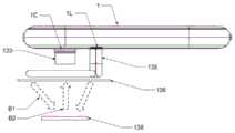
图1-A、1-B和1-C是在荧光照明模式下测试样品的系统19的示意图。具体地,图1-B和1-C分别是从前侧和后侧示出的系统19的分解图。系统19包含智能手机1;安装在智能电话1的上部上的光学适配器装置18;样品载片5,其插入到装置18的接收器槽4中,使得样品载片5上的样品位于智能手机1中的相机模块1C的视场和焦点范围内。控制杆8完全压入装置18中,使得系统19以荧光照明模式操作。在样品载片5进入之后,连接到装置18的橡胶门16盖住接收器槽4,以防止环境光进入接收器槽4而影响测试。1-A, 1-B, and 1-C are schematic diagrams of a
安装在智能电话1中的软件(未示出)在智能电话1中的光源1L发光的同时分析由相机模块1C收集的图像,以便获得样品的一些属性,并且将结果输出到智能电话1中的显示屏1f。Software (not shown) installed in the
图2-A、2-B和2-C是在明场照明模式下测试样品的系统20的示意图。具体地,图2-B和2-C分别是从前侧和后侧示出的系统20的分解图。系统20包含智能手机1;安装在智能电话1的上部上的光学适配器装置18;样品载片5,其插入到装置18的接收器槽4中,使得样品载片5上的样品位于智能手机1中的相机模块1C的视场和焦点范围内。控制杆8从装置18中向外拉动并由在装置18中的预定位置处的制动器(未示出)停止,使得系统20在明场照明模式下操作。2-A, 2-B, and 2-C are schematic diagrams of a
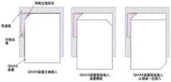
图3是系统19和系统20中的光学适配器装置18的示意性分解图。装置18包括安装在智能手机1上部的支架壳体2;附接到壳体2的光学盒3,包括接收器槽4、光学室3C、允许控制杆8滑入的轨道6b和6t,以及插入沟槽4s中以覆盖所述接收器槽4的橡胶门16。光学插件7装配到光学室3C的顶部,其中的出口光圈7L和进口光圈7C与智能电话1中的光源1L和相机1C对准(如图2-B所示)。透镜11安装在光学插件7中的进口光圈7C中,并且构造成使得插入所述接收器槽4中的样品载片5中的样品位于相机1C的工作距离内(如图2-B和1-B所示)。透镜11用于放大由相机1C(如图2-B和1-B所示)捕获的样品的图像。长通光学滤波器12安装在透镜11顶部的进口光圈7C中。一对直角反射镜13和14安装在光学室3C的底部上,并且被配置为使得反射镜13和反射镜14与光源1L和相机1C分别对准(如图2-B和1-B所示)。在下面的图4中描述与装置18中的明场照明光学器件的操作一样的反射镜13和反射镜14。控制杆8包括两个水平杆:上水平杆包括安装在槽8a中的带通滤波器15,而下水平杆包括安装在水平平面8b上的光吸收器9和安装在倾斜平面8c上的反射镜10。在下面的图5中描述操作与装置18中的荧光照明光学器件一样的滤波器15、光吸收器9和反射镜10。控制杆8的上水平杆在盒子3中沿轨道6t滑动,而下水平杆8b和8c在盒子3中沿轨道6b滑动。控制杆8停止在盒3中的两个不同位置,以在明场照明光学器件和荧光照明光学器件之间切换。控制杆8完全插入盒3中以切换装置18与荧光照明光学器件一起工作。球塞17安装在轨道6t的侧壁上,以便当控制杆8从箱3向外拉到开关装置18以与明场照明光学器件一起工作时,使控制杆8停止在预定位置。FIG. 3 is a schematic exploded view of
图4是示出在明场照明模式下测试样品的系统20的细节,特别是装置18的细节的示意性截面图。该图示出了以上参考图3描述的元件的功能。从装置18向外拉动控制杆8(如图3所示)并且由止动件17停止(如图3所示)在预定位置处,使得反射镜13和反射镜14暴露于相机1C和光源1L并与相机1C和光源1L对准。光源1L远离智能手机1发射光束BB1。光束BB1被反射镜14偏转90度至光束BB2,该光束BB2被反射镜13进一步偏转90度至光束BB3。光束BB3以垂直入射角反向照射样品载片5中的样品。透镜11在相机1C的图像传感器平面上产生样品的放大图像。智能手机1捕获并处理图像以获得样品的某些属性。FIG. 4 is a schematic cross-sectional view showing details of the
图5是示出在荧光照明模式下测试样品的系统19的细节,特别是装置18的细节的示意性截面图。该图示出了以上参考图3描述的元件的功能。控制杆8(如图3所示)完全插入到装置18中,使得光吸收器9和倾斜反射镜10处于相机1C和光源1L的视野下,并且阻挡光源1L与反射镜对13和14之间的光路。带通滤波器15位于光源1L正下方。光源1L远离智能手机1发射光束BF1。滤波器15允许具有与样品载片5中的荧光样品的激发波长相匹配的特定波长范围的光束BF1通过。光束BF1的一部分照射在透明样品载片5的边缘上,并且联接到在样品载片5中行进的波导光束BF3,并且照射透镜11下面的样品区域。光束BF1的一部分照射在反射镜10上。倾斜反射镜10将光束BF1偏转到光束BF2,并以大的倾斜角向后照射透镜11正下方的样品载片5中的样品区域。具有大发散角的光束BF1的剩余部分(即光束BF4)照射在吸收器9上并被吸收,使得光束BF4的反射光不会以小入射角进入相机1C。来自透镜11下面的样品区域的光穿过透镜11并被长通滤波器12滤波,使得只有样品载片5中的荧光样品发射的特定波长范围内的光进入相机1C以形成图像。智能手机1捕获并处理图像以获得样品的某些属性。橡胶门16插入装置18中以覆盖样品载片5,以防止环境光进入装置18影响测试。Figure 5 is a schematic cross-sectional view showing details of the
图6-A和图6-B是示意性剖视图,示出了当从装置18向外拉动控制杆8时使控制杆8停止在预定位置的设计。球塞17安装在轨道槽6t的侧壁中,并且在控制杆8的侧壁上钻出凹槽8g,该凹槽8g的形状与球塞17中的球的形状相匹配。当从装置18向外拉动控制杆8并且还没有到达如图2所示的预定位置时,如图6-A所示,球塞17中的球被控制杆8的侧壁压入其主体中,从而控制杆8可以沿着轨道6t滑动。如图6-B所示,当控制杆8上的凹槽8g到达球塞17的位置时,球塞17中的球跳到凹槽8g中以让控制杆8停止。FIGS. 6-A and 6-B are schematic cross-sectional views showing designs for stopping the
图7是保持QMAX装置的样品滑块的结构的示意图。样品滑块包含:轨道框架,其具有轨道槽以使QMAX装置沿其滑动;活动臂,其预先设置在轨道槽中,与QMAX装置一起移动以引导其移动。活动臂装备有止动机构,以使QMAX装置停止在两个预定的停止位置处。轨道槽的宽度和高度被仔细地构造成使得QMAX装置在垂直于滑动平面中的滑动方向的方向上移动小于0.5mm,并且沿着QMAX装置的厚度方向移动小于0.2mm。FIG. 7 is a schematic diagram of the structure of the sample slide holding the QMAX device. The sample slide includes: a track frame with track grooves to slide the QMAX device along it; and a movable arm, pre-set in the track groove, that moves with the QMAX device to guide its movement. The movable arm is equipped with a stop mechanism to stop the QMAX device at two predetermined stop positions. The width and height of the track grooves are carefully constructed so that the QMAX device travels less than 0.5 mm in the direction perpendicular to the sliding direction in the sliding plane and less than 0.2 mm along the thickness of the QMAX device.
图8是活动臂在两个预定停止位置之间切换的示意图。通过将QMAX装置和活动臂一起按压到轨道槽的端部然后释放,QMAX卡可以停止在位置1或位置2,在位置1,样品区域在用于容易地从滑块取出QMAX装置的智能电话相机的视野之外,在位置2,样品区域正好在用于捕获图像的智能电话相机的视野之下。Figure 8 is a schematic diagram of the movable arm switching between two predetermined stop positions. By pressing the QMAX device together with the movable arm to the end of the track slot and then releasing, the QMAX card can be stopped in
图9是滑块如何指示QMAX装置是否沿正确方向插入的示意图。QMAX装置的一个角的形状被配置成不同于其他三个直角。动臂的形状与特定形状的转角形状相匹配,使QMAX装置只能沿正确方向滑动到轨道槽中的正确位置。如果从错误侧翻转或插入QMAX装置,则滑块外部的QMAX装置部分比正确插入QMAX装置时更长。Figure 9 is a schematic diagram of how the slider indicates whether the QMAX device is inserted in the correct orientation. The shape of one corner of the QMAX device is configured to be different from the other three right angles. The shape of the boom matches the shape of the specific shaped corners so that the QMAX unit can only slide in the correct direction into the correct position in the track slot. If the QMAX unit is turned over or inserted from the wrong side, the portion of the QMAX unit outside the slider will be longer than when the QMAX unit was inserted correctly.
当荧光图像和明场图像都可用时,可以利用荧光图像的知识来处理明场图像,或者利用明场图像的知识来处理荧光图像,或者共同处理两个图像。荧光图像和明场图像的视场可以不同;因此,两个图像不是像素到像素地空间对准的。When both fluorescence images and brightfield images are available, knowledge of the fluorescence image can be used to process the brightfield image, knowledge of the brightfield image can be used to process the fluorescence image, or both images can be processed together. The field of view of the fluorescence image and the brightfield image can be different; thus, the two images are not spatially aligned pixel-to-pixel.
为了解决荧光图像和明场图像之间的不对准,可以将图像配准应用于这两个图像。图像配准找到使从一个图像到另一个图像的空间位置相关的几何变换。各种图像配准算法可用于对准荧光图像和明场图像,包含但不限于基于特征点、基于互相关、基于傅立叶对准等。图像配准输出将一个图像的空间位置(坐标)映射到另一个图像的几何变换。To resolve the misalignment between the fluorescence image and the brightfield image, image registration can be applied to both images. Image registration finds geometric transformations that correlate spatial positions from one image to another. Various image registration algorithms can be used to align the fluorescence and brightfield images, including but not limited to feature point-based, cross-correlation-based, Fourier-based alignment, and the like. Image registration outputs a geometric transformation that maps the spatial location (coordinates) of one image to another.
在荧光图像和明场图像对准之后,可以利用来自两个图像的信息来改进一个图像的处理,或者共同处理两个图像。After the fluorescence and brightfield images are aligned, information from both images can be used to improve processing of one image, or to process both images together.
实施例:Example:
A1.一种光学适配器,包含:A1. An optical adapter comprising:
i.支架框架,以及i. Bracket frame, and
ii.可移除地附接至支架框架上的光学盒,ii. an optical box removably attached to the stand frame,
其中支架框架被配置成用于可移除地装配在一个移动装置上并且将光学盒与整合在移动装置中的相机和照明源对齐;wherein the stand frame is configured for removably mounting on a mobile device and aligning the optical box with a camera and illumination source integrated in the mobile device;
其中光学盒包含样品接收器槽和照明光学器件。The optics box contains the sample receiver well and illumination optics.
B1.一种光学系统,包含:B1. An optical system, comprising:
i.实施方案A1的光学适配器;以及i. The optical adapter of Embodiment Al; and
ii.QMAX卡,其包含第一板和第二板,其中第一板和第二板将液体样品压缩成小于200μm的均匀厚度层;以及ii. a QMAX card comprising a first plate and a second plate, wherein the first plate and the second plate compress the liquid sample into a layer of uniform thickness less than 200 μm; and
iii.滑块,其被配置成容纳QMAX卡并且被插入到所述光盒中。iii. A slider configured to receive a QMAX card and inserted into the optical box.
C1.根据前述任一实施方案所述的适配器或系统,其中移动装置是智能电话。C1. The adapter or system of any preceding embodiment, wherein the mobile device is a smartphone.
C2.根据前述任一实施方案所述的适配器或系统,其中保持器框架包含构造成可替换为用于不同移动装置的具有不同尺寸的其他保持器壳体的保持器壳体。C2. The adapter or system of any preceding embodiment, wherein the holder frame includes a holder housing configured to be replaceable with other holder housings of different sizes for different mobile devices.
C3.根据前述任一实施方案所述的适配器或系统,其中保持器框架的大小适于将光学适配器可移除地装配到移动装置的上部。C3. The adapter or system of any preceding embodiment, wherein the holder frame is sized to removably fit the optical adapter to the upper portion of the mobile device.
C4.根据前述任一实施方案所述的适配器或系统,其中光学适配器的光学盒包含:C4. The adapter or system of any preceding embodiment, wherein the optical case of the optical adapter comprises:
i.接收器槽,其被配置成用于在相机的视场和焦点范围中将QMAX卡接收和定位在样品载片中;i. a receiver slot configured to receive and position the QMAX card in the sample slide within the camera's field of view and focus;
ii.明场照明光学器件,其被配置成捕获样品的明场显微图像;ii. Brightfield illumination optics configured to capture brightfield microscopic images of the sample;
iii.荧光照明光学器件,其被配置成捕获样品的荧光显微图像;以及iii. Fluorescence illumination optics configured to capture a fluorescence microscopic image of the sample; and
iv.控制杆,其被配置成通过在光学箱中向内和向外滑动而在明场照明光学器件与荧光照明光学器件之间切换。iv. A lever configured to switch between brightfield illumination optics and fluorescence illumination optics by sliding inward and outward in the optics box.
C5.根据前述任一实施方案所述的适配器或系统,其中接收器槽包含可完全覆盖槽以防止环境光进入光盒中以由相机收集的橡胶门。C5. The adapter or system of any preceding embodiment, wherein the receiver slot includes a rubber door that fully covers the slot to prevent ambient light from entering the light box for collection by the camera.
C6.根据前述任一实施方案所述的适配器或系统,其中适配器中的明场照明光学器件构造成接收并转向由光源发射的光束,以便以垂直入射角对样品进行背照明C6. The adapter or system of any preceding embodiment, wherein the brightfield illumination optics in the adapter are configured to receive and turn the light beam emitted by the light source to back-illuminate the sample at a normal angle of incidence
C7.根据前述任一实施方案所述的适配器或系统,其中光学盒进一步包含安装在其中且与移动装置的相机对准的透镜,该透镜放大由相机捕获的图像。C7. The adapter or system of any preceding embodiment, wherein the optical box further comprises a lens mounted therein and aligned with a camera of the mobile device, the lens magnifying an image captured by the camera.
C8.根据前述任一实施方案所述的适配器或系统,其中由相机捕获的图像进一步由移动装置的处理器处理且在移动装置的屏幕上输出分析结果。C8. The adapter or system of any preceding embodiment, wherein the images captured by the camera are further processed by the mobile device's processor and the analysis results are output on the mobile device's screen.
C9.根据前述任一实施方案所述的适配器或系统,其中控制杆是可滑动的,并且被配置成在同一光学适配器中实现明场照明和荧光照明光学器件两者。C9. The adapter or system of any preceding embodiment, wherein the lever is slidable and configured to implement both brightfield illumination and fluorescence illumination optics in the same optical adapter.
C10.根据前述任一实施方案所述的适配器或系统,其中荧光照明光学器件的光学元件安装在控制杆上,并且当控制杆完全滑动到光学盒中时,C10. The adapter or system of any preceding embodiment, wherein the optical element of the fluorescent illumination optics is mounted on a lever, and when the lever is fully slid into the optics box,
C11.根据前述任一实施方案所述的适配器或系统,其中具有荧光照明光学元件的控制杆阻挡明场照明光学器件的光路并且将照明光学器件切换到荧光照明光学器件。C11. The adapter or system of any preceding embodiment, wherein the lever with the fluorescence illumination optics blocks the light path of the brightfield illumination optics and switches the illumination optics to the fluorescence illumination optics.
C12.根据前述任一实施方案所述的适配器或系统,其中当控制杆滑出时,安装在控制杆上的荧光照明光学元件移出光学路径并将照明光学器件切换到明场照明光学器件。C12. The adapter or system of any preceding embodiment, wherein when the lever is slid out, the lever-mounted fluorescent illumination optics move out of the optical path and switch the illumination optics to brightfield illumination optics.
C13.根据前述任一实施方案所述的适配器或系统,其中控制杆包含处于不同高度的两个平面。C13. The adapter or system of any preceding embodiment, wherein the lever comprises two planes at different heights.
C14.根据前述任一实施方案所述的适配器或系统,其中两个平面通过竖直杆连接在一起,并且一起移入或移出光学盒。C14. The adapter or system of any preceding embodiment, wherein the two planes are connected together by a vertical rod and move in and out of the optical box together.
C15.根据前述任一实施方案所述的适配器或系统,其中两个平面可分离且每一平面可个别地移入或移出光学盒。C15. The adapter or system of any preceding embodiment, wherein the two planes are separable and each plane is individually moveable into or out of the optical box.
C16.根据前述任一实施方案所述的适配器或系统,其中上部控制杆平面包括至少一个光学元件,该光学元件可以是但不限于滤波器。C16. The adapter or system of any preceding embodiment, wherein the upper lever plane includes at least one optical element, which may be, but is not limited to, a filter.
C17.根据前述任一实施方案所述的适配器或系统,其中上部杆平面在光源下方移动,并且上部杆平面与光源之间的优选距离在0至5mm的范围内。C17. The adapter or system of any preceding embodiment, wherein the upper rod plane moves below the light source and the preferred distance between the upper rod plane and the light source is in the range of 0 to 5 mm.
C18.根据前述任一实施方案所述的适配器或系统,其中底部控制杆平面的一部分不平行于图像平面。C18. The adapter or system of any preceding embodiment, wherein a portion of the bottom lever plane is not parallel to the image plane.
C19.根据前述任一实施方案所述的适配器或系统,其中底部控制杆平面的非平行部分的表面具有反射率大于95%的镜面光洁度。C19. The adapter or system of any preceding embodiment, wherein the surface of the non-parallel portion of the bottom lever plane has a specular finish with a reflectivity greater than 95%.
C20.根据前述任一实施方案所述的适配器或系统,其中底部控制杆平面的非平行部分在光源下方移动且使从光源发射的光偏转以在相机正下方对样品区域进行反向照明。C20. The adapter or system of any preceding embodiment, wherein the non-parallel portion of the bottom lever plane moves under the light source and deflects light emitted from the light source to back-illuminate the sample area directly below the camera.
C21.根据任何前述实施例的适配器或系统,其中底部控制杆平面的非平行部分的优选倾斜角在45度到65度的范围内,并且倾斜角被定义为非平行底部平面和竖直平面之间的角度。C21. The adapter or system of any preceding embodiment, wherein the preferred angle of inclination of the non-parallel portion of the bottom lever plane is in the range of 45 degrees to 65 degrees, and the angle of inclination is defined as the difference between the non-parallel bottom plane and the vertical plane angle between.
C22.根据前述任一实施方案所述的适配器或系统,其中底部控制杆平面的一部分平行于图像平面,且位于样品下方且远离样品1mm到10mm处。C22. The adapter or system of any of the preceding embodiments, wherein a portion of the bottom lever plane is parallel to the image plane and is located below and 1 mm to 10 mm away from the sample.
C23.根据前述任一实施方案所述的适配器或系统,其中底部控制杆平面的平行部分的表面是高度光吸收的,具有大于95%的光吸收。C23. The adapter or system of any preceding embodiment, wherein the surface of the parallel portion of the bottom lever plane is highly light absorbing, having greater than 95% light absorption.
C24.根据前述任一实施方案所述的适配器或系统,其中吸收表面用于消除以小入射角在样品上反向照射的反射光。C24. The adapter or system of any of the preceding embodiments, wherein an absorbing surface is used to eliminate reflected light backlit on the sample at small angles of incidence.
C25.根据前述任一实施方案所述的适配器或系统,其中控制杆包括构造成使杆停止的止动件。C25. The adapter or system of any preceding embodiment, wherein the lever includes a stop configured to stop the lever.
C26.根据前述任一实施方案所述的适配器或系统,其中止挡件包含球塞,并且使用控制杆上的凹槽,以便在从适配器向外拉动时将控制杆停止在预定位置处。C26. The adapter or system of any preceding embodiment, wherein the stop comprises a ball plug and uses a groove on the lever to stop the lever at a predetermined position when pulled outward from the adapter.
C27.根据前述任一实施方案所述的适配器或系统,其中止挡件构造成允许用户使用任意力来拉动控制杆,但使控制杆停止在固定位置处,光学适配器的工作模式在该固定位置处切换到明场照明。C27. The adapter or system of any preceding embodiment, wherein the stopper is configured to allow the user to pull the lever with any force, but stop the lever at a fixed position where the optical adapter's operating mode is Switch to brightfield illumination.
C28.根据前述任一实施方案所述的适配器或系统,其中样品滑块安装在接收器槽内以接收QMAX装置并将样品定位在智能手机相机的视场和焦点范围内的QMAX装置中。C28. The adapter or system of any preceding embodiment, wherein the sample slide is mounted in the receiver slot to receive the QMAX device and position the sample in the QMAX device within the field of view and focus of the smartphone camera.
C29.根据前述任一实施方案所述的适配器或系统,其中通过将QMAX装置和活动臂一起按压到轨道槽的端部然后释放,该活动臂在两个停止位置之间切换。C29. The adapter or system of any preceding embodiment, wherein the movable arm switches between two rest positions by pressing the QMAX device together with the movable arm to the end of the track slot and then releasing.
C30.根据前述任一实施方案所述的适配器或系统,其中活动臂可指示QMAX装置是否沿正确方向插入。C30. The adapter or system of any preceding embodiment, wherein the movable arm indicates whether the QMAX device is inserted in the correct orientation.
C31.根据前述任一实施方案所述的适配器或系统,其中QMAX装置的一个角的形状构造成不同于其他三个直角转角。C31. The adapter or system of any preceding embodiment, wherein one corner of the QMAX device is shaped to be different from the other three right angle corners.
C31.活动臂的形状与特定形状的转角形状相匹配,使QMAX装置只能沿正确方向滑动到轨道槽中的正确位置处。C31. The shape of the movable arm matches the shape of the corner of the specific shape, so that the QMAX device can only slide in the correct direction to the correct position in the track groove.
C32.根据前述任一实施方案所述的适配器或系统,其中样品滑块包含固定轨道框架和活动臂:C32. The adapter or system of any preceding embodiment, wherein the sample slide comprises a fixed track frame and a movable arm:
C33.根据前述任一实施方案所述的适配器或系统,其中框架轨道固定地安装在光学盒的接收器槽中;轨道框架具有与QMAX装置的宽度和厚度相匹配的滑轨槽,使得QMAX装置能够沿着轨道滑动。轨道槽的宽度和高度被仔细地构造为使得QMAX装置在垂直于滑动平面中的滑动方向的方向上移动小于0.5mm,并且沿着QMAX装置的厚度方向移动小于0.2mm。C33. The adapter or system of any one of the preceding embodiments, wherein the frame rails are fixedly mounted in the receiver slots of the optical box; the rail frame has slide rail slots that match the width and thickness of the QMAX device such that the QMAX device Able to slide along the track. The width and height of the track grooves are carefully constructed so that the QMAX device travels less than 0.5 mm in the direction perpendicular to the sliding direction in the sliding plane and less than 0.2 mm along the thickness of the QMAX device.
C34.根据前述任一实施方案所述的适配器或系统,其中框架轨道在智能电话的相机的视场下方具有开放的窗口以允许光背照样品。C34. The adapter or system of any preceding embodiment, wherein the frame rail has an open window below the smartphone's camera's field of view to allow light to back-illuminate the sample.
C35.根据前述任一实施方案所述的适配器或系统,其中活动臂预先构建在轨道框架的滑轨槽中,且与QMAX装置一起移动以引导QMAX装置在轨道框架中的移动。C35. The adapter or system of any preceding embodiment, wherein the movable arm is pre-built in a rail slot of the track frame and moves with the QMAX device to guide movement of the QMAX device in the track frame.
C36.根据前述任一实施方案所述的适配器或系统,其中活动臂装备有具有两个预定停止位置的止动机构。C36. The adapter or system of any preceding embodiment, wherein the movable arm is equipped with a stop mechanism having two predetermined stop positions.
B.用于将比色读取器连接到智能手机的光学适配器(倾斜光纤端照明)。B. Optical adapter for connecting the colorimetric reader to the smartphone (tilted fiber end illumination).
比色测定是一项非常强大的技术,在健康监测、疾病诊断、化学测定等方面有着广泛的应用。获得准确比色测定结果的关键因素是准确定量颜色变化。通常,通过将颜色变化与标准色卡进行比较来分析比色测试条的颜色变化。但是这种比较是由人眼完成的,并且容易受到环境光条件的影响,这限制了量化颜色变化的精确度。Colorimetric assays are a very powerful technique with a wide range of applications in health monitoring, disease diagnosis, chemical assays, and more. A critical factor in obtaining accurate colorimetric assay results is accurate quantification of color change. Typically, colorimetric test strips are analyzed for color change by comparing the color change to a standard color chart. But this comparison is done by the human eye and is susceptible to ambient light conditions, which limits the accuracy with which color changes can be quantified.
本文描述的本发明通过提供一种包括光学适配器和手机的系统来解决这个问题。光学适配器装置安装在将其转换为比色读取器的手机上,比色读取器可提供一致且均匀的照明以照射比色测试卡的前表面并捕获样品的图像以分析颜色变化。系统可由普通人员在任何地点方便可靠地操作。光学适配器利用智能电话的现有资源,包含相机、光源、处理器和显示屏,这提供了低成本的解决方案以精确地量化比色测定的颜色变化。The invention described herein addresses this problem by providing a system that includes an optical adapter and a cell phone. The optical adapter unit is mounted on a cell phone that converts it into a colorimetric reader that provides consistent and uniform illumination to illuminate the front surface of the colorimetric test card and capture an image of the sample to analyze color changes. The system can be easily and reliably operated by ordinary personnel from any location. The optical adapter utilizes the existing resources of the smartphone, including the camera, light source, processor and display, which provides a low-cost solution to accurately quantify color change in colorimetric assays.
在本发明中,光学适配装置包括配合在手机上部上的保持器框架和附接到保持器的光学盒,光学盒具有样品接收器槽和照明光学器件。在一些现有技术的用于手机的连接适配器中,它们的适配器设计是包括配合在手机上的夹持机械零件和功能元件的整体件。这种设计存在的问题是,它们需要为每种特定型号的智能手机重新设计整体适配器。但是在本发明中,光学适配器被分成仅用于装配智能电话的保持框架和包含所有功能部件的通用光学盒。对于不同尺寸的智能手机,只要摄像头和光源的相对位置相同,只需重新设计固定架,节省了大量的设计和制造成本。In the present invention, the optical adapter includes a holder frame fitted on the upper part of the cell phone and an optical box attached to the holder, the optical box having a sample receiver slot and illumination optics. In some prior art connection adapters for cell phones, their adapter design is a unitary piece including gripping mechanical parts and functional elements that fit on the cell phone. The problem with this design is that they require a redesign of the overall adapter for each specific model of smartphone. In the present invention, however, the optical adapter is divided into a holding frame only for assembling a smartphone and a general-purpose optical case containing all functional parts. For smartphones of different sizes, as long as the relative positions of the camera and the light source are the same, it is only necessary to redesign the fixing frame, which saves a lot of design and manufacturing costs.
光学适配器的光学盒包括:接收器槽,其接收比色样品并将其定位在智能手机相机的视场和焦点范围内;照明和成像光学器件,其独立于任何外部条件在样品上产生均匀一致的照明并捕获样品图像。The optics box for the optical adapter includes: a receiver slot, which receives a colorimetric sample and positions it within the smartphone camera's field of view and focus; illumination and imaging optics, which produces a uniform consistency across the sample independent of any external conditions illumination and capture an image of the sample.
为了捕获样品图像以准确地表示颜色变化,希望相机下的样品区域被均匀地照明。但是对于所有普通的智能电话,光源和相机之间总是存在距离。当样品非常靠近智能电话的相机放置时,在没有附加照明光学器件的情况下,由光源均匀地正面照明的区域正好在光源下面,但不在相机的视场内。为了解决这个问题,在本发明中,使用倾斜的大芯光纤来转动从光源发出的光束,以便均匀地照射相机正下方的样品区域。In order to capture an image of the sample to accurately represent color changes, it is desirable that the area of the sample under the camera is illuminated uniformly. But with all ordinary smartphones, there is always a distance between the light source and the camera. When the sample is placed very close to the smartphone's camera, without additional illumination optics, the area uniformly front-illuminated by the light source is just below the light source, but not within the camera's field of view. To solve this problem, in the present invention, an inclined large-core fiber is used to turn the light beam emitted from the light source so as to uniformly illuminate the sample area directly below the camera.
并且为了产生更均匀的照明,希望来自区域光源而不是来自智能电话的LED点源的光束。为此目的,可以提供放置在光纤端面前面的单独的漫射器,但是这种方法增加了光学适配器中的元件并且增加了成本。取而代之的是,在本发明中,使光纤的两个端面具有无光泽的光洁度以用作漫射器,使得朝向样品的端面可以成为面光源以在样品上产生更均匀的照明。And in order to produce more uniform illumination, beams from an area light source rather than a smartphone's LED point source are desired. For this purpose, it is possible to provide a separate diffuser placed in front of the fiber end face, but this approach adds components and cost to the optical adapter. Instead, in the present invention, both end faces of the fiber are given a matte finish to act as diffusers, so that the end face facing the sample can be a surface light source to produce more uniform illumination on the sample.
通常,光学盒还包含安装在其中的透镜,透镜与智能电话的相机对准,这使得样品在相机的焦点范围内。由相机捕获的图像将由智能电话的处理器进一步处理以分析颜色变化并在智能电话的屏幕上输出分析结果。Typically, the optics box also contains a lens mounted in it that is aligned with the smartphone's camera, which brings the sample into focus of the camera. The images captured by the camera will be further processed by the smartphone's processor to analyze the color changes and output the analysis results on the smartphone's screen.
样品滑块安装在接收器槽内以接收QMAX装置并将样品定位在智能手机相机的视场和焦点范围内的QMAX装置中。The sample slide is mounted in the receiver slot to receive the QMAX device and position the sample in the QMAX device within the field of view and focus of the smartphone camera.
样品滑块包括固定轨道架和活动臂:The sample slide includes a fixed rail holder and movable arm:
框架轨道固定安装在光学盒的接收器槽中。轨道框架具有与QMAX装置的宽度和厚度相匹配的滑轨槽,使得QMAX装置能够沿着轨道滑动。轨道槽的宽度和高度被仔细地构造为使得QMAX装置在垂直于滑动平面中的滑动方向的方向上移动小于0.5mm,并且沿着QMAX装置的厚度方向移动小于0.2mm。The frame rails fit securely in the receiver slots of the optics box. The track frame has rail grooves that match the width and thickness of the QMAX device, allowing the QMAX device to slide along the track. The width and height of the track grooves are carefully constructed so that the QMAX device travels less than 0.5 mm in the direction perpendicular to the sliding direction in the sliding plane and less than 0.2 mm along the thickness of the QMAX device.
框架轨道在手机的相机的视野下具有开放的窗口,以允许光回照样品。The frame rail has an open window under the field of view of the phone's camera to allow light to back illuminate the sample.
活动臂预先设置在轨道框架的滑轨槽中,并与QMAX装置一起运动,以引导QMAX装置在轨道框架中的运动。The movable arm is pre-set in the sliding rail groove of the track frame, and moves together with the QMAX device to guide the movement of the QMAX device in the track frame.
活动臂装备有具有两个预定停止位置的止动机构。对于一个位置,臂将使QMAX装置停止在QMAX装置上的固定采样区域正好在智能电话的相机下面的位置。对于另一个位置,臂将使QMAX装置停止在QMAX装置上的采样区域在智能电话的视场之外的位置,并且QMAX装置可以容易地从轨道槽中取出。The movable arm is equipped with a stop mechanism with two predetermined stop positions. For one position, the arm will stop the QMAX unit at a location on the QMAX unit where the fixed sampling area is just below the smartphone's camera. For another position, the arm will stop the QMAX device where the sampling area on the QMAX device is outside the smartphone's field of view, and the QMAX device can be easily removed from the track slot.
通过将QMAX装置和活动臂一起按压到轨道槽的端部然后释放,活动臂在两个停止位置之间切换。By pressing the QMAX device together with the movable arm to the end of the track slot and then releasing, the movable arm switches between the two stop positions.
活动臂可以指示QMAX装置是否沿正确的方向插入。QMAX装置的一个角的形状被配置成不同于其他三个直角。动臂的形状与特定形状的转角形状相匹配,使QMAX装置只能沿正确方向滑动到轨道槽中的正确位置。The movable arm can indicate whether the QMAX unit is inserted in the correct direction. The shape of one corner of the QMAX device is configured to be different from the other three right angles. The shape of the boom matches the shape of the specific shaped corners so that the QMAX unit can only slide in the correct direction into the correct position in the track slot.
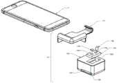
图10-A、10-B和10-C是用于智能手机比色读取器的系统10的示意图。具体地,图10-B和10-C分别是从前侧和后侧示出的系统10的分解图。系统10包含智能手机1;安装在智能电话1的上部上的光学适配器装置13;插入到装置13的接收器槽136中,使得样本卡137上的样品区域位于智能手机1中的相机模块1C的视场和焦点范围内的比色测试卡137。安装在智能电话1中的软件(未示出)在智能电话1中的光源1L发光的同时分析由相机模块1C收集的图像,以便分析色度测试的颜色变化,并且将结果输出到智能电话1中的显示屏1f。10-A, 10-B, and 10-C are schematic diagrams of a
图11是系统10中的光学适配器装置13的示意性分解图。装置13包括安装在智能手机1上部的支架壳体131;附接到壳体131上的光学盒132包含接收器槽136、光学腔室132C。光学插件134装配到光学腔室132C的顶部中,其中具有与智能手机1中的光源1L和相机1C(在图10-B中示出)对准的出口光圈134L和入光圈134C。透镜133安装在光学插件134中的入光圈134C中,并且被配置成使得插入接收器槽136中的比色样本卡137上的样本区域位于相机1C的工作距离内(如图10-B所示)。大芯光纤135以倾斜角度安装在出口光圈134L中。使纤维135的两个端面具有无光泽的光洁度。下面在图2中描述作为装置13中的照明光学器件的操作的光纤135。FIG. 11 is a schematic exploded view of the
图12是示出读取比色卡,特别是装置13的系统10的细节的示意性剖视图。该图示出了以上参考图11描述的元件的功能。光源1L远离智能电话1发射光束B1。光束B1通过第一端面联接到光纤135中,并且沿着光纤135的方向行进,并且从第二端面发射出来以变成光束B2。光束B2从前侧照射相机1C正下方的比色样本卡137的样品区域以产生均匀的照射。因为光纤135的端面被制成无光泽和漫射光洁度,所以光束B2可以被认为是从面光源发射的,这有助于产生更均匀的照明。安装光纤135的倾斜角被设置成使光束B2的中心射线照射在相机正下方的样本卡137上的区域上。透镜11在相机1C的图像传感器平面上产生样品区域的图像。智能电话1捕获并处理图像以分析图像中的颜色信息,从而量化比色测定的颜色变化。FIG. 12 is a schematic cross-sectional view showing details of the
C.用于将比色读取器连接到智能手机的光学适配器(光纤环照明)。C. Optical adapter (fiber optic ring illumination) for connecting the colorimetric reader to the smartphone.
比色测定是一项非常强大的技术,在健康监测、疾病诊断、化学测定等方面有着广泛的应用。获得准确比色测定结果的关键因素是准确定量颜色变化。通常,通过将颜色变化与标准色卡进行比较来分析比色测试条的颜色变化。但是这种比较是由人眼完成的,并且容易受到环境光条件的影响,这限制了量化颜色变化的精确度。Colorimetric assays are a very powerful technique with a wide range of applications in health monitoring, disease diagnosis, chemical assays, and more. A key factor in obtaining accurate colorimetric assay results is accurate quantification of color change. Typically, colorimetric test strips are analyzed for color change by comparing the color change to a standard color chart. But this comparison is done by the human eye and is susceptible to ambient light conditions, which limits the accuracy with which color changes can be quantified.
本文描述的本发明通过提供一种包括光学适配器和手机的系统来解决这个问题。光学适配器装置安装在将其转换为比色读取器的手机上,比色读取器可提供一致且均匀的照明以照射比色测试卡的前表面并捕获样品的图像以分析颜色变化。系统可由普通人员在任何地点方便可靠地操作。光学适配器利用智能电话的现有资源,包含相机、光源、处理器和显示屏,这提供了低成本的解决方案以精确地量化比色测定的颜色变化。The invention described herein addresses this problem by providing a system that includes an optical adapter and a cell phone. The optical adapter unit is mounted on a cell phone that converts it into a colorimetric reader that provides consistent and uniform illumination to illuminate the front surface of the colorimetric test card and capture an image of the sample to analyze color changes. The system can be easily and reliably operated by ordinary personnel from any location. The optical adapter utilizes the existing resources of the smartphone, including the camera, light source, processor and display, which provides a low-cost solution to accurately quantify color change in colorimetric assays.
在本发明中,光学适配装置包括配合在手机上部上的保持器框架和附接到保持器的光学盒,光学盒具有样品接收器槽和照明光学器件。在一些现有技术的用于手机的连接适配器中,它们的适配器设计是包括配合在手机上的夹持机械零件和功能元件的整体件。这种设计存在的问题是,它们需要为每种特定型号的智能手机重新设计整体适配器。但是在本发明中,光学适配器被分成仅用于装配智能电话的保持框架和包含所有功能部件的通用光学盒。对于不同尺寸的智能手机,只要摄像头和光源的相对位置相同,只需重新设计固定架,节省了大量的设计和制造成本。In the present invention, the optical adapter includes a holder frame fitted on the upper part of the cell phone and an optical box attached to the holder, the optical box having a sample receiver slot and illumination optics. In some prior art connection adapters for cell phones, their adapter design is a unitary piece including gripping mechanical parts and functional elements that fit on the cell phone. The problem with this design is that they require a redesign of the overall adapter for each specific model of smartphone. In the present invention, however, the optical adapter is divided into a holding frame only for assembling a smartphone and a general-purpose optical case containing all functional parts. For smartphones of different sizes, as long as the relative positions of the camera and the light source are the same, it is only necessary to redesign the fixing frame, which saves a lot of design and manufacturing costs.
光学适配器的光学盒包括:接收器槽,其接收比色样品并将其定位在智能手机相机的视场和焦点范围内;照明和成像光学器件,其独立于任何外部条件在样品上产生均匀一致的照明并捕获样品图像。The optics box for the optical adapter includes: a receiver slot, which receives a colorimetric sample and positions it within the smartphone camera's field of view and focus; illumination and imaging optics, which produces a uniform consistency across the sample independent of any external conditions illumination and capture an image of the sample.
为了捕获样品图像以准确地表示颜色变化,希望相机下的样品区域被均匀地照明。但是对于所有普通的智能电话,光源总是点源并且以一定距离安装在相机附近,这意味着光源相对于相机不是中心对称的。这导致以下问题:当样品非常靠近智能电话的相机放置时,在没有附加照明光学器件的帮助下,相机视场中样品前表面上的照明图案将在线性方向上具有梯度强度变化。因此,希望产生具有大发射面积和与相机中心对称的光源。为了实现这个目的,在本发明中,塑料侧发射光纤环围绕智能手机相机放置,以使光纤环相对于相机中心对称。光纤环的两个端面朝向智能手机的光源安装。这将把原始单点源转换成无限数量的小光源,这些小光源具有分布在距智能手机相机等距离的圆上的几乎相等的发光强度。从环形光纤的侧壁发射的光进一步穿过漫射膜以增加发射面积并使照明更均匀。相机正下方的样品区域被设计的基于侧发射光纤环的照明光学器件均匀地正面照明。In order to capture an image of the sample to accurately represent color changes, it is desirable that the area of the sample under the camera is illuminated uniformly. But for all normal smartphones, the light source is always a point source and mounted near the camera at a distance, which means that the light source is not centrosymmetric with respect to the camera. This leads to the following problem: when the sample is placed very close to the smartphone's camera, without the help of additional illumination optics, the illumination pattern on the front surface of the sample in the camera's field of view will have gradient intensity changes in a linear direction. Therefore, it is desirable to produce a light source with a large emitting area and symmetry with the center of the camera. To achieve this, in the present invention, a plastic side-emitting fiber optic ring is placed around the smartphone camera so that the fiber optic ring is symmetrical with respect to the center of the camera. The two end faces of the fiber optic ring are installed towards the light source of the smartphone. This will convert the original single point source into an infinite number of small light sources with nearly equal luminous intensities distributed on a circle equidistant from the smartphone camera. Light emitted from the sidewalls of the ring fiber passes further through the diffusing film to increase the emission area and make the illumination more uniform. The sample area directly below the camera is uniformly front-illuminated by designed illumination optics based on a ring of side-emitting fiber optics.
因为如何表示比色样品的颜色很大程度上取决于照明条件,所以重要的是独立于任何外部光条件来控制光盒中的照明一致。为了解决这个问题,接收器槽具有连接到其上的橡胶门,该橡胶门可以完全覆盖槽,以防止环境光进入光学盒,从而导致照明条件的改变。Because how the color of a colorimetric sample is represented is highly dependent on the lighting conditions, it is important to control the lighting consistently in the light box independently of any external light conditions. To address this, the receiver slot has a rubber door attached to it that completely covers the slot to prevent ambient light from entering the optics box, which could cause changes in lighting conditions.
通常,光学盒还包含安装在其中的透镜,透镜与智能电话的相机对准,这使得样品在相机的焦点范围内。由相机捕获的图像将由智能电话的处理器进一步处理以分析颜色变化并在智能电话的屏幕上输出分析结果。Typically, the optics box also contains a lens mounted in it that is aligned with the smartphone's camera, which brings the sample into focus of the camera. The images captured by the camera will be further processed by the smartphone's processor to analyze the color changes and output the analysis results on the smartphone's screen.
样品滑块安装在接收器槽内以接收QMAX装置并将样品定位在智能手机相机的视场和焦点范围内的QMAX装置中。The sample slide is mounted in the receiver slot to receive the QMAX device and position the sample in the QMAX device within the field of view and focus of the smartphone camera.
样品滑块包括固定轨道架和活动臂:The sample slide includes a fixed rail holder and movable arm:
框架轨道固定安装在光学盒的接收器槽中。轨道框架具有与QMAX装置的宽度和厚度相匹配的滑轨槽,使得QMAX装置能够沿着轨道滑动。轨道槽的宽度和高度被仔细地构造为使得QMAX装置在垂直于滑动平面中的滑动方向的方向上移动小于0.5mm,并且沿着QMAX装置的厚度方向移动小于0.2mm。The frame rails fit securely in the receiver slots of the optics box. The track frame has rail grooves that match the width and thickness of the QMAX device, allowing the QMAX device to slide along the track. The width and height of the track grooves are carefully constructed so that the QMAX device travels less than 0.5 mm in the direction perpendicular to the sliding direction in the sliding plane and less than 0.2 mm along the thickness of the QMAX device.
框架轨道在手机的相机的视野下具有开放的窗口,以允许光回照样品。The frame rail has an open window under the field of view of the phone's camera to allow light to back illuminate the sample.
活动臂预先设置在轨道框架的滑轨槽中,并与QMAX装置一起运动,以引导QMAX装置在轨道框架中的运动。The movable arm is pre-set in the sliding rail groove of the track frame, and moves together with the QMAX device to guide the movement of the QMAX device in the track frame.
活动臂装备有具有两个预定停止位置的止动机构。对于一个位置,臂将使QMAX装置停止在QMAX装置上的固定采样区域正好在智能电话的相机下面的位置。对于另一个位置,臂将使QMAX装置停止在QMAX装置上的采样区域在智能电话的视场之外的位置,并且QMAX装置可以容易地从轨道槽中取出。The movable arm is equipped with a stop mechanism with two predetermined stop positions. For one position, the arm will stop the QMAX unit at a location on the QMAX unit where the fixed sampling area is just below the smartphone's camera. For another position, the arm will stop the QMAX device where the sampling area on the QMAX device is outside the smartphone's field of view, and the QMAX device can be easily removed from the track slot.
通过将QMAX装置和活动臂一起按压到轨道槽的端部然后释放,活动臂在两个停止位置之间切换。By pressing the QMAX device together with the movable arm to the end of the track slot and then releasing, the movable arm switches between the two stop positions.
活动臂可以指示QMAX装置是否沿正确的方向插入。QMAX装置的一个转角的形状被配置成不同于其他三个直角。动臂的形状与特定形状的转角形状相匹配,使QMAX装置只能沿正确方向滑动到轨道槽中的正确位置。The movable arm can indicate whether the QMAX unit is inserted in the correct direction. The shape of one corner of the QMAX device is configured to be different from the other three right angles. The shape of the boom matches the shape of the specific shaped corners so that the QMAX unit can only slide in the correct direction into the correct position in the track slot.
一些实施方案some embodiments
1.光纤环形照明器1. Fiber Ring Illuminator
在光学组件的一些实施方案中,其中In some embodiments of the optical assembly, wherein
a.侧发光光纤的半径为10mm;a. The radius of the side-emitting fiber is 10mm;
b.环形光纤的直径可以是至少5mm、10mm、15mm、20mm、25mm、30mm、40mm、50mm、60mm、80mm或100mm,或在任意两个值之间的范围内;b. The diameter of the ring fiber may be at least 5mm, 10mm, 15mm, 20mm, 25mm, 30mm, 40mm, 50mm, 60mm, 80mm or 100mm, or within a range between any two values;
c.环形光纤的横截面的直径可以是至少0.5mm、1.0mm、1.5mm、2.0mm、2.5mm、3mm、4mm、5mm、6mm、8mm或10mm,或在任意两个值之间的范围内。c. The diameter of the cross-section of the ring fiber may be at least 0.5mm, 1.0mm, 1.5mm, 2.0mm, 2.5mm, 3mm, 4mm, 5mm, 6mm, 8mm, or 10mm, or within a range between any two values .
在光学组件的一些实施方案中,其中In some embodiments of the optical assembly, wherein
d.外部成像器透镜的直径为6mm;d. The diameter of the external imager lens is 6mm;
e.所述成像器透镜的直径可为至少2mm、3mm、4mm、5mm、10mm、15mm、20mm、25mm、30mm、40mm或50mm,或在任意两个值之间的范围内。e. The diameter of the imager lens may be at least 2mm, 3mm, 4mm, 5mm, 10mm, 15mm, 20mm, 25mm, 30mm, 40mm, or 50mm, or within a range between any two values.
在光学组件的一些实施方案中,其中环形光纤可以与微透镜阵列结合使用或由微透镜阵列代替;In some embodiments of the optical assembly, wherein the annular optical fiber can be used in conjunction with or replaced by a microlens array;
在光学组件的一些实施方案中,其中光学组件包括在样品与所述环形光纤之间的光漫射板,其中光漫射板具有被配置成与相机对准的光圈。In some embodiments of the optical assembly, wherein the optical assembly includes a light diffusing plate between the sample and the annular optical fiber, wherein the light diffusing plate has an aperture configured to align with the camera.
在光学组件的一些实施方案中,其中所述光漫射板的一侧的长度可以是至少5mm、10mm、15mm、20mm、25mm、30mm、40mm、50mm、100mm、150mm或200mm,或在任意两个值之间的范围内,其中所述漫射板的厚度可以是至少2mm、3mm、4mm、5mm、10mm、15mm或20mm、或在任意两个值之间的范围内。In some embodiments of the optical assembly, wherein the length of one side of the light diffusing plate may be at least 5mm, 10mm, 15mm, 20mm, 25mm, 30mm, 40mm, 50mm, 100mm, 150mm, or 200mm, or between any two range between values, wherein the thickness of the diffuser plate may be at least 2mm, 3mm, 4mm, 5mm, 10mm, 15mm or 20mm, or a range between any two values.
在光学组件的一些实施方案中,其中所述光漫射板与身上环形光纤之间的距离可以是至少1mm、10mm、15mm、20mm、25mm、30mm、40mm、50mm、100mm,或在任意两个值之间的范围内。In some embodiments of the optical assembly, wherein the distance between the light diffusing plate and the body ring optical fiber may be at least 1 mm, 10 mm, 15 mm, 20 mm, 25 mm, 30 mm, 40 mm, 50 mm, 100 mm, or between any two range between values.
权利要求2所述的光学组件,其中样品与所述环形纤维之间的距离可以是至少2mm、10mm、15mm、20mm、25mm、30mm、40mm、50mm、100mm、150mm、200mm,或在任意两个值之间的范围内。The optical assembly of
控制杆:Controller:
1.权利要求3所述的光学组件,其中活动臂上的第一平面与光源之间的距离可以是至少0.5mm、2mm、4mm、8mm、10mm、20mm、50mm、100mm或在任意两个值之间的范围内。1. The optical assembly of
2.权利要求3所述的光学组件,其中活动臂的第一平面与第二平面之间的距离可以是至少5mm、10mm、15mm、20mm、40mm、100mm、200mm,或在任意两个值之间的范围内。2. The optical assembly of
3.权利要求5所述的光学组件,其中活动臂需要移动以在不同位置之间切换的距离可以是至少1mm、5mm、15mm、20mm、40mm、100mm,或在任意两个值之间的范围内。3. The optical assembly of
4.权利要求3所述的光学组件,其中第二平面被连接到倾斜平面,其中反射镜被安装在倾斜平面上5.权利要求4的光学组件,其中倾斜平面的优选倾斜角可以是至少10度、30度、60度、80度,或在在任意两个值之间的范围内,并且倾斜角被定义为第二平面与倾斜平面之间的角度。4. The optical assembly of
图13-A、13-B和13-C是用于智能手机比色读取器的系统10的示意图。特别地,图13-B和图13-C分别是从前侧和后侧示出的系统10的分解图。系统10包含智能手机1;安装在智能电话1的上部上的光学适配器装置13;比色样品卡138,其插入到装置13的接收器槽137中,使得样品卡138上的样品区域位于智能手机1中的相机模块1C的视场和焦点范围内。在样品卡138进入之后,附接到装置18的橡胶门139覆盖接收器槽137,以防止环境光进入光学适配器13影响测试。安装在智能电话1中的软件(未示出)在智能电话1中的光源1L发光的同时分析由相机模块1C收集的图像,以便分析色度测试的颜色变化,并且将结果输出到智能电话1中的显示屏1f。13-A, 13-B and 13-C are schematic diagrams of a
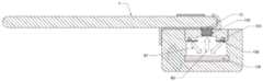
图14是系统10中的光学适配器装置13的示意性分解图。装置13包括安装在智能手机1上部的支架壳体131;附接到壳体131的光学盒132包括接收器槽137、光学腔室132C和橡胶门139,橡胶门139插入沟槽137s中以覆盖接收器槽137。光学插件134装配到光学腔室132C的顶部中,其中具有与智能电话1中的光源1L和相机1C(图13-B中示出)对准的出口光圈134L和入光圈134C。透镜133安装在光学插件134中的入光圈134C中,并且被配置成使得插入接收器槽137中的比色样品卡138上的样本区域位于相机1C的工作距离内(如图13-B所示)。侧面发射光纤环135安装在光学插件134中,光学插件134构造成使相机1C位于所述光纤环135的中心。所述光纤环135的两个端面安装在面向光源1L的出口光圈134L中。光漫射膜136放置在所述光纤环135之下,并具有为透镜光圈开放的孔。其操作作为装置13中的照明光的光纤环135在下面的图15-Ac中描述。FIG. 14 is a schematic exploded view of the
图15-A、15-B和15-C示出了读取比色卡的系统10,特别是装置13的细节的示意图。图15-A是示出装置13的细节的截面图。以及图15-B和图15-C是仅示出装置13中的光学元件的构造的示意图。这些图示出了以上参考图14描述的元件的功能。从光源1L发射的光从光纤环135的两个端面联接到侧面发射光纤环135中,并且沿着环在内部传播。光束B1从光纤环的侧壁射出并穿过漫射膜136。光束B1从前侧照射相机1C正下方的比色样品卡138的样品区域以产生均匀的照射。被照射的样品区域吸收部分光束B1并将光束B1反射到光束B2。光束B2由透镜133收集并进入相机1C。透镜133在相机1C的图像传感器平面上产生样品区域的图像。智能电话1捕获并处理图像以分析图像中的颜色信息,从而量化比色测定的颜色变化。Figures 15-A, 15-B and 15-C show schematic diagrams of details of the
D.断层摄像装置与系统D. Tomography devices and systems
D-1.QMAX结构断层摄像装置D-1.QMAX structural tomography device
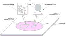
公开了一种以纳米尺度的最高分辨率重建生物样本的可切片虚拟三维拷贝的断层摄像装置。装置包括成像传感器、透镜和QMAX装置,如图16-A所示。A tomography device that reconstructs sliceable virtual three-dimensional copies of biological samples at the highest resolution at the nanoscale is disclosed. The device includes an imaging sensor, a lens, and a QMAX device, as shown in Figure 16-A.
QMAX装置具有周期性柱阵列。生物样本包含在QMAX装置中。折射率匹配液体可用于减少光的散射,并减少整个样品折射率的不均匀性。QMAX结构增强了六(或更多)个数量级的检测灵敏度。QMAX devices have periodic arrays of pillars. Biological samples are contained in the QMAX device. Refractive index matching liquids can be used to reduce light scattering and reduce inhomogeneity in refractive index across the sample. The QMAX structure enhances detection sensitivity by six (or more) orders of magnitude.
D-2.基于QMAX结构的标定D-2. Calibration based on QMAX structure
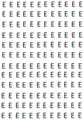
柱阵列在每个柱的顶部具有金属盘。金属盘提供用于由成像传感器捕获的图像的空间和高度校准的校准信号。金属盘的形状可以设计成便于快速校准。例如,金属盘的形状可以像字母E;这种柱阵列在图16-B中示出。Column arrays have metal disks on top of each column. The metal disk provides a calibration signal for spatial and height calibration of the image captured by the imaging sensor. The shape of the metal disc can be designed to facilitate quick calibration. For example, a metal disk can be shaped like the letter E; such an array of posts is shown in Figure 16-B.
当成像传感器捕获QMAX结构上具有或不具有生物样本的图像时,可以在空间上校准捕获的图像,并且还可以定量地校准相机的焦距。When the imaging sensor captures images with or without biological samples on the QMAX structure, the captured images can be spatially calibrated, and the camera's focus can also be calibrated quantitatively.
对于空间校准,捕获的图像经过对象检测。对象检测方案可以是模板匹配、光学字符识别、形状检测或者本领域中使用的其他方案。物体检测检索检测到的图案的方向,在图16-B的实施例中,方向是字母E。利用方位参数,通过二维几何变换实现空间标定。For spatial calibration, captured images are subject to object detection. The object detection scheme may be template matching, optical character recognition, shape detection, or other schemes used in the art. Object detection retrieves the direction of the detected pattern, which in the embodiment of Figure 16-B is the letter E. Using azimuth parameters, space calibration is achieved through two-dimensional geometric transformation.
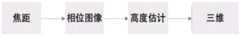
我们公开了利用柱阵列的焦距的定量校准。焦距对捕获图像的影响可以通过薄透镜模型来解释,如图16-C所示。如果感测设备与聚焦平面相距一定距离,则点Q将被投影到直径kσ的圆上,并且其辐射率将在圆上扩展,其中Q被散焦。焦平面的位置v取决于透镜的焦距f和离物体的距离u。这三个变量之间的关系由众所周知的高斯透镜定律或薄透镜方程给出:
我们测量捕获图像上的聚焦程度,并推导出聚焦平面位置。聚焦度测量整个图像或每个图像像素的聚焦级别。文献中已经提出了多种算法和算子来测量聚焦度,例如基于梯度的、基于拉普拉斯算子的、基于小波的、基于统计的、基于余弦变换/傅立叶变换的等等。We measure the degree of focus on the captured image and derive the focus plane position. Focus measures the level of focus of the entire image or each image pixel. Various algorithms and operators have been proposed in the literature to measure focusing, such as gradient-based, Laplacian-based, wavelet-based, statistics-based, cosine/Fourier transform-based, and so on.
可以预先测量在不同聚焦平面处捕获的柱阵列的聚焦度,并将其存储在查找表中。例如,当成像传感器捕获柱阵列的新图像时,图16-D示出了图16-B中的示例柱阵列的捕获图像,我们计算新捕获图像的聚焦度,将聚焦度参考查找表,并找到其相应的焦平面位置。The focus of the column array captured at different focus planes can be measured in advance and stored in a look-up table. For example, when the imaging sensor captures a new image of the column array, Fig. 16-D shows the captured image of the example column array in Fig. 16-B, we calculate the degree of focus of the newly captured image, refer the degree of focus to the look-up table, and Find its corresponding focal plane position.
D-3.断层摄像系统D-3. Tomography system
断层摄像的目的是通过生物样本的多个投影重建生物样本的三维体积。端到端断层摄像系统包括光源、成像和三维重建。The purpose of tomography is to reconstruct the three-dimensional volume of the biological sample from multiple projections of the biological sample. The end-to-end tomography system includes light source, imaging and 3D reconstruction.
光源light source
由成像传感器捕获的光可以从样品折射、从样品发射等。Light captured by the imaging sensor may be refracted from the sample, emitted from the sample, and the like.
成像imaging
成像部分捕获成像传感器上的投影。可以在不同的焦距、不同的角度、来自不同的照明等处捕获投影。The imaging portion captures the projection on the imaging sensor. Projections can be captured at different focal lengths, at different angles, from different lighting, etc.
可以在不同的焦距处捕获若干图像。透镜以步长或多个步长朝向或向后移动QMAX结构。步长的值和透镜的移动可以由硬件或软件通过应用程序接口来控制。图像传感器记录捕获的图像。Several images can be captured at different focal lengths. The lens moves towards or back the QMAX structure in a step or multiple steps. The value of the step size and the movement of the lens can be controlled by hardware or software through an application programming interface. The image sensor records the captured image.
可以以不同角度捕获若干图像。旋转样品并捕获通过其近似直线投影的光学图像。将样品旋转到一系列角位置,并且在每个方位捕获图像。仔细地对准装置以确保旋转轴垂直于光轴,从而由成像传感器收集关于每个平面的投影数据。焦平面可以位于旋转轴和最靠近透镜的QMAX卡之间的中间。这意味着每个图像既包含来自样品前半部分(最靠近透镜的一半)的聚焦数据,又包含来自样品后半部分的散焦数据。聚焦数据将被用于三维体积重建,而散焦数据将不被使用。可以配备带通滤波器来选择聚焦数据。Several images can be captured at different angles. Rotate the sample and capture an optical image projected through it approximately in a straight line. The sample is rotated to a series of angular positions and images are captured at each orientation. The device is carefully aligned to ensure that the axis of rotation is perpendicular to the optical axis so that projection data for each plane is collected by the imaging sensor. The focal plane can be halfway between the axis of rotation and the QMAX card closest to the lens. This means that each image contains both focus data from the front half of the sample (half closest to the lens) and defocus data from the back half of the sample. Focus data will be used for 3D volume reconstruction, while defocus data will not be used. A bandpass filter can be equipped to select focus data.
使用标准断层摄像算法执行光学投影断层摄像。由于焦平面相对于旋转轴的位置,彼此相隔180度拍摄的两个图像将聚焦在样品的不同部分上。将背投限制到对应于样品的聚焦部分的区域改善了结果的质量。当对于通过样品的各种取向积累数据时,可以旋转用作带通滤波器的半盘掩模,以确保只有聚焦的数据被反向投影。Optical projection tomography was performed using standard tomography algorithms. Due to the position of the focal plane relative to the axis of rotation, two images taken 180 degrees apart from each other will focus on different parts of the sample. Restricting rear projection to the area corresponding to the focused portion of the sample improves the quality of the results. When accumulating data for various orientations through the sample, the half-disk mask, which acts as a bandpass filter, can be rotated to ensure that only focused data is back-projected.
可以在不同的照明下捕获若干图像。可以从由参考光束相对于样品光束的频移引起的时间相关干涉图案获得定量相位图像。安装有检流计的倾斜反射镜可用于改变照明角度。激光束穿过移动激光束频率的两个声光调制器。第二分束器重新组合样本和参考激光束,形成在成像传感器处捕获的干涉图案。然后通过应用相移干涉测量法计算相位图像。对于折射率对比度小的薄样品的近平面波照明,透射场的相位很好地近似等于沿光束传播路径的折射率的线积分。因此,相位图像可以简单地解释为折射率的投影。Several images can be captured under different lighting. Quantitative phase images can be obtained from time-dependent interference patterns caused by frequency shifts of the reference beam relative to the sample beam. A galvanometer-mounted tilting mirror can be used to vary the illumination angle. The laser beam passes through two acousto-optic modulators that shift the frequency of the laser beam. The second beam splitter recombines the sample and reference laser beams to form an interference pattern captured at the imaging sensor. The phase image is then calculated by applying phase-shift interferometry. For near-plane wave illumination of thin samples with small refractive index contrast, the phase of the transmitted field is well approximately equal to the line integral of the refractive index along the beam propagation path. Therefore, the phase image can be simply interpreted as a projection of the refractive index.
除了带通滤波器之外,为了(包括但不限于)以下目的,在图像捕获期间可以使用各种成像滤波器:In addition to bandpass filters, various imaging filters may be used during image capture for purposes including but not limited to:
(1)信号选择,由此选择捕获图像的一部分;(1) Signal selection, whereby a portion of the captured image is selected;
(2)信号增强,由此增强捕获图像的部分或全部;(2) signal enhancement, thereby enhancing part or all of the captured image;
(3)信号变换,由此捕获图像的部分或全部被变换为另一表示,比如频率表示、多尺度表示等;(3) Signal transformation, whereby part or all of the captured image is transformed into another representation, such as a frequency representation, a multi-scale representation, etc.;
(4)信号复制,由此捕获图像的一部分被捕获图像的另一部分替换,或者被捕获图像的另一部分的表示替换;(4) Signal duplication whereby a portion of the captured image is replaced by another portion of the captured image, or is replaced by a representation of another portion of the captured image;
(5)或(1)-(4)的任何组合。(5) or any combination of (1)-(4).
通过对采集到的图像进行对比度增强、色彩增强、降噪等滤波处理,可以提高像素亮度的动态范围、调整色温、提高信噪比等。By performing filtering processing such as contrast enhancement, color enhancement, and noise reduction on the collected image, the dynamic range of pixel brightness can be improved, the color temperature can be adjusted, and the signal-to-noise ratio can be improved.
捕获的图像可以被转换为可以更适合于三维重构的另一种表示。它可以被转换成不同的格式(8位到16位、整数到浮点等)、不同的颜色空间(RGB到HSV等)、不同的域(空间域到频域等)等。The captured image can be transformed into another representation that can be more suitable for three-dimensional reconstruction. It can be converted into different formats (8-bit to 16-bit, integer to floating point, etc.), different color spaces (RGB to HSV, etc.), different domains (spatial domain to frequency domain, etc.), etc.
捕获图像的一部分可以由捕获图像的另一部分(或另一部分的变换)代替。它可以是被另一个区域的变换所代替的空间区域,例如围绕边界的反射扩展,等等,它可以是另一个频率子带的变换所代替的一个频率子带,例如高频子带被来自低频子带的估计所代替,等等。A portion of the captured image may be replaced by another portion of the captured image (or a transformation of another portion). It can be a region of space replaced by a transformation of another region, such as a reflection spread around a boundary, etc. It can be a frequency subband replaced by a transformation of another frequency subband, e.g. a high frequency subband is derived from are replaced by estimates of low frequency subbands, and so on.
三维重建Three-dimensional reconstruction
从生物样本的投影重建生物样本的三维体积是一个反问题。三维体积重建可以采用相位图像检索方案、反投影方案、非线性近似方案、优化方案等。Reconstructing the three-dimensional volume of a biological sample from its projection is an inverse problem. Three-dimensional volume reconstruction can adopt phase image retrieval scheme, back-projection scheme, nonlinear approximation scheme, optimization scheme, etc.
当在不同的聚焦距离处捕获几个图像时,我们计算这些图像的聚焦度,并将这些聚焦度作为向量列出。然后,我们用查找表参考矢量,并找到它们相应的焦平面距离。相应的可以是基于距离、基于相关性或选择最佳匹配的其他标准。When several images are captured at different focus distances, we compute the degrees of focus of these images and list these degrees of focus as a vector. We then reference the vectors with a lookup table and find their corresponding focal plane distances. Correspondingly, distance-based, correlation-based, or other criteria for selecting the best match may be used.
图16-E示出了基于相位图像检索的方案的图。它由四个组分组成:Figure 16-E shows a diagram of a phase image retrieval based scheme. It consists of four components:
·焦距计算·Focal length calculation
·相位图像检索· Phase image retrieval
·高度估计· Height estimation
·三维体积重建3D volume reconstruction
第二分量、相位恢复是通过基于强度传输(TIE)方程的定量相位成像技术。TIE方程状态The second component, phase recovery, is through quantitative phase imaging techniques based on the Intensity Transfer (TIE) equation. TIE equation state

其中

所述TIE方程可用快速傅里叶变换、离散余弦变换求解;例如“利用强度方程的传输进行无边界伪影相位检索:使用离散余弦变换的快速求解”,C.Zuo,Q.Chen,和A.Asundi,Optics Express,第22卷,第8期,2014年4月。从TIE方程中检索相位图像
给定相位图像,我们估计生物样本的高度(厚度)。回想一下,对于厚度为t且折射率为n的样品,相应的光程长度Lp为Lp=t×nGiven a phase image, we estimate the height (thickness) of the biological sample. Recall that for a sample of thickness t and refractive index n, the corresponding optical path length Lp isLp = t×n
可以用已知的折射率计算生物样本的高度。The height of the biological sample can be calculated using the known refractive index.
进一步,可以重建生物样本的三维体积。Further, the three-dimensional volume of the biological sample can be reconstructed.
背投算法通常用于断层摄像中的三维重建。它包括傅里叶变换基算法、滤波反投影算法、反投影滤波算法和迭代算法。Backprojection algorithms are commonly used for 3D reconstruction in tomography. It includes Fourier transform basis algorithm, filtered back-projection algorithm, back-projection filtering algorithm and iterative algorithm.
当焦平面相对于旋转轴的位置不同时,彼此相隔180度拍摄的两个图像将聚焦在样品的不同部分上。为了补偿,可以采用半平面调整的背投算法。因此,将背投限制到对应于样品的聚焦部分的区域将改进结果的质量。当对于通过样品的各种取向积累数据时,可以旋转半盘掩模以确保只有聚焦的数据被反向投影。When the position of the focal plane relative to the axis of rotation is different, two images taken 180 degrees apart from each other will focus on different parts of the sample. To compensate, a rear projection algorithm with half-plane adjustment can be used. Therefore, limiting the rear projection to the area corresponding to the focused portion of the sample will improve the quality of the results. When accumulating data for various orientations through the sample, the half-disk mask can be rotated to ensure that only focused data is back-projected.
作为反投影算法的另一个实施方案,可以应用基于滤波反投影方法的过程。对光束旋转方向上的每个x切片应用离散逆Radon变换,其中x是倾斜方向上的坐标,以及激光束方向与物镜光轴的相对角度。为了补偿成像和照明方向之间的角度,将x值除以。为了减少缺失投影的影响,可以应用迭代约束方法。As another embodiment of the backprojection algorithm, a process based on the filtered backprojection method can be applied. A discrete inverse Radon transform is applied to each x slice in the beam rotation direction, where x is the coordinate in the tilt direction and the relative angle of the laser beam direction to the optical axis of the objective. To compensate for the angle between the imaging and illumination directions, divide the x value by . To reduce the effect of missing projections, iterative constraint methods can be applied.
对于从其投影重建三维体积的反转问题,所得到的三维体积可能是模糊的。斜坡滤波器可用于消除或减少模糊。For the inverse problem of reconstructing a 3D volume from its projection, the resulting 3D volume may be ambiguous. A ramp filter can be used to remove or reduce blur.
除了去模糊滤波器之外,各种成像滤波器可用于三维体积重建,例如(包括但不限于):In addition to deblurring filters, various imaging filters can be used for 3D volume reconstruction, such as (including but not limited to):
(1)信号选择,其中选择图像或图像体积的一部分;(1) Signal selection, where an image or a portion of an image volume is selected;
(2)信号增强,其中图像或图像体积的一部分或全部被增强;(2) signal enhancement, wherein part or all of the image or image volume is enhanced;
(3)信号变换,由此捕获图像的部分或全部被变换为另一表示,比如频率表示、多尺度表示等;(3) Signal transformation, whereby part or all of the captured image is transformed into another representation, such as a frequency representation, a multi-scale representation, etc.;
(4)信号复制,由此捕获图像的一部分被捕获图像的另一部分替换,或者被捕获图像的另一部分的表示替换;(4) Signal duplication whereby a portion of the captured image is replaced by another portion of the captured image, or is replaced by a representation of another portion of the captured image;
(5)或(1)-(4)的任何组合。(5) or any combination of (1)-(4).
D-4.本发明的实施例D-4. Embodiments of the present invention
DA1.一种用于样品成像的装置,包含QMAX装置和成像器,其中:DA1. An apparatus for imaging a sample, comprising a QMAX apparatus and an imager, wherein:
(1)QMAX装置包含:(1) The QMAX device includes:
第一板、第二板以及间隔件,其中:A first plate, a second plate, and a spacer, wherein:
i.板可相对于彼此移动成不同的构造;i. The plates can be moved into different configurations relative to each other;
ii.一个或两个板是柔性的;ii. One or both plates are flexible;
iii.每个板在其内表面上包括用于接触流体样品的样品接触区域;iii. each plate includes a sample contact area on its inner surface for contacting the fluid sample;
iv.一个或两个板包含固定于相应板的间隔件;iv. One or both of the panels contain spacers secured to the respective panels;
v.间隔件具有预定的基本上均匀的高度和预定的间隔件间距,并且v. the spacers have a predetermined substantially uniform height and a predetermined spacer spacing, and
vi.至少一个间隔件位于样品接触区域内部;vi. at least one spacer is located inside the sample contact area;
其中这些构造之一是开放构造,其中:这两个板是分开的,这些板之间的间距不受这些间隔件调节,并且样品被沉积在这些一个或两个板上;以及wherein one of the configurations is an open configuration, wherein: the two plates are separated, the spacing between the plates is not adjusted by the spacers, and the sample is deposited on one or both of the plates; and
其中构造中的另一个是在样品沉积之后以开放构造进行构造的闭合构造;以及在闭合构造中:样品的至少一部分被两个板压缩为均匀厚度的层,其中层的均匀厚度被两个板的内表面限制并且被板和间隔件调节。wherein the other of the configurations is a closed configuration constructed in an open configuration after deposition of the sample; and in a closed configuration: at least a portion of the sample is compressed into a layer of uniform thickness by the two plates, wherein the uniform thickness of the layer is compressed by the two plates The inner surface of the is limited and regulated by the plates and spacers.
(2)成像器被配置成捕获从均匀厚度层的至少一部分发出的信号的图像。(2) The imager is configured to capture images of signals emanating from at least a portion of the layer of uniform thickness.
DB1.一种用于断层摄像的系统,包含QMAX装置、成像器、保持器和控制装置,其中:DB1. A system for tomography comprising a QMAX device, an imager, a holder and a control device, wherein:
(1)QMAX装置包括:第一板、第二板和间隔件,其中:(1) The QMAX device includes: a first plate, a second plate and a spacer, wherein:
i.板可相对于彼此移动成不同的构造;i. The plates can be moved into different configurations relative to each other;
ii.一个或两个板是柔性的;ii. One or both plates are flexible;
iii.每个板在其内表面上包括用于接触流体样品的样品接触区域;iii. each plate includes a sample contact area on its inner surface for contacting the fluid sample;
iv.一个或两个板包含固定于相应板的间隔件;iv. One or both of the panels contain spacers secured to the respective panels;
v.间隔件具有预定的基本上均匀的高度和预定的间隔件间距,并且v. the spacers have a predetermined substantially uniform height and a predetermined spacer spacing, and
vi.至少一个间隔件位于样品接触区域内部;vi. at least one spacer is located inside the sample contact area;
其中这些构造之一是开放构造,其中:这两个板是分开的,这些板之间的间距不受这些间隔件调节,并且该样品被沉积在这些一个或两个板上;以及wherein one of the configurations is an open configuration, wherein: the two plates are separated, the spacing between the plates is not adjusted by the spacers, and the sample is deposited on one or both of the plates; and
其中构造中的另一个是在样品沉积之后以开放构造进行构造的闭合构造;以及在闭合构造中:样品的至少一部分被两个板压缩为均匀厚度的层,其中层的均匀厚度被两个板的内表面限制并且被板和间隔件调节。wherein the other of the configurations is a closed configuration constructed in an open configuration after deposition of the sample; and in a closed configuration: at least a portion of the sample is compressed into a layer of uniform thickness by the two plates, wherein the uniform thickness of the layer is compressed by the two plates The inner surface of the is limited and regulated by the plates and spacers.
(2)该成像器包括图像传感器和透镜,其中:(2) The imager includes an image sensor and a lens, wherein:
i.透镜被配置成用于聚焦从均匀厚度的层的至少一部分发出的信号并且将聚焦的信号投射到图像传感器上,并且i. the lens is configured to focus the signal emanating from at least a portion of the layer of uniform thickness and project the focused signal onto the image sensor, and
ii.图像传感器被配置为捕获所述聚焦信号的图像;ii. an image sensor configured to capture an image of the focus signal;
(3)保持器被配置成调整QMAX装置和成像器之间的相对位置;以及(3) The holder is configured to adjust the relative position between the QMAX device and the imager; and
(4)控制装置包括硬件和软件,用于控制和/或推导由支架进行的位置调整,以及接收所述图像并将其重构为三维体积。(4) The control device includes hardware and software for controlling and/or deriving the positional adjustments made by the stand, as well as receiving and reconstructing the images into a three-dimensional volume.
DBB1.一种用于断层摄像的系统,包含QMAX装置、成像器、保持器和控制装置,其中:DBB1. A system for tomography, comprising a QMAX device, an imager, a holder and a control device, wherein:
(1)QMAX装置包括:第一板、第二板和间隔件,其中:(1) The QMAX device includes: a first plate, a second plate and a spacer, wherein:
i.板可相对于彼此移动成不同的构造;i. The plates can be moved into different configurations relative to each other;
ii.一个或两个板是柔性的;ii. One or both plates are flexible;
iii.每个板在其内表面上包括用于接触流体样品的样品接触区域;iii. each plate includes a sample contact area on its inner surface for contacting the fluid sample;
iv.一个或两个板包含固定于相应板的间隔件;iv. One or both of the panels contain spacers secured to the respective panels;
v.间隔件具有预定的基本上均匀的高度和预定的间隔件间距,并且v. the spacers have a predetermined substantially uniform height and a predetermined spacer spacing, and
vi.至少一个间隔件位于样品接触区域内部;vi. at least one spacer is located inside the sample contact area;
其中这些构造之一是开放构造,其中:所述两个板是分开的,板之间的间距不受所述间隔件调节,并所述样品被沉积在一个或两个所述板上;以及wherein one of these configurations is an open configuration, wherein: the two plates are separated, the spacing between the plates is not adjusted by the spacer, and the sample is deposited on one or both of the plates; and
其中构造中的另一个是闭合构造,其在样品沉积在开放构造之后构造而成;并且在所述闭合构造中:样品的至少一部分被两个板压缩为均匀厚度的层,其中层的均匀厚度被所述两个板的内表面定义并且被所述板和所述间隔件调节。wherein the other of the configurations is a closed configuration, which is constructed after the sample is deposited in the open configuration; and in the closed configuration: at least a portion of the sample is compressed by the two plates into a layer of uniform thickness, wherein the uniform thickness of the layer Defined by the inner surfaces of the two plates and conditioned by the plates and the spacers.
(2)所述成像器能够改变焦平面,包含图像传感器和透镜,其中:(2) The imager is capable of changing the focal plane and includes an image sensor and a lens, wherein:
i.所述透镜被配置以用于聚焦从所述均匀厚度的层的至少一部分发出的信号并且将聚焦的信号投射到所述图像传感器上,并且i. the lens is configured to focus signals emanating from at least a portion of the layer of uniform thickness and project the focused signals onto the image sensor, and
ii.所述图像传感器被配置以捕获所述聚焦信号的图像;ii. the image sensor is configured to capture an image of the focus signal;
iii.透镜为单一透镜或由若干透镜组成的复合透镜;iii. The lens is a single lens or a compound lens composed of several lenses;
iv.透镜中的至少一个元件透镜可移动以改变距图像传感器的距离,从而改变成像器的焦平面;以及iv. At least one element of the lenses is movable to change the distance from the image sensor, thereby changing the focal plane of the imager; and
v.可移动透镜可以由步进电机和/或电磁力驱动,所述步进电机和/或电磁力是计算机化的或手动控制的。v. The movable lens can be driven by a stepper motor and/or electromagnetic force, which is computerized or manually controlled.
以及as well as
(4)所述控制装置包括硬件和软件,用于控制和/或推导由支架进行的位置调整,以及接收所述图像并将其重构为三维体积。(4) The control device includes hardware and software for controlling and/or deriving the positional adjustments made by the stand, as well as receiving and reconstructing the image into a three-dimensional volume.
DC1.一种断层摄像方法,包含以下步骤:DC1. A tomography method, comprising the following steps:
(a)将样品沉积到任何前述装置或系统实施例中的QMAX装置上;(a) depositing the sample onto the QMAX device of any of the foregoing device or system embodiments;
(b)在(a)之后,使用这两个板来将该样品的至少一部分压缩成基本上均匀厚度的层,该层由这些板的样品接触表面限制,其中该层的均匀厚度是由这些间隔件和这些板调节的,其中该压缩包含:(b) following (a), using the two plates to compress at least a portion of the sample into a layer of substantially uniform thickness bounded by the sample contacting surfaces of the plates, wherein the uniform thickness of the layer is determined by the Spacers and these plates are regulated, wherein the compression contains:
将两块板放在一起;以及put the two boards together; and
平行地或顺序地适形按压这些板中的至少一个板的一个区域以将这些板按压在一起至一个闭合构造,其中该适形按压在这些板上在样品的至少一部分上产生基本上均匀的压力,并且该按压使样品的至少一部分在这些板的这些样品接触表面之间横向地展开,并且其中该闭合构造是其中在均匀厚度区域层中的这些板之间的间距是由这些间隔件调节的构造;Conformally pressing a region of at least one of the plates in parallel or sequentially to press the plates together to a closed configuration, wherein the conformal pressing produces a substantially uniform over at least a portion of the sample on the plates pressure, and the pressing causes at least a portion of the sample to spread laterally between the sample contacting surfaces of the plates, and wherein the closed configuration is wherein the spacing between the plates in the uniform thickness region layer is adjusted by the spacers structure;
(c)使用任何前述装置或系统实施例的成像器捕获从所述均匀厚度的层的至少一部分发出的信号的图像;(c) capturing images of signals emanating from at least a portion of the layer of uniform thickness using the imager of any preceding device or system embodiment;
(d)调整QMAX装置与成像器的相对位置,重复步骤(c);以及(d) adjusting the relative position of the QMAX device to the imager, repeating step (c); and
(e)在一系列步骤(c)之后,将所捕获的图像重建成所述均匀厚度的层的至少一部分的三维体积,(e) after a series of steps (c), reconstructing the captured image into a three-dimensional volume of at least a portion of the layer of uniform thickness,
其中适形按压是使施加在区域上的压力基本上恒定而与板的外表面的形状变化无关的方法;以及wherein conformal pressing is a method of making the pressure exerted on the area substantially constant regardless of changes in the shape of the outer surface of the plate; and
其中该平行按压同时将所述压力施加在该预期区域上,并且顺序按压将所述压力施加在预定区域的一部分上并且逐渐移动到其他区域。Wherein the parallel pressing simultaneously applies the pressure on the intended area, and the sequential pressing applies the pressure on a part of the predetermined area and gradually moves to other areas.
DCC1.一种在不同焦平面拍摄图像的方法,包含以下步骤:DCC1. A method of capturing images at different focal planes, comprising the steps of:
(a)计算机控制或手动控制成像器中的可移动透镜到初始位置;(a) Computer control or manual control of the movable lens in the imager to the initial position;
(b)将所述可移动透镜位置对应于焦平面的位置;(b) corresponding the movable lens position to the position of the focal plane;
(c)使用成像器中的图像传感器捕获图像并记录焦平面的位置;(c) using the image sensor in the imager to capture the image and record the position of the focal plane;
(d)计算机化或手动添加步进位移以将可移动透镜移动到下一位置;(d) Computerized or manual addition of step displacements to move the movable lens to the next position;
(e)重复步骤(b)到(d);(e) repeat steps (b) to (d);
(f)在一系列步骤(e)之后,捕获不同焦平面处的若干图像。(f) After a series of steps (e), several images at different focal planes are captured.
DA21.根据前述任一实施方案所述的装置,其中QMAX装置进一步包含涂覆在一个或两个板上的干燥试剂,该干燥试剂对样品进行染色。DA21. The device of any preceding embodiment, wherein the QMAX device further comprises a dry reagent coated on one or both plates, the dry reagent staining the sample.
DA22.根据前述任一实施方案所述的装置,其中:DA22. The device of any preceding embodiment, wherein:
i.一个或两个板样品接触区域包括一个或多个结合位点,每个结合位点结合并固定各自的分析物;或者i. One or both of the plate sample contact areas include one or more binding sites, each binding site binding and immobilizing a respective analyte; or
ii.一个或两个板样品接触区域包含一个或多个存储位点,每个存储位点存储一种或多种试剂;其中试剂在步骤(c)期间或之后溶解和分散在样品中,并且其中样品含有一种或多种分析物;或者ii. one or both of the plate sample contact areas comprise one or more storage sites, each storage site storing one or more reagents; wherein the reagents are dissolved and dispersed in the sample during or after step (c), and wherein the sample contains one or more analytes; or
iii.一个或多个扩增位点,当所述分析物或标记距所述扩增位点500nm时,每个所述扩增位点能够扩增来自分析物或分析物的标记的信号。;iii. One or more amplification sites, each of which is capable of amplifying a signal from an analyte or a label of an analyte when the analyte or label is 500 nm from the amplification site. ;
iv.i至iii的任意组合。iv. Any combination of i to iii.
DA23.根据前述任一实施方案所述的装置,其中成像器进一步包含光源,该光源提供用于成像的均匀厚度层的照明或激发的光。DA23. The device of any preceding embodiment, wherein the imager further comprises a light source that provides illumination or excitation light of the uniform thickness layer for imaging.
DA24.根据实施方案DA23所述的装置,其中光源选自由以下组成的群组:LED、激光器、白炽灯及其任意组合。DA24. The device of embodiment DA23, wherein the light source is selected from the group consisting of LEDs, lasers, incandescent lamps, and any combination thereof.
DB2.根据实施方案DB1所述的系统,其中信号包括选自由以下信号组成的群组的光学信号:光反射、光折射、光透射、发光信号及其任何组合。DB2. The system of embodiment DB1, wherein the signal comprises an optical signal selected from the group consisting of: light reflection, light refraction, light transmission, luminescence signal, and any combination thereof.
DB3.根据前述任一实施方案所述的系统,其中成像器进一步包含光源,该光源提供照射用于成像的均匀厚度层的光,其中光源选自由白炽光、LED、CFL、激光器及其任何组合组成的群组。DB3. The system of any preceding embodiment, wherein the imager further comprises a light source that provides light to illuminate the layer of uniform thickness for imaging, wherein the light source is selected from incandescent light, LEDs, CFLs, lasers, and any combination thereof formed group.
DB4.根据前述任一实施方案所述的系统,其中成像器进一步包含提供激发光的光源,该激发光激发来自用于成像的均匀厚度的所述层的荧光发射,其中光源是LED和/或激光器。DB4. The system of any preceding embodiment, wherein the imager further comprises a light source providing excitation light that excites fluorescence emission from the layer of uniform thickness for imaging, wherein the light source is an LED and/or laser.
DB5.根据前述任一实施方案所述的系统,其中保持器能够沿着QMAX装置的光轴调节透镜与QMAX装置的相对位置以改变透镜的焦平面位置。DB5. The system of any preceding embodiment, wherein the holder is capable of adjusting the relative position of the lens to the QMAX device along the optical axis of the QMAX device to change the focal plane position of the lens.
DB6.根据前述任一实施方案所述的系统,其中保持器能够调节透镜与QMAX装置之间的相对位置以改变成像角度,其中成像角度是透镜的焦平面与均匀厚度层之间的角度。DB6. The system of any preceding embodiment, wherein the holder is capable of adjusting the relative position between the lens and the QMAX device to vary the imaging angle, wherein the imaging angle is the angle between the focal plane of the lens and the layer of uniform thickness.
DB7.根据前述任一实施方案所述的系统,其中成像器进一步包含为成像提供照明光的光源,且其中保持器能够调节光源与QMAX装置之间的相对位置以改变照明光的入射角,其中入射角是照明光与垂直于均匀厚度层的线之间的角度。DB7. The system of any preceding embodiment, wherein the imager further comprises a light source that provides illumination light for imaging, and wherein the holder is capable of adjusting the relative position between the light source and the QMAX device to vary the angle of incidence of the illumination light, wherein The angle of incidence is the angle between the illuminating light and a line perpendicular to the layer of uniform thickness.
DB8.根据前述任一实施方案所述的系统,其中所述控制装置包括用于向保持器发送界定位置调整的命令的硬件和软件,且其中保持器构造成接收命令并以不超过10%的偏差进行调整。DB8. The system of any preceding embodiment, wherein the control device includes hardware and software for sending commands to the holder defining the position adjustment, and wherein the holder is configured to receive the command and receive the command at a rate of no more than 10% deviations are adjusted.
DB9.根据前述任一实施方案所述的系统,其中所述控制装置包括硬件和软件用于向保持器发送界定位置调整的命令,且其中所述保持器构造以接收所述命令并以不超过1%的偏差进行调整。DB9. The system of any preceding embodiment, wherein the control means includes hardware and software for sending commands to the retainer defining position adjustments, and wherein the retainer is configured to receive the commands without exceeding 1% deviation to adjust.
DB10.根据实施例DB8-DB9中的任一项所述的系统,其中所述控制装置包括硬件和软件,用于接收定义位置调节的输入,并且将所述输入转换为用于保持器进行调节的命令。DB10. The system of any one of embodiments DB8-DB9, wherein the control means includes hardware and software for receiving input defining position adjustment and converting the input for the holder to adjust The command.
DB11.根据前述任一实施方案所述的系统,其中系统进一步包含多个校准柱,并且其中:DB11. The system of any preceding embodiment, wherein the system further comprises a plurality of calibration columns, and wherein:
(1)多个校准柱位于闭合构造的两块板的样品接触区域之间,且彼此具有不同的高度,均小于间隔件的均匀高度;(1) A plurality of calibration columns are located between the sample contact areas of the two plates in the closed configuration, and have different heights from each other, all less than the uniform height of the spacers;
(2)在沿公共光轴的不同焦平面处捕获所述图像;以及(2) capturing the images at different focal planes along a common optical axis; and
(3)所述控制装置包括硬件和软件,用于:(a)计算所述图像中的每一个的聚焦分数;以及(b)通过将所述聚焦得分与查找表进行比较来推断捕获图像中的每一者的聚焦平面位置,其中聚焦得分是针对所捕获图像的每一像素计算的聚焦度矩阵,其中查找表是预定的并且包括沿着公共光轴的一行预定聚焦平面位置和相应行的校准聚焦得分,每个校准聚焦得分是基于在相应预定聚焦平面处捕获的校准柱的图像来计算的。(3) The control device includes hardware and software for: (a) calculating a focus score for each of the images; and (b) inferring the focus score in the captured image by comparing the focus score with a look-up table The focal plane positions of each of the , where the focus score is a focus degree matrix calculated for each pixel of the captured image, where the lookup table is predetermined and includes a row of predetermined focal plane positions along a common optical axis and a corresponding row of Calibration focus scores, each of which is calculated based on images of the calibration column captured at the corresponding predetermined focus plane.
DB12.根据前述任一实施方案所述的系统,其中所述图像在沿公共光轴的不同焦平面处被捕获,并且其中所述控制装置包含硬件和软件,用于:(a)为所述层的至少一部分中的生物实体生成相位图像,其中该相位图像是基于用于成像的照明光的波长,包含来自该生物实体的信号的图像的至少一部分,以及分别捕获图像的聚焦平面位置而计算的相位分布;以及(b)基于相位图像和样品的折射率来估计生物实体的厚度,其中生物实体是所述层的至少一部分的一部分或全部。DB12. The system of any preceding embodiment, wherein the images are captured at different focal planes along a common optical axis, and wherein the control means comprises hardware and software for: (a) providing the The biological entity in at least a portion of the layer generates a phase image, wherein the phase image is calculated based on the wavelength of the illumination light used for imaging, at least a portion of the image containing the signal from the biological entity, and the position of the focal plane at which the image was captured, respectively and (b) estimating the thickness of the biological entity based on the phase image and the refractive index of the sample, wherein the biological entity is part or all of at least a portion of the layer.
DB13.根据实施例DB8的系统,其中控制装置包含用于基于所估计的厚度将图像的所述至少一部分重建为生物实体的三维体积的硬件和软件。DB13. The system of embodiment DB8, wherein the control means comprises hardware and software for reconstructing the at least a portion of the image into a three-dimensional volume of the biological entity based on the estimated thickness.
DB14.根据前述任一实施方案所述的系统,其中所述图像以不同成像角度捕获,其中控制装置包含硬件和软件,用于:(1)知道或推导每个所述图像的成像角度;以及(2)使用背投算法基于已知/推导出的成像角度将所述图像重构为三维体积,并且其中成像角度是透镜的焦平面与所述均匀厚度的层之间的角度。DB14. The system of any preceding embodiment, wherein the images are captured at different imaging angles, wherein the control means comprises hardware and software for: (1) knowing or deriving the imaging angle for each of the images; and (2) Reconstructing the image into a three-dimensional volume based on a known/derived imaging angle using a rear projection algorithm, and where the imaging angle is the angle between the focal plane of the lens and the layer of uniform thickness.
DB15.根据前述任一实施方案所述的系统,其中图像以照明光的不同入射角捕获,其中控制装置包括硬件和软件,用于:(1)知道或推导每个图像的入射角;以及(2)使用背投算法基于已知/推导出的入射角将图像重构为三维体积,并且其中照明光的入射角是照明光与垂直于均匀厚度层的线之间的角度。DB15. The system of any preceding embodiment, wherein the images are captured at different angles of incidence of the illumination light, wherein the control means includes hardware and software for: (1) knowing or deriving the angle of incidence for each image; and ( 2) Reconstruct the image into a three-dimensional volume using a rear projection algorithm based on a known/derived angle of incidence, and where the angle of incidence of the illumination light is the angle between the illumination light and a line normal to the layer of uniform thickness.
DB16.根据实施方案DB14-DB15中的任一项所述的系统,其中反投影算法选自由以下算法组成的群组:傅立叶变换基算法、滤波反投影算法、反投影和滤波算法、迭代算法及其任意组合。DB16. The system of any one of embodiments DB14-DB15, wherein the backprojection algorithm is selected from the group consisting of a Fourier transform basis algorithm, a filtered backprojection algorithm, a backprojection and filtering algorithm, an iterative algorithm, and any combination thereof.
DB17.根据前述任一实施方案所述的系统,其中成像器配备有成像滤波器,并且其中所捕获的图像由成像滤波器和/或控制装置的软件滤波,用于:(1)信号选择,由此选择捕获图像的一部分;(2)信号增强,从而增强捕获图像的部分或全部;(3)信号变换,将捕获图像的部分或全部变换为另一种表示,如频率表示、多尺度表示等;(4)信号复制,由此捕获图像的一部分被捕获图像的另一部分替换,或者被捕获图像的另一部分的表示替换;或(1)-(4)的任何组合。DB17. The system of any preceding embodiment, wherein the imager is equipped with an imaging filter, and wherein the captured image is filtered by the imaging filter and/or software of the control device for: (1) signal selection, A part of the captured image is thus selected; (2) signal enhancement, thereby enhancing part or all of the captured image; (3) signal transformation, transforming part or all of the captured image into another representation, such as frequency representation, multi-scale representation etc.; (4) signal duplication whereby a portion of the captured image is replaced by another portion of the captured image, or a representation of another portion of the captured image; or any combination of (1)-(4).
DB18.根据前述任一实施方案所述的系统,其中控制装置进一步包含用于将图像的至少一部分重构为三维体积的硬件和软件,其中在三维体积重构期间,图像和三维体积由软件滤波以用于:(1)信号选择,其中选择图像或图像体积的一部分;(2)信号增强,其中图像或图像体积的一部分或全部被增强;(3)信号变换,其中图像或图像体积的一部分或全部被变换成另一种表示,例如频率表示,多尺度表示等;(4)信号复制,其中图像或图像体积的一部分被捕获图像的另一部分替代,或被捕获图像的另一部分的表示替代;或(1)-(4)的任何组合。DB18. The system of any preceding embodiment, wherein the control means further comprises hardware and software for reconstructing at least a portion of the image into a three-dimensional volume, wherein the image and the three-dimensional volume are filtered by the software during reconstruction of the three-dimensional volume To be used for: (1) signal selection, in which an image or part of an image volume is selected; (2) signal enhancement, in which part or all of an image or image volume is enhanced; (3) signal transformation, in which an image or part of an image volume is enhanced or all are transformed into another representation, such as a frequency representation, a multiscale representation, etc.; (4) signal replication, in which part of an image or image volume is replaced by another part of the captured image, or by a representation of another part of the captured image ; or any combination of (1)-(4).
DC2.根据实施方案DC1所述的方法,进一步包含:在步骤(c)之前,用染色剂染色样品。DC2. The method of embodiment DC1, further comprising: prior to step (c), staining the sample with a stain.
DC3.根据前述任一方法实施方案所述的方法,其中在步骤(b)期间,适形按压是通过人手进行的。DC3. The method of any preceding method embodiment, wherein during step (b), the conformal compression is performed by a human hand.
DC4.根据前述任一方法实施方案所述的方法,其中步骤(d)的适形按压由加压液体、加压气体或适形材料提供。DC4. The method of any of the preceding method embodiments, wherein the conformal pressing of step (d) is provided by a pressurized liquid, a pressurized gas, or a conformal material.
DC5.根据前述任一方法实施方案所述的方法,其中所述调整步骤(d)包含沿着所述QMAX装置的光轴调整所述透镜与所述QMAX装置的相对位置以改变所述透镜的焦平面位置。DC5. The method of any preceding method embodiment, wherein the adjusting step (d) comprises adjusting the relative position of the lens and the QMAX device along an optical axis of the QMAX device to change the lens's relative position focal plane position.
DC6.根据前述任一方法实施方案所述的方法,其中所述调整步骤(d)包含调整所述透镜与所述QMAX装置之间的相对位置以改变成像角度,其中所述成像角度是焦平面与所述均匀厚度层之间的角度。DC6. The method of any preceding method embodiment, wherein the adjusting step (d) comprises adjusting a relative position between the lens and the QMAX device to change an imaging angle, wherein the imaging angle is a focal plane and the uniform thickness layer.
DC7.根据前述任一方法实施方案所述的方法,其中成像器进一步包含提供用于成像的照明光的光源,且其中调整步骤(d)包含调整光源与QMAX装置的相对位置以改变照明光的入射角,其中入射角是照明光与垂直于均匀厚度层的线之间的角度。DC7. The method of any of the preceding method embodiments, wherein the imager further comprises a light source that provides illumination light for imaging, and wherein adjusting step (d) comprises adjusting the relative position of the light source and the QMAX device to change the amount of illumination light The angle of incidence, where the angle of incidence is the angle between the illuminating light and a line normal to the layer of uniform thickness.
DC8.根据前述任一方法实施方案所述的方法,其中调整步骤(d)是手动执行的。DC8. The method of any preceding method embodiment, wherein adjusting step (d) is performed manually.
DC9.根据前述任一方法实施方案所述的方法,其中调节步骤(d)通过可操作地联接到保持器的控制装置来执行,其中控制装置包含用于接收界定位置调节的输入并将命令发送到保持器的硬件和软件,且其中保持器构造成接收命令并使调节具有不大于10%的偏差。DC9. The method of any preceding method embodiment, wherein adjusting step (d) is performed by a control device operably coupled to the holder, wherein the control device includes an input for receiving an input defining the position adjustment and sending a command to the hardware and software of the keeper, and wherein the keeper is configured to receive commands and make adjustments with a deviation of no more than 10%.
DC10.根据前述任一方法实施方案所述的方法,其中调节步骤(d)通过可操作地耦合到保持器的控制装置来执行,其中控制装置包含用于接收界定位置调节的输入并将命令发送到保持器的硬件和软件,且其中保持器构造成接收命令并使调节具有不大于1%的偏差。DC10. The method of any preceding method embodiment, wherein adjusting step (d) is performed by a control device operably coupled to the holder, wherein the control device includes an input for receiving an input defining the position adjustment and sending a command to the hardware and software of the keeper, and wherein the keeper is configured to receive commands and make adjustments with a deviation of no more than 1%.
DC11.根据任何先前方法实施方案所述的方法,其中所述图像是在沿着公共光轴的不同焦平面处捕获的,并且其中重建步骤(e)包括:(i)计算所述图像中的每一个的聚焦分数;以及(ii)通过将所述聚焦得分与查找表进行比较来推断捕获所述图像中的每一者的焦平面位置,其中聚焦得分是针对所捕获图像的每一像素计算的聚焦度矩阵,其中查找表是预定的并且包括沿着所述公共光轴的一行预定焦平面位置和相应行的校准聚焦得分,每个校准聚焦得分是基于在相应预定焦平面处捕获的校准柱的图像来计算的。DC11. The method of any preceding method embodiment, wherein the images are captured at different focal planes along a common optical axis, and wherein reconstructing step (e) comprises: (i) computing the a focus score for each; and (ii) inferring a focal plane position for capturing each of the images by comparing the focus score to a look-up table, where the focus score is calculated for each pixel of the captured image a focus matrix, wherein the look-up table is predetermined and includes a row of predetermined focal plane positions along the common optical axis and a corresponding row of calibrated focus scores, each calibrated focus score based on a calibration captured at the corresponding predetermined focal plane Column image to calculate.
DC12.根据任何先前方法实施方案所述的方法,其中所述图像是在沿着公共光轴的不同焦平面处捕获的,并且其中重建步骤(e)包括:(i)为所述层的所述至少一部分中的生物实体生成相位图像,其中该相位图像是基于用于成像的照明光的波长,包含来自该生物实体的信号的所述图像的至少一部分,以及捕获所述图像的焦平面位置而计算的相位分布;以及(ii)基于所述相位图像和所述样品的折射率来估计所述生物实体的厚度,其中所述生物实体是所述层的至少一部分的一部分或全部。DC12. The method of any preceding method embodiment, wherein the images are captured at different focal planes along a common optical axis, and wherein reconstructing step (e) comprises: (i) all of the layers generating a phase image of the biological entity in the at least a portion, wherein the phase image is based on the wavelength of the illumination light used for imaging, at least a portion of the image containing the signal from the biological entity, and the focal plane position at which the image was captured and (ii) estimating the thickness of the biological entity based on the phase image and the refractive index of the sample, wherein the biological entity is part or all of at least a portion of the layer.
DC13.根据实施方案DC12所述的方法,其中重建步骤(e)进一步包含基于所估计的厚度将所述图像的至少一部分重建为生物实体的三维体积。DC13. The method of embodiment DC12, wherein reconstructing step (e) further comprises reconstructing at least a portion of the image as a three-dimensional volume of a biological entity based on the estimated thickness.
DC14.根据前述任一方法实施方案所述的方法,其中所述图像是以不同成像角度捕获的,其中重建步骤(e)包含:(i)知道或推导每个所述图像的成像角度;以及(ii)使用背投算法基于已知/推导出的成像角度将所述图像重构为三维体积,并且其中成像角度是透镜的焦平面与均匀厚度的层之间的角度。DC14. The method of any preceding method embodiment, wherein the images are captured at different imaging angles, wherein reconstructing step (e) comprises: (i) knowing or deriving the imaging angle for each of the images; and (ii) Reconstructing the image into a three-dimensional volume based on a known/derived imaging angle using a rear projection algorithm, and where the imaging angle is the angle between the focal plane of the lens and the layer of uniform thickness.
DC15.根据前述任一方法实施方案所述的方法,其中所述图像是以照明光的不同入射角捕获的,其中重建步骤(e)包含:(i)知道或推导每个所述图像的入射角;以及(ii)使用背投算法基于已知/推导出的入射角将所述图像重构为三维体积,并且其中照明光的入射角是照明光与垂直于均匀厚度层的线之间的角度。DC15. The method of any preceding method embodiment, wherein the images are captured at different angles of incidence of illumination light, wherein reconstructing step (e) comprises: (i) knowing or deriving the incidence of each of the images and (ii) using a rear projection algorithm to reconstruct the image into a three-dimensional volume based on the known/derived angle of incidence, and wherein the angle of incidence of the illuminating light is between the illuminating light and a line normal to the layer of uniform thickness angle.
DC16.根据实施方案DC14-DC15中任一项所述的方法,其中反投影算法选自由以下各项组成的群组:傅立叶变换基算法、滤波反投影算法、反投影和滤波算法、迭代算法及其任意组合。DC16. The method of any one of embodiments DC14-DC15, wherein the backprojection algorithm is selected from the group consisting of a Fourier transform basis algorithm, a filtered backprojection algorithm, a backprojection and filtering algorithm, an iterative algorithm, and any combination thereof.
DC17.根据前述任一方法实施方案所述的方法,其中样品是选自细胞、组织、体液、粪便及其任意组合的生物样本。DC17. The method of any of the preceding method embodiments, wherein the sample is a biological sample selected from the group consisting of cells, tissues, body fluids, feces, and any combination thereof.
DC18.根据前述任一方法实施方案所述的方法,其中样品是来自环境来源的环境样品,该环境来源选自由河流、湖泊、池塘、海洋、冰川、冰山、雨、雪、污水、水库、自来水、饮用水等;来自土壤、堆肥、沙、岩石、混凝土、木材、砖、污水、空气、水下通风口、工业废气、车辆废气及其任意组合的固体样品组成的群组。DC18. The method according to any preceding method embodiment, wherein the sample is an environmental sample from an environmental source selected from the group consisting of rivers, lakes, ponds, oceans, glaciers, icebergs, rain, snow, sewage, reservoirs, tap water , drinking water, etc.; groups of solid samples from soil, compost, sand, rock, concrete, wood, brick, sewage, air, underwater vents, industrial exhaust, vehicle exhaust, and any combination thereof.
DC19.根据前述任一方法实施方案所述的方法,其中该样品是选自由以下组成的群组的一种食物样品:原料、熟食、植物和动物食物源、经预处理的食物、部分或完全处理的食物,以及它们的任何组合。DC19. The method according to any preceding method embodiment, wherein the sample is a food sample selected from the group consisting of raw, cooked food, plant and animal food sources, pretreated food, partially or completely processed foods, and any combination of them.
DC20.根据前述方法实施方案中任一项所述的方法,其中样品是血液,并且生物实体是红细胞、白细胞和/或血小板。DC20. The method of any one of the preceding method embodiments, wherein the sample is blood and the biological entity is red blood cells, white blood cells and/or platelets.
DC21.根据实施方案DC20所述的方法,还包含:DC21. The method of embodiment DC20, further comprising:
(f)基于它们各自重建的三维体积计算红细胞、白细胞和/或血小板的体积。(f) Calculate the volumes of red blood cells, white blood cells and/or platelets based on their respective reconstructed three-dimensional volumes.
DC22.根据实施方案DC21的方法,还包含:DC22. The method of embodiment DC21, further comprising:
(g)基于所计算的体积,确定选自由以下群组组成的血液测试读数:平均红细胞体积(MCV)、血细胞比容、红细胞分布宽度(RDW)、平均血小板体积(MPV)、血小板分布宽度(PDW)、未成熟血小板级分(IPF)及其任意组合。(g) Based on the calculated volume, determine a blood test reading selected from the group consisting of: mean corpuscular volume (MCV), hematocrit, erythrocyte distribution width (RDW), mean platelet volume (MPV), platelet distribution width ( PDW), immature platelet fraction (IPF), and any combination thereof.
E.机器学习辅助的测定和成像E. Machine Learning-Assisted Assays and Imaging
E-1.用于分析和成像的QMAX装置E-1. QMAX device for analysis and imaging
公开了一种用于生物分析物检测和定位的装置,包含QMAX装置、成像器和计算单元。生物样本在QMAX装置上待测。通过本公开获得在样品中包含的分析物的计数和位置。A device for biological analyte detection and localization is disclosed, comprising a QMAX device, an imager, and a computing unit. Biological samples were tested on the QMAX device. Counts and locations of analytes contained in a sample are obtained by the present disclosure.
所述成像器捕获所述生物样本的图像。图像被提交给计算单元。计算单元可以物理上直接连接到所述成像器、通过网络连接,或间接通过图像传送。The imager captures an image of the biological sample. The image is submitted to the computing unit. The computing unit may be physically connected directly to the imager, via a network connection, or indirectly via image transfer.
E-2.工作流E-2. Workflow
所公开的分析物检测和定位采用机器学习深度学习。机器学习算法是能够从数据学习的算法。机器学习的更严格的定义是“如果由P测量的计算机程序在T中的任务处的性能随着经验E而改善,则称计算机程序从经验E学习关于某类任务T和性能测量P”。它探索了能够从数据学习和对数据进行预测的算法的研究和构造,这种算法通过从样品输入建立模型来进行数据驱动预测或决策,从而克服了遵循严格静态程序指令的问题。The disclosed analyte detection and localization employs machine learning deep learning. Machine learning algorithms are algorithms that are able to learn from data. A more rigorous definition of machine learning is "a computer program is said to learn from experience E about some class of task T and performance measure P if its performance at a task in T, as measured by P, improves with experience E." It explores the study and construction of algorithms capable of learning from and making predictions from data that overcome the problem of following strictly static program instructions by building models from sample inputs to make data-driven predictions or decisions.
深度学习是一种基于一组试图对数据中的高级抽象建模的算法的特定类型的机器学习。在简单的情况下,可能有两组神经元:一组接收输入信号、一组发送输出信号。当输入层接收输入时,它将输入的修改版本传递给下一层。在深度网络中,在输入和输出之间存在许多层(并且这些层不是由神经元构成的,但是它可以帮助考虑这种方式),从而允许算法使用由多个线性变换和非线性变换组成的多个处理层。Deep learning is a specific type of machine learning based on a set of algorithms that attempt to model high-level abstractions in data. In simple cases, there may be two sets of neurons: one that receives input signals and one that sends output signals. When the input layer receives input, it passes a modified version of the input to the next layer. In deep networks, there are many layers between the input and output (and these layers are not made of neurons, but it can help to think of this way), allowing algorithms to use a combination of multiple linear and non-linear transformations Multiple processing layers.
所公开的分析物检测和定位工作流程包括两个阶段、训练和预测,如图17-A所示。我们将在以下段落中描述训练和预测阶段。The disclosed analyte detection and localization workflow includes two stages, training and prediction, as shown in Figure 17-A. We will describe the training and prediction phases in the following paragraphs.
训练在训练阶段,将带有注释的训练数据馈送到卷积神经网络中。卷积神经网络是一种用于处理具有已知网格状拓扑结构的数据的专用神经网络。示例包括时间序列数据和图像数据,所述时间序列数据可以被认为是以规则时间间隔采样的1D网格,图像数据可以被认为是像素的2D网格。卷积网络在实际应用中已经非常成功。名称“卷积神经网络”表示该网络采用称为卷积的数学运算。卷积是一种特殊的线性运算。卷积网络是简单的神经网络,其在它们的至少一层中使用卷积代替一般矩阵乘法。Training During the training phase, the annotated training data is fed into the convolutional neural network. A convolutional neural network is a specialized neural network for processing data with a known grid-like topology. Examples include time series data, which can be thought of as a 1D grid sampled at regular time intervals, and image data, which can be thought of as a 2D grid of pixels. Convolutional networks have been very successful in practical applications. The name "convolutional neural network" means that the network employs a mathematical operation called convolution. Convolution is a special kind of linear operation. Convolutional networks are simple neural networks that use convolution in at least one of their layers instead of general matrix multiplication.
对待检测的分析物注释训练数据。注释指示分析物是否存在于训练数据中。注释可以以完全包含分析物或分析物的中心位置的边界框的形式进行。在后一种情况下,中心位置进一步被转化为覆盖分析物的圈。Annotate the training data for the analyte to be detected. Annotations indicate whether the analyte was present in the training data. Annotation can be in the form of a bounding box that completely encloses the analyte or the central location of the analyte. In the latter case, the central position is further translated into a circle covering the analyte.
当训练数据的大小很大时,存在两个挑战:注释(通常由人完成)是耗时的,并且训练时计算昂贵的。为了克服这些挑战,可以将所述训练数据划分为小尺寸的补丁,然后对这些补丁或这些补丁的一部分进行注释和训练。When the size of training data is large, there are two challenges: annotation (usually done by humans) is time-consuming and computationally expensive to train. To overcome these challenges, the training data can be divided into patches of small size, and then these patches or parts of these patches are annotated and trained.
带注释的训练数据被馈送到卷积神经网络用于模型训练。输出是可用于对图像进行像素级预测的模型。我们使用带有完全卷积网络(FCN)的Caffe库。也可以使用其他卷积神经网络结构,例如张量流(TensorFlow)。Annotated training data is fed to a convolutional neural network for model training. The output is a model that can be used to make pixel-level predictions on the image. We use the Caffe library with fully convolutional networks (FCN). Other convolutional neural network architectures such as tensorflow (TensorFlow) can also be used.
训练阶段生成将在预测阶段中使用的模型。该模型可以在输入图像的预测阶段中重复使用。因此,计算单元仅需要访问所生成的模型。它不需要访问训练数据,也不必在计算单元上运行训练阶段。The training phase produces the model that will be used in the prediction phase. The model can be reused in the prediction stage of the input image. Therefore, the computational unit only needs to access the generated model. It does not require access to training data, nor does it have to run the training phase on compute units.
预测predict
在预测阶段,将检测组件应用于输入图像,其后是定位组件。预测阶段的输出是样品中所含分析物的计数以及每种分析物的位置。In the prediction stage, a detection component is applied to the input image, followed by a localization component. The output of the prediction stage is the count of analytes contained in the sample and the location of each analyte.
在检测组件中,将输入图像连同从训练阶段生成的模型一起馈送到卷积神经网络中。检测级的输出是热图形式的像素级预测。热图可以具有与输入图像相同的尺寸,或者它可以是输入图像的缩小版本。热图中的每个像素具有从0到1的值,该值可以被认为是像素是否属于分析物的可能性(信念)。该值越高,它属于分析物的机会越大。In the detection component, the input image is fed into the convolutional neural network along with the model generated from the training phase. The output of the detection stage is a pixel-level prediction in the form of a heatmap. The heatmap can have the same dimensions as the input image, or it can be a scaled-down version of the input image. Each pixel in the heatmap has a value from 0 to 1, which can be thought of as the likelihood (belief) of whether the pixel belongs to the analyte. The higher the value, the greater the chance that it belongs to the analyte.
热图是所述定位组件的输入。我们公开了一种定位分析物中心的算法。主要思想是从热图迭代地检测局部峰值。在我们找到峰值之后,我们计算峰值周围具有较小数值的局部区域。我们从热图中去除这个区域,并从剩余像素中找到下一个峰值。重复该过程,直到从热图中去除所有像素。A heatmap is the input to the positioning component. We disclose an algorithm for locating analyte centers. The main idea is to iteratively detect local peaks from the heatmap. After we find the peak, we calculate the local area around the peak with smaller values. We remove this area from the heatmap and find the next peak from the remaining pixels. Repeat this process until all pixels are removed from the heatmap.
定位算法的一个实施例是将热图值从最高值到最低值排序成一维顺序列表。然后选取具有最高值的像素,将该像素连同其相邻像素一起从列表中移除。重复该过程以选择列表中具有最高值的像素,直到从列表中删除所有像素为止。One embodiment of a localization algorithm is to sort the heatmap values from highest to lowest into a one-dimensional sequential list. The pixel with the highest value is then picked and removed from the list along with its neighbors. This process is repeated to select the pixel with the highest value in the list until all pixels are removed from the list.
算法全局搜索(热图)Algorithm global search (heatmap)
输入:enter:
热图(heatmap)heatmap
输出:output:
lociloci
loci←{}loci←{}
排序(热图)Sort (heatmap)
同时(热图不为空){while (heatmap is not empty) {
s←pop(热图)s←pop (heat map)
D←{磁盘中心为s,半径为R}D←{disk center is s, radius is R}
热图=热图\D//从热图中删除Dheatmap = heatmap\D // remove D from heatmap
将s添加到lociadd s to loci
}}
排序之后,热图是一维排序列表,其中热图值从最高到最低排序。每个热图值与其对应的像素坐标相关联。热图中的第一项是具有最高值的项,它是pop(热图)函数的输出。创建一个盘,其中心是具有最高热图值的盘的像素坐标。然后,将其像素坐标位于盘内的所有热图值从热图中移除。该算法重复弹出当前热图中的最高值,移除其周围的盘,直到项目从热图中移除为止。After sorting, the heatmap is a one-dimensional sorted list in which the heatmap values are sorted from highest to lowest. Each heatmap value is associated with its corresponding pixel coordinate. The first item in the heatmap is the item with the highest value, which is the output of the pop(heatmap) function. Creates a disk whose center is the pixel coordinate of the disk with the highest heatmap value. Then, all heatmap values whose pixel coordinates lie within the disk are removed from the heatmap. The algorithm repeatedly pops the highest value in the current heatmap, removing the disks around it, until the item is removed from the heatmap.
在所述排序列表热图中,每个项目都知道后续项目和随后的项目。当从排序列表中删除项目时,我们进行以下更改,如图17-B所示:In the sorted list heatmap, each item is aware of subsequent items and subsequent items. When removing items from the sorted list, we make the following changes, as shown in Figure 17-B:
·假设删除项是xr,其继续项是xp,而其跟随项是xf。• Suppose the deletion term isxr , its continuation term isxp , and its follower term isxf .
·对于继续项xp,重新定义其跟随项为删除项的跟随项。因此,xp的跟随项是现在的xf。• For the continuation itemxp , redefine its follower to be the follower of the delete item. Therefore, the follower ofxp is now xf.
·对于删除项xr,不定义继续项和跟随项,然后将其从排序列表中删除。• For the delete itemxr , the continuation and follower items are not defined and then removed from the sorted list.
·对于跟随项xf,重新将其继续项重定义为已删除项的继续项。因此,xf的继续项现在是xp。• For a follower xf , redefine its continuation as a continuation of the deleted item. Therefore, the continuation term ofxf is nowxp .
在从排序列表中删除所有项目之后,定位算法完成。该组loci中元素的数目将是分析物的计数,并且位置信息是该组loci中每s的像素坐标。After removing all items from the sorted list, the positioning algorithm completes. The number of elements in the set of loci will be the counts of analytes, and the location information is the pixel coordinates per s in the set of loci.
另一实施方案搜索局部峰值,该局部峰值不是具有最高热图值的局部峰值所必需的。为了检测每个局部峰值,我们从随机起始点开始,并且搜索局部最大值。在找到峰值之后,计算峰值周围具有较小数值的局部区域。从热图中去除这个区域,并从剩余像素中找到下一个峰值。重复该过程,直到从热图中去除所有像素。Another embodiment searches for local peaks that are not necessary for the local peak with the highest heatmap value. To detect each local peak, we start from a random starting point and search for local maxima. After finding the peak, calculate the local area around the peak with smaller values. Remove this area from the heatmap and find the next peak from the remaining pixels. Repeat this process until all pixels are removed from the heatmap.
算法局部搜索(s,热图)Algorithm local search(s, heatmap)
输入:enter:
s:起始位置(x,y)s: starting position (x, y)
热图heatmap
输出:output:
s:局部峰的位置。s: The position of the local peak.
我们只考虑值>0的像素。We only consider pixels with value > 0.
算法覆盖(s,热图)Algorithm Coverage(s, heatmap)
输入:enter:
s:局部峰的位置。s: The position of the local peak.
热图:Heatmap:
输出:output:
覆盖:由峰值覆盖的一组像素:Coverage: A set of pixels covered by the peak:
这是一种从s开始的广度优先搜索算法,具有一个改变条件的访问点:只有当热图[p]>0并且热图[p]<=热图[q]时,才添加当前位置q的相邻p来覆盖。因此,被覆盖的每个像素具有通向局部峰值s的非下降路径。This is a breadth-first search algorithm starting at s with an access point that changes the condition: add the current position q only if heatmap[p] > 0 and heatmap[p] <= heatmap[q] to cover the adjacent p of . Therefore, each pixel covered has a non-descent path to the local peak s.
算法定位(热图)输入:Algorithm localization (heatmap) input:
热图heatmap
输出:output:
lociloci
loci←{}loci←{}
像素←{热图中的所有像素}pixels←{all pixels in heatmap}
而像素不为空{while pixel is not empty {
s←来自像素的任何像素s← any pixel from pixel
s←局部搜索(s,热图)//s现在是局部峰值s ← local search(s, heatmap) // s is now a local peak
探测围绕s的半径为R的局部区域以获得更好的局部峰值Probe a local area of radius R around s for better local peaks
r←覆盖(s,热图)r←cover(s, heatmap)
像素←像素\r//删除所覆盖的所有像素Pixel←Pixel\r//Remove all pixels covered
将s添加到loci中add s to loci
E-3.本发明的实施例E-3. Embodiments of the present invention
EA1.一种用于数据分析的深度学习方法,包含:EA1. A deep learning method for data analysis, comprising:
(a)接收测试样品的图像,其中样品被装载到QMAX装置中并且图像由连接到QMAX装置的成像器拍摄,其中图像包括来自测试样品中的分析物的可检测信号;(a) receiving an image of the test sample, wherein the sample is loaded into the QMAX device and the image is taken by an imager connected to the QMAX device, wherein the image includes a detectable signal from an analyte in the test sample;
(b)用检测模型分析图像并生成图像的2-D数据阵列,其中2-D数据阵列包括图像中每个位置的分析物的概率数据,并且通过训练过程建立检测模型,该训练过程包含:(b) analyzing the image with the detection model and generating a 2-D data array of the image, wherein the 2-D data array includes probabilistic data for the analyte at each location in the image, and building the detection model through a training process comprising:
i.将注释数据集馈送到卷积神经网络,其中该注释数据集来自与测试样品类型相同且针对相同分析物的样品;i. Feeding an annotation dataset to a convolutional neural network, wherein the annotation dataset is from a sample of the same type and for the same analyte as the test sample;
ii.通过卷积训练和建立检测模型;ii. Train and build detection models through convolution;
(c)通过以下步骤分析2-D数据阵列以检测局部信号峰值:(c) Analyze the 2-D data array to detect local signal peaks by the following steps:
i.信号列表过程,或i. Signal List Process, or
ii.局部搜索过程;以及ii. the local search process; and
(d)基于局部信号峰信息计算分析物的量。(d) Calculate the amount of analyte based on local signal peak information.
EB1.一种用于数据分析的系统,包含:EB1. A system for data analysis, comprising:
QMAX装置、成像器和计算单元,其中:QMAX device, imager, and computing unit, where:
(a)QMAX装置被配置成将测试样品的至少一部分压缩成高度均匀厚度的层;(a) the QMAX device is configured to compress at least a portion of the test sample into a layer of highly uniform thickness;
(b)成像器被配置成在均匀厚度的层处产生样品的图像,其中图像包括来自测试样品中的分析物的可检测信号;(b) the imager is configured to generate an image of the sample at the layer of uniform thickness, wherein the image includes a detectable signal from an analyte in the test sample;
(c)计算单元被配置成:(c) The computing unit is configured to:
i.从成像器接收图像;i. receive an image from the imager;
ii.用检测模型分析图像并生成图像的2-D数据阵列,其中2-Dii. Analyze the image with the detection model and generate a 2-D data array of the image, where the 2-D
数据阵列包括图像中每个位置的分析物的概率数据,并且通过训练过程建立检测模型,该训练过程包括:The data array includes probabilistic data for the analyte at each location in the image, and a detection model is built through a training process that includes:
将注释数据集馈送到卷积神经网络,其中该注释数据集来自与测试样品类型相同且针对相同分析物的样品;feeding an annotation dataset to a convolutional neural network, wherein the annotation dataset is from a sample of the same type and for the same analyte as the test sample;
通过卷积训练和建立所述检测模型;Train and build the detection model by convolution;
iii.(c)利用信号列表过程或局部搜索过程对2-D数据阵列进行分析,以检测局部信号峰值;以及iii. (c) analyzing the 2-D data array using a signal listing process or a local search process to detect local signal peaks; and
iv.基于局部信号峰信息计算分析物的量。iv. Calculate the amount of analyte based on the local signal peak information.
EA2.根据实施方案EA1所述的方法,其中信号列表处理包含:EA2. The method of embodiment EA1, wherein the signal list processing comprises:
i.通过迭代检测二维数据阵列中的局部峰值建立信号列表,计算检测到的局部峰值周围的局部区域,并将检测到的峰值和局部区域数据依次去除到信号列表中;以及i. Build a signal list by iteratively detecting local peaks in the two-dimensional data array, calculate the local area around the detected local peak, and sequentially remove the detected peak and local area data into the signal list; and
ii.依次重复地从信号列表中去除最高信号和从最高信号周围去除信号,从而检测局部信号峰值。ii. Repeatedly removing the highest signal from the list of signals and removing signals around the highest signal in order to detect local signal peaks.
EA3.根据实施方案EA中的任一项所述的方法,其中本地搜索过程包含:EA3. The method of any of embodiments EA, wherein the local search process comprises:
i.从随机点开始搜索2-D数据阵列中的局部最大值;i. Searching for local maxima in the 2-D data array starting from a random point;
ii.计算峰值周围但数值较小的局部区域;ii. Calculate the local area around the peak but with a small value;
iii.从2-D数据阵列中去除局部最大值及其周围的较小值;以及iii. remove the local maxima and their surrounding smaller values from the 2-D data array; and
iv.重复步骤i-iii以检测局部信号峰值。iv. Repeat steps i-iii to detect local signal peaks.
EA4.根据前述任一实施方案EA所述的方法,其中经注释数据集在注释之前被分区。EA4. The method of any preceding embodiment EA, wherein the annotated dataset is partitioned prior to annotation.
EB2.根据实施方案EB1所述的系统,其中成像器包含相机。EB2. The system of embodiment EB1, wherein the imager comprises a camera.
EB3.根据实施方案EB2所述的系统,其中相机是移动通信装置的一部分。EB3. The system of embodiment EB2, wherein the camera is part of a mobile communication device.
EB4.根据前述任一实施方案EB所述的系统,其中计算单元是移动通信装置的一部分。EB4. The system of any preceding embodiment EB, wherein the computing unit is part of a mobile communication device.
F.用于组织染色和细胞成像的装置和方法F. Devices and Methods for Tissue Staining and Cell Imaging
F-1.用于组织染色和细胞成像的QMAX装置的实施例F-1. Example of a QMAX device for tissue staining and cell imaging
图18示出了具有或不具有铰链的通用QMAX装置的实施方案,其中Q:量化;M:扩增;A:加入试剂;X:加速;也称为压缩调节开放流(CROF)装置。通用QMAX装置包括第一板10和第二板20。特别地,图(A)示出了第一板10和第二板20的透视图,其中第一板具有间隔件。然而,应当注意,间隔件也固定在第二板20(未示出)上或第一板10和第二板20(未示出)两者上。图(B)示出了在开放构造下将样品90沉积在第一板10上的透视图和截面图;然而,应当注意,样品90也沉积在第二板20(未示出)上,或者沉积在第一板10和第二板20(未示出)两者上。图(C)示出了(i)使用第一板10和第二板20延展样品90(样品在板的内表面之间流动)并减小样品厚度,以及(ii)在QMAX装置的闭合构造中使用间隔件和板调节样品厚度。每个板的内表面具有一个或多个结合位点和/或存储位点(未示出)。Figure 18 shows an embodiment of a universal QMAX device with or without a hinge, where Q: quantification; M: amplification; A: reagent addition; X: acceleration; also known as a compression regulated open flow (CROF) device. The generic QMAX device includes a
在一些实施方案中,间隔件40具有预定的均匀高度和预定的均匀间隔件间距。平均LOD-8ppb。在闭合构造中,如图1(C)所示,板之间的间距以及因此样品90的厚度由间隔件40调节。在一些实施方案中,样品90的均匀厚度基本上类似于间隔件40的均匀高度。应当注意,尽管图18-A示出了要固定在板中的一个上的间隔件40,但是在一些实施方案中,间隔件不是固定的。例如,在某些实施方案中,将间隔件与样品混合,使得当将样品压缩成薄层时,作为具有均匀尺寸的刚性珠粒或颗粒的间隔件调节样品层的厚度。In some embodiments, the
图18-A示出了用于细胞成像的QMAX装置的实施例。如图所示,该装置包括第一板10、第二板20和间隔件40。板可相对于彼此移动成不同的构造,一个或两个板是柔性的。每个板在其各自的内表面上具有用于接触染色液910和/或怀疑含有目标分析物的组织样品90的样品接触区域(未示出)。第二板20包含与其内表面21固定的间隔件40;间隔件40具有预定的基本均匀的高度和预定的间隔件间距,并且至少一个间隔件位于样品接触区域内。Figure 18-A shows an embodiment of a QMAX device for cellular imaging. As shown, the device includes a
图18-A面图(A)和(B)示出了一种构造,即开放构造。如图所示,在开放构造中,两个板部分或完全分开,板之间的间隔102不由间隔件40调节,并且染色液910和样品90沉积在第一板10上。应当注意,染色液910和样品90也可以沉积在第二板20或两个板上。Figures 18-A panels (A) and (B) show one configuration, the open configuration. As shown, in the open configuration, the two plates are partially or completely separated, the spacing 102 between the plates is not adjusted by the
图18-A图(C)示出了两个板的另一种构造,即闭合构造。如图(B)所示,在染色液910和样品90以开放构造沉积之后构造闭合构造。并且在闭合构造中,样品90的至少一部分在这两个板之间并且染色液910的至少一部分的层在样品90的至少一部分与第二板20之间,其中该厚度染色液层的至少一部分由板、样品90和间隔件40调节,并且样品表面和第二板表面之间的平均距离等于或小于250μm,变化很小。Figure 18-A panel (C) shows another configuration of the two panels, the closed configuration. As shown in Figure (B), a closed configuration is constructed after the
在一些实施方案中,样品可以以开放构造在其上干燥,并且其中样品包含选自以下的体液:羊水、房水、玻璃体液、血液(例如全血、分级分离的血液、血浆或血清)、母乳、脑脊髓液(CSF)、耳垢(耳屎)、乳糜、食糜、内淋巴、外淋巴、粪便、呼吸、胃酸、胃液、淋巴、粘液(包括鼻引流和痰)、心包液、腹膜液、胸膜液、脓、风湿、唾液、呼出的呼吸冷凝物、皮脂、精液、痰、汗、滑液、泪液、呕吐物、尿液及其任意组合。In some embodiments, the sample may be dried thereon in an open configuration, and wherein the sample comprises a body fluid selected from the group consisting of amniotic fluid, aqueous humor, vitreous humor, blood (eg, whole blood, fractionated blood, plasma, or serum), Breast milk, cerebrospinal fluid (CSF), earwax (earwax), chyle, chyme, endolymph, perilymph, feces, respiration, gastric acid, gastric juice, lymph, mucus (including nasal drainage and sputum), pericardial fluid, peritoneal fluid, Pleural fluid, pus, rheumatism, saliva, exhaled breath condensate, sebum, semen, phlegm, sweat, synovial fluid, tears, vomit, urine, and any combination thereof.
在一些实施方案中,这些一个或两个板板的样品接触区域被配置成使得该样品可以在开放构造下在其上干燥,并且该样品包含血液涂片并且在一个或两个板上干燥。In some embodiments, the sample contact areas of the one or both plates are configured such that the sample can be dried thereon in an open configuration, and the sample contains a blood smear and is dried on one or both plates.
在一些实施方案中,样品是厚度为1-200μm的固体组织切片,并且一个或两个板的样品接触区域与样品粘合。在一些实施方案中,样品是石蜡包埋的。在一些实施方案中,样品是血液。In some embodiments, the sample is a solid tissue section having a thickness of 1-200 μm, and the sample contacting areas of one or both plates are bonded to the sample. In some embodiments, the sample is paraffin embedded. In some embodiments, the sample is blood.
在一些实施方案中,染色液是不特别包含能够改变样品性质的组分的纯缓冲溶液。在一些实施方案中,染色液包含能够固定样品的固定剂。在一些实施方案中,染色液包含封闭剂,其中封闭剂被配置成使样品中的非特异性内源物种不能与用于特异性标记目标分析物的检测剂反应。在一些实施方案中,染色液包含能够除去样品中石蜡的脱蜡剂。在一些实施方案中,染色液包含能够透化含有目标分析物的组织样品中的细胞的透化试剂。在一些实施方案中,染色液包含能够促进抗原修复的抗原修复剂。在一些实施方案中,染色液包含特异性标记样品中目标分析物的检测试剂。在一些实施方案中,一个或两个板的样品接触区域包含含有封端剂的存储位点,其中封端剂被配置成使样品中的非特异性内源物种不能与用于特异性标记目标分析物的检测剂反应。在一些实施方案中,一个或两个板的样品接触区域包含含有能够除去样品中石蜡的脱蜡剂的存储位点。在一些实施方案中,一个或两个板的样品接触区域包含含有透化试剂的存储位点,该透化试剂能够透化含有目标分析物的组织样品中的细胞。在一些实施方案中,一个或两个板的样品接触区域包括含有能够促进抗原修复的抗原修复剂的存储位点。在一些实施方案中,一个或两个板的样品接触区域包括含有特异性标记样品中的目标分析物的检测试剂的存储位点。在一些实施方案中,一个或两个板的样品接触区域包含含有捕获剂的结合位点,其中捕获剂构造成结合到样品中的细胞表面上的目标分析物并固定细胞。In some embodiments, the staining solution is a pure buffer solution that does not specifically contain components that can alter the properties of the sample. In some embodiments, the staining solution contains a fixative capable of fixing the sample. In some embodiments, the staining solution comprises a blocking agent, wherein the blocking agent is configured such that non-specific endogenous species in the sample cannot react with the detection agent used to specifically label the analyte of interest. In some embodiments, the staining solution contains a dewaxing agent capable of removing paraffin from the sample. In some embodiments, the staining solution comprises a permeabilizing reagent capable of permeabilizing cells in a tissue sample containing the analyte of interest. In some embodiments, the staining solution comprises an antigen retrieval agent capable of promoting antigen retrieval. In some embodiments, the staining solution comprises a detection reagent that specifically labels the target analyte in the sample. In some embodiments, the sample contacting area of one or both plates comprises storage sites containing capping agents, wherein the capping agents are configured to render nonspecific endogenous species in the sample incapable of interacting with the target for the specific labeling assay Detector reaction of the substance. In some embodiments, the sample contacting area of one or both plates includes a storage site containing a dewaxing agent capable of removing paraffin from the sample. In some embodiments, the sample contacting areas of one or both plates comprise storage sites containing permeabilizing reagents capable of permeabilizing cells in a tissue sample containing the analyte of interest. In some embodiments, the sample contact area of one or both plates includes storage sites containing antigen retrieval agents capable of promoting antigen retrieval. In some embodiments, the sample contacting area of one or both plates includes storage sites containing detection reagents that specifically label the analyte of interest in the sample. In some embodiments, the sample contacting area of one or both plates comprises binding sites containing capture agents, wherein the capture agents are configured to bind to the target analyte on the surface of cells in the sample and immobilize the cells.
在一些实施方案中,检测剂包含用于染色剂的染色剂,该染色剂选自由一下组成的群组:酸性品红、阿尔新蓝8GX、茜素红S、苯胺蓝WS、碱性嫩黄O、偶氮胭脂红B、偶氮胭脂红G、天青A、天青B、天青C、碱性品红、俾斯麦棕Y、灿烂甲酚蓝、亮绿、胭脂红、氯霉素黑E、刚果红、CI甲酚紫、结晶紫、达红、曙红B、曙红Y、赤藓红、乙基曙红、乙基绿、快绿FCF、异硫氰酸荧光素、吉姆萨染色、苏木精、苏木精和伊红、靛蓝胭脂红、健那绿B、哲纳尔氏染色素1899、浅绿色SF、孔雀石绿、马休黄、甲基橙、甲基紫2B、亚甲基蓝、亚甲基蓝、亚甲基紫、(亚甲基青莲)、中性红、苯胺黑、尼罗蓝A、核快红、油红、橙G、橙II、地衣红、副蔷薇苯胺、副蔷薇苯胺、副蔷薇苯胺、吡口罗红B、派若宁、刃天青、玫瑰红、番红O、苏丹黑B、苏丹三号、苏丹五号、四铬染色、硫堇、甲苯胺蓝、威格特、瑞氏染色,以及它们的任何组合。In some embodiments, the detection agent comprises a stain for a stain selected from the group consisting of Acid Fuchsin, Alcian Blue 8GX, Alizarin Red S, Aniline Blue WS, Basic Bright Yellow O , azo carmine B, azo carmine G, azure A, azure B, azure C, basic fuchsin, Bismarck brown Y, brilliant cresyl blue, brilliant green, carmine, chloramphenicol black E , Congo red, CI cresyl violet, crystal violet, Dart red, eosin B, eosin Y, erythrosine, ethyl eosin, ethyl green, fast green FCF, fluorescein isothiocyanate, Giemsa stain , Hematoxylin, Hematoxylin and Eosin, Indigo Carmine, Gena Green B, Jenner's Chromatin 1899, Light Green SF, Malachite Green, Macheu Yellow, Methyl Orange, Methyl Violet 2B, Methylene blue, methylene blue, methylene violet, (methylene green lotus), neutral red, aniline black, Nile blue A, nuclear fast red, oil red, orange G, orange II, lichen red, pararosaniline, paraben Rosaniline, Pararosaniline, Pikouro Red B, Pyronine, Resazurin, Rose Bengal, Safranin O, Sudan Black B, Sudan No. 3, Sudan No. 5, Tetrachrome Stain, Thionine, Toluidine Blue , Wigt, Wright stain, and any combination of them.
在一些实施方案中,检测剂包含构造成特异性结合样品中的蛋白质分析物的抗体。In some embodiments, the detection agent comprises an antibody configured to specifically bind to a protein analyte in the sample.
在一些实施方案中,检测剂包含寡核苷酸探针,其被配置成特异性结合样品中的DNA和/或RNA。In some embodiments, the detection agent comprises an oligonucleotide probe configured to specifically bind to DNA and/or RNA in the sample.
在一些实施方案中,检测试剂用报告分子标记,其中报告分子被配置成提供待读取和分析的可检测信号。In some embodiments, the detection reagent is labeled with a reporter molecule, wherein the reporter molecule is configured to provide a detectable signal to be read and analyzed.
在一些实施例中,该信号选自由以下各项组成的群组:In some embodiments, the signal is selected from the group consisting of:
i.选自光致发光、电致发光、以及电化学发光的发光;i. Luminescence selected from photoluminescence, electroluminescence, and electrochemiluminescence;
ii.光吸收、反射、透射、衍射、散射或扩散;ii. Light absorption, reflection, transmission, diffraction, scattering or diffusion;
iii.表面拉曼散射;iii. Surface Raman scattering;
iv.选自电阻、电容和电感的电阻抗;iv. Electrical impedance selected from resistance, capacitance and inductance;
v.磁弛豫性;v. Magnetic relaxation;
vi.以及i-v的任意组合。vi. and any combination of i-v.
F-2.免疫组织化学F-2. Immunohistochemistry
在一些实施方案中,本发明的装置和方法可用于对样品进行免疫组织化学。In some embodiments, the devices and methods of the present invention can be used to perform immunohistochemistry on samples.
在免疫组织化学(IHC)染色方法中,将组织样品固定(例如,在多聚甲醛中),任选地包埋在蜡中,切成小于100μm厚(例如,2μm至6μm厚)的薄切片,然后固定在支持物如载玻片上。一旦固定,组织切片可使用浓度增加的醇洗液脱水,并使用去污剂比如二甲苯澄清。In immunohistochemical (IHC) staining methods, tissue samples are fixed (eg, in paraformaldehyde), optionally embedded in wax, and cut into thin sections less than 100 μm thick (eg, 2 μm to 6 μm thick) , and then immobilized on a support such as a glass slide. Once fixed, tissue sections can be dehydrated using increasing concentrations of alcohol washes and clarified using a detergent such as xylene.
在大多数IHC方法中,可以使用第一和第二抗体。在这些方法中,第一抗体结合感兴趣的抗原(例如生物标记)并且未标记。第二抗体与第一抗体结合并直接与报告分子或可以收集溶液中的报告分子的接头分子(例如生物素)缀合。或者,第一抗体本身可直接与报告分子或可收集溶液中的报告分子的接头分子(例如生物素)缀合。报告分子包括荧光团(例如FITC、TRITC、AMCA、荧光素和罗丹明)和酶如碱性磷酸酶(AP)和辣根过氧化物酶(HRP),其中有多种荧光、显色和化学发光底物比如DAB或BCIP/NBT。In most IHC methods, primary and secondary antibodies can be used. In these methods, the primary antibody binds the antigen of interest (eg, a biomarker) and is unlabeled. The second antibody binds to the first antibody and is conjugated directly to the reporter molecule or a linker molecule (eg, biotin) that can collect the reporter molecule in solution. Alternatively, the primary antibody itself can be directly conjugated to the reporter molecule or a linker molecule (eg, biotin) that can collect the reporter molecule in solution. Reporters include fluorophores (e.g. FITC, TRITC, AMCA, fluorescein, and rhodamine) and enzymes such as alkaline phosphatase (AP) and horseradish peroxidase (HRP), of which a variety of fluorescent, chromogenic, and chemical Luminescent substrates such as DAB or BCIP/NBT.
在直接方法中,将组织切片与标记的第一抗体(例如FITC缀合的抗体)在结合缓冲液中孵育。第一抗体直接与组织切片中的抗原结合,并且在洗涤组织切片以除去任何未结合的第一抗体之后,通过显微镜分析切片。In the direct method, tissue sections are incubated with labeled primary antibodies (eg, FITC-conjugated antibodies) in binding buffer. The primary antibody binds directly to the antigen in the tissue section, and after washing the tissue section to remove any unbound primary antibody, the section is analyzed microscopically.
在间接方法中,组织切片与结合组织中目标抗原的未标记的一抗孵育。洗涤组织切片以除去未结合的一抗后,将组织切片与结合一抗的标记二抗孵育。In the indirect method, tissue sections are incubated with unlabeled primary antibodies that bind to the antigen of interest in the tissue. After washing the tissue section to remove unbound primary antibody, the tissue section is incubated with a labeled secondary antibody bound to the primary antibody.
在抗原的免疫组织化学染色之后,组织样品可以用另一种染色剂,例如苏木精、Hoechst染色剂和DAPI染色,以提供对比和/或鉴定其他特征。Following immunohistochemical staining of the antigen, the tissue sample can be stained with another stain, such as hematoxylin, Hoechst stain, and DAPI, to provide contrast and/or identify other features.
本装置可用于组织样品的免疫组织化学(IHC)染色。在这些实施方案中,该装置可以包括第一板和第二板,其中:这些板可以相对于彼此移动成不同的构造;一个或两个板是柔性的;每个板在其各自的表面上具有用于接触组织样品或IHC染色液的样品接触区域;第一板中的样品接触区域光滑平整;第二板中的样品接触区域包括间隔件,该间隔件固定在表面上并且具有预定的基本均匀的高度和预定的恒定间隔件间距,所述间隔件间距在7μm至200μm的范围内;This device can be used for immunohistochemical (IHC) staining of tissue samples. In these embodiments, the device can include a first plate and a second plate, wherein: the plates can move into different configurations relative to each other; one or both plates are flexible; each plate is on its respective surface There is a sample contact area for contacting the tissue sample or IHC staining solution; the sample contact area in the first plate is smooth and flat; the sample contact area in the second plate includes a spacer, the spacer is fixed on the surface and has a predetermined basic Uniform height and predetermined constant spacer spacing in the range of 7 μm to 200 μm;
其中这些构造之一是开放构造,其中:这两个板是完全或部分分开的,这些板之间的间距不受这些间隔件调节;并且其中该构造中的另一个是闭合构造,该闭合构造是在样品和IHC染色液以开放构造沉积之后构造的;并且在该闭合构造中:该样品的至少一部分是在这两个板之间并且该样品的至少一部分与该第二板之间是一层至少部分的染色液,其中该厚度染色液层的至少一部分由板、样品和间隔件调节,并且样品表面和第二板表面之间的平均距离等于或小于250μm,变化很小。wherein one of the configurations is an open configuration, wherein: the two panels are fully or partially separated, and the spacing between the panels is not adjusted by the spacers; and wherein the other of the configurations is a closed configuration, the closed configuration is constructed after the sample and the IHC staining solution are deposited in an open configuration; and in the closed configuration: at least a portion of the sample is between the two plates and between at least a portion of the sample and the second plate is a layer at least part of the dye liquor, wherein the thickness of at least a part of the dye liquor layer is adjusted by the plate, the sample and the spacer, and the average distance between the sample surface and the second plate surface is equal to or less than 250 μm with little variation.
在一些实施方案中,装置可以包含涂覆在一个或两个板的样品接触区域上的干燥IHC染色剂。在一些实施方案中,装置可以包含涂覆在第二板的样品接触区域上的干燥IHC染色剂,并且IHC染色液包含溶解干燥IHC染色剂的液体。权利要求1所述的装置,其中该样品的厚度是2μm至6μm。In some embodiments, the device may comprise a dry IHC stain coated on the sample contact area of one or both plates. In some embodiments, the device may comprise a dry IHC stain coated on the sample contact area of the second plate, and the IHC stain solution comprises a liquid that dissolves the dry IHC stain. The device of
F-3.H&E与特殊染色F-3.H&E and special dyeing
在一些实施方案中,本发明的装置和方法可用于进行H&E染色和特殊染色。In some embodiments, the devices and methods of the present invention can be used to perform H&E staining and specialty staining.
苏木紫和曙红染色或苏木精和曙红染色(H&E染色或HE染色)是组织学中的主要染色之一。它是医学诊断中应用最广泛的染色剂,通常是黄金标准;例如,当病理学家观察可疑癌症的活组织检查时,组织学切片可能被H&E染色并被称为“H&E切片”、“H+E切片”或“HE切片”。苏木紫和曙红的组合产生蓝色、紫罗兰色和红色。Hematoxylin and eosin staining or hematoxylin and eosin staining (H&E staining or HE staining) is one of the main stains in histology. It is the most widely used stain in medical diagnosis and is often the gold standard; for example, when a pathologist looks at a biopsy of suspected cancer, a histological section may be stained with H&E and referred to as "H&E section", "H&E section", "H&E section", "H&E section" +E slice" or "HE slice". The combination of hematoxylin and eosin produces blue, violet and red.
在诊断病理学中,“特殊染色”术语最常用于临床环境中,并且仅仅是指除了H&E方法之外的用于赋予样品颜色的任何技术。这还包含免疫组织化学和原位杂交染色。另一方面,H&E染色是组织学和医学诊断实验室中最常用的染色方法。In diagnostic pathology, the term "special staining" is most commonly used in clinical settings, and refers only to any technique other than the H&E method used to impart color to a sample. This also includes immunohistochemistry and in situ hybridization staining. On the other hand, H&E staining is the most commonly used staining method in histological and medical diagnostic laboratories.
在任一实施方案中,干结合位点可以包含捕获剂,比如抗体或核酸。在一些实施方案中,可释放的干燥试剂可以是标记的试剂,比如荧光标记的试剂,例如荧光标记的抗体或细胞染色剂,比如罗曼诺夫斯基染色剂、雷氏曼着色剂、迈格林华染色剂、吉姆沙染色剂、哲纳尔氏染色剂、赖特氏染色剂或其任何组合(例如瑞氏姬姆沙混合染色剂)。这种染色剂可以包含曙红Y或含有亚甲蓝的曙红B。在某些实施方案中,染色剂可以是碱性染色剂,比如苏木精。In either embodiment, the dry binding site may comprise a capture agent, such as an antibody or nucleic acid. In some embodiments, the releasable dry reagent can be a labeled reagent, such as a fluorescently labeled reagent, such as a fluorescently labeled antibody, or a cell stain, such as Romanovsky stain, Reichmann stain, Maigreen Hua stain, Giemsa stain, Jenner's stain, Wright's stain, or any combination thereof (eg, Wright's Giemsa stain). Such stains may comprise eosin Y or eosin B with methylene blue. In certain embodiments, the stain may be an alkaline stain, such as hematoxylin.
在一些实施方案中,特殊染色剂包括但不限于,酸性品红、阿尔新蓝8GX、茜素红S、苯胺蓝WS、碱性嫩黄O、偶氮胭脂红B、偶氮胭脂红G、天青A、天青B、天青C、碱性品红、俾斯麦棕Y、灿烂甲酚蓝、亮绿、胭脂红、氯霉素黑E、刚果红、CI甲酚紫、结晶紫、达红、曙红B、曙红Y、赤藓红、乙基曙红、乙基绿、快绿FCF、异硫氰酸荧光素、吉姆萨染色、苏木精、苏木精和伊红、靛蓝胭脂红、健那绿B、哲纳尔氏染色素1899、浅绿色SF、孔雀石绿、马休黄、甲基橙、甲基紫2B、亚甲基蓝、亚甲基蓝、亚甲基紫、(亚甲基青莲)、中性红、苯胺黑、尼罗蓝A、核快红、油红、橙G、橙II、地衣红、副蔷薇苯胺、副蔷薇苯胺、副蔷薇苯胺、吡口罗红B、派若宁、刃天青、玫瑰红、番红O、苏丹黑B、苏丹三号、苏丹五号、四铬染色、硫堇、甲苯胺蓝、威格特、瑞氏染色,以及它们的任何组合。In some embodiments, special stains include, but are not limited to, Acid Fuchsin, Alcian Blue 8GX, Alizarin Red S, Aniline Blue WS, Basic Bright Yellow O, Azocarmine B, Azocarmine G, Sky Cyan A, Azure B, Azure C, Basic Fuchsin, Bismarck Brown Y, Brilliant Cresyl Blue, Brilliant Green, Carmine, Chloramphenicol Black E, Congo Red, CI Cresyl Violet, Crystal Violet, Dart Red , Eosin B, Eosin Y, Erythrosine, Ethyl Eosin, Ethyl Green, Fast Green FCF, Fluorescein Isothiocyanate, Giemsa Stain, Hematoxylin, Hematoxylin and Eosin, Indigo Carmine Red, Kena Green B, Jenner's 1899, Light Green SF, Malachite Green, Macheu Yellow, Methyl Orange, Methyl Violet 2B, Methylene Blue, Methylene Blue, Methylene Violet, (Methylene Cyan) Lotus), Neutral Red, Nigrosine, Nile Blue A, Nuclear Fast Red, Oil Red, Orange G, Orange II, Lichen Red, Pararosaniline, Pararosaniline, Pararosaniline, Pikouro Red B, Pie Rhonin, Resazurin, Rose Bengal, Safranin O, Sudan Black B, Sudan No. 3, Sudan No. 5, Tetrachrome Stain, Thioline, Toluidine Blue, Wigt, Wright's Stain, and any combination thereof .
F-4.原位杂交技术F-4. In situ hybridization technique
在一些实施方案中,本发明的装置和方法可用于对组织样品进行原位杂交(ISH)。In some embodiments, the devices and methods of the present invention can be used to perform in situ hybridization (ISH) on tissue samples.
原位杂交(ISH)是一种杂交类型,其使用标记的互补DNA、RNA或修饰的核酸链(即探针)将特定DNA或RNA序列定位在组织的一部分或部分中(原位),或者如果组织足够小(例如植物种子、果蝇胚胎),定位在整个组织(整装ISH)、细胞和循环肿瘤细胞(CTC)中。In situ hybridization (ISH) is a type of hybridization that uses labeled complementary DNA, RNA or modified nucleic acid strands (i.e. probes) to localize specific DNA or RNA sequences in a part or parts of tissue (in situ), or If the tissue is small enough (eg plant seeds, Drosophila embryos), localize in whole tissue (whole mount ISH), cells and circulating tumor cells (CTCs).
原位杂交用于揭示特定核酸序列在染色体上或组织中的位置,这是理解基因的组织、调节和功能的关键步骤。目前使用的关键技术包括:用寡核苷酸和RNA探针(放射标记的和半抗原标记的)与mRNA原位杂交;用光和电子显微镜分析;整体原位杂交;RNA和RNA加蛋白的双重检测;荧光原位杂交检测染色体序列。DNA ISH可用于确定染色体的结构。荧光DNAISH(FISH)可例如用于医学诊断以评估染色体完整性。RNA ISH(RNA原位杂交)用于测量和定位组织切片、细胞、整体支架和循环肿瘤细胞(CTC)内的RNA(mRNA、lncRNA和miRNA)。In situ hybridization is used to reveal the location of specific nucleic acid sequences on chromosomes or in tissues, which is a key step in understanding the organization, regulation and function of genes. Key techniques currently used include: in situ hybridization to mRNA with oligonucleotides and RNA probes (radiolabeled and hapten-labeled); analysis with light and electron microscopy; whole-mount in situ hybridization; RNA and RNA plus protein Double detection; fluorescence in situ hybridization to detect chromosomal sequences. DNA ISH can be used to determine the structure of chromosomes. Fluorescent DNAISH (FISH) can be used, for example, in medical diagnostics to assess chromosomal integrity. RNA ISH (RNA in situ hybridization) is used to measure and localize RNA (mRNA, lncRNA, and miRNA) within tissue sections, cells, whole-mount scaffolds, and circulating tumor cells (CTCs).
在一些实施方案中,检测剂包含用于原位杂交染色的核酸探针。核酸探针包括但不限于被配置成特异性结合样品中的DNA和/或RNA的寡核苷酸探针。In some embodiments, the detection agent comprises a nucleic acid probe for in situ hybridization staining. Nucleic acid probes include, but are not limited to, oligonucleotide probes configured to specifically bind DNA and/or RNA in a sample.
F-5.用于组织染色和细胞成像的系统和方法F-5. Systems and Methods for Tissue Staining and Cell Imaging
还提供了一种使用移动电话对组织样品进行快速染色和分析的系统,包含:Also provided is a system for rapid staining and analysis of tissue samples using a mobile phone, comprising:
(a)如上所述的样品、染色液和装置,(a) samples, staining solutions and devices as described above,
(b)移动通信(b) Mobile communication
一种装置,包含:A device comprising:
i.一个或多个相机,其用于检测和/或成像样品;i. one or more cameras for detecting and/or imaging the sample;
ii.用于接收和/或处理所检测的信号和/或样品的图像并且用于远程通信的电子装置、信号处理器、硬件和软件;ii. Electronics, signal processors, hardware and software for receiving and/or processing detected signals and/or images of samples and for telecommunication;
(c)来自移动通信装置或外部源的光源。(c) A light source from a mobile communication device or an external source.
还提供了一种使用移动电话快速染色和分析组织样品的方法,包含:Also provided is a method of rapidly staining and analyzing tissue samples using a mobile phone, comprising:
(a)将组织样品和染色液沉积在上述系统的装置上,并将两块板置于闭合构造;(a) depositing the tissue sample and staining solution on the device of the above system and placing the two plates in a closed configuration;
(b)获取具有成像、数据处理和通信的硬件和软件的手机;(b) acquisition of mobile phones with hardware and software for imaging, data processing and communication;
(c)通过移动电话沉积在CROF装置上的组织样品进行测定以产生结果;和(c) assayed by mobile phone on tissue samples deposited on the CROF device to generate results; and
(d)将结果从移动电话传送到远离移动电话的位置处。(d) Transmitting the results from the mobile phone to a location remote from the mobile phone.
还提供了一种用于对组织样品进行染色的方法,包含:Also provided is a method for staining a tissue sample, comprising:
(a)获取组织样品;(a) obtaining a tissue sample;
(b)获取染色液;(b) obtaining staining solution;
(c)第一板和第二板,其中:(c) A first plate and a second plate, wherein:
板可相对于彼此移动成不同的构造;The plates can be moved relative to each other into different configurations;
一个或两个板是柔性的;one or both plates are flexible;
每个板在其各自的表面上具有用于接触组织样品或IHC染色液的样品接触区域;Each plate has a sample contact area on its respective surface for contacting the tissue sample or IHC staining solution;
第一板中的样品接触区域光滑并且平坦;The sample contact area in the first plate is smooth and flat;
第二板中的样品接触区域包括间隔件,间隔件固定在表面上并且具有预定的基本上均匀的高度和预定的恒定间隔件间距,间隔件间距在7μm至200μm的范围内;The sample contact area in the second plate includes spacers fixed to the surface and having a predetermined substantially uniform height and a predetermined constant spacer spacing, the spacer spacing being in the range of 7 μm to 200 μm;
(c)将样品沉积在一个或两个板上;当板被配置成开放构造时,其中开放构造是其中两个板部分地或完全地分开并且板之间的间距不通过间隔件调节的构造;(c) depositing the sample on one or both plates; when the plates are configured in an open configuration, where the open configuration is a configuration in which the two plates are partially or fully separated and the spacing between the plates is not adjusted by spacers ;
(d)在(c)后,用两块板将至少部分组织样品和至少部分染色液压缩成闭合构造;(d) following (c), using two plates to compress at least a portion of the tissue sample and at least a portion of the staining solution into a closed configuration;
其中在该闭合构造中:该样品的至少一部分是在这两个板之间并且至少部分染色液的层是在该样品的至少一部分与该第二板之间,其中染色液层的至少一部分由板、样品和间隔件调节,并且样品表面和第二板表面之间的平均距离等于或小于250μm,变化很小。wherein in the closed configuration: at least a portion of the sample is between the two plates and at least a portion of the layer of staining solution is between at least a portion of the sample and the second plate, wherein at least a portion of the layer of staining solution is formed by The plate, sample and spacer were adjusted and the average distance between the sample surface and the second plate surface was equal to or less than 250 μm with little variation.
其他实施方案所述的所有益处和优点(例如,加速的反应、更快的结果等)可以应用于该装置、系统和方法。All of the benefits and advantages described in the other embodiments (eg, accelerated reactions, faster results, etc.) can be applied to the device, system and method.
此外,上文在其他实施例的上下文中描述的所有参数(例如,间隔件的大小、间距和形状、间隔件和板的柔性,以及可如何使用装置和系统等)可并入本节中描述的IHC实施方案中。Furthermore, all parameters described above in the context of other embodiments (eg, size, spacing and shape of spacers, flexibility of spacers and plates, and how devices and systems may be used, etc.) may be incorporated into the description in this section in the IHC embodiment.
例如,在一些实施方案中,调节均匀厚度的层的间隔件(即,在层中将板彼此隔开的间隔件)具有至少1%,例如至少2%或至少5%的“填充因子”,其中填充因子是与均匀厚度的层接触的间隔件面积与均匀厚度的层接触的总板面积的比率。在一些实施方案中,对于调节均匀厚度的层的间隔件,间隔件的杨氏模量乘以间隔件的填充因子等于或大于10MPa,例如,至少15MPa或至少20MPa,其中填充因子是与均匀厚度的层接触的间隔件面积与均匀厚度的层接触的总板面积的比率。在一些实施方案中,柔性板的厚度乘以柔性板的杨氏模量在60至300GPaμm的范围内。在一些实施方案中,对于柔性板,间隔件间距(ISD)的四次方除以柔性板的厚度(h)和柔性板的杨氏模量(E),ISD4/(ΔE)等于或小于106μm3/GPa,例如小于105μm3/Gpa、小于104μm3/GPa或小于103μm3/GPa。For example, in some embodiments, the spacers of the conditioned layer of uniform thickness (ie, the spacers that separate the plates from each other in the layer) have a "fill factor" of at least 1%, such as at least 2% or at least 5%, where the fill factor is the ratio of the spacer area in contact with the layer of uniform thickness to the total plate area in contact with the layer of uniform thickness. In some embodiments, for a spacer that adjusts a layer of uniform thickness, the Young's modulus of the spacer times the fill factor of the spacer is equal to or greater than 10 MPa, eg, at least 15 MPa or at least 20 MPa, where the fill factor is related to the uniform thickness The ratio of the spacer area in contact with the layer to the total plate area in contact with the layer of uniform thickness. In some embodiments, the thickness of the flexible sheet times the Young's modulus of the flexible sheet is in the range of 60 to 300 GPa μm. In some embodiments, for a flex board, the fourth power of the spacer spacing (ISD) divided by the thickness (h) of the flex board and the Young's modulus (E) of the flex board, ISD4 /(ΔE) is equal to or less than 106 μm3 /GPa, for example less than 105 μm3 /Gpa, less than 104 μm3 /GPa or less than 103 μm3 /GPa.
在一些实施方案中,一个或两个板包含在板的表面上或内部的位置标记,该位置标记提供板的位置,例如将要分析的位置或应该在其上沉积区段的位置的信息。在一些情况下,一个或两个板可以在板的表面上或内部包括刻度标记,该刻度标记提供区段和/或板的结构的横向尺寸的信息。在一些实施方案中,一个或两个板在板的表面上或内部包含成像标记,其有助于样品的成像。例如,成像标记可以帮助聚焦成像装置或将成像装置引导到装置上的位置。在一些实施方案中,间隔件可用作位置标记、比例标记、成像标记或它们的任何组合。In some embodiments, one or both plates contain location markers on or within the surface of the plates that provide information on the location of the plates, eg, where to analyze or where segments should be deposited. In some cases, one or both panels may include graduated markings on or within the surface of the panels that provide information on the lateral dimensions of the sections and/or the structure of the panels. In some embodiments, one or both plates contain imaging indicia on or within the surface of the plates that facilitate imaging of the sample. For example, imaging markers can help focus the imaging device or guide the imaging device to a location on the device. In some embodiments, the spacers can be used as position markers, scale markers, imaging markers, or any combination thereof.
在一些实施方案中,间隔件间距可以基本上是周期性的。在一些情况下,间隔件可呈规则图案且相邻间隔件之间的间隔可大致相同。在一些实施方案中,间隔件为具有选自圆形、多边形、圆形、正方形、矩形、椭圆形或其任何组合的横截面形状的柱,且在一些实施方案中,间隔件可具有大体上平坦的顶表面,其中对于每一间隔件,间隔件的横向尺寸与其高度的比率为至少1。在一些实施方案中,间隔件的最小横向尺寸小于或基本上等于流体样品中分析物的最小尺寸。间隔件的最小横向尺寸在0.5μm至100μm的范围内,例如在2μm至50μm或0.5μm至10μm的范围内。In some embodiments, the spacer pitch can be substantially periodic. In some cases, the spacers can be in a regular pattern and the spacing between adjacent spacers can be approximately the same. In some embodiments, the spacer is a post having a cross-sectional shape selected from circular, polygonal, circular, square, rectangular, oval, or any combination thereof, and in some embodiments, the spacer may have a substantially A flat top surface, wherein for each spacer the ratio of the lateral dimension of the spacer to its height is at least 1. In some embodiments, the smallest lateral dimension of the spacer is less than or substantially equal to the smallest dimension of the analyte in the fluid sample. The smallest lateral dimension of the spacers is in the range of 0.5 μm to 100 μm, for example in the range of 2 μm to 50 μm or 0.5 μm to 10 μm.
在一些实施方案中,间隔件具有柱形形状,并且间隔件的侧壁拐角具有曲率半径为至少1μm,例如至少1.2μm、至少1.5μm或至少2.0μm的圆形形状。间隔件可以具有任何方便的密度,例如至少1000/mm2的密度、例如至少1000/mm2的密度、至少2000/mm2的密度、至少5,000/mm2的密度或至少10,000/mm2的密度。In some embodiments, the spacer has a cylindrical shape, and the sidewall corners of the spacer have a circular shape with a radius of curvature of at least 1 μm, eg, at least 1.2 μm, at least 1.5 μm, or at least 2.0 μm. The spacers may have any convenient density, such as a density of at least 1000/mm2 , such as a density of at least 1000/mm2 , a density of at least 2000/mm2 , a density of at least 5,000/mm2 , or a density of at least 10,000/mm2 .
在该装置中,至少一个板可以是透明的,从而允许光学读取测定。同样地,在该装置中,至少一个板可以由柔性聚合物制成,从而允许通过将板压缩在一起而有效地展开样品。在一些实施方案中,对于压缩这些板的压力,这些间隔件是不可压缩的和/或独立地,这些板中只有一个是柔性的。柔性板可以具有20μm至200μm,例如50μm至150μm范围内的厚度。如上所述,在闭合位置,均匀厚度层的厚度可以具有小的变化。In the device, at least one of the plates may be transparent to allow optical readout of the assay. Likewise, in this device, at least one of the plates can be made of a flexible polymer, allowing for efficient expansion of the sample by compressing the plates together. In some embodiments, the spacers are incompressible to the pressure that compresses the plates and/or independently only one of the plates is flexible. The flexible board may have a thickness in the range of 20 μm to 200 μm, eg, 50 μm to 150 μm. As mentioned above, in the closed position, the thickness of the uniform thickness layer may have small variations.
在一些实施方案中,该变化可以小于10%、小于5%或小于2%,这意味着该区域的厚度不超过平均厚度的+/-10%、+/-5%或+/-2%。In some embodiments, the variation may be less than 10%, less than 5%, or less than 2%, meaning that the thickness of the region does not exceed +/- 10%, +/- 5%, or +/- 2% of the average thickness .
在一些实施方案中,该第一板和第二板是连接的并且该装置可以通过折叠这些板来从该开放构造改变成闭合构造。在一些实施方案中,该第一板和第二板可以通过铰链连接,并且该装置可以通过折叠这些板以使得该装置沿着该铰链弯曲而从该开放构造改变成闭合构造。铰链可以是附接到板上的单独材料,或者在一些情况下,板可以与板一体形成。In some embodiments, the first and second panels are connected and the device can be changed from the open configuration to the closed configuration by folding the panels. In some embodiments, the first and second panels can be connected by a hinge, and the device can be changed from the open configuration to the closed configuration by folding the panels so that the device flexes along the hinge. The hinge may be a separate material attached to the panel, or in some cases the panel may be integrally formed with the panel.
在一些实施方案中,该装置能够非常快速地分析该部分。在一些情况下,可以在60秒或更短、30秒、20秒或15秒或10秒或更短的时间内进行分析。In some embodiments, the device is capable of analyzing the portion very quickly. In some cases, the analysis can be performed in 60 seconds or less, 30 seconds, 20 seconds or 15 seconds or 10 seconds or less.
在一些实施方案中,该系统可以另外包括(d)壳体,该壳体被配置成用于保持该样品并且安装到该移动通信装置上。外壳可以包含用于促进移动通信装置对样品的成像和/或信号处理的光学器件,以及被配置成将光学器件保持在移动通信装置上的底座。在一些情况下,装置的光学元件(例如透镜、滤波器、反射镜、棱镜或分束器)可以是可移动的,使得样品可以在至少两个通道中成像。In some embodiments, the system may additionally include (d) a housing configured to hold the sample and mounted to the mobile communication device. The housing may contain optics for facilitating imaging and/or signal processing of the sample by the mobile communication device, and a mount configured to retain the optics on the mobile communication device. In some cases, the optical elements of the device (eg, lenses, filters, mirrors, prisms, or beam splitters) can be movable so that the sample can be imaged in at least two channels.
在一些实施方案中,移动通信装置可以被配置成将测试结果传送给医务人员(例如MD)、医疗机构(例如医院或测试实验室)或保险公司。此外,移动通信装置可以被配置成与医疗专业人员,医疗机构或保险公司通信关于对象的信息(例如,对象的年龄、性别、体重、地址、姓名、先前测试结果、先前医疗历史等)。在某些实施方案中,移动通信装置可以被配置成从医务人员接收处方、诊断或建议。例如,在一些实施方案中,移动通信装置可以将化验结果发送到医务人员给出诊断的远程位置。可以经由移动通信装置将诊断传送到对象。In some embodiments, the mobile communication device may be configured to communicate test results to medical personnel (eg, MDs), medical facilities (eg, hospitals or testing labs), or insurance companies. Additionally, the mobile communication device may be configured to communicate information about the subject (eg, subject's age, gender, weight, address, name, previous test results, previous medical history, etc.) with a medical professional, medical institution, or insurance company. In some embodiments, the mobile communication device may be configured to receive prescriptions, diagnoses or recommendations from medical personnel. For example, in some embodiments, the mobile communication device can transmit the test results to a remote location where a medical staff member makes a diagnosis. The diagnosis can be communicated to the subject via the mobile communication device.
在一些实施方案中,移动通信装置可以包含允许其(a)捕获样品的图像的硬件和软件;(b)分析图像中的测试位置和控制位置;以及(c)将从测试位置的分析获得的值与表征快速诊断测试的阈值进行比较。在一些情况下,移动通信装置经由无线或蜂窝网络与远程位置通信。In some embodiments, the mobile communication device may contain hardware and software that allows it to (a) capture an image of the sample; (b) analyze the test location and control location in the image; and (c) obtain the results from the analysis of the test location Values are compared to thresholds that characterize rapid diagnostic tests. In some cases, the mobile communication device communicates with the remote location via a wireless or cellular network.
在任一实施方案中,移动通信装置可以是移动电话。In either embodiment, the mobile communication device may be a mobile phone.
该系统可以用在一种方法中,该方法包括(a)在该系统的装置上的样品;(b)测定沉积在装置上的样品以产生结果;以及(c)将结果从移动通信装置传送到远离移动通信装置的位置。该方法可以包括在远程位置分析结果以提供分析结果;以及将分析结果从远程位置传送到移动通信装置。如上所述,可以由远程位置的医务人员完成分析。并且,在一些实施方案中,移动通信装置可以从远程位置处的医务人员处接收处方、诊断或建议。The system may be used in a method comprising (a) a sample on a device of the system; (b) assaying a sample deposited on the device to produce a result; and (c) transmitting the result from a mobile communication device to a location away from the mobile communication device. The method may include analyzing the results at the remote location to provide the analysis results; and transmitting the analysis results from the remote location to the mobile communication device. As mentioned above, the analysis can be done by medical personnel at a remote location. Also, in some embodiments, the mobile communication device may receive prescriptions, diagnoses or recommendations from medical personnel at remote locations.
还提供了一种用于分析组织切片的方法。在一些实施方案中,该方法可以包含获得如上所述的装置,将该部分沉积到该装置的一个或两个板上;将这些板置于闭合构造中并且在这些板的至少一部分上施加外力;当板为闭合构造时,分析均匀厚度层中的样品。Also provided is a method for analyzing tissue sections. In some embodiments, the method may comprise obtaining a device as described above, depositing the portion onto one or both plates of the device; placing the plates in a closed configuration and applying an external force on at least a portion of the plates ; When the plate is in closed configuration, analyze samples in layers of uniform thickness.
在一些实施方案中,该装置包含:In some embodiments, the device comprises:
(a)获取组织切片;(a) obtaining tissue sections;
(b)获得可相对于彼此移动成不同构造的第一板和第二板,其中每个板具有基本上平坦的样品接触表面,一个或两个板是柔性的,并且所述一个或两个板包含与相应的样品接触表面固定的间隔件,并且其中所述间隔件具有:(b) obtaining a first plate and a second plate that are movable relative to each other into different configurations, wherein each plate has a substantially flat sample contact surface, one or both plates are flexible, and the one or both plates The plate contains spacers secured to the respective sample contact surfaces, and wherein the spacers have:
i.预定的基本上均匀的高度,i. a predetermined substantially uniform height,
ii.具有基本上均匀的截面和平坦的顶表面的柱的形状;ii. The shape of the column with a substantially uniform cross-section and a flat top surface;
iii.宽度与高度之比等于或大于1;iii. The ratio of width to height is equal to or greater than 1;
iv.在10μm至200μm的范围内的预定恒定间隔间距离;iv. a predetermined constant inter-interval distance in the range of 10 μm to 200 μm;
v.填充因子等于或大于1%;v. Fill factor equal to or greater than 1%;
(c)将样品沉积在一个或两个板上;当板被配置成开放构造时,其中开放构造是其中两个板部分地或完全地分开并且板之间的间距不通过间隔件调节的构造;(c) depositing the sample on one or both plates; when the plates are configured in an open configuration, where the open configuration is a configuration in which the two plates are partially or fully separated and the spacing between the plates is not adjusted by spacers ;
(d)(c)之后,使用这两个板来将该区段的至少一部分压缩成基本上均匀厚度的层,该层是由这些板的样品接触表面限制的,其中该层的均匀厚度是由这些间隔件和这些板调节的,并且具有在1.8μm至3μm的范围内变化小于10%的平均值,其中该压缩包含:After (d)(c), the two plates are used to compress at least a portion of the section into a layer of substantially uniform thickness bounded by the sample contacting surfaces of the plates, wherein the uniform thickness of the layer is adjusted by the spacers and the plates, and having an average value varying less than 10% in the range of 1.8 μm to 3 μm, wherein the compression comprises:
将两块板放在一起;以及put the two boards together; and
平行地或顺序地适形按压这些板中的至少一个板的一个区域以将这些板按压在一起至一个闭合构造,其中该适形按压在这些板上在样品的至少一部分上产生基本上均匀的压力,并且该按压使样品的至少一部分在这些板的这些样品接触表面之间侧向地展开,并且其中该闭合构造是其中在均匀厚度区域层中的这些板之间的间距是由这些间隔件调节的构造;Conformally pressing a region of at least one of the plates in parallel or sequentially to press the plates together to a closed configuration, wherein the conformal pressing produces a substantially uniform over at least a portion of the sample on the plates pressure, and the pressing causes at least a portion of the sample to spread laterally between the sample contacting surfaces of the plates, and wherein the closed configuration is wherein the spacing between the plates in the uniform thickness region layer is determined by the spacers adjustment structure;
(e)在板闭合时分析均匀厚度层中的截面(e) Analysis of a section in a layer of uniform thickness when the plate is closed
构造;structure;
其中填充因子是间隔件接触面积与总板面积之比;where the fill factor is the ratio of the spacer contact area to the total plate area;
其中适形按压是使施加在区域上的压力基本上恒定而与板的外表面的形状变化无关的方法;以及wherein conformal pressing is a method of making the pressure exerted on the area substantially constant regardless of changes in the shape of the outer surface of the plate; and
其中该平行按压同时将这些压力施加在该预期区域上,并且顺序按压将该压力施加在该预期区域的一部分上并且逐渐移动到其他区域。Wherein the parallel pressing applies the pressures simultaneously on the intended area, and the sequential pressing applies the pressure on a part of the intended area and gradually moves to other areas.
在一些实施方案中,该方法可以包括:在这些板处于闭合构造之后移除外力;当板为闭合构造时,使厚度均匀的层中的部分成像。如上所述,在这些实施方案中,间隔件间距可以在20μm到200μm或5μm到20μm的范围内。在这些实施方案中,填充因子与间隔件的杨氏模量的乘积为2MPa或更大。在一些实施方案中,表面变化小于30nm。In some embodiments, the method can include: removing the external force after the panels are in the closed configuration; imaging the portion of the layer of uniform thickness when the panels are in the closed configuration. As mentioned above, in these embodiments, the spacer pitch may be in the range of 20 μm to 200 μm or 5 μm to 20 μm. In these embodiments, the product of the fill factor and the Young's modulus of the spacer is 2 MPa or greater. In some embodiments, the surface variation is less than 30 nm.
在这些实施方案所述的任何一个中,成像和计数可以通过以下步骤完成:i.照射均匀厚度层中的截面;ii.使用CCD或CMOS传感器拍摄该部分的一个或多个图像。In any of these embodiments, imaging and counting can be accomplished by: i. illuminating a section in a layer of uniform thickness; ii. taking one or more images of the section using a CCD or CMOS sensor.
在一些实施方案中,外力可例如,通过使用比如拇指的手指向下按压,或在同一手上的拇指和比如食指的另一手指之间夹紧由人5手提供。In some embodiments, the external force may be provided by a human hand, for example, by pressing down with a finger, such as the thumb, or clamping between the thumb and another finger, such as the index finger, on the same hand.
在一些实施方案中,一个或多个板可包含涂布在一个或两个板上的干燥试剂(例如,结合剂、染色剂、检测剂或测定反应物)。In some embodiments, one or more plates may contain dry reagents (eg, binding agents, stains, detection agents, or assay reagents) coated on one or both of the plates.
在一些实施方案中,均匀厚度样品层的厚度均匀性可为至多+/-5%,例如至多+/-2%或至多+/-1%。In some embodiments, the thickness uniformity of a uniform thickness sample layer may be at most +/- 5%, eg, at most +/- 2% or at most +/- 1%.
在一些实施方案中,间隔件是截面形状选自圆形、多边形、环形、正方形、矩形、卵形、椭圆形或它们的任何组合的柱。In some embodiments, the spacers are posts with a cross-sectional shape selected from the group consisting of circular, polygonal, annular, square, rectangular, oval, elliptical, or any combination thereof.
F-6.本发明的实施例F-6. Embodiments of the present invention
FA1.一种用于分析组织样品的装置,包含:FA1. A device for analyzing a tissue sample, comprising:
第一板、第二板以及间隔件,其中:A first plate, a second plate, and a spacer, wherein:
i.板可相对于彼此移动成不同的构造;i. The plates can be moved into different configurations relative to each other;
ii.一个或两个板是柔性的;ii. One or both plates are flexible;
iii.每个板在其各自的内表面上具有用于接触染色液和/或怀疑含有目标分析物的组织样品的样品接触区域;iii. Each plate has a sample contact area on its respective inner surface for contacting the staining solution and/or tissue sample suspected of containing the analyte of interest;
iv.一个或两个板包含固定于相应板的间隔件;iv. One or both of the panels contain spacers secured to the respective panels;
v.间隔件具有预定的基本上均匀的高度和预定的间隔件间距,并且v. the spacers have a predetermined substantially uniform height and a predetermined spacer spacing, and
vi.至少一个间隔件位于样品接触区域内部;vi. at least one spacer is located inside the sample contact area;
其中这些构造之一是开放构造,其中:这两个板是部分或完全分开的,这些板之间的间距不受这些间隔件调节,并且该样品被沉积在这些一个或两个板上;wherein one of the configurations is an open configuration, wherein: the two plates are partially or fully separated, the spacing between the plates is not adjusted by the spacers, and the sample is deposited on one or both of the plates;
其中构造中的另一个是闭合构造,其在染色液和样品沉积之后被配置为处于开放构造,并且处于闭合构造:样品的至少一部分在两个板之间,并且染色液的至少一部分的层在两个板之间至少部分样品和第二板,其中至少部分染色液层的厚度由板,样品和间隔件调节,并且样品表面和第二板表面之间的平均距离等于或小于250μm,变化小。Wherein the other of the configurations is a closed configuration, which is configured to be in an open configuration after deposition of the staining solution and the sample, and in a closed configuration: at least a portion of the sample is between the two plates, and the layer of at least a portion of the staining solution is in the At least part of the sample and the second plate between the two plates, wherein the thickness of at least part of the dyeing liquid layer is adjusted by the plate, the sample and the spacer, and the average distance between the surface of the sample and the surface of the second plate is equal to or less than 250 μm, with little variation .
FAA1.一种用于分析组织样品的装置,包含:FAA1. A device for analyzing a tissue sample, comprising:
第一板、第二板以及间隔件,其中:A first plate, a second plate, and a spacer, wherein:
i.板可相对于彼此移动成不同的构造;i. The plates can be moved into different configurations relative to each other;
ii.ii.一个或两个板是柔性的;ii.ii. one or both plates are flexible;
iii.每个板在其各自的内表面上具有用于接触转移溶液和/或怀疑含有目标分析物的组织样品的样品接触区域;iii. Each plate has a sample contact area on its respective inner surface for contacting the transfer solution and/or the tissue sample suspected of containing the analyte of interest;
iv.一个或两个板包含染色剂,该染色剂在相应的样品接触区域上干燥并且被配置成在接触转移溶液时溶解在转移溶液中并且染色组织样品;iv. one or both plates contain a stain that is dried on the respective sample contact area and configured to dissolve in the transfer solution and stain the tissue sample upon contact with the transfer solution;
v.一个或两个板包含固定于相应板的间隔件;v. one or both of the panels contain spacers secured to the respective panels;
vi.间隔件具有预定的基本上均匀的高度和预定的间隔件间距,并且vi. the spacers have a predetermined substantially uniform height and a predetermined spacer spacing, and
vii.至少一个间隔件位于样品接触区域内部;vii. at least one spacer is located inside the sample contact area;
其中这些构造之一是开放构造,其中:这两个板是部分或完全分开的,这些板之间的间距不受这些间隔件调节,并且该样品被沉积在这些一个或两个板上;wherein one of the configurations is an open configuration, wherein: the two plates are partially or fully separated, the spacing between the plates is not adjusted by the spacers, and the sample is deposited on one or both of the plates;
其中所述构造中的另一个是闭合构造,其在所述染色液和所述样品沉积之后以所述开放构造进行构造,并且在闭合构造中:至少部分样品在两个板之间,并且至少部分转移溶液层在样品的至少一部分和第二板之间,其中转移溶液层的至少一部分的厚度由板、样品和间隔件调节,并且样品表面和第二板表面之间的平均距离等于或小于250μm,变化很小。wherein the other of the configurations is a closed configuration, which is configured in the open configuration after deposition of the staining solution and the sample, and in the closed configuration: at least part of the sample is between two plates, and at least A portion of the transfer solution layer is between at least a portion of the sample and the second plate, wherein the thickness of at least a portion of the transfer solution layer is adjusted by the plate, the sample, and the spacer, and the average distance between the sample surface and the second plate surface is equal to or less than 250μm, the change is very small.
FB1.一种用于分析组织样品的方法,包含以下步骤:FB1. A method for analyzing a tissue sample comprising the steps of:
(a)获取疑似含有目标分析物和染色液的组织样品;(a) Obtain tissue samples suspected of containing the target analyte and staining solution;
(b)获取第一板、第二板以及间隔件,其中:(b) obtaining the first plate, the second plate, and the spacer, wherein:
i.板可相对于彼此移动成不同的构造;i. The plates can be moved into different configurations relative to each other;
ii.一个或两个板是柔性的;ii. One or both plates are flexible;
iii.每个板在其各自的内表面上具有用于接触染色液和/或组织样品的样品接触区域;iii. each plate has a sample contact area on its respective inner surface for contacting the staining solution and/or tissue sample;
iv.一个或两个板包含固定于相应板的间隔件;iv. One or both of the panels contain spacers secured to the respective panels;
v.间隔件具有预定的基本上均匀的高度和预定的间隔件间距,并且v. the spacers have a predetermined substantially uniform height and a predetermined spacer spacing, and
vi.至少一个间隔件位于样品接触区域内部;vi. at least one spacer is located inside the sample contact area;
(c)当板处于开放构造时,将染色液和组织样品沉积在一个或两个板上,(c) depositing staining solution and tissue sample on one or both plates when the plates are in the open configuration,
其中开放构造是其中两个板部分或完全分离的构造,两个板之间的间距不受间隔件调节,并且样品和染色液沉积在一个或两个板上;wherein an open configuration is a configuration in which the two plates are partially or completely separated, the spacing between the two plates is not regulated by a spacer, and the sample and staining solution are deposited on one or both plates;
(d)在(c)之后,将这两个板结合在一起并且将这些板压制成闭合构造,(d) after (c), joining the two panels together and pressing the panels into a closed configuration,
其中按压包括平行地或顺序地适形按压板中的至少一个板的一个区域以将板按压在一起成为闭合构造,其中该适形按压在样品的至少一部分上方在板上产生基本上均匀的压力,并且按压使样品的至少一部分在板的内表面之间横向展开;wherein pressing includes conformally pressing an area of at least one of the plates in parallel or sequentially to press the plates together into a closed configuration, wherein the conforming pressing produces a substantially uniform pressure on the plate over at least a portion of the sample , and pressing to spread at least a portion of the sample laterally between the inner surfaces of the plates;
其中该构造中的另一个是闭合构造,该闭合构造是在该染色液和该样品沉积之后在该开放构造中构造的,并且在该闭合构造中:该样品的至少一部分是在这两个板之间并且染色液的至少一部分的层是在这两个板之间至少部分样品和第二板,其中至少部分染色液层的厚度由板,样品和间隔件调节,并且样品表面和第二板表面之间的平均距离等于或小于250μm,变化小;wherein the other of the configurations is a closed configuration, the closed configuration is configured in the open configuration after deposition of the staining solution and the sample, and in the closed configuration: at least a portion of the sample is on both plates The layer between and at least part of the staining solution is between the two plates at least part of the sample and the second plate, wherein the thickness of at least part of the layer of staining solution is adjusted by the plate, the sample and the spacer, and the surface of the sample and the second plate The average distance between surfaces is equal to or less than 250 μm, with small variation;
以及as well as
(e)当板处于闭合构造时分析目标分析物。(e) Analysis of the target analyte when the plate is in the closed configuration.
FBB1.一种用于分析组织样品的方法,包含以下步骤:FBB1. A method for analyzing a tissue sample, comprising the steps of:
(a)获取疑似含有目标分析物和转移溶液的组织样品;(a) Obtain a tissue sample suspected of containing the target analyte and transfer solution;
(b)获取第一板、第二板以及间隔件,其中:(b) obtaining the first plate, the second plate, and the spacer, wherein:
i.板可相对于彼此移动成不同的构造;i. The plates can be moved into different configurations relative to each other;
ii.一个或两个板是柔性的;ii. One or both plates are flexible;
iii.每个板在其各自的内表面上具有用于接触染色液和/或怀疑含有目标分析物的组织样品的样品接触区域;iii. Each plate has a sample contact area on its respective inner surface for contacting the staining solution and/or tissue sample suspected of containing the analyte of interest;
iv.一个或两个板包含染色剂,该染色剂被涂覆在相应的样品接触区域上并且被配置成在接触转移溶液时溶解在转移溶液中并且染色组织样品;iv. one or both plates comprising a stain that is coated on the respective sample contact area and configured to dissolve in the transfer solution and stain the tissue sample upon contact with the transfer solution;
v.一个或两个板包含固定于相应板的间隔件;v. one or both of the panels contain spacers secured to the respective panels;
vi.间隔件具有预定的基本上均匀的高度和预定的间隔件间距,并且vi. the spacers have a predetermined substantially uniform height and a predetermined spacer spacing, and
vii.至少一个间隔件位于样品接触区域内部;vii. at least one spacer is located inside the sample contact area;
(c)当板处于开放构造时,将染色液和组织样品沉积在一个或两个板上,(c) depositing staining solution and tissue sample on one or both plates when the plates are in the open configuration,
其中开放构造是其中两个板部分或完全分离的构造,两个板之间的间距不受间隔件调节,并且样品和染色液沉积在一个或两个板上;wherein an open configuration is a configuration in which the two plates are partially or completely separated, the spacing between the two plates is not regulated by a spacer, and the sample and staining solution are deposited on one or both plates;
(d)在(c)之后,将这两个板结合在一起并且将这些板压制成闭合构造,(d) after (c), joining the two panels together and pressing the panels into a closed configuration,
其中按压包括平行地或顺序地适形按压板中的至少一个板的一个区域以将板按压在一起成为闭合构造,其中该适形按压在样品的至少一部分上方在板上产生基本上均匀的压力,并且按压使样品的至少一部分在板的内表面之间横向展开;wherein pressing includes conformally pressing an area of at least one of the plates in parallel or sequentially to press the plates together into a closed configuration, wherein the conforming pressing produces a substantially uniform pressure on the plate over at least a portion of the sample , and pressing to spread at least a portion of the sample laterally between the inner surfaces of the plates;
其中该构造中的另一个是闭合构造,该闭合构造是在该染色液和该样品沉积之后在该开放构造中构造的,并且在该闭合构造中:该样品的至少一部分是在这两个板之间并且染色液的至少一部分的层是在这两个板之间至少部分样品和第二板,其中至少部分染色液层的厚度由板,样品和间隔件调节,并且样品表面和第二板表面之间的平均距离等于或小于250μm,变化小;wherein the other of the configurations is a closed configuration, the closed configuration is configured in the open configuration after deposition of the staining solution and the sample, and in the closed configuration: at least a portion of the sample is on both plates The layer between and at least part of the staining solution is between the two plates at least part of the sample and the second plate, wherein the thickness of at least part of the layer of staining solution is adjusted by the plate, the sample and the spacer, and the surface of the sample and the second plate The average distance between surfaces is equal to or less than 250 μm, with small variation;
以及as well as
(e)当板处于闭合构造时分析目标分析物。(e) Analysis of the target analyte when the plate is in the closed configuration.
FA2.在一些实施方案中,样品可以以开放构造在其上干燥,并且其中样品包含选自以下的体液:羊水、房水、玻璃体液、血液(例如全血、分级分离的血液、血浆或血清)、母乳、脑脊髓液(CSF)、耳垢(耳屎)、乳糜、食糜、内淋巴、外淋巴、粪便、呼吸、胃酸、胃液、淋巴、粘液(包括鼻引流和痰)、心包液、腹膜液、胸膜液、脓、风湿、唾液、呼出的呼吸冷凝物、皮脂、精液、痰、汗、滑液、泪液、呕吐物、尿液及其任意组合。FA2. In some embodiments, the sample can be dried thereon in an open configuration, and wherein the sample comprises a body fluid selected from the group consisting of amniotic fluid, aqueous humor, vitreous humor, blood (eg, whole blood, fractionated blood, plasma, or serum) ), breast milk, cerebrospinal fluid (CSF), ear wax (earwax), chyle, chyme, endolymph, perilymph, feces, respiration, gastric acid, gastric juice, lymph, mucus (including nasal drainage and phlegm), pericardial fluid, peritoneum Fluid, pleural fluid, pus, rheumatism, saliva, exhaled breath condensate, sebum, semen, phlegm, sweat, synovial fluid, tears, vomit, urine, and any combination thereof.
FAA2.根据前述任一实施方案所述的装置,其中染色液具有在0.1mPa S到3.5mPaS范围内的粘度。FAA2. The device of any preceding embodiment, wherein the dye liquor has a viscosity in the range of 0.1 mPaS to 3.5 mPaS.
FA3.根据前述任一实施方案所述的装置,其中所述一个或两个板的所述样品接触区域构造成使得所述样品可在其上在所述开放构造下干燥,且其中所述样品包含血液涂片且在一个或两个板上干燥。FA3. The device of any preceding embodiment, wherein the sample contacting area of the one or both plates is configured such that the sample can be dried thereon in the open configuration, and wherein the sample Blood smears are included and dried on one or both plates.
FA4.根据前述任一实施方案所述的装置,其中所述一个或两个板的所述样品接触区域粘附到所述样品,且其中所述样品是厚度在1-200μm范围内的组织区段。FA4. The device of any one of the preceding embodiments, wherein the sample-contacting regions of the one or both plates are adhered to the sample, and wherein the sample is an area of tissue having a thickness in the range of 1-200 μm part.
FA5.根据实施方案FA4所述的装置,其中样品是石蜡包埋的。FA5. The device of embodiment FA4, wherein the sample is paraffin embedded.
FA6.根据前述任一实施方案所述的装置,其中所述样品是固定的。FA6. The device of any preceding embodiment, wherein the sample is immobilized.
FA7.根据前述任一实施方案所述的装置,其中所述染色液包含能够固定所述样品的固定剂。FA7. The device of any preceding embodiment, wherein the staining solution comprises a fixative capable of immobilizing the sample.
FA8.根据前述任一实施方案所述的装置,其中所述染色液包含封闭剂,其中所述封闭剂构造成使所述样品中的非特异性内源物种不能与用于特异性标记所述目标分析物的检测剂反应。FA8. The device of any preceding embodiment, wherein the staining solution comprises a blocking agent, wherein the blocking agent is configured to render nonspecific endogenous species in the sample incapable of interacting with the target for specific labeling. Detector response of the analyte.
FA9.根据前述任一实施方案所述的装置,其中所述染色液包含能够移除所述样品中的石蜡的脱石蜡剂。FA9. The device of any preceding embodiment, wherein the staining solution comprises a deparaffinizing agent capable of removing paraffin from the sample.
FA10.根据前述任一实施方案所述的装置,其中染色液包含透化试剂,该透化试剂能够透化含有目标分析物的组织样品中的细胞。FA10. The device of any preceding embodiment, wherein the staining solution comprises a permeabilizing reagent capable of permeabilizing cells in a tissue sample containing the analyte of interest.
FA11.根据前述任一实施方案所述的装置,其中染色液包含能够促进抗原修复的抗原修复剂。FA11. The device of any preceding embodiment, wherein the staining solution comprises an antigen retrieval agent capable of promoting antigen retrieval.
FA12.根据前述任一实施方案所述的装置,其中染色液包含特异性标记样品中的目标分析物的检测剂。FA12. The device of any preceding embodiment, wherein the staining solution comprises a detection agent that specifically labels the analyte of interest in the sample.
FA13.根据前述任一实施方案所述的装置,其中一个或两个板的样品接触区域包含含有封闭剂的存储位置,其中封闭剂构造成使样品中的非特异性内源物种不能与用于特异性标记目标分析物的检测剂反应。FA13. The device of any one of the preceding embodiments, wherein the sample contacting area of one or both plates comprises a storage location containing a blocking agent, wherein the blocking agent is configured such that non-specific endogenous species in the sample cannot interact with the specific Detector reaction for the sexually labeled target analyte.
FA14.根据前述任一实施方案所述的装置,其中一个或两个板的样品接触区域包含存储位置,存储位置含有能够移除样品中的石蜡的脱蜡剂。FA14. The apparatus of any preceding embodiment, wherein the sample contacting area of one or both plates comprises a storage location containing a dewaxing agent capable of removing paraffin from the sample.
FA15.根据前述任一实施方案所述的装置,其中一个或两个板的样品接触区域包含存储位点,该存储位点含有透化试剂,该透化试剂能够透化含有目标分析物的组织样品中的细胞。FA15. The device of any preceding embodiment, wherein the sample contacting area of one or both plates comprises a storage site containing a permeabilizing reagent capable of permeabilizing tissue containing the analyte of interest cells in the sample.
FA16.根据前述任一实施方案所述的装置,其中一个或两个板的样品接触区域包含存储位点,该存储位点含有能够促进抗原修复的抗原修复剂。FA16. The device of any preceding embodiment, wherein the sample contacting area of one or both plates comprises a storage site containing an antigen retrieval agent capable of promoting antigen retrieval.
FA17.根据前述任一实施方案所述的装置,其中一个或两个板的样品接触区域包含存储位置,存储位置含有特异性标记样品中的目标分析物的检测剂。FA17. The device of any one of the preceding embodiments, wherein the sample contacting area of one or both plates comprises a storage location containing a detection agent that specifically labels an analyte of interest in the sample.
FA18.根据前述任一实施方案所述的装置,其中检测剂包含用于选自由以下染色剂组成的群组的染色剂:酸性品红、阿尔新蓝8GX、茜素红S、苯胺蓝WS、碱性嫩黄O、偶氮胭脂红B、偶氮胭脂红G、天青A、天青B、天青C、碱性品红、俾斯麦棕Y、灿烂甲酚蓝、亮绿、胭脂红、氯霉素黑E、刚果红、CI甲酚紫、结晶紫、达红、曙红B、曙红Y、赤藓红、乙基曙红、乙基绿、快绿FCF、异硫氰酸荧光素、吉姆萨染色、苏木精、苏木精和伊红、靛蓝胭脂红、健那绿B、哲纳尔氏染色素1899、浅绿色SF、孔雀石绿、马休黄、甲基橙、甲基紫2B、亚甲基蓝、亚甲基蓝、亚甲基紫、(亚甲基青莲)、中性红、苯胺黑、尼罗蓝A、核快红、油红、橙G、橙II、地衣红、副蔷薇苯胺、副蔷薇苯胺、副蔷薇苯胺、吡口罗红B、派若宁、刃天青、玫瑰红、番红O、苏丹黑B、苏丹三号、苏丹五号、四铬染色、硫堇、甲苯胺蓝、威格特、瑞氏染色,以及它们的任何组合。FA18. The device of any one of the preceding embodiments, wherein the detection agent comprises a stain for a stain selected from the group consisting of Acid Fuchsin, Alcian Blue 8GX, Alizarin Red S, Aniline Blue WS, Alkaline bright yellow O, azo carmine B, azo carmine G, azure A, azure B, azure C, basic magenta, Bismarck brown Y, brilliant cresyl blue, brilliant green, carmine, chlorine Mycin Black E, Congo Red, CI Cresyl Violet, Crystal Violet, Dart Red, Eosin B, Eosin Y, Erythrosine, Ethyl Eosin, Ethyl Green, Fast Green FCF, Fluorescein Isothiocyanate , Giemsa stain, hematoxylin, hematoxylin and eosin, indigo carmine, kena green B, Jenner's 1899, light green SF, malachite green, machu yellow, methyl orange, Base violet 2B, methylene blue, methylene blue, methylene violet, (methylene green lotus), neutral red, aniline black, Nile blue A, nuclear fast red, oil red, orange G, orange II, lichen red, paraffin Rosaniline, Pararosaniline, Pararosaniline, Pikouro Red B, Pyronine, Resazurin, Rose Bengal, Safranin O, Sudan Black B, Sudan No. 3, Sudan No. 5, Tetrachrome Stain, Thionine , toluidine blue, Wigt, Wright's stain, and any combination thereof.
FA19.根据前述任一实施方案所述的装置,其中检测剂包含构造成特异性结合到样品中的蛋白质分析物的抗体。FA19. The device of any preceding embodiment, wherein the detection agent comprises an antibody configured to specifically bind to a protein analyte in the sample.
FA20.根据前述任一实施方案所述的装置,其中检测剂包含构造成特异性结合样品中的DNA和/或RNA的寡核苷酸探针。FA20. The device of any preceding embodiment, wherein the detection agent comprises an oligonucleotide probe configured to specifically bind to DNA and/or RNA in the sample.
FA21.根据前述任一实施方案所述的装置,其中检测剂用报告分子标记,其中报告分子构造成提供待读取和分析的可检测信号。FA21. The device of any preceding embodiment, wherein the detection agent is labeled with a reporter molecule, wherein the reporter molecule is configured to provide a detectable signal to be read and analyzed.
FA22.根据实施方案FA21所述的装置,其中该信号选自由以下各项组成的群组:FA22. The apparatus of embodiment FA21, wherein the signal is selected from the group consisting of:
i.选自光致发光、电致发光、以及电化学发光的发光;i. Luminescence selected from photoluminescence, electroluminescence, and electrochemiluminescence;
ii.光吸收、反射、透射、衍射、散射或扩散;ii. Light absorption, reflection, transmission, diffraction, scattering or diffusion;
iii.表面拉曼散射;iii. Surface Raman scattering;
iv.选自电阻、电容和电感的电阻抗;iv. Electrical impedance selected from resistance, capacitance and inductance;
v.磁弛豫性;v. Magnetic relaxation;
vi.以及i-v的任意组合。vi. and any combination of i-v.
FA23.根据前述任一实施方案所述的装置,其中一个或两个板的样品接触区域包含含有捕获剂的结合位点,其中捕获剂构造成结合到样品中的细胞表面上的目标分析物并固定细胞。FA23. The device of any one of the preceding embodiments, wherein the sample contacting area of one or both plates comprises a binding site comprising a capture agent, wherein the capture agent is configured to bind to a target analyte on the surface of cells in the sample and Fixed cells.
FB2.根据实施方案FB1所述的方法,其中沉积步骤(c)包含在将染色液的剩余部分沉积在干燥样品的顶部之前,将样品在一个或两个板上沉积和干燥,其中样品可以以开放构造在其上干燥,并且其中样品包含选自以下的体液:羊水、房水、玻璃体液、血液(例如全血、分级分离的血液、血浆或血清)、母乳、脑脊髓液(CSF)、耳垢(耳屎)、乳糜、食糜、内淋巴、外淋巴、粪便、呼吸、胃酸、胃液、淋巴、粘液(包括鼻引流和痰)、心包液、腹膜液、胸膜液、脓、风湿、唾液、呼出的呼吸冷凝物、皮脂、精液、痰、汗、滑液、泪液、呕吐物、尿液及其任意组合。FB2. The method of embodiment FB1, wherein depositing step (c) comprises depositing and drying the sample on one or both plates before depositing the remainder of the staining solution on top of the dried sample, wherein the sample may be The open structure is dried thereon and wherein the sample comprises a body fluid selected from the group consisting of amniotic fluid, aqueous humor, vitreous humor, blood (eg whole blood, fractionated blood, plasma or serum), breast milk, cerebrospinal fluid (CSF), Earwax (earwax), chyle, chyme, endolymph, perilymph, feces, respiration, gastric acid, gastric juice, lymph, mucus (including nasal drainage and phlegm), pericardial fluid, peritoneal fluid, pleural fluid, pus, rheumatism, saliva, Exhaled breath condensate, sebum, semen, phlegm, sweat, synovial fluid, tears, vomit, urine, and any combination thereof.
FBB2.根据前述任一实施方案所述的方法,其中染色液具有在0.1mPa S到3.5mPaS范围内的粘度。FBB2. The method of any preceding embodiment, wherein the dye liquor has a viscosity in the range of 0.1 mPaS to 3.5 mPaS.
FB3.根据前述任一实施方案所述的方法,其中沉积步骤(c)包含在将染色液的剩余部分沉积在干燥样品的顶部上之前将样品沉积并干燥在一个或两个板上,且其中样品包含血液涂片且在一个或两个板上干燥。FB3. The method of any of the preceding embodiments, wherein depositing step (c) comprises depositing and drying the sample on one or both plates prior to depositing the remainder of the dye liquor on top of the dried sample, and wherein The samples contained blood smears and were dried on one or both plates.
FB4.根据前述任一实施方案所述的方法,其中沉积步骤(c)包含在将染色液沉积在样品顶部上之前将样品沉积并附接到一个或两个板,其中一个或两个板的样品接触区域粘附到样品,且其中样品是厚度在1-200m范围内的组织切片。FB4. The method of any preceding embodiment, wherein depositing step (c) comprises depositing and attaching the sample to one or both plates prior to depositing the staining solution on top of the sample, wherein the The sample contact area is adhered to the sample, and wherein the sample is a tissue section with a thickness in the range of 1-200 m.
FB5.根据实施方案FA4所述的装置,其中样品是石蜡包埋的。FB5. The device of embodiment FA4, wherein the sample is paraffin embedded.
FB6.根据前述任一实施方案所述的方法,其中样品是固定的。FB6. The method of any preceding embodiment, wherein the sample is immobilized.
FB7.根据前述任一实施方案所述的方法,其中染色液包含能够固定样品的固定剂。FB7. The method of any preceding embodiment, wherein the staining solution comprises a fixative capable of fixing the sample.
FB8.根据前述任一实施方案所述的方法,其中染色液包含封闭剂,其中封闭剂构造成使样品中的非特异性内源物种不能与用于特异性标记目标分析物的检测剂反应。FB8. The method of any one of the preceding embodiments, wherein the staining solution comprises a blocking agent, wherein the blocking agent is configured such that non-specific endogenous species in the sample cannot react with the detection agent used to specifically label the analyte of interest.
FB9.根据前述任一实施方案所述的方法,其中染色液包含能够去除样品中的石蜡的脱石蜡剂。FB9. The method of any preceding embodiment, wherein the staining solution comprises a deparaffinizing agent capable of removing paraffin from the sample.
B10.根据前述任一实施方案所述的方法,其中染色液包含透化试剂,该透化试剂能够透化含有目标分析物的组织样品中的细胞。B10. The method of any preceding embodiment, wherein the staining solution comprises a permeabilizing reagent capable of permeabilizing cells in a tissue sample containing the analyte of interest.
FB11.根据前述任一实施方案所述的方法,其中染色液包含能够促进抗原修复的抗原修复剂。FB11. The method of any preceding embodiment, wherein the staining solution comprises an antigen retrieval agent capable of promoting antigen retrieval.
FB12.根据前述任一实施方案所述的方法,其中染色液包含特异性标记样品中的目标分析物的检测剂。FB12. The method of any preceding embodiment, wherein the staining solution comprises a detection agent that specifically labels the analyte of interest in the sample.
FB13.根据前述任一实施方案所述的方法,其中一个或两个板的样品接触区域包含含有封闭剂的存储位点,其中封闭剂构造成使样品中的非特异性内源物种不能与用于特异性标记目标分析物的检测剂反应。FB13. The method of any one of the preceding embodiments, wherein the sample contacting area of one or both plates comprises a storage site containing a blocking agent, wherein the blocking agent is configured such that non-specific endogenous species in the sample cannot interact with the A detector reaction that specifically labels the analyte of interest.
FB14.根据前述任一实施方案所述的方法,其中一个或两个板的样品接触区域包含存储位点,该存储位点含有能够除去样品中的石蜡的脱蜡剂。FB14. The method of any preceding embodiment, wherein the sample contacting area of one or both plates comprises a storage site containing a dewaxing agent capable of removing paraffin from the sample.
FB15.根据前述任一实施方案所述的方法,其中一个或两个板的样品接触区域包含存储位点,该存储位点含有透化试剂,该透化试剂能够透化含有目标分析物的组织样品中的细胞。FB15. The method of any one of the preceding embodiments, wherein the sample contacting area of one or both plates comprises a storage site containing a permeabilizing reagent capable of permeabilizing tissue containing the analyte of interest cells in the sample.
FB16.根据前述任一实施方案所述的方法,其中一个或两个板的样品接触区域包含存储位点,该存储位点含有能够促进抗原修复的抗原修复剂。FB16. The method of any one of the preceding embodiments, wherein the sample contact area of one or both plates comprises a storage site containing an antigen retrieval agent capable of promoting antigen retrieval.
FB17.根据前述任一实施方案所述的方法,其中一个或两个板的样品接触区域包含存储位点,该存储位点含有特异性标记样品中的目标分析物的检测剂。FB17. The method of any preceding embodiment, wherein the sample contact area of one or both plates comprises a storage site containing a detection agent that specifically labels an analyte of interest in the sample.
FB18.根据前述任一实施方案所述的方法,其中检测剂包含用于选自由以下染色剂组成的群组的染色剂:酸性品红、阿尔新蓝8GX、茜素红S、苯胺蓝WS、碱性嫩黄O、偶氮胭脂红B、偶氮胭脂红G、天青A、天青B、天青C、碱性品红、俾斯麦棕Y、灿烂甲酚蓝、亮绿、胭脂红、氯霉素黑E、刚果红、CI甲酚紫、结晶紫、达红、曙红B、曙红Y、赤藓红、乙基曙红、乙基绿、快绿FCF、异硫氰酸荧光素、吉姆萨染色、苏木精、苏木精和伊红、靛蓝胭脂红、健那绿B、哲纳尔氏染色素1899、浅绿色SF、孔雀石绿、马休黄、甲基橙、甲基紫2B、亚甲基蓝、亚甲基蓝、亚甲基紫、(亚甲基青莲)、中性红、苯胺黑、尼罗蓝A、核快红、油红、橙G、橙II、地衣红、副蔷薇苯胺、副蔷薇苯胺、副蔷薇苯胺、吡口罗红B、派若宁、刃天青、玫瑰红、番红O、苏丹黑B、苏丹三号、苏丹五号、四铬染色、硫堇、甲苯胺蓝、威格特、瑞氏染色,以及它们的任何组合。FB18. The method of any one of the preceding embodiments, wherein the detection agent comprises a stain for a stain selected from the group consisting of Acid Fuchsin, Alcian Blue 8GX, Alizarin Red S, Aniline Blue WS, Alkaline bright yellow O, azo carmine B, azo carmine G, azure A, azure B, azure C, basic magenta, Bismarck brown Y, brilliant cresyl blue, brilliant green, carmine, chlorine Mycin Black E, Congo Red, CI Cresyl Violet, Crystal Violet, Dart Red, Eosin B, Eosin Y, Erythrosine, Ethyl Eosin, Ethyl Green, Fast Green FCF, Fluorescein Isothiocyanate , Giemsa stain, hematoxylin, hematoxylin and eosin, indigo carmine, kena green B, Jenner's 1899, light green SF, malachite green, machu yellow, methyl orange, Base violet 2B, methylene blue, methylene blue, methylene violet, (methylene green lotus), neutral red, aniline black, Nile blue A, nuclear fast red, oil red, orange G, orange II, lichen red, paraffin Rosaniline, Pararosaniline, Pararosaniline, Pikouro Red B, Pyronine, Resazurin, Rose Bengal, Safranin O, Sudan Black B, Sudan No. 3, Sudan No. 5, Tetrachrome Stain, Thionine , toluidine blue, Wigt, Wright's stain, and any combination thereof.
FB19.根据前述任一实施方案所述的方法,其中检测剂包含构造成特异性结合到样品中的蛋白质分析物的抗体。FB19. The method of any preceding embodiment, wherein the detection agent comprises an antibody configured to specifically bind to a protein analyte in the sample.
FB20.根据前述任一实施方案所述的方法,其中检测剂包含构造成特异性结合样品中的DNA和/或RNA的寡核苷酸探针。FB20. The method of any preceding embodiment, wherein the detection agent comprises an oligonucleotide probe configured to specifically bind to DNA and/or RNA in the sample.
FB21.根据前述任一实施方案所述的方法,其中检测剂用报告分子标记,其中报告分子构造成提供待读取和分析的可检测信号。FB21. The method of any preceding embodiment, wherein the detection agent is labeled with a reporter molecule, wherein the reporter molecule is configured to provide a detectable signal to be read and analyzed.
FB22.在一些实施例中FB21,该信号选自由以下各项组成的群组:FB22. In some embodiments FB21, the signal is selected from the group consisting of:
i.选自光致发光、电致发光、以及电化学发光的发光;i. Luminescence selected from photoluminescence, electroluminescence, and electrochemiluminescence;
ii.光吸收、反射、透射、衍射、散射或扩散;ii. Light absorption, reflection, transmission, diffraction, scattering or diffusion;
iii.表面拉曼散射;iii. Surface Raman scattering;
iv.选自电阻、电容和电感的电阻抗;iv. Electrical impedance selected from resistance, capacitance and inductance;
v.磁弛豫性;v. Magnetic relaxation;
vi.以及i-v的任意组合。vi. and any combination of i-v.
FB23.根据前述任一实施方案所述的方法,其中一个或两个板的样品接触区域包含含有捕获剂的结合位点,其中捕获剂构造成结合到样品中的细胞表面上的目标分析物并固定细胞。FB23. The method of any one of the preceding embodiments, wherein the sample contacting area of one or both plates comprises a binding site comprising a capture agent, wherein the capture agent is configured to bind to an analyte of interest on the surface of cells in the sample and to Fixed cells.
FB24.根据前述任一实施方案所述的方法,在步骤(e)之前,还包含:在闭合构造下孵育样品一段时间,一段时间长于检测剂漫射穿过均匀厚度层和样品所花费的时间。FB24. The method of any preceding embodiment, prior to step (e), further comprising: incubating the sample in a closed configuration for a period of time that is longer than the time it takes for the detection agent to diffuse through the layer of uniform thickness and the sample .
FB25.根据前述任一实施方案所述的方法,在步骤(e)之前,进一步包含:在30-75℃范围内的预定温度下以闭合构造孵育样品。FB25. The method of any preceding embodiment, prior to step (e), further comprising: incubating the sample in a closed configuration at a predetermined temperature in the range of 30-75°C.
FB26.根据前述任一实施方案所述的方法,其中染色液包含转移溶液。FB26. The method of any preceding embodiment, wherein the staining solution comprises a transfer solution.
G.双透镜成像系统G. Dual Lens Imaging System
但是现在,双相机在现有技术的智能电话上越来越普遍,这提供了基于智能电话的成像的更多可能性。通过使用两个相机,样品的两个不同区域可以同时成像,这相当于大得多的视场。此外,每个相机可以用于以不同的分辨率进行显微成像。例如,一个相机可以进行具有较低分辨率但较大视场的显微镜检查以对样品中的大物体成像,而另一个相机可以进行具有较高分辨率但较小视场的显微镜检查以对小物体成像。当用于成像的样品具有混合的小物体和大物体时,这是有用的。因此,向用户提供基于双相机的智能电话成像系统非常值得期待。But now, dual cameras are becoming more common on state-of-the-art smartphones, opening up more possibilities for smartphone-based imaging. By using two cameras, two different areas of the sample can be imaged simultaneously, which equates to a much larger field of view. Additionally, each camera can be used for microscopic imaging at different resolutions. For example, one camera can perform microscopy with a lower resolution but a larger field of view to image large objects in the sample, while another camera can perform microscopy with a higher resolution but a smaller field of view for small Object imaging. This is useful when the sample used for imaging has a mixture of small and large objects. Therefore, it is worth looking forward to providing users with a dual-camera-based smartphone imaging system.
双相机成像系统Dual camera imaging system
图19-A是双相机成像系统的示意图。双相机成像系统包括具有两个内置相机模块的移动计算装置(例如智能电话)、两个外部透镜、QMAX装置和光源。每个相机模块具有内部透镜和图像传感器。QMAX装置位于两个相机模块之下。每个外部透镜放置在QMAX装置和其相应的内部透镜之间的适当高度处,其中QMAX装置中的样品可以清楚地聚焦在图像传感器上。每个外部透镜与其相应的内部透镜对准。由成像传感器捕获的光可以从样品折射、从样品发射等。由成像传感器捕获的光覆盖可见波长,并且可以以垂直或倾斜入射角从后侧或顶侧照射QMAX装置中的样品。Figure 19-A is a schematic diagram of a dual camera imaging system. A dual camera imaging system includes a mobile computing device (eg, a smartphone) with two built-in camera modules, two external lenses, a QMAX device, and a light source. Each camera module has an internal lens and image sensor. The QMAX unit sits beneath the two camera modules. Each outer lens is placed at an appropriate height between the QMAX device and its corresponding inner lens, where the sample in the QMAX device can be clearly focused on the image sensor. Each outer lens is aligned with its corresponding inner lens. Light captured by the imaging sensor may be refracted from the sample, emitted from the sample, and the like. The light captured by the imaging sensor covers visible wavelengths and can illuminate the sample in the QMAX device from the back or top side at normal or oblique incidence angles.
用于大视场成像的双相机成像系统Dual Camera Imaging System for Large Field of View Imaging
一个实施方案是双相机成像系统用于大FOV成像。在该实施方案中,由两个相机拍摄的图像具有相同的比例或光学放大率。为此,外部透镜1fE1的焦距,内部透镜1fN1的焦距,外部透镜2fE2的焦距和内部透镜2fN2的焦距满足以下关系:
将两个相机之间的距离选择为适当的值,使得两个相机的FOV重叠。如图19-B所示,字母“A”表示样品,由于两个相机的FOV之间的重叠,字母“A”的一部分存在于相机1的FOV和相机2的FOV中。The distance between the two cameras is chosen to be an appropriate value such that the FOVs of the two cameras overlap. As shown in Fig. 19-B, the letter "A" represents the sample, and a portion of the letter "A" exists in the FOV of
另一图像处理步骤用于通过匹配由相机1和相机2拍摄的两个图像共享的相同特征,将两个图像合并成一个大图像。Another image processing step is used to merge the two images taken by
用于双分辨率成像的双相机成像系统Dual Camera Imaging System for Dual Resolution Imaging
基于透镜的成像系统具有内在的缺点,即它在FOV的尺寸和分辨率之间具有折衷。为了获得大的FOV,需要牺牲成像系统的分辨率。当样品混合具有显著不同尺寸尺度的小物体和大物体时,这个问题更受关注。为了对足够数量的大物体成像,FOV需要足够大,但是这会损失分辨率以获得小物体的细节。为了解决这个问题,在这个实施例中,双相机成像系统用于在同一样品上实现双分辨率成像,其中相机1(或2)用于低分辨率和大FOV成像,相机2(或1)用于高分辨率和小FOV成像。Lens-based imaging systems have the inherent disadvantage that they have a trade-off between the size and resolution of the FOV. In order to obtain a large FOV, the resolution of the imaging system needs to be sacrificed. This problem is of more concern when the sample mixes small and large objects with significantly different size scales. In order to image a sufficient number of large objects, the FOV needs to be large enough, but this loses resolution to gain detail in small objects. To address this issue, in this embodiment, a dual camera imaging system is used to achieve dual resolution imaging on the same sample, where camera 1 (or 2) is used for low resolution and large FOV imaging, and camera 2 (or 1) For high resolution and small FOV imaging.
成像系统的分辨率取决于光学扩放大率,并且光学放大率等于外部透镜的焦距与内部长度的焦距之比。例如,在本实施例中,相机1用于低分辨率成像,相机2用于高分辨率成像,则外部透镜1fE1的焦距、内部透镜1fN1的焦距、外部透镜2fE2的焦距和内部透镜2fN2的焦距满足以下关系:
两个相机的FOV可以重叠或不重叠。The FOVs of the two cameras may or may not overlap.
如图19-C所示,相机1拍摄的样品图像覆盖更大的FOV,并且在单个FOV中包含更多的对象,但是不能分辨小对象的细节。并且由相机2拍摄的图像覆盖相对小的FOV,并且在单个FOV中包含较少的对象,但是具有能够分辨小对象中的细节的较高分辨率。As shown in Fig. 19-C, the sample image captured by
本发明的实施例Embodiments of the present invention
A1.一种双镜头成像装置,包含:A1. A dual-lens imaging device, comprising:
第一外部透镜、第二外部透镜、壳体单元和卡单元,其中:a first external lens, a second external lens, a housing unit, and a card unit, wherein:
i.外壳单元构造成容纳第一外部透镜和第二外部透镜以及卡单元,且将双长度成像装置与移动装置连接;i. The housing unit is configured to accommodate the first and second external lenses and the card unit, and to connect the dual-length imaging device with the mobile device;
ii.第一外部透镜和第二外部透镜被配置成分别与移动装置中的两个内部透镜对准;以及ii. The first outer lens and the second outer lens are configured to align with the two inner lenses in the mobile device, respectively; and
iii.该卡单元被配置成容纳样本卡,该样本卡包含样品,iii. the card unit is configured to receive a sample card containing the sample,
其中卡单元位于外透镜和内透镜之间;wherein the card unit is located between the outer lens and the inner lens;
其中外部透镜被配置成将从样本卡折射或发射的照明光聚焦到移动装置中的图像传感器上,从而允许图像传感器捕获样品的图像。Wherein the external lens is configured to focus illumination light refracted or emitted from the sample card onto the image sensor in the mobile device, thereby allowing the image sensor to capture an image of the sample.
B1.一种双镜头成像系统,包含:B1. A dual-lens imaging system, comprising:
(a)实施方案所述A1的双透镜成像装置,(a) the dual-lens imaging device of A1 in the embodiment,
(b)该移动装置包含通过双透镜成像装置捕获和处理样品图像的硬件和软件。(b) The mobile device contains hardware and software to capture and process images of the sample through the dual-lens imaging device.
C1.根据前述任一实施方案所述的装置或系统,其中样本卡是QMAX卡。C1. The device or system of any preceding embodiment, wherein the sample card is a QMAX card.
C2.根据前述任一实施方案所述的装置或系统,其中移动装置是移动通信装置。C2. The device or system of any preceding embodiment, wherein the mobile device is a mobile communication device.
C3.根据前述任一实施方案所述的装置或系统,其中移动装置是智能电话。C3. The device or system of any preceding embodiment, wherein the mobile device is a smartphone.
C4.根据前述任一实施方案所述的装置或系统,其中移动装置包含向样本卡提供光的光源。C4. The device or system of any preceding embodiment, wherein the mobile device comprises a light source that provides light to the sample card.
C5.根据前述任一实施方案所述的装置或系统,其中两个外部透镜构造成捕获至少部分重叠的重叠图像。C5. The device or system of any preceding embodiment, wherein the two outer lenses are configured to capture at least partially overlapping overlapping images.
C6.根据实施方案C5所述的装置或系统,其中重叠图像具有相同的分辨率。C6. The device or system of embodiment C5, wherein the overlapping images have the same resolution.
C7.根据实施方案C6所述的装置或系统,其中软件被配置成处理重叠图像以生成样品的组合图像。C7. The device or system of embodiment C6, wherein the software is configured to process the overlapping images to generate a combined image of the sample.
C8.根据实施方案C5所述的装置或系统,其中重叠图像具有不同的分辨率。C8. The device or system of embodiment C5, wherein the overlay images have different resolutions.
C9.根据实施方案C8所述的装置或系统,其中软件构造成处理重叠图像说明图像的具有较低分辨率的特定部分。C9. The apparatus or system of embodiment C8, wherein the software is configured to process the overlay image to illustrate a particular portion of the image having a lower resolution.
C10.根据前述任一实施方案所述的装置或系统,其中两个外部透镜构造成对Q卡的样品区域的两个不同位置成像。C10. The device or system of any preceding embodiment, wherein the two external lenses are configured to image two different locations of the sample area of the Q-card.
C11.根据前述任一实施方案所述的装置或系统,其中两个外部透镜构造成具有不同大小的视场。C11. The device or system of any preceding embodiment, wherein the two outer lenses are configured to have fields of view of different sizes.
C12.根据前述任一实施方案所述的装置或系统,其中两个外部透镜构造成具有不同大小的FoV(彼此间视野),且其中两个不同FoV的比率为1.1、1.2、1.5、2、5、10、15、20、30、50、100、200、1000或在两者的任何值的范围内。优选的比率是1.2、1.5、2、5、10、20,或在两者的任何值的范围内。C12. The device or system of any preceding embodiment, wherein the two outer lenses are configured to have different sizes of FoV (field of view from each other), and wherein the ratio of the two different FoVs is 1.1, 1.2, 1.5, 2, 5, 10, 15, 20, 30, 50, 100, 200, 1000, or within a range of any value of both. Preferred ratios are 1.2, 1.5, 2, 5, 10, 20, or a range of any value of both.
C13.根据前述任一实施方案所述的装置或系统,其中两个外部透镜的FoV的重叠经构造成约1%、5%、10%、20%、50%、60%、70%、80%、90%或这些值中的任一者之间的范围。C13. The device or system of any preceding embodiment, wherein the overlap of the FoV of the two outer lenses is configured to be about 1%, 5%, 10%, 20%, 50%, 60%, 70%, 80% %, 90%, or a range between any of these values.
C14.根据前述任一实施方案所述的装置或系统,其中两个外部透镜与不同的滤波器和/或偏振器光学联接。C14. The device or system of any preceding embodiment, wherein the two outer lenses are optically coupled with different filters and/or polarizers.
其他实施方案Other implementations
一种光学适配器,用于使用具有光源、单个相机和计算机处理器的手持式成像装置对样品进行成像,光学适配器包含:An optical adapter for imaging a sample using a hand-held imaging device having a light source, a single camera, and a computer processor, the optical adapter comprising:
外壳;shell;
外壳内的空腔;以及a cavity within the enclosure; and
在空腔内的控制杆,the lever in the cavity,
其中控制杆包含至少一个光学元件并且被配置成能够在第一位置与第二位置之间移动,其中(i)在第一位置中,所述成像装置能够以明场模式对样品进行成像,并且(ii)在第二位置中,所述成像装置能够以荧光激发模式对样品进行成像。wherein the lever includes at least one optical element and is configured to be movable between a first position and a second position, wherein (i) in the first position, the imaging device is capable of imaging the sample in brightfield mode, and (ii) In the second position, the imaging device is capable of imaging the sample in a fluorescence excitation mode.
一种光学适配器,用于使用具有光源、单个相机和计算机处理器的手持式成像装置对样品进行成像,光学适配器包含:An optical adapter for imaging a sample using a hand-held imaging device having a light source, a single camera, and a computer processor, the optical adapter comprising:
外壳;shell;
透镜,被布置为向相机提供视场;a lens arranged to provide a field of view to the camera;
外壳内的空腔,用于接收样品并且将样品定位在相机的视场内,其中透镜被定位成当处于相机的视场内时接收由样品折射的或由样品发射的光;以及a cavity within the housing for receiving the sample and positioning the sample within the camera's field of view, wherein the lens is positioned to receive light refracted or emitted by the sample when within the camera's field of view; and
在空腔内的控制杆,the lever in the cavity,
其中控制杆包含至少一个光学元件并且被配置成能够在第一位置与第二位置之间移动,其中(i)在第一位置中,所述成像装置能够以明场模式对样品进行成像,并且(ii)在第二位置中,所述成像装置能够以荧光激发模式对样品进行成像。wherein the lever includes at least one optical element and is configured to be movable between a first position and a second position, wherein (i) in the first position, the imaging device is capable of imaging the sample in brightfield mode, and (ii) In the second position, the imaging device is capable of imaging the sample in a fluorescence excitation mode.
一种光学适配器,用于使用具有光源、单个相机和计算机处理器的手持式成像装置对样品进行成像,光学适配器包含:An optical adapter for imaging a sample using a hand-held imaging device having a light source, a single camera, and a computer processor, the optical adapter comprising:
外壳;shell;
外壳内的空腔,用于接收样品并将样品定位在相机的视场内;以及a cavity within the housing for receiving and positioning the sample within the camera's field of view; and
在空腔内的控制杆,the lever in the cavity,
其中控制杆包含至少一个光学元件并且被配置成能够在第一位置与第二位置之间移动,其中(i)在第一位置中,所述成像装置能够以明场模式对样品进行成像,并且(ii)在第二位置中,所述成像装置能够以荧光激发模式对样品进行成像,并且wherein the lever includes at least one optical element and is configured to be movable between a first position and a second position, wherein (i) in the first position, the imaging device is capable of imaging the sample in brightfield mode, and (ii) in the second position, the imaging device is capable of imaging the sample in a fluorescence excitation mode, and
其中控制杆包含沿着第一平面延伸的第一平面区域以及沿着第一方向从第一平面区域横向移位并且沿着第二平面延伸的第二平面区域,第一平面被布置在沿着第二方向与第二平面不同的高度处,第二方向垂直于第一方向。wherein the lever includes a first planar area extending along a first plane and a second planar area laterally displaced from the first planar area along a first direction and extending along a second plane, the first plane being arranged along the Where the second direction is at a different height from the second plane, the second direction is perpendicular to the first direction.
一种光学适配器,用于使用具有光源、单个相机和计算机处理器的手持式成像装置对样品进行成像,光学适配器包含:An optical adapter for imaging a sample using a hand-held imaging device having a light source, a single camera, and a computer processor, the optical adapter comprising:
外壳;shell;
外壳内的空腔,用于接收样品并将样品定位在相机的视场内;以及a cavity within the housing for receiving and positioning the sample within the camera's field of view; and
在空腔内的控制杆,the lever in the cavity,
其中控制杆包含至少一个光学元件并且被配置成能够在第一位置与第二位置之间移动,其中(i)在第一位置中,所述成像装置能够以明场模式对样品进行成像,并且(ii)在第二位置中,所述成像装置能够以荧光激发模式对样品进行成像,并且wherein the lever includes at least one optical element and is configured to be movable between a first position and a second position, wherein (i) in the first position, the imaging device is capable of imaging the sample in brightfield mode, and (ii) in the second position, the imaging device is capable of imaging the sample in a fluorescence excitation mode, and
其中控制杆包含沿着第一平面延伸的第一平面区域以及沿着第一方向从第一平面区域横向移位并且沿着第二平面延伸的第二平面区域,第一平面被布置在沿着第二方向与第二平面不同的高度处,第二方向垂直于第一方向,并且wherein the lever includes a first planar area extending along a first plane and a second planar area laterally displaced from the first planar area along a first direction and extending along a second plane, the first plane being arranged along the the second direction is at a different height from the second plane, the second direction is perpendicular to the first direction, and
其中第一平面区域包含至少一个光学元件,并且第二平面区域包含至少一个光学元件。Wherein the first planar area contains at least one optical element and the second planar area contains at least one optical element.
一种光学适配器,用于使用具有光源、单个相机和计算机处理器的手持式成像装置对样品进行成像,光学适配器包含:An optical adapter for imaging a sample using a hand-held imaging device having a light source, a single camera, and a computer processor, the optical adapter comprising:
外壳;shell;
外壳内的空腔;以及a cavity within the enclosure; and
在空腔内的控制杆,the lever in the cavity,
其中控制杆包含至少一个光学元件并且被配置成能够在至少三个不同位置之间移动,其中(i)在第一位置中,成像装置能够以明场模式对样品进行成像,(ii)在第二位置中,成像装置能够以荧光激发模式对样品进行成像,并且(iii)在第三位置中,成像装置能够测量样品的光吸收。wherein the lever contains at least one optical element and is configured to be movable between at least three different positions, wherein (i) in the first position the imaging device is capable of imaging the sample in brightfield mode, (ii) in the first position In the second position, the imaging device is capable of imaging the sample in a fluorescence excitation mode, and (iii) in the third position, the imaging device is capable of measuring the light absorption of the sample.
一种光学适配器,用于使用具有光源、单个相机和计算机处理器的手持式成像装置对样品进行成像,光学适配器包含:An optical adapter for imaging a sample using a hand-held imaging device having a light source, a single camera, and a computer processor, the optical adapter comprising:
外壳;shell;
透镜,构造成提供相机的视场;a lens configured to provide the camera's field of view;
外壳内的空腔,用于接收样品并将样品定位在相机的视场内;A cavity within the housing for receiving and positioning the sample within the camera's field of view;
外壳内的光圈,其中光圈被布置成用于接收来自光源的用于照射样品的源光;以及an aperture within the housing, wherein the aperture is arranged to receive source light from the light source for illuminating the sample; and
空腔内的控制杆,the control rod in the cavity,
其中控制杆包含至少一个光学元件并且被配置成能够在第一位置与第二位置之间移动,其中(i)在第一位置中,成像装置能够以明场模式对样品进行成像,(ii)在第二位置中,成像装置能够以荧光激发模式对样品成像,其中在荧光激发模式中,透镜被布置成当样品被光源照射时接收由样品发射的光。wherein the lever includes at least one optical element and is configured to be movable between a first position and a second position, wherein (i) in the first position the imaging device is capable of imaging the sample in brightfield mode, (ii) In the second position, the imaging device is capable of imaging the sample in a fluorescence excitation mode, wherein in the fluorescence excitation mode the lens is arranged to receive light emitted by the sample when the sample is illuminated by the light source.
一种光学适配器,用于使用具有光源、单个相机和计算机处理器的手持式成像装置对样品进行成像,光学适配器包含:An optical adapter for imaging a sample using a hand-held imaging device having a light source, a single camera, and a computer processor, the optical adapter comprising:
外壳;shell;
透镜,构造成提供相机的视场;a lens configured to provide the camera's field of view;
外壳内的空腔,用于接收样品并将样品定位在相机的视场内;A cavity within the housing for receiving and positioning the sample within the camera's field of view;
空腔内的控制杆,the control rod in the cavity,
其中控制杆包含至少一个光学元件并且被配置成能够在第一位置与第二位置之间移动,其中(i)在第一位置中,所述成像装置能够以明场模式对样品进行成像,并且(ii)在第二位置中,所述成像装置能够以荧光激发模式对样品进行成像。wherein the lever includes at least one optical element and is configured to be movable between a first position and a second position, wherein (i) in the first position, the imaging device is capable of imaging the sample in brightfield mode, and (ii) In the second position, the imaging device is capable of imaging the sample in a fluorescence excitation mode.
一种光学组件,可附接至具有光源、相机和计算机处理器的手持式电子装置上,其中光学组件被配置成使得能够在通过来自光源的光照射样品的情况下通过相机对样品进行显微成像,光学组件包含:An optical assembly attachable to a handheld electronic device having a light source, a camera, and a computer processor, wherein the optical assembly is configured to enable microscopy of a sample through the camera while the sample is illuminated by light from the light source Imaging, optical components include:
外壳;shell;
外壳内的空腔;a cavity within the enclosure;
构造成向相机提供显微视场的透镜;以及a lens configured to provide a microscopic field of view to the camera; and
空腔内的活动臂,其中活动臂配置成在第一位置与第二位置之间可切换,其中当活动臂处于第一位置时,光学组件处于明场模式,且当活动臂处于第二位置时,光学组件处于荧光激发模式。a movable arm within the cavity, wherein the movable arm is configured to be switchable between a first position and a second position, wherein when the movable arm is in the first position, the optical assembly is in a brightfield mode, and when the movable arm is in the second position , the optical assembly is in fluorescence excitation mode.
任一实施方案所述的光学组件,其中外壳包含:The optical assembly of any embodiment, wherein the housing comprises:
空腔内的样品接收区域;以及a sample receiving area within the cavity; and
在外壳的一侧上的狭槽,其中狭槽被布置成用于在样品接收区域内接收样品基底并且将样品定位在相机的视场内。A slot on one side of the housing, wherein the slot is arranged for receiving the sample substrate in the sample receiving area and positioning the sample within the field of view of the camera.
实施方案所述的光学组件,进一步包含第一组的一个或多个光学元件,第一组的一个或多个光学元件被布置成接收从外壳中的对应于光源的第一光圈进入的光并且将从第一光圈进入的光沿着第一路径重定向到外壳中的对应于相机的第二光圈以在活动臂处于第一位置时提供样品的明场照明。The optical assembly of an embodiment, further comprising a first set of one or more optical elements arranged to receive light entering from a first aperture in the housing corresponding to the light source and Light entering from the first aperture is redirected along a first path to a second aperture in the housing corresponding to the camera to provide brightfield illumination of the sample when the movable arm is in the first position.
实施方案所述的光学组件,其中第一组一个或多个光学元件包含第一直角镜和第二直角镜,其中第一直角镜和第二直角镜在第一路径中并且被布置成反射来自光源的光以便垂直地入射到相机中,The optical assembly of an embodiment, wherein the first set of one or more optical elements comprises a first right angle mirror and a second right angle mirror, wherein the first right angle mirror and the second right angle mirror are in the first path and arranged to reflect the light from the light source so as to be incident vertically into the camera,
实施方案所述的光学组件,其中光源是点源,以通过用相同的波前照射样品来实现透明样品的干涉成像。The optical assembly of embodiments wherein the light source is a point source to achieve interferometric imaging of a transparent sample by illuminating the sample with the same wavefront.
实施方案所述的光学组件,光学组件进一步包含第二组一个或多个光学元件,第二组一个或多个光学元件机械地联接到活动臂上并且被布置成用于接收从第一光圈进入的光并且使从第一光圈进入的光沿着第二路径改变方向以倾斜地照射样品以便在活动臂处于第二位置时提供样品的荧光照射,The optical assembly of the embodiment, the optical assembly further comprising a second set of one or more optical elements mechanically coupled to the movable arm and arranged to receive input from the first aperture and redirect the light entering from the first aperture along a second path to illuminate the sample obliquely to provide fluorescence illumination of the sample when the movable arm is in the second position,
实施方案所述的光学组件,其中斜角大于透镜的收集角,透镜构造成提供相机的视场。Embodiments of the optical assembly wherein the oblique angle is greater than the collection angle of the lens configured to provide the camera's field of view.
实施方案所述的光学组件,其中第二组的一个或多个光学元件包含反射镜和光学吸收器,其中反射镜反射光以倾斜地照射样品并且光学吸收器吸收来自第一光圈的外来光,否则这些外来光将穿过外壳的第二光圈并且在荧光激发模式中覆盖相机。The optical assembly of an embodiment wherein the one or more optical elements of the second set comprises a mirror and an optical absorber, wherein the mirror reflects light to illuminate the sample obliquely and the optical absorber absorbs extraneous light from the first aperture, Otherwise this extraneous light would pass through the second aperture of the housing and cover the camera in the fluorescence excitation mode.
实施方案所述的光学组件,其中吸收器吸收在穿过第一光圈之后未入射到反射镜上的光,其中光吸收器是薄膜光吸收器。Embodiments of the optical assembly wherein the absorber absorbs light not incident on the mirror after passing through the first aperture, wherein the light absorber is a thin film light absorber.
实施方案所述的光学组件,进一步包含第三组的一个或多个光学元件,第三组的一个或多个光学元件被布置成用于接收从第一光圈进入的光并且将进入活动臂中的第二光圈中的光重定向并且沿着第一路径朝向活动臂上的光漫射器行进以便在正常方向上照射样品以便测量样品的光吸收。The optical assembly of an embodiment, further comprising a third set of one or more optical elements, the third set of one or more optical elements arranged to receive light entering from the first aperture and to enter the movable arm The light in the second aperture of the is redirected and travels along the first path towards the light diffuser on the movable arm to illuminate the sample in the normal direction for measuring the light absorption of the sample.
实施方案所述的光学组件,其中第三组的一个或多个光学元件包含光漫射器、第一直角镜和第二直角镜,其中第一直角镜和第二直角镜位于第一路径中,并且被布置成将来自光源的光反射到光漫射器,然后垂直入射到相机中;The optical assembly of an embodiment, wherein the one or more optical elements of the third group comprise a light diffuser, a first right angle mirror, and a second right angle mirror, wherein the first right angle mirror and the second right angle mirror are located in the first path , and is arranged to reflect the light from the light source to the light diffuser and then to the camera at normal incidence;
实施方案所述的光学组件,其中光漫射器是不透明度在10%至90%范围内的半透明漫射器。The optical assembly of embodiments wherein the light diffuser is a translucent diffuser with an opacity in the range of 10% to 90%.
实施方案所述的光学组件,进一步包含橡胶门以覆盖样品接收器以防止环境光进入空腔。The optical assembly of an embodiment, further comprising a rubber door to cover the sample receiver to prevent ambient light from entering the cavity.
前述任一实施方案所述的光学组件,其中光源和相机以彼此固定的距离定位在手持式电子装置的同一侧上。The optical assembly of any preceding embodiment, wherein the light source and the camera are positioned at a fixed distance from each other on the same side of the handheld electronic device.
一种系统,包含:前述任一实施方案所述的光学组件,以及移动电话附件,移动电话附件包含被配置成联接到光学组件的第一侧和被配置成联接到手持式电子装置的第二相对侧,其中手持式电子装置是移动电话。A system comprising: the optical assembly of any of the preceding embodiments, and a mobile phone accessory comprising a first side configured to couple to the optical assembly and a second side configured to couple to a handheld electronic device On the opposite side, where the handheld electronic device is a mobile phone.
任一实施方案所述的系统,其中移动电话附件是可更换的,以便为不同尺寸移动电话提供附件。The system of any embodiment wherein the mobile phone accessories are replaceable to provide accessories for mobile phones of different sizes.
任一实施方案所述的系统,其中移动电话附件的尺寸是可调节的。The system of any embodiment, wherein the size of the mobile phone accessory is adjustable.
一种用于手持移动电子装置的光学组件,光学组件包含:An optical assembly for a handheld mobile electronic device, the optical assembly comprises:
外壳;shell;
在外壳内的空腔;a cavity within the enclosure;
在空腔内的多个光学元件,其中多个光学元件被布置成用于接收从外壳中的第一光圈进入的光并且将从第一光圈进入的光沿着第一路径朝向外壳中的第二光圈重定向;a plurality of optical elements within the cavity, wherein the plurality of optical elements are arranged to receive light entering from the first aperture in the housing and to follow a first path towards the first aperture in the housing Two aperture redirection;
在外壳内的至少两个不同位置中可构造的活动臂、在外壳内的至少三个不同位置中可构造的活动臂,A movable arm configurable in at least two different positions within the housing, a movable arm configurable in at least three different positions within the housing,
其中活动臂包含用于反射光的光反射器部分,where the movable arm contains a light reflector portion for reflecting light,
其中活动臂包含光漫射器以使光均匀化并且破坏光的相干性,where the movable arm contains a light diffuser to homogenize the light and destroy the coherence of the light,
其中活动臂包含与外壳中的进口光圈对齐的光圈,where the movable arm contains an aperture that aligns with the entrance aperture in the housing,
其中当活动臂处于外壳内的第一位置中时,光反射器部分被定位在外壳中的进口光圈与多个光学元件之间,使得光反射器部分阻挡从第一开口进入的光入射到多个光学元件上,并且wherein the light reflector portion is positioned between the entrance aperture in the housing and the plurality of optical elements when the movable arm is in the first position within the housing such that the light reflector portion blocks light entering from the first opening from being incident to the plurality of optical elements on an optical element, and
其中当活动臂处于外壳内的第二位置时,从第一开口进入的光入射到多个光学元件上,并且其中当活动臂处于外壳内的第三位置时,从第一开口进入的光穿过活动臂上的光圈,然后入射到光漫射器上;wherein light entering from the first opening is incident on the plurality of optical elements when the movable arm is in the second position within the housing, and wherein light entering from the first opening passes through the first opening when the movable arm is in the third position within the housing through the aperture on the movable arm, and then incident on the light diffuser;
任一实施方案所述的光学组件,包含在外壳的一侧上的狭槽,其中狭槽被布置成用于接收样品底物,使得:The optical assembly of any embodiment, comprising a slot on one side of the housing, wherein the slot is arranged to receive the sample substrate such that:
当样品底物完全插入槽内且活动臂处于外壳内的第二位置时,第一路径与样品底物相交;以及the first path intersects the sample substrate when the sample substrate is fully inserted into the slot and the movable arm is in the second position within the housing; and
当样品底物完全插入槽内并且活动臂处于外壳内的第一位置时,由光反射器部分反射的光被重定向到样品底物;以及When the sample substrate is fully inserted into the slot and the movable arm is in the first position within the housing, light partially reflected by the light reflector is redirected to the sample substrate; and
当样品底物完全插入槽中并且活动臂处于外壳内的第三位置时,光沿着第一路径朝向光漫射器行进,然后在样品底物上照射。When the sample substrate is fully inserted into the slot and the movable arm is in the third position within the housing, the light travels along the first path towards the light diffuser and then impinges on the sample substrate.
任一实施方案所述的光学组合件,其中活动臂包含光吸收部分以吸收在穿过第一光圈之后未入射到反射镜上的光。The optical assembly of any embodiment, wherein the movable arm includes a light absorbing portion to absorb light that is not incident on the mirror after passing through the first aperture.
任一实施方案所述的光学组件,其中活动臂包含:The optical assembly of any embodiment, wherein the movable arm comprises:
定位在光反射器部分上方的第一接收器;以及a first receiver positioned over the light reflector portion; and
光学滤波器,光学滤波器被安置在接收器中;以及位于光圈部分上方的第二容座;以及位于接收器中的滤波器。an optical filter positioned in the receiver; and a second receptacle positioned above the aperture portion; and a filter positioned in the receiver.
任一实施方案所述的光学组件,其中当活动臂处于第一位置时,安置在接收器中的滤波器被定位成接收从外壳中的第一光圈进入的光;以及当活动臂处于第三位置时,位于接收器中的滤波器定位成接收从外壳中的第一光圈进入的光。The optical assembly of any of the embodiments, wherein the filter disposed in the receiver is positioned to receive light entering from the first aperture in the housing when the movable arm is in the first position; and when the movable arm is in the third position In position, the filter in the receiver is positioned to receive light entering from the first aperture in the housing.
任一实施方案所述的光学组件,其中当活动臂处于第一位置时,在所述样品底物完全插入到所述槽内的情况下,位于接收器中的滤波器与一部分样品底物所处的区域重叠。The optical assembly of any embodiment, wherein the filter in the receiver is in contact with a portion of the sample substrate with the sample substrate fully inserted into the slot when the movable arm is in the first position. area overlaps.
一种系统,包含:A system that includes:
任一实施方案所述的光学组件;以及The optical assembly of any embodiment; and
移动电话附件,移动电话附件包含被配置成联接到光学组件上的第一侧并且包含被配置成联接到移动电话上的第二相反侧,其中移动电话附件的大小是可调节的。A mobile phone accessory including a first side configured to couple to the optical assembly and including a second opposite side configured to couple to the mobile phone, wherein the size of the mobile phone accessory is adjustable.
一种光学组件,光学组件可附接至具有光源、相机和计算机处理器的手持式电子装置,其中光学组件被配置成使得能够在通过来自光源的光照射样品的情况下通过相机对样品进行显微成像,光学组件包含:An optical assembly attachable to a handheld electronic device having a light source, a camera, and a computer processor, wherein the optical assembly is configured to enable visualization of a sample by the camera when the sample is illuminated by light from the light source. Micro-imaging, optical components include:
构造成向相机提供显微视场的透镜;a lens configured to provide a microscopic field of view to the camera;
用于接收样品并将样品定位在显微视场内的接收器;A receiver for receiving and positioning the sample within the field of view of the microscope;
光纤,构造成接收来自光源的光并照射接收器。An optical fiber configured to receive light from the light source and illuminate the receiver.
任一实施方案所述的光学组件,其中当光学组件附接到手持式电子装置时,透镜和相机界定光轴,且其中光纤围绕光轴。The optical assembly of any embodiment, wherein the lens and camera define an optical axis when the optical assembly is attached to the handheld electronic device, and wherein the optical fiber surrounds the optical axis.
任一实施方案所述的光学组件,其中光纤是环形的。The optical assembly of any of the embodiments, wherein the optical fiber is annular.
任一实施方案所述的光学组件,其中光纤是侧发射光纤。The optical assembly of any of the embodiments, wherein the optical fiber is a side-emitting optical fiber.
任一实施方案所述的光学组件,其中光学组件包含限定接收器的外壳,其中环形光纤位于外壳的凹槽中,其中外壳包含被配置成与光源和环形光纤的两个端面对准以接收来自光源的光的光圈。The optical assembly of any embodiment, wherein the optical assembly includes a housing defining a receiver, wherein the annular optical fiber is located in a groove in the housing, wherein the housing includes two end faces configured to align with the light source and the annular optical fiber for receiving Aperture of light from a light source.
任一实施方案所述的光学组件,其中光从环形光纤的侧面发射以在光轴上照射正好在相机下方的样品区域。The optical assembly of any of the embodiments wherein light is emitted from the side of the ring fiber to illuminate the sample area just below the camera on the optical axis.
任一实施方案所述的光学组件,其中光学组件包含限定接收器的外壳,其中外壳包含被配置成与光源对齐的第一光圈,并且光纤的第一端面被定位在第一光圈中以接收来自光源的光。The optical assembly of any embodiment, wherein the optical assembly includes a housing defining a receiver, wherein the housing includes a first aperture configured to align with the light source, and the first end face of the optical fiber is positioned in the first aperture to receive the light source.
任一实施方案所述的光学组件,其中外壳包含被配置成与相机对准的第二光圈,并且其中光纤包含定位在第一光圈中的第一端并且包含定位在第二光圈中的第二端。The optical assembly of any of the embodiments, wherein the housing includes a second aperture configured to align with the camera, and wherein the optical fiber includes a first end positioned in the first aperture and includes a second aperture positioned in the second aperture end.
任一实施方案所述的光学组件,其中光纤的第一端面和光纤的第二端面中的至少一个是缠结的。The optical assembly of any embodiment, wherein at least one of the first end face of the optical fiber and the second end face of the optical fiber is entangled.
任一实施方案所述的光学组件,其中当光学组件被附接到手持式电子装置上时,光纤相对于光源倾斜,并且The optical assembly of any of the embodiments, wherein the optical fiber is tilted relative to the light source when the optical assembly is attached to the handheld electronic device, and
其中光纤的第二端面被布置成用于照射直接位于透镜下方的样品的区域。Wherein the second end face of the optical fiber is arranged for illuminating the area of the sample directly under the lens.
任一实施方案所述的光学组件,其中光学组件包含限定接收器的外壳,外壳包含其中布置有光纤的凹槽。The optical assembly of any of the embodiments, wherein the optical assembly includes a housing defining the receiver, the housing including a groove in which the optical fiber is disposed.
一种光学组件,光学组件可附接至具有光源、相机和计算机处理器的手持式电子装置,其中光学组件被配置成使得能够在通过来自光源的光照射样品的情况下通过相机对样品进行显微成像,光学组件包含:An optical assembly attachable to a handheld electronic device having a light source, a camera, and a computer processor, wherein the optical assembly is configured to enable visualization of a sample by the camera when the sample is illuminated by light from the light source. Micro-imaging, optical components include:
构造成向相机提供显微视场的透镜;a lens configured to provide a microscopic field of view to the camera;
用于接收样品并将样品定位在显微视场内的接收器;A receiver for receiving and positioning the sample within the field of view of the microscope;
反射镜,其偏离透镜的光轴并且被定位为反射来自光源的光并且在相对于光轴的倾斜角的范围内照射样品;以及a mirror offset from the optical axis of the lens and positioned to reflect light from the light source and illuminate the sample over a range of tilt angles relative to the optical axis; and
波长滤波器,波长滤波器被定位在样品与相机之间以便响应于斜照射而使样品发射的荧光通过。A wavelength filter positioned between the sample and the camera to pass fluorescence emitted by the sample in response to oblique illumination.
任一实施方案所述的光学组件,其中透镜被定位在样品的前侧上并且反射镜被定位成从样品的后侧倾斜地照射样品,其中倾斜角度大于透镜的收集角度。The optical assembly of any embodiment wherein the lens is positioned on the front side of the sample and the mirror is positioned to illuminate the sample obliquely from the rear side of the sample, wherein the angle of inclination is greater than the collection angle of the lens.
任一实施方案所述的光学组件还包含光学吸收器,光学吸收器定位在邻近反射镜的光轴上以吸收来自光源的未被反射镜反射的光。The optical assembly of any embodiment further includes an optical absorber positioned adjacent the optical axis of the mirror to absorb light from the light source that is not reflected by the mirror.
任一实施方案所述的光学组件,其中反射镜和光学吸收器安装在共同结构上且相对于彼此倾斜。The optical assembly of any of the embodiments, wherein the mirror and the optical absorber are mounted on a common structure and tilted relative to each other.
任一实施方案所述的光学组件,进一步包含第二波长滤波器,第二波长滤波器定位在光源和反射镜之间的照明光的路径中,以选择用于照明样品的某些波长。The optical assembly of any embodiment, further comprising a second wavelength filter positioned in the path of the illumination light between the light source and the mirror to select certain wavelengths for illuminating the sample.
前述任一实施方案所述的光学组件,其中样品由包含平面结构的样品保持器支撑,且其中接收器构造成定位平面结构以部分地延伸到来自光源的照明光的路径中以将照明光联接到平面结构中。The optical assembly of any of the preceding embodiments, wherein the sample is supported by a sample holder comprising a planar structure, and wherein the receiver is configured to position the planar structure to extend partially into the path of the illumination light from the light source to couple the illumination light into a flat structure.
实施例6所述的光学组件,其中接收器被配置成用于定位平面结构,使得照明光路径入射到平面结构的边缘上,其中边缘沿着垂直于包含视场的平面的平面延伸。The optical assembly of embodiment 6, wherein the receiver is configured to position the planar structure such that the illumination light path is incident on an edge of the planar structure, wherein the edge extends along a plane perpendicular to the plane containing the field of view.
任一实施方案所述的光学组件,其中反射镜经布置以反射光以从平面结构的后侧部分地倾斜地照明样品且部分地照明平面结构的边缘以将照明光联接到平面结构中。The optical assembly of any embodiment, wherein the mirror is arranged to reflect light to illuminate the sample partially obliquely from the rear side of the planar structure and partially illuminate edges of the planar structure to couple the illumination light into the planar structure.
任一实施方案所述的光学组件,进一步包含橡胶门以覆盖样品接收器以防止环境光进入光学组件和进入相机。The optical assembly of any embodiment, further comprising a rubber door to cover the sample receiver to prevent ambient light from entering the optical assembly and into the camera.
任一实施方案所述的光学组件,其中平面结构被配置以将联接的照明光波导到样品以照射样品并致使样品发射荧光。The optical assembly of any embodiment, wherein the planar structure is configured to waveguide the coupled illumination light to the sample to illuminate the sample and cause the sample to emit fluorescence.
任一实施方案所述的光学组件,进一步包含样品保持器,The optical assembly of any embodiment, further comprising a sample holder,
任一实施方案所述的光学组件,其中样品是液体样品并且样品保持器包含夹持液体样品的第一板和第二板。The optical assembly of any of the embodiments, wherein the sample is a liquid sample and the sample holder comprises first and second plates that hold the liquid sample.
前述任一实施方案所述的光学组件,其中透镜、接收器、反射镜和波长滤波器支撑在共用光学盒中,且光学组件进一步包含用于将光学盒附接到手持式电子装置的可更换保持器框架。The optical assembly of any of the preceding embodiments, wherein the lens, receiver, mirror, and wavelength filter are supported in a common optical case, and the optical assembly further comprises a replaceable optical case for attaching the optical case to the handheld electronic device retainer frame.
任一实施方案所述的光学组件,其中光源和相机被定位在手持式电子装置的同一侧上并且彼此相距固定距离。The optical assembly of any of the embodiments, wherein the light source and the camera are positioned on the same side of the handheld electronic device and at a fixed distance from each other.
任一实施方案所述的光学组件,其中手持式电子装置是手机。The optical assembly of any embodiment, wherein the handheld electronic device is a cell phone.
一种装置,包含前述任一实施方案所述的光学组件和手持式电子装置。A device comprising the optical assembly of any of the preceding embodiments and a handheld electronic device.
一种光学组件,可附接至具有光源、相机和计算机处理器的手持式电子装置,其中光学组件被配置成使得能够在通过来自光源的光照射样品的情况下通过相机对样品进行显微成像,光学组件包含:An optical assembly attachable to a handheld electronic device having a light source, a camera, and a computer processor, wherein the optical assembly is configured to enable microscopic imaging of a sample by the camera when the sample is illuminated by light from the light source , the optical components include:
被配置成向相机提供显微视场的透镜;a lens configured to provide a microscopic field of view to the camera;
接收器,接收器用于接收样品并且将样品定位在显微镜视场内,a receiver for receiving the sample and positioning the sample within the microscope field of view,
其中样品由包含平面结构的样品保持器支撑,并且其中接收器被配置成定位平面结构以部分地延伸到来自光源的照明光的路径中,以将照明光联接到平面结构中并且使得样品发射荧光;以及wherein the sample is supported by a sample holder comprising the planar structure, and wherein the receiver is configured to position the planar structure to extend partially into the path of illumination light from the light source to couple the illumination light into the planar structure and cause the sample to emit fluorescence ;as well as
波长滤波器,波长滤波器被定位在样品与相机之间以便使由样品响应于照射而发射的荧光通过。A wavelength filter is positioned between the sample and the camera to pass fluorescence emitted by the sample in response to illumination.
任一实施方案所述的光学组件进一步包含覆盖样品接收器的橡胶门,以防止环境光通过接收器进入光学组件。The optical assembly of any embodiment further comprises a rubber door covering the sample receiver to prevent ambient light from entering the optical assembly through the receiver.
任一实施方案所述的光学组件,其中平面结构被配置成将联接的照明光波导到样品以照射样品并致使样品发射荧光。The optical assembly of any embodiment, wherein the planar structure is configured to waveguide the coupled illumination light to the sample to illuminate the sample and cause the sample to emit fluorescence.
任一实施方案所述的光学组件,进一步包含样品保持器,The optical assembly of any embodiment, further comprising a sample holder,
任一实施方案所述的光学组件,其中样品是液体样品并且样品保持器包含夹持液体样品的第一板和第二板。The optical assembly of any of the embodiments, wherein the sample is a liquid sample and the sample holder comprises first and second plates that hold the liquid sample.
任一实施方案所述的光学组件,进一步包含第二波长滤波器,第二波长滤波器位于光源和部分延伸到光路中的样品保持器的部分之间的照明光的路径中。The optical assembly of any embodiment, further comprising a second wavelength filter in the path of the illumination light between the light source and the portion of the sample holder extending partially into the optical path.
前述任一实施方案所述的光学组件,其中透镜、接收器和波长滤波器支撑在共用光学盒中,且光学组件进一步包含用于将光学盒附接到手持式电子装置的可更换保持器框架。The optical assembly of any of the preceding embodiments, wherein the lens, receiver, and wavelength filter are supported in a common optical case, and the optical assembly further comprises a replaceable holder frame for attaching the optical case to the handheld electronic device .
任一实施方案所述的光学组件,其中光源和相机彼此以固定距离定位在手持式电子装置的同一侧上。The optical assembly of any embodiment, wherein the light source and the camera are positioned a fixed distance from each other on the same side of the handheld electronic device.
任一实施方案所述的光学组件,其中手持式电子装置是手机。The optical assembly of any embodiment, wherein the handheld electronic device is a cell phone.
一种装置,包含前述任一实施方案所述的光学组件和手持式电子装置。A device comprising the optical assembly of any of the preceding embodiments and a handheld electronic device.
一种光学组件,光学组件可附接至具有光源、相机和计算机处理器的手持式电子装置,其中光学组件被配置成使得能够在通过来自光源的光照射样品的情况下通过相机对样品进行显微成像,光学组件包含:An optical assembly attachable to a handheld electronic device having a light source, a camera, and a computer processor, wherein the optical assembly is configured to enable visualization of a sample by the camera when the sample is illuminated by light from the light source. Micro-imaging, optical components include:
第一装配透镜,其被配置成向第一相机模块提供第一显微视场;a first mounting lens configured to provide a first microscopic field of view to the first camera module;
第二装配透镜,其被配置成向第二相机模块提供第二显微视场;以及a second mounting lens configured to provide a second microscopic field of view to the second camera module; and
接收器,接收器用于接收样品并且将样品定位在第一显微视场内以及第二显微视场内。A receiver for receiving the sample and positioning the sample within the first microscopic field of view and within the second microscopic field of view.
任一实施方案所述的光学组件,其中第一相机模块包含第一内部透镜,第二相机模块包含第二内部透镜,其中由第一装配透镜和第一内部透镜提供的第一光学放大率与由第二装配透镜和第二内部透镜提供的第二光学放大率相同。The optical assembly of any embodiment, wherein the first camera module includes a first inner lens and the second camera module includes a second inner lens, wherein the first optical magnification provided by the first mounting lens and the first inner lens is the same as The second optical magnification provided by the second mounting lens and the second inner lens is the same.
任一实施方案所述的光学组件,其中第一装配透镜的焦距与第一内部透镜的焦距的第一比率等于第二装配透镜的焦距与第二内部透镜的焦距的第二比率。The optical assembly of any embodiment, wherein the first ratio of the focal length of the first mount lens to the focal length of the first inner lens is equal to the second ratio of the focal length of the second mount lens to the focal length of the second inner lens.
任一实施方案所述的光学组件,其中由第一相机模块和第一装配透镜提供的第一图像分辨率与由第二相机模块和第二装配透镜提供的第二图像分辨率相同。The optical assembly of any embodiment, wherein the first image resolution provided by the first camera module and the first mount lens is the same as the second image resolution provided by the second camera module and the second mount lens.
任一实施方案所述的光学组件,其中第一相机模块包含第一内部透镜,第二相机模块包含第二内部透镜,其中由第一装配透镜和第一内部透镜提供的第一光学放大率不同于由第二装配透镜和第二内部透镜提供的第二光学放大率。The optical assembly of any embodiment, wherein the first camera module includes a first inner lens and the second camera module includes a second inner lens, wherein the first optical magnification provided by the first mount lens and the first inner lens is different due to the second optical magnification provided by the second mounting lens and the second inner lens.
任一实施方案所述的光学组件,其中第一装配透镜的焦距与第一内部透镜的焦距的第一比率小于第二装配透镜的焦距与第二内部透镜的焦距的第二比率。The optical assembly of any embodiment, wherein the first ratio of the focal length of the first mount lens to the focal length of the first inner lens is less than the second ratio of the focal length of the second mount lens to the focal length of the second inner lens.
任一实施方案所述的光学组件,其中由第一相机模块和第一装配透镜提供的第一图像分辨率小于由第二相机模块和第二装配透镜提供的第二图像分辨率。The optical assembly of any embodiment, wherein the first image resolution provided by the first camera module and the first mount lens is less than the second image resolution provided by the second camera module and the second mount lens.
前述任一实施方案所述的光学组件,其中第一显微视场与第二显微视场重叠。The optical assembly of any preceding embodiment, wherein the first microscopic field of view overlaps the second microscopic field of view.
任一实施方案所述的光学组件,其中第一显微视场与第二显微视场的重叠量在1%与90%之间。The optical assembly of any embodiment, wherein the first microscopic field of view and the second microscopic field of view overlap by between 1% and 90%.
任一实施方案所述的光学组件,其中第一显微视场不与第二显微视场重叠。The optical assembly of any embodiment, wherein the first microscopic field of view does not overlap the second microscopic field of view.
前述任一实施方案所述的光学组件,其中第一装配透镜和第二装配透镜中的每一者经布置以接收由样品散射或由样品发射的光。The optical assembly of any of the preceding implementations, wherein each of the first mount lens and the second mount lens are arranged to receive light scattered by or emitted by the sample.
前述任一实施方案所述的光学组件,其中第一显微视场小于第二显微视场。The optical assembly of any preceding embodiment, wherein the first microscopic field of view is smaller than the second microscopic field of view.
前述任一实施方案所述的光学组件,其中第一装配透镜的角视场小于第二装配透镜的角视场。The optical assembly of any preceding embodiment, wherein the angular field of view of the first mount lens is smaller than the angular field of view of the second mount lens.
任一实施方案所述的光学组件,其中第一装配透镜的角视场与第二装配透镜的角视场的比率在1.1和1000之间。The optical assembly of any embodiment wherein the ratio of the angular field of view of the first mount lens to the angular field of view of the second mount lens is between 1.1 and 1000.
任一实施方案所述的光学组件,包含:The optical assembly of any embodiment, comprising:
第一滤波器,第一滤波器被布置在去往或来自第一装配透镜的第一照明路径中;以及a first filter arranged in the first illumination path to or from the first mounting lens; and
第二滤波器,第二滤波器被布置在去往或来自第二组装透镜的第二照明路径中。A second filter, the second filter is arranged in the second illumination path to or from the second assembled lens.
任一实施方案所述的光学组件,其中第一滤波器被配置成过滤第一波长范围,第二滤波器被配置成过滤第二波长范围,并且第一波长范围不同于第二波长范围。The optical assembly of any embodiment, wherein the first filter is configured to filter a first wavelength range, the second filter is configured to filter a second wavelength range, and the first wavelength range is different from the second wavelength range.
任一实施方案所述的光学组件,包含:The optical assembly of any embodiment, comprising:
第一偏振器,其被布置在去往或来自第一组装透镜的第一照明路径中;以及a first polarizer disposed in the first illumination path to or from the first assembled lens; and
第二偏振器,其被布置在去往或来自第二组装透镜的第二照明路径中。A second polarizer arranged in the second illumination path to or from the second assembled lens.
任一实施方案所述的光学组件,其中第一偏振器和第二偏振器具有不同的偏振相关光透射和阻挡特性。The optical assembly of any embodiment, wherein the first polarizer and the second polarizer have different polarization-dependent light transmission and blocking properties.
一种装置,包含前述任一实施方案所述的光学组件和手持式电子装置。A device comprising the optical assembly of any of the preceding embodiments and a handheld electronic device.
任一实施方案所述的光学组件,其中手持式电子装置是手机。The optical assembly of any embodiment, wherein the handheld electronic device is a cell phone.
任一实施方案所述的装置,其中手持式电子装置构造成计算地合并从第一相机模块获得的第一图像与从第二相机模块获得的第二图像。The device of any embodiment, wherein the handheld electronic device is configured to computationally merge the first image obtained from the first camera module with the second image obtained from the second camera module.
一种成像方法,包含:An imaging method comprising:
在两个板之间压缩样品,其中两个板通过间隔件阵列彼此分离,至少一个具有参考标记;compressing the sample between two plates, wherein the two plates are separated from each other by an array of spacers, at least one having a reference mark;
使用包含相机和至少一个透镜的成像系统获取样品的多个图像,其中每个图像对应于样品厚度内的不同物面;acquiring a plurality of images of the sample using an imaging system comprising a camera and at least one lens, wherein each image corresponds to a different object plane within the thickness of the sample;
基于一个或多个参考标记,计算分析每个图像以确定关于相应物平面的信息;以及Based on the one or more reference markers, computationally analyzing each image to determine information about the corresponding object plane; and
基于多个图像和关于相应对象平面的信息计算地构造样品的三维图像。A three-dimensional image of the sample is computationally constructed based on the plurality of images and information about the corresponding object plane.
前述任一实施方案所述的成像方法,其中所确定的关于相应物面的信息包含物面相对于成像系统的深度。The imaging method of any preceding embodiment, wherein the determined information about the corresponding object plane includes a depth of the object plane relative to the imaging system.
任一实施方案2所述的成像方法,其中至少一些间隔件各自具有参考标记。The imaging method of any of
前述任一实施方案所述的成像方法,其中所确定的关于相应物平面的信息包含物平面相对于成像系统的深度和取向。The imaging method of any preceding embodiment, wherein the determined information about the respective object plane includes the depth and orientation of the object plane relative to the imaging system.
前述任一实施方案所述的成像方法,其中每个图像的计算分析包含确定一个或多个参考标记的散焦程度。The imaging method of any preceding embodiment, wherein the computational analysis of each image comprises determining the degree of defocus of one or more reference markers.
前述任一实施方案所述的成像方法,其中每个图像的计算分析包含:基于每个参考标记的散焦度来确定多个参考标记中的每个参考标记的深度;以及基于所确定的参考标记的深度来确定相应物平面相对于成像系统的深度和取向。The imaging method of any preceding embodiment, wherein the computational analysis of each image comprises: determining a depth of each of the plurality of reference markers based on a degree of defocus of each reference marker; and based on the determined reference The depth of the mark determines the depth and orientation of the corresponding object plane relative to the imaging system.
前述任一实施方案所述的成像方法,其中参考标记相对于垂直于板中的至少一者的轴不旋转对称。The imaging method of any of the preceding embodiments, wherein the reference marks are not rotationally symmetric with respect to an axis perpendicular to at least one of the plates.
前述任一实施方案所述的成像方法,其中每个图像的计算分析包含确定一个或多个参考标记相对于成像系统的所述轴旋转取向。The imaging method of any preceding embodiment, wherein the computational analysis of each image comprises determining a rotational orientation of one or more reference markers relative to the axis of the imaging system.
前述任一实施方案所述的成像方法,其中每个图像的计算分析包含将关于参考标记的图像信息与关于参考标记的先验知识进行比较。The imaging method of any preceding embodiment, wherein the computational analysis of each image comprises comparing image information about the reference marker with prior knowledge about the reference marker.
前述任一实施方案所述的成像方法,其中关于参考标记的先验知识基于每个参考标记的形状和每个参考标记相对于板的位置中的一个或多个。The imaging method of any of the preceding embodiments, wherein the prior knowledge about the reference marks is based on one or more of a shape of each reference mark and a position of each reference mark relative to the plate.
前述任一实施方案所述的成像方法,其中间隔件是柱状体。The imaging method of any of the preceding embodiments, wherein the spacer is a cylindrical body.
前述任一实施方案所述的成像方法,其中获取多个图像包含相对于夹持样品的板移动成像系统的一个或一个以上组件。The imaging method of any of the preceding embodiments, wherein acquiring the plurality of images comprises moving one or more components of the imaging system relative to the plate holding the sample.
前述任一实施方案所述的成像方法,其中三维图像的计算构造包含处理每一获取的图像以移除散焦特征。The imaging method of any of the preceding embodiments, wherein the computational construction of the three-dimensional image comprises processing each acquired image to remove out-of-focus features.
前述任一实施方案所述的成像方法,其中处理每个获取的图像以去除散焦特征包含使用带通滤波器。The imaging method of any of the preceding embodiments, wherein processing each acquired image to remove out-of-focus features comprises using a bandpass filter.
前述任一实施方案所述的成像方法,其中所获取的图像对应于通过将来自样品的光与未引导到相机上的样品的参考光组合而形成的干涉图像。The imaging method of any of the preceding embodiments, wherein the acquired image corresponds to an interference image formed by combining light from the sample with reference light from the sample that is not directed onto the camera.
一种成像装置,包含:An imaging device comprising:
成像系统,包含相机和至少一个透镜;an imaging system, including a camera and at least one lens;
用于相对于成像系统支撑样品盒的样品保持器,样品盒包含通过间隔件阵列彼此分离的两个板,至少一个具有参考标记,其中待成像的样品被配置成在两个板之间被压缩;以及A sample holder for supporting a sample cartridge relative to the imaging system, the sample cartridge comprising two plates separated from each other by an array of spacers, at least one having a reference mark, wherein the sample to be imaged is configured to be compressed between the two plates ;as well as
处理和控制系统,处理和控制系统被联接至样品保持器和相机上并且被配置成使用成像系统来采集样品的多个图像,其中每个图像对应于样品的厚度内的一个不同的物平面,并且a processing and control system coupled to the sample holder and the camera and configured to use the imaging system to acquire a plurality of images of the sample, wherein each image corresponds to a different object plane within the thickness of the sample, and
其中处理和控制系统进一步被配置成:wherein the processing and control system is further configured to:
基于一个或多个参考标记计算分析每个图像以确定关于相应物平面的信息;以及Computationally analyzing each image based on one or more reference markers to determine information about the corresponding object plane; and
基于多个图像和关于相应对象平面的信息计算地构造样品的三维图像。A three-dimensional image of the sample is computationally constructed based on the plurality of images and information about the corresponding object plane.
前述任一实施方案所述的成像装置,其中所确定的关于相应物面的信息包含物面相对于成像系统的深度。The imaging device of any preceding embodiment, wherein the determined information about the corresponding object plane includes a depth of the object plane relative to the imaging system.
任一实施方案所述的成像装置,或其中间隔件中的至少一些间隔件各自具有参考标记。The imaging device of any embodiment, or wherein at least some of the spacers each have a reference mark.
前述任一实施方案所述的成像装置,其中所确定的关于相应物平面的信息包含物平面相对于成像系统的深度和取向。The imaging device of any preceding embodiment, wherein the determined information about the respective object plane includes the depth and orientation of the object plane relative to the imaging system.
前述任一实施方案所述的装置,其中每个图像的计算分析包含确定参考标记中的一者或一者以上的散焦程度。The device of any preceding implementation, wherein the computational analysis of each image comprises determining a degree of defocus for one or more of the reference markers.
实施方案20所述的装置,其中每个图像的计算分析包含:基于每个参考标记的散焦度来确定多个参考标记中的每个参考标记的深度;以及基于所确定的参考标记的深度来确定相应物平面相对于成像系统的深度和取向。The apparatus of
前述任一实施方案所述的装置,其中参考标记相对于垂直于板中的至少一者的轴不旋转对称。The device of any of the preceding embodiments, wherein the reference marks are not rotationally symmetric with respect to an axis perpendicular to at least one of the plates.
任一实施方案所述的装置,其中每个图像的计算分析包含确定参考标记中的一者或一者以上相对于成像系统的所述轴的旋转定向。The device of any implementation, wherein the computational analysis of each image comprises determining a rotational orientation of one or more of the reference markers relative to the axis of the imaging system.
前述任一实施方案所述的装置,其中对每个图像的计算分析包含将关于参考标记的图像信息与关于参考标记的先验知识进行比较。The apparatus of any preceding embodiment, wherein the computational analysis of each image comprises comparing image information about the reference markers to prior knowledge about the reference markers.
前述任一实施方案所述的装置,其中关于参考标记的先验知识基于每个参考标记的形状和每个参考标记相对于板的位置中的一个或多个。The apparatus of any preceding embodiment, wherein the prior knowledge about the reference marks is based on one or more of a shape of each reference mark and a position of each reference mark relative to the plate.
前述任一实施方案所述的装置,其中间隔件是柱状体。The device of any of the preceding embodiments, wherein the spacer is a cylindrical body.
前述任一实施方案所述的装置,其中控制系统构造成相对于夹持样品的板移动成像系统的一个或一个以上组件以获取多个图像。The apparatus of any of the preceding embodiments, wherein the control system is configured to move one or more components of the imaging system relative to the plate holding the sample to acquire the plurality of images.
前述任一实施方案所述的装置,其中三维图像的计算构造包含处理每一获取的图像以移除散焦特征。The device of any preceding implementation, wherein the computational construction of the three-dimensional image comprises processing each acquired image to remove out-of-focus features.
任一实施方案所述的装置,其中处理每一获取的图像以移除散焦特征包含使用带通滤波器。The device of any implementation, wherein processing each acquired image to remove out-of-focus features includes using a bandpass filter.
前述任一实施方案所述的装置,其中获取的图像对应于通过将来自样品的光与未引导到相机上的样品的参考光组合而形成的干涉图像。The apparatus of any of the preceding embodiments, wherein the acquired image corresponds to an interference image formed by combining light from the sample with reference light from the sample not directed onto the camera.
更多其他实施方案More other implementations
本发明包括只要各种组分彼此不矛盾就可以以多种方式组合的各种实施方案。实施方案应当被认为是单个发明文件:每个申请具有作为参考文献的其他申请,并且也出于所有目的而整体地引用,而不是作为离散的独立文件。这些实施方案不仅包括当前文件中的公开内容,而且包括在此引用、并入或要求优先权的文件。The present invention includes various embodiments that can be combined in various ways as long as the various components do not contradict each other. The embodiments should be considered a single invention document: each application has other applications by reference, and is also cited in its entirety for all purposes, rather than as discrete independent documents. These embodiments include not only the disclosure in the current document, but also documents cited, incorporated into or claiming priority herein.
(1)定义(1)Definition
在本申请中或在分别于2016年8月10日和2016年9月14日提交的PCT申请(指定美国)号PCT/US2016/045437和PCT/US0216/051775、2017年2月7日提交的美国临时申请号62/456065、2017年2月8日提交的美国临时申请号62/426065、2017年2月8日提交的美国临时申请号62/456504中定义了用于描述本文公开的装置、系统和方法的术语,所有这些申请的全部内容出于所有目的并入本文。In this application or in PCT applications (designated United States) Nos. PCT/US2016/045437 and PCT/US0216/051775, filed on August 10, 2016 and September 14, 2016, respectively, on February 7, 2017 U.S. Provisional Application No. 62/456065; system and method terminology, the entire contents of all of these applications are incorporated herein for all purposes.
术语“CROF卡(或卡)”、“COF卡”、“QMAX卡”、Q卡”、“CROF装置”、“COF装置”、“QMAX装置”、“CROF板”、“COF板”以及“QMAX-板”是可互换的,除了在一些实施方案中,COF卡不包括间隔件;并且这些术语是指一种装置,该装置包括第一板和第二板,第一板和第二板可相对于彼此移动成不同构造(包括开放构造和闭合构造),并且该装置包括调节板之间的间距的间隔件(COF的一些实施方案除外)。术语“X-板”是指CROF卡中的两个板之一,其中间隔件固定到该板。COF卡、CROF卡和X-板的更多描述在2017年2月7日提交的临时申请序列号62/456065中进行描述,所有这些申请出于所有目的以其整体并入本文。The terms "CROF card (or card)", "COF card", "QMAX card", Q card", "CROF device", "COF device", "QMAX device", "CROF board", "COF board" and " QMAX-board" is interchangeable, except that in some embodiments, the COF card does not include spacers; and these terms refer to a device that includes a first board and a second board, a first board and a second board The plates are movable relative to each other into different configurations (including open and closed), and the device includes spacers (except for some embodiments of COF) that adjust the spacing between the plates. The term "X-plate" refers to a CROF card One of the two boards in the to which the spacer is secured to the board. More descriptions of COF cards, CROF cards and X-boards are described in Provisional Application Serial No. 62/456065, filed February 7, 2017, all These applications are incorporated herein in their entirety for all purposes.
(2)Q卡、间隔件和均匀样品厚度(2)Q-cards, spacers and uniform sample thickness
本文所公开的装置、系统和方法可包括或使用Q卡、间隔件和用于样品检测、分析和量化的均匀样品厚度实施方案。在一些实施方案中,Q卡包括间隔件,其有助于使样品的至少一部分成为高度均匀的层。在本文中公开了或在分别于2016年8月10日和2016年9月14日提交的PCT申请(指定美国)号PCT/US2016/045437和PCT/US0216/051775、2017年2月7日提交的美国临时申请号62/456065、2017年2月8日提交的美国临时申请号62/426065、2017年2月8日提交的美国临时申请号62/456504中列出、描述和总结了间隔件的结构、材料、功能、变化和尺寸以及间隔件和样品层的均匀性,所有这些申请的全部内容出于所有目的并入本文。The devices, systems, and methods disclosed herein may include or use Q-cards, spacers, and uniform sample thickness embodiments for sample detection, analysis, and quantification. In some embodiments, the Q-card includes spacers that help make at least a portion of the sample a highly uniform layer. Published herein or in PCT applications (designated US) Nos. PCT/US2016/045437 and PCT/US0216/051775, filed on August 10, 2016 and September 14, 2016, respectively, filed February 7, 2017 Spacers are listed, described and summarized in US Provisional Application No. 62/456065, US Provisional Application No. 62/426065, filed February 8, 2017, and US Provisional Application No. 62/456,504, filed February 8, 2017 The structure, materials, functions, variations and dimensions, and uniformity of the spacer and sample layers, all of these applications are incorporated herein in their entirety for all purposes.
(3)铰链、开放凹口、凹陷边缘和滑块(3)Hinges, open notches, recessed edges and sliders
本文公开的装置、系统和方法可以包括或使用用于样品检测、分析和量化的Q卡。在一些实施方案中,Q卡包括铰链、凹口、凹陷和滑块,其有助于促进Q卡的操作和样品的测量。在本文中公开了或在分别于2016年8月10日和2016年9月14日提交的PCT申请(指定美国)号PCT/US2016/045437和PCT/US0216/051775、2017年2月7日提交的美国临时申请号62/456065、2017年2月8日提交的美国临时申请号62/426065、2017年2月8日提交的美国临时申请号62/456504中列出、描述和总结了铰链、槽口、凹槽和滑动件的结构、材料、功能、变化和尺寸,所有这些申请的全部内容出于所有目的并入本文。The devices, systems, and methods disclosed herein can include or use a Q-card for sample detection, analysis, and quantification. In some embodiments, the Q-card includes hinges, notches, depressions, and sliders that help facilitate operation of the Q-card and measurement of samples. Published herein or in PCT applications (designated US) Nos. PCT/US2016/045437 and PCT/US0216/051775, filed on August 10, 2016 and September 14, 2016, respectively, filed February 7, 2017 The hinges, The structures, materials, functions, variations and dimensions of the notches, grooves and slides, all of these applications are incorporated herein in their entirety for all purposes.
(4)Q卡、滑块和智能手机检测系统(4)Q card, slider and smartphone detection system
本文公开的装置、系统和方法可以包括或使用用于样品检测、分析和量化的Q卡。在一些实施方案中,Q卡与允许智能手机检测系统读取的卡的滑块一起使用。在本文中公开了或在分别于2016年8月10日和2016年9月14日提交的PCT申请(指定美国)号PCT/US2016/045437和PCT/US0216/051775、2017年2月7日提交的美国临时申请号62/456065、2017年2月8日提交的美国临时申请号62/426065、2017年2月8日提交的美国临时申请号62/456504中列出、描述和总结了Q卡、滑动件和手机检测系统的结构、材料、功能、变化、尺寸和连接,所有这些申请的全部内容出于所有目的并入本文。The devices, systems, and methods disclosed herein can include or use a Q-card for sample detection, analysis, and quantification. In some embodiments, the Q card is used with a slider that allows the smartphone detection system to read the card. Published herein or in PCT applications (designated US) Nos. PCT/US2016/045437 and PCT/US0216/051775, filed on August 10, 2016 and September 14, 2016, respectively, filed February 7, 2017 Q cards are listed, described, and summarized in U.S. Provisional Application No. 62/456065, filed Feb. 8, 2017, U.S. Provisional Application No. 62/426065, filed Feb. 8, 2017, U.S. Provisional Application No. 62/456504, filed Feb. 8, 2017 , Slider and Cell Phone Inspection Systems, Structure, Materials, Functions, Variations, Dimensions and Connections, all of which are incorporated herein in their entirety for all purposes.
在QMAX的一些实施方案中,一个或两个板的样品接触区域包括一个压缩开放流动监测表面结构(MSS),该压缩开放流动监测表面结构被配置成监测在COF之后已经发生了多少流动。例如,在一些实施方案中,MSS包括浅正方形阵列,其将引起对样品中的组分(例如血液中的血细胞)的摩擦。通过检查样品的一些组分的分布,可以获得与样品及其组分在COF下的流动相关的信息。In some embodiments of the QMAX, the sample contact area of one or both plates includes a compressed open flow monitoring surface structure (MSS) configured to monitor how much flow has occurred after COF. For example, in some embodiments, the MSS includes an array of shallow squares that will cause friction to components in the sample (eg, blood cells in blood). By examining the distribution of some components of the sample, information related to the flow of the sample and its components under COF can be obtained.
MSS的深度可以是间隔件高度的1/1000、1/100、1/100、1/5、1/2或在任何两个值的范围内,并且呈突出或孔形。The depth of the MSS can be 1/1000, 1/100, 1/100, 1/5, 1/2 of the spacer height, or within a range of any two values, and be in the shape of a protrusion or hole.
(5)检测方法(5)Detection method
本文公开的装置、系统和方法可包括或用于各种类型的检测方法。在本文中公开了或在分别于2016年8月10日和2016年9月14日提交的PCT申请(指定美国)号PCT/US2016/045437和PCT/US0216/051775、2017年2月7日提交的美国临时申请号62/456065、2017年2月8日提交的美国临时申请号62/426065、2017年2月8日提交的美国临时申请号62/456504中列出、描述和总结了检测方法,所有这些申请的全部内容出于所有目的并入本文。The devices, systems and methods disclosed herein can include or be used in various types of detection methods. Published herein or in PCT applications (designated US) Nos. PCT/US2016/045437 and PCT/US0216/051775, filed on August 10, 2016 and September 14, 2016, respectively, filed February 7, 2017 The detection methods are listed, described and summarized in US Provisional Application No. 62/456065, US Provisional Application No. 62/426065, filed February 8, 2017, and US Provisional Application No. 62/456504, filed February 8, 2017 , all of these applications are incorporated herein in their entirety for all purposes.
(6)标记(6)Mark
本文公开的装置、系统和方法可以采用用于分析物检测的各种类型的标记。在本文中公开了或在分别于2016年8月10日和2016年9月14日提交的PCT申请(指定美国)号PCT/US2016/045437和PCT/US0216/051775、2017年2月7日提交的美国临时申请号62/456065、2017年2月8日提交的美国临时申请号62/426065、2017年2月8日提交的美国临时申请号62/456504中列出、描述和总结了标记,所有这些申请的全部内容出于所有目的并入本文。The devices, systems and methods disclosed herein can employ various types of labels for analyte detection. Published herein or in PCT applications (designated US) Nos. PCT/US2016/045437 and PCT/US0216/051775, filed on August 10, 2016 and September 14, 2016, respectively, filed February 7, 2017 The markings are listed, described and summarized in U.S. Provisional Application No. 62/456065, filed Feb. 8, 2017, U.S. Provisional Application No. 62/426065, filed Feb. 8, 2017, and U.S. Provisional Application No. 62/456504, filed Feb. 8, 2017, The entire contents of all of these applications are incorporated herein for all purposes.
(7)分析物(7)Analytes
本文公开的装置、系统和方法可用于操作和检测各种类型的分析物(包括生物标记)。在本文中公开了或在分别于2016年8月10日和2016年9月14日提交的PCT申请(指定美国)号PCT/US2016/045437和PCT/US0216/051775、2017年2月7日提交的美国临时申请号62/456065、2017年2月8日提交的美国临时申请号62/426065、2017年2月8日提交的美国临时申请号62/456504中列出、描述和总结了分析物,所有这些申请的全部内容出于所有目的并入本文。The devices, systems and methods disclosed herein can be used to manipulate and detect various types of analytes, including biomarkers. Published herein or in PCT applications (designated US) Nos. PCT/US2016/045437 and PCT/US0216/051775, filed on August 10, 2016 and September 14, 2016, respectively, filed February 7, 2017 The analytes are listed, described and summarized in US Provisional Application No. 62/456065, US Provisional Application No. 62/426065, filed February 8, 2017, and US Provisional Application No. 62/456504, filed February 8, 2017 , all of these applications are incorporated herein in their entirety for all purposes.
(8)应用(领域和样品)(8)Applications (fields and samples)
本文公开的装置、系统和方法可以用于各种应用(领域和样品)。在本文中公开了或在分别于2016年8月10日和2016年9月14日提交的PCT申请(指定美国)号PCT/US2016/045437和PCT/US0216/051775、2017年2月7日提交的美国临时申请号62/456065、2017年2月8日提交的美国临时申请号62/426065、2017年2月8日提交的美国临时申请号62/456504中列出、描述和总结了应用,所有这些申请的全部内容出于所有目的并入本文。The devices, systems and methods disclosed herein can be used in a variety of applications (fields and samples). Published herein or in PCT applications (designated US) Nos. PCT/US2016/045437 and PCT/US0216/051775, filed on August 10, 2016 and September 14, 2016, respectively, filed February 7, 2017 Applications are listed, described and summarized in U.S. Provisional Application No. 62/456065, filed Feb. 8, 2017, U.S. Provisional Application No. 62/426065, filed Feb. 8, 2017, and U.S. Provisional Application No. 62/456504, filed Feb. 8, 2017, The entire contents of all of these applications are incorporated herein for all purposes.
(9)云(9)Cloud
本文公开的装置、系统和方法可以采用云技术进行数据传输、存储和/或分析。在本文中公开了或在分别于2016年8月10日和2016年9月14日提交的PCT申请(指定美国)号PCT/US2016/045437和PCT/US0216/051775、2017年2月7日提交的美国临时申请号62/456065、2017年2月8日提交的美国临时申请号62/426065、2017年2月8日提交的美国临时申请号62/456504中列出、描述和总结了相关的云技术,所有这些申请的全部内容出于所有目的并入本文。The apparatuses, systems and methods disclosed herein may employ cloud technology for data transmission, storage and/or analysis. Published herein or in PCT applications (designated US) Nos. PCT/US2016/045437 and PCT/US0216/051775, filed on August 10, 2016 and September 14, 2016, respectively, filed February 7, 2017 The related U.S. Provisional Application No. 62/456065, U.S. Provisional Application No. 62/426065, filed Feb. 8, 2017, and U.S. Provisional Application No. 62/456504, filed Feb. 8, 2017, are listed, described, and summarized. Cloud Technology, the entire contents of all of these applications are incorporated herein for all purposes.
其他注释other notes
在以下列举的段落中描述了根据本公开的发明主题的其他实施方案。Other embodiments of the inventive subject matter according to the present disclosure are described in the paragraphs listed below.
必须注意,如本文和所附权利要求中所使用的,单数形式“一”、“一个”和“该”包括复数指示对象,除非上下文另外明确指出,例如当使用词语“单个”时。例如,提及“分析物”包括单个分析物和多个分析物,提及“捕获剂”包括单个捕获剂和多个捕获剂,提及“检测剂”包括单个检测剂和多个检测剂,提及“试剂”包括单个试剂和多个试剂。It must be noted that, as used herein and in the appended claims, the singular forms "a," "an," and "the" include plural referents unless the context clearly dictates otherwise, such as when the word "single" is used. For example, reference to an "analyte" includes a single analyte and a plurality of analytes, reference to a "capture agent" includes a single capture agent and a plurality of capture agents, and reference to a "detector" includes a single detection agent and a plurality of detection agents, Reference to "a reagent" includes both a single reagent and a plurality of reagents.
如本文所用,术语“适配”和“构造”意味着元件、部件或其他主题被设计和/或旨在执行给定功能。因此,术语“适配”和“构造”的使用不应被解释为意味着给定的元件、组件或其他主题简单地“能够”执行给定的功能。类似地,被陈述为构造为执行特定功能的主题可以附加地或可选地描述为可操作以执行该功能。As used herein, the terms "adapted" and "configured" mean that an element, component or other subject matter is designed and/or intended to perform a given function. Thus, use of the terms "adapted" and "configured" should not be construed to mean that a given element, component or other subject matter is simply "capable" of performing a given function. Similarly, subject matter stated as configured to perform a particular function may additionally or alternatively be described as operable to perform that function.
如本文所用,当在提及根据本公开的一个或多个部件、特征、细节、结构、实施方案和/或方法时,使用短语“例如”、短语“作为示例”和/或简称为术语“示例”和“示例性”旨在传达所描述的部件、特征、细节、结构、实施方案、和/或方法是根据本公开的部件、特征、细节、结构、实施方案、和/或方法的说明性的非排他示例。因此,所描述的部件、特征、细节、结构、实施方案、和/或方法不旨在是限制性的、必需的或排他性的/穷尽的;并且其他部件、特征、细节、结构、实施方案、和/或方法,包含结构上和/或功能上类似和/或等效的部件、特征、细节、结构、实施方案、和/或方法,也在本公开的范围内。As used herein, when referring to one or more components, features, details, structures, embodiments and/or methods in accordance with the present disclosure, the phrase "such as", the phrase "by way of example" and/or simply the term " "Example" and "exemplary" are intended to convey that the described components, features, details, structures, embodiments, and/or methods are illustrations of components, features, details, structures, embodiments, and/or methods according to the present disclosure A non-exclusive example of sex. Accordingly, the described components, features, details, structures, embodiments, and/or methods are not intended to be limiting, required, or exclusive/exhaustive; and other components, features, details, structures, embodiments, and/or methods, including structurally and/or functionally similar and/or equivalent components, features, details, structures, embodiments, and/or methods, are also within the scope of the present disclosure.
如本文所用,关于多于一个实体的列表的短语“至少一个”和“一个或多个”是指实体列表中的任何一个或多个实体,并且不限于实体列表中具体列出的每个(each)和每个(every)实体中的至少一个。例如,“A和B中的至少一个”(或等效地,“A或B中的至少一个”,或等效地,“A和/或B中的至少一个”)可指单独的A、单独的B,或A和B的组合。As used herein, the phrases "at least one" and "one or more" in reference to a list of more than one entity refer to any one or more of the entities in the list of entities, and are not limited to each specifically listed in the list of entities ( each) and at least one of each (every) entity. For example, "at least one of A and B" (or equivalently, "at least one of A or B", or equivalently, "at least one of A and/or B") may refer to A, B alone, or a combination of A and B.
如这里所使用的,置于第一实体和第二实体之间的术语“和/或”是指(1)第一实体,(2)第二实体,以及(3)第一实体和第二实体中的一个。使用“和/或”列出的多个实体应当以相同的方式来解释,即如此结合的实体的“一个或多个”。除了由“和/或”子句具体标识的实体之外,可以任选地存在其他实体,无论其与具体标识的那些实体相关还是无关。As used herein, the term "and/or" placed between a first entity and a second entity refers to (1) the first entity, (2) the second entity, and (3) the first entity and the second entity one of the entities. Multiple entities listed with "and/or" should be construed in the same fashion, ie, "one or more" of the entities so conjoined. In addition to the entities specifically identified by the "and/or" clause, other entities may optionally be present, whether related or unrelated to those entities specifically identified.
当本文提及数值范围时,本发明包括其中包括端点的实施方案,其中排除两个端点的实施方案,以及其中包括一个端点而排除另一个端点的实施方案。应当假定包括两个端点,除非另有说明。此外,除非另有说明或本领域普通技术人员从上下文和理解中明显看出。When a numerical range is referred to herein, the invention includes embodiments in which the endpoints are included, embodiments in which both endpoints are excluded, and embodiments in which one endpoint is included and the other endpoint is excluded. Both endpoints should be assumed to be included unless otherwise stated. Furthermore, unless otherwise specified or apparent from the context and understanding by one of ordinary skill in the art.
在任何专利、专利申请或其他参考文献通过引用并入本文并且(1)定义术语的方式与本公开的未并入部分或其他并入的参考文献不一致和/或(2)以其他方式与本公开的未并入部分或其他并入的参考文献不一致的情况下,应当以本公开的未并入部分为准,并且术语或其中并入的公开应当仅对于其中术语被定义和/或并入的公开最初存在的参考文献为准。In any patent, patent application or other reference incorporated herein by reference and (1) the terms are defined in a manner inconsistent with non-incorporated portions of this disclosure or other incorporated references and/or (2) otherwise inconsistent with this disclosure In the event of an inconsistency between an unincorporated part of the disclosure or other incorporated references, the unincorporated part of this disclosure shall control, and the term or the disclosure incorporated therein shall only refer to the term in which the term is defined and/or incorporated The publication of the original existing reference shall prevail.
其他实施方案Other implementations
应当理解,虽然已经结合本发明的详细描述描述了本发明,但是前面的描述旨在说明而不是限制本发明的范围,本发明的范围由所附权利要求的范围限定。It should be understood that while the invention has been described in conjunction with the detailed description of the invention, the foregoing description is intended to illustrate rather than limit the scope of the invention, which is defined by the scope of the appended claims.
Claims (119)
Translated fromChinesePriority Applications (2)
| Application Number | Priority Date | Filing Date | Title |
|---|---|---|---|
| CN202310507084.7ACN116794819A (en) | 2017-02-08 | 2018-02-08 | Optical device, apparatus and system for assay |
| CN202411624401.4ACN119738560A (en) | 2017-02-08 | 2018-02-15 | Determination with rapid temperature change |
Applications Claiming Priority (17)
| Application Number | Priority Date | Filing Date | Title |
|---|---|---|---|
| US201762456504P | 2017-02-08 | 2017-02-08 | |
| US201762456598P | 2017-02-08 | 2017-02-08 | |
| US201762456590P | 2017-02-08 | 2017-02-08 | |
| US62/456,598 | 2017-02-08 | ||
| US62/456,504 | 2017-02-08 | ||
| US62/456,590 | 2017-02-08 | ||
| US201762457133P | 2017-02-09 | 2017-02-09 | |
| US201762456904P | 2017-02-09 | 2017-02-09 | |
| US62/456,904 | 2017-02-09 | ||
| US62/457,133 | 2017-02-09 | ||
| US201762459554P | 2017-02-15 | 2017-02-15 | |
| US62/459,554 | 2017-02-15 | ||
| US201762460075P | 2017-02-16 | 2017-02-16 | |
| US201762460062P | 2017-02-16 | 2017-02-16 | |
| US62/460,075 | 2017-02-16 | ||
| US62/460,062 | 2017-02-16 | ||
| PCT/US2018/017504WO2018148471A2 (en) | 2017-02-08 | 2018-02-08 | Optics, device, and system for assaying |
Related Child Applications (1)
| Application Number | Title | Priority Date | Filing Date |
|---|---|---|---|
| CN202310507084.7ADivisionCN116794819A (en) | 2017-02-08 | 2018-02-08 | Optical device, apparatus and system for assay |
Publications (2)
| Publication Number | Publication Date |
|---|---|
| CN111465882Atrue CN111465882A (en) | 2020-07-28 |
| CN111465882B CN111465882B (en) | 2023-05-26 |
Family
ID=63107057
Family Applications (2)
| Application Number | Title | Priority Date | Filing Date |
|---|---|---|---|
| CN201880020973.8AActiveCN111465882B (en) | 2017-02-08 | 2018-02-08 | Optical device, apparatus and system for assay |
| CN202310507084.7APendingCN116794819A (en) | 2017-02-08 | 2018-02-08 | Optical device, apparatus and system for assay |
Family Applications After (1)
| Application Number | Title | Priority Date | Filing Date |
|---|---|---|---|
| CN202310507084.7APendingCN116794819A (en) | 2017-02-08 | 2018-02-08 | Optical device, apparatus and system for assay |
Country Status (5)
| Country | Link |
|---|---|
| EP (1) | EP3580597A4 (en) |
| JP (1) | JP7177073B2 (en) |
| CN (2) | CN111465882B (en) |
| CA (1) | CA3053009A1 (en) |
| WO (1) | WO2018148471A2 (en) |
Cited By (3)
| Publication number | Priority date | Publication date | Assignee | Title |
|---|---|---|---|---|
| US10991190B1 (en) | 2020-07-20 | 2021-04-27 | Abbott Laboratories | Digital pass verification systems and methods |
| EP4199810A4 (en)* | 2020-08-23 | 2024-03-13 | My-Or Diagnostics Ltd. | DEVICE AND METHOD FOR DETERMINING THE HEMOGLOBIN LEVEL |
| WO2024092682A1 (en)* | 2022-11-04 | 2024-05-10 | 深圳华大智造科技股份有限公司 | Optical system and detection method |
Families Citing this family (46)
| Publication number | Priority date | Publication date | Assignee | Title |
|---|---|---|---|---|
| CA2982146A1 (en) | 2015-04-10 | 2016-10-13 | Spatial Transcriptomics Ab | Spatially distinguished, multiplex nucleic acid analysis of biological specimens |
| CN113227755B (en)* | 2018-08-28 | 2025-06-27 | 上海宜晟生物科技有限公司 | Improved measurement accuracy |
| USD898224S1 (en) | 2018-11-15 | 2020-10-06 | Essenlix Corporation | Assay plate with sample landing mark |
| USD898221S1 (en) | 2018-11-15 | 2020-10-06 | Essenlix Corporation | Assay plate |
| USD897555S1 (en) | 2018-11-15 | 2020-09-29 | Essenlix Corporation | Assay card |
| USD898939S1 (en) | 2018-11-20 | 2020-10-13 | Essenlix Corporation | Assay plate with sample landing mark |
| USD893469S1 (en) | 2018-11-21 | 2020-08-18 | Essenlix Corporation | Phone holder |
| USD910202S1 (en) | 2018-11-21 | 2021-02-09 | Essenlix Corporation | Assay plate with sample landing mark |
| USD910203S1 (en) | 2018-11-27 | 2021-02-09 | Essenlix Corporation | Assay plate with sample landing mark |
| USD893470S1 (en) | 2018-11-28 | 2020-08-18 | Essenlix Corporation | Phone holder |
| USD912842S1 (en) | 2018-11-29 | 2021-03-09 | Essenlix Corporation | Assay plate |
| WO2020123319A2 (en) | 2018-12-10 | 2020-06-18 | 10X Genomics, Inc. | Methods of using master / copy arrays for spatial detection |
| USD898222S1 (en) | 2019-01-18 | 2020-10-06 | Essenlix Corporation | Assay card |
| CN110297322A (en)* | 2019-01-21 | 2019-10-01 | 福鼎市一雄光学仪器有限公司 | The mobile phone high definition close-shot enlarging lens device of built-in fiber illumination |
| JP2022528693A (en)* | 2019-04-05 | 2022-06-15 | エッセンリックス コーポレーション | Improved assay accuracy and reliability |
| CN111289443A (en)* | 2019-07-03 | 2020-06-16 | 无锡市人民医院 | A kind of drainage liquid colorimetric card which is convenient for recycling and colorimetric method thereof |
| CN114365024A (en)* | 2019-07-11 | 2022-04-15 | 感知力私人有限公司 | Mobile phone imaging system and analysis method based on machine learning |
| US20220276235A1 (en)* | 2019-07-18 | 2022-09-01 | Essenlix Corporation | Imaging based homogeneous assay |
| EP3798614B1 (en)* | 2019-09-30 | 2024-11-20 | IAssay, Inc. | Modular multiplex analysis devices and platforms |
| US12157124B2 (en) | 2019-11-06 | 2024-12-03 | 10X Genomics, Inc. | Imaging system hardware |
| GB201918948D0 (en) | 2019-12-20 | 2020-02-05 | Oxford Immune Algorithmics Ltd | Portable device for imaging biological sample |
| US12405264B2 (en) | 2020-01-17 | 2025-09-02 | 10X Genomics, Inc. | Electrophoretic system and method for analyte capture |
| CN113155555B (en)* | 2020-01-23 | 2023-04-11 | 天津市政工程设计研究总院有限公司 | Manufacturing method of magnesium alloy model for simulating concrete pipe gallery |
| US11768175B1 (en) | 2020-03-04 | 2023-09-26 | 10X Genomics, Inc. | Electrophoretic methods for spatial analysis |
| WO2021236625A1 (en) | 2020-05-19 | 2021-11-25 | 10X Genomics, Inc. | Electrophoresis cassettes and instrumentation |
| EP4414459B1 (en) | 2020-05-22 | 2025-09-03 | 10X Genomics, Inc. | Simultaneous spatio-temporal measurement of gene expression and cellular activity |
| US12031177B1 (en) | 2020-06-04 | 2024-07-09 | 10X Genomics, Inc. | Methods of enhancing spatial resolution of transcripts |
| EP4162074B1 (en) | 2020-06-08 | 2024-04-24 | 10X Genomics, Inc. | Methods of determining a surgical margin and methods of use thereof |
| AU2021288090A1 (en) | 2020-06-10 | 2023-01-19 | 10X Genomics, Inc. | Fluid delivery methods |
| CN111715319A (en)* | 2020-07-27 | 2020-09-29 | 苏州市职业大学 | Multifunctional machine vision experimental platform |
| WO2022061152A2 (en)* | 2020-09-18 | 2022-03-24 | 10X Genomics, Inc. | Sample handling apparatus and fluid delivery methods |
| EP4491742A3 (en) | 2020-09-18 | 2025-05-21 | 10x Genomics, Inc. | Sample handling apparatus and image registration methods |
| CN112276370B (en)* | 2020-11-27 | 2021-10-08 | 华中科技大学 | A three-dimensional code laser marking method and system based on spatial light modulator |
| WO2022119459A1 (en)* | 2020-12-03 | 2022-06-09 | Pictor Limited | Device and method of analyte detection |
| AU2021409136A1 (en) | 2020-12-21 | 2023-06-29 | 10X Genomics, Inc. | Methods, compositions, and systems for capturing probes and/or barcodes |
| JP2024502159A (en)* | 2021-01-06 | 2024-01-17 | スコップゲンクス・プライベート・リミテッド | Compact, portable multimode microscope |
| US20240151737A1 (en)* | 2021-02-18 | 2024-05-09 | Peek Technologies, Inc. | Configurable diagnostic platform systems and methods for performing chemical test assays |
| WO2022178267A2 (en) | 2021-02-19 | 2022-08-25 | 10X Genomics, Inc. | Modular assay support devices |
| EP4347879B1 (en) | 2021-06-03 | 2025-02-19 | 10X Genomics, Inc. | Methods, compositions, kits, and systems for enhancing analyte capture for spatial analysis |
| CN113504235B (en)* | 2021-08-04 | 2025-02-14 | 杭州衡利电子技术有限公司 | Portable trace evidence detection equipment |
| USD1064308S1 (en) | 2021-09-17 | 2025-02-25 | 10X Genomics, Inc. | Sample handling device |
| CN113984759B (en)* | 2021-09-24 | 2023-12-12 | 暨南大学 | Optical instant detection system and body fluid slide crystallization and detection method |
| EP4305195A2 (en) | 2021-12-01 | 2024-01-17 | 10X Genomics, Inc. | Methods, compositions, and systems for improved in situ detection of analytes and spatial analysis |
| EP4441711A1 (en) | 2021-12-20 | 2024-10-09 | 10X Genomics, Inc. | Self-test for pathology/histology slide imaging device |
| WO2025069340A1 (en)* | 2023-09-28 | 2025-04-03 | 株式会社セルフメディカル | Lighting device for smart device |
| CN120467240B (en)* | 2025-07-11 | 2025-09-23 | 西安爱德华测量设备股份有限公司 | Contour degree detection device |
Citations (6)
| Publication number | Priority date | Publication date | Assignee | Title |
|---|---|---|---|---|
| US20120157160A1 (en)* | 2010-12-21 | 2012-06-21 | The Regents Of The University Of California | Compact wide-field fluorescent imaging on a mobile device |
| CN103181154A (en)* | 2010-10-29 | 2013-06-26 | 加利福尼亚大学董事会 | Mobile phone microscope apparatus and method for imaging |
| CN204439554U (en)* | 2015-03-27 | 2015-07-01 | 华南师范大学 | Smart mobile phone wide field fluoroscope imager |
| TW201536255A (en)* | 2014-03-21 | 2015-10-01 | Ind Tech Res Inst | Portable analytical device and system |
| CN105793694A (en)* | 2013-12-12 | 2016-07-20 | 梅斯医疗电子系统有限公司 | Home Test Equipment |
| JP2016161550A (en)* | 2015-03-05 | 2016-09-05 | アークレイ株式会社 | Colorimetric measuring adapter |
Family Cites Families (9)
| Publication number | Priority date | Publication date | Assignee | Title |
|---|---|---|---|---|
| US7372985B2 (en)* | 2003-08-15 | 2008-05-13 | Massachusetts Institute Of Technology | Systems and methods for volumetric tissue scanning microscopy |
| JP2005099786A (en)* | 2003-08-29 | 2005-04-14 | Olympus Corp | Optical element switching device of microscope |
| DE102004034975A1 (en)* | 2004-07-16 | 2006-02-16 | Carl Zeiss Jena Gmbh | Method for acquiring images of a sample with a microscope |
| JP5080186B2 (en)* | 2007-09-26 | 2012-11-21 | 富士フイルム株式会社 | Molecular analysis photodetection method, molecular analysis photodetection device used therefor, and sample plate |
| EP2227711A4 (en)* | 2008-01-02 | 2014-01-22 | Univ California | TELEMICROSCOPY APPARATUS WITH HIGH DIGITAL OPENING |
| ES2974570T3 (en)* | 2013-07-12 | 2024-06-27 | Nowdiagnostics Inc | A universal rapid diagnostic test reader with trans-visual sensitivity |
| TWI533025B (en)* | 2014-07-07 | 2016-05-11 | 億觀生物科技股份有限公司 | Portable microscope |
| JP6769949B2 (en)* | 2014-07-24 | 2020-10-14 | ユニバーシティー ヘルス ネットワーク | Data collection and analysis for diagnostic purposes |
| US10068145B2 (en)* | 2015-07-30 | 2018-09-04 | Fuji Xerox Co., Ltd. | Photographing system configured to hold a pill |
- 2018
- 2018-02-08WOPCT/US2018/017504patent/WO2018148471A2/ennot_activeCeased
- 2018-02-08EPEP18751754.5Apatent/EP3580597A4/enactivePending
- 2018-02-08CACA3053009Apatent/CA3053009A1/enactivePending
- 2018-02-08CNCN201880020973.8Apatent/CN111465882B/enactiveActive
- 2018-02-08CNCN202310507084.7Apatent/CN116794819A/enactivePending
- 2018-02-08JPJP2019543054Apatent/JP7177073B2/enactiveActive
Patent Citations (6)
| Publication number | Priority date | Publication date | Assignee | Title |
|---|---|---|---|---|
| CN103181154A (en)* | 2010-10-29 | 2013-06-26 | 加利福尼亚大学董事会 | Mobile phone microscope apparatus and method for imaging |
| US20120157160A1 (en)* | 2010-12-21 | 2012-06-21 | The Regents Of The University Of California | Compact wide-field fluorescent imaging on a mobile device |
| CN105793694A (en)* | 2013-12-12 | 2016-07-20 | 梅斯医疗电子系统有限公司 | Home Test Equipment |
| TW201536255A (en)* | 2014-03-21 | 2015-10-01 | Ind Tech Res Inst | Portable analytical device and system |
| JP2016161550A (en)* | 2015-03-05 | 2016-09-05 | アークレイ株式会社 | Colorimetric measuring adapter |
| CN204439554U (en)* | 2015-03-27 | 2015-07-01 | 华南师范大学 | Smart mobile phone wide field fluoroscope imager |
Cited By (7)
| Publication number | Priority date | Publication date | Assignee | Title |
|---|---|---|---|---|
| US10991190B1 (en) | 2020-07-20 | 2021-04-27 | Abbott Laboratories | Digital pass verification systems and methods |
| US10991185B1 (en) | 2020-07-20 | 2021-04-27 | Abbott Laboratories | Digital pass verification systems and methods |
| US11514737B2 (en) | 2020-07-20 | 2022-11-29 | Abbott Laboratories | Digital pass verification systems and methods |
| US11514738B2 (en) | 2020-07-20 | 2022-11-29 | Abbott Laboratories | Digital pass verification systems and methods |
| US11574514B2 (en) | 2020-07-20 | 2023-02-07 | Abbott Laboratories | Digital pass verification systems and methods |
| EP4199810A4 (en)* | 2020-08-23 | 2024-03-13 | My-Or Diagnostics Ltd. | DEVICE AND METHOD FOR DETERMINING THE HEMOGLOBIN LEVEL |
| WO2024092682A1 (en)* | 2022-11-04 | 2024-05-10 | 深圳华大智造科技股份有限公司 | Optical system and detection method |
Also Published As
| Publication number | Publication date |
|---|---|
| JP7177073B2 (en) | 2022-11-22 |
| WO2018148471A2 (en) | 2018-08-16 |
| CA3053009A1 (en) | 2018-08-16 |
| JP2020509403A (en) | 2020-03-26 |
| EP3580597A4 (en) | 2021-05-05 |
| EP3580597A2 (en) | 2019-12-18 |
| CN116794819A (en) | 2023-09-22 |
| CN111465882B (en) | 2023-05-26 |
| WO2018148471A3 (en) | 2018-10-04 |
Similar Documents
| Publication | Publication Date | Title |
|---|---|---|
| CN111465882B (en) | Optical device, apparatus and system for assay | |
| US11463608B2 (en) | Image-based assay using mark-assisted machine learning | |
| US12120407B2 (en) | Image-based assay using mark-assisted machine learning | |
| JP7084959B2 (en) | Devices and systems for analyzing samples, especially blood, and how to use them. | |
| US12293517B2 (en) | Systems for cell shape estimation | |
| US11526984B2 (en) | Method of computing tumor spatial and inter-marker heterogeneity | |
| US11885952B2 (en) | Optics, device, and system for assaying and imaging | |
| Liu et al. | Pocket MUSE: an affordable, versatile and high-performance fluorescence microscope using a smartphone | |
| US20230014031A1 (en) | Devices and methods for tissue and cell staining | |
| KR20190057446A (en) | Device and system for collecting and analyzing vapor condensate, particularly exhaled breath condensate, as well as method of using the same | |
| CN110824165B (en) | Lung cancer tumor marker detection device and method based on microfluidic chip and mobile phone | |
| US9255884B2 (en) | Fluorescence strip, fluorescence excitation device and portable fluorescence analysis system with the same | |
| Deshmukh et al. | A confirmatory test for sperm in sexual assault samples using a microfluidic-integrated cell phone imaging system | |
| US20220065795A1 (en) | System and methods for remote assessment of a sample assay for disease diagnostics | |
| US20090041633A1 (en) | Apparatus and method for performing ligand binding assays on microarrays in multiwell plates | |
| US20080207463A1 (en) | Apparatus and method for performing ligand binding assays on microarrays in multiwell plates | |
| Gordon et al. | Low cost microscope for malarial parasitemia quantification in microfluidically generated blood smears | |
| WO2025188900A1 (en) | Systems and methods for biological sample imaging and analysis |
Legal Events
| Date | Code | Title | Description |
|---|---|---|---|
| PB01 | Publication | ||
| PB01 | Publication | ||
| SE01 | Entry into force of request for substantive examination | ||
| SE01 | Entry into force of request for substantive examination | ||
| GR01 | Patent grant | ||
| GR01 | Patent grant | ||
| TR01 | Transfer of patent right | ||
| TR01 | Transfer of patent right | Effective date of registration:20231101 Address after:Room 409, 4th Floor, Building 2, No. 1508 Kunyang Road, Minhang District, Shanghai Patentee after:Shanghai Yisheng Biotechnology Co.,Ltd. Address before:Unit B, 14th Floor, Wing Cheung Commercial Building, 19-25 Suhang Street, Sheung Wan, Hong Kong, China Patentee before:Yewei Co.,Ltd. Effective date of registration:20231101 Address after:Unit B, 14th Floor, Wing Cheung Commercial Building, 19-25 Suhang Street, Sheung Wan, Hong Kong, China Patentee after:Yewei Co.,Ltd. Address before:Zhang Keshen, New Jersey, USA Patentee before:Essenlix |