CN111440239B - Nano antibody B3 resisting human transforming growth factor beta 1 and preparation method and application thereof - Google Patents
Nano antibody B3 resisting human transforming growth factor beta 1 and preparation method and application thereofDownload PDFInfo
- Publication number
- CN111440239B CN111440239BCN202010227938.2ACN202010227938ACN111440239BCN 111440239 BCN111440239 BCN 111440239BCN 202010227938 ACN202010227938 ACN 202010227938ACN 111440239 BCN111440239 BCN 111440239B
- Authority
- CN
- China
- Prior art keywords
- tgf
- nanobody
- sequence
- protein
- growth factor
- Prior art date
- Legal status (The legal status is an assumption and is not a legal conclusion. Google has not performed a legal analysis and makes no representation as to the accuracy of the status listed.)
- Active
Links
- 101500025614Homo sapiens Transforming growth factor beta-1Proteins0.000titleclaimsabstractdescription8
- 238000002360preparation methodMethods0.000titleabstractdescription8
- 230000027455bindingEffects0.000claimsabstractdescription23
- 239000002243precursorSubstances0.000claimsabstractdescription11
- 239000000178monomerSubstances0.000claimsabstractdescription9
- 239000000539dimerSubstances0.000claimsabstractdescription8
- 239000013604expression vectorSubstances0.000claimsdescription15
- 239000002773nucleotideSubstances0.000claimsdescription14
- 125000003729nucleotide groupChemical group0.000claimsdescription14
- 108091026890Coding regionProteins0.000claimsdescription10
- 102000004887Transforming Growth Factor betaHuman genes0.000claimsdescription6
- 108090001012Transforming Growth Factor betaProteins0.000claimsdescription6
- 230000009465prokaryotic expressionEffects0.000claimsdescription6
- ZRKFYGHZFMAOKI-QMGMOQQFSA-NtgfbetaChemical compoundC([C@H](NC(=O)[C@H](C(C)C)NC(=O)CNC(=O)[C@H](CCC(O)=O)NC(=O)[C@H](CCCNC(N)=N)NC(=O)[C@H](CC(N)=O)NC(=O)[C@H](CC(C)C)NC(=O)[C@H]([C@@H](C)O)NC(=O)[C@H](CCC(O)=O)NC(=O)[C@H]([C@@H](C)O)NC(=O)[C@H](CC(C)C)NC(=O)CNC(=O)[C@H](C)NC(=O)[C@H](CO)NC(=O)[C@H](CCC(N)=O)NC(=O)[C@@H](NC(=O)[C@H](C)NC(=O)[C@H](C)NC(=O)[C@@H](NC(=O)[C@H](CC(C)C)NC(=O)[C@@H](N)CCSC)C(C)C)[C@@H](C)CC)C(=O)N[C@@H]([C@@H](C)O)C(=O)N[C@@H](C(C)C)C(=O)N[C@@H](CC=1C=CC=CC=1)C(=O)N[C@@H](C)C(=O)N1[C@@H](CCC1)C(=O)N[C@@H]([C@@H](C)O)C(=O)N[C@@H](CC(N)=O)C(=O)N[C@@H](CCC(O)=O)C(=O)N[C@@H](C)C(=O)N[C@@H](CC=1C=CC=CC=1)C(=O)N[C@@H](CCCNC(N)=N)C(=O)N[C@@H](C)C(=O)N[C@@H](CC(C)C)C(=O)N1[C@@H](CCC1)C(=O)N1[C@@H](CCC1)C(=O)N[C@@H](CCCNC(N)=N)C(=O)N[C@@H](CCC(O)=O)C(=O)N[C@@H](CCCNC(N)=N)C(=O)N[C@@H](CO)C(=O)N[C@@H](CCCNC(N)=N)C(=O)N[C@@H](CC(C)C)C(=O)N[C@@H](CC(C)C)C(O)=O)C1=CC=C(O)C=C1ZRKFYGHZFMAOKI-QMGMOQQFSA-N0.000claimsdescription6
- 241000588724Escherichia coliSpecies0.000claimsdescription5
- 108091028043Nucleic acid sequenceProteins0.000claimsdescription5
- 239000012528membraneSubstances0.000claimsdescription2
- 238000005406washingMethods0.000claimsdescription2
- 125000003275alpha amino acid groupChemical group0.000claims2
- 230000000295complement effectEffects0.000claims1
- 238000009396hybridizationMethods0.000claims1
- 102000046299Transforming Growth Factor beta1Human genes0.000abstractdescription65
- 101800002279Transforming growth factor beta-1Proteins0.000abstractdescription65
- 206010028980NeoplasmDiseases0.000abstractdescription21
- 239000000427antigenSubstances0.000abstractdescription9
- 102000036639antigensHuman genes0.000abstractdescription9
- 108091007433antigensProteins0.000abstractdescription9
- 238000005516engineering processMethods0.000abstractdescription5
- 229940099456transforming growth factor beta 1Drugs0.000abstractdescription4
- 230000003305autocrineEffects0.000abstractdescription3
- 230000013020embryo developmentEffects0.000abstractdescription3
- 238000009169immunotherapyMethods0.000abstractdescription3
- 230000003076paracrineEffects0.000abstractdescription3
- 230000029663wound healingEffects0.000abstractdescription3
- 229960000074biopharmaceuticalDrugs0.000abstractdescription2
- 238000002823phage displayMethods0.000abstractdescription2
- 230000017423tissue regenerationEffects0.000abstractdescription2
- 230000001737promoting effectEffects0.000abstract2
- 230000002068genetic effectEffects0.000abstract1
- 108090000623proteins and genesProteins0.000description31
- 210000004027cellAnatomy0.000description27
- 102000004169proteins and genesHuman genes0.000description27
- 241001416177Vicugna pacosSpecies0.000description17
- 108020004414DNAProteins0.000description15
- 238000001514detection methodMethods0.000description15
- 239000006228supernatantSubstances0.000description15
- 230000014509gene expressionEffects0.000description14
- 239000000243solutionSubstances0.000description14
- 239000013612plasmidSubstances0.000description13
- 108010003723Single-Domain AntibodiesProteins0.000description12
- 238000000746purificationMethods0.000description11
- 239000000047productSubstances0.000description10
- 238000001962electrophoresisMethods0.000description9
- 102000037865fusion proteinsHuman genes0.000description9
- 108020001507fusion proteinsProteins0.000description9
- 238000000034methodMethods0.000description9
- 238000012216screeningMethods0.000description9
- 239000012634fragmentSubstances0.000description8
- 210000003000inclusion bodyAnatomy0.000description8
- NKLPQNGYXWVELD-UHFFFAOYSA-Mcoomassie brilliant blueChemical compound[Na+].C1=CC(OCC)=CC=C1NC1=CC=C(C(=C2C=CC(C=C2)=[N+](CC)CC=2C=C(C=CC=2)S([O-])(=O)=O)C=2C=CC(=CC=2)N(CC)CC=2C=C(C=CC=2)S([O-])(=O)=O)C=C1NKLPQNGYXWVELD-UHFFFAOYSA-M0.000description7
- 238000004091panningMethods0.000description7
- 239000013598vectorSubstances0.000description7
- 238000001262western blotMethods0.000description7
- 150000001413amino acidsChemical group0.000description6
- RAXXELZNTBOGNW-UHFFFAOYSA-NimidazoleNatural productsC1=CNC=N1RAXXELZNTBOGNW-UHFFFAOYSA-N0.000description6
- 102000004190EnzymesHuman genes0.000description5
- 108090000790EnzymesProteins0.000description5
- 108010043121Green Fluorescent ProteinsProteins0.000description5
- 102000004144Green Fluorescent ProteinsHuman genes0.000description5
- 230000001580bacterial effectEffects0.000description5
- 230000000903blocking effectEffects0.000description5
- 239000003795chemical substances by applicationSubstances0.000description5
- 238000010276constructionMethods0.000description5
- 239000012228culture supernatantSubstances0.000description5
- 238000010790dilutionMethods0.000description5
- 239000012895dilutionSubstances0.000description5
- 239000005090green fluorescent proteinSubstances0.000description5
- 238000010186stainingMethods0.000description5
- 229940126585therapeutic drugDrugs0.000description5
- 238000011144upstream manufacturingMethods0.000description5
- 206010058467Lung neoplasm malignantDiseases0.000description4
- 101000702488Rattus norvegicus High affinity cationic amino acid transporter 1Proteins0.000description4
- 230000008901benefitEffects0.000description4
- 238000006243chemical reactionMethods0.000description4
- 230000003053immunizationEffects0.000description4
- 238000002649immunizationMethods0.000description4
- 230000005847immunogenicityEffects0.000description4
- 239000007788liquidSubstances0.000description4
- 201000005202lung cancerDiseases0.000description4
- 208000020816lung neoplasmDiseases0.000description4
- 239000013642negative controlSubstances0.000description4
- 239000013641positive controlSubstances0.000description4
- 238000002415sodium dodecyl sulfate polyacrylamide gel electrophoresisMethods0.000description4
- 239000007787solidSubstances0.000description4
- 241000620209Escherichia coli DH5[alpha]Species0.000description3
- 102000003960LigasesHuman genes0.000description3
- 108090000364LigasesProteins0.000description3
- OKKJLVBELUTLKV-UHFFFAOYSA-NMethanolChemical compoundOCOKKJLVBELUTLKV-UHFFFAOYSA-N0.000description3
- 238000012408PCR amplificationMethods0.000description3
- 102000007056Recombinant Fusion ProteinsHuman genes0.000description3
- 108010008281Recombinant Fusion ProteinsProteins0.000description3
- 238000001042affinity chromatographyMethods0.000description3
- 238000000137annealingMethods0.000description3
- 230000015556catabolic processEffects0.000description3
- 238000004113cell cultureMethods0.000description3
- 239000013592cell lysateSubstances0.000description3
- 239000002299complementary DNASubstances0.000description3
- 238000006731degradation reactionMethods0.000description3
- 239000003814drugSubstances0.000description3
- 239000003937drug carrierSubstances0.000description3
- 239000013613expression plasmidSubstances0.000description3
- 238000001502gel electrophoresisMethods0.000description3
- 238000011534incubationMethods0.000description3
- 230000006698inductionEffects0.000description3
- 238000011068loading methodMethods0.000description3
- 239000002609mediumSubstances0.000description3
- 210000005259peripheral bloodAnatomy0.000description3
- 239000011886peripheral bloodSubstances0.000description3
- 230000008569processEffects0.000description3
- 230000019491signal transductionEffects0.000description3
- 239000001488sodium phosphateSubstances0.000description3
- 229910000162sodium phosphateInorganic materials0.000description3
- 238000001890transfectionMethods0.000description3
- RYFMWSXOAZQYPI-UHFFFAOYSA-Ktrisodium phosphateChemical compound[Na+].[Na+].[Na+].[O-]P([O-])([O-])=ORYFMWSXOAZQYPI-UHFFFAOYSA-K0.000description3
- 210000004881tumor cellAnatomy0.000description3
- -11-39aa)Proteins0.000description2
- 229920000936AgarosePolymers0.000description2
- 239000012099Alexa Fluor familySubstances0.000description2
- 206010006187Breast cancerDiseases0.000description2
- 208000026310Breast neoplasmDiseases0.000description2
- 241000283707CapraSpecies0.000description2
- 206010008342Cervix carcinomaDiseases0.000description2
- 108010047041Complementarity Determining RegionsProteins0.000description2
- 102000004127CytokinesHuman genes0.000description2
- 108090000695CytokinesProteins0.000description2
- 101710112752CytotoxinProteins0.000description2
- 208000032612Glial tumorDiseases0.000description2
- 206010018338GliomaDiseases0.000description2
- YWAQATDNEKZFFK-BYPYZUCNSA-NGly-Gly-SerChemical compoundNCC(=O)NCC(=O)N[C@@H](CO)C(O)=OYWAQATDNEKZFFK-BYPYZUCNSA-N0.000description2
- DHMQDGOQFOQNFH-UHFFFAOYSA-NGlycineChemical compoundNCC(O)=ODHMQDGOQFOQNFH-UHFFFAOYSA-N0.000description2
- 206010025323LymphomasDiseases0.000description2
- 241000283973Oryctolagus cuniculusSpecies0.000description2
- 206010033128Ovarian cancerDiseases0.000description2
- 206010061535Ovarian neoplasmDiseases0.000description2
- 102000035195PeptidasesHuman genes0.000description2
- 108091005804PeptidasesProteins0.000description2
- 239000004365ProteaseSubstances0.000description2
- 108010076504Protein Sorting SignalsProteins0.000description2
- 239000012980RPMI-1640 mediumSubstances0.000description2
- FAPWRFPIFSIZLT-UHFFFAOYSA-MSodium chlorideChemical compound[Na+].[Cl-]FAPWRFPIFSIZLT-UHFFFAOYSA-M0.000description2
- 208000005718Stomach NeoplasmsDiseases0.000description2
- 108010006785Taq PolymeraseProteins0.000description2
- 208000006105Uterine Cervical NeoplasmsDiseases0.000description2
- 238000004115adherent cultureMethods0.000description2
- 239000002671adjuvantSubstances0.000description2
- 238000000246agarose gel electrophoresisMethods0.000description2
- 230000002776aggregationEffects0.000description2
- 238000004220aggregationMethods0.000description2
- ROOXNKNUYICQNP-UHFFFAOYSA-Nammonium persulfateChemical compound[NH4+].[NH4+].[O-]S(=O)(=O)OOS([O-])(=O)=OROOXNKNUYICQNP-UHFFFAOYSA-N0.000description2
- 239000004037angiogenesis inhibitorSubstances0.000description2
- 230000001028anti-proliverative effectEffects0.000description2
- 239000002246antineoplastic agentSubstances0.000description2
- 238000003556assayMethods0.000description2
- 230000015572biosynthetic processEffects0.000description2
- 239000000872bufferSubstances0.000description2
- 230000024245cell differentiationEffects0.000description2
- 201000010881cervical cancerDiseases0.000description2
- 239000003153chemical reaction reagentSubstances0.000description2
- 239000013599cloning vectorSubstances0.000description2
- 229940127089cytotoxic agentDrugs0.000description2
- 231100000599cytotoxic agentToxicity0.000description2
- 239000002619cytotoxinSubstances0.000description2
- 230000007547defectEffects0.000description2
- 238000011161developmentMethods0.000description2
- 230000018109developmental processEffects0.000description2
- UQLDLKMNUJERMK-UHFFFAOYSA-Ldi(octadecanoyloxy)leadChemical compound[Pb+2].CCCCCCCCCCCCCCCCCC([O-])=O.CCCCCCCCCCCCCCCCCC([O-])=OUQLDLKMNUJERMK-UHFFFAOYSA-L0.000description2
- 230000029087digestionEffects0.000description2
- 230000000694effectsEffects0.000description2
- 238000010828elutionMethods0.000description2
- 239000012149elution bufferSubstances0.000description2
- 230000017188evasion or tolerance of host immune responseEffects0.000description2
- 206010017758gastric cancerDiseases0.000description2
- 239000000499gelSubstances0.000description2
- 238000003384imaging methodMethods0.000description2
- BPHPUYQFMNQIOC-NXRLNHOXSA-Nisopropyl beta-D-thiogalactopyranosideChemical compoundCC(C)S[C@@H]1O[C@H](CO)[C@H](O)[C@H](O)[C@H]1OBPHPUYQFMNQIOC-NXRLNHOXSA-N0.000description2
- 201000007270liver cancerDiseases0.000description2
- 208000014018liver neoplasmDiseases0.000description2
- 239000012160loading bufferSubstances0.000description2
- 239000006166lysateSubstances0.000description2
- 238000004519manufacturing processMethods0.000description2
- 239000000463materialSubstances0.000description2
- 230000004048modificationEffects0.000description2
- 238000012986modificationMethods0.000description2
- 238000007857nested PCRMethods0.000description2
- 108020004707nucleic acidsProteins0.000description2
- 102000039446nucleic acidsHuman genes0.000description2
- 150000007523nucleic acidsChemical class0.000description2
- 239000002245particleSubstances0.000description2
- 230000000861pro-apoptotic effectEffects0.000description2
- 108020003175receptorsProteins0.000description2
- 102000005962receptorsHuman genes0.000description2
- 230000009870specific bindingEffects0.000description2
- 201000011549stomach cancerDiseases0.000description2
- 238000003786synthesis reactionMethods0.000description2
- 230000008685targetingEffects0.000description2
- 238000012360testing methodMethods0.000description2
- 229940124597therapeutic agentDrugs0.000description2
- 230000001225therapeutic effectEffects0.000description2
- 230000004614tumor growthEffects0.000description2
- MZOFCQQQCNRIBI-VMXHOPILSA-N(3s)-4-[[(2s)-1-[[(2s)-1-[[(1s)-1-carboxy-2-hydroxyethyl]amino]-4-methyl-1-oxopentan-2-yl]amino]-5-(diaminomethylideneamino)-1-oxopentan-2-yl]amino]-3-[[2-[[(2s)-2,6-diaminohexanoyl]amino]acetyl]amino]-4-oxobutanoic acidChemical compoundOC[C@@H](C(O)=O)NC(=O)[C@H](CC(C)C)NC(=O)[C@H](CCCN=C(N)N)NC(=O)[C@H](CC(O)=O)NC(=O)CNC(=O)[C@@H](N)CCCCNMZOFCQQQCNRIBI-VMXHOPILSA-N0.000description1
- 108091032973(ribonucleotides)n+mProteins0.000description1
- QKNYBSVHEMOAJP-UHFFFAOYSA-N2-amino-2-(hydroxymethyl)propane-1,3-diol;hydron;chlorideChemical compoundCl.OCC(N)(CO)COQKNYBSVHEMOAJP-UHFFFAOYSA-N0.000description1
- FWBHETKCLVMNFS-UHFFFAOYSA-N4',6-Diamino-2-phenylindolChemical compoundC1=CC(C(=N)N)=CC=C1C1=CC2=CC=C(C(N)=N)C=C2N1FWBHETKCLVMNFS-UHFFFAOYSA-N0.000description1
- HRPVXLWXLXDGHG-UHFFFAOYSA-NAcrylamideChemical compoundNC(=O)C=CHRPVXLWXLXDGHG-UHFFFAOYSA-N0.000description1
- MKZCBYZBCINNJN-DLOVCJGASA-NAla-Asp-PheChemical compoundC[C@H](N)C(=O)N[C@@H](CC(O)=O)C(=O)N[C@H](C(O)=O)CC1=CC=CC=C1MKZCBYZBCINNJN-DLOVCJGASA-N0.000description1
- LGFCAXJBAZESCF-ACZMJKKPSA-NAla-Gln-AlaChemical compound[H]N[C@@H](C)C(=O)N[C@@H](CCC(N)=O)C(=O)N[C@@H](C)C(O)=OLGFCAXJBAZESCF-ACZMJKKPSA-N0.000description1
- SUHLZMHFRALVSY-YUMQZZPRSA-NAla-Lys-GlyChemical compoundNCCCC[C@H](NC(=O)[C@@H](N)C)C(=O)NCC(O)=OSUHLZMHFRALVSY-YUMQZZPRSA-N0.000description1
- COXMUHNBYCVVRG-DCAQKATOSA-NArg-Leu-SerChemical compound[H]N[C@@H](CCCNC(N)=N)C(=O)N[C@@H](CC(C)C)C(=O)N[C@@H](CO)C(O)=OCOXMUHNBYCVVRG-DCAQKATOSA-N0.000description1
- PRLPSDIHSRITSF-UNQGMJICSA-NArg-Phe-ThrChemical compound[H]N[C@@H](CCCNC(N)=N)C(=O)N[C@@H](CC1=CC=CC=C1)C(=O)N[C@@H]([C@@H](C)O)C(O)=OPRLPSDIHSRITSF-UNQGMJICSA-N0.000description1
- XRNXPIGJPQHCPC-RCWTZXSCSA-NArg-Thr-ValChemical compoundCC(C)[C@H](NC(=O)[C@@H](NC(=O)[C@@H](N)CCCNC(N)=N)[C@@H](C)O)C(O)=OXRNXPIGJPQHCPC-RCWTZXSCSA-N0.000description1
- SLKLLQWZQHXYSV-CIUDSAMLSA-NAsn-Ala-LysChemical compoundNC(=O)C[C@H](N)C(=O)N[C@@H](C)C(=O)N[C@@H](CCCCN)C(O)=OSLKLLQWZQHXYSV-CIUDSAMLSA-N0.000description1
- BCADFFUQHIMQAA-KKHAAJSZSA-NAsn-Thr-ValChemical compound[H]N[C@@H](CC(N)=O)C(=O)N[C@@H]([C@@H](C)O)C(=O)N[C@@H](C(C)C)C(O)=OBCADFFUQHIMQAA-KKHAAJSZSA-N0.000description1
- MNQMTYSEKZHIDF-GCJQMDKQSA-NAsp-Thr-AlaChemical compound[H]N[C@@H](CC(O)=O)C(=O)N[C@@H]([C@@H](C)O)C(=O)N[C@@H](C)C(O)=OMNQMTYSEKZHIDF-GCJQMDKQSA-N0.000description1
- 108091003079Bovine Serum AlbuminProteins0.000description1
- 101100282617Bovine herpesvirus 1.1 (strain Cooper) gC geneProteins0.000description1
- 101100394003Butyrivibrio fibrisolvens end1 geneProteins0.000description1
- TVYMKYUSZSVOAG-ZLUOBGJFSA-NCys-Ala-AlaChemical compound[H]N[C@@H](CS)C(=O)N[C@@H](C)C(=O)N[C@@H](C)C(O)=OTVYMKYUSZSVOAG-ZLUOBGJFSA-N0.000description1
- GEEXORWTBTUOHC-FXQIFTODSA-NCys-Arg-SerChemical compoundC(C[C@@H](C(=O)N[C@@H](CO)C(=O)O)NC(=O)[C@H](CS)N)CN=C(N)NGEEXORWTBTUOHC-FXQIFTODSA-N0.000description1
- 102000012410DNA LigasesHuman genes0.000description1
- 108010061982DNA LigasesProteins0.000description1
- 108010014303DNA-directed DNA polymeraseProteins0.000description1
- 102000016928DNA-directed DNA polymeraseHuman genes0.000description1
- 239000006144Dulbecco’s modified Eagle's mediumSubstances0.000description1
- 241001198387Escherichia coli BL21(DE3)Species0.000description1
- 102000010834Extracellular Matrix ProteinsHuman genes0.000description1
- 108010037362Extracellular Matrix ProteinsProteins0.000description1
- 241000729176Fagopyrum dibotrysSpecies0.000description1
- LZRMPXRYLLTAJX-GUBZILKMSA-NGln-Arg-GluChemical compound[H]N[C@@H](CCC(N)=O)C(=O)N[C@@H](CCCNC(N)=N)C(=O)N[C@@H](CCC(O)=O)C(O)=OLZRMPXRYLLTAJX-GUBZILKMSA-N0.000description1
- RFTVTKBHDXCEEX-WDSKDSINSA-NGlu-Ser-GlyChemical compound[H]N[C@@H](CCC(O)=O)C(=O)N[C@@H](CO)C(=O)NCC(O)=ORFTVTKBHDXCEEX-WDSKDSINSA-N0.000description1
- 108010024636GlutathioneProteins0.000description1
- XPJBQTCXPJNIFE-ZETCQYMHSA-NGly-Gly-LeuChemical compoundCC(C)C[C@@H](C(O)=O)NC(=O)CNC(=O)CNXPJBQTCXPJNIFE-ZETCQYMHSA-N0.000description1
- CQMFNTVQVLQRLT-JHEQGTHGSA-NGly-Thr-GlnChemical compound[H]NCC(=O)N[C@@H]([C@@H](C)O)C(=O)N[C@@H](CCC(N)=O)C(O)=OCQMFNTVQVLQRLT-JHEQGTHGSA-N0.000description1
- 102100031181Glyceraldehyde-3-phosphate dehydrogenaseHuman genes0.000description1
- 239000004471GlycineSubstances0.000description1
- 108060003393GranulinProteins0.000description1
- 101710088172HTH-type transcriptional regulator RipAProteins0.000description1
- NCSIQAFSIPHVAN-IUKAMOBKSA-NIle-Asn-ThrChemical compoundCC[C@H](C)[C@@H](C(=O)N[C@@H](CC(=O)N)C(=O)N[C@@H]([C@@H](C)O)C(=O)O)NNCSIQAFSIPHVAN-IUKAMOBKSA-N0.000description1
- 102100034343IntegraseHuman genes0.000description1
- 102100037850Interferon gammaHuman genes0.000description1
- 108010074328Interferon-gammaProteins0.000description1
- HRTRLSRYZZKPCO-BJDJZHNGSA-NLeu-Ile-SerChemical compound[H]N[C@@H](CC(C)C)C(=O)N[C@@H]([C@@H](C)CC)C(=O)N[C@@H](CO)C(O)=OHRTRLSRYZZKPCO-BJDJZHNGSA-N0.000description1
- DTUZCYRNEJDKSR-NHCYSSNCSA-NLys-Gly-IleChemical compoundCC[C@H](C)[C@@H](C(O)=O)NC(=O)CNC(=O)[C@@H](N)CCCCNDTUZCYRNEJDKSR-NHCYSSNCSA-N0.000description1
- CNGOEHJCLVCJHN-SRVKXCTJSA-NLys-Pro-GluChemical compoundNCCCC[C@H](N)C(=O)N1CCC[C@H]1C(=O)N[C@@H](CCC(O)=O)C(O)=OCNGOEHJCLVCJHN-SRVKXCTJSA-N0.000description1
- CAODKDAPYGUMLK-FXQIFTODSA-NMet-Asn-SerChemical compoundCSCC[C@H](N)C(=O)N[C@@H](CC(N)=O)C(=O)N[C@@H](CO)C(O)=OCAODKDAPYGUMLK-FXQIFTODSA-N0.000description1
- MHQXIBRPDKXDGZ-ZFWWWQNUSA-NMet-Gly-TrpChemical compoundC1=CC=C2C(C[C@H](NC(=O)CNC(=O)[C@@H](N)CCSC)C(O)=O)=CNC2=C1MHQXIBRPDKXDGZ-ZFWWWQNUSA-N0.000description1
- 206010027476MetastasesDiseases0.000description1
- 101100301239Myxococcus xanthus recA1 geneProteins0.000description1
- YBAFDPFAUTYYRW-UHFFFAOYSA-NN-L-alpha-glutamyl-L-leucineNatural productsCC(C)CC(C(O)=O)NC(=O)C(N)CCC(O)=OYBAFDPFAUTYYRW-UHFFFAOYSA-N0.000description1
- 229930040373ParaformaldehydeNatural products0.000description1
- 229920001213Polysorbate 20Polymers0.000description1
- UIMCLYYSUCIUJM-UWVGGRQHSA-NPro-Gly-LysChemical compoundNCCCC[C@@H](C(O)=O)NC(=O)CNC(=O)[C@@H]1CCCN1UIMCLYYSUCIUJM-UWVGGRQHSA-N0.000description1
- 229940124158Protease/peptidase inhibitorDrugs0.000description1
- 108010092799RNA-directed DNA polymeraseProteins0.000description1
- QEDMOZUJTGEIBF-FXQIFTODSA-NSer-Arg-AspChemical compound[H]N[C@@H](CO)C(=O)N[C@@H](CCCNC(N)=N)C(=O)N[C@@H](CC(O)=O)C(O)=OQEDMOZUJTGEIBF-FXQIFTODSA-N0.000description1
- UIGMAMGZOJVTDN-WHFBIAKZSA-NSer-Gly-SerChemical compoundOC[C@H](N)C(=O)NCC(=O)N[C@@H](CO)C(O)=OUIGMAMGZOJVTDN-WHFBIAKZSA-N0.000description1
- 108091081024Start codonProteins0.000description1
- 208000037065Subacute sclerosing leukoencephalitisDiseases0.000description1
- 206010042297Subacute sclerosing panencephalitisDiseases0.000description1
- 210000001744T-lymphocyteAnatomy0.000description1
- IJVNLNRVDUTWDD-MEYUZBJRSA-NThr-Leu-TyrChemical compound[H]N[C@@H]([C@@H](C)O)C(=O)N[C@@H](CC(C)C)C(=O)N[C@@H](CC1=CC=C(O)C=C1)C(O)=OIJVNLNRVDUTWDD-MEYUZBJRSA-N0.000description1
- ABWNZPOIUJMNKT-IXOXFDKPSA-NThr-Phe-SerChemical compound[H]N[C@@H]([C@@H](C)O)C(=O)N[C@@H](CC1=CC=CC=C1)C(=O)N[C@@H](CO)C(O)=OABWNZPOIUJMNKT-IXOXFDKPSA-N0.000description1
- 102000011117Transforming Growth Factor beta2Human genes0.000description1
- 108010009583Transforming Growth FactorsProteins0.000description1
- 102000009618Transforming Growth FactorsHuman genes0.000description1
- 101800000304Transforming growth factor beta-2Proteins0.000description1
- 102000056172Transforming growth factor beta-3Human genes0.000description1
- 108090000097Transforming growth factor beta-3Proteins0.000description1
- 108700019146TransgenesProteins0.000description1
- SVGAWGVHFIYAEE-JSGCOSHPSA-NTrp-Gly-GlnChemical compoundC1=CC=C2C(C[C@H](N)C(=O)NCC(=O)N[C@@H](CCC(N)=O)C(O)=O)=CNC2=C1SVGAWGVHFIYAEE-JSGCOSHPSA-N0.000description1
- 101100208365Trypanosoma brucei brucei KRET2 geneProteins0.000description1
- 108060008682Tumor Necrosis FactorProteins0.000description1
- 102000000852Tumor Necrosis Factor-alphaHuman genes0.000description1
- 102000001742Tumor Suppressor ProteinsHuman genes0.000description1
- 108010040002Tumor Suppressor ProteinsProteins0.000description1
- TZXFLDNBYYGLKA-BZSNNMDCSA-NTyr-Asp-TyrChemical compoundC([C@H](N)C(=O)N[C@@H](CC(O)=O)C(=O)N[C@@H](CC=1C=CC(O)=CC=1)C(O)=O)C1=CC=C(O)C=C1TZXFLDNBYYGLKA-BZSNNMDCSA-N0.000description1
- NKUGCYDFQKFVOJ-JYJNAYRXSA-NTyr-Leu-GlnChemical compoundNC(=O)CC[C@@H](C(O)=O)NC(=O)[C@H](CC(C)C)NC(=O)[C@@H](N)CC1=CC=C(O)C=C1NKUGCYDFQKFVOJ-JYJNAYRXSA-N0.000description1
- XSQUKJJJFZCRTK-UHFFFAOYSA-NUreaChemical compoundNC(N)=OXSQUKJJJFZCRTK-UHFFFAOYSA-N0.000description1
- HTONZBWRYUKUKC-RCWTZXSCSA-NVal-Thr-ValChemical compoundCC(C)[C@H](N)C(=O)N[C@@H]([C@@H](C)O)C(=O)N[C@@H](C(C)C)C(O)=OHTONZBWRYUKUKC-RCWTZXSCSA-N0.000description1
- OWFGFHQMSBTKLX-UFYCRDLUSA-NVal-Tyr-TyrChemical compoundCC(C)[C@@H](C(=O)N[C@@H](CC1=CC=C(C=C1)O)C(=O)N[C@@H](CC2=CC=C(C=C2)O)C(=O)O)NOWFGFHQMSBTKLX-UFYCRDLUSA-N0.000description1
- 230000009471actionEffects0.000description1
- 108010076324alanyl-glycyl-glycineProteins0.000description1
- 108010086434alanyl-seryl-glycineProteins0.000description1
- 125000000539amino acid groupChemical group0.000description1
- 229910001870ammonium persulfateInorganic materials0.000description1
- AVKUERGKIZMTKX-NJBDSQKTSA-NampicillinChemical compoundC1([C@@H](N)C(=O)N[C@H]2[C@H]3SC([C@@H](N3C2=O)C(O)=O)(C)C)=CC=CC=C1AVKUERGKIZMTKX-NJBDSQKTSA-N0.000description1
- 229960000723ampicillinDrugs0.000description1
- 230000003321amplificationEffects0.000description1
- 230000033115angiogenesisEffects0.000description1
- 238000010171animal modelMethods0.000description1
- 239000003146anticoagulant agentSubstances0.000description1
- 229940127219anticoagulant drugDrugs0.000description1
- 230000010100anticoagulationEffects0.000description1
- 230000005975antitumor immune responseEffects0.000description1
- 238000012575bio-layer interferometryMethods0.000description1
- UDSAIICHUKSCKT-UHFFFAOYSA-Nbromophenol blueChemical compoundC1=C(Br)C(O)=C(Br)C=C1C1(C=2C=C(Br)C(O)=C(Br)C=2)C2=CC=CC=C2S(=O)(=O)O1UDSAIICHUKSCKT-UHFFFAOYSA-N0.000description1
- 210000004899c-terminal regionAnatomy0.000description1
- 201000011510cancerDiseases0.000description1
- 230000009400cancer invasionEffects0.000description1
- 239000004202carbamideSubstances0.000description1
- 230000010261cell growthEffects0.000description1
- 230000004663cell proliferationEffects0.000description1
- 230000008859changeEffects0.000description1
- 238000010367cloningMethods0.000description1
- 230000005757colony formationEffects0.000description1
- 230000001276controlling effectEffects0.000description1
- 238000007796conventional methodMethods0.000description1
- 238000012258culturingMethods0.000description1
- 125000004122cyclic groupChemical group0.000description1
- 210000001151cytotoxic T lymphocyteAnatomy0.000description1
- 210000004443dendritic cellAnatomy0.000description1
- 238000013461designMethods0.000description1
- 238000010586diagramMethods0.000description1
- 239000003085diluting agentSubstances0.000description1
- 238000010494dissociation reactionMethods0.000description1
- 230000005593dissociationsEffects0.000description1
- 229940079593drugDrugs0.000description1
- 238000004520electroporationMethods0.000description1
- 230000002708enhancing effectEffects0.000description1
- 230000007705epithelial mesenchymal transitionEffects0.000description1
- 238000011067equilibrationMethods0.000description1
- 239000006167equilibration bufferSubstances0.000description1
- 238000002474experimental methodMethods0.000description1
- 235000013861fat-freeNutrition0.000description1
- 239000012091fetal bovine serumSubstances0.000description1
- 239000000945fillerSubstances0.000description1
- 239000012530fluidSubstances0.000description1
- 238000001917fluorescence detectionMethods0.000description1
- 230000008014freezingEffects0.000description1
- 238000007710freezingMethods0.000description1
- 229950004003fresolimumabDrugs0.000description1
- 230000006870functionEffects0.000description1
- 108010063718gamma-glutamylaspartic acidProteins0.000description1
- 238000012215gene cloningMethods0.000description1
- 108010078144glutaminyl-glycineProteins0.000description1
- RWSXRVCMGQZWBV-WDSKDSINSA-NglutathioneChemical compoundOC(=O)[C@@H](N)CCC(=O)N[C@@H](CS)C(=O)NCC(O)=ORWSXRVCMGQZWBV-WDSKDSINSA-N0.000description1
- 108020004445glyceraldehyde-3-phosphate dehydrogenaseProteins0.000description1
- 108010067216glycyl-glycyl-glycineProteins0.000description1
- XKUKSGPZAADMRA-UHFFFAOYSA-Nglycyl-glycyl-glycineNatural productsNCC(=O)NCC(=O)NCC(O)=OXKUKSGPZAADMRA-UHFFFAOYSA-N0.000description1
- 239000001963growth mediumSubstances0.000description1
- 238000000703high-speed centrifugationMethods0.000description1
- 230000002519immonomodulatory effectEffects0.000description1
- 230000001900immune effectEffects0.000description1
- 230000036737immune functionEffects0.000description1
- 238000010166immunofluorescenceMethods0.000description1
- 239000002955immunomodulating agentSubstances0.000description1
- 230000002584immunomodulatorEffects0.000description1
- 229940121354immunomodulatorDrugs0.000description1
- 230000006872improvementEffects0.000description1
- 230000002401inhibitory effectEffects0.000description1
- 238000003780insertionMethods0.000description1
- 230000037431insertionEffects0.000description1
- 230000003993interactionEffects0.000description1
- 210000004731jugular veinAnatomy0.000description1
- 210000001985kidney epithelial cellAnatomy0.000description1
- 210000004698lymphocyteAnatomy0.000description1
- 230000002934lysing effectEffects0.000description1
- 239000012139lysis bufferSubstances0.000description1
- 230000036210malignancyEffects0.000description1
- 230000003211malignant effectEffects0.000description1
- 239000003550markerSubstances0.000description1
- 239000011159matrix materialSubstances0.000description1
- 230000009401metastasisEffects0.000description1
- 230000005012migrationEffects0.000description1
- 238000013508migrationMethods0.000description1
- 235000013336milkNutrition0.000description1
- 239000008267milkSubstances0.000description1
- 210000004080milkAnatomy0.000description1
- 239000000203mixtureSubstances0.000description1
- 230000003472neutralizing effectEffects0.000description1
- 238000003199nucleic acid amplification methodMethods0.000description1
- 230000011164ossificationEffects0.000description1
- 229920002866paraformaldehydePolymers0.000description1
- 230000035515penetrationEffects0.000description1
- 102000023856peptide binding proteinsHuman genes0.000description1
- 108091008399peptide binding proteinsProteins0.000description1
- 239000000137peptide hydrolase inhibitorSubstances0.000description1
- 210000005105peripheral blood lymphocyteAnatomy0.000description1
- 108010074082phenylalanyl-alanyl-lysineProteins0.000description1
- 239000013600plasmid vectorSubstances0.000description1
- 238000002264polyacrylamide gel electrophoresisMethods0.000description1
- 238000003752polymerase chain reactionMethods0.000description1
- 239000000256polyoxyethylene sorbitan monolaurateSubstances0.000description1
- 235000010486polyoxyethylene sorbitan monolaurateNutrition0.000description1
- 239000011148porous materialSubstances0.000description1
- 239000000843powderSubstances0.000description1
- 239000002244precipitateSubstances0.000description1
- 238000004393prognosisMethods0.000description1
- 230000000644propagated effectEffects0.000description1
- 230000001681protective effectEffects0.000description1
- 238000002331protein detectionMethods0.000description1
- 230000012846protein foldingEffects0.000description1
- 239000012264purified productSubstances0.000description1
- 238000010814radioimmunoprecipitation assayMethods0.000description1
- 238000003259recombinant expressionMethods0.000description1
- 230000006798recombinationEffects0.000description1
- 238000005215recombinationMethods0.000description1
- 238000011084recoveryMethods0.000description1
- 230000008929regenerationEffects0.000description1
- 238000011069regeneration methodMethods0.000description1
- 230000001105regulatory effectEffects0.000description1
- 108091008146restriction endonucleasesProteins0.000description1
- 238000007789sealingMethods0.000description1
- 238000012163sequencing techniqueMethods0.000description1
- 210000002966serumAnatomy0.000description1
- 235000020183skimmed milkNutrition0.000description1
- 229940126586small molecule drugDrugs0.000description1
- 239000011780sodium chlorideSubstances0.000description1
- 238000007711solidificationMethods0.000description1
- 230000008023solidificationEffects0.000description1
- 241000894007speciesSpecies0.000description1
- 238000007447staining methodMethods0.000description1
- 239000011550stock solutionSubstances0.000description1
- 239000012089stop solutionSubstances0.000description1
- 238000003860storageMethods0.000description1
- 238000012916structural analysisMethods0.000description1
- 238000010254subcutaneous injectionMethods0.000description1
- 239000007929subcutaneous injectionSubstances0.000description1
- 238000010257thawingMethods0.000description1
- 210000001519tissueAnatomy0.000description1
- 230000007838tissue remodelingEffects0.000description1
- 230000010474transient expressionEffects0.000description1
- 108010044292tryptophyltyrosineProteins0.000description1
- 108010009962valyltyrosineProteins0.000description1
- 239000011534wash bufferSubstances0.000description1
Images


Classifications
- C—CHEMISTRY; METALLURGY
- C07—ORGANIC CHEMISTRY
- C07K—PEPTIDES
- C07K16/00—Immunoglobulins [IGs], e.g. monoclonal or polyclonal antibodies
- C07K16/18—Immunoglobulins [IGs], e.g. monoclonal or polyclonal antibodies against material from animals or humans
- C07K16/28—Immunoglobulins [IGs], e.g. monoclonal or polyclonal antibodies against material from animals or humans against receptors, cell surface antigens or cell surface determinants
- C07K16/2863—Immunoglobulins [IGs], e.g. monoclonal or polyclonal antibodies against material from animals or humans against receptors, cell surface antigens or cell surface determinants against receptors for growth factors, growth regulators
- A—HUMAN NECESSITIES
- A61—MEDICAL OR VETERINARY SCIENCE; HYGIENE
- A61P—SPECIFIC THERAPEUTIC ACTIVITY OF CHEMICAL COMPOUNDS OR MEDICINAL PREPARATIONS
- A61P35/00—Antineoplastic agents
- C—CHEMISTRY; METALLURGY
- C12—BIOCHEMISTRY; BEER; SPIRITS; WINE; VINEGAR; MICROBIOLOGY; ENZYMOLOGY; MUTATION OR GENETIC ENGINEERING
- C12N—MICROORGANISMS OR ENZYMES; COMPOSITIONS THEREOF; PROPAGATING, PRESERVING, OR MAINTAINING MICROORGANISMS; MUTATION OR GENETIC ENGINEERING; CULTURE MEDIA
- C12N15/00—Mutation or genetic engineering; DNA or RNA concerning genetic engineering, vectors, e.g. plasmids, or their isolation, preparation or purification; Use of hosts therefor
- C12N15/09—Recombinant DNA-technology
- C12N15/63—Introduction of foreign genetic material using vectors; Vectors; Use of hosts therefor; Regulation of expression
- C12N15/70—Vectors or expression systems specially adapted for E. coli
- A—HUMAN NECESSITIES
- A61—MEDICAL OR VETERINARY SCIENCE; HYGIENE
- A61K—PREPARATIONS FOR MEDICAL, DENTAL OR TOILETRY PURPOSES
- A61K39/00—Medicinal preparations containing antigens or antibodies
- A61K2039/505—Medicinal preparations containing antigens or antibodies comprising antibodies
- C—CHEMISTRY; METALLURGY
- C07—ORGANIC CHEMISTRY
- C07K—PEPTIDES
- C07K2317/00—Immunoglobulins specific features
- C07K2317/50—Immunoglobulins specific features characterized by immunoglobulin fragments
- C07K2317/56—Immunoglobulins specific features characterized by immunoglobulin fragments variable (Fv) region, i.e. VH and/or VL
- C07K2317/569—Single domain, e.g. dAb, sdAb, VHH, VNAR or nanobody®
- C—CHEMISTRY; METALLURGY
- C07—ORGANIC CHEMISTRY
- C07K—PEPTIDES
- C07K2317/00—Immunoglobulins specific features
- C07K2317/90—Immunoglobulins specific features characterized by (pharmaco)kinetic aspects or by stability of the immunoglobulin
- C07K2317/92—Affinity (KD), association rate (Ka), dissociation rate (Kd) or EC50 value
Landscapes
- Health & Medical Sciences (AREA)
- Chemical & Material Sciences (AREA)
- Genetics & Genomics (AREA)
- Organic Chemistry (AREA)
- Life Sciences & Earth Sciences (AREA)
- Engineering & Computer Science (AREA)
- General Health & Medical Sciences (AREA)
- Zoology (AREA)
- General Engineering & Computer Science (AREA)
- Molecular Biology (AREA)
- Immunology (AREA)
- Bioinformatics & Cheminformatics (AREA)
- Medicinal Chemistry (AREA)
- Wood Science & Technology (AREA)
- Biotechnology (AREA)
- Biophysics (AREA)
- Biochemistry (AREA)
- Biomedical Technology (AREA)
- Physics & Mathematics (AREA)
- Chemical Kinetics & Catalysis (AREA)
- General Chemical & Material Sciences (AREA)
- Microbiology (AREA)
- Nuclear Medicine, Radiotherapy & Molecular Imaging (AREA)
- Pharmacology & Pharmacy (AREA)
- Animal Behavior & Ethology (AREA)
- Public Health (AREA)
- Veterinary Medicine (AREA)
- Plant Pathology (AREA)
- Proteomics, Peptides & Aminoacids (AREA)
- Peptides Or Proteins (AREA)
- Micro-Organisms Or Cultivation Processes Thereof (AREA)
Abstract
Translated fromChinese
Description
Translated fromChinese技术领域technical field
本发明属于生物制药领域,涉及一种抗人转化生长因子β1的纳米抗体及其制备方法应用,具体涉及一种对人转化生长因子β1具有高特异性识别和高亲和力的抗人转化生长因子β1的纳米抗体及其制备方法和应用。The invention belongs to the field of biopharmaceuticals, relates to an anti-human transforming
背景技术Background technique
转化生长因子β(TGF-β)是调节细胞生长、分化和免疫功能的细胞因子之一。目前人类TGF-β超家族有TGF-β1、TGF-β2、TGF-β3、TGF-β1β2四个成员,其中TGF-β1兼具调节细胞增殖分化、控制细胞外基质蛋白的合成与降解等功能,其还可参与胚胎与骨骼的发生、组织重塑和伤口愈合等过程。然而,在肿瘤恶性进展期间,几乎所有肿瘤细胞内可监测到TGF-β1的高度活化表达,其作用由抑癌转为促癌。临床上,肿瘤患者血清中TGF-β1的水平与肿瘤恶性程度及预后也密切相关。TGF-β1与其受体结合后,能通过加速血管生成和上皮-间充质转化、增强基质修饰等方式促进肿瘤的生长及侵袭转移;TGF-β1信号通路还可阻止T细胞分化成细胞毒性T淋巴细胞、降低树突状细胞的抗原呈递能力、抑制IFN-γ和TNF-α的产生等。由此可见,TGF-β1能抑制抗肿瘤免疫应答,在肿瘤免疫逃逸过程中扮演着重要角色,故通过中和抗体封闭TGF-β1是肿瘤免疫治疗的关键点之一。Transforming growth factor beta (TGF-beta) is one of the cytokines that regulates cell growth, differentiation and immune function. At present, the human TGF-β superfamily has four members: TGF-β1, TGF-β2, TGF-β3, and TGF-β1β2. Among them, TGF-β1 has functions such as regulating cell proliferation and differentiation, and controlling the synthesis and degradation of extracellular matrix proteins. It is also involved in embryogenesis and bone formation, tissue remodeling and wound healing. However, during the malignant progression of tumors, the highly activated expression of TGF-β1 can be detected in almost all tumor cells, and its role changes from tumor suppressor to tumor promotion. Clinically, the level of TGF-β1 in the serum of tumor patients is also closely related to the degree of tumor malignancy and prognosis. After TGF-β1 binds to its receptor, it can promote tumor growth, invasion and metastasis by accelerating angiogenesis, epithelial-mesenchymal transition, and enhancing matrix modification; TGF-β1 signaling pathway can also prevent T cells from differentiating into cytotoxic T cells. Lymphocytes, reduce the antigen presenting ability of dendritic cells, inhibit the production of IFN-γ and TNF-α, etc. It can be seen that TGF-β1 can inhibit the anti-tumor immune response and play an important role in the process of tumor immune escape. Therefore, blocking TGF-β1 by neutralizing antibodies is one of the key points of tumor immunotherapy.
已有研究表明,靶向TGF-β1信号通路的封闭抗体,如抗TGF-β1抗体Fresolimumab(GC1008)、抗TGF-β1受体抗体Lapatimab(AB0213)等,可以显著减少肿瘤基质蛋白的合成和成纤维细胞的数量,进而抑制肿瘤的生长和迁移;然而现有大部分完整抗体存在免疫原性高、溶解性差、易沾粘聚集和易被蛋白酶降解等缺点,限制了这类蛋白抗体在封闭TGF-β1进而抑制肿瘤生长和迁移中的应用。Studies have shown that blocking antibodies targeting the TGF-β1 signaling pathway, such as anti-TGF-β1 antibody Fresolimumab (GC1008), anti-TGF-β1 receptor antibody Lapatimab (AB0213), etc., can significantly reduce the synthesis and production of tumor matrix proteins. However, most of the existing intact antibodies have shortcomings such as high immunogenicity, poor solubility, easy adhesion and aggregation, and easy degradation by proteases, which limit the use of such protein antibodies in blocking TGF -β1 in turn inhibits the application of tumor growth and migration.
发明内容SUMMARY OF THE INVENTION
针对现有技术中存在的问题的一个或多个,本发明一个方面提供一种对人转化生长因子β1具有高特异性和高亲和力的抗人转化生长因子β1的纳米抗体,所述纳米抗体B3的氨基酸序列包含序列表中SEQ ID NO:10所示的氨基酸序列,且所述纳米抗体能够特异性识别并结合TGF-β1的三种形式:LAP前体、TGF-β1单体及二聚体。In view of one or more of the problems in the prior art, one aspect of the present invention provides an anti-human transforming
本发明另一方面提供了编码上述的纳米抗体的氨基酸序列的核苷酸编码序列。Another aspect of the present invention provides a nucleotide coding sequence encoding the amino acid sequence of the aforementioned Nanobody.
上述核苷酸编码序列如序列表中SEQ ID NO:11所示或在高严谨条件下可与序列表中SEQ ID NO:11所示的DNA序列杂交的核苷酸序列;所述高严谨条件为在0.1×SSPE(或0.1×SSC)、0.1%SDS的溶液中,65℃条件下杂交并洗膜。The above-mentioned nucleotide coding sequence is shown as SEQ ID NO: 11 in the sequence listing or a nucleotide sequence that can hybridize with the DNA sequence shown in SEQ ID NO: 11 in the sequence listing under high stringency conditions; the high stringency conditions In order to hybridize and wash the membrane in a solution of 0.1×SSPE (or 0.1×SSC) and 0.1% SDS at 65°C.
本发明又一方面提供一种含有上述的核苷酸编码序列的表达载体。Another aspect of the present invention provides an expression vector containing the above-mentioned nucleotide coding sequence.
上述表达载体为将上述的核苷酸编码序列插入pGEX6P.1原核表达载体中获得。The above expression vector is obtained by inserting the above nucleotide coding sequence into the pGEX6P.1 prokaryotic expression vector.
含有上述的表达载体的宿主细胞也属于本发明的内容,所述宿主细胞包括但不限于大肠杆菌DH5α感受态细胞、大肠杆菌BL21(DE3)感受态细胞。Host cells containing the above-mentioned expression vector also belong to the content of the present invention, and the host cells include but are not limited to Escherichia coli DH5α competent cells and Escherichia coli BL21 (DE3) competent cells.
本发明再一方面提供一种肿瘤治疗药物,其包含上述的纳米抗体和/或上述的核苷酸编码序列和/或上述的表达载体和/或上述的宿主细胞;Another aspect of the present invention provides a tumor therapeutic drug, which comprises the above-mentioned nanobody and/or the above-mentioned nucleotide coding sequence and/or the above-mentioned expression vector and/or the above-mentioned host cell;
优选地,所述肿瘤治疗药物还包含药学上可接受的载剂,所述药学上可接受的载剂包括但不限于:细胞毒素、放射性同位素、免疫调节剂、抗血管生成剂、抗增殖剂、促凋亡剂、化学治疗剂或治疗性核酸的治疗剂。Preferably, the tumor therapeutic drug further comprises a pharmaceutically acceptable carrier, the pharmaceutically acceptable carrier includes but is not limited to: cytotoxin, radioisotope, immunomodulatory agent, anti-angiogenic agent, anti-proliferative agent , a pro-apoptotic agent, a chemotherapeutic agent, or a therapeutic agent of a therapeutic nucleic acid.
上述肿瘤包括但不限于:肺癌、卵巢癌、乳腺癌、淋巴瘤、宫颈癌、肝癌、胃癌、胶质瘤。The above tumors include but are not limited to: lung cancer, ovarian cancer, breast cancer, lymphoma, cervical cancer, liver cancer, gastric cancer, and glioma.
本发明再一方面还提供了上述的纳米抗体的制备方法,其包括以下步骤:Another aspect of the present invention also provides the preparation method of the above-mentioned Nanobody, which comprises the following steps:
1)构建表达融合蛋白TGF-β1的重组质粒,表达并纯化获得经纯化的融合蛋白TGF-β1;1) construct a recombinant plasmid expressing fusion protein TGF-β1, express and purify to obtain purified fusion protein TGF-β1;
2)使用步骤1)获得经纯化的融合蛋白TGF-β1作为抗原免疫羊驼;2) use step 1) to obtain purified fusion protein TGF-β1 as antigen to immunize alpacas;
3)筛选能够与经纯化的融合蛋白TGF-β1具有高结合力的基因序列;3) Screening the gene sequence that can have high binding force with the purified fusion protein TGF-β1;
4)将步骤3)获得的基因序列进行蛋白诱导表达。4) Induce protein expression of the gene sequence obtained in step 3).
上述步骤3)中所述筛选采用亲和淘筛的方法。The screening described in the above step 3) adopts the method of affinity panning.
基于以上技术方案提供的纳米抗体TGF-β1-VHH的制备方法采用噬菌体展示技术对纳米抗体TGF-β1-VHH进行表达;通过生物淘筛技术筛选出与抗原具有较高结合力的纳米抗体。数据表明,本发明获得的纳米抗体TGF-β1-VHH能够特异性地和天然TGF-β1蛋白的三种形式:LAP前体、TGF-β1单体及二聚体进行结合,并且具有特异性强和亲和力高的特点,能够有效中和肿瘤微环境中自分泌或旁分泌TGF-β1,为肿瘤的免疫治疗提供一种新的有效手段。Based on the preparation method of the nanobody TGF-β1-VHH provided by the above technical scheme, the nanobody TGF-β1-VHH is expressed by phage display technology; the nanobody with high binding force to the antigen is screened by biopanning technology. The data show that the nanobody TGF-β1-VHH obtained by the present invention can specifically bind to three forms of natural TGF-β1 protein: LAP precursor, TGF-β1 monomer and dimer, and has strong specificity. With the characteristics of high affinity and high affinity, it can effectively neutralize autocrine or paracrine TGF-β1 in the tumor microenvironment, providing a new and effective means for tumor immunotherapy.
附图说明Description of drawings
图1为真核表达质粒cFUGW-TGF-β1的构建与鉴定的琼脂糖凝胶电泳图像;Figure 1 is an agarose gel electrophoresis image of the construction and identification of the eukaryotic expression plasmid cFUGW-TGF-β1;
图2为重组TGF-β1蛋白在HEK-293T细胞中的瞬时表达图像,其中A和B幅表示绿色荧光蛋白表达荧光检测图像,C幅和D幅表示聚丙烯酰胺凝胶电泳检测重组TGF-β1蛋白表达图像;Figure 2 shows the transient expression images of recombinant TGF-β1 protein in HEK-293T cells, wherein A and B represent the fluorescence detection images of green fluorescent protein expression, and C and D represent the detection of recombinant TGF-β1 by polyacrylamide gel electrophoresis protein expression images;
图3为重组TGF-β1的亲和层析纯化蛋白的凝胶电泳考马斯亮蓝鉴定图像;Fig. 3 is a gel electrophoresis image of Coomassie brilliant blue identification of recombinant TGF-β1 purified protein by affinity chromatography;
图4为噬菌体VHH展示文库的构建中琼脂糖凝胶电泳检测图像和噬菌体库菌板照片;Figure 4 is an image of agarose gel electrophoresis detection and a photo of a phage library bacterial plate in the construction of a phage VHH display library;
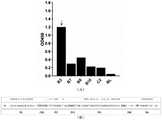
图5为纳米抗体TGF-β1-VHH与重组TGF-β1蛋白的ELLSA结合检测柱状图及序列结构图;Figure 5 is a bar graph and a sequence structure diagram of the ELLSA binding detection of nanobody TGF-β1-VHH and recombinant TGF-β1 protein;
图6为纳米抗体TGF-β1-VHH的原核诱导表达与纯化的凝胶电泳图像;Figure 6 is a gel electrophoresis image of prokaryotic induction and purification of Nanobody TGF-β1-VHH;
图7为纳米抗体TGF-β1-VHH与肿瘤细胞TGF-β1结合特异性检测图像;Figure 7 is an image of the specificity detection of nanobody TGF-β1-VHH binding to tumor cell TGF-β1;
图8为纳米抗体TGF-β1-VHH与重组TGF-β1的亲和力检测曲线图。Figure 8 is a graph showing the affinity detection curve of nanobody TGF-β1-VHH and recombinant TGF-β1.
具体实施方式Detailed ways
纳米抗体是羊驼外周血中存在一种天然缺失轻链的特殊抗体,该抗体为目前已知的天然来源的最小抗原结合单位,被称之为纳米抗体,其仅由VHH重链可变区和CH2、CH3恒定区组成,且单独的VHH区具有同样特异的抗原结合能力。此外它还兼具小分子药物与传抗抗体的优势,具有溶解性好、免疫原性低、穿透力强和人源化简单等优点,适用于多种表达系统。针对临床应用中靶向TGF-β1信号通路的封闭抗体多为完整抗体,存在免疫原性高、溶解性差、易沾粘聚集和易被蛋白酶降解等缺陷,本发明以该种纳米抗体为基础,制备一种羊驼来源的抗TGF-β1纳米抗体。Nanobody is a special antibody that naturally lacks light chain in the peripheral blood of alpaca. It is composed of constant regions of CH2 and CH3, and the VHH region alone has the same specific antigen-binding ability. In addition, it also combines the advantages of small molecule drugs and anti-antibodies, and has the advantages of good solubility, low immunogenicity, strong penetration and simple humanization, and is suitable for a variety of expression systems. In view of the fact that the blocking antibodies targeting the TGF-β1 signaling pathway in clinical applications are mostly complete antibodies, which have defects such as high immunogenicity, poor solubility, easy adhesion and aggregation, and easy degradation by proteases, the present invention is based on this nanobody. An alpaca-derived anti-TGF-β1 nanobody was prepared.
下面结合具体实施例和附图对本发明作进一步详细说明。The present invention will be further described in detail below with reference to specific embodiments and accompanying drawings.
下文的公开提供了许多不同的实施方式或例子用来实现本发明的不同方面。本发明提供了各种特定的工艺和材料的例子,但是本领域普通技术人员可以意识到其他工艺的应用和/或其他材料的使用。The following disclosure provides many different embodiments or examples for implementing different aspects of the invention. Various examples of specific processes and materials are provided herein, but one of ordinary skill in the art will recognize the application of other processes and/or the use of other materials.
实施例在以本发明技术方案为前提下进行实施,给出了详细的实施方式和具体的操作过程,但本发明的公开内容不限于下述的实施例。The examples are implemented on the premise of the technical solutions of the present invention, and detailed embodiments and specific operation procedures are given, but the disclosure content of the present invention is not limited to the following examples.
下述实施例中所用方法如无特别说明均为常规方法。The methods used in the following examples are conventional methods unless otherwise specified.
本发明实施例中使用的细胞系:Cell lines used in the examples of the present invention:
NCI-H520肺癌细胞:以含10%FCS的RPMI-1640培养基进行贴壁培养;HEK-293T人胚肾上皮细胞系:以含10%胎牛血清的DMEM培养基进行贴壁培养;上述细胞系均购自中国医学科学院细胞中心。NCI-H520 lung cancer cells: adherent culture in RPMI-1640 medium containing 10% FCS; HEK-293T human embryonic kidney epithelial cell line: adherent culture in DMEM medium containing 10% fetal bovine serum; the above cells All lines were purchased from the Cell Center of the Chinese Academy of Medical Sciences.
本发明实施例中使用的实验动物羊驼为3岁成年健康羊驼,购自内蒙古养殖场。The experimental animal alpaca used in the examples of the present invention is a 3-year-old adult healthy alpaca, purchased from a farm in Inner Mongolia.
本发明实施例中使用的菌株和质粒载体:Strains and plasmid vectors used in the examples of the present invention:
大肠杆菌DH5α感受态细胞、大肠杆菌TG1细胞,购自宝生物工程有限公司。基因型为:supE44ΔlacU169
M13K07辅助噬菌体购自北京全式金生物技术有限公司。The M13K07 helper phage was purchased from Beijing Quanshijin Biotechnology Co., Ltd.
cFUGW真核表达载体(Lois等.Science 2002 Feb 1:295(5556):868-72 Germlinetransmission and tissue-specific expression of transgenes delivered bylentiviral vectors)、pGEX6P.1原核表达载体、噬菌体pMECS载体均为国典(北京)医药科技有限公司实验室保存。cFUGW eukaryotic expression vector (Lois et al. Science 2002 Feb 1:295(5556):868-72 Germlinetransmission and tissue-specific expression of transgenes delivered by lentiviral vectors), pGEX6P.1 prokaryotic expression vector, phage pMECS vector are all national codes (Beijing ) Laboratory of Pharmaceutical Technology Co., Ltd.
本发明实施例中使用的主要工具酶和试剂:Main tool enzymes and reagents used in the examples of the present invention:
FastStart Taq DNA聚合酶(瑞士罗氏生物有限公司);Phusion高保DNA聚合酶、Nde I、Xho I、No tI、EcoR I、Age I、T4 DNA连接酶(美国New England Biolabs生物工程有限公司);IPTG、蛋白酶抑制剂、TaqMix(日本Toyobo公司);甘氨酸、溴酚蓝(美国Amresco公司);过硫酸铵、丙烯酰胺、甲醇、蛋白膜印迹再生液(中国康为世纪有限公司);封闭专用脱脂奶粉(中国普利莱公司)。FastStart Taq DNA polymerase (Roche Biotech, Switzerland); Phusion DNA polymerase, Nde I, Xho I, NotI, EcoR I, Age I, T4 DNA ligase (New England Biolabs Bioengineering Co., Ltd., USA); IPTG , protease inhibitor, TaqMix (Toyobo, Japan); Glycine, bromophenol blue (Amresco, USA); Ammonium persulfate, acrylamide, methanol, Western blot regeneration solution (China Kangwei Century Co., Ltd.); special non-fat milk powder for sealing (China Pulilai Company).
本发明实施例中使用的主要仪器:Main instruments used in the embodiments of the present invention:
OptimaL-100XP超速冷冻离心机(美国Beckman);LSM780激光共聚焦显微镜(德国Zeiss);AccuriC6流式细胞仪(美国BD);热循环仪、紫外线透射仪、Transblot SD半干转膜仪、电泳仪(美国BioRad);ECL自显影仪(CLiNx Science Instruments);电穿孔仪和0.1cm电穿孔比色皿(Gene Pulser)。OptimaL-100XP ultra-fast refrigerated centrifuge (Beckman, USA); LSM780 laser confocal microscope (Zeiss, Germany); AccuriC6 flow cytometer (BD, USA); (BioRad, USA); ECL autograph (CLiNx Science Instruments); electroporator and 0.1 cm electroporation cuvette (Gene Pulser).
实施例1、真核表达质粒cFUGW-TGF-β1的构建与蛋白表达纯化Example 1. Construction of eukaryotic expression plasmid cFUGW-TGF-β1 and protein expression and purification
该实施例中使用的人TGF-β1标准全长DNA序列(包含N端信号肽(signalpeptides,1~39aa)、LAP前体结构(30~278aa)和C端TGF-β单体(279~390aa)三部分,编码核苷酸序列长度为1173bp,如序列表中SEQ ID NO:1所示)保存于Gateway Cloning载体(通过常规Gateway基因克隆技术获得)内。The standard full-length DNA sequence of human TGF-β1 used in this example (including N-terminal signal peptide (signalpeptides, 1-39aa), LAP precursor structure (30-278aa) and C-terminal TGF-β monomer (279-390aa) ) three parts, the length of the coding nucleotide sequence is 1173 bp, as shown in SEQ ID NO: 1 in the sequence listing) and stored in the Gateway Cloning vector (obtained by conventional Gateway gene cloning technology).
该实施例具体包括以下步骤:This embodiment specifically includes the following steps:
1.1、首先利用Snapgene程序设计用于扩增获得TGF-β1标准全长DNA序列的上游引物F和下游引物R,并分别引入Age I和EcoR I酶切位点(下述上游引物F和下游引物R序列中下划线部分)及其保护碱基;此外目的序列起始密码子前添加了Kozak序列(上游引物F序列中黑色加粗部分),且在下游引入6His序列(下游引物R中黑色加粗部分)用于后续目的蛋白的纯化。1.1. First use Snapgene program to design upstream primer F and downstream primer R for amplifying the standard full-length DNA sequence of TGF-β1, and introduce Age I and EcoR I restriction sites (the following upstream primer F and downstream primer respectively) The underlined part in the R sequence) and its protective base; in addition, the Kozak sequence (the black bold part in the upstream primer F sequence) was added before the start codon of the target sequence, and the 6His sequence was introduced downstream (the black bold part in the downstream primer R) part) for subsequent purification of the target protein.
上游引物F:
下游引物R:Downstream primer R:


1.2、以Gateway Cloning载体中的人TGF-β1标准全长DNA序列为模板,以2×Phusion Master PCR酶、上游引物F及下游引物R为体系进行聚合酶链式反应,退火温度为70℃/65℃。收集上述PCR产物,并对产物进行凝胶电泳试验,如图1中A幅所示,其中lane1~2分别表示退火温度为70℃和65℃时的PCR产物的电泳结果,M表示为Marker,可见在退火温度70℃和65℃条件下均获得了片段大小为1188bp左右(如图中箭头指示)的目的PCR产物条带。使用柱式DNA回收试剂盒(购自生工生物工程有限公司)纯化上述PCR产物,并利用限制性内切酶Age I和EcoR I分别对cFUGW空载载体及PCR纯化产物进行双酶切反应,得到两条具有相同粘性末端的线性化片段,在T4连接酶存在下,将二者酶切产物于16℃过夜连接并转化后得到重组质粒,命名为cFUGW-TGF-β1重组质粒,其中在cFUGW载体中的原始GFP片段被TGF-β1片段替换。对得到的cFUGW-TGF-β1重组质粒进行Pac I和Ahd I双酶切鉴定,如图1中B幅所示,其中lane1~3表示为cFUGW-TGF-β1/PacI+AhdI(3518bp+6895bp),lane4表示为cFUGW-TGF-β1未酶切阴性对照,M表示1kb DNA Ladder。可见与上述设计引物时Snapgene软件预测结果相同;进一步测序结果显示cFUGW-TGF-β1重组质粒构建成功。1.2. The standard full-length DNA sequence of human TGF-β1 in the Gateway Cloning vector was used as the template, and 2 × Phusion Master PCR enzyme, upstream primer F and downstream primer R were used as the system to carry out polymerase chain reaction, and the annealing temperature was 70 ℃/ 65°C. Collect the above PCR products, and perform gel electrophoresis test on the products, as shown in panel A in Figure 1, where lanes 1 to 2 represent the electrophoresis results of the PCR products when the annealing temperature is 70°C and 65°C, respectively, M represents Marker, It can be seen that the target PCR product band with a fragment size of about 1188 bp (indicated by the arrow in the figure) was obtained at the annealing temperature of 70°C and 65°C. Use a column DNA recovery kit (purchased from Sangon Bioengineering Co., Ltd.) to purify the above PCR product, and use restriction endonucleases Age I and EcoR I to carry out a double digestion reaction on the cFUGW empty vector and the PCR purified product, respectively, to obtain Two linearized fragments with the same cohesive ends, in the presence of T4 ligase, the two digested products were ligated at 16 °C overnight and transformed to obtain a recombinant plasmid, named cFUGW-TGF-β1 recombinant plasmid, in which cFUGW vector The original GFP fragment in is replaced by a TGF-β1 fragment. The obtained cFUGW-TGF-β1 recombinant plasmid was identified by double digestion with Pac I and Ahd I, as shown in panel B in Figure 1, where
1.3、按照jetPRIME Polyplus转染试剂盒的要求,利用HEK-293T细胞瞬时转染上述cFUGW-TGF-β1重组质粒作为试验组,并在37℃、5%CO2条件下继续培养细胞,同时设置cFUGW空载质粒作为阴性对照组。培养24小时后对试验组和对照组的绿色荧光蛋白(GFP)进行观察,如图2所示,可见在cFUGW-TGF-β1重组质粒中,由于cFUGW载体的原始GFP已被TGF-β1片段替换,故无荧光出现(图2中B幅),而cFUGW空载质粒可以正常检测到绿色荧光(图2中A幅)。1.3. According to the requirements of jetPRIME Polyplus transfection kit, HEK-293T cells were transiently transfected with the above cFUGW-TGF-β1 recombinant plasmid as the experimental group, and the cells were continued to culture at 37°C and 5% CO2 , while setting cFUGW Empty plasmid was used as negative control. After 24 hours of culture, the green fluorescent protein (GFP) of the test group and the control group was observed, as shown in Figure 2, it can be seen that in the cFUGW-TGF-β1 recombinant plasmid, the original GFP of the cFUGW vector has been replaced by the TGF-β1 fragment. , so no fluorescence appeared (panel B in Figure 2), while the cFUGW empty plasmid could normally detect green fluorescence (panel A in Figure 2).
培养48~72h后,无菌收集细胞培养上清并用0.45μm滤膜过滤,获得带有6His标签的重组融合蛋白粗样。由于获得的融合蛋白TGF-β1末端引入的6His标签分子量极小,且具备免疫原性低、不改变自身可溶性、不影响蛋白折叠、亲和层析操作简易等优点,故可使用HisTrap-Ni SephaorseExcel系列填料纯化该重组融合蛋白,可最大限度保持该重组融合蛋白的免疫学活性。该实施例中使用HisTrap Column对上述获得的带有6His标签的融合蛋白粗样进行上机纯化。其中,平衡缓冲液为pH 7.2的20mM磷酸钠溶液,洗涤缓冲液为含20mM咪唑的磷酸钠溶液,洗脱缓冲液为含500mM咪唑的磷酸钠溶液;上样及洗脱流速分别设置为2mL/min和4mL/min。收集上述纯化前后蛋白样本及流穿液,分别用考马斯亮蓝染色检测蛋白纯度,Western Blot检测蛋白表达。其中配制12%SDS-PAGE凝胶,电泳,加入一抗(CST/兔源His-Tag,D3I10XPO)及相应种属二抗(羊抗兔IgG-HRP(购自北京三联博悦生物技术有限公司))(1:2000稀释)孵育1h后置入ECL自显影仪中曝光成像。After culturing for 48-72 hours, the cell culture supernatant was collected aseptically and filtered through a 0.45 μm filter to obtain a crude sample of recombinant fusion protein with a 6His tag. Because the molecular weight of the 6His tag introduced at the end of the obtained fusion protein TGF-β1 is extremely small, and it has the advantages of low immunogenicity, no change in self-solubility, no influence on protein folding, and simple operation of affinity chromatography, HisTrap-Ni SephaorseExcel can be used. A series of fillers purify the recombinant fusion protein, which can maximize the immunological activity of the recombinant fusion protein. In this example, a HisTrap Column was used to purify the crude fusion protein with a 6His tag obtained above. Among them, the equilibration buffer is a 20mM sodium phosphate solution with pH 7.2, the washing buffer is a sodium phosphate solution containing 20mM imidazole, and the elution buffer is a sodium phosphate solution containing 500mM imidazole; the sample loading and elution flow rates are respectively set to 2mL/ min and 4 mL/min. The protein samples and flow-through fluid before and after purification were collected, and the protein purity was detected by Coomassie brilliant blue staining, and the protein expression was detected by Western Blot. Prepare 12% SDS-PAGE gel, electrophoresis, add primary antibody (CST/rabbit-derived His-Tag, D3I10XPO) and corresponding secondary antibody (goat anti-rabbit IgG-HRP (purchased from Beijing Sanlian Boyue Biotechnology Co., Ltd.) )) (1:2000 dilution) after incubation for 1 h, it was placed in the ECL autograph for exposure imaging.
如图2中C幅和D幅所示,为Western blot检测蛋白表达结果,其中Lane1/2表示未转染重组质粒的细胞培养上清/细胞裂解液,lane3/4~5/6表示转染重组质粒的细胞培养上清/细胞裂解液,M表示蛋白分子量标准,GAPDH作为内参。结果显示重组质粒转染HEK-293T细胞后的培养上清中可以成功检测到LAP前体(55kDa)、TGF-β1单体(12kDa)及少量二聚体(25kDa);而细胞裂解液中仅检测到LAP前体。由于TGF-β1是绝大多数细胞均可分泌的多能效细胞因子,故未转染HEK-293T细胞上清也可检测到少量LAP前体的表达。Panels C and D in Figure 2 are the results of Western blot detection of protein expression, where Lane1/2 represents the cell culture supernatant/cell lysate without recombinant plasmid transfection, and
如图3(A幅和B幅)所示,为考马斯亮蓝染色检测亲和层析蛋白纯化效果的结果,其中lane1表示纯化前上清,2表示流穿液,3~7表示不同纯化洗脱孔,M表示蛋白分子标准。结果显示Lane3/4孔内三种形式的TGF-β1均得到较好的纯化。As shown in Figure 3 (panel A and panel B), it is the result of Coomassie brilliant blue staining to detect the purification effect of affinity chromatography protein, where lane1 represents the supernatant before purification, 2 represents the flow-through liquid, and 3-7 represent different purification washes De-pore, M indicates protein molecular standard. The results showed that the three forms of TGF-β1 were well purified in Lane3/4 wells.
根据上述融合蛋白检测结果,证明该实施例获得纯度良好的末端引入有6His标签的融合蛋白,命名为TGF-β1纯化蛋白,其中包含TGF-β1的三种形式:LAP前体、TGF-β1单体及其二聚体。According to the above fusion protein detection results, it is proved that this example obtains a fusion protein with a 6His tag introduced at the end of good purity, named TGF-β1 purified protein, which contains three forms of TGF-β1: LAP precursor, TGF-β1 single body and its dimer.
实施例2:羊驼噬菌体VHH展示文库的构建Example 2: Construction of alpaca phage VHH display library
该实施例使用上述实施例1获得的TGF-β1纯化蛋白对羊驼进行免疫,构建VHH基因文库,具体包括以下步骤:This example uses the purified protein of TGF-β1 obtained in the above Example 1 to immunize alpacas to construct a VHH gene library, which specifically includes the following steps:
2.1、初次免疫将1mg TGF-β1纯化蛋白(实施例1获得)与完全弗氏佐剂1:1等体积混匀并充分乳化,采用皮下注射的方式免疫羊驼;其后用0.5mg TGF-β1纯化蛋白1:1等体积混合不完全弗氏佐剂进行4次加强免疫;共免疫5次,单次间隔为20天。抽取少量羊驼外周血进行效价检测,待免疫效价大于1:60000时,再次进行加强免疫,24h后,在被免疫羊驼颈静脉处采集100mL抗凝血。从该抗凝血中提取外周血淋巴细胞,并使用TRIZOL试剂分离总RNA,在SuperScriptTMII反转录酶及oligo-dT引物的作用下获得其cDNA。2.1.
2.2、以上述获得的cDNA为模板,利用下表1所示的CALL001引物、CALL002引物和FastStart Taq DNA聚合酶进行第一次PCR扩增,反应体系均为50μL,在95℃下孵育7分钟,然后进行30~35个PCR循环,使cDNA变性并激活聚合酶,每个循环包括94℃-60s,55℃-60s和72℃-60s。在最后一个PCR循环后,在72℃下进行10分钟的最终DNA延伸步骤。随后将第一次PCR产物进行琼脂糖电泳,如图4中A幅所示,为电泳结果,将700bp处的条带进行回收合并,用作第二次PCR(巢式PCR)的模板;随后利用下表1所示的VHH-BACK引物、PMCF引物进行第二次PCR扩增,反应体系同第一次PCR扩增,通过在95℃下孵育7分钟使DNA模板变性并激活聚合酶,然后进行17~20个PCR循环,每个循环包括94℃-45s,55℃-45s和72℃-45s。在最后一个PCR循环后,在72℃进行10分钟的DNA延伸步骤。随后将获得的PCR产物进行琼脂糖电泳,如图4中B幅所示,为电泳结果,收集并纯化400bp的预估扩增条带,即得到羊驼外周血液中天然缺失轻链的抗体的重链可变区(VHH)片段。2.2. Using the cDNA obtained above as a template, use the CALL001 primers, CALL002 primers and FastStart Taq DNA polymerase shown in Table 1 below to carry out the first PCR amplification, the reaction systems are all 50 μL, incubate at 95 ℃ for 7 minutes, Then 30-35 PCR cycles were performed to denature the cDNA and activate the polymerase, each cycle including 94°C-60s, 55°C-60s and 72°C-60s. After the last PCR cycle, a final DNA extension step was performed at 72 °C for 10 min. Subsequently, the first PCR product was subjected to agarose electrophoresis, as shown in panel A in Figure 4, for the electrophoresis result, the band at 700bp was recovered and merged, and used as the template for the second PCR (nested PCR); then The second PCR amplification was performed using the VHH-BACK primers and PMCF primers shown in Table 1 below. The reaction system was the same as the first PCR amplification. The DNA template was denatured and the polymerase was activated by incubating at 95°C for 7 minutes, and then 17-20 PCR cycles were performed, each cycle including 94°C-45s, 55°C-45s and 72°C-45s. After the last PCR cycle, a DNA extension step was performed at 72°C for 10 minutes. The obtained PCR product was then subjected to agarose electrophoresis, as shown in panel B in Figure 4, as the result of electrophoresis, the estimated amplification band of 400 bp was collected and purified, that is, to obtain an antibody that naturally lacks light chains in the peripheral blood of alpaca Heavy chain variable region (VHH) fragments.
表1:羊驼噬菌体VHH展示文库的构建所用引物Table 1: Primers used for construction of alpaca phage VHH display library

2.3、将纯化的巢式PCR产物(400bp的扩增条带)和噬菌体pMECS载体经Pst I、NotI双酶切后,以摩尔比1:3将二者的酶切产物在T4连接酶的存在下,16℃连接后电转化至大肠杆菌TG1感受态细胞。将电转化后的菌液在LB液体培养基中于37℃摇床培养1小时,取1微升稀释103、106、109倍后涂布LB固体培养皿表面用于库容量测定,其余菌液涂布在LB固体培养皿表面,过夜于37℃培养箱中培养TG1细胞,次日收集所有菌落为VHH抗体文库。用菌落PCR方法鉴定VHH片段的阳性克隆效率(即VHH片段插入率),其中菌落PCR使用的引物为:MP57:TTATGCTTCCGGCTCGTATG(SEQ ID NO:8);GIII:CCACAGACAGCCCTCATAG(SEQ ID NO:9),终浓度均为0.4m M,图4中C幅结果显示VHH片段的阳性克隆率在70%左右,证明获得VHH抗体文库。根据PCR阳性克隆率利用以下公式:库容量=测定培养皿上克隆数×稀释倍数×PCR鉴定阳性克隆率×菌液体积(单位:微升)推算羊驼噬菌体VHH展示文库的库容量为1.2×10⁸。2.3. After the purified nested PCR product (amplified band of 400bp) and the phage pMECS vector were double digested by Pst I and NotI, the digested products of the two were digested in the presence of T4 ligase at a molar ratio of 1:3. ligated at 16°C and electrotransformed into E. coli TG1 competent cells. The electrotransformed bacterial liquid was cultured in LB liquid medium at 37°C on a shaker for 1 hour, and 1 microliter was diluted 103 , 106 , and 109 times and then coated on the surface of LB solid petri dish for storage capacity determination. The rest of the bacterial solution was coated on the surface of the LB solid petri dish, and the TG1 cells were cultured in a 37°C incubator overnight, and all colonies were collected the next day as a VHH antibody library. The positive cloning efficiency of VHH fragments (ie VHH fragment insertion rate) was identified by colony PCR method, wherein the primers used in colony PCR were: MP57: TTATGCTTCCGGCTCGTATG (SEQ ID NO: 8); GIII: CCACAGACAGCCCTCATAG (SEQ ID NO: 9), and finally The concentrations were all 0.4 mM. The results of panel C in Figure 4 showed that the positive clone rate of VHH fragments was about 70%, which proved that the VHH antibody library was obtained. According to the PCR positive clone rate, the following formula is used: library capacity = the number of clones on the assay dish × dilution factor × PCR identification positive clone rate × bacterial liquid volume (unit: microliter), the library capacity of the alpaca phage VHH display library is calculated to be 1.2 × 10⁸ .
2.4、在2YT固体培养基上划线接种M13K07辅助噬菌体。挑取相隔良好的噬菌斑进行扩繁,获得滴度为1×10¹²cfu/mL的辅助噬菌体M13K07。用该辅助噬菌体M13K07感染上述步骤2.3获得的VHH抗体文库,过夜培养后上清用PEG/NaCl(20%,2.5M)溶液进行沉淀,无菌PBS悬浮沉淀,分离重组噬菌体颗粒,即为羊驼噬菌体VHH展示文库。2.4. Streak M13K07 helper phage on 2YT solid medium. The well-spaced plaques were picked and propagated to obtain helper phage M13K07 with a titer of 1×10¹² cfu/mL. Infect the VHH antibody library obtained in the above step 2.3 with the helper phage M13K07. After overnight culture, the supernatant is precipitated with a PEG/NaCl (20%, 2.5M) solution, suspended in sterile PBS, and the recombinant phage particles are separated, which is alpaca Phage VHH Display Libraries.
测定羊驼噬菌体VHH展示文库滴度:将获得的重组噬菌体颗粒原液进行按照1:106、1:107、1:108等进行梯度稀释,随后各取1微升感染TG1细胞15分钟,涂布氨苄抗性LB固体培养皿表面,次日观察培养板中菌落形成情况,根据公式(菌落数×稀释倍数×103)计算噬菌体滴度(pfu)。如图4中D幅所示,左右两幅图分别为1:10000000和1:1000000稀释倍数下的噬菌斑数,因此估算羊驼噬菌体VHH展示文库的滴度可达1.2×10¹³pfu。Determination of the titer of the alpaca phage VHH display library: the obtained recombinant phage particle stock solution was serially diluted according to 1:106 , 1:107 , 1:108 , etc., and then 1 μl of each was used to infect TG1 cells for 15 minutes, The surface of the ampicillin-resistant LB solid petri dish was coated, the colony formation in the culture plate was observed the next day, and the phage titer (pfu) was calculated according to the formula (colony number×dilution factor×103 ). As shown in panel D in Figure 4, the left and right panels are the plaque numbers at 1:10,000,000 and 1:1,000,000 dilutions, respectively, so the estimated titer of the alpaca phage VHH display library can reach 1.2×10¹³ pfu.
实施例3:表达纳米抗体TGF-β1-VHH的阳性克隆株的筛选Example 3: Screening of positive clones expressing Nanobody TGF-β1-VHH
3.1:亲和淘选的简化步骤:3.1: Simplified steps for affinity panning:
(1)用实施例1获得的TGF-β1纯化蛋白包被免疫管,4℃过夜。(1) Coat the immune tube with the purified TGF-β1 protein obtained in Example 1, overnight at 4°C.
(2)PBS洗管3次,拍干。(2) Wash the
(3)3%MPBS(3%脱脂牛奶加入PBS中)封闭,37℃孵育2h,倾去封闭液,用PBS洗管3次,拍干。(3) Block with 3% MPBS (3% skim milk added to PBS), incubate at 37° C. for 2 h, pour off the blocking solution, wash the tube three times with PBS, and pat dry.
(4)向封闭好的免疫管加入上述实施例2制备的羊驼噬菌体VHH展示文库(噬菌体文库),2ml/管,轻摇温育30min后,再静置温育1.5h。(4) Add the alpaca phage VHH display library (phage library) prepared in Example 2 to the sealed immune tube, 2 ml/tube, shake gently for 30 min, and then incubate for 1.5 h.
(5)弃管内噬菌体文库,用PBST洗涤3次,再用PBS洗涤3次,拍干。(5) Discard the phage library in the tube, wash 3 times with PBST, then 3 times with PBS, and pat dry.
(6)加入宿主菌TG1对结合噬菌体文库进行洗脱。此即完成第一轮淘筛,获得一级筛选抗体库。(6) Add host strain TG1 to elute the bound phage library. This completes the first round of panning and obtains the primary screening antibody library.
(7)重复上述淘筛步骤4)、5)和6),进行循环淘筛,最终经过4轮淘筛,即获得四级筛选抗体库。(7) Repeat the above-mentioned panning steps 4), 5) and 6), carry out cyclic panning, and finally pass through 4 rounds of panning to obtain a quaternary screening antibody library.
3.2、间接Phage Elisa初步筛选抗原阳性纳米抗体:3.2. Indirect Phage Elisa preliminary screening of antigen-positive nanobodies:
(1)挑取4轮淘筛后涂布在2YTAG平板上长出的单个菌落,接种至72孔培养板中,该板标记为MasterPlate,30℃,振荡培养过夜。(1) Pick a single colony grown on a 2YTAG plate after 4 rounds of panning, and inoculate it into a 72-well culture plate, marked as MasterPlate, at 30°C for overnight shaking culture.
(2)次日,另取72孔培养板,取400μL含辅助M13K07至每孔,此板标为P1Plate。(2) The next day, another 72-well culture plate was taken, and 400 μL of auxiliary M13K07 was taken into each well, and this plate was marked as P1Plate.
(3)从过夜培养的MasterPlate上每孔取40μL培养液至P1Plate,37℃振荡培养过夜;1500g离心20min,小心留上清待用,即为重组表达抗体。(3) Take 40 μL of culture medium from each well of the overnight cultured MasterPlate to P1Plate, shake and culture at 37°C overnight; centrifuge at 1500g for 20 minutes, carefully save the supernatant for later use, which is the recombinant expression antibody.
(4)用TGF-β1纯化蛋白包被96孔酶标板。(4) Coat 96-well microtiter plate with TGF-β1 purified protein.
(5)提前将上述重组表达抗体160μL与40μLMPBS混匀,室温孵育20min。加入封闭好的酶标孔中,37℃结合反应2小时。(5) Mix 160 μL of the above recombinantly expressed antibody with 40 μL of MPBS in advance, and incubate at room temperature for 20 min. Add to the blocked enzyme-labeled wells, and react at 37°C for 2 hours.
(6)洗涤及加酶标二抗:将酶标抗M13K07抗体用PBS做1:4000稀释,200μL/孔,37℃孵育反应1小时。(6) Washing and adding enzyme-labeled secondary antibody: The enzyme-labeled anti-M13K07 antibody was diluted 1:4000 with PBS, 200 μL/well, and incubated at 37° C. for 1 hour.
(7)加入200μL/孔TMB显色液,37℃孵育显色45min左右显色及用100μL/孔终止液,终止显色,在405nm处读值。读数大于阴性对照至少2倍视为阳性克隆株。(7) Add 200 μL/well of TMB chromogenic solution, incubate at 37°C for about 45 minutes, and use 100 μL/well of stop solution to stop the color development, and read the value at 405 nm. Reads at least 2 times greater than the negative control were considered positive clones.
经间接Phage Elisa初筛,与阴性对照比较,结果如图5中A幅所示,在随机挑选的6株克隆中共筛选得到编号为B3的1株阳性克隆,其可以表达与TGF-β1纯化蛋白高度特异性结合的纳米抗体,命名为纳米抗体TGF-β1-VHH(B3阳性克隆株表达的纳米抗体TGF-β1-VHH命名为B3纳米抗体)。对上述1株阳性克隆表达的纳米抗体TGF-β1-VHH(即VHH区域)进行测序,该株阳性克隆表达的纳米抗体TGF-β1-VHH(即B3纳米抗体)的氨基酸序列为序列表中SEQ ID NO:10所示,对应的核苷酸序列为序列表中SEQ ID NO:11所示。对该氨基酸序列进行结构分析,如图5中B幅所示,该氨基酸序列长度为110个氨基酸残基,其结构为:FR1-CDR1-FR2-CDR2-FR3-CDR3-FR4,其中CDRl、CDR2和CDR3为互补决定区(complementaritydetermining region,CDR);CDR共同组成抗体的抗原结合部位(antigen-binding site),决定抗体的特异性,是抗体识别及结合抗原的部位。FR1、FR2、FR3和FR4为骨架区(framework region,FR)。After indirect Phage Elisa primary screening, compared with the negative control, the results are shown in panel A in Figure 5. A total of 6 randomly selected clones were screened to obtain 1 positive clone numbered B3, which can express the purified protein of TGF-β1. The highly specific binding Nanobody was named Nanobody TGF-β1-VHH (the Nanobody TGF-β1-VHH expressed by B3 positive clones was named B3 Nanobody). Sequence the nanobody TGF-β1-VHH (ie VHH region) expressed by the positive clone above, and the amino acid sequence of the nanobody TGF-β1-VHH (ie B3 nanobody) expressed by this positive clone is SEQ in the sequence table ID NO: 10, the corresponding nucleotide sequence is shown in SEQ ID NO: 11 in the sequence listing. Structural analysis is carried out to this amino acid sequence, as shown in B in Figure 5, the amino acid sequence length is 110 amino acid residues, and its structure is: FR1-CDR1-FR2-CDR2-FR3-CDR3-FR4, wherein CDR1, CDR2 and CDR3 are complementarity determining regions (complementarity determining regions, CDRs); CDRs together constitute the antigen-binding site of the antibody, which determines the specificity of the antibody and is the site where the antibody recognizes and binds to the antigen. FR1, FR2, FR3 and FR4 are framework regions (FR).
实施例4:纳米抗体TGF-β1-VHH的原核诱导表达与纯化Example 4: Prokaryotic induction and purification of nanobody TGF-β1-VHH
该实施例首先利用上述实施例3筛选获得的1株阳性克隆,使用GenElute PlasmidMiniprep Kit按照说明书要求制备重组pMECS噬菌粒。利用该重组pMECS噬菌粒构建纳米抗体TGF-β1-VHH的表达质粒,转入大肠杆菌中进行诱导表达获得B3纳米抗体,具体包括以下步骤:In this example, one positive clone obtained by screening in the above Example 3 was firstly used, and the recombinant pMECS phagemid was prepared by using the GenElute Plasmid Miniprep Kit according to the instructions. The recombinant pMECS phagemid is used to construct the expression plasmid of the nanobody TGF-β1-VHH, which is transferred into Escherichia coli for induced expression to obtain the B3 nanobody, which specifically includes the following steps:
4.1、将重组pMECS噬菌粒与pGEX6P.1-GST原核表达载体分别经BamH I、EcoR I双酶切后,获得VHH目的片段和pGEX6P.1线性化载体,在T4连接酶存在下进行连接,连接后构建得到pGEX6P.1-VHH原核表达载体。将该pGEX6P.1-VHH原核表达载体转入大肠杆菌DH5α感受态细胞中进行表达,筛选获得阳性菌株,命名为B3阳性菌株。4.1. After the recombinant pMECS phagemid and pGEX6P.1-GST prokaryotic expression vector were digested with BamH I and EcoR I respectively, the VHH target fragment and pGEX6P.1 linearized vector were obtained, and were connected in the presence of T4 ligase. After ligation, the pGEX6P.1-VHH prokaryotic expression vector was constructed. The pGEX6P.1-VHH prokaryotic expression vector was transferred into Escherichia coli DH5α competent cells for expression, and a positive strain was obtained by screening, which was named as the B3 positive strain.
4.2、对B3阳性菌株进行1mM IPTG,22℃的过夜原核诱导,高速离心富集菌体,反复冻融4至6次后超声彻底裂解菌体(工作2s、间歇6s,功率为40%)。随后将相对澄清的裂解液进行12000rpm高速离心,收获上清液,沉淀使用8M尿素复溶,分别得到上清及包涵体中的纳米抗体,使用考马斯亮蓝染色方法和Western blot方法(电泳及抗GST-Western blot)检测纳米抗体表达(CST/鼠源GST单克隆抗体;中杉金桥/山羊抗小鼠二抗)。4.2. Prokaryotic induction of B3 positive strains at 22°C overnight with 1 mM IPTG, high-speed centrifugation to enrich the cells, repeated freezing and thawing for 4 to 6 times, and then ultrasonically lysing the cells (working 2s, intermittently 6s, power 40%). The relatively clear lysate was then centrifuged at 12,000 rpm at a high speed, the supernatant was harvested, and the precipitate was reconstituted with 8M urea to obtain the nanobodies in the supernatant and inclusion bodies, respectively, using Coomassie brilliant blue staining method and Western blot method (electrophoresis and antibody GST-Western blot) to detect the expression of nanobody (CST/mouse GST monoclonal antibody; Zhongshan Jinqiao/goat anti-mouse secondary antibody).
结果如图6中A幅和B幅所示,其中A幅表示考马斯亮蓝染色结果,B幅表示Westernblot检测结果,A和B中的Lane1/2均表示B3阳性菌株的上清/包涵体中纳米抗体的检测结果,M表示蛋白分子标准。可见B3纳米抗体与GST融合蛋白(分子量为36kDa)在B3阳性菌株的培养上清和包涵体中均得到了表达,其中上清表达效果更好。The results are shown in panels A and B in Figure 6, where panel A represents the result of Coomassie brilliant blue staining, panel B represents the result of Western blot detection, and Lane1/2 in both A and B represents the supernatant/inclusion body of the B3 positive strain The detection result of the nanobody, M represents the protein molecular standard. It can be seen that the B3 nanobody and GST fusion protein (molecular weight 36kDa) were expressed in both the culture supernatant and inclusion bodies of B3 positive strains, and the supernatant expression effect was better.
4.3、对B3阳性菌株处理之后的细菌裂解液上清和包涵体复溶液进行GSTrapTM FF1mL色谱柱纯化,色谱柱由生物相容性聚丙烯制成,赋予其与GST标签阳性蛋白和其他谷胱甘肽结合蛋白的强结合能力。其中,洗脱缓冲液为含10mM还原型谷胱甘肽的Tris-HCl溶液(pH8.0)。使用
结果如图6中C幅和D幅所示,其中C幅表示考马斯亮蓝染色结果,D幅表示SDS-PAGE电泳检测结果,在C幅和D幅中,Lane1/2均表示为纯化前上清/包涵体中的纳米抗体检测结果,Lane3/4均表示为纯化后上清/包涵体中的纳米抗体检测结果,M表示蛋白分子标准。可见纯化后上清中表达的纳米抗体得到了有效的富集。The results are shown in panels C and D in Figure 6, where panel C represents the result of Coomassie brilliant blue staining, panel D represents the detection result of SDS-PAGE electrophoresis, and in panels C and D, Lane1/2 is shown as the upper part before purification The nanobody detection results in the supernatant/inclusion body, Lane3/4 are expressed as the nanobody detection result in the purified supernatant/inclusion body, and M is the protein molecular standard. It can be seen that the Nanobodies expressed in the supernatant after purification are effectively enriched.
实施例5:纳米抗体TGF-β1-VHH的结合特异性检测Example 5: Detection of binding specificity of Nanobody TGF-β1-VHH
该实施例收集上述实施例4经纯化后上清中的B3纳米抗体,以市售TGF-β1抗体作为对照,检测与天然TGF-β1蛋白的结合特异性。In this example, the B3 nanobody in the purified supernatant of the above Example 4 was collected, and the binding specificity to the natural TGF-β1 protein was detected by using the commercially available TGF-β1 antibody as a control.
5.1、Western Blot检测纳米抗体对TGF-β1的结合特异性5.1. Western Blot to detect the binding specificity of nanobodies to TGF-β1
在RPMI-1640培养基中扩增培养人肺癌细胞系NCI-H520,用RIPA裂解液充分释放细胞全蛋白,每孔定量10μg上样至预先配制的12%SDS-PAGE胶内。分别用一抗(分别为B3纳米抗体(B3-VHH)、市售兔源TGF-β1单抗(阳性对照))进行孵育,4℃过夜,随后用HRP标记的相应种属来源二抗(Alexa FluorR555)(1:2000稀释)进行二抗孵育,室温孵育时间1h,随后放入ECL自显影仪(CLinX公司)中曝光成像。The human lung cancer cell line NCI-H520 was expanded and cultured in RPMI-1640 medium, and the whole cell protein was fully released with RIPA lysis buffer, and 10 μg of each well was quantitatively loaded onto a pre-prepared 12% SDS-PAGE gel. The cells were incubated with primary antibodies (respectively B3 nanobody (B3-VHH), commercially available rabbit-derived TGF-β1 mAb (positive control)) at 4°C overnight, followed by HRP-labeled secondary antibodies from the corresponding species (Alexa FluorR 555) (1:2000 dilution) for secondary antibody incubation, incubation time at room temperature for 1 h, and then placed in an ECL autograph (CLinX company) for exposure imaging.
结果如图7中A幅所示,样本为NCI-H520肿瘤细胞裂解液/10μg,其中Lane1~2分别为实验组B3纳米抗体、阳性对照组市售TGF-β1单抗;M:蛋白分子量标准。显示两者对不同形式的TGF-β1(前体、单体和二聚体)均有较好的结合,市售抗体组非特异性条带较少。The results are shown in panel A in Figure 7. The sample is NCI-H520 tumor cell lysate/10 μg, of which Lane1-2 are the B3 nanobody in the experimental group and the commercially available TGF-β1 monoclonal antibody in the positive control group; M: protein molecular weight standard . It is shown that both of them have good binding to different forms of TGF-β1 (precursor, monomer and dimer), and the commercial antibody group has less non-specific bands.
5.2、免疫荧光检测纳米抗体对TGF-β1的结合特异性5.2. Immunofluorescence detection of the binding specificity of nanobodies to TGF-β1
制备NCI-H520细胞爬片,37℃,5%CO2孵箱内过夜贴壁,4%多聚甲醛固定后进行抗原特异性反应:一抗分别为B3纳米抗体(B3-VHH)、市售TGF-β1单抗(阳性对照),室温孵育4h;其后分别使用同种属荧光二抗(Alexa FluorR555)再次进行特异性结合;两组爬片经DAPI染核15min后封片,于Zeiss共聚焦显微镜上采集图像。Prepare NCI-H520 cell slides, adhere overnight in a 37°C, 5% CO2 incubator, fix with 4% paraformaldehyde, and perform antigen-specific reactions: primary antibodies are B3 nanobody (B3-VHH), commercially available TGF-β1 monoclonal antibody (positive control) was incubated at room temperature for 4 hours; after that, specific binding was carried out again using the same fluorescent secondary antibody (Alexa FluorR 555); the two groups of slides were stained with DAPI for 15 minutes, then mounted and placed in Images were acquired on a Zeiss confocal microscope.
结果如图7中B幅所示,其中由上至下一抗分别为阳性对照组市售TGF-β1单抗、实验组B3纳米抗体,显示两者均能够特异性识别NCI-H520细胞表面TGF-β1。The results are shown in panel B in Figure 7, in which the antibodies from top to bottom are the commercially available TGF-β1 monoclonal antibody in the positive control group and the B3 nanobody in the experimental group, showing that both can specifically recognize TGF-β on the surface of NCI-H520 cells -β1.
5.3、Fortebio实验检测纳米抗体与TGF-β1蛋白的结合亲和性5.3. Fortebio assay to detect the binding affinity of nanobodies to TGF-β1 protein
Fortebio Octet实验利用生物膜干涉技术(Bio-Layer Interferometry),预装GST传感器与纳米抗体TGF-β1-VHH非共价结合,检测与目标蛋白TGF-β1的相互作用。上样稀释液为含0.02%Tween-20及0.1%BSA的PBST溶液,平衡/固化缓冲液与上样缓冲液保持一致。将TGF-β1纯化蛋白(实施例1获得)由1000nM梯度稀释6个单位,同时设置缓冲液组作为背景扣除。样品板(Greiner PN655209)中分别加入上样缓冲液、固化样品(B3纳米抗体)、结合样品(梯度稀释的TGF-β1纯化蛋白);程序设置包括固化、结合、解离三个步骤,时间分别为5min、10min、20min。亲和力判断依据如下:KD>10-8M属于结合能力较弱;10-9M<KD<10-8M属于结合能力较强;10-11M<KD<10-9M属于结合能力强;KD<10-11M属于结合能力很强。The Fortebio Octet experiment uses Bio-Layer Interferometry to non-covalently bind the preloaded GST sensor to the nanobody TGF-β1-VHH to detect the interaction with the target protein TGF-β1. The loading diluent was PBST solution containing 0.02% Tween-20 and 0.1% BSA, and the equilibration/solidification buffer was the same as the loading buffer. The TGF-β1 purified protein (obtained in Example 1) was serially diluted by 6 units from 1000 nM, and the buffer group was set as background subtraction. The sample plate (Greiner PN655209) was added with loading buffer, immobilized sample (B3 nanobody), and binding sample (gradually diluted TGF-β1 purified protein). 5min, 10min, 20min. The affinity judgment is based on the following: KD>10-8 M indicates weak binding ability; 10-9 M<KD<10-8 M indicates strong binding ability; 10-11 M<KD<10-9 M indicates strong binding ability; KD<10-11 M belongs to strong binding ability.
结果如图8所示,结果显示B3纳米抗体属于慢结合和慢解离模式,亲和力常数KD(M)为9×10⁻⁹mol/L,属于较强结合。The results are shown in Figure 8. The results show that the B3 Nanobody belongs to slow binding and slow dissociation modes, and the affinity constant KD(M) is 9×10⁻⁹ mol/L, which is a strong binding.
综上所述,本发明制备获得的抗TGF-β1纳米抗体(B3纳米抗体)具有高特异性识别能力和高亲和力结合能力,能够有效中和肿瘤微环境中自分泌或旁分泌TGF-β1(包含LAP前体(LAP原基和TGF-β1单体以非共价键接合)、TGF-β1单体及二聚体),弥补传统TGF-β1抗体的固有缺陷,可在抵消肿瘤免疫逃逸、抑制肿瘤的发生发展中发挥重要作用。因此,制备得到的抗TGF-β1纳米抗体可以用于制备肿瘤治疗药物,在该肿瘤治疗药物中还可以包含药学上可接受的载剂,例如细胞毒素、放射性同位素、免疫调节剂、抗血管生成剂、抗增殖剂、促凋亡剂、化学治疗剂或治疗性核酸的治疗剂等。利用该肿瘤治疗药物可以治疗包括但不限于肺癌、卵巢癌、乳腺癌、淋巴瘤、宫颈癌、肝癌、胃癌、胶质瘤等肿瘤,并能够促进伤口愈合以及促进组织修复和胚胎发育等。To sum up, the anti-TGF-β1 nanobody (B3 nanobody) prepared by the present invention has high specific recognition ability and high affinity binding ability, and can effectively neutralize autocrine or paracrine TGF-β1 ( It contains LAP precursor (LAP primordium and TGF-β1 monomer are joined by non-covalent bonds), TGF-β1 monomer and dimer), which makes up for the inherent defects of traditional TGF-β1 antibodies and can counteract tumor immune escape, It plays an important role in inhibiting the occurrence and development of tumors. Therefore, the prepared anti-TGF-β1 nanobody can be used to prepare a tumor therapeutic drug, and the tumor therapeutic drug can also contain a pharmaceutically acceptable carrier, such as cytotoxin, radioisotope, immunomodulator, anti-angiogenesis agents, anti-proliferative agents, pro-apoptotic agents, chemotherapeutic agents, or therapeutic agents for therapeutic nucleic acids, and the like. The tumor therapeutic drug can be used to treat tumors including but not limited to lung cancer, ovarian cancer, breast cancer, lymphoma, cervical cancer, liver cancer, gastric cancer, glioma, etc., and can promote wound healing, tissue repair and embryonic development.
最后应说明的是:以上所述仅为本发明的优选实施例而已,并不用于限制本发明,尽管参照前述实施例对本发明进行了详细的说明,对于本领域的技术人员来说,其依然可以对前述各实施例所记载的技术方案进行修改,或者对其中部分技术特征进行等同替换。凡在本发明的精神和原则之内,所作的任何修改、等同替换、改进等,均应包含在本发明的保护范围之内。Finally, it should be noted that the above are only preferred embodiments of the present invention, and are not intended to limit the present invention. Although the present invention has been described in detail with reference to the foregoing embodiments, for those skilled in the art, it is still The technical solutions described in the foregoing embodiments may be modified, or some technical features thereof may be equivalently replaced. Any modification, equivalent replacement, improvement, etc. made within the spirit and principle of the present invention shall be included within the protection scope of the present invention.
序列表sequence listing
<110> 中国医学科学院基础医学研究所<110> Institute of Basic Medicine, Chinese Academy of Medical Sciences
<120> 抗人转化生长因子β1的纳米抗体B3及其制备方法和应用<120> Nanobody B3 against human transforming
<160> 11<160> 11
<170> SIPOSequenceListing 1.0<170> SIPOSequenceListing 1.0
<210> 1<210> 1
<211> 1173<211> 1173
<212> DNA<212> DNA
<213> 人工序列(Artificial Sequence)<213> Artificial Sequence
<400> 1<400> 1
atgccgccct ccgggctgcg gctgctgccg ctgctgctac cgctgctgtg gctactggtg 60atgccgccct ccgggctgcg gctgctgccg ctgctgctac cgctgctgtg gctactggtg 60
ctgacgcctg gccggccggc cgcgggacta tccacctgca agactatcga catggagctg 120ctgacgcctg gccggccggc cgcgggacta tccacctgca agactatcga catggagctg 120
gtgaagcgga agcgcatcga ggccatccgc ggccagatcc tgtccaagct gcggctcgcc 180gtgaagcgga agcgcatcga ggccatccgc ggccagatcc tgtccaagct gcggctcgcc 180
agccccccga gccaggggga ggtgccgccc ggcccgctgc ccgaggccgt gctcgccctg 240agccccccga gccaggggga ggtgccgccc ggcccgctgc ccgaggccgt gctcgccctg 240
tacaacagca cccgcgaccg ggtggccggg gagagtgcag aaccggagcc cgagcctgag 300tacaacagca cccgcgaccg ggtggccggg gagagtgcag aaccggagcc cgagcctgag 300
gccgactact acgccaagga ggtcacccgc gtgctaatgg tggaaaccca caacgaaatc 360gccgactact acgccaagga ggtcacccgc gtgctaatgg tggaaaccca caacgaaatc 360
tatgacaagt tcaagcagag tacacacagc atatatatgt tcttcaacac atcagagctc 420tatgacaagt tcaagcagag tacacacagc atatatatgt tcttcaacac atcagagctc 420
cgagaagcgg tacctgaacc cgtgttgctc tcccgggcag agctgcgtct gctgaggctc 480cgagaagcgg tacctgaacc cgtgttgctc tcccgggcag agctgcgtct gctgaggctc 480
aagttaaaag tggagcagca cgtggagctg taccagaaat acagcaacaa ttcctggcga 540aagttaaaag tggagcagca cgtggagctg taccagaaat acagcaacaa ttcctggcga 540
tacctcagca accggctgct ggcacccagc gactcgccag agtggttatc ttttgatgtc 600tacctcagca accggctgct ggcacccagc gactcgccag agtggttatc ttttgatgtc 600
accggagttg tgcggcagtg gttgagccgt ggaggggaaa ttgagggctt tcgccttagc 660accggagttg tgcggcagtg gttgagccgt ggaggggaaa ttgagggctt tcgccttagc 660
gcccactgct cctgtgacag cagggataac acactgcaag tggacatcaa cgggttcact 720gcccactgct cctgtgacag cagggataac acactgcaag tggacatcaa cgggttcact 720
accggccgcc gaggtgacct ggccaccatt catggcatga accggccttt cctgcttctc 780accggccgcc gaggtgacct ggccaccatt catggcatga accggccttt cctgcttctc 780
atggccaccc cgctggagag ggcccagcat ctgcaaagct cccggcaccg ccgagccctg 840atggccaccc cgctggagag ggcccagcat ctgcaaagct cccggcaccg ccgagccctg 840
gacaccaact attgcttcag ctccacggag aagaactgct gcgtgcggca gctgtacatt 900gacaccaact attgcttcag ctccacggag aagaactgct gcgtgcggca gctgtacatt 900
gacttccgca aggacctcgg ctggaagtgg atccacgagc ccaagggcta ccatgccaac 960gacttccgca aggacctcgg ctggaagtgg atccacgagc ccaagggcta ccatgccaac 960
ttctgcctcg ggccctgccc ctacatttgg agcctggaca cgcagtacag caaggtcctg 1020ttctgcctcg ggccctgccc ctacatttgg agcctggaca cgcagtacag caaggtcctg 1020
gccctgtaca accagcataa cccgggcgcc tcggcggcgc cgtgctgcgt gccgcaggcg 1080gccctgtaca accagcataa cccgggcgcc tcggcggcgc cgtgctgcgt gccgcaggcg 1080
ctggagccgc tgcccatcgt gtactacgtg ggccgcaagc ccaaggtgga gcagctgtcc 1140ctggagccgc tgcccatcgt gtactacgtg ggccgcaagc ccaaggtgga gcagctgtcc 1140
aacatgatcg tgcgctcctg caagtgcagc tga 1173aacatgatcg tgcgctcctg caagtgcagc tga 1173
<210> 2<210> 2
<211> 32<211> 32
<212> DNA<212> DNA
<213> 人工序列(Artificial Sequence)<213> Artificial Sequence
<400> 2<400> 2
ggaccggtgc caccatgccg ccctccgggc tg 32ggaccggtgc caccatgccg ccctccgggc tg 32
<210> 3<210> 3
<211> 59<211> 59
<212> DNA<212> DNA
<213> 人工序列(Artificial Sequence)<213> Artificial Sequence
<400> 3<400> 3
ccggaattct agagtcgcgg ccgcttcagt ggtgatggtg atgatggctg cacttgcag 59ccggaattct agagtcgcgg ccgcttcagt ggtgatggtg atgatggctg cacttgcag 59
<210> 4<210> 4
<211> 23<211> 23
<212> DNA<212> DNA
<213> 人工序列(Artificial Sequence)<213> Artificial Sequence
<400> 4<400> 4
gtcctggctg ctcttctaca agg 23gtcctggctg ctcttctaca agg 23
<210> 5<210> 5
<211> 23<211> 23
<212> DNA<212> DNA
<213> 人工序列(Artificial Sequence)<213> Artificial Sequence
<400> 5<400> 5
ggtacgtgct gttgaactgt tcc 23ggtacgtgct gttgaactgt tcc 23
<210> 6<210> 6
<211> 29<211> 29
<212> DNA<212> DNA
<213> 人工序列(Artificial Sequence)<213> Artificial Sequence
<400> 6<400> 6
gatgtgcagc tgcaggagtc tggrggagg 29gatgtgcagc tgcaggagtc tggrggagg 29
<210> 7<210> 7
<211> 34<211> 34
<212> DNA<212> DNA
<213> 人工序列(Artificial Sequence)<213> Artificial Sequence
<400> 7<400> 7
ctagtgcggc cgctgaggag acggtgacct gggt 34ctagtgcggc cgctgaggag acggtgacct gggt 34
<210> 8<210> 8
<211> 20<211> 20
<212> DNA<212> DNA
<213> 人工序列(Artificial Sequence)<213> Artificial Sequence
<400> 8<400> 8
ttatgcttcc ggctcgtatg 20ttatgcttcc ggctcgtatg 20
<210> 9<210> 9
<211> 19<211> 19
<212> DNA<212> DNA
<213> 人工序列(Artificial Sequence)<213> Artificial Sequence
<400> 9<400> 9
ccacagacag ccctcatag 19ccacagacag ccctcatag 19
<210> 10<210> 10
<211> 110<211> 110
<212> PRT<212> PRT
<213> 人工序列(Artificial Sequence)<213> Artificial Sequence
<400> 10<400> 10
Glu Ser Gly Gly Gly Leu Ala Gln Ala Gly Gly Ser Leu Arg Leu SerGlu Ser Gly Gly Gly Leu Ala Gln Ala Gly Gly Ser Leu Arg Leu Ser
1 5 10 151 5 10 15
Cys Ala Ala Ser Gly Ser Thr Phe Ser Ile Asn Thr Met Gly Trp TyrCys Ala Ala Ser Gly Ser Thr Phe Ser Ile Asn Thr Met Gly Trp Tyr
20 25 30 20 25 30
Arg Thr Val Pro Gly Lys Gln Arg Glu Leu Val Ala Leu Ile Ser SerArg Thr Val Pro Gly Lys Gln Arg Glu Leu Val Ala Leu Ile Ser Ser
35 40 45 35 40 45
Gly Gly Ser Thr Leu Tyr Ala Asp Phe Ala Lys Gly Arg Phe Thr IleGly Gly Ser Thr Leu Tyr Ala Asp Phe Ala Lys Gly Arg Phe Thr Ile
50 55 60 50 55 60
Ser Arg Asp Asn Ala Lys Asn Thr Val Tyr Leu Gln Met Asn Ser LeuSer Arg Asp Asn Ala Lys Asn Thr Val Tyr Leu Gln Met Asn Ser Leu
65 70 75 8065 70 75 80
Lys Pro Glu Asp Thr Ala Val Tyr Tyr Cys Arg Ser Lys Gly Ile GlyLys Pro Glu Asp Thr Ala Val Tyr Tyr Cys Arg Ser Lys Gly Ile Gly
85 90 95 85 90 95
Tyr Asp Tyr Trp Gly Gln Gly Thr Gln Val Thr Val Ser SerTyr Asp Tyr Trp Gly Gln Gly Thr Gln Val Thr Val Ser Ser
100 105 110 100 105 110
<210> 11<210> 11
<211> 330<211> 330
<212> DNA<212> DNA
<213> 人工序列(Artificial Sequence)<213> Artificial Sequence
<400> 11<400> 11
gagtctgggg gaggcttggc gcaggctggg gggtctctga gactctcctg tgcagcctct 60gagtctgggg gaggcttggc gcaggctggg gggtctctga gactctcctg tgcagcctct 60
ggaagcacct tcagtatcaa taccatgggc tggtaccgca cggttccagg gaagcagcgc 120ggaagcacct tcagtatcaa taccatgggc tggtaccgca cggttccagg gaagcagcgc 120
gagttggtcg cacttattag tagtggtggt agtacattat atgcagactt cgcgaagggc 180gagttggtcg cacttattag tagtggtggt agtacattat atgcagactt cgcgaagggc 180
cgattcacca tctccagaga caacgccaag aacacagtgt atctgcaaat gaacagcctg 240cgattcacca tctccagaga caacgccaag aacacagtgt atctgcaaat gaacagcctg 240
aaacctgagg acacggccgt ctattactgc cgctcgaagg gcataggtta tgactactgg 300aaacctgagg acacggccgt ctattactgc cgctcgaagg gcataggtta tgactactgg 300
ggccagggga cccaggtcac cgtctcctca 330ggccagggga cccaggtcac cgtctcctca 330
Claims (6)
Priority Applications (1)
| Application Number | Priority Date | Filing Date | Title |
|---|---|---|---|
| CN202010227938.2ACN111440239B (en) | 2020-03-27 | 2020-03-27 | Nano antibody B3 resisting human transforming growth factor beta 1 and preparation method and application thereof |
Applications Claiming Priority (1)
| Application Number | Priority Date | Filing Date | Title |
|---|---|---|---|
| CN202010227938.2ACN111440239B (en) | 2020-03-27 | 2020-03-27 | Nano antibody B3 resisting human transforming growth factor beta 1 and preparation method and application thereof |
Publications (2)
| Publication Number | Publication Date |
|---|---|
| CN111440239A CN111440239A (en) | 2020-07-24 |
| CN111440239Btrue CN111440239B (en) | 2022-07-15 |
Family
ID=71649160
Family Applications (1)
| Application Number | Title | Priority Date | Filing Date |
|---|---|---|---|
| CN202010227938.2AActiveCN111440239B (en) | 2020-03-27 | 2020-03-27 | Nano antibody B3 resisting human transforming growth factor beta 1 and preparation method and application thereof |
Country Status (1)
| Country | Link |
|---|---|
| CN (1) | CN111440239B (en) |
Families Citing this family (2)
| Publication number | Priority date | Publication date | Assignee | Title |
|---|---|---|---|---|
| CN118355028A (en)* | 2021-08-19 | 2024-07-16 | 武汉友芝友生物制药股份有限公司 | Anti-TGF beta antibody and preparation method and application thereof |
| CN117106082B (en)* | 2023-09-15 | 2024-10-29 | 成都蓉生药业有限责任公司 | Humanized nanometer antibody against TGF beta |
Family Cites Families (2)
| Publication number | Priority date | Publication date | Assignee | Title |
|---|---|---|---|---|
| SI2714735T1 (en)* | 2011-06-03 | 2021-12-31 | Xoma Technology Ltd. | Antibodies specific for tgf-beta |
| CA3033614A1 (en)* | 2016-08-11 | 2018-02-15 | Precithera, Inc. | Tgf-.beta. antagonist conjugates |
- 2020
- 2020-03-27CNCN202010227938.2Apatent/CN111440239B/enactiveActive
Non-Patent Citations (2)
| Title |
|---|
| Kevin A Henry et al..Isolation of TGF-β-neutralizing single-domain antibodies of predetermined epitope specificity using next-generation DNA sequencing..《Protein Eng Des Sel.》.2016,第29卷(第10期),439-443.* |
| 王玥 等.抗人转化生长因子β1纳米抗体的制备及功能鉴定.《基础医学与临床》.2020,第40卷(第5期),632-638.* |
Also Published As
| Publication number | Publication date |
|---|---|
| CN111440239A (en) | 2020-07-24 |
Similar Documents
| Publication | Publication Date | Title |
|---|---|---|
| CN112094342B (en) | Alpaca source nano antibody combined with SARS-CoV-2RBD | |
| CN112094343B (en) | Alpaca source nano antibody combined with SARS-CoV-2 RBD | |
| TWI701259B (en) | 4-1BB antibody and its preparation method and application | |
| WO2021244089A1 (en) | Sars-cov-2 spike protein binding molecule and application thereof | |
| CN111793132A (en) | Monoclonal antibody of human procalcitonin and preparation method and application thereof | |
| WO2022061594A1 (en) | Sars-cov-2 spike protein binding molecule and use thereof | |
| WO2024124637A1 (en) | Anti-cll1 single-domain antibody and use thereof | |
| CN111440239B (en) | Nano antibody B3 resisting human transforming growth factor beta 1 and preparation method and application thereof | |
| CN119306838B (en) | Shark nanobody for resisting human PD-1 and application thereof | |
| TW201619194A (en) | Antibody that specifically binds to human VASA protein, antibody preparation, isolated nucleic acid molecule, cell, and method of isolating cell expressing VASA protein | |
| CN113150141B (en) | Anti-recombinant human basic fibroblast growth factor nano antibody and application thereof | |
| WO2023231888A1 (en) | Anti-human growth hormone single-domain antibody and use thereof | |
| CN111440238B (en) | Nano antibody of anti-human transforming growth factor beta 1 and preparation method and application thereof | |
| CN118878669B (en) | Anti-chikungunya heat bispecific antibody 8D1-10D5 and application thereof | |
| CN108727488B (en) | Preparation and Application of Anti-Norovirus GII.17 Monoclonal Antibody | |
| KR102314157B1 (en) | Antibody against transforming growth factor beta receptor and uses thereof | |
| CN111763255B (en) | Genetically modified VEGFA protein, monoclonal antibody thereof and application | |
| CN114591436B (en) | Specific antibody to pepsinogen I and its preparation method and application | |
| CN110642947A (en) | Anti-human CD147 monoclonal antibody, expression vector, cell strain and application thereof | |
| CN113512538B (en) | Anti-porcine aminopeptidase N protein monoclonal antibody and its application | |
| CN116813765A (en) | Specific antibody of Rab8 protein and preparation method and application thereof | |
| WO2013097470A1 (en) | Anti-bfgf humanized double-stranded antibody with stable disulfide bond, preparation method and application thereof | |
| CN119638838B (en) | A nano antibody against programmed death molecule 1 and its application | |
| CN117567625B (en) | Claudin 18.2-resistant monoclonal antibody and application thereof | |
| CN119285786B (en) | A nanobody against FAP and its application |
Legal Events
| Date | Code | Title | Description |
|---|---|---|---|
| PB01 | Publication | ||
| PB01 | Publication | ||
| SE01 | Entry into force of request for substantive examination | ||
| SE01 | Entry into force of request for substantive examination | ||
| GR01 | Patent grant | ||
| GR01 | Patent grant |