CN111429474A - Mammary gland DCE-MRI image focus segmentation model establishment and segmentation method based on mixed convolution - Google Patents
Mammary gland DCE-MRI image focus segmentation model establishment and segmentation method based on mixed convolutionDownload PDFInfo
- Publication number
- CN111429474A CN111429474ACN202010125719.3ACN202010125719ACN111429474ACN 111429474 ACN111429474 ACN 111429474ACN 202010125719 ACN202010125719 ACN 202010125719ACN 111429474 ACN111429474 ACN 111429474A
- Authority
- CN
- China
- Prior art keywords
- image
- module
- dce
- segmentation
- mri
- Prior art date
- Legal status (The legal status is an assumption and is not a legal conclusion. Google has not performed a legal analysis and makes no representation as to the accuracy of the status listed.)
- Granted
Links
Images


Classifications
- G—PHYSICS
- G06—COMPUTING OR CALCULATING; COUNTING
- G06T—IMAGE DATA PROCESSING OR GENERATION, IN GENERAL
- G06T7/00—Image analysis
- G06T7/10—Segmentation; Edge detection
- G06T7/174—Segmentation; Edge detection involving the use of two or more images
- G—PHYSICS
- G06—COMPUTING OR CALCULATING; COUNTING
- G06F—ELECTRIC DIGITAL DATA PROCESSING
- G06F18/00—Pattern recognition
- G06F18/20—Analysing
- G06F18/25—Fusion techniques
- G06F18/253—Fusion techniques of extracted features
- G—PHYSICS
- G06—COMPUTING OR CALCULATING; COUNTING
- G06N—COMPUTING ARRANGEMENTS BASED ON SPECIFIC COMPUTATIONAL MODELS
- G06N3/00—Computing arrangements based on biological models
- G06N3/02—Neural networks
- G06N3/04—Architecture, e.g. interconnection topology
- G06N3/045—Combinations of networks
- G—PHYSICS
- G06—COMPUTING OR CALCULATING; COUNTING
- G06N—COMPUTING ARRANGEMENTS BASED ON SPECIFIC COMPUTATIONAL MODELS
- G06N3/00—Computing arrangements based on biological models
- G06N3/02—Neural networks
- G06N3/08—Learning methods
- G—PHYSICS
- G06—COMPUTING OR CALCULATING; COUNTING
- G06T—IMAGE DATA PROCESSING OR GENERATION, IN GENERAL
- G06T7/00—Image analysis
- G06T7/0002—Inspection of images, e.g. flaw detection
- G06T7/0012—Biomedical image inspection
- G—PHYSICS
- G06—COMPUTING OR CALCULATING; COUNTING
- G06T—IMAGE DATA PROCESSING OR GENERATION, IN GENERAL
- G06T2207/00—Indexing scheme for image analysis or image enhancement
- G06T2207/10—Image acquisition modality
- G06T2207/10016—Video; Image sequence
- G—PHYSICS
- G06—COMPUTING OR CALCULATING; COUNTING
- G06T—IMAGE DATA PROCESSING OR GENERATION, IN GENERAL
- G06T2207/00—Indexing scheme for image analysis or image enhancement
- G06T2207/10—Image acquisition modality
- G06T2207/10072—Tomographic images
- G06T2207/10088—Magnetic resonance imaging [MRI]
- G06T2207/10096—Dynamic contrast-enhanced magnetic resonance imaging [DCE-MRI]
- G—PHYSICS
- G06—COMPUTING OR CALCULATING; COUNTING
- G06T—IMAGE DATA PROCESSING OR GENERATION, IN GENERAL
- G06T2207/00—Indexing scheme for image analysis or image enhancement
- G06T2207/30—Subject of image; Context of image processing
- G06T2207/30004—Biomedical image processing
- G06T2207/30068—Mammography; Breast
Landscapes
- Engineering & Computer Science (AREA)
- Theoretical Computer Science (AREA)
- Physics & Mathematics (AREA)
- General Physics & Mathematics (AREA)
- Data Mining & Analysis (AREA)
- General Engineering & Computer Science (AREA)
- Health & Medical Sciences (AREA)
- Life Sciences & Earth Sciences (AREA)
- Artificial Intelligence (AREA)
- Computer Vision & Pattern Recognition (AREA)
- Evolutionary Computation (AREA)
- General Health & Medical Sciences (AREA)
- Computational Linguistics (AREA)
- Computing Systems (AREA)
- Molecular Biology (AREA)
- Mathematical Physics (AREA)
- Software Systems (AREA)
- Biophysics (AREA)
- Biomedical Technology (AREA)
- Bioinformatics & Computational Biology (AREA)
- Bioinformatics & Cheminformatics (AREA)
- Evolutionary Biology (AREA)
- Medical Informatics (AREA)
- Nuclear Medicine, Radiotherapy & Molecular Imaging (AREA)
- Radiology & Medical Imaging (AREA)
- Quality & Reliability (AREA)
- Image Processing (AREA)
- Magnetic Resonance Imaging Apparatus (AREA)
Abstract
Translated fromChinese
Description
Translated fromChinese技术领域technical field
本发明属于医学图像分析技术领域,涉及一种基于混合卷积的乳腺DCE-MRI图像病灶分割模型建立及分割方法。The invention belongs to the technical field of medical image analysis, and relates to a hybrid convolution-based breast DCE-MRI image lesion segmentation model establishment and segmentation method.
背景技术Background technique
随着科学技术的进步,医学影像技术也有了很大的进步,并成为乳腺癌筛查与诊治必不可缺的手段之一。核磁共振成像(Magnetic Resonance Imaging,MRI)能够获取患者多角度,更全面的断层成像,作为乳腺疾病常规筛查技术之一,对乳腺癌诊断与治疗具有重要的价值。其中,动态增强核磁共振成像(Dynamic contrast-enhanced magneticresonance imaging,DCE-MRI)序列具有高分辨率图像和动态信息表达能力,是观察病灶内部结构和边缘形态的主要序列图像。乳腺DCE-MRI病灶感兴趣区域的分割是医学应用的重要课题,也是病灶分析和诊断的重要步骤。但是,由于临床上每天都会产生大量的影像数据,医生手动分割病灶是一项非常有挑战性的工作,不仅依赖于高超的专业知识,而且费时费力。这极大地推动了计算机辅助分割方法的研究与发展。With the advancement of science and technology, medical imaging technology has also made great progress, and has become one of the indispensable means of breast cancer screening, diagnosis and treatment. Magnetic resonance imaging (Magnetic Resonance Imaging, MRI) can obtain multi-angle and more comprehensive tomographic imaging of patients. As one of the routine screening techniques for breast diseases, it has important value in the diagnosis and treatment of breast cancer. Among them, dynamic contrast-enhanced magnetic resonance imaging (DCE-MRI) sequence has high-resolution image and dynamic information expression ability, and is the main sequence image to observe the internal structure and edge morphology of lesions. The segmentation of the region of interest of breast DCE-MRI lesions is an important topic in medical applications and an important step in the analysis and diagnosis of lesions. However, due to the large amount of image data generated every day in clinic, it is a very challenging task for doctors to manually segment lesions, which not only relies on superb professional knowledge, but also is time-consuming and labor-intensive. This greatly promotes the research and development of computer-aided segmentation methods.
早期乳腺病灶分割算法主要依赖于图像的灰度特征和纹理特征。近年来,由于计算机计算能力的强大以及带有注释的数据集的可用性,深度神经网络,比如卷积神经网络(Convolutional Neural Network,CNN),已经成为分割任务的强大工具。2018年Benjelloun等人成功的应用了一个全卷积网络结构(U-Net)建立了一个集乳腺DCE-MRI影像病变检测与分割于一体的网络。该算法在84例DCE-MRI的5452张切片上进行训练和测试,最终取得了超过人类的分割效果。2019年Piantadosi等人探索了深度学习对于医学图像处理的实用性同时考虑了MRI图像的生理遗传性,提出来一种利用动态增强三个时期图像的“3TP U-Net”网络来进行乳腺MRI影像病变区域分割。上述的方法都是在乳腺MRI的二维切片来进行训练与测试了,并没有考虑到病变的三维立体信息。Early breast lesion segmentation algorithms mainly rely on the grayscale features and texture features of images. In recent years, deep neural networks, such as Convolutional Neural Networks (CNN), have become powerful tools for segmentation tasks due to the powerful computing power of computers and the availability of annotated datasets. In 2018, Benjelloun et al. successfully applied a fully convolutional network structure (U-Net) to establish a network integrating breast DCE-MRI image lesion detection and segmentation. The algorithm is trained and tested on 5452 slices of 84 DCE-MRIs, and finally achieves segmentation results that exceed humans. In 2019, Piantadosi et al. explored the practicality of deep learning for medical image processing while considering the physiological heritability of MRI images, and proposed a "3TP U-Net" network that uses dynamic enhancement of three-period images to perform breast MRI imaging. Lesion area segmentation. The above methods are all trained and tested on two-dimensional slices of breast MRI, and do not take into account the three-dimensional information of lesions.
为了利用MRI三维的信息来获得更为精准的分割结果,一些研究将2D网络中的2D卷积换为3D卷积,从而同时获取在三个维度的图像特征。2018年Zhang等人提出了一种分级的乳腺区域面具指导(Mask-guided Hierarchical Learning,MHL)的乳腺MRI病变分割的方法,该方法使用了3个3D的FCN来进行乳房区域、病变粗分割以及细分割任务,从而提高了分割的精度。2018年Chen等人提出了一种结合CNN与循环神经网络(Recurrent NeuralNetwork,RNN)的DCE-MRI病变分割算法。该算法使用RNN来提取动态增强时序特征,同时使用3D的FCN获取三维空间特征,通过空间特征与时间特征的融合的得到最终的病变分割结果。通过3D卷积的使用,网络可以直接获取病变的三维信息,这对于获得更为精确的分割结果是有意义的。In order to use the three-dimensional information of MRI to obtain more accurate segmentation results, some studies replace the 2D convolution in the 2D network with 3D convolution, so as to obtain image features in three dimensions at the same time. In 2018, Zhang et al. proposed a graded breast region mask-guided (Mask-guided Hierarchical Learning, MHL) breast MRI lesion segmentation method, which used three 3D FCNs for breast region, lesion coarse segmentation and fine-grained segmentation tasks, thereby improving the accuracy of segmentation. In 2018, Chen et al. proposed a DCE-MRI lesion segmentation algorithm combining CNN and Recurrent Neural Network (RNN). The algorithm uses RNN to extract dynamic enhanced time series features, and uses 3D FCN to obtain three-dimensional spatial features, and obtains the final lesion segmentation result through the fusion of spatial features and temporal features. Through the use of 3D convolution, the network can directly obtain the three-dimensional information of the lesion, which is meaningful for obtaining more accurate segmentation results.
综上所述,在现有的基于深度网络的乳腺MRI图像病灶分割方法中,主要是使用2D网络或者3D网络。相较于2D网络,3D的网络可以提取三维的图像特征,提高网络分割性能。但是,3D卷积的使用会增加网络的参数以及计算量,带来网络优化难度增大的困扰。除此之外,乳腺MRI图像在三个维度上的分辨率是不同的,尤其是对于一些深度分辨率较低的MRI图像,直接使用3D卷积在分辨率不一致的3D数据上提取空间特征可能会影响特征的有效性。除此之外,乳腺病变尺寸变化范围比较大,很多研究没有考虑多尺度的图像信息,无法同时兼顾大小尺寸病灶分割任务,所以导致最终的效果不理想。To sum up, in the existing breast MRI image lesion segmentation methods based on deep networks, 2D networks or 3D networks are mainly used. Compared with 2D network, 3D network can extract three-dimensional image features and improve network segmentation performance. However, the use of 3D convolution will increase the network parameters and computational complexity, which will increase the difficulty of network optimization. In addition, the resolution of breast MRI images is different in three dimensions, especially for some MRI images with low depth resolution, it is possible to directly use 3D convolution to extract spatial features on 3D data with inconsistent resolution. will affect the validity of the feature. In addition, the size of breast lesions varies widely, and many studies do not consider multi-scale image information, and cannot take into account the task of segmentation of large and small lesions at the same time, resulting in unsatisfactory final results.
发明内容SUMMARY OF THE INVENTION
为解决现有技术中存在的不足,本发明提供了一种基于混合卷积的乳腺DCE-MRI图像病灶分割模型建立及分割方法,解决现有研究无法同时兼顾大小尺寸病灶分割任务的问题。In order to solve the deficiencies in the prior art, the present invention provides a hybrid convolution-based breast DCE-MRI image lesion segmentation model establishment and segmentation method, which solves the problem that the existing research cannot take into account both the size and size lesion segmentation tasks.
为了解决上述技术问题,本发明采用如下技术方案予以实现:In order to solve the above-mentioned technical problems, the present invention adopts the following technical solutions to realize:
基于混合卷积的乳腺DCE-MRI图像病灶分割模型建立方法,包括以下步骤:A method for establishing a lesion segmentation model in breast DCE-MRI images based on hybrid convolution includes the following steps:
步骤1,对于乳腺DCE-MRI图像集中每个DCE-MRI序列图像进行以下处理:Step 1: Perform the following processing on each DCE-MRI sequence image in the breast DCE-MRI image set:
步骤1.1,获取剪影图像,其中剪影图像是注射造影剂后拍摄图像中的增强高峰图像与注射造影剂前拍摄的一期图像的差值;Step 1.1, acquiring a silhouette image, wherein the silhouette image is the difference between the enhancement peak image in the image captured after the injection of the contrast agent and the first-phase image captured before the injection of the contrast agent;
步骤1.2,将注射造影剂后拍摄第一期图像作为增强初期图像,注射造影剂后拍摄的最后一期图像作为增强晚期图像,将剪影图像、增强初期图像与增强晚期图像构建成三通道图像;Step 1.2, taking the first-phase image taken after the injection of the contrast agent as the initial enhancement image, and the last phase image taken after the injection of the contrast agent as the enhanced late-phase image, and constructing the silhouette image, the enhanced initial image and the enhanced late-phase image into a three-channel image;
步骤2,构建基于混合卷积与ASPP网络的乳腺DCE-MRI图像病灶分割网络,所述的分割网络包括2D空间特征提取模块、3D空间特征提取模块、2D与3D特征融合模块、多尺度特征提取模块、上采样模块;
其中,2D空间特征提取模块用于使用U-Net编码结构提取步骤1.2得到的三通道图像中每一幅断层切片的2D空间特征图;Among them, the 2D spatial feature extraction module is used to extract the 2D spatial feature map of each tomographic slice in the three-channel image obtained in step 1.2 using the U-Net coding structure;
3D空间特征提取模块用于使用2D与3D卷积混合模块提取步骤1.2得到的三通道图像中每一幅断层切片的3D空间特征图;其中,3D空间特征提取模块包括四个2D与3D卷积混合模块,每一个2D与3D卷积混合模块都包括2个2D卷积和一个3D卷积;The 3D spatial feature extraction module is used to use the 2D and 3D convolution hybrid module to extract the 3D spatial feature map of each slice in the three-channel image obtained in step 1.2; wherein, the 3D spatial feature extraction module includes four 2D and 3D convolutions Mixing module, each 2D and 3D convolution mixing module includes two 2D convolutions and one 3D convolution;
2D与3D特征融合模块用于将2D空间特征提取模块中的每一幅断层切片的2D空间特征图与对应的3D空间特征提取模块中的每一幅断层切片的3D特征图进行融合,得到包含有2D空间特征和3D空间特征的特征图;The 2D and 3D feature fusion module is used to fuse the 2D spatial feature map of each tomographic slice in the 2D spatial feature extraction module with the 3D feature map of each tomographic slice in the corresponding 3D spatial feature extraction module to obtain a Feature maps with 2D spatial features and 3D spatial features;
多尺度特征提取模块用于使用ASPP结构对2D与3D特征融合模块中的特征图进行处理,得到具有多尺度图像信息的特征图;The multi-scale feature extraction module is used to process the feature map in the 2D and 3D feature fusion module using the ASPP structure to obtain a feature map with multi-scale image information;
上采样模块用于使用U-Net的解码结构对多尺度特征提取模块得到的具有多尺度图像信息的特征图进行上采样处理,得到与其对应的断层切片的输入尺寸一致的分割概率图;The upsampling module is used to perform upsampling processing on the feature map with multi-scale image information obtained by the multi-scale feature extraction module using the decoding structure of U-Net, and obtain a segmentation probability map consistent with the input size of the corresponding tomographic slice;
步骤3,使用步骤1得到的三通道的图像训练步骤2得到的分割网络,得到训练好的分割模型。Step 3, use the three-channel image obtained in step 1 to train the segmentation network obtained in
具体的,所述的步骤1.1中,将注射造影剂后拍摄的图像中像素均值最高的一期作为增强高峰图像。Specifically, in the step 1.1, the phase with the highest pixel mean value in the image captured after the injection of the contrast agent is used as the enhancement peak image.
具体的,在进行步骤1.1之前,对所述的DCE-MRI序列图像进行归一化处理,将灰度值映射到0-1或0-255之间。Specifically, before performing step 1.1, the DCE-MRI sequence image is normalized, and the gray value is mapped between 0-1 or 0-255.
具体的,所述的3D空间特征提取模块中,2D与3D卷积混合模块输入的是当前断层切片及其上下层切片。Specifically, in the 3D spatial feature extraction module, the input of the 2D and 3D convolution hybrid module is the current tomographic slice and its upper and lower layer slices.
本发明还公开基于混合卷积的乳腺DCE-MRI图像病灶分割模型建立系统,包括以下模块:The invention also discloses a hybrid convolution-based breast DCE-MRI image lesion segmentation model establishment system, including the following modules:
图像预处理模块,用于对于乳腺DCE-MRI图像集中每个DCE-MRI序列图像进行处理,包括剪影图像获取模块以及三通道图像构建模块,其中:The image preprocessing module is used to process each DCE-MRI sequence image in the breast DCE-MRI image set, including a silhouette image acquisition module and a three-channel image building module, wherein:
剪影图像获取模块,用于获取剪影图像,其中剪影图像是注射造影剂后拍摄图像中的增强高峰图像与注射造影剂前拍摄的一期图像的差值;a silhouette image acquisition module, configured to acquire a silhouette image, wherein the silhouette image is the difference between the enhancement peak image in the image captured after the injection of the contrast agent and the first-phase image captured before the injection of the contrast agent;
三通道图像构建模块,用于将注射造影剂后拍摄第一期图像作为增强初期图像,注射造影剂后拍摄的最后一期图像作为增强晚期图像,将剪影图像、增强初期图像与增强晚期图像构建成三通道图像;The three-channel image building module is used to take the first-phase image taken after contrast agent injection as the enhancement initial image, the last phase image taken after contrast agent injection as the enhanced late-phase image, and construct the silhouette image, the enhanced initial image and the enhanced late-phase image into a three-channel image;
分割网络构建模块,用于构建基于混合卷积与ASPP网络的乳腺DCE-MRI图像病灶分割网络,所述的分割网络包括:The segmentation network building module is used to construct a breast DCE-MRI image lesion segmentation network based on hybrid convolution and ASPP network, and the segmentation network includes:
2D空间特征提取模块,用于使用U-Net编码结构提取三通道图像中每一幅断层切片的2D空间特征图;The 2D spatial feature extraction module is used to extract the 2D spatial feature map of each slice in the three-channel image using the U-Net coding structure;
3D空间特征提取模块,用于使用2D与3D卷积混合模块提取三通道图像中每一幅断层切片的3D空间特征图;其中,3D空间特征提取模块包括四个2D与3D卷积混合模块,每一个2D与3D卷积混合模块都包括2个2D卷积和一个3D卷积;The 3D spatial feature extraction module is used to extract the 3D spatial feature map of each slice in the three-channel image by using the 2D and 3D convolution hybrid module; wherein, the 3D spatial feature extraction module includes four 2D and 3D convolution hybrid modules, Each 2D and 3D convolution hybrid module includes two 2D convolutions and one 3D convolution;
2D与3D特征融合模块,用于将2D空间特征提取模块中的每一幅断层切片的2D空间特征图与对应的3D空间特征提取模块中的每一幅断层切片的3D特征图进行融合,得到包含有2D空间特征和3D空间特征的特征图;The 2D and 3D feature fusion module is used to fuse the 2D spatial feature map of each tomographic slice in the 2D spatial feature extraction module with the 3D feature map of each tomographic slice in the corresponding 3D spatial feature extraction module to obtain Feature maps containing 2D spatial features and 3D spatial features;
多尺度特征提取模块,用于使用ASPP结构对2D与3D特征融合模块中的特征图进行处理,得到具有多尺度图像信息的特征图;The multi-scale feature extraction module is used to process the feature map in the 2D and 3D feature fusion module by using the ASPP structure to obtain a feature map with multi-scale image information;
上采样模块,用于使用U-Net的解码结构对多尺度特征提取模块得到的具有多尺度图像信息的特征图进行上采样处理,得到与其对应的断层切片的输入尺寸一致的分割概率图;The upsampling module is used for upsampling the feature map with multi-scale image information obtained by the multi-scale feature extraction module by using the decoding structure of U-Net to obtain a segmentation probability map consistent with the input size of the corresponding tomographic slice;
分割模型构建模块,用于使用步骤1得到的三通道的图像训练步骤2得到的分割网络,得到训练好的分割模型。The segmentation model building module is used to train the segmentation network obtained in
具体的,所述的剪影图像获取模块中,将注射造影剂后拍摄的图像中像素均值最高的一期作为增强高峰图像。Specifically, in the silhouette image acquisition module, the phase with the highest pixel mean value in the image captured after the injection of the contrast agent is used as the enhancement peak image.
具体的,图像预处理模块中,还包括对所述的DCE-MRI序列图像进行归一化处理,将灰度值映射到0-1或0-255之间。Specifically, the image preprocessing module further includes normalizing the DCE-MRI sequence image, and mapping the gray value to between 0-1 or 0-255.
具体的,所述的3D空间特征提取模块中,2D与3D卷积混合模块输入的是当前断层切片及其上下层切片。Specifically, in the 3D spatial feature extraction module, the input of the 2D and 3D convolution hybrid module is the current tomographic slice and its upper and lower layer slices.
本发明还公开了基于混合卷积的乳腺DCE-MRI图像病灶分割方法,包括以下步骤:The invention also discloses a breast DCE-MRI image lesion segmentation method based on hybrid convolution, comprising the following steps:
步骤1,对于待处理的任一DCE-MRI序列图像,进行上述步骤1的预处理,得到三通道图像;Step 1, for any DCE-MRI sequence image to be processed, perform the preprocessing of the above step 1 to obtain a three-channel image;
步骤2,将步骤1得到的三通道图像输入到分割模型中,得到病灶分割结果。In
本发明还公开基于混合卷积的乳腺DCE-MRI图像病灶分割系统,包括以下模块:The invention also discloses a breast DCE-MRI image lesion segmentation system based on hybrid convolution, including the following modules:
图像预处理模块,用于对于待处理的DCE-MRI序列图像,使用本发明步骤1或图像预处理模块进行处理,得到三通道图像;The image preprocessing module is used to process the DCE-MRI sequence image to be processed by using step 1 or the image preprocessing module of the present invention to obtain a three-channel image;
病灶分割模块,用于将图像预处理模块的三通道图像输入到分割模型中,得到病灶分割结果。The lesion segmentation module is used to input the three-channel image of the image preprocessing module into the segmentation model to obtain the lesion segmentation result.
与现有技术相比,本发明的有益效果是:Compared with the prior art, the beneficial effects of the present invention are:
(1)本发明结合乳腺DCE-MRI序列图像特征,提出了DCE-MRI图像多通道表达方法,能够将动态增强时序信息融入到单幅图像中作为分割网络输入,从而提高分割效果。(1) The present invention proposes a multi-channel expression method for DCE-MRI images in combination with the features of breast DCE-MRI sequences, which can incorporate dynamic enhanced time series information into a single image as the input of the segmentation network, thereby improving the segmentation effect.
(2)本发明针对现有2D网络无法利用3D信息,而3D分割网络在分辨率不一致的MRI图像上提取空间特征性能受限和计算量大的问题,提出了使用混合2D与3D卷积来提取切片间的3D空间特征,并与2D的空间特征融合,来提高乳腺病灶分割的精度。(2) The present invention proposes to use hybrid 2D and 3D convolution to solve the problem that the existing 2D network cannot utilize 3D information, and the 3D segmentation network has limited performance and large amount of computation in extracting spatial features from MRI images with inconsistent resolutions. 3D spatial features between slices are extracted and fused with 2D spatial features to improve the accuracy of breast lesion segmentation.
(3)本发明采用ASPP在具有高层语义的特征图上提取多尺度的图像特征,从而有效地解决病灶尺寸差异对分割结果的影响。(3) The present invention uses ASPP to extract multi-scale image features on the feature map with high-level semantics, thereby effectively solving the influence of the difference in lesion size on the segmentation result.
附图说明Description of drawings
图1是本方法乳腺MRI分割的整体流程图;Fig. 1 is the overall flow chart of this method of breast MRI segmentation;
图2是本方法中基于混合卷积的网络结构图;Fig. 2 is the network structure diagram based on hybrid convolution in this method;
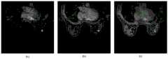
图3是本方法中使用的乳腺DCE-MRI不同时期图像;其中(a)为未注射造影剂的图像、(b)为增强初期图像、(c)为增强高峰图像、(d)为增强晚期图像;Figure 3 is the images of breast DCE-MRI in different periods used in this method; (a) is the image without contrast agent injection, (b) is the image at the early stage of enhancement, (c) is the image at the peak of enhancement, (d) is the image at the late stage of enhancement image;
图4是本方法中DCE-MRI序列三通道图像;其中(a)为剪影图像、(b)为增强初期图像、(c)为增强晚期图像;Figure 4 is a three-channel image of the DCE-MRI sequence in this method; wherein (a) is a silhouette image, (b) is an early enhancement image, and (c) is an enhanced late image;
图5为本方法中两个DCE-MRI图像的分割结果示意图;其中(a)和(b)为两个样例的金标准和分割结果图。Figure 5 is a schematic diagram of the segmentation results of two DCE-MRI images in the method; (a) and (b) are the gold standard and segmentation results of the two samples.
以下给出本发明的具体实施方式,需要说明的是,本发明并不局限于以下具体实施例,凡在本申请技术方案基础上做的等同变换均落入本发明的保护范围。Specific embodiments of the present invention are given below. It should be noted that the present invention is not limited to the following specific embodiments, and all equivalent transformations made on the basis of the technical solutions of the present application fall into the protection scope of the present invention.
具体实施方式Detailed ways
一个乳腺DCE-MRI序列图像中包含n期图像,包括注射造影剂前拍摄的一期图像和注射造影剂后拍摄的n-1期图像,n一般为7~9,在本发明中将注射造影剂后拍摄的第一期图像作为增强初期图像,注射造影剂后拍摄的最后一期图像作为增强晚期图像。A breast DCE-MRI sequence image includes n-phase images, including the first-phase image taken before the injection of the contrast agent and the n-1 phase image taken after the injection of the contrast agent, n is generally 7 to 9, in the present invention, the contrast-injected contrast agent will be injected. The first phase image taken after the injection of the contrast agent was taken as the early enhancement image, and the last phase image taken after the injection of the contrast agent was taken as the enhanced late image.
本发明的增强高峰图像是指注射造影剂后拍摄的n-1期图像中像素均值最高的一期图像。The enhanced peak image in the present invention refers to the first-phase image with the highest pixel mean value among the n-1 phase images captured after the injection of the contrast agent.
本发明中的“三通道图像”是指将三个单通道图(即剪影图像、增强初期图像与增强晚期图像)分别作为图像的一个通道,构成一幅具有三个通道的图像。The "three-channel image" in the present invention refers to taking three single-channel images (ie, silhouette image, enhanced early image and enhanced late image) as one channel of the image respectively to form an image with three channels.
本发明具体实施例中使用的乳腺DCE-MRI图像集是从医院获得的已有的图像,每个图像集包含多例DCE-MRI序列图像,一个DCE-MRI序列图像一般扫描7-9期图像,而每期图像包含96-188幅断层切片。The breast DCE-MRI image set used in the specific embodiment of the present invention is the existing image obtained from the hospital, each image set contains multiple DCE-MRI sequence images, and one DCE-MRI sequence image generally scans 7-9 images , while each image contains 96-188 slices.
DCE-MRI序列图像包含了多期图像,能够记录乳房组织结构在注射造影剂前后的信号变化信息。乳腺DCE-MRI病理学相关研究表明,乳腺病灶的生长很大程度上依赖于内部的血管,因此相较于正常组织,病变内血管渗透性更强。由于不同病灶间毛细血管的通透性和组织细胞外间隙的不同,当静脉注射对比剂后,乳腺病灶区域会呈现不同的信号强度变化,但是正常组织就不会有明显和多样的变化。考虑到DCE-MRI序列图像的这一特性,本发明提出了一种DCE-MRI图像的多通道表达,作为网络的输入,从而提高分割的性能。The DCE-MRI sequence image contains multi-phase images, which can record the signal change information of the breast tissue structure before and after the injection of contrast agent. Breast DCE-MRI pathological studies have shown that the growth of breast lesions is largely dependent on the internal blood vessels, so compared with normal tissue, the blood vessels in the lesions are more permeable. Due to the difference in capillary permeability and tissue extracellular space between different lesions, after intravenous injection of contrast agent, the breast lesions will show different signal intensity changes, but there will be no obvious and diverse changes in normal tissues. Considering this characteristic of DCE-MRI sequence images, the present invention proposes a multi-channel representation of DCE-MRI images as the input of the network, thereby improving the performance of segmentation.
本发明的具体实施例中公开的基于混合卷积和ASPP的乳腺DCE-MRI图像病灶分割模型建立方法,具体包括以下步骤:The method for establishing a breast DCE-MRI image lesion segmentation model based on hybrid convolution and ASPP disclosed in the specific embodiment of the present invention specifically includes the following steps:
步骤1,对于乳腺DCE-MRI图像集中每个DCE-MRI序列图像进行以下处理:Step 1: Perform the following processing on each DCE-MRI sequence image in the breast DCE-MRI image set:
步骤1.1,本实施例中使用的DCE-MRI序列图像为DICOM格式,因此需要对原始DICOM格式的DCE-MRI序列所有期图像进行归一化,将灰度值映射到0-1或0-255,可以将图像保存为PNG形式。Step 1.1, the DCE-MRI sequence image used in this example is in DICOM format, so it is necessary to normalize all phase images of the DCE-MRI sequence in the original DICOM format, and map the gray value to 0-1 or 0-255 , you can save the image as PNG.
然后获取剪影图像,其中剪影图像是DCE-MRI序列图像中的增强高峰图像与未注射造影剂的一期图像的差值。每个DCE-MRI序列图像包括多期图像,首先统计注射造影剂后的每期所有断层切片中亮度最高的32个像素的值,计算均值,最后选择注射造影剂后的多期图像中像素均值最高的一期作为增强高峰图像。增强高峰图像如图3中(c)图所示,未注射造影剂图像如图3中(a)图所示。A silhouette image is then acquired, where the silhouette image is the difference between the peak enhancement image in the DCE-MRI sequence and the primary image without contrast injection. Each DCE-MRI sequence image includes multi-phase images. First, the values of the 32 pixels with the highest brightness in all tomographic slices in each phase after contrast agent injection are counted, and the mean value is calculated. Finally, the pixel mean value in the multi-phase images after contrast agent injection is selected. The highest phase is used as an enhanced peak image. The enhanced peak image is shown in Figure 3 (c), and the image without contrast agent is shown in Figure 3 (a).
步骤1.2,将注射造影剂后拍摄第一期图像作为增强初期图像,如图3中(b)图所示,注射造影剂后拍摄的最后一期图像作为增强晚期图像,如图3中(d)图所示;将剪影图像、增强初期图像与增强晚期图像构建成三通道图像,如图4所示,作为分隔网络的输入。In step 1.2, the first-phase image taken after the injection of the contrast agent is used as the enhanced initial image, as shown in (b) in Figure 3, and the last phase of the image taken after the contrast agent is injected as the enhanced late-phase image, as shown in Figure 3 (d) ) as shown in the figure; the silhouette image, the enhanced initial image and the enhanced late image are constructed into a three-channel image, as shown in Figure 4, as the input of the separation network.
步骤2,构建基于混合卷积与ASPP网络的乳腺DCE-MRI图像病灶分割网络,所述的分割网络包括2D空间特征提取模块、3D空间特征提取模块、2D与3D特征融合模块、多尺度特征提取模块、上采样模块。分隔网络结构如图2所示。
其中,2D空间特征提取模块用于使用U-Net编码结构提取三通道的图像中每一幅断层切片的2D空间特征图,输入U-Net编码结构的是当前断层切片。本实施例采用python语言和pytorch深度网络框架实现分割网络模型的构建,其中卷积、池化和上采样操作均为调用框架相应函数所得。考虑到U-Net网络能够使用小样本数据集进行训练和测试,并在生物医学分割领域取得了巨大的成功,本实施例使用U-Net作为2D切片基础分割网络,构建基于混合卷积与ASPP网络的病灶区域分割模型。Among them, the 2D spatial feature extraction module is used to extract the 2D spatial feature map of each tomographic slice in the three-channel image by using the U-Net coding structure, and the input to the U-Net coding structure is the current tomographic slice. This embodiment adopts the python language and the pytorch deep network framework to realize the construction of the segmentation network model, in which the convolution, pooling and upsampling operations are obtained by calling the corresponding functions of the framework. Considering that the U-Net network can be trained and tested with small sample datasets, and has achieved great success in the field of biomedical segmentation, this example uses U-Net as the 2D slice basis segmentation network, and builds a network based on hybrid convolution and ASPP. Lesion area segmentation model of the network.
3D空间特征提取模块用于使用2D与3D卷积混合模块提取三通道的图像中每一幅断层切片的3D空间特征图,2D与3D卷积混合模块输入的是当前断层切片及其上下层切片,为了保证不同切片的特征图在相应通道上的空间一致性,在三个切片上使用的2D卷积参数是相同的。The 3D spatial feature extraction module is used to use the 2D and 3D convolution hybrid module to extract the 3D spatial feature map of each tomographic slice in the three-channel image. The 2D and 3D convolution hybrid module inputs the current tomographic slice and its upper and lower layers. , in order to ensure the spatial consistency of the feature maps of different slices on the corresponding channels, the 2D convolution parameters used on the three slices are the same.
其中,3D空间特征提取模块包括四个2D与3D卷积混合模块,每一个2D与3D卷积混合模块都包括2个2D卷积和一个3D卷积。本实施例中,2D与3D卷积混合模块的原理是:首先使用2个3×3的2D卷积来获取二维的特征图;其次,将三个二维特征图拼接成一个三维的特征图,同时这三个特征图作为下一个2D与3D卷积混合模块的输入;然后,使用一个1×1×3的3D的卷积来提取第三个维度上的空间特征。Among them, the 3D spatial feature extraction module includes four 2D and 3D convolution hybrid modules, and each 2D and 3D convolution hybrid module includes two 2D convolutions and one 3D convolution. In this embodiment, the principle of the 2D and 3D convolution hybrid module is: first, two 3×3 2D convolutions are used to obtain a two-dimensional feature map; secondly, three two-dimensional feature maps are spliced into a three-dimensional feature At the same time, these three feature maps are used as the input of the next 2D and 3D convolution mixing module; then, a 1 × 1 × 3 3D convolution is used to extract the spatial features in the third dimension.
2D与3D特征融合模块用于将2D空间特征提取模块中的每一幅断层切片的2D空间特征图与对应的3D空间特征提取模块中的每一幅断层切片的3D特征图进行融合。本实施例采用图像相加方式进行融合,得到包含有2D空间特征和3D空间特征的特征图,特征图的个数与断层切片数量一致;The 2D and 3D feature fusion module is used to fuse the 2D spatial feature map of each tomographic slice in the 2D spatial feature extraction module with the 3D feature map of each tomographic slice in the corresponding 3D spatial feature extraction module. In this embodiment, the image addition method is used for fusion to obtain a feature map including 2D spatial features and 3D spatial features, and the number of feature maps is consistent with the number of tomographic slices;
多尺度特征提取模块用于使用ASPP结构对2D与3D特征融合模块中的特征图进行处理,得到具有多尺度图像信息的特征图。ASPP结构包括两个3×3的正常卷积,两个采样率为2的3×3的空洞卷积以及两个采样率为6的3×3的空洞卷积。输入的特征图通过三种并行卷积,获得三个尺度的特征图。然后将不同尺度下的特征图进行拼接,并使用1×1的卷积来变换通道,使其与输入特征图的通道数量保持一致。The multi-scale feature extraction module is used to process the feature maps in the 2D and 3D feature fusion module using the ASPP structure to obtain feature maps with multi-scale image information. The ASPP structure consists of two 3×3 normal convolutions, two 3×3 atrous convolutions with a sampling rate of 2, and two 3×3 atrous convolutions with a sampling rate of 6. The input feature map is passed through three parallel convolutions to obtain feature maps of three scales. The feature maps at different scales are then stitched together, and 1×1 convolution is used to transform the channels so that it is consistent with the number of channels in the input feature map.
上采样模块用于使用U-Net的解码结构对多尺度特征提取模块得到的具有多尺度图像信息的特征图进行上采样处理,得到与其对应的断层切片的输入尺寸一致的分割概率图。The upsampling module is used for upsampling the feature map with multi-scale image information obtained by the multi-scale feature extraction module using the decoding structure of U-Net to obtain a segmentation probability map consistent with the input size of the corresponding tomographic slice.
步骤3,使用步骤1得到的三通道的图像训练步骤2得到的分割网络,得到训练好的分割模型。Step 3, use the three-channel image obtained in step 1 to train the segmentation network obtained in
在本实施例中,构建DSC作为网络的目标函数,网络训练损失函数为Diss loss函数,步骤1得到的三通道的图像送入构建好的分割网络中,使用该损失函数端到端的训练分隔网络,得到训练好的分割模型。In this embodiment, the DSC is constructed as the objective function of the network, the network training loss function is the Diss loss function, the three-channel image obtained in step 1 is sent to the constructed segmentation network, and the end-to-end training segmentation network is performed using this loss function. , to get the trained segmentation model.
本发明实施例还公开了基于混合卷积的乳腺DCE-MRI图像病灶分割模型建立系统,该系统包括以下模块:The embodiment of the present invention also discloses a hybrid convolution-based breast DCE-MRI image lesion segmentation model establishment system, which includes the following modules:
图像预处理模块,用于对于乳腺DCE-MRI图像集中每个DCE-MRI序列图像进行处理,包括剪影图像获取模块以及三通道图像构建模块,其中:The image preprocessing module is used to process each DCE-MRI sequence image in the breast DCE-MRI image set, including a silhouette image acquisition module and a three-channel image building module, wherein:
剪影图像获取模块,用于获取剪影图像,其中剪影图像是DCE-MRI序列图像中的增强高峰图像与未注射造影剂的一期图像的差值;每个DCE-MRI序列图像包括多期图像,将DCE-MRI序列图像中像素均值最高的一期作为增强高峰图像。The silhouette image acquisition module is used for acquiring silhouette images, wherein the silhouette image is the difference between the enhanced peak image in the DCE-MRI sequence image and the first-phase image without contrast agent injection; each DCE-MRI sequence image includes multi-phase images, The phase with the highest pixel mean value in the DCE-MRI sequence images was taken as the peak enhancement image.
三通道图像构建模块,用于将注射造影剂后拍摄第一期图像作为增强初期图像,注射造影剂后拍摄的最后一期图像为增强晚期图像,将剪影图像、增强初期图像与增强晚期图像构建成三通道图像。The three-channel image building module is used to take the first-phase image taken after the injection of the contrast agent as the enhanced initial image, and the last phase of the image taken after the contrast agent is injected as the enhanced late-phase image. into a three-channel image.
分割网络构建模块,用于构建基于混合卷积与ASPP网络的乳腺DCE-MRI图像病灶分割网络,所述的分割网络包括:The segmentation network building module is used to construct a breast DCE-MRI image lesion segmentation network based on hybrid convolution and ASPP network, and the segmentation network includes:
2D空间特征提取模块,用于使用U-Net编码结构提取三通道的图像中每一幅断层切片的2D空间特征图,U-Net编码结构输入为当前断层切片;The 2D spatial feature extraction module is used to extract the 2D spatial feature map of each tomographic slice in the three-channel image using the U-Net coding structure, and the U-Net coding structure input is the current tomographic slice;
3D空间特征提取模块,用于使用2D与3D卷积混合模块提取三通道的图像中每一幅断层切片的3D空间特征图,2D与3D卷积混合模块输入的是当前断层切片及其上下层切片,为了保证不同切片的特征图在相应通道上的空间一致性,在三个切片上使用的2D卷积参数是相同的。The 3D spatial feature extraction module is used to use the 2D and 3D convolution hybrid module to extract the 3D spatial feature map of each tomographic slice in the three-channel image. The input of the 2D and 3D convolution hybrid module is the current tomographic slice and its upper and lower layers. Slices, in order to ensure the spatial consistency of the feature maps of different slices on the corresponding channels, the 2D convolution parameters used on the three slices are the same.
其中,3D空间特征提取模块包括四个2D与3D卷积混合模块,每一个2D与3D卷积混合模块都包括2个2D卷积和一个3D卷积;本实施例2D与3D卷积混合模块的原理同上述实施例。The 3D spatial feature extraction module includes four 2D and 3D convolution hybrid modules, and each 2D and 3D convolution hybrid module includes two 2D convolutions and one 3D convolution; the 2D and 3D convolution hybrid modules in this embodiment The principle is the same as the above-mentioned embodiment.
2D与3D特征融合模块,用于将2D空间特征提取模块中的每一幅断层切片的2D空间特征图与对应的3D空间特征提取模块中的每一幅断层切片的3D特征图进行融合,得到包含有2D空间特征和3D空间特征的特征图;本实施例采用图像相加方式进行融合,特征图的个数与断层切片数量一致;The 2D and 3D feature fusion module is used to fuse the 2D spatial feature map of each tomographic slice in the 2D spatial feature extraction module with the 3D feature map of each tomographic slice in the corresponding 3D spatial feature extraction module to obtain A feature map containing 2D spatial features and 3D spatial features; in this embodiment, image addition is used for fusion, and the number of feature maps is consistent with the number of slices;
多尺度特征提取模块,用于使用ASPP结构对2D与3D特征融合模块中的特征图进行处理,得到具有多尺度图像信息的特征图。其中,本实施例的ASPP结构同上述实施例。The multi-scale feature extraction module is used to process the feature map in the 2D and 3D feature fusion module using the ASPP structure to obtain a feature map with multi-scale image information. The ASPP structure of this embodiment is the same as that of the above-mentioned embodiment.
上采样模块,用于使用U-Net的解码结构对多尺度特征提取模块得到的具有多尺度图像信息的特征图进行上采样处理,得到与其对应的断层切片的输入尺寸一致的分割概率图;The upsampling module is used for upsampling the feature map with multi-scale image information obtained by the multi-scale feature extraction module by using the decoding structure of U-Net to obtain a segmentation probability map consistent with the input size of the corresponding tomographic slice;
分割模型构建模块,用于使用步骤1得到的三通道的图像训练步骤2得到的分割网络,得到训练好的分割模型。The segmentation model building module is used to train the segmentation network obtained in
通过上述实施例可得到训练好的分割模型,利用该分割模型对需要处理的DCE-MRI序列图像病灶区域进行分割,因此,A trained segmentation model can be obtained through the above embodiment, and the segmentation model is used to segment the lesion area of the DCE-MRI sequence image that needs to be processed. Therefore,
本发明的另一个实施例中公开了一种基于混合卷积的乳腺DCE-MRI图像病灶分割方法,该方法包括以下步骤:Another embodiment of the present invention discloses a hybrid convolution-based breast DCE-MRI image lesion segmentation method, which includes the following steps:
步骤1,对于待处理的一个DCE-MRI序列图像,经过上述实施例中步骤1的预处理,得到三通道图像;Step 1, for a DCE-MRI sequence image to be processed, through the preprocessing of step 1 in the above embodiment, a three-channel image is obtained;
步骤2,将得到的三通道图像输入到分割模型中,即可得到病灶分割结果。本实施例中得到的二维分割结果,可将二维的分割结果按照对应输入切片的在三维DCE-MRI中的顺序拼接成三维立体图。In
如图5所示为本实施例中其中两个DCE-MRI图像的分割结果。FIG. 5 shows the segmentation results of two DCE-MRI images in this embodiment.
本发明实施例还公开一种基于混合卷积的乳腺DCE-MRI图像病灶分割系统,该系统包括以下模块:The embodiment of the present invention also discloses a hybrid convolution-based breast DCE-MRI image lesion segmentation system, which includes the following modules:
图像预处理模块,用于对于待处理的DCE-MRI序列图像,使用分割模型建立方法实施例中的步骤1或分割模型建立系统中的图像预处理模块进行处理,得到三通道图像;The image preprocessing module is used to process the DCE-MRI sequence images to be processed by using step 1 in the embodiment of the segmentation model establishment method or the image preprocessing module in the segmentation model establishment system to obtain a three-channel image;
病灶分割模块,用于将图像预处理模块的三通道图像输入到分割模型建立方法实施例得到的分割模型或分割模型建立系统的分割模型构建模块中,得到病灶分割结果。The lesion segmentation module is used for inputting the three-channel image of the image preprocessing module into the segmentation model obtained in the embodiment of the segmentation model establishment method or the segmentation model establishment module of the segmentation model establishment system to obtain the lesion segmentation result.
本发明还对比了本发明方法与下面4种不同方法之间的分割效果:The present invention also contrasts the segmentation effect between the method of the present invention and the following 4 different methods:
N1:DCE-MRI序列图像不进行步骤1.1~1.2的处理,直接使用单通道的图像作为输入;分割网络包括2D空间特征提取模块和上采样模块;采用端到端的方式训练仅包含2D空间特征提取的U-Net模型,然后使用训练好的模型分割DCE-MRI图像病灶区域。N1: DCE-MRI sequence images are not processed in steps 1.1 to 1.2, and single-channel images are directly used as input; the segmentation network includes a 2D spatial feature extraction module and an upsampling module; the end-to-end training only includes 2D spatial feature extraction The U-Net model is then used to segment the DCE-MRI image lesion area using the trained model.
N2:使用三通道图像作为输入;分割网络包括2D空间特征提取模块和上采样模块;采用端到端的方式训练仅包含2D空间特征提取的U-Net模型,然后使用训练好的模型分割DCE-MRI图像病灶区域。N2: Use a three-channel image as input; the segmentation network includes a 2D spatial feature extraction module and an upsampling module; an end-to-end U-Net model containing only 2D spatial feature extraction is trained, and then the trained model is used to segment DCE-MRI Image the lesion area.
N3:使用三通道图像作为输入;分割网络包括2D空间特征提取模块、多尺度特征提取模块和上采样模块;采用端到端的方式对整个网络进行训练,使用训练好的模型分割病灶区域。N3: Use three-channel images as input; the segmentation network includes a 2D spatial feature extraction module, a multi-scale feature extraction module, and an upsampling module; the entire network is trained in an end-to-end manner, and the trained model is used to segment the lesion area.
N4:使用三通道图像作为输入;分割网络包括2D空间特征提取模块、3D空间特征提取模块、2D与3D特征融合模块和上采样模块;采用端到端的方式对整个网络进行训练,使用训练好的模型分割病灶区域。N4: Use a three-channel image as input; the segmentation network includes a 2D spatial feature extraction module, a 3D spatial feature extraction module, a 2D and 3D feature fusion module, and an upsampling module; the entire network is trained in an end-to-end manner, using the trained The model segments the lesion area.
表1所示为本发明实施例中90个DCE-MRI图像样本的的实验结果的均值,其中戴斯系数(Dice Similarity Coefficient,DSC)与阳性预测值(Positive Predictive Value,PPV)为实验结果的评价指标,评价指标的取值范围为[0,1],取值越高,代表分割性能越好。由表1可知,本发明中的各个部分均可以对最终的结果产生有利的影响,并最终得到一个较优的结果。Table 1 shows the mean value of the experimental results of 90 DCE-MRI image samples in the embodiment of the present invention, wherein the Dice Similarity Coefficient (DSC) and the Positive Predictive Value (Positive Predictive Value, PPV) are the values of the experimental results. Evaluation index. The value range of the evaluation index is [0, 1]. The higher the value, the better the segmentation performance. It can be seen from Table 1 that each part of the present invention can have a favorable effect on the final result, and finally obtain a better result.
表1不同方法之间的效果对比Table 1 Comparison of effects between different methods
Claims (10)
Priority Applications (1)
| Application Number | Priority Date | Filing Date | Title |
|---|---|---|---|
| CN202010125719.3ACN111429474B (en) | 2020-02-27 | 2020-02-27 | Mammary gland DCE-MRI image focus segmentation model establishment and segmentation method based on mixed convolution |
Applications Claiming Priority (1)
| Application Number | Priority Date | Filing Date | Title |
|---|---|---|---|
| CN202010125719.3ACN111429474B (en) | 2020-02-27 | 2020-02-27 | Mammary gland DCE-MRI image focus segmentation model establishment and segmentation method based on mixed convolution |
Publications (2)
| Publication Number | Publication Date |
|---|---|
| CN111429474Atrue CN111429474A (en) | 2020-07-17 |
| CN111429474B CN111429474B (en) | 2023-04-07 |
Family
ID=71547308
Family Applications (1)
| Application Number | Title | Priority Date | Filing Date |
|---|---|---|---|
| CN202010125719.3AActiveCN111429474B (en) | 2020-02-27 | 2020-02-27 | Mammary gland DCE-MRI image focus segmentation model establishment and segmentation method based on mixed convolution |
Country Status (1)
| Country | Link |
|---|---|
| CN (1) | CN111429474B (en) |
Cited By (13)
| Publication number | Priority date | Publication date | Assignee | Title |
|---|---|---|---|---|
| CN112085736A (en)* | 2020-09-04 | 2020-12-15 | 厦门大学 | Mixed-dimension convolution-based renal tumor segmentation method |
| CN112132790A (en)* | 2020-09-02 | 2020-12-25 | 西安国际医学中心有限公司 | DAC-GAN model construction method and application in mammary gland MR image |
| CN112529914A (en)* | 2020-12-18 | 2021-03-19 | 北京中科深智科技有限公司 | Real-time hair segmentation method and system |
| CN113192035A (en)* | 2021-04-30 | 2021-07-30 | 哈尔滨理工大学 | Improved mammary gland MRI segmentation method based on U-Net network |
| CN113421633A (en)* | 2021-06-25 | 2021-09-21 | 上海联影智能医疗科技有限公司 | Feature classification method, computer device, and storage medium |
| CN113469229A (en)* | 2021-06-18 | 2021-10-01 | 中山大学孙逸仙纪念医院 | Method and device for automatically labeling breast cancer focus based on deep learning |
| CN113657480A (en)* | 2021-08-13 | 2021-11-16 | 江南大学 | A clothing analysis method based on feature fusion network model |
| CN114066904A (en)* | 2021-11-19 | 2022-02-18 | 西安交通大学医学院第二附属医院 | Skin lesion image segmentation method and device based on deep learning and storage medium |
| CN114581701A (en)* | 2022-02-24 | 2022-06-03 | 杭州电子科技大学 | Method for generating dynamic enhanced image features by weighting image features through T2 |
| CN114820584A (en)* | 2022-05-27 | 2022-07-29 | 北京安德医智科技有限公司 | Lung focus positioner |
| CN115018862A (en)* | 2022-05-26 | 2022-09-06 | 杭州深睿博联科技有限公司 | A method and device for liver tumor segmentation based on hybrid neural network |
| CN115953781A (en)* | 2023-03-14 | 2023-04-11 | 武汉昊博科技有限公司 | Mammary gland artificial intelligence analysis system and method based on thermal chromatography image |
| CN116778470A (en)* | 2023-06-30 | 2023-09-19 | 京东方科技集团股份有限公司 | Object recognition and object recognition model training method, device, equipment and medium |
Citations (9)
| Publication number | Priority date | Publication date | Assignee | Title |
|---|---|---|---|---|
| CN108038517A (en)* | 2018-01-02 | 2018-05-15 | 东北农业大学 | Based on the maize leaf disease recognition method for improving convolutional neural networks MODEL C ifar10 |
| CN109063710A (en)* | 2018-08-09 | 2018-12-21 | 成都信息工程大学 | Based on the pyramidal 3D CNN nasopharyngeal carcinoma dividing method of Analysis On Multi-scale Features |
| US10304193B1 (en)* | 2018-08-17 | 2019-05-28 | 12 Sigma Technologies | Image segmentation and object detection using fully convolutional neural network |
| CN109840913A (en)* | 2019-01-21 | 2019-06-04 | 中南民族大学 | The method and system of lump segmentation in a kind of mammography X |
| US20190236782A1 (en)* | 2018-01-30 | 2019-08-01 | International Business Machines Corporation | Systems and methods for detecting an indication of malignancy in a sequence of anatomical images |
| WO2019200747A1 (en)* | 2018-04-20 | 2019-10-24 | 平安科技(深圳)有限公司 | Method and device for segmenting proximal femur, computer apparatus, and storage medium |
| CN110458249A (en)* | 2019-10-10 | 2019-11-15 | 点内(上海)生物科技有限公司 | A kind of lesion categorizing system based on deep learning Yu probability image group |
| CN110490851A (en)* | 2019-02-15 | 2019-11-22 | 腾讯科技(深圳)有限公司 | Galactophore image dividing method, apparatus and system based on artificial intelligence |
| CN110674866A (en)* | 2019-09-23 | 2020-01-10 | 兰州理工大学 | Method for detecting X-ray breast lesion images by using transfer learning characteristic pyramid network |
- 2020
- 2020-02-27CNCN202010125719.3Apatent/CN111429474B/enactiveActive
Patent Citations (9)
| Publication number | Priority date | Publication date | Assignee | Title |
|---|---|---|---|---|
| CN108038517A (en)* | 2018-01-02 | 2018-05-15 | 东北农业大学 | Based on the maize leaf disease recognition method for improving convolutional neural networks MODEL C ifar10 |
| US20190236782A1 (en)* | 2018-01-30 | 2019-08-01 | International Business Machines Corporation | Systems and methods for detecting an indication of malignancy in a sequence of anatomical images |
| WO2019200747A1 (en)* | 2018-04-20 | 2019-10-24 | 平安科技(深圳)有限公司 | Method and device for segmenting proximal femur, computer apparatus, and storage medium |
| CN109063710A (en)* | 2018-08-09 | 2018-12-21 | 成都信息工程大学 | Based on the pyramidal 3D CNN nasopharyngeal carcinoma dividing method of Analysis On Multi-scale Features |
| US10304193B1 (en)* | 2018-08-17 | 2019-05-28 | 12 Sigma Technologies | Image segmentation and object detection using fully convolutional neural network |
| CN109840913A (en)* | 2019-01-21 | 2019-06-04 | 中南民族大学 | The method and system of lump segmentation in a kind of mammography X |
| CN110490851A (en)* | 2019-02-15 | 2019-11-22 | 腾讯科技(深圳)有限公司 | Galactophore image dividing method, apparatus and system based on artificial intelligence |
| CN110674866A (en)* | 2019-09-23 | 2020-01-10 | 兰州理工大学 | Method for detecting X-ray breast lesion images by using transfer learning characteristic pyramid network |
| CN110458249A (en)* | 2019-10-10 | 2019-11-15 | 点内(上海)生物科技有限公司 | A kind of lesion categorizing system based on deep learning Yu probability image group |
Non-Patent Citations (2)
| Title |
|---|
| 任湘等: "基于卷积神经网络的乳腺癌分子分型预测研究", 《杭州电子科技大学学报(自然科学版)》* |
| 杨珍等: "利用DCE-MRI结合改进卷积神经网络的MR图像自动分割与分类方法", 《重庆理工大学学报(自然科学)》* |
Cited By (17)
| Publication number | Priority date | Publication date | Assignee | Title |
|---|---|---|---|---|
| CN112132790A (en)* | 2020-09-02 | 2020-12-25 | 西安国际医学中心有限公司 | DAC-GAN model construction method and application in mammary gland MR image |
| CN112132790B (en)* | 2020-09-02 | 2024-05-14 | 西安国际医学中心有限公司 | DAC-GAN model construction method and application thereof in mammary gland MR image |
| CN112085736B (en)* | 2020-09-04 | 2024-02-02 | 厦门大学 | Kidney tumor segmentation method based on mixed-dimension convolution |
| CN112085736A (en)* | 2020-09-04 | 2020-12-15 | 厦门大学 | Mixed-dimension convolution-based renal tumor segmentation method |
| CN112529914A (en)* | 2020-12-18 | 2021-03-19 | 北京中科深智科技有限公司 | Real-time hair segmentation method and system |
| CN113192035A (en)* | 2021-04-30 | 2021-07-30 | 哈尔滨理工大学 | Improved mammary gland MRI segmentation method based on U-Net network |
| CN113469229A (en)* | 2021-06-18 | 2021-10-01 | 中山大学孙逸仙纪念医院 | Method and device for automatically labeling breast cancer focus based on deep learning |
| CN113421633A (en)* | 2021-06-25 | 2021-09-21 | 上海联影智能医疗科技有限公司 | Feature classification method, computer device, and storage medium |
| CN113657480A (en)* | 2021-08-13 | 2021-11-16 | 江南大学 | A clothing analysis method based on feature fusion network model |
| CN114066904A (en)* | 2021-11-19 | 2022-02-18 | 西安交通大学医学院第二附属医院 | Skin lesion image segmentation method and device based on deep learning and storage medium |
| CN114066904B (en)* | 2021-11-19 | 2024-08-13 | 西安交通大学医学院第二附属医院 | Deep learning-based skin lesion image segmentation method, equipment and storage medium |
| CN114581701A (en)* | 2022-02-24 | 2022-06-03 | 杭州电子科技大学 | Method for generating dynamic enhanced image features by weighting image features through T2 |
| CN115018862A (en)* | 2022-05-26 | 2022-09-06 | 杭州深睿博联科技有限公司 | A method and device for liver tumor segmentation based on hybrid neural network |
| CN114820584B (en)* | 2022-05-27 | 2023-02-21 | 北京安德医智科技有限公司 | Lung focus positioner |
| CN114820584A (en)* | 2022-05-27 | 2022-07-29 | 北京安德医智科技有限公司 | Lung focus positioner |
| CN115953781A (en)* | 2023-03-14 | 2023-04-11 | 武汉昊博科技有限公司 | Mammary gland artificial intelligence analysis system and method based on thermal chromatography image |
| CN116778470A (en)* | 2023-06-30 | 2023-09-19 | 京东方科技集团股份有限公司 | Object recognition and object recognition model training method, device, equipment and medium |
Also Published As
| Publication number | Publication date |
|---|---|
| CN111429474B (en) | 2023-04-07 |
Similar Documents
| Publication | Publication Date | Title |
|---|---|---|
| CN111429474B (en) | Mammary gland DCE-MRI image focus segmentation model establishment and segmentation method based on mixed convolution | |
| Ni et al. | GC-Net: Global context network for medical image segmentation | |
| Yang et al. | Research on feature extraction of tumor image based on convolutional neural network | |
| CN110310281B (en) | Mask-RCNN deep learning-based pulmonary nodule detection and segmentation method in virtual medical treatment | |
| CN110996789B (en) | Systems and methods for performing screening, diagnostic, or other image-based analysis tasks | |
| CN108629816B (en) | A method for thin-slice magnetic resonance image reconstruction based on deep learning | |
| CN111951288A (en) | A deep learning-based segmentation method for skin cancer lesions | |
| CN113420826B (en) | Liver focus image processing system and image processing method | |
| CN109523521A (en) | Lung neoplasm classification and lesion localization method and system based on more slice CT images | |
| CN109389584A (en) | Multiple dimensioned rhinopharyngeal neoplasm dividing method based on CNN | |
| Chen et al. | Skin lesion segmentation using recurrent attentional convolutional networks | |
| CN117078692B (en) | Medical ultrasonic image segmentation method and system based on self-adaptive feature fusion | |
| CN115546605A (en) | Training method and device based on image labeling and segmentation model | |
| CN114565601A (en) | Improved liver CT image segmentation algorithm based on DeepLabV3+ | |
| CN112862805B (en) | Acoustic neuroma image automatic segmentation method and system | |
| CN114663445A (en) | A 3D Cardiac Image Segmentation Method Based on Multiscale Edge Perception | |
| Aslam et al. | Liver-Tumor Detection Using CNN ResUNet. | |
| CN112150564B (en) | Medical image fusion algorithm based on deep convolution neural network | |
| Honghan et al. | Rms-se-unet: A segmentation method for tumors in breast ultrasound images | |
| CN118037615A (en) | A tumor segmentation-guided magnetic resonance image translation method, system, device and medium | |
| CN119251250A (en) | Transformer-based segmentation method and system for cancerous tissue in cervical cancer pathological images | |
| Yueyuan et al. | Swin transformer combined with convolutional encoder for cephalometric landmarks detection | |
| Lu et al. | AugMS-Net: Augmented multiscale network for small cervical tumor segmentation from MRI volumes | |
| CN115984257A (en) | Multi-modal medical image fusion method based on multi-scale transform | |
| Zhang et al. | SAMS-Net: Fusion of attention mechanism and multi-scale features network for tumor infiltrating lymphocytes segmentation |
Legal Events
| Date | Code | Title | Description |
|---|---|---|---|
| PB01 | Publication | ||
| PB01 | Publication | ||
| SE01 | Entry into force of request for substantive examination | ||
| SE01 | Entry into force of request for substantive examination | ||
| GR01 | Patent grant | ||
| GR01 | Patent grant |