CN111420121A - Composite biological ink based on methacrylated hydrogel/nanoclay/acellular matrix and preparation method and application thereof - Google Patents
Composite biological ink based on methacrylated hydrogel/nanoclay/acellular matrix and preparation method and application thereofDownload PDFInfo
- Publication number
- CN111420121A CN111420121ACN202010260929.3ACN202010260929ACN111420121ACN 111420121 ACN111420121 ACN 111420121ACN 202010260929 ACN202010260929 ACN 202010260929ACN 111420121 ACN111420121 ACN 111420121A
- Authority
- CN
- China
- Prior art keywords
- acellular matrix
- nanoclay
- ink
- hydrogel
- methacrylated hydrogel
- Prior art date
- Legal status (The legal status is an assumption and is not a legal conclusion. Google has not performed a legal analysis and makes no representation as to the accuracy of the status listed.)
- Pending
Links
- 239000011159matrix materialSubstances0.000titleclaimsabstractdescription57
- 239000012802nanoclaySubstances0.000titleclaimsabstractdescription41
- 239000000017hydrogelSubstances0.000titleclaimsabstractdescription30
- 239000002131composite materialSubstances0.000titleclaimsabstractdescription27
- 238000002360preparation methodMethods0.000titleabstractdescription9
- CERQOIWHTDAKMF-UHFFFAOYSA-NMethacrylic acidChemical compoundCC(=C)C(O)=OCERQOIWHTDAKMF-UHFFFAOYSA-N0.000claimsabstract2
- 210000004185liverAnatomy0.000claimsdescription20
- 238000007639printingMethods0.000claimsdescription17
- 210000001519tissueAnatomy0.000claimsdescription13
- 210000004027cellAnatomy0.000claimsdescription12
- 230000010412perfusionEffects0.000claimsdescription11
- 102000057297Pepsin AHuman genes0.000claimsdescription10
- 108090000284Pepsin AProteins0.000claimsdescription10
- 229940111202pepsinDrugs0.000claimsdescription10
- 238000000034methodMethods0.000claimsdescription9
- 210000004204blood vesselAnatomy0.000claimsdescription5
- 210000002216heartAnatomy0.000claimsdescription5
- 210000003734kidneyAnatomy0.000claimsdescription5
- 210000004072lungAnatomy0.000claimsdescription5
- 210000003205muscleAnatomy0.000claimsdescription5
- 210000000496pancreasAnatomy0.000claimsdescription5
- 210000000952spleenAnatomy0.000claimsdescription5
- 210000002784stomachAnatomy0.000claimsdescription5
- VEXZGXHMUGYJMC-UHFFFAOYSA-NHydrochloric acidChemical compoundClVEXZGXHMUGYJMC-UHFFFAOYSA-N0.000claimsdescription4
- 210000004556brainAnatomy0.000claimsdescription4
- 210000000232gallbladderAnatomy0.000claimsdescription4
- 210000000936intestineAnatomy0.000claimsdescription4
- 210000002751lymphAnatomy0.000claimsdescription4
- 210000005036nerveAnatomy0.000claimsdescription4
- 230000002572peristaltic effectEffects0.000claimsdescription4
- 238000005406washingMethods0.000claimsdescription4
- APKFDSVGJQXUKY-KKGHZKTASA-NAmphotericin-BNatural productsO[C@H]1[C@@H](N)[C@H](O)[C@@H](C)O[C@H]1O[C@H]1C=CC=CC=CC=CC=CC=CC=C[C@H](C)[C@@H](O)[C@@H](C)[C@H](C)OC(=O)C[C@H](O)C[C@H](O)CC[C@@H](O)[C@H](O)C[C@H](O)C[C@](O)(C[C@H](O)[C@H]2C(O)=O)O[C@H]2C1APKFDSVGJQXUKY-KKGHZKTASA-N0.000claimsdescription3
- 102000016911DeoxyribonucleasesHuman genes0.000claimsdescription3
- 108010053770DeoxyribonucleasesProteins0.000claimsdescription3
- 102000006382RibonucleasesHuman genes0.000claimsdescription3
- 108010083644RibonucleasesProteins0.000claimsdescription3
- 229920004890Triton X-100Polymers0.000claimsdescription3
- 239000013504Triton X-100Substances0.000claimsdescription3
- APKFDSVGJQXUKY-INPOYWNPSA-Namphotericin BChemical compoundO[C@H]1[C@@H](N)[C@H](O)[C@@H](C)O[C@H]1O[C@H]1/C=C/C=C/C=C/C=C/C=C/C=C/C=C/[C@H](C)[C@@H](O)[C@@H](C)[C@H](C)OC(=O)C[C@H](O)C[C@H](O)CC[C@@H](O)[C@H](O)C[C@H](O)C[C@](O)(C[C@H](O)[C@H]2C(O)=O)O[C@H]2C1APKFDSVGJQXUKY-INPOYWNPSA-N0.000claimsdescription3
- 229960003942amphotericin bDrugs0.000claimsdescription3
- GUJOJGAPFQRJSV-UHFFFAOYSA-Ndialuminum;dioxosilane;oxygen(2-);hydrateChemical compoundO.[O-2].[O-2].[O-2].[Al+3].[Al+3].O=[Si]=O.O=[Si]=O.O=[Si]=O.O=[Si]=OGUJOJGAPFQRJSV-UHFFFAOYSA-N0.000claimsdescription3
- 229910052901montmorilloniteInorganic materials0.000claimsdescription3
- 229960005322streptomycinDrugs0.000claimsdescription3
- 241001465754MetazoaSpecies0.000claimsdescription2
- HPTYUNKZVDYXLP-UHFFFAOYSA-Naluminum;trihydroxy(trihydroxysilyloxy)silane;hydrateChemical compoundO.[Al].[Al].O[Si](O)(O)O[Si](O)(O)OHPTYUNKZVDYXLP-UHFFFAOYSA-N0.000claimsdescription2
- 229910000278bentoniteInorganic materials0.000claimsdescription2
- 239000000440bentoniteSubstances0.000claimsdescription2
- SVPXDRXYRYOSEX-UHFFFAOYSA-NbentoquatamChemical compoundO.O=[Si]=O.O=[Al]O[Al]=OSVPXDRXYRYOSEX-UHFFFAOYSA-N0.000claimsdescription2
- 238000000227grindingMethods0.000claimsdescription2
- 229910052621halloysiteInorganic materials0.000claimsdescription2
- 229910000271hectoriteInorganic materials0.000claimsdescription2
- KWLMIXQRALPRBC-UHFFFAOYSA-LhectoriteChemical compound[Li+].[OH-].[OH-].[Na+].[Mg+2].O1[Si]2([O-])O[Si]1([O-])O[Si]([O-])(O1)O[Si]1([O-])O2KWLMIXQRALPRBC-UHFFFAOYSA-L0.000claimsdescription2
- NLYAJNPCOHFWQQ-UHFFFAOYSA-NkaolinChemical compoundO.O.O=[Al]O[Si](=O)O[Si](=O)O[Al]=ONLYAJNPCOHFWQQ-UHFFFAOYSA-N0.000claimsdescription2
- 229910052622kaoliniteInorganic materials0.000claimsdescription2
- 210000001508eyeAnatomy0.000claims2
- 238000010276constructionMethods0.000claims1
- 238000004132cross linkingMethods0.000claims1
- 210000005069earsAnatomy0.000claims1
- 238000004108freeze dryingMethods0.000claims1
- 210000001331noseAnatomy0.000claims1
- 230000000694effectsEffects0.000abstractdescription8
- 239000000976inkSubstances0.000description11
- 239000003102growth factorSubstances0.000description7
- 210000000056organAnatomy0.000description5
- 108010010803GelatinProteins0.000description4
- 230000010261cell growthEffects0.000description4
- 238000012512characterization methodMethods0.000description4
- 239000008273gelatinSubstances0.000description4
- 229920000159gelatinPolymers0.000description4
- 235000019322gelatineNutrition0.000description4
- 235000011852gelatine dessertsNutrition0.000description4
- 239000000463materialSubstances0.000description4
- 230000008569processEffects0.000description4
- 206010028980NeoplasmDiseases0.000description3
- 241000700159RattusSpecies0.000description3
- 238000005516engineering processMethods0.000description3
- DCUFMVPCXCSVNP-UHFFFAOYSA-Nmethacrylic anhydrideChemical compoundCC(=C)C(=O)OC(=O)C(C)=CDCUFMVPCXCSVNP-UHFFFAOYSA-N0.000description3
- 239000000203mixtureSubstances0.000description3
- 238000012360testing methodMethods0.000description3
- 230000009286beneficial effectEffects0.000description2
- 230000000975bioactive effectEffects0.000description2
- 230000021164cell adhesionEffects0.000description2
- 238000011161developmentMethods0.000description2
- 230000018109developmental processEffects0.000description2
- 239000003814drugSubstances0.000description2
- 238000007877drug screeningMethods0.000description2
- 238000007490hematoxylin and eosin (H&E) stainingMethods0.000description2
- 238000000338in vitroMethods0.000description2
- 238000004519manufacturing processMethods0.000description2
- FQPSGWSUVKBHSU-UHFFFAOYSA-NmethacrylamideChemical compoundCC(=C)C(N)=OFQPSGWSUVKBHSU-UHFFFAOYSA-N0.000description2
- 238000012216screeningMethods0.000description2
- 2380000101463D printingMethods0.000description1
- 102000008186CollagenHuman genes0.000description1
- 108010035532CollagenProteins0.000description1
- 102100024785Fibroblast growth factor 2Human genes0.000description1
- 108090000379Fibroblast growth factor 2Proteins0.000description1
- 241000699670Mus sp.Species0.000description1
- BPQQTUXANYXVAA-UHFFFAOYSA-NOrthosilicateChemical compound[O-][Si]([O-])([O-])[O-]BPQQTUXANYXVAA-UHFFFAOYSA-N0.000description1
- 239000000654additiveSubstances0.000description1
- 230000000996additive effectEffects0.000description1
- 230000000890antigenic effectEffects0.000description1
- 230000004071biological effectEffects0.000description1
- 239000012620biological materialSubstances0.000description1
- 230000003592biomimetic effectEffects0.000description1
- 230000024245cell differentiationEffects0.000description1
- 230000003915cell functionEffects0.000description1
- 230000008859changeEffects0.000description1
- 238000006243chemical reactionMethods0.000description1
- 229920001436collagenPolymers0.000description1
- 230000003247decreasing effectEffects0.000description1
- 238000013461designMethods0.000description1
- 238000001514detection methodMethods0.000description1
- 229940079593drugDrugs0.000description1
- 238000001125extrusionMethods0.000description1
- 239000000835fiberSubstances0.000description1
- 230000004927fusionEffects0.000description1
- 239000000499gelSubstances0.000description1
- 230000012010growthEffects0.000description1
- 230000002440hepatic effectEffects0.000description1
- 230000008676importEffects0.000description1
- 230000010354integrationEffects0.000description1
- 238000002032lab-on-a-chipMethods0.000description1
- 210000005229liver cellAnatomy0.000description1
- 239000002135nanosheetSubstances0.000description1
- 239000002245particleSubstances0.000description1
- 210000003240portal veinAnatomy0.000description1
- 230000001737promoting effectEffects0.000description1
- 238000011160researchMethods0.000description1
- 238000003860storageMethods0.000description1
- 238000006467substitution reactionMethods0.000description1
- 230000009466transformationEffects0.000description1
- 238000000844transformationMethods0.000description1
Images
Classifications
- A—HUMAN NECESSITIES
- A61—MEDICAL OR VETERINARY SCIENCE; HYGIENE
- A61L—METHODS OR APPARATUS FOR STERILISING MATERIALS OR OBJECTS IN GENERAL; DISINFECTION, STERILISATION OR DEODORISATION OF AIR; CHEMICAL ASPECTS OF BANDAGES, DRESSINGS, ABSORBENT PADS OR SURGICAL ARTICLES; MATERIALS FOR BANDAGES, DRESSINGS, ABSORBENT PADS OR SURGICAL ARTICLES
- A61L27/00—Materials for grafts or prostheses or for coating grafts or prostheses
- A61L27/36—Materials for grafts or prostheses or for coating grafts or prostheses containing ingredients of undetermined constitution or reaction products thereof, e.g. transplant tissue, natural bone, extracellular matrix
- A61L27/3604—Materials for grafts or prostheses or for coating grafts or prostheses containing ingredients of undetermined constitution or reaction products thereof, e.g. transplant tissue, natural bone, extracellular matrix characterised by the human or animal origin of the biological material, e.g. hair, fascia, fish scales, silk, shellac, pericardium, pleura, renal tissue, amniotic membrane, parenchymal tissue, fetal tissue, muscle tissue, fat tissue, enamel
- A—HUMAN NECESSITIES
- A61—MEDICAL OR VETERINARY SCIENCE; HYGIENE
- A61L—METHODS OR APPARATUS FOR STERILISING MATERIALS OR OBJECTS IN GENERAL; DISINFECTION, STERILISATION OR DEODORISATION OF AIR; CHEMICAL ASPECTS OF BANDAGES, DRESSINGS, ABSORBENT PADS OR SURGICAL ARTICLES; MATERIALS FOR BANDAGES, DRESSINGS, ABSORBENT PADS OR SURGICAL ARTICLES
- A61L27/00—Materials for grafts or prostheses or for coating grafts or prostheses
- A61L27/36—Materials for grafts or prostheses or for coating grafts or prostheses containing ingredients of undetermined constitution or reaction products thereof, e.g. transplant tissue, natural bone, extracellular matrix
- A61L27/3683—Materials for grafts or prostheses or for coating grafts or prostheses containing ingredients of undetermined constitution or reaction products thereof, e.g. transplant tissue, natural bone, extracellular matrix subjected to a specific treatment prior to implantation, e.g. decellularising, demineralising, grinding, cellular disruption/non-collagenous protein removal, anti-calcification, crosslinking, supercritical fluid extraction, enzyme treatment
- A61L27/3691—Materials for grafts or prostheses or for coating grafts or prostheses containing ingredients of undetermined constitution or reaction products thereof, e.g. transplant tissue, natural bone, extracellular matrix subjected to a specific treatment prior to implantation, e.g. decellularising, demineralising, grinding, cellular disruption/non-collagenous protein removal, anti-calcification, crosslinking, supercritical fluid extraction, enzyme treatment characterised by physical conditions of the treatment, e.g. applying a compressive force to the composition, pressure cycles, ultrasonic/sonication or microwave treatment, lyophilisation
- A—HUMAN NECESSITIES
- A61—MEDICAL OR VETERINARY SCIENCE; HYGIENE
- A61L—METHODS OR APPARATUS FOR STERILISING MATERIALS OR OBJECTS IN GENERAL; DISINFECTION, STERILISATION OR DEODORISATION OF AIR; CHEMICAL ASPECTS OF BANDAGES, DRESSINGS, ABSORBENT PADS OR SURGICAL ARTICLES; MATERIALS FOR BANDAGES, DRESSINGS, ABSORBENT PADS OR SURGICAL ARTICLES
- A61L27/00—Materials for grafts or prostheses or for coating grafts or prostheses
- A61L27/40—Composite materials, i.e. containing one material dispersed in a matrix of the same or different material
- A61L27/44—Composite materials, i.e. containing one material dispersed in a matrix of the same or different material having a macromolecular matrix
- A61L27/446—Composite materials, i.e. containing one material dispersed in a matrix of the same or different material having a macromolecular matrix with other specific inorganic fillers other than those covered by A61L27/443 or A61L27/46
- B—PERFORMING OPERATIONS; TRANSPORTING
- B33—ADDITIVE MANUFACTURING TECHNOLOGY
- B33Y—ADDITIVE MANUFACTURING, i.e. MANUFACTURING OF THREE-DIMENSIONAL [3-D] OBJECTS BY ADDITIVE DEPOSITION, ADDITIVE AGGLOMERATION OR ADDITIVE LAYERING, e.g. BY 3-D PRINTING, STEREOLITHOGRAPHY OR SELECTIVE LASER SINTERING
- B33Y10/00—Processes of additive manufacturing
- B—PERFORMING OPERATIONS; TRANSPORTING
- B33—ADDITIVE MANUFACTURING TECHNOLOGY
- B33Y—ADDITIVE MANUFACTURING, i.e. MANUFACTURING OF THREE-DIMENSIONAL [3-D] OBJECTS BY ADDITIVE DEPOSITION, ADDITIVE AGGLOMERATION OR ADDITIVE LAYERING, e.g. BY 3-D PRINTING, STEREOLITHOGRAPHY OR SELECTIVE LASER SINTERING
- B33Y70/00—Materials specially adapted for additive manufacturing
- B—PERFORMING OPERATIONS; TRANSPORTING
- B33—ADDITIVE MANUFACTURING TECHNOLOGY
- B33Y—ADDITIVE MANUFACTURING, i.e. MANUFACTURING OF THREE-DIMENSIONAL [3-D] OBJECTS BY ADDITIVE DEPOSITION, ADDITIVE AGGLOMERATION OR ADDITIVE LAYERING, e.g. BY 3-D PRINTING, STEREOLITHOGRAPHY OR SELECTIVE LASER SINTERING
- B33Y80/00—Products made by additive manufacturing
Landscapes
- Health & Medical Sciences (AREA)
- Chemical & Material Sciences (AREA)
- Life Sciences & Earth Sciences (AREA)
- Engineering & Computer Science (AREA)
- Materials Engineering (AREA)
- Dermatology (AREA)
- General Health & Medical Sciences (AREA)
- Veterinary Medicine (AREA)
- Public Health (AREA)
- Biomedical Technology (AREA)
- Animal Behavior & Ethology (AREA)
- Epidemiology (AREA)
- Manufacturing & Machinery (AREA)
- Medicinal Chemistry (AREA)
- Oral & Maxillofacial Surgery (AREA)
- Transplantation (AREA)
- Chemical Kinetics & Catalysis (AREA)
- Botany (AREA)
- Molecular Biology (AREA)
- Zoology (AREA)
- Urology & Nephrology (AREA)
- Inorganic Chemistry (AREA)
- Composite Materials (AREA)
- Materials For Medical Uses (AREA)
Abstract
Description
Translated fromChinese技术领域technical field
本发明涉及生物墨水技术领域,具体涉及一种用于体外细胞打印的高活性复合生物墨水及其制备方法与应用。The invention relates to the technical field of biological inks, in particular to a highly active composite biological ink used for in vitro cell printing and a preparation method and application thereof.
背景技术Background technique
3D生物打印是一种能在数字三维模式驱动下按照增材制造原理定位装配生物材料或细胞单元,制造医疗器械、组织工程支架和组织器官的技术。近年来,3D打印技术和微流控技术的逐渐成熟,各式新型的器官组织芯片不断涌现,在筛选药物、推动个性化和精准化医疗快速发展方面具有重大的作用。既可以降低药物筛选的成本,也可以减少测试时间,节约人力和财力资源;更重要的是可以提高被测试对象的一致性,使实验结果更具有说服力。利用“生物墨水”作为打印材料,模拟天然的3D ECM,结合细胞以构建含有活性的组织或器官。虽然目前已研制出来的生物墨水非常类似于ECM的活性,但由于体外3D培养系统仍然缺乏促进细胞生长和维持细胞功能的合适天然生长因子,导致3D生物打印构建的组织活性相对原型具有较大的差距。因此使用天然生长因子对3D ECM进行模拟是当前3D生物打印实现高仿生性的当务之急。3D bioprinting is a technology that can position and assemble biological materials or cell units according to the principle of additive manufacturing under the driving of digital three-dimensional mode, and manufacture medical devices, tissue engineering scaffolds and tissues and organs. In recent years, with the gradual maturity of 3D printing technology and microfluidic technology, various new types of organ and tissue chips have emerged, which play an important role in screening drugs and promoting the rapid development of personalized and precision medicine. It can not only reduce the cost of drug screening, but also reduce the testing time, saving human and financial resources; more importantly, it can improve the consistency of the tested objects and make the experimental results more convincing. Using "bioink" as a printing material to simulate natural 3D ECM, bind cells to construct active tissues or organs. Although the bioinks that have been developed so far are very similar to the activity of ECM, the in vitro 3D culture system still lacks suitable natural growth factors to promote cell growth and maintain cell function, resulting in tissue activity constructed by 3D bioprinting. gap. Therefore, the use of natural growth factors to simulate 3D ECM is an urgent task for 3D bioprinting to achieve high biomimeticity.
具有复杂天然组成和仿生结构的脱细胞基质是生物墨水的首选。脱细胞基质是指将组织经过脱细胞工艺处理后,去除能够引起免疫排斥的抗原部分。具有良好的机械力学性能,良好的生物相容性,在体内起着支撑、连接细胞的作用,同时其三维空间的结构及生长因子有利于细胞的黏附和生长。Acellular matrices with complex natural compositions and biomimetic structures are the first choice for bioinks. Acellular matrix refers to the removal of antigenic parts that can cause immune rejection after the tissue is treated by acellular process. It has good mechanical properties, good biocompatibility, and plays the role of supporting and connecting cells in the body. At the same time, its three-dimensional structure and growth factors are conducive to cell adhesion and growth.
国内外对于脱细胞生物墨水的研究逐渐深入,2018年在Lab On A Chip上的一篇文章指出,基于肝脏脱细胞基质(DLM)的基本成分和明胶甲基丙烯酰胺(GelMA)整合之后制成肿瘤芯片,形成3D微流控结构动态培养系统,实验结果表明肿瘤性能得到了改善,这与脱细胞基质提供了生长因子以及相关生物活性成分,更好的再现3D肝脏的天然肿瘤微环境有关。The research on acellular bioinks at home and abroad is gradually deepening. In 2018, an article on Lab On A Chip pointed out that it was made based on the integration of the basic components of liver acellular matrix (DLM) and gelatin methacrylamide (GelMA). The tumor chip forms a 3D microfluidic structure dynamic culture system. The experimental results show that the tumor performance has been improved, which is related to the acellular matrix provides growth factors and related bioactive components, and better reproduces the natural tumor microenvironment of the 3D liver.
发明内容SUMMARY OF THE INVENTION
本发明要解决的技术问题是提供一种新型的用于细胞打印的高活性复合生物墨水,该生物墨水以脱细胞基质为生物活性成分,并加入纳米黏土,提高了可打印性能。The technical problem to be solved by the present invention is to provide a new type of high-activity composite bio-ink for cell printing. The bio-ink uses acellular matrix as the bioactive component and adds nanoclay to improve the printability.
为了解决上述技术问题,本发明提供了一种用于细胞打印的基于甲基丙烯酸化水凝胶/纳米粘土/脱细胞基质的复合生物墨水,所述复合生物墨水的组分包括脱细胞基质溶液(DECM)、光交联甲基丙烯酸化水凝胶(GelMA)以及纳米黏土(Nanoclay)。In order to solve the above technical problems, the present invention provides a methacrylated hydrogel/nanoclay/acellular matrix-based composite bioink for cell printing, the components of the composite bioink include acellular matrix solution (DECM), photocrosslinked methacrylated hydrogel (GelMA), and nanoclay (Nanoclay).
进一步地,所述脱细胞基质溶液为脱细胞基质的胃蛋白酶溶液,其浓度优选为1-20mg/ml,更优选为1-10mg/ml。Further, the acellular matrix solution is a pepsin solution of acellular matrix, and its concentration is preferably 1-20 mg/ml, more preferably 1-10 mg/ml.
本发明中,脱细胞基质可由动物或人的肝脏、肾脏、神经、脑、肠、胰腺、心脏、肺、胃、脾、胆、血管、肌肉、淋巴、耳朵、鼻子、眼等器官组织经脱细胞工艺处理得到,具有良好的机械力学性能以及生物相容性,其三维的空间结构以及生长因子也有利于细胞的粘附和生长。甲基丙烯酸酐化水凝胶是由甲基丙烯酸酐(MA)与明胶(Gelatin)制备获得,是一种光敏性的生物水凝胶材料,具有优异的生物相容性,且可由紫外光或可见光激发固化反应,形成适于细胞生长与分化且有一定强度的三维结构。两种材料的结合可显著提高生物墨水的生物活性,但也降低了生物墨水的可打印性。In the present invention, the decellularized matrix can be obtained from animal or human organs and tissues such as liver, kidney, nerve, brain, intestine, pancreas, heart, lung, stomach, spleen, gallbladder, blood vessel, muscle, lymph, ear, nose, eye, etc. The cell process treatment has good mechanical and mechanical properties and biocompatibility, and its three-dimensional spatial structure and growth factors are also conducive to cell adhesion and growth. Methacrylic anhydride hydrogel is prepared from methacrylic anhydride (MA) and gelatin (Gelatin). It is a photosensitive biohydrogel material with excellent biocompatibility. Visible light stimulates the curing reaction to form a three-dimensional structure suitable for cell growth and differentiation with a certain strength. The combination of the two materials can significantly improve the bioactivity of the bioink, but also reduce the printability of the bioink.
进一步地,所述光交联甲基丙烯酸化水凝胶的浓度为1%-50%。Further, the concentration of the photo-crosslinked methacrylated hydrogel is 1%-50%.
纳米黏土具有分层结构,通过在体系中加入纳米黏土辅助打印,使得纳米黏土中的硅酸盐纳米片分散到水凝胶中,增强了生物墨水的机械强度。另外,纳米黏土具有凝胶引导的性质,其加入显著地提高了生物墨水的流变力学性能(储能模量),使生物墨水的凝胶状态更好,保证了打印结构的形状保真度,并避免打印中不同层的生物墨水纤维的融合问题,有效提高了生物墨水的打印性能。进一步地,所述纳米粘土为以下种类中的任意一种或多种:膨润土、蒙脱石、高岭石、锂蒙脱石和埃洛石。更进一步地,所述纳米黏土为LaponiteXLG系列的蒙脱土纳米黏土。The nanoclay has a layered structure. By adding nanoclay to the system to assist printing, the silicate nanosheets in the nanoclay are dispersed into the hydrogel, which enhances the mechanical strength of the bioink. In addition, the nanoclay has a gel-guided property, and its addition significantly improves the rheological mechanical properties (storage modulus) of the bioink, making the gel state of the bioink better and ensuring the shape fidelity of the printed structure. , and avoid the fusion problem of different layers of bio-ink fibers during printing, which effectively improves the printing performance of bio-ink. Further, the nanoclay is any one or more of the following types: bentonite, montmorillonite, kaolinite, hectorite and halloysite. Further, the nanoclay is montmorillonite nanoclay of LaponiteXLG series.
进一步地,所述光交联甲基丙烯酸化水凝胶与脱细胞基质溶液的体积比为5:1~1:5,优选为1:1。Further, the volume ratio of the photocrosslinked methacrylated hydrogel to the acellular matrix solution is 5:1 to 1:5, preferably 1:1.
进一步地,所述纳米黏土的添加量为脱细胞基质溶液与光交联甲基丙烯酸化水凝胶总质量的1%~5%,优选为1%。Further, the added amount of the nanoclay is 1% to 5% of the total mass of the acellular matrix solution and the photocrosslinked methacrylated hydrogel, preferably 1%.
进一步地,所述复合生物墨水用于打印的细胞包括肝脏、肾脏、神经、脑、肠、胰腺、心脏、肺、胃、脾、胆、血管、肌肉、淋巴、耳朵、鼻子、眼等器官组织的细胞。Further, the cells used for printing the composite bioink include liver, kidney, nerve, brain, intestine, pancreas, heart, lung, stomach, spleen, gallbladder, blood vessel, muscle, lymph, ear, nose, eye and other organs and tissues. cells.
本发明另一方面提供了所述的基于甲基丙烯酸化水凝胶/纳米粘土/脱细胞基质的复合生物墨水的制备方法,包括:Another aspect of the present invention provides a method for preparing the composite bioink based on methacrylated hydrogel/nanoclay/acellular matrix, comprising:
S1、将脱细胞基质冻干、研磨,加入胃蛋白酶溶液溶解,制成脱细胞基质溶液;S1. Freeze-dry the acellular matrix, grind it, add a pepsin solution to dissolve it, and prepare an acellular matrix solution;
S2、将得到的脱细胞基质溶液与光交联甲基丙烯酸化水凝胶混合,再加入纳米黏土,混合均匀,即得到所述复合生物墨水。S2. Mix the obtained acellular matrix solution with the photo-crosslinked methacrylated hydrogel, then add nanoclay, and mix evenly to obtain the composite bioink.
进一步地,步骤S1中,所述脱细胞基质是由解冻后的组织连接蠕动泵进行脱细胞灌注而得到的,所述脱细胞灌注具体依次包括:Further, in step S1, the decellularized matrix is obtained by connecting the thawed tissue to a peristaltic pump for decellularization perfusion, and the decellularization perfusion specifically includes:
(1)0.01mol/L PBS灌注1h;(1) 0.01mol/L PBS perfusion for 1h;
(2)1%Triton X-100灌注1h;(2) 1% Triton X-100 perfusion for 1 hour;
(3)PBS冲洗15min后,0.1%SDS灌注30min;(3) After washing with PBS for 15 minutes, perfuse with 0.1% SDS for 30 minutes;
(4)PBS冲洗15min后,80U/ml DNase、5U/ml RNase灌注30min;(4) After washing with PBS for 15min, 80U/ml DNase and 5U/ml RNase were perfused for 30min;
(5)2%青霉素-链霉素、2.5μg/ml两性霉素B的PBS灌注30min。(5) 2% penicillin-streptomycin, 2.5 μg/ml amphotericin B in PBS for 30 min.
进一步地,蠕动泵的灌注速率为10ml/min。Further, the perfusion rate of the peristaltic pump was 10 ml/min.
进一步地,制备脱细胞基质后,还需要对其进行表征,所述表征包括:扫描电镜SEM,HE染色,残留DNA浓度验证脱细胞过程。Further, after preparing the decellularized matrix, it also needs to be characterized, and the characterization includes: scanning electron microscope SEM, HE staining, and residual DNA concentration to verify the decellularization process.
进一步地,步骤S1中,所述胃蛋白酶溶液是由胃蛋白酶溶解于盐酸中得到的,浓度为0.5-10mg/mL,优选为1mg/mL。Further, in step S1, the pepsin solution is obtained by dissolving pepsin in hydrochloric acid, and the concentration is 0.5-10 mg/mL, preferably 1 mg/mL.
进一步地,步骤S2中,还包括对制备的生物墨水进行可打印性和生物相容性的验证的步骤。Further, in step S2, the step of verifying the printability and biocompatibility of the prepared bioink is also included.
本发明还提供了所述的复合生物墨水在构建组织工程支架中的应用。The invention also provides the application of the composite bio-ink in constructing a tissue engineering scaffold.
本发明的有益效果:Beneficial effects of the present invention:
(1)本发明的制备方法简单易操大鼠,小鼠或新生鼠的肝脏、肾脏、胰腺、心脏、肺、胃、脾、血管、肌肉等其他组织作为脱细胞基质的来源。(1) The preparation method of the present invention is simple and easy to operate in rats, and the liver, kidney, pancreas, heart, lung, stomach, spleen, blood vessel, muscle and other tissues of mice or neonatal rats are used as the source of acellular matrix.
(2)由于本发明在制备生物墨水的过程当中,加入了脱细胞基质和纳米黏土,所以制备出来的生物墨水表征效果好,可打印性强,生物相容性好。(2) Since the present invention adds acellular matrix and nanoclay in the process of preparing the bioink, the prepared bioink has good characterization effect, strong printability and good biocompatibility.
(3)本发明制备的新型的用于细胞打印的高活性复合生物墨水,用于医疗或制药行业,将在一定程度上降低药物筛选成本,减少筛选测试时间,节约人力和财力资源,对于未来医疗事业的发展具有奠基作用。(3) The novel high-activity composite bio-ink for cell printing prepared by the present invention, used in medical or pharmaceutical industries, will reduce the cost of drug screening to a certain extent, reduce the time of screening and testing, save human and financial resources, and provide future benefits for the future. The development of medical undertakings plays a foundational role.
附图说明Description of drawings
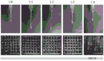
图1是1%的纳米粘土(Nanoclay)加入甲基丙烯酸化水凝胶与脱细胞基质(DECM)不同的体积比中形成的生物墨水在喷头处的挤出形态及各支架的打印效果图。Figure 1 shows the extrusion morphology of the bioink at the nozzle and the printing effect of each scaffold formed by adding 1% nanoclay (Nanoclay) to the methacrylated hydrogel and acellular matrix (DECM) in different volume ratios.
具体实施方式Detailed ways
下面结合附图和具体实施例对本发明作进一步说明,以使本领域的技术人员可以更好地理解本发明并能予以实施,但所举实施例不作为对本发明的限定。The present invention will be further described below with reference to the accompanying drawings and specific embodiments, so that those skilled in the art can better understand the present invention and implement it, but the embodiments are not intended to limit the present invention.
实施例1Example 1
一、制备基于肝脏脱细胞基质的复合生物墨水1. Preparation of composite bioink based on liver acellular matrix
(1)肝脏准备:将留置针插入天然大鼠的肝门静脉,取出肝脏,-80℃冷冻12h;(1) Liver preparation: Insert an indwelling needle into the hepatic portal vein of a natural rat, take out the liver, and freeze at -80°C for 12 hours;
(2)肝脏脱细胞基质的制备:肝脏常温解冻,连接蠕动泵进行灌注(灌注速率为10ml/min)①0.01mol/L PBS灌注1h;②1%Triton X-100灌注1h;PBS冲洗15min;③0.1%SDS灌注30min;PBS冲洗15min;④80U/ml DNase,5U/ml Rnase灌注30min;⑤含有2%青霉素-链霉素和2.5μg/ml两性霉素B的PBS灌注30min;当脱细胞基质出现完全半透明形态时,可将脱细胞基质进行冷冻干燥处理;(2) Preparation of liver decellularized matrix: the liver was thawed at room temperature and perfused with a peristaltic pump (perfusion rate was 10 ml/min) ① 0.01mol/L PBS perfused for 1 h; ② 1% Triton X-100 perfused for 1 h; washed with PBS for 15 min; ③ 0 .1% SDS perfusion for 30min; PBS rinse for 15min; ④80U/ml DNase, 5U/ml RNase perfusion for 30min; ⑤PBS containing 2% penicillin-streptomycin and 2.5μg/ml amphotericin B for 30min; When fully translucent morphology appears, the acellular matrix can be freeze-dried;
(3)肝脏脱细胞基质的表征:肝脏脱细胞基质经历冷冻干燥后,进行SEM检测,HE染色以及DNA含量等一系列表征,证明脱细胞较完全以后,可以进行下一步;(3) Characterization of liver acellular matrix: After the liver acellular matrix is freeze-dried, a series of characterizations such as SEM detection, HE staining and DNA content are carried out, which proves that after the decellularization is relatively complete, the next step can be performed;
(4)①肝脏脱细胞基质溶液的制备:将肝脏脱细胞基质冻干12h,取冻干后的脱细胞基质进行剪碎、研磨,使其易于溶解,研磨至合适颗粒大小,溶于浓度为1mg/mL的胃蛋白酶中制成溶液;②肝脏脱细胞基质溶液的表征:检测溶液中胶原蛋白的浓度,以及溶液中GAG、生长因子HGF和bFGF的含量,确保检测的含量尽量与未脱细胞处理的天然肝脏对比,没有极为显著的变化,保证制成的生物墨水具有可提供肝原代细胞生长的生长因子;(4) ① Preparation of liver acellular matrix solution: freeze-dry the liver acellular matrix for 12 hours, take the freeze-dried acellular matrix for shredding and grinding to make it easy to dissolve, and grind to a suitable particle size. 1mg/mL of pepsin to make a solution; ②Characterization of liver decellularized matrix solution: Detect the concentration of collagen in the solution, as well as the content of GAG, growth factor HGF and bFGF in the solution, and ensure that the detected content is as close as possible to those without decellularization. Compared with the treated natural liver, there is no extremely significant change, ensuring that the prepared bioink has growth factors that can provide the growth of primary liver cells;
(5)肝脏脱细胞基质生物墨水的制备:将肝脏脱细胞基质与明胶甲基丙烯酰胺(GelMA)1:1混合,并加入1%的纳米黏土,混合均匀,制成生物墨水。(5) Preparation of liver acellular matrix bioink: The liver acellular matrix was mixed with gelatin methacrylamide (GelMA) 1:1, and 1% of nanoclay was added, and mixed evenly to prepare a bioink.
二、生物墨水打印性能的检测2. Testing the printing performance of bio-ink
首先,利用AUTODESK 3DS MAX 2010软件设计一个10mm×10mm×1mm的长方体模型,用Repetier-Host切片软件对模型进行切片处理,将切片处理后的模型以G-code文件格式导入3D生物打印机中。按照表1的配比配制生物墨水,然后将配制好的生物墨水放置在3D生物打印机的打印喷头中,其中打印喷头直径为200μm,打印速度为7mm/s,底板及喷头温度设置为25℃,在材料逐层打印堆积成型后,将打印的结构暴露在紫外线下固化20s,产生稳定的支架。First, use AUTODESK 3DS MAX 2010 software to design a 10mm × 10mm × 1mm cuboid model, use Repetier-Host slicing software to slice the model, and import the sliced model into the 3D bioprinter in G-code file format. The bio-ink was prepared according to the ratio in Table 1, and then the prepared bio-ink was placed in the print head of the 3D bioprinter. The diameter of the print head was 200 μm, the printing speed was 7 mm/s, and the temperature of the bottom plate and the print head was set to 25 °C. After the material was printed layer-by-layer, the printed structure was cured by exposure to UV light for 20 s, resulting in a stable scaffold.
表1不同配比的生物墨水Table 1 Bioinks with different ratios
附图1示出了1%的nanoclay加入GelMA与脱细胞基质(DECM)不同的体积比中形成的生物墨水在喷头处的挤出形态及各支架的打印效果如下图所示。Figure 1 shows the extruded shape of the bioink at the nozzle and the printing effect of each scaffold formed by adding 1% nanoclay to GelMA and acellular matrix (DECM) in different volume ratios as shown in the figure below.
从图1中可以看出,当GelMA:DECM的比例为1:0~1:3时,得到的生物墨水都显示出了良好的打印性能。当GelMA:DECM的比例上升至1:4时,生物墨水的打印性能出现明显的下降。It can be seen from Figure 1 that when the ratio of GelMA:DECM is 1:0 to 1:3, the obtained bioinks all show good printing performance. When the ratio of GelMA:DECM increased to 1:4, the printing performance of the bioink decreased significantly.
现有技术中,未加nanoclay时,生物墨水中GelMA:DECM最大比例为1:1,即DECM的上限为50%。本实施例中,通过在生物墨水中引入了1%的nanoclay,明显的提高生物墨水的打印性能,使得DECM的可打印含量增加至75%,有利于提高打印支架的生物活性。In the prior art, when nanoclay is not added, the maximum ratio of GelMA:DECM in the bioink is 1:1, that is, the upper limit of DECM is 50%. In this example, by introducing 1% nanoclay into the bioink, the printing performance of the bioink is significantly improved, so that the printable content of the DECM is increased to 75%, which is beneficial to improve the biological activity of the printed scaffold.
以上所述实施例仅是为充分说明本发明而所举的较佳的实施例,本发明的保护范围不限于此。本技术领域的技术人员在本发明基础上所作的等同替代或变换,均在本发明的保护范围之内。本发明的保护范围以权利要求书为准。The above-mentioned embodiments are only preferred embodiments for fully illustrating the present invention, and the protection scope of the present invention is not limited thereto. Equivalent substitutions or transformations made by those skilled in the art on the basis of the present invention are all within the protection scope of the present invention. The protection scope of the present invention is subject to the claims.
Claims (12)
Priority Applications (1)
| Application Number | Priority Date | Filing Date | Title |
|---|---|---|---|
| CN202010260929.3ACN111420121A (en) | 2020-04-03 | 2020-04-03 | Composite biological ink based on methacrylated hydrogel/nanoclay/acellular matrix and preparation method and application thereof |
Applications Claiming Priority (1)
| Application Number | Priority Date | Filing Date | Title |
|---|---|---|---|
| CN202010260929.3ACN111420121A (en) | 2020-04-03 | 2020-04-03 | Composite biological ink based on methacrylated hydrogel/nanoclay/acellular matrix and preparation method and application thereof |
Publications (1)
| Publication Number | Publication Date |
|---|---|
| CN111420121Atrue CN111420121A (en) | 2020-07-17 |
Family
ID=71555724
Family Applications (1)
| Application Number | Title | Priority Date | Filing Date |
|---|---|---|---|
| CN202010260929.3APendingCN111420121A (en) | 2020-04-03 | 2020-04-03 | Composite biological ink based on methacrylated hydrogel/nanoclay/acellular matrix and preparation method and application thereof |
Country Status (1)
| Country | Link |
|---|---|
| CN (1) | CN111420121A (en) |
Cited By (2)
| Publication number | Priority date | Publication date | Assignee | Title |
|---|---|---|---|---|
| CN113599570A (en)* | 2021-07-22 | 2021-11-05 | 华南理工大学 | DNA nano composite hydrogel adhesive and preparation and application thereof |
| CN119971139A (en)* | 2025-02-10 | 2025-05-13 | 中国人民解放军总医院第三医学中心 | A 3D bioprinted vascularized bladder patch and its preparation method |
Citations (5)
| Publication number | Priority date | Publication date | Assignee | Title |
|---|---|---|---|---|
| KR20000003304U (en)* | 1998-07-22 | 2000-02-15 | 윤종용 | Optical axis adjusting device of image reading apparatus |
| CN105238132A (en)* | 2015-10-20 | 2016-01-13 | 中山大学 | Biological ink for 3D printing |
| CN109054496A (en)* | 2018-06-22 | 2018-12-21 | 中山大学附属第医院 | Composite biological ink and preparation method thereof |
| CN110585483A (en)* | 2019-09-26 | 2019-12-20 | 东华大学 | Novel biological ink capable of being crosslinked by multiple methods and preparation method thereof |
| WO2019245110A1 (en)* | 2018-06-18 | 2019-12-26 | 주식회사 티앤알바이오팹 | Hybrid bio-ink, method of preparing same, and method of preparing artificial tissue using same |
- 2020
- 2020-04-03CNCN202010260929.3Apatent/CN111420121A/enactivePending
Patent Citations (5)
| Publication number | Priority date | Publication date | Assignee | Title |
|---|---|---|---|---|
| KR20000003304U (en)* | 1998-07-22 | 2000-02-15 | 윤종용 | Optical axis adjusting device of image reading apparatus |
| CN105238132A (en)* | 2015-10-20 | 2016-01-13 | 中山大学 | Biological ink for 3D printing |
| WO2019245110A1 (en)* | 2018-06-18 | 2019-12-26 | 주식회사 티앤알바이오팹 | Hybrid bio-ink, method of preparing same, and method of preparing artificial tissue using same |
| CN109054496A (en)* | 2018-06-22 | 2018-12-21 | 中山大学附属第医院 | Composite biological ink and preparation method thereof |
| CN110585483A (en)* | 2019-09-26 | 2019-12-20 | 东华大学 | Novel biological ink capable of being crosslinked by multiple methods and preparation method thereof |
Non-Patent Citations (4)
| Title |
|---|
| (日)冈野光夫等: "《组织工程学》", 30 April 2016, 沈阳:辽宁科学技术出版社* |
| DONALD BEJLERI ET AL: "A Bioprinted Cardiac Patch Composed of Cardiac-Specific Extracellular Matrix and Progenitor Cells for Heart Repair", 《ADVANCED HEALTHCARE MATERIALS》* |
| QING GAO ET AL: "3D Printing of Complex GelMA-based Scaffolds with Nanoclay", 《BIOFABRICATION》* |
| 贺永等: "《生物3D打印 从医疗辅具制造到细胞打印》", 31 January 2019, 武汉:华中科技大学出版社* |
Cited By (2)
| Publication number | Priority date | Publication date | Assignee | Title |
|---|---|---|---|---|
| CN113599570A (en)* | 2021-07-22 | 2021-11-05 | 华南理工大学 | DNA nano composite hydrogel adhesive and preparation and application thereof |
| CN119971139A (en)* | 2025-02-10 | 2025-05-13 | 中国人民解放军总医院第三医学中心 | A 3D bioprinted vascularized bladder patch and its preparation method |
Similar Documents
| Publication | Publication Date | Title |
|---|---|---|
| Fang et al. | Hydrogels for 3D bioprinting in tissue engineering and regenerative medicine: Current progress and challenges | |
| Yang et al. | Emerging 3D bioprinting applications in plastic surgery | |
| Zhang et al. | Strategies for improving the 3D printability of decellularized extracellular matrix bioink | |
| CN106039415B (en) | Method for preparing bio-brick containing oxidized alginate and bio-brick prepared thereby | |
| CN113679888A (en) | Photocuring forming composite hydrogel matrix precursor, preparation method thereof and stent with same | |
| Babu et al. | Controlling structure with injectable biomaterials to better mimic tissue heterogeneity and anisotropy | |
| US12318996B2 (en) | Cross-linkable microgel composite matrix bath for embedded bioprinting of perfusable tissue constructs | |
| CN106474560B (en) | A kind of hydrogel material and the preparation method and application thereof for 3D biometric print | |
| CN106039421B (en) | A biobrick comprising endothelial cells and its use | |
| Liu et al. | Recent advances in decellularized matrix-derived materials for bioink and 3D bioprinting | |
| CN113559328B (en) | Biological ink and preparation method thereof | |
| CN111388758A (en) | Composite biological ink based on methacrylated hydrogel/hydroxyethyl cellulose/acellular matrix and preparation method thereof | |
| CN114904056B (en) | Composite hydrogel based on human placenta acellular matrix and preparation method thereof | |
| Rosellini et al. | Mending a broken heart by biomimetic 3D printed natural biomaterial-based cardiac patches: a review | |
| Kim et al. | Advanced strategies in 3D bioprinting for vascular tissue engineering and disease modelling using smart bioinks | |
| CN111748526A (en) | A kind of preparation method of imitating extracellular matrix printable composite bioink | |
| CN111420121A (en) | Composite biological ink based on methacrylated hydrogel/nanoclay/acellular matrix and preparation method and application thereof | |
| Sun et al. | Recent advances in 3D bioprinting of tissues and organs for transplantation and drug screening | |
| Du et al. | Bioactive polymer composite scaffolds fabricated from 3D printed negative molds enable bone formation and vascularization | |
| CN118987239A (en) | Composite hydrogel and preparation method thereof | |
| CN117510911A (en) | A neurobioprinting ink and its preparation method and application | |
| CN115894964B (en) | Photo-cured porous hydrogel cell preparation and preparation method thereof | |
| CN117838923A (en) | Preparation method and application of tricarboxylic acid circulating metabolite salt ion modified bone repair hydrogel | |
| JP7696580B2 (en) | Bioink, molded body, product, and method for manufacturing molded body | |
| Wang et al. | A soft receiving platform for coaxial bioprinting cell-laden microtubes with uniform wall thickness |
Legal Events
| Date | Code | Title | Description |
|---|---|---|---|
| PB01 | Publication | ||
| PB01 | Publication | ||
| SE01 | Entry into force of request for substantive examination | ||
| SE01 | Entry into force of request for substantive examination | ||
| RJ01 | Rejection of invention patent application after publication | Application publication date:20200717 | |
| RJ01 | Rejection of invention patent application after publication |