CN111408072B - Device for evaluating radiation dose delivery and system for verifying radiation dose delivery - Google Patents
Device for evaluating radiation dose delivery and system for verifying radiation dose deliveryDownload PDFInfo
- Publication number
- CN111408072B CN111408072BCN202010242209.4ACN202010242209ACN111408072BCN 111408072 BCN111408072 BCN 111408072BCN 202010242209 ACN202010242209 ACN 202010242209ACN 111408072 BCN111408072 BCN 111408072B
- Authority
- CN
- China
- Prior art keywords
- dose
- points
- patient
- radiation
- gamma
- Prior art date
- Legal status (The legal status is an assumption and is not a legal conclusion. Google has not performed a legal analysis and makes no representation as to the accuracy of the status listed.)
- Active
Links
- 230000005855radiationEffects0.000titleclaimsdescription130
- 238000011282treatmentMethods0.000claimsabstractdescription147
- 238000011156evaluationMethods0.000claimsabstractdescription94
- 238000001959radiotherapyMethods0.000claimsabstractdescription53
- 238000009826distributionMethods0.000claimsdescription109
- 238000003384imaging methodMethods0.000claimsdescription23
- 238000012545processingMethods0.000claimsdescription16
- 238000004364calculation methodMethods0.000claimsdescription11
- 238000004458analytical methodMethods0.000claimsdescription3
- 238000006243chemical reactionMethods0.000claimsdescription3
- 238000000034methodMethods0.000abstractdescription64
- 238000005259measurementMethods0.000abstractdescription60
- 238000010200validation analysisMethods0.000abstractdescription23
- 238000004590computer programMethods0.000abstractdescription12
- 210000000056organAnatomy0.000description42
- 230000000875corresponding effectEffects0.000description41
- 210000003484anatomyAnatomy0.000description29
- 238000004980dosimetryMethods0.000description28
- 210000000920organ at riskAnatomy0.000description23
- 238000002560therapeutic procedureMethods0.000description19
- 206010028980NeoplasmDiseases0.000description17
- 238000004422calculation algorithmMethods0.000description15
- 230000004907fluxEffects0.000description15
- 238000002721intensity-modulated radiation therapyMethods0.000description14
- 238000002203pretreatmentMethods0.000description14
- 238000012795verificationMethods0.000description14
- 230000008569processEffects0.000description13
- 238000002591computed tomographyMethods0.000description11
- 238000000275quality assuranceMethods0.000description9
- 230000033001locomotionEffects0.000description7
- 230000005540biological transmissionEffects0.000description6
- 238000005286illuminationMethods0.000description6
- 229910021417amorphous siliconInorganic materials0.000description5
- 230000006870functionEffects0.000description5
- 238000001727in vivoMethods0.000description5
- 230000001225therapeutic effectEffects0.000description5
- 238000001514detection methodMethods0.000description4
- 230000007717exclusionEffects0.000description4
- 238000003908quality control methodMethods0.000description4
- 230000004044responseEffects0.000description4
- 238000003860storageMethods0.000description4
- 230000005856abnormalityEffects0.000description3
- 230000003872anastomosisEffects0.000description3
- 230000006378damageEffects0.000description3
- 230000001678irradiating effectEffects0.000description3
- 238000012986modificationMethods0.000description3
- 230000004048modificationEffects0.000description3
- 230000008859changeEffects0.000description2
- 238000013170computed tomography imagingMethods0.000description2
- 230000001627detrimental effectEffects0.000description2
- 238000010586diagramMethods0.000description2
- 210000002216heartAnatomy0.000description2
- 230000004807localizationEffects0.000description2
- 210000004072lungAnatomy0.000description2
- 230000001575pathological effectEffects0.000description2
- 230000000149penetrating effectEffects0.000description2
- 238000002673radiosurgeryMethods0.000description2
- 231100000628reference doseToxicity0.000description2
- 230000035945sensitivityEffects0.000description2
- 238000004088simulationMethods0.000description2
- 210000000278spinal cordAnatomy0.000description2
- 230000003068static effectEffects0.000description2
- 230000001360synchronised effectEffects0.000description2
- 238000011287therapeutic doseMethods0.000description2
- 238000012546transferMethods0.000description2
- 238000000342Monte Carlo simulationMethods0.000description1
- 241000699670Mus sp.Species0.000description1
- 208000012902Nervous system diseaseDiseases0.000description1
- 208000025966Neurological diseaseDiseases0.000description1
- 208000009443Vascular MalformationsDiseases0.000description1
- 238000010521absorption reactionMethods0.000description1
- 238000013459approachMethods0.000description1
- 230000009286beneficial effectEffects0.000description1
- 230000008901benefitEffects0.000description1
- 238000004891communicationMethods0.000description1
- 239000012141concentrateSubstances0.000description1
- 239000002872contrast mediaSubstances0.000description1
- 238000012937correctionMethods0.000description1
- 230000002596correlated effectEffects0.000description1
- 238000011161developmentMethods0.000description1
- 238000006073displacement reactionMethods0.000description1
- 238000010894electron beam technologyMethods0.000description1
- 238000012854evaluation processMethods0.000description1
- 230000001747exhibiting effectEffects0.000description1
- 238000013213extrapolationMethods0.000description1
- 210000001508eyeAnatomy0.000description1
- 238000005194fractionationMethods0.000description1
- 201000010536head and neck cancerDiseases0.000description1
- 208000014829head and neck neoplasmDiseases0.000description1
- 230000036541healthEffects0.000description1
- 238000001802infusionMethods0.000description1
- 230000000977initiatory effectEffects0.000description1
- 230000003993interactionEffects0.000description1
- 150000002500ionsChemical class0.000description1
- 230000001788irregularEffects0.000description1
- 230000003902lesionEffects0.000description1
- 238000013178mathematical modelMethods0.000description1
- 239000011159matrix materialSubstances0.000description1
- 239000012528membraneSubstances0.000description1
- 239000002184metalSubstances0.000description1
- 238000012544monitoring processMethods0.000description1
- 230000003287optical effectEffects0.000description1
- 238000005457optimizationMethods0.000description1
- -1or a small phantomSubstances0.000description1
- 210000004279orbitAnatomy0.000description1
- 210000003681parotid glandAnatomy0.000description1
- 238000002727particle therapyMethods0.000description1
- 230000009528severe injuryEffects0.000description1
- 238000007493shaping processMethods0.000description1
- 238000002603single-photon emission computed tomographyMethods0.000description1
- 238000006467substitution reactionMethods0.000description1
- 238000001356surgical procedureMethods0.000description1
- 230000009466transformationEffects0.000description1
- 230000002792vascularEffects0.000description1
- 210000001835visceraAnatomy0.000description1
- 230000000007visual effectEffects0.000description1
- 238000012800visualizationMethods0.000description1
- XLYOFNOQVPJJNP-UHFFFAOYSA-NwaterSubstancesOXLYOFNOQVPJJNP-UHFFFAOYSA-N0.000description1
Images


Classifications
- A—HUMAN NECESSITIES
- A61—MEDICAL OR VETERINARY SCIENCE; HYGIENE
- A61N—ELECTROTHERAPY; MAGNETOTHERAPY; RADIATION THERAPY; ULTRASOUND THERAPY
- A61N5/00—Radiation therapy
- A61N5/10—X-ray therapy; Gamma-ray therapy; Particle-irradiation therapy
- A61N5/1048—Monitoring, verifying, controlling systems and methods
- A61N5/1071—Monitoring, verifying, controlling systems and methods for verifying the dose delivered by the treatment plan
- A—HUMAN NECESSITIES
- A61—MEDICAL OR VETERINARY SCIENCE; HYGIENE
- A61N—ELECTROTHERAPY; MAGNETOTHERAPY; RADIATION THERAPY; ULTRASOUND THERAPY
- A61N5/00—Radiation therapy
- A61N5/10—X-ray therapy; Gamma-ray therapy; Particle-irradiation therapy
- A61N5/103—Treatment planning systems
- A—HUMAN NECESSITIES
- A61—MEDICAL OR VETERINARY SCIENCE; HYGIENE
- A61N—ELECTROTHERAPY; MAGNETOTHERAPY; RADIATION THERAPY; ULTRASOUND THERAPY
- A61N5/00—Radiation therapy
- A61N5/10—X-ray therapy; Gamma-ray therapy; Particle-irradiation therapy
- A61N5/1048—Monitoring, verifying, controlling systems and methods
- A61N5/1049—Monitoring, verifying, controlling systems and methods for verifying the position of the patient with respect to the radiation beam
- A61N2005/1054—Monitoring, verifying, controlling systems and methods for verifying the position of the patient with respect to the radiation beam using a portal imaging system
- A—HUMAN NECESSITIES
- A61—MEDICAL OR VETERINARY SCIENCE; HYGIENE
- A61N—ELECTROTHERAPY; MAGNETOTHERAPY; RADIATION THERAPY; ULTRASOUND THERAPY
- A61N5/00—Radiation therapy
- A61N5/10—X-ray therapy; Gamma-ray therapy; Particle-irradiation therapy
- A61N5/1048—Monitoring, verifying, controlling systems and methods
- A61N2005/1074—Details of the control system, e.g. user interfaces
- A—HUMAN NECESSITIES
- A61—MEDICAL OR VETERINARY SCIENCE; HYGIENE
- A61N—ELECTROTHERAPY; MAGNETOTHERAPY; RADIATION THERAPY; ULTRASOUND THERAPY
- A61N5/00—Radiation therapy
- A61N5/10—X-ray therapy; Gamma-ray therapy; Particle-irradiation therapy
- A61N2005/1092—Details
Landscapes
- Health & Medical Sciences (AREA)
- Engineering & Computer Science (AREA)
- Biomedical Technology (AREA)
- Pathology (AREA)
- Nuclear Medicine, Radiotherapy & Molecular Imaging (AREA)
- Radiology & Medical Imaging (AREA)
- Life Sciences & Earth Sciences (AREA)
- Animal Behavior & Ethology (AREA)
- General Health & Medical Sciences (AREA)
- Public Health (AREA)
- Veterinary Medicine (AREA)
- Radiation-Therapy Devices (AREA)
Abstract
Translated fromChinese
Description
Translated fromChinese本申请是申请号为201610201152.7、申请日为2016年03月31日、发明名称为“射野剂量测定系统、设备和方法”的发明专利申请的分案申请。This application is a divisional application of the invention patent application with the application number of 201610201152.7, the application date of March 31, 2016, and the invention name of "field dosimetry system, equipment and method".
技术领域technical field
本公开一般涉及向患者递送放射,并且更具体地,用于通过选择性评价测量点来提供放射疗法治疗的剂量测定(dosimetry)验证的系统、方法和计算机程序产品。本公开还涉及用于通过使用不同的评价标准评价测量点来提供放射疗法治疗的剂量测定验证的系统、方法和计算机程序产品。本公开还涉及用于在放射治疗之前和在放射治疗期间用于执行质量控制测量的系统、方法和计算机程序产品。The present disclosure relates generally to the delivery of radiation to a patient, and more particularly, to systems, methods and computer program products for providing dosimetry validation of radiation therapy treatment by selectively evaluating measurement points. The present disclosure also relates to systems, methods, and computer program products for providing dosimetric validation of radiation therapy treatments by evaluating measurement points using different evaluation criteria. The present disclosure also relates to systems, methods and computer program products for performing quality control measurements prior to and during radiation therapy.
背景技术Background technique
一般而言,放射手术治疗和放射疗法治疗包括几个阶段。首先,感兴趣区域(头部,身体等)中的解剖结构的精确的三维(3D)映射图(map)被构造成确定解剖结构内的靶(target)的准确坐标,即,定位身体内的肿瘤或异常并且限定其准确形状和大小。第二,计算用于放射射束的运动路径,以递送将多种医疗限制考虑在内的外科医生认为可以接受的剂量分布。在该阶段期间,专家小组使用特殊的计算机软件开发了一种治疗计划来通过设计放射射束最佳地照射肿瘤并且最小化到周围正常组织的剂量以从不同的角度和平面汇聚在靶区域上。第三阶段是执行放射治疗计划的地方。在该阶段期间,根据使用放射治疗技术(诸如例如强度调制放射疗法(IMRT)和体积调制弧形疗法(VMAT))所规定的治疗计划向患者递送放射剂量。这些技术通常与配备有多叶准直器(MLC)的放射疗法系统(诸如线性加速器(linac))一起用来通过向病理解剖体递送所规定的放射剂量(X射线、γ射线、电子、质子和/或离子)来治疗病理解剖体(肿瘤、病灶、血管畸形、神经紊乱等),同时使得对周围组织和关键解剖结构的放射暴露最小化。In general, radiosurgery and radiotherapy treatment consists of several stages. First, an accurate three-dimensional (3D) map of the anatomy in the region of interest (head, body, etc.) is constructed to determine the exact coordinates of the target within the anatomy, ie to locate the target within the body Tumor or abnormality and define its exact shape and size. Second, the motion path for the radiation beam is calculated to deliver a dose distribution that the surgeon considers acceptable, taking into account various medical constraints. During this phase, a team of experts uses special computer software to develop a treatment plan to optimally irradiate the tumor by designing the radiation beam and minimize the dose to surrounding normal tissue to converge on the target area from different angles and planes . The third stage is where the radiation therapy plan is performed. During this phase, the radiation dose is delivered to the patient according to a prescribed treatment plan using radiation therapy techniques such as, for example, intensity-modulated radiation therapy (IMRT) and volume-modulated arc therapy (VMAT). These techniques are commonly used with multi-leaf collimator (MLC) equipped radiation therapy systems such as linear accelerators (linacs) by delivering prescribed radiation doses (X-rays, gamma rays, electrons, protons) to the pathological anatomy and/or ions) to treat pathological anatomy (tumors, lesions, vascular malformations, neurological disorders, etc.) while minimizing radiation exposure to surrounding tissues and critical anatomical structures.
存在许多导致所规定的放射剂量分布和所递送的实际剂量(即,在放射治疗期间递送给靶的实际剂量)之间差异的因素。一个这样的因素是患者在放射疗法系统中的位置的不确定性。其它因素包括由可以在患者治疗过程期间发生的改变引入的不确定性。这样的改变可以包括随机误差(诸如患者设置位置中的小差异)。其它来源归结于如果患者肿瘤消退或者如果患者在治疗期间体重减轻可能发生的生理改变。另一类别的不确定性包括运动。因为有些动作可能更加随意和不可预测,而其它的运动可以更加有规律,所以运动可能与任一类别重叠。存在许多其它不确定性的来源,诸如缺少大丸剂或固定设备(人为误差)、错误的患者、机械故障/校准误差/改变是放射输出、破坏的数据(方案与所计算的剂量不吻合)、错误递送机(例如,在原来的那个此刻不能运转的情况下,患者可能在另一递送机上治疗)。这些不确定性可能会影响患者的治疗质量以及递送给靶的实际放射剂量。There are a number of factors that contribute to the discrepancy between the prescribed radiation dose distribution and the actual dose delivered (ie, the actual dose delivered to the target during radiation therapy). One such factor is the uncertainty of the patient's position in the radiation therapy system. Other factors include uncertainty introduced by changes that can occur during the course of a patient's treatment. Such changes may include random errors (such as small differences in patient setting positions). Other sources are attributed to physiological changes that may occur if the patient's tumor regresses or if the patient loses weight during treatment. Another category of uncertainty includes motion. Because some movements can be more random and unpredictable, while others can be more regular, movements can overlap with either category. There are many other sources of uncertainty, such as missing bolus or fixtures (human error), wrong patient, mechanical failure/calibration error/change is radiation output, corrupted data (protocol does not match calculated dose), The wrong delivery machine (eg, the patient may be treated on another delivery machine if the original one is not operational at the moment). These uncertainties can affect the quality of treatment for patients and the actual radiation dose delivered to the target.
因此,基于预先确定的治疗计划向靶递送预测放射剂量的准确性在放射治疗的最终成功或未通过中起到重要作用。不准确的剂量递送可能会导致放射不足以治愈或对附近的健康组织和有风险器官有风险器官(OAR)的过多的放射。过高的放射剂量可能导致严重损害肿瘤周围的健康组织以及位于邻近的器官,而剂量太低可能危及治愈的可能性。因此,递送放射剂量的相对小的误差会严重伤害患者。因此,质量保证工具和协议都需要验证在没有危及有风险器官和健康组织的情况下,将所规定的放射剂量递送给靶。Thus, the accuracy of the predicted radiation dose delivered to the target based on a predetermined treatment plan plays an important role in the ultimate success or failure of radiation therapy. Inaccurate dose delivery may result in insufficient radiation to heal or too much radiation to nearby healthy tissue and organs at risk (OAR). Radiation doses that are too high can cause severe damage to healthy tissue surrounding the tumor as well as adjacent organs, while doses that are too low can jeopardize the possibility of a cure. Thus, relatively small errors in delivering radiation doses can seriously harm patients. Therefore, both quality assurance tools and protocols need to verify that the prescribed radiation dose is delivered to the target without endangering at-risk organs and healthy tissue.
因为治疗计划的高复杂性和唯一性,所以患者特定的治疗前(即,在射束中没有患者的情况下)验证通常被认为是患者治疗的必要的先决条件。治疗前验证包括程序,其比较预期的治疗计划的全部或至少部分与由患者治疗时间以外的线性加速器(linac)递送的对应的放射射束的测量。Because of the high complexity and uniqueness of treatment plans, patient-specific pre-treatment (ie, in the absence of a patient in the beam) validation is often considered a necessary prerequisite for patient treatment. Pre-treatment validation includes a procedure that compares all or at least part of an expected treatment plan with measurements of corresponding radiation beams delivered by a linear accelerator (linac) outside of the patient's treatment time.
剂量测定验证是对于放射疗法治疗实施的治疗前协议之一。剂量测定验证包括:验证所递送的剂量分布实际上是预测要递送给患者的剂量分布。因为由一些放射疗法(诸如(IMRT)和(VMAT)治疗)所提供的射束递送复杂性的增加,所以用于治疗的剂量测定验证需要放射剂量递送的严格验证。Dosimetry validation is one of the pre-treatment protocols implemented for radiotherapy treatments. Dosimetric verification includes verifying that the delivered dose distribution is actually the predicted dose distribution to be delivered to the patient. Because of the increased complexity of beam delivery provided by some radiation therapies, such as (IMRT) and (VMAT) treatments, dosimetric validation for therapy requires rigorous validation of radiation dose delivery.
在所建立的剂量验证方法中,使用伽玛评价方法比较整合的剂量分布图像与由治疗计划系统(TPS)所预测的剂量图像。伽玛评价方法广泛用于剂量测量,因为它将空间误差和剂量水平误差组合在单个值中。然而,这样的评价的劣势是所有测量点都基于相同的标准进行评价,即使评价标准对于某些点可能太宽松或太严格。即使所检测的剂量差异对于有风险器官有风险器官可能太高(例如,在有风险器官有风险器官中生成的过度剂量(overdose)或热点要比在靶或健康组织中的过度剂量或热点更加严重),宽松评价标准也可以确认剂量递送,而即使所检测的剂量差异不影响患者,更严格的评价标准也可以拒绝剂量递送。In the established dose verification method, a gamma evaluation method was used to compare the integrated dose distribution image with the dose image predicted by the treatment planning system (TPS). The gamma evaluation method is widely used in dosimetry because it combines spatial error and dose level error in a single value. However, the disadvantage of such an evaluation is that all measurement points are evaluated based on the same criteria, even though the evaluation criteria may be too loose or too strict for some points. Even though the detected dose difference may be too high for the at-risk organ (eg, an overdose or hotspot generated in the at-risk organ is more likely than the overdose or hotspot in the target or healthy tissue) Severe), looser evaluation criteria can also confirm dose delivery, while more stringent evaluation criteria can deny dose delivery even if the detected dose difference does not affect the patient.
进一步地,在所建立的剂量验证方法中,如果所测量的放射与所预期的放射不同,则治疗停止。然而,如果放射射束与患者相切,则患者轮廓的小改变可以在所测量的剂量中产生显著差异,而并不显著影响患者中的实际剂量。事实上,在一些实例中,放射场有意地制得比靶大。在弧形疗法治疗中,例如,因为使用了平面中的所有放射方向,所以切向场更可能发生。因此,当由射束照射的所有点用于治疗的实时评价时,所建立的剂量评价方法可能错误地检测到剂量误差并且触发停止放射治疗。Further, in the established dose verification method, if the measured radiation is different from the expected radiation, the treatment is stopped. However, if the radiation beam is tangential to the patient, small changes in the patient profile can make a significant difference in the measured dose without significantly affecting the actual dose in the patient. In fact, in some instances, the radiation field is intentionally made larger than the target. In arc therapy treatments, for example, tangential fields are more likely to occur because all radiation directions in the plane are used. Therefore, when all points irradiated by the beam are used for real-time evaluation of treatment, established dose evaluation methods may falsely detect dose errors and trigger cessation of radiotherapy.
发明内容SUMMARY OF THE INVENTION
本公开提供了用于放射治疗前、治疗、以及体内(in-vivo)剂量测定验证的系统、方法、设备和计算机程序产品。剂量测定验证包括:对于测量平面中的不同点,使用不同的评价标准来评价剂量分布。The present disclosure provides systems, methods, apparatus, and computer program products for pre-radiotherapy, therapeutic, and in-vivo dosimetry validation. Dosimetric validation involves evaluating the dose distribution using different evaluation criteria for different points in the measurement plane.
本公开还提供了用于对于测量平面中的不同点使用不同的伽玛标准来评价剂量分布的系统、方法和计算机程序产品。测量平面中的每个点都可以与对应的伽玛标准相关联。可以基于投影到测量平面中的点上的解剖结构的类型限定用于不同的伽玛标准的值。The present disclosure also provides systems, methods and computer program products for evaluating dose distribution using different gamma criteria for different points in the measurement plane. Each point in the measurement plane can be associated with a corresponding gamma standard. Values for different gamma standards can be defined based on the type of anatomy projected onto the point in the measurement plane.
本公开还提供了用于选择性地评价测量平面中的点的系统、方法和计算机程序产品。选择性评价排除了由在患者体内不相交的放射射束、和/或在患者的表面附近行进的放射射束、和/或从正在被评价的穿过患者表面的深度大约1cm的放射射束照射的点。评价可以包括:使用伽玛标准来评价所选择的点。评价还可以包括:使用不同的伽玛标准来评价所选择的点。The present disclosure also provides systems, methods, and computer program products for selectively evaluating points in a measurement plane. Selective evaluation excludes radiation beams from disjoint radiation beams within the patient, and/or radiation beams traveling near the patient's surface, and/or radiation beams that are approximately 1 cm deep from the patient surface being evaluated irradiated point. The evaluation may include using a gamma criterion to evaluate the selected points. The evaluation may also include evaluating the selected points using different gamma criteria.
本公开提供了用于治疗计划的基于电子射野成像设备(EPID)的治疗前剂量验证的系统、方法和计算机程序产品。The present disclosure provides systems, methods, and computer program products for electronic portal imaging device (EPID)-based pre-treatment dose verification for treatment planning.
本公开还提供用于使用电子射野成像设备(EPID)进行实时放射剂量验证的系统、方法、设备和计算机程序产品,其中,只有测量平面内的所选择的点用于治疗验证。The present disclosure also provides systems, methods, devices, and computer program products for real-time radiation dose verification using an electronic portal imaging device (EPID), wherein only selected points within the measurement plane are used for treatment verification.
本公开还提供了用于将所测量的剂量分布转换成绝对剂量分布的(EPID)校准模型。The present disclosure also provides (EPID) calibration models for converting measured dose distributions to absolute dose distributions.
本公开还提供了用于定量评价剂量分布的系统、方法和计算机程序产品。The present disclosure also provides systems, methods and computer program products for quantitative assessment of dose distribution.
本公开还提供了在其上体现用于在如本文中所公开的放射疗法治疗系统的质量控制的编程指令序列的非暂态计算机可读存储介质,其执行在计算机可读存储介质上体现的编程指令序列以使计算机处理系统执行如本文所公开的方法的步骤,该放射疗法治疗系统包括如本文所公开的计算机处理系统。The present disclosure also provides a non-transitory computer-readable storage medium having embodied thereon a sequence of programming instructions for quality control of a radiation therapy treatment system as disclosed herein, executing the sequences embodied on the computer-readable storage medium. A sequence of instructions is programmed to cause a computer processing system to perform the steps of the method as disclosed herein, the radiation therapy treatment system comprising the computer processing system as disclosed herein.
附图说明Description of drawings
本文中所描述的附图仅用于说明的目的,并不旨在以任何方式限制本公开的范围。通过结合附图阅读随后的说明书,本发明得以更好地理解,其中,相同的元件用相同的附图标记表示。如本文中所使用的,各种实施例可以是指部分或全部的实施例。The drawings described herein are for illustration purposes only and are not intended to limit the scope of the present disclosure in any way. The invention may be better understood by reading the ensuing specification in conjunction with the accompanying drawings, wherein like elements are designated by like reference numerals. As used herein, various embodiments may refer to some or all of the embodiments.
图1是根据本发明的各种实施例的放射疗法系统的透视图。1 is a perspective view of a radiation therapy system according to various embodiments of the present invention.
图2图示了根据各种实施例的靶体积定义。2 illustrates target volume definitions according to various embodiments.
图3图示了像素-体素关系。Figure 3 illustrates the pixel-voxel relationship.
图4图示了根据本发明的各种实施例的剂量验证过程。Figure 4 illustrates a dose verification process according to various embodiments of the present invention.
图5图示了根据本发明的各种实施例的3D剂量分布验证过程和结果。Figure 5 illustrates the 3D dose distribution validation process and results according to various embodiments of the present invention.
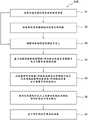
图6是根据本发明的各种实施例的使用不同的伽玛标准来图示剂量评价过程的流程图。6 is a flow diagram illustrating a dose evaluation process using different gamma standards, according to various embodiments of the present invention.
图7图示了照射患者表面的切线和表面射束。Figure 7 illustrates tangential and surface beams illuminating the patient surface.
图8是根据本发明的各种实施例的选择性剂量评价方法的流程图。8 is a flow diagram of a selective dose assessment method according to various embodiments of the present invention.
具体实施方式Detailed ways
为了验证放射治疗程序被正确地应用,可以在放射疗法的对应阶段实施用于治疗计划的剂量测定验证以及体内剂量测定的质量保证协议。实施质量保证协议以验证所开发的治疗计划是准确的,治疗递送是准确的,并且递送给患者的实际剂量是所计划的剂量。特别在先进的放射疗法技术(诸如强度调制放射疗法(IMRT)或者弧形疗法)中需要质量保证,其中,为了在肿瘤内部集中剂量,而避开有风险器官有风险器官(OAR),治疗计划往往具有高梯度剂量分布。To verify that radiotherapy procedures are being applied correctly, dosimetry validation for treatment planning and quality assurance protocols for in vivo dosimetry can be implemented at the corresponding phase of radiotherapy. Quality assurance protocols are implemented to verify that the developed treatment plan is accurate, that the treatment delivery is accurate, and that the actual dose delivered to the patient is the planned dose. Quality assurance is particularly required in advanced radiation therapy techniques such as intensity-modulated radiation therapy (IMRT) or arc therapy, where, in order to concentrate the dose within the tumor, while avoiding the organs at risk (OAR), treatment planning Often has a high gradient dose distribution.
治疗执行验证可以包括两个步骤。第一步骤包括治疗前测量并且第二步骤包括测量期间或者治疗中测量。执行治疗前测量以在患者的第一次治疗之前,检查来自计划阶段的治疗参数到特定放射治疗设备的适当传递。它还确保了治疗计划由设备执行的正确性。因此,治疗前验证是将所预期的治疗计划的全部或至少一部分与患者治疗时间之外(即,具有开放场或者幻像(phantom))的由线性加速器所递送的对应的放射射束的测量进行比较的程序。这种比较关注于所预测的和所测量的叶片(leaf)位置、递送给检测器或者幻像的剂量、或者提取用于测量的入射能量通量(fluence)。Treatment execution verification can consist of two steps. The first step includes pre-treatment measurements and the second step includes during or during treatment measurements. Pre-treatment measurements are performed to check for proper delivery of treatment parameters from the planning phase to a particular radiation therapy device prior to the patient's first treatment. It also ensures that the treatment plan is correctly executed by the device. Thus, pre-treatment validation is the measurement of all or at least a portion of the intended treatment plan with the measurement of the corresponding radiation beam delivered by the linear accelerator outside the patient treatment time (ie, with an open field or phantom). program for comparison. This comparison focuses on predicted and measured leaf positions, dose delivered to detectors or phantoms, or incident energy fluence extracted for measurement.
所述治疗验证期间(或“中”)是关注于基于在患者治疗期间获取的测量对所计划的和所递送的剂量分布的全部或部分进行比较的程序。然后,这些测量可以用来确定递送给检测器或患者的剂量、或者从测量获得的入射能量通量。The treatment validation period (or "mid") is a procedure focused on comparing all or part of the planned and delivered dose distribution based on measurements taken during the treatment of the patient. These measurements can then be used to determine the dose delivered to the detector or patient, or the incident energy flux obtained from the measurements.
图1图示了可以向位于治疗床1上的患者5提供放射疗法并且允许实施用于质量保证(QA)协议的各种治疗前和治疗中射野剂量测定验证的示例性放射疗法治疗系统100。放射疗法治疗可以包括基于光子的放射疗法、粒子疗法、电子射束疗法、或者任何其它类型的治疗疗法。在实施例中,放射疗法治疗系统100包括放射治疗设备10,诸如但不限于放射疗法或放射手术设备,其可以包括支撑放射模块8的台架(gantry)7,该放射模块8包括一个或多个放射源3和线性加速器(linac)2,该线性加速器可操作以生成kV或MV的X射线放射射束。台架7可以是环形台架(即,其延伸通过整个360度弧来创建完整的环或圈),但还可以使用其它类型的安装布置。例如,可以使用静态射束、或C型部分环形台架、或机械臂。还可以使用能够在相对于患者5的各种转动和/或轴向位置定位放射模块8的任何其它框架。FIG. 1 illustrates an exemplary radiation
放射模块8还可以包括调制设备(未示出),其可操作以调制放射射束以及将治疗放射射束朝向患者5和朝向期望被照射的患者的一部分引导。期望被照射的部分被称为靶或靶区域或感兴趣区域。患者5可以具有需要被照射的一个或多个感兴趣区域。准直设备(未示出)可以被包括在调制设备中以限定和调整孔隙(aperture)的大小,通过该孔隙放射射束可以从源3朝向患者5通过。准直设备可以由致动器(未示出)控制,该致动器可以由计算机处理系统40和/或控制器30控制。The radiation module 8 may also include a modulation device (not shown) operable to modulate the radiation beam and direct the therapeutic radiation beam towards the patient 5 and towards a portion of the patient desired to be irradiated. The portion that is desired to be irradiated is called the target or target area or region of interest. The patient 5 may have one or more regions of interest that need to be irradiated. A collimation device (not shown) may be included in the modulation device to define and adjust the size of the aperture through which the radiation beam may pass from the
在实施例中,放射疗法设备是kV或MV能量强度调制放射疗法(IMRT)设备。这样的系统中的强度简档(profile)是针对个体患者的治疗要求而定制的。强度调制放射疗法场用多叶准直器(MLC)递送,该多叶准直器可以是附接到直线加速器的头部的计算机控制的机械射束成形设备并且包括金属指状物(fingers)或叶片的组件。(MLC)可以例如由叶片宽度为0.5和/或1.0cm的120个可移动叶片制成。对于每个射束方向,通过依序递送形状和重量优化的各种子场来实现优化的强度简档。从一个子场到下一个,叶片可以在放射射束开启时(即,动态多叶准直(DMLC))移动,或者在放射射束移动关断时(即,分段多叶准直(SMLC))移动。In an embodiment, the radiation therapy device is a kV or MV energy intensity modulated radiation therapy (IMRT) device. Intensity profiles in such systems are tailored to the treatment requirements of individual patients. The intensity modulated radiation therapy field is delivered with a multi-leaf collimator (MLC), which may be a computer-controlled mechanical beam shaping device attached to the head of the linac and including metal fingers or components of the blades. (MLC) may eg be made of 120 movable vanes with vane widths of 0.5 and/or 1.0 cm. An optimized intensity profile is achieved by sequentially delivering shape and weight optimized various subfields for each beam direction. From one subfield to the next, the vanes can move when the radiation beam is on (ie, dynamic multi-leaf collimation (DMLC)), or when the radiation beam movement is off (ie, segmented multi-leaf collimation (SMLC) ))move.
设备10还可以是其中用在计算机控制下开启和关闭的二进制准直器实现的强度调制的断层放射疗法设备。当台架围绕患者连续转动时,射束的小宽度的暴露时间可以用二进制准直器的开启和关闭来调整,从而允许放射通过患者的最优选的方向和位置递送给肿瘤。Device 10 may also be an intensity modulated tomotherapy device in which intensity modulation is implemented with binary collimators that are switched on and off under computer control. As the gantry rotates continuously around the patient, the exposure time of the narrow width of the beam can be adjusted with the binary collimator on and off, allowing radiation to be delivered to the tumor through the most preferred direction and location of the patient.
设备10还可以是螺旋断层放射疗法设备,其包括滑环式转动台架。设备10还可以是强度调制弧形疗法设备(IMAT),其中,替代使用转动扇形(fan)射束,不同形状的转动锥形射束用来实现强度调制。设备10还可以是使用多个弧的简化的强度调制弧形疗法(SIMAT)设备、或者扫窗弧形疗法设备(SWAT),其中,(MLC)叶片位置在转动的情况下扫掠穿过靶计划体积(TPV)。设备10还可以是体积调制弧形疗法(VMAT)设备,其中剂量率、射束孔隙形状和转动速度可以连续变化以向靶计划体积(TPV)提供所规定的剂量。Apparatus 10 may also be a helical tomotherapy apparatus comprising a slip ring type rotating gantry. The device 10 may also be an intensity modulated arc therapy device (IMAT) wherein, instead of using a rotating fan beam, a different shape of rotating cone beam is used to achieve intensity modulation. The device 10 may also be a simplified intensity modulated arc therapy (SIMAT) device using multiple arcs, or a swept window arc therapy device (SWAT) in which the (MLC) blade position is swept across the target with rotation Planned Volume (TPV). The device 10 may also be a volume modulated arc therapy (VMAT) device in which the dose rate, beam aperture shape and rotational speed can be continuously varied to deliver the prescribed dose to the target planning volume (TPV).
设备10还包括用于获取数字图像以用于射野剂量验证的射野剂量成像设备20。射野剂量成像设备20可以是电子射野成像设备(EPID)。射野剂量成像设备20可以被放置在不同位置处,诸如例如,在治疗床1的顶部上或者附接到加速器头部2。射野剂量成像设备20能够生成即时2D数字信息。它可以是基于相机的设备(诸如基于相机的(EPID))或者基于非晶硅的设备(诸如非晶硅(EPID))。(EPID)20还可以是基于CCD相机的(EPID),其实际上是例如同时整合停滞时间介于大约0.1ms的所获取的帧之间的剂量测定器(dosimeter)的阵列。另一备选是平板成像器(或非晶硅EPID),其提供了良好的图像质量、高光学传递效率、大成像区域和放射抗性。The device 10 also includes a portal
可以使用的示例性非晶硅成像设备是aSi1000 EPID成像器,其具有在40×30cm2有源检测器区域4中布置的光敏非晶硅光电二极管阵列,并且具有最大帧速率为9.574fps,每一帧是检测器元件的扫描。平板成像器通常由图像元素(像素)组成,该图像元素寄存落在它们上的放射量并且将所接收的放射量转换成对应数量的电子。电子被转换成使用成像设备20或计算机40进一步处理的电信号。这样的配置(即,与(多个)治疗源相对定位的(多个)数字成像检测器)提供连续且立即捕获从每个弧形场段传送的和/或在连续弧形射束递送期间的治疗放射的能量和强度的能力,以便生成数字化的X射线测量的二维(2D)图像。因为射野剂量成像设备20生成即时2D数字信息,所以它便于以任何台架角度进行2D剂量测定。An exemplary amorphous silicon imaging device that can be used is the aSi1000 EPID imager, which has an array of photosensitive amorphous silicon photodiodes arranged in a 40 x 30cm2
计算机40包括典型的硬件(诸如处理器)以及用于运行各种软件程序和/或通信应用程序的操作系统。计算机可以包括操作以与放射疗法设备10通信的软件程序,并且该软件程序还可操作以从任何外部的软件程序和硬件来接收数据。该计算机40还可以包括适于由医疗人员访问的任何合适的输入/输出设备、以及I/O接口、存储设备、存储器、键盘、鼠标、监视器、打印机、扫描仪等。计算机40还可以与其它计算机和放射疗法系统联网。放射疗法设备10和计算机40两者均可以与网络以及数据库和服务器通信。该计算机40还适于在不同的医疗设备之间传递医疗图像相关的数据。
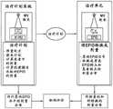
系统100还可以包括包含编程指令的多个模块,其互相通信并且当被执行时如本文所讨论的使系统100执行与放射疗法/手术有关的不同功能。例如,系统100可以包括:治疗计划模块,该治疗计划模块可操作以基于由医疗人员输入系统的多个数据来生成用于患者5的治疗计划,治疗计划包括所预测的放射剂量分布;患者定位模块,其可操作以针对特定放射疗法治疗相对于台架7的等中心点定位并对准患者5;图像获取模块,其可操作以指令放射疗法设备10在放射疗法治疗之前和/或在放射疗法治疗期间获取患者5的图像(即,体内图像),和/或指令其它成像设备或系统获取患者5的图像。The
系统100可以进一步包括:治疗递送模块,其可操作以指令放射疗法设备10在患者5处于或不处于适当位置的情况下递送治疗计划;转换模块,其可操作以将2D射野图像(EPI)转换成2D射野剂量;分析模块,其可操作以计算所预测的和所测量的剂量分布之间的比较;选择模块,其可操作以基于选择标准来选择用于剂量评价的测量点;评价模块,其基于适用于不同的点的不同的评价标准来评价用于剂量差异的测量点;计算模块,其可操作以计算剂量递送误差;以及执行模块,其可操作以初始化停止放射过程、或者向医师发送警报信号、或者发起报警程序。例如,这些模块可以用C或C++编程语言书写。用于执行如本文所描述的本发明的操作的计算机程序代码还可以用其它编程语言书写。The
作为对质量控制协议的一部分,对于治疗前射野剂量测定验证,在开始患者治疗之前验证由治疗场递送的放射剂量分布。患者治疗包括:根据所规定的递送治疗计划用治疗射束(即例如,X射线)照射患者。As part of the quality control protocol, for pre-treatment field dosimetry validation, the radiation dose distribution delivered by the treatment field is validated prior to initiating patient treatment. Patient treatment involves irradiating the patient with a treatment beam (ie, eg, X-rays) according to a prescribed delivery treatment plan.
在治疗阶段之前,使用治疗计划系统(TPS)开发所规定的递送方案,并且包括:使用特殊的计算机软件开发方案以最佳地照射肿瘤并且从不同的角度和平面将到周围的正常组织的剂量最小化。首先,使用计算机断层扫描(CT)、锥形射束CBCT、核磁共振成像(MRI)、正电子发射断层扫描(PET)、3D转动血管造影(3DRA)或超声技术中的任一项来构建感兴趣区域(头部、身体等)中的解剖结构的精确的三维(3D)映射图。这确定了解剖结构内的靶的准确坐标,即,将肿瘤或异常定位在身体内并且限定其准确形状和大小。例如,为了获得CT图像,机动台子通过CT成像系统中的圆形开口移动患者。当患者通过CT成像系统时,x射线源围绕圆形开口内部转动。单次转动花费大约1秒。x射线源产生用来照射患者身体的一段(segment)的狭窄的扇形x射线射束。扇形射束的厚度可以小至1毫米或者大至10毫米。在典型的检查中有几个阶段,每个阶段由x射线管围绕配合台子通过圆形开口移动的患者的10~50次转动组成。患者可以接收造影材料的注入,以便于血管结构的可视化。Before the treatment phase, the prescribed delivery protocol is developed using a treatment planning system (TPS) and includes: using special computer software to develop a protocol to optimally irradiate the tumor and dose to surrounding normal tissue from different angles and planes minimize. First, construct a sense of Accurate three-dimensional (3D) maps of anatomical structures in regions of interest (head, body, etc.). This determines the exact coordinates of the target within the anatomy, ie locates the tumor or abnormality within the body and defines its exact shape and size. For example, to obtain CT images, a motorized table moves the patient through a circular opening in the CT imaging system. As the patient passes through the CT imaging system, the x-ray source rotates around the inside of the circular opening. A single spin takes about 1 second. The x-ray source produces a narrow fan-shaped beam of x-rays that is used to irradiate a segment of the patient's body. The thickness of the fan beam can be as small as 1 mm or as large as 10 mm. In a typical exam there are several phases, each consisting of 10 to 50 rotations of the x-ray tube around the patient with a cooperating table moving through a circular opening. The patient may receive an infusion of contrast material to facilitate visualization of vascular structures.
患者的出口侧上的一个或多个检测器(诸如EPID)记录从作为x射线源的一个位置(角度)处的x射线“快照(snapshot)”被照射的患者身体的段离开的x射线。在一个完整的转动期间收集许多不同的“快照”(角度)。然后,对于x射线源的每个完整的转动,向计算机发送数据以将所有的个体“快照”重建成内部器官和组织的横截面图像(切片)。可以从用检测器检测到的吸收信号计算CT图像,而源和检测器通过背投影环绕在患者周围。为此,所检测的信号的强度从检测器投影回到源并且在x射线照射的对象区域中重叠。One or more detectors (such as EPIDs) on the exit side of the patient record x-rays exiting from the segment of the patient's body that is irradiated as an x-ray "snapshot" at a location (angle) of the x-ray source. Collect many different "snapshots" (angles) during one full turn. Then, for each complete rotation of the x-ray source, data is sent to a computer to reconstruct all individual "snapshots" into cross-sectional images (slices) of internal organs and tissues. A CT image can be calculated from the absorption signal detected with a detector, while the source and detector surround the patient by backprojection. To this end, the intensity of the detected signal is projected from the detector back to the source and overlapped in the area of the object irradiated by the x-rays.
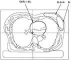
治疗计划基于个体患者的解剖特点。解剖特点包括外部几何结构、定位、肿瘤和有风险器官有风险器官的范围、以及组织密度的变化。解剖描述(即,解剖模型)可以例如从该组CT图像(或MRI、SPECT、PET等)中推导出,但还可以使用与模拟器图像组合的一组轮廓。如图2所示,基于CT图像和/或轮廓,生成指定TPS中的患者解剖的解剖结构(即,点、轮廓、和体积)。每个解剖描述唯一链接到一个或多个射束布置。The treatment plan is based on the anatomical characteristics of the individual patient. Anatomical features include external geometry, localization, extent of tumor and organs at risk, and changes in tissue density. An anatomical description (ie, an anatomical model) can be derived, for example, from the set of CT images (or MRI, SPECT, PET, etc.), but also a set of contours combined with simulator images can be used. As shown in FIG. 2, based on the CT images and/or contours, anatomical structures (ie, points, contours, and volumes) that specify the patient's anatomy in the TPS are generated. Each anatomical description is uniquely linked to one or more beam arrangements.
然后,计算用于放射射束的运动路径以递送将包括有风险器官有风险器官(OAR)的位置和类型的多种医疗约束考虑在内的外科医生认为可以接受的剂量分布。OAR是位于靠近必须严格约束用于其的放射剂量的靶的关键结构。用放射对关键结构的过度剂量可能导致医疗并发症。OAR还被称为“敏感结构”或“关键结构”。在该阶段期间,专家小组使用特殊的计算机软件开发治疗计划,以通过设计放射射束以从不同的角度和平面会聚在靶区域上最佳地照射肿瘤并且将到周围的正常组织和OAR的剂量最小化。治疗计划包括用于计算以递送治疗医师认为可以接受的剂量分布的放射射束的轨迹(运动路径)。基于以下知识来开发射束轨迹:解剖结构内的靶的准确坐标、邻近靶的OAR的准确坐标、不直接邻近靶但是对其而言甚至少许剂量也会是有害的OAR的坐标、以及身体内的肿瘤或异常的准确形状和大小。The motion path for the radiation beam is then calculated to deliver a dose distribution that the surgeon deems acceptable taking into account various medical constraints including the location and type of the organ at risk (OAR). The OAR is a critical structure located near the target for which the radiation dose must be tightly constrained. Overdose of critical structures with radiation can lead to medical complications. OARs are also known as "sensitive structures" or "critical structures". During this phase, a team of experts uses special computer software to develop a treatment plan to optimally irradiate the tumor and dose to the surrounding normal tissue and OAR by designing the radiation beam to converge on the target area from different angles and planes minimize. The treatment plan includes the trajectory (path of motion) of the radiation beam used to calculate to deliver a dose distribution deemed acceptable by the treating physician. Beam trajectories are developed based on the knowledge of: the exact coordinates of the target within the anatomy, the exact coordinates of the OAR adjacent to the target, the coordinates of the OAR not directly adjacent to the target but for which even a small dose would be detrimental, and the coordinates of the OAR within the body the exact shape and size of the tumor or abnormality.
放射治疗计划的目标包括均匀性、一致性、避免性(avoidance)和简单性。均匀性要求是要照射特定剂量水平内的肿瘤体积。对于治疗计划而言重要的是在靶上具有均匀的剂量分布,以使“冷点(cold spots)”可以被最小化。“冷点”是在其所需剂量水平下接收的结构的一部分,诸如例如,器官、肿瘤或组织。另一方面,术语“热点(hot spot)”用来表示其接收比所需剂量水平更高的结构的一部分。一致性要求用来实现靶剂量控制,同时将对OAR或者健康正常组织的损伤最小化。避免性要求可以是限制递送给OAR的剂量。简单性要求是要提供尽可能简单的治疗计划。简单的治疗计划通常减少治疗时间以及实施误差。在优化治疗计划中,感兴趣区域的三维体积可以由体素网格表示,并且治疗计划可以包括从放射源到每个体素的所期望的剂量分布。治疗计划也可以包括医师感兴趣的该组器官几何结构、以及用于每个感兴趣器官所期望的剂量水平。The goals of radiation therapy planning include homogeneity, consistency, avoidance, and simplicity. The homogeneity requirement is to irradiate the tumor volume within a specific dose level. It is important for treatment planning to have a uniform dose distribution on the target so that "cold spots" can be minimized. A "cold spot" is a portion of a structure, such as, for example, an organ, tumor, or tissue, received at its desired dosage level. On the other hand, the term "hot spot" is used to denote a portion of a structure that receives a higher dose level than required. Consistency requirements are used to achieve target dose control while minimizing damage to the OAR or healthy normal tissue. An avoidance requirement may be to limit the dose delivered to the OAR. The requirement for simplicity is to provide the simplest possible treatment plan. Simple treatment planning generally reduces treatment time and implementation errors. In optimizing the treatment plan, the three-dimensional volume of the region of interest can be represented by a grid of voxels, and the treatment plan can include the desired dose distribution from the radiation source to each voxel. The treatment plan may also include the set of organ geometries of interest to the physician, and the desired dose levels for each organ of interest.
为了优化弧形递送治疗计划(对于弧形疗法,轨迹可以是由治疗台架绕患者转动形成的弧形的,通常单个360度或单个180度旋转),在治疗计划过程一开始,可以指定沿着轨迹的若干个控制点。每个控制点与一组治疗参数相关联,包括但不限于一组(MLC)叶片位置、(MLC)形状、台架转动速度、台架位置、剂量率、和/或任何其它参数。可以以任何方便的方式设定控制点的数量和位置,诸如但不限于,通过使用治疗计划软件或通过系统操作员。在示例性实施例中,轨迹可以包括单个180度的弧形轨迹和大约177个顺序控制点,其意味着有线性加速器(2)应该符合其以便递送所计划的治疗的177种配置。基于治疗参数,针对每个控制点,通过任意数量的技术(诸如但不限于,笔形射束卷积或者任何其它合适的算法)计算治疗体积内的剂量分布。使用实际射野成像参数(包括场大小、CIAO(完全照射的孔隙轮廓)孔隙、监视器单元(MU)的总数量、台架角度、准直器角度、床角度和能量)计算剂量分布。In order to optimize the arc-delivery treatment plan (for arc therapy, the trajectory can be an arc formed by the rotation of the treatment table around the patient, usually a single 360-degree or single 180-degree rotation), at the beginning of the treatment planning process, it is possible to specify along the several control points of the trajectory. Each control point is associated with a set of treatment parameters including, but not limited to, a set of (MLC) blade positions, (MLC) shapes, gantry rotational speed, gantry position, dose rate, and/or any other parameter. The number and location of control points may be set in any convenient manner, such as, but not limited to, by using treatment planning software or by a system operator. In an exemplary embodiment, the trajectory may include a single 180 degree arc trajectory and approximately 177 sequential control points, which means there are 177 configurations that the linear accelerator (2) should conform to in order to deliver the planned treatment. Based on the treatment parameters, for each control point, the dose distribution within the treatment volume is calculated by any number of techniques, such as, but not limited to, pencil beam convolution or any other suitable algorithm. Dose distribution was calculated using actual portal imaging parameters including field size, CIAO (Completely Illuminated Aperture Profile) aperture, total number of monitor units (MU), gantry angle, collimator angle, bed angle and energy.
一旦完成治疗计划,患者的OAR和靶几何结构以及治疗计划参数被存储在计算机40的数据库中以备将来使用。存储在数据库中的信息可以用作基于知识的计划模型的基础。基于知识的模型包含用于某种类型的患者的剂量和体积信息。然后,该患者中的体积和剂量之间的关系可以应用于将来情况。基于知识的模型使临床医生能够使用来自数据库的剂量和患者解剖结构信息,以估计新患者中的剂量分布以及反映优选的治疗方法和协议。Once the treatment plan is completed, the patient's OAR and target geometry and treatment plan parameters are stored in the database of
可替代地,可以通过使用患者的OAR、靶几何结构和先前所计划的类似患者的数据库基于先前所生成的基于知识的模型来生成用于当前患者的治疗计划。Alternatively, a treatment plan for the current patient may be generated based on a previously generated knowledge-based model using the patient's OAR, target geometry, and a previously planned database of similar patients.
一旦完成治疗计划,用于每个段的放射剂量分布与对应的台架角度、(MLC)配置、和从系统的RTPLAN文件提取的监视器单元(MU)相关联。RTPLAN是治疗计划模块,其可以包括与处理器40相关联的多个放射疗法(RT)模块,其一起工作以在治疗过程(治疗中)之前或期间满足传递治疗计划的要求。这些模块可以包括关于一般治疗计划、处方、耐受表、患者设置、分次(fraction)方案、射束等的信息。通过从RTPLAN文件提取用于每个控制点的台架角度、(MLC)配置和监视器单元(MU)并且将所提取的参数与用于每个段的对应的计算的剂量分布相关联,可以生成用于每个段/场的所预测的射野剂量图像。Once the treatment plan is completed, the radiation dose distribution for each segment is associated with the corresponding gantry angle, (MLC) configuration, and monitor unit (MU) extracted from the system's RTPLAN file. The RTPLAN is a treatment planning module that may include multiple radiation therapy (RT) modules associated with the
可以应用各种方法来生成所预测的射野剂量图像,即,用于位于连续控制点之间的弧形段的所预测的射野剂量图像。例如,先前所确定的射野剂量图像预测(PDIP)算法(诸如在商业上用于Varian系统的那个)可以用来基于理论TPS光子强度矩阵、主准直器位置和总的监视器单元(MU)计算所预测的剂量图像。可替代地,基于在Pinnacle TSP中使用的能量通量模型的EPID剂量预测模型还可以用来生成所预测的图像。在又一个实施例中,可以基于从所获得的CT图像推导出的预测算法生成所预测的图像。在这样的情况下,如在计划CT扫描中观察到的患者几何结构被转换成等同的均匀幻像,并且执行一有限组的EPID测量以推导出该模型的输入参数。然后,保存所推导出的模型以备将来使用。在一个备选实施例中,基于预测算法生成所预测的图像,该预测算法基于患者的计划CT扫描并且基于如在治疗计划中确定的照射几何结构用作参数数据。在又一实施例中,可以使用基于通量的预测模型,其利用Monte Carlo模拟和MLC的linac特定的工程简图来创建预测模型。使用EPID特定的剂量内核的叠加将能量通量转换成剂量。使用Monte Carlo计算的散射通量内核来近似来自患者或幻像的散射。预测模型被再次保存以备将来使用。在又一实施例中,射野剂量图像预测(PDIP)软件(诸如在Varian Eclipse治疗计划系统中使用的那个)还可以用来生成所预测的图像。在又一实施例中,如通过引用整体并入本文的由Van Esch等人在“Optimized Varian aSi portal dosimetry:development of datasets for collectiveuse”所描述的预测算法可以用来生成所预测的射野剂量图像。任何其它预测算法可以用来生成所预测的射野剂量图像。该算法和相关联的数据(即,照射几何结构、照射场和照射能量)也可能被组合在数据集中以备后来使用。Various methods may be applied to generate predicted portal dose images, ie, predicted portal dose images for arcuate segments located between consecutive control points. For example, a previously determined Portal Dose Image Prediction (PDIP) algorithm, such as the one used commercially for Varian systems, can be used based on the theoretical TPS photon intensity matrix, the master collimator position and the total monitor unit (MU ) to calculate the predicted dose image. Alternatively, an EPID dose prediction model based on the energy flux model used in the Pinnacle TSP can also be used to generate the predicted images. In yet another embodiment, the predicted image may be generated based on a prediction algorithm derived from the obtained CT images. In such cases, the patient geometry as observed in the planned CT scan is transformed into an equivalent homogeneous phantom, and a limited set of EPID measurements are performed to derive the input parameters of the model. Then, save the derived model for future use. In an alternative embodiment, the predicted images are generated based on a predictive algorithm that is based on a planned CT scan of the patient and used as parametric data based on the irradiation geometry as determined in the treatment plan. In yet another embodiment, a flux-based predictive model may be used that utilizes Monte Carlo simulations and linac-specific engineering schemas of MLC to create predictive models. The energy flux is converted to dose using a superposition of EPID-specific dose kernels. Scatter from the patient or phantom was approximated using a Monte Carlo calculated scatter flux kernel. The predictive model is saved again for future use. In yet another embodiment, Portal Dose Image Prediction (PDIP) software, such as that used in the Varian Eclipse treatment planning system, may also be used to generate the predicted images. In yet another embodiment, a prediction algorithm as described by Van Esch et al in "Optimized Varian aSi portal dosimetry: development of datasets for collective use", incorporated herein by reference in its entirety, may be used to generate predicted portal dose images . Any other prediction algorithm can be used to generate the predicted portal dose image. The algorithm and associated data (ie, illumination geometry, illumination field, and illumination energy) may also be combined in a dataset for later use.
通过生成用于每一段/场的所预测的剂量图像,获得2D预测射野剂量图像的序列。用于单个射束的各种段可以被整合成每射束(即,每台架角度)的单个2D数字图像。所生成的预测射野剂量的图像序列可以被存储在计算机处理器40中。By generating predicted dose images for each segment/field, a sequence of 2D predicted portal dose images is obtained. The various segments for a single beam can be integrated into a single 2D digital image per beam (ie, per gantry angle). The generated image sequence of predicted portal dose may be stored in
对于IMRT,MLC用来将放射射束成形成每射束角度的多个段,从而创建不同强度的通量映射图。递送时,三维(3D)中求和通量调制的射束以创建高度适形的剂量分布。这种技术提高了用处方剂量覆盖不规则形状的肿瘤靶而避开附近的正常组织和有风险器官有风险器官的能力。为了创建这些适形剂量分布,IMRT利用将大型射束分成被称为“子射束(beamlets)”的几个较小射束的栅格(grid)的技术,并且子射束被赋予的强度权重介于总射束强度的0%和100%之间。然后,子射束被组合以创建被称为强度映射图的强度的图案,其表示从递送剂量给靶并且避开其它组织所需的该射束的特定入射角度输出的放射。对在IMRT治疗计划中使用的射束的每一个射束执行该过程,并且然后在3D中求和所有强度映射图以创建所期望的剂量分布。强度映射图被转换成用于每个射束的被称为段的可递送的MLC配置。然后,IMRT治疗可以以步进-拍摄(step-and-shoot)方法或者使用动态方法被递送,在步进-拍摄方法期间放射射束在段之间被关断,在动态方法期间放射射束保持开启而MLC形成不同的段。For IMRT, MLC is used to form the radiation beam into multiple segments per beam angle, thereby creating flux maps of different intensities. Upon delivery, the flux-modulated beams are summed in three dimensions (3D) to create a highly conformal dose distribution. This technique improves the ability to cover irregularly shaped tumor targets with prescribed doses while avoiding nearby normal tissue and organs at risk. To create these conformal dose distributions, IMRT utilizes a technique that divides a large beam into a grid of several smaller beamlets called "beamlets", and the beamlets are assigned an intensity of The weight is between 0% and 100% of the total beam intensity. The sub-beams are then combined to create a pattern of intensities called an intensity map, which represents the radiation output from the beam's particular angle of incidence required to deliver a dose to the target and avoid other tissue. This process is performed for each of the beams used in IMRT treatment planning, and then all intensity maps are summed in 3D to create the desired dose distribution. The intensity maps are converted into deliverable MLC configurations called segments for each beam. IMRT therapy can then be delivered in a step-and-shoot method, during which the radiation beam is switched off between segments, or using a dynamic method, during which the radiation beam is switched off Leave it on while the MLC forms a different segment.
在治疗计划之后,在DICOM-RT中导出包括CT图像集在内的治疗计划,该CT图像集包括处于治疗位置的患者的3D图像、解剖特点(诸如外部几何结构、定位、以及肿瘤和有风险器官有风险器官的范围)、以及组织密度的变化、和相关联的解剖结构(即,点、轮廓和在TPS中指定患者解剖结构的体积)。Following treatment planning, a treatment plan is derived in DICOM-RT including a CT image set including 3D images of the patient at the treatment site, anatomical features such as external geometry, localization, and tumor and at-risk Organs (range of at-risk organs), and changes in tissue density, and associated anatomy (ie, points, contours, and volumes specifying the patient's anatomy in TPS).
在治疗计划之后和在治疗递送之前,执行作为质量保证协议的一部分的本文中所描述的治疗前剂量验证。用于验证(用于治疗前和治疗中验证)的剂量测定方法可以包括:非传输剂量测定,其包括确定检测器、患者或幻像中的剂量,或在放射源和检测器(即,幻像或患者)之间没有衰减介质的情况下,基于测量来确定入射能量通量;传输剂量测定,其包括确定检测器、患者或幻像的位置处的剂量,或者基于通过患者或幻像传送的放射来确定入射能量通量;幻像中剂量测定,其包括确定幻像内部的剂量(剂量可以在点、线、平面、或幻像内的体积处);和体内剂量测定,其包括测量或确定患者体内的剂量(这可以侵入式地(即,患者体内)或者非侵入式地(即,患者身上或者距离患者一定距离)执行,由此通过外推获得感兴趣点处的体内剂量)。Following treatment planning and prior to treatment delivery, perform pre-treatment dose verification as described herein as part of the quality assurance protocol. Dosimetry methods for validation (for pre- and in-treatment validation) may include: non-transmission dosimetry, which involves determining dose in the detector, patient, or phantom, or between the source and detector (i.e., phantom or phantom). Determination of incident energy flux based on measurements without an attenuating medium between patients); transmission dosimetry, which involves determining the dose at the location of the detector, patient or phantom, or based on radiation delivered through the patient or phantom Incident energy flux; in-phantom dosimetry, which involves determining the dose inside the phantom (the dose can be at a point, line, plane, or volume within the phantom); and in vivo dosimetry, which involves measuring or determining the dose in the patient ( This can be performed invasively (ie, in the patient) or non-invasively (ie, on or at a distance from the patient, whereby the in vivo dose at the point of interest is obtained by extrapolation).
可以用剂量计的不同配置在不同位置进行剂量验证。当电子射野剂量成像器(EPID)用作剂量计时,以下剂量验证选项是可用的:Dose verification can be performed at different locations with different configurations of dosimeters. When an Electronic Portal Dose Imager (EPID) is used as a dosimeter, the following dose verification options are available:
(a)非传输治疗前剂量测定:在射束中没有患者或幻像的情况下获取用于每个场的图像,并且:(a) Non-transmission pre-treatment dosimetry: Images for each field are acquired without the patient or phantom in the beam, and:
-比较所获取的图像(原始图像或被转换为剂量分布图像)和在成像器(射野剂量测定)的水平处所预测的EPID响应或所预测的剂量图像(PDI);或者- comparing the acquired image (original image or converted to dose distribution image) with the predicted EPID response or predicted dose image (PDI) at the level of the imager (portal dosimetry); or
-比较在患者/幻像CT扫描(将图像转换成能量通量,用作用于剂量计算算法的输入)内部重建的剂量和用患者/幻像CT扫描计算的方案。- Compare the dose reconstructed inside the patient/phantom CT scan (converting the images into energy flux used as input for the dose calculation algorithm) with the protocol calculated with the patient/phantom CT scan.
(b)非传输治疗剂量测定:在治疗期间用位于源和患者之间的检测器获取用于每个场的图像,并且:(b) Non-transmission therapy dosimetry: Images for each field are acquired with a detector located between the source and the patient during therapy, and:
-比较所获取的图像(原始图像或被转换为剂量分布图像)和在治疗时间(射野剂量测定)期间在成像器的水平处所预测的EPID响应或所预测的剂量图像(PDI);或者- comparing the acquired image (original image or converted to dose distribution image) with the predicted EPID response or predicted dose image (PDI) at the level of the imager during the treatment time (portal dosimetry); or
-比较在患者/幻像CT扫描(将治疗图像转换成能量通量,用作用于剂量计算算法的输入)内部重建的剂量和用患者/幻像CT扫描计算的方案。- Compare the dose reconstructed inside the patient/phantom CT scan (converting the treatment images into energy flux used as input for the dose calculation algorithm) with the protocol calculated with the patient/phantom CT scan.
(c)传输治疗剂量测定:用位于患者或者幻像后面的检测器获取用于每个场的图像,并且:(c) Transmission therapy dosimetry: Images for each field are acquired with a detector located behind the patient or phantom, and:
-比较所获取的图像(原始图像或被转换为剂量分布图像)和在患者/幻像(射野剂量测定)后面在成像器的水平处所预测的EPID响应或所预测的剂量图像(PDI);或者- comparing the acquired image (original image or converted to dose distribution image) with the predicted EPID response or predicted dose image (PDI) at the level of the imager behind the patient/phantom (portal dosimetry); or
-比较在患者CT扫描(或者背投影初级信号(使用基于校正的算法)或者将图像转换成能量通量,用作用于剂量计算算法的输入)内部重建的剂量和用患者CT扫描计算的方案。- Compare the dose reconstructed inside the patient CT scan (either back-projecting the primary signal (using a correction based algorithm) or converting the image to energy flux, used as input for the dose calculation algorithm) with the scheme calculated with the patient CT scan.
在示例性实施例中,治疗前剂量测定验证过程包括:按照治疗计划在没有患者的情况下将放射射束递送到EPID 20上,测量所递送的放射剂量,并且比较所测量的剂量和所预测的剂量。可以通过评价针对靶体积内少数显著点、患者的2D轮廓或图像上方的点的栅格、或覆盖患者的解剖结构的3D点阵列的剂量分布来验证剂量分布。In an exemplary embodiment, the pre-treatment dosimetry validation process includes delivering a radiation beam onto the
在操作中,对于每个治疗射束,在用以所计划的台架角度θ的放射射束进行完全放射递送期间获取EPID图像。当线性加速器2绕台架7转动时,EPID 20从不同投影角度(0≤θ≤360°)接收数据。EPID 20从每段中收集所传送的放射。整合用于单个射束的各种段并且生成每射束(即,每台架角度)单个2D数字图像。原始2D图像被发送到计算机40用于进一步处理。可以在没有同步射束脉冲和EPID读出的情况下以连续剂量测定方式捕获EPID图像,以便提供多个原始2D射野图像(即,荧光检查图像序列)。系统100还可以包括帧捕捉卡(grabber card,未示出)和相关联的硬件和软件工具(未示出),该硬件和软件工具在应用任何校正之前允许原始图像帧从EPID被直接导出到计算机40。系统100还包括被配置成将所获取的图像帧与治疗信息(即,方案识别、方案参数等)相关联的同步模块。因此,在治疗前验证期间,在没有患者的情况下,对于每个弧形场段,使用电子射野剂量成像设备(EPID)20获取2D射野图像(EPI)。作为实际治疗但是患者不在射束中的情况下,在相同的条件下测量每个射野图像(EPI)。In operation, for each treatment beam, an EPID image is acquired during full radiation delivery with the radiation beam at the planned gantry angle Θ. As the
所获取的EPID图像可以使用剂量测定校准模型被转换成2D绝对剂量图像(PDI)(即,射野剂量图像)。射野剂量图像(PDI)表示EPID的平面处的绝对剂量分布,并且通过将灰度像素值剂量转换成剂量值或灰度像素值的模拟而被获得。为了将射野图像转换成射野剂量图像,可以使用经验或模拟模型的任一个。在第一模型中,EPID信号使用校准的检测器(诸如但不限于水内部的电离室、或小型幻像、或膜)被转换成剂量。第二途径通过MonteCarlo或其它实验模拟技术模拟或建模检测器响应。该转换提供了独特的像素体素关系,即,特定体素Rxyz处的剂量和对应的检测器像素或点Rpi处的剂量之间的关系。如图3所示,患者体积中的每个体素与EPID平面中的特定点Rpi(即,像素)相关联。The acquired EPID images can be converted into 2D absolute dose images (PDIs) (ie, portal dose images) using a dosimetry calibration model. A Portal Dose Image (PDI) represents the absolute dose distribution at the plane of the EPID and is obtained by converting the grayscale pixel value dose into dose values or simulations of grayscale pixel values. To convert the portal image to the portal dose image, either an empirical or a simulated model can be used. In a first model, the EPID signal is converted to dose using a calibrated detector such as, but not limited to, an ionization chamber inside the water, or a small phantom, or a membrane. The second approach simulates or models the detector response by MonteCarlo or other experimental simulation techniques. This transformation provides a unique pixel voxel relationship, ie the relationship between the dose at a particular voxel Rxyz and the dose at the corresponding detector pixel or point Rpi . As shown in Figure 3, each voxel in the patient volume is associated with a specific pointRpi (ie, a pixel) in the EPID plane.
通过将所测量的EPID图像连续转换成绝对射野剂量图像(PDI),获得所测量的绝对射野剂量图像的序列。可以整合所测量的绝对射野剂量图像的序列以生成每射束(即,每台架角度)的单个2D数字图像。所生成的2D数字图像可以存储在计算机处理器40中。由于多个所测量的射野剂量图像表示不同的台架角度θ的一系列图像点地点/位置,多个射野剂量图像可以被存储为在具有图像点的三维(3D)位置信息的阵列中映射的数据集,而射束递送角度是参数中的其中一个参数。所预测的射野剂量图像还可以存储为在类似于用于存储多个所测量的射野剂量图像中的一个的在阵列中映射的数据集。A sequence of measured absolute portal dose images is obtained by successively converting the measured EPID images into absolute portal dose images (PDI). The sequence of measured absolute portal dose images can be integrated to generate a single 2D digital image per beam (ie, per gantry angle). The generated 2D digital images may be stored in
如图4所示,为了确定所测量的和所预测的剂量之间的差异,治疗计划数据和所测量的射野剂量图像首先被配准(register)到单个坐标系。这可以使用任何可用的程序(包括例如MATLAB)进行,其允许观察并且分析标准格式的所有治疗计划。然后,使用射野成像器记录的所测量的剂量被配准到CT图像集。接着,在多个正交方向上通过靶体积取得剂量简档,并且评价每个简档的所预测的和所测量的剂量之间的剂量变化。As shown in Figure 4, in order to determine the difference between the measured and predicted doses, the treatment plan data and the measured portal dose images are first registered to a single coordinate system. This can be done using any available program, including eg MATLAB, which allows viewing and analysis of all treatment plans in a standard format. The measured dose recorded using the portal imager is then registered to a set of CT images. Next, dose profiles are taken through the target volume in multiple orthogonal directions, and the dose variation between the predicted and measured doses for each profile is evaluated.
剂量差异还可以通过评价剂量分布来确定。用于每个射束的3D剂量分布可以通过重建平行于EPID的多个平面中的患者体积内的剂量来获得。重建可以使用任何可用的重建算法/模型来进行。重建模型可以包括台架角度以及作为变量的患者的位置和外部轮廓。为了重建患者体积内的剂量,所测量的射野剂量图像首先被转换成能量通量,然后通过重建体积来背投影能量通量,随后计算递送给患者的3D剂量分布。Dose differences can also be determined by evaluating the dose distribution. The 3D dose distribution for each beam can be obtained by reconstructing the dose within the patient volume in multiple planes parallel to the EPID. Reconstruction can be performed using any available reconstruction algorithm/model. The reconstructed model may include the gantry angle as well as the patient's position and external contour as variables. To reconstruct the dose within the patient volume, the measured portal dose images are first converted into energy fluxes, then the energy fluxes are back-projected through the reconstructed volume, and the 3D dose distribution delivered to the patient is then calculated.
伽玛评价是通常用来定量比较剂量/剂量分布的方法。如图4和图5所示,伽玛方法使用所测量的和所预测的剂量/剂量分布之间的比较。图5通过剂量分布叠加在来自计划的CT扫描数据的对应的切片上的通过等中心(白色标记)的三个正交平面来图示从EPID传输图像重建的3D剂量分布、来自TPS的所计划的3D剂量分布、和所得的3D伽玛分布。Gamma evaluation is a method commonly used to quantitatively compare doses/dose distributions. As shown in Figures 4 and 5, the gamma method uses a comparison between the measured and predicted dose/dose distribution. Figure 5 illustrates the 3D dose distribution reconstructed from the EPID transmission image, the planned dose distribution from the TPS, by three orthogonal planes through the isocenter (white markers) with the dose distribution superimposed on the corresponding slices from the planned CT scan data. 3D dose distribution, and the resulting 3D gamma distribution.
通常,伽玛评价方法将剂量差异标准和使其对于低剂量梯度区域和高剂量梯度区域两者而言是合适的方法的距离吻合(distance to agreement,DTA)标准进行组合。剂量分布可以细分为低剂量梯度区域和高剂量梯度区域,每个区域具有不同接受标准。例如,高剂量梯度可以是定义为对于邻近像素而言最大相对剂量差异高于10%的像素的区域。在高剂量梯度区域中,计算或测量中的小空间误差产生测量和计算之间的大剂量差异。因此,高剂量梯度区域中的剂量差异可能是不重要的,并且距离吻合(DTA)分布的概念用来确定剂量计算的可接受性。距离吻合(即,几何)(DTA)标准(即,参数)是展示相同剂量的所预测的剂量分布中的所测量的数据点和最近点之间的距离。Typically, gamma evaluation methods combine a dose difference criterion with a distance to agreement (DTA) criterion that makes it a suitable method for both low dose gradient regions and high dose gradient regions. The dose distribution can be subdivided into low dose gradient regions and high dose gradient regions, each with different acceptance criteria. For example, a high dose gradient may be a region defined as a pixel with a maximum relative dose difference higher than 10% for adjacent pixels. In regions of high dose gradients, small spatial errors in calculations or measurements produce large dose differences between measurements and calculations. Therefore, dose differences in regions of high dose gradients may be unimportant, and the concept of distance anastomosis (DTA) distribution is used to determine the acceptability of dose calculations. A distance fit (ie, geometric) (DTA) criterion (ie, parameter) is the distance between the measured data point and the closest point in the predicted dose distribution exhibiting the same dose.
为了使用伽玛评价方法来确定剂量变化,通过比较所测量的剂量图像中的每个点和所预测的剂量图像中的相同点来计算射野剂量图像(PDI)之间的相对剂量差异。伽玛评价方法是使用接受标准将剂量分布比较联合的技术。可接受性的测量是剂量和物理距离两者之间的测量和所预测的点之间的多维距离。伽玛值是用作未通过接受标准的区域中的不吻合测量并且指示通过接受标准的区域中的计算质量的数字质量指数。下文的伽玛值联合(unity)指示通过标准内的吻合。用于剂量差异标准(DD)和几何(距离吻合,DTA)标准的通过标准通常分别为3%和3mm。基于这些标准来计算伽玛值。因此,对于常规的双分量伽玛函数,在所测量的剂量中取一个点,并且与在落入由(DTA)限定的几何搜索框之内的所预测的剂量中的所有点进行比较。具有最低伽玛指数的所预测的剂量中的点被认为是最佳匹配。To determine dose variation using the gamma evaluation method, the relative dose difference between portal dose images (PDI) is calculated by comparing each point in the measured dose image with the same point in the predicted dose image. The gamma evaluation method is a technique for combining dose distribution comparisons using acceptance criteria. The measure of acceptability is the measurement of both dose and physical distance and the multidimensional distance between the predicted points. The gamma value is a numerical quality index used as a measure of misfit in regions that fail the acceptance criteria and indicating the quality of the calculation in regions that pass the acceptance criteria. The gamma value unity below indicates a fit within the passing criteria. Pass criteria for dose difference criteria (DD) and geometric (distance anastomosis, DTA) criteria are typically 3% and 3 mm, respectively. Gamma values are calculated based on these criteria. Thus, for a conventional two-component gamma function, a point is taken in the measured dose and compared to all points in the predicted dose that fall within the geometric search box defined by (DTA). The point in the predicted dose with the lowest gamma index was considered the best match.
对于两个静态3D剂量分布,所预测的并且因此被标记为参照剂量(或搜索剂量)的剂量、以及被标记为比较剂量的所测量的剂量,可以经由等式1获得比较剂量中的点pcom的伽玛指数(γ):For two static 3D dose distributions, the dose predicted and thus labeled as the reference dose (or search dose), and the measured dose labeled as the comparison dose, the point p in the comparison dose can be obtained via Equation 1 Gamma index (γ) ofcom :

其中,pcom是比较剂量中的体素的固定几何点;spref是参照剂量中的搜索球(search sphere)
在高梯度区域中,伽玛评价参数Δx和Δy分别用来确定位移,其中,Δx和Δy分别是在水平和垂直方向上所测量的和所预测的剂量点之间的空间距离。在低梯度区域中,剂量直接与放置在所测量的和所计算的剂量之间的差异上的接受公差比较。如果最大相对剂量差异低于所有相邻像素的大约5%,则像素被选择为低剂量梯度。为了确定剂量变化,通过比较所测量的剂量分布中的每个点和所预测的剂量分布中的相同点来计算两个PDI的之间的相对剂量差异。In regions of high gradient, the gamma evaluation parameters Δx and Δy, respectively, are used to determine the displacement, where Δx and Δy are the spatial distances between the measured and predicted dose points in the horizontal and vertical directions, respectively. In the low gradient region, the dose is compared directly to the acceptance tolerance placed on the difference between the measured and calculated dose. If the maximum relative dose difference is less than about 5% of all adjacent pixels, a pixel is selected for a low dose gradient. To determine dose change, the relative dose difference between the two PDIs was calculated by comparing each point in the measured dose distribution with the same point in the predicted dose distribution.
伽玛值或距离度量Г是用作未通过接受标准的区域中的不吻合测量并且指示通过接受标准的区域中的计算质量的数字质量指数。用于剂量差异准则(DD)和距离吻合标准(DTA)的通过标准通常分别为3%和3mm。伽玛值Г被计算并且与这些标准进行比较。The gamma or distance metric Г is a numerical quality index that is used as a measure of misfit in regions that fail the acceptance criteria and indicates the quality of the computation in regions that pass the acceptance criteria. Pass criteria for the Dose Difference Criterion (DD) and Distance Agreement Criterion (DTA) are typically 3% and 3 mm, respectively. Gamma values Г are calculated and compared to these standards.
用于确定将剂量差异和距离标准两者考虑在内的接受标准的方法的一般表示如下:A general representation of the method used to determine acceptance criteria that takes into account both dose difference and distance criteria is as follows:

其中
δ(rp,rm)=Dp(rp)-Dm(rm) (4)δ(rp , rm )=Dp (rp )-Dm (rm ) (4)
其中,r是所预测的射野剂量分布中的预测点rp和所测量的剂量分布中的对应的测量点rm之间的空间距离;X和Y表示沿着测量平面的X轴和Y轴(即,水平方向和垂直方向)的预测点rp和测量点rm的空间位置,而xp和xm分别指示沿着预测点rp和测量点rm的X轴的位置,并且yp和ym分别指示沿着预测点rp和测量点rm的Y轴的位置;δ指示剂量测定差异,即,所预测的和所测量的分布剂量值之间的差异,而Dp表示预测剂量值并且Dm表示测量剂量值。计算测量图像中的特定预测点的伽玛值Г。比较相同的预测点与测量图像中的其它点。对于所有的点,伽玛值Г被计算并且这些值的最小值是属于预测点rp的伽玛指数或伽玛误差值γ:where r is thespatial distance between the predicted pointrp in the predicted portal dose distribution and the corresponding measurement point rm in the measured dose distribution; X and Y represent the X-axis and Y along the measurement plane the spatial positions of the predicted pointrp and the measurement point rm along the axes (i.e., the horizontal and vertical directions), whilexp andxm indicate the positions along the X-axis of the predicted pointrp and the measurement pointrm ,respectively , and yp and ym indicate the position along the Y-axis of the predicted pointrp and the measured point rm, respectively; δ indicates the dosimetric difference, that is, the difference between the predicted and measured distribution dosevalues , and Dp represents the predicted dose value andDm represents the measured dose value. Calculate the gamma value Г for a specific predicted point in the measurement image. Compare the same predicted point with other points in the measured image. For all points, a gamma value Г is calculated and the minimum of these values is the gamma index or gamma error value γ belonging to the predicted point rp :

这意味着伽玛值是该组评价值中的最小广义伽玛值Г。对于预测图像中的所有的点,进行该计算。因此,确定了伽玛函数γ。通过-未通过(pass-fail)标准然后通过以下各项进行确定:This means that the gamma value is the smallest generalized gamma value Г in the set of evaluation values. This calculation is performed for all points in the predicted image. Therefore, the gamma function γ is determined. Pass-fail criteria are then determined by:
γ(rp)≤1,计算通过 (6)γ(rp )≤1, the calculation passes (6)
γ(rp)>1,计算未通过 (7)γ(rp )>1, the calculation fails (7)
这意味着如果伽玛指数γ小于或等于1,则测量点在接受的椭球内并且当与计算剂量可接受地吻合时,通过标准。在测量分布中的所有的点处找到伽玛指数,并且通过的点的百分比可以用来评估测量剂量和预测剂量之间的总体吻合。通常,如果至少大约90%~98%的评价点通过单个伽玛标准,即例如,通过3%的DD和3mm的DTA,则所递送的剂量被认为是与预测剂量吻合,并且质量保证测量被接受。This means that if the gamma index γ is less than or equal to 1, the measurement point is within the accepted ellipsoid and passes the criterion when it agrees with the calculated dose acceptably. Gamma indices are found at all points in the measurement distribution, and the percentage of points that pass can be used to assess the overall agreement between the measured and predicted doses. Generally, if at least about 90-98% of the evaluation points pass a single gamma criterion, i.e., for example, pass a DD of 3% and a DTA of 3 mm, the delivered dose is considered to be in agreement with the predicted dose, and quality assurance measurements are accept.
用单个伽玛标准评价所有点的缺点(即例如,对所有的点应用相同的DD和DTA通过值)是每个点(而不管它表示关键器官、靶或健康组织中的剂量强度)正在被相同地评价。因此,如果单个伽玛标准过于宽松,即,用于剂量DD和距离吻合DTA标准的值过高,则即使现有的剂量差别(discrepancy)对于关键器官可能太多(例如,在关键器官中生成的热点的后果比在靶或健康组织中的热点严重得多),点和最终的治疗计划也可以通过剂量检查(即,通过伽玛指数)。另一方面,如果单个伽玛标准过于严格,即,用于剂量DD和距离吻合DTA标准的值太低,则即使所检测的剂量差别可能不会对患者具有任何有害影响,点和最终的治疗计划可能未通过剂量检查。在这样的情况下,如果根据方案递送,则即使剂量不会伤害患者,治疗计划也可能未通过质量保证。The disadvantage of evaluating all points with a single gamma criterion (ie, for example, applying the same DD and DTA pass values to all points) is that each point (regardless of whether it represents dose intensity in critical organs, targets, or healthy tissue) is being evaluated. Evaluate the same. Therefore, if the individual gamma criteria are too loose, ie, the values for the dose DD and distance anastomosis DTA criteria are too high, even the existing dose discrepancy may be too much for critical organs (eg, to generate in critical organs). The consequences of hotspots are much more severe than hotspots in target or healthy tissue), and the point and final treatment plan can also be checked by dose (ie, by gamma index). On the other hand, if the individual gamma criteria are too strict, i.e. the values for the dose DD and distance fit DTA criteria are too low, then even the detected dose differences may not have any detrimental effect on the patient, point and eventual treatment The plan may have failed the dose check. In such cases, if delivered according to the protocol, the treatment plan may fail quality assurance even if the dose does not harm the patient.
替代使用单个伽玛标准评价测量平面中的点,在本公开中,应用一种方法,由此使用不同的评价标准来评价不同的点。因此,一些点可以使用严格的伽玛标准进行评价,而一些点可以使用较不严格的标准进行评价。例如,与关键器官相关联的点可以使用比用来评价与靶或正常组织相关联的点的伽玛标准更严格的伽玛标准进行评价。另一方面,与靶相关联的点可以使用比与关键器官相关联的伽玛标准更不严格、但比用来评价与正常组织相关联的点的伽玛标准更严格的伽玛标准进行评价。Instead of using a single gamma criterion to evaluate points in the measurement plane, in the present disclosure, a method is applied whereby different evaluation criteria are used to evaluate different points. Therefore, some points can be evaluated using strict gamma criteria, while some points can be evaluated using less stringent criteria. For example, points associated with critical organs may be evaluated using stricter gamma criteria than those used to evaluate points associated with target or normal tissue. On the other hand, points associated with the target can be evaluated using gamma criteria that are less stringent than those associated with critical organs, but more stringent than those used to evaluate points associated with normal tissue .
可替代地,与靶和正常组织相关联的点可以使用可能与用于评价与关键器官相关联的点的伽玛标准相比更不严格的相同的伽玛标准进行评价。Alternatively, spots associated with target and normal tissue can be evaluated using the same gamma criteria that may be less stringent than those used to evaluate spots associated with critical organs.
可替代地,与关键器官和靶相关联的点可以使用比用于与正常组织相关联的点的伽玛标准更严格的相同的伽玛标准进行评价。Alternatively, spots associated with critical organs and targets can be evaluated using the same gamma criteria that are more stringent than those used for spots associated with normal tissue.
在备选实施例中,每个点可以使用其自身的伽玛基准进行评价。因此,每个点可以使用对应的伽玛标准进行评价。伽玛标准可以彼此不同。In an alternative embodiment, each point may be evaluated using its own gamma reference. Therefore, each point can be evaluated using the corresponding gamma criterion. Gamma standards can be different from each other.
在备选实施例中,与关键器官相关联的点可以利用第一评价标准进行评价,并且与靶相关联的点可以利用第二评价标准进行评价,其中,第一评价标准基于预先确定的最小绝对剂量值,并且第二评价标准基于预先确定的最大绝对剂量值。In an alternative embodiment, points associated with critical organs may be evaluated using a first evaluation criterion and points associated with a target may be evaluated using a second evaluation criterion, wherein the first evaluation criterion is based on a predetermined minimum absolute dose value, and the second evaluation criterion is based on a predetermined maximum absolute dose value.
在备选实施例中,与关键器官相关联的点可以利用第一评价标准进行评价,与靶相关联的点可以利用第二评价标准进行评价,并且可以基于第三评价标准评价与正常组织相关联的点,其中,第一评价标准基于预先确定的最小绝对剂量值,第二评价标准基于预先确定的最大绝对剂量值,并且第三评价标准基于介于最大绝对剂量值和最小绝对剂量值之间的绝对剂量值。In an alternative embodiment, points associated with critical organs may be evaluated using a first evaluation criterion, points associated with a target may be evaluated using a second evaluation criterion, and associated with normal tissue may be evaluated based on a third evaluation criterion point, wherein the first evaluation criterion is based on a predetermined minimum absolute dose value, the second evaluation criterion is based on a predetermined maximum absolute dose value, and the third evaluation criterion is based on a value between the maximum absolute dose value and the minimum absolute dose value absolute dose values.
在示例性实施例中,应用评价方法,其中,测量平面中的点rpi使用对应的伽玛标准进行评价。因此,例如,剂量分布图像中的一些点rpi可以使用第一伽玛标准γ1进行评价,其中,通过误差值是a%的DD并且b mm的DTA,一些点rpi可以使用第二伽玛标准γ2进行评价,其中,通过误差值是a′%的DD并且b′mm的DTA,并且一些点rpi可以使用第三伽玛标准γ3进行评价,其中,例如通过误差值是a〃%的DD并且b〃mm的DTA。用于剂量差异的值a-a〃的范围可以例如是2%~4%,并且用于距离吻合标准的值b-b〃的范围可以例如是2mm~4mm。不同的伽玛标准的数量和所公开的相关联的通过误差值的数量仅仅是示例性的,并且可以使用任何数量的不同的伽玛标准和任何数量的通过误差值组合。对于测量平面中的不同的点使用不同的伽玛标准会增加剂量误差评价的灵活性,并且因此增加关键热点检测(即,用于其的检测远比其它点更为关键的热点)的准确度。In an exemplary embodiment, an evaluation method is applied, wherein the points rpi in the measurement plane are evaluated using the corresponding gamma criteria. Thus, for example, some points rpi in the dose distribution image can be evaluated using a first gamma criterion γ1 , where, with error values being a % of DD and b mm of DTA, some points rpi can be evaluated using a second gamma gamma standard γ2 for evaluation, where the pass error value is DD of a'% and b'mm for DTA, and some points rpi can be evaluated using a third gamma criterion γ3 , where, for example, by error value is a "% DD and b" mm DTA. The value aa″ for the dose difference may range, for example, from 2% to 4%, and the value bb″ for the distance fit criterion may range, for example, from 2mm to 4mm. The number of different gamma standards and the disclosed number of associated pass error values are merely exemplary, and any number of different gamma standards and any number of combinations of pass error values may be used. Using different gamma standards for different points in the measurement plane increases the flexibility of dose error assessment and thus increases the accuracy of critical hotspot detection (ie, hotspots for which detection is much more critical than other points) .
因此,对于每个点rpi,伽玛值Гi被计算并且这些值的最小值表示属于预测点rpi的伽玛指数或者伽玛误差值γ:Thus, for each point rpi , a gamma value Гi is calculated and the minimum of these values represents the gamma index or gamma error value γ belonging to the predicted point rpi :

其中

δ(rpi,rmi)=Dp(rpi)-Dm(rmi) (11)δ(rpi , rmi )=Dp (rpi )−Dm (rmi ) (11)
其中,r是所预测的射野剂量分布中的预测点rpi和所测量的剂量分布中的对应的测量点rmi之间的空间距离;X和Y表示沿着测量平面的X轴和Y轴(即,水平方向和垂直方向)的预测点rpi和测量点rmi的空间位置,而xpi和xmi分别指示沿着预测点rpi和测量点rmi的X轴的位置,并且ypi和ymi分别指示沿着预测点rpi和测量点rmi的Y轴的位置;δ指示剂量测定差异,即,所预测的和所测量的分布剂量值之间的差异,而Dp表示预测剂量值并且Dm表示测量剂量值。计算测量图像中的特定预测点rpi的伽玛值Гi,并且通过以下各项独立地确定用于每个点rpi的通过-未通过标准:where r is the spatial distance between the predicted point rpi in the predicted portal dose distribution and the corresponding measurement point rmi in the measured dose distribution; X and Y represent the X-axis and Y along the measurement plane the spatial positions of the predicted point rpi and the measured point rmi along the axes (i.e., the horizontal and vertical directions), while xpi and xmi indicate the position along the X-axis of the predicted point rpi and the measured point rmi , respectively, and ypi and ymi indicate the position along the Y-axis of the predicted point rpi and the measured point rmi , respectively; δ indicates the dosimetric difference, that is, the difference between the predicted and measured distribution dose values, and Dp represents the predicted dose value andDm represents the measured dose value. Calculate the gamma value Гi for a particular predicted point rpi in the measured image, and independently determine the pass-fail criteria for each point rpi by:
γi(rpi)≤1,计算通过 (12)γi (rpi )≤1, the calculation passes (12)
γi(rpi)>1,计算未通过 (13)γi (rpi )>1, the calculation fails (13)
由于伽玛指数γi对于所测量的分布剂量中的不同的点rpi可能不同,因此每个点rpi基于适合于特定点rpi的伽玛标准进行评价。所测量的剂量分布图像中的每个评价点rpi、对应的伽玛标准(γi)、用于剂量差异标准(DDi)的对应的通过标准、以及对应的距离吻合标准(DTAi)之间的对应关系因此可以被配置如下:Since the gamma index γi may be different for different points rpi in the measured distribution dose, each point rpi is evaluated based on a gamma criterion suitable for the particular point rpi . Between each evaluation point rpi in the measured dose distribution image, the corresponding gamma criterion (γi ), the corresponding passing criterion for the dose difference criterion (DDi), and the corresponding distance fit criterion (DTAi) The correspondence can thus be configured as follows:
表1Table 1

因此,所测量的剂量分布图像中的每个点使用其自身的伽玛标准(γi)进行评价。每个伽玛标准(γi)具有特定的剂量差异标准DDi和特定的距离吻合标准DTAi,以使每个点(rpi)可以基于更严格或更宽松的通过标准独立地进行评价。Therefore, each point in the measured dose distribution image is evaluated using its own gamma criterion (γi ). Each gamma criterion (γi ) has a specific dose difference criterion DDi and a specific distance fit criterion DTAi so that each point (rpi ) can be independently evaluated based on a stricter or looser pass criterion.
在示例性实施例中,不同的伽玛标准γi和因此的用于相关联的剂量差异DDi和距离吻合DTAi的不同的值可以用来评价与不同解剖结构(诸如靶(t)、关键器官(co)和正常组织(nt))相关联的点rpi。如此,与关键器官相关联的点rpi可以使用第一伽玛标准(γco)进行评价,与靶相关联的点rpi可以使用第二伽玛标准(γt)进行评价,并且与健康正常的组织相关联的点rpi可以使用第三伽玛标准(γnt)进行评价。关键器官可以包括其对放射的敏感度使得从治疗计划接收的剂量与其耐受相比较为显著的器官。关键器官可以包括紧邻(接近)靶的器官、以及还有虽然不是紧邻靶但是具有非常低的耐受剂量的器官。关键器官的示例包括但不限于脊髓、心脏、肺、眼眶、腮腺、眼晶体等。靶包括要被照射的肿瘤,并且正常组织是围绕靶的并且既不是靶也不是关键器官的健康组织。In an exemplary embodiment, different gamma criteria γi and thus different values for the associated dose difference DDi and distance fit DTAi can be used to evaluate different anatomical structures such as target(t), Pointrpi associated with critical organ (co) and normal tissue (nt)). As such, points rpi associated with critical organs can be evaluated using a first gamma criterion (γco ), points rpi associated with targets can be evaluated using a second gamma criterion (γt ), and correlated with health The normal tissue associated pointrpi can be evaluated using a third gamma criterion (γnt) . Critical organs may include organs whose sensitivity to radiation makes the dose received from the treatment plan significant compared to their tolerance. Critical organs may include organs in close proximity (proximity) to the target, and also organs that are not in close proximity to the target but have very low tolerated doses. Examples of critical organs include, but are not limited to, the spinal cord, heart, lung, orbit, parotid gland, eye lens, and the like. The target includes the tumor to be irradiated, and normal tissue is the healthy tissue that surrounds the target and is neither the target nor the critical organ.
第一伽玛标准(γco)可以比第二伽玛标准(γt)和第三伽玛标准(γnt)更严格,并且第二伽玛标准(γt)可以比第三伽玛标准(γnt)更严格。剂量差异标准(DDco)和与第一伽玛标准相关联的对应的距离吻合标准(DTAco)可以例如是2%和2mm,剂量差异标准(DDt)和与第二伽玛标准相关联的对应的距离吻合标准(DTAt)可以例如是3%和3mm,剂量差异标准(DDnt)和与第三伽玛标准相关联的对应的距离吻合标准(DTAnt)可以例如是4%和4mm。用于剂量差异的值和对应的距离吻合标准仅仅是示例性的,并且可以使用任何其它适用值和值的组合。The first gamma standard (γco ) may be stricter than the second gamma standard (γt ) and the third gamma standard (γnt ), and the second gamma standard (γt ) may be stricter than the third gamma standard (γnt ) is stricter. The dose difference criterion (DDco ) and the corresponding distance agreement criterion (DTAco ) associated with the first gamma criterion may be, for example, 2% and 2 mm, the dose difference criterion (DDt ) and associated with the second gamma criterion The corresponding distance fit criteria (DTAt ) may be, for example, 3% and 3 mm, the dose difference criteria (DDnt ) and the corresponding distance fit criteria (DTAnt ) associated with the third gamma criteria may be, for example, 4% and 4mm. The values for dose differences and corresponding distance fit criteria are merely exemplary, and any other suitable values and combinations of values may be used.
因此,与关键器官的点(体素)相关联的所测量的剂量分布中的点rpi(即,EPID的平面中的像素)可以使用第一伽玛标准(γco)进行评价,第一伽玛标准更适合于评估关键器官中剂量误差,与靶的点(体素)相关联的点rpi可以使用更适合于评估靶中的剂量误差的第二伽玛标准(γt)进行评价,并且与正常组织的点(体素)相关联的点rpi可以利用更适合于评价正常组织的第三伽玛标准(γnt)进行评价。因为用于评估关键器官的伽玛标准比用于评估靶和正常组织的伽玛标准更严格,所以该评价方法允许更准确地评价治疗剂量递送。Therefore, points rpi in the measured dose distribution associated with points (voxels) of critical organs (ie, pixels in the plane of the EPID) can be evaluated using a first gamma criterion (γco ), the first The gamma criterion is more suitable for assessing dose error in critical organs, the point rpi associated with the point (voxel) of the target can be evaluated using a second gamma criterion (γt ) which is more suitable for assessing dose error in the target , and points rpi associated with points (voxels) of normal tissue can be evaluated using a third gamma criterion (γnt ) that is more suitable for evaluating normal tissue. Because the gamma criteria used to evaluate critical organs are more stringent than those used to evaluate target and normal tissues, this evaluation method allows for a more accurate evaluation of therapeutic dose delivery.
为了将测量平面中的点rpi与不同解剖结构的点(体素)相关联,解剖结构内的三维点Rxyz使用任何适用的投影方法被投影到EPID的二维(2D)平面(即,测量平面)上。通过将三维(3D)点Rxyz投影到测量平面上,所投影的点Rxyz中的每个点将与测量平面内的对应的点rpi相关联。要投影到测量平面上的点Rxyz的数量是可变的,并且可以包括点Rxyz的全部或一部分,这些点Rxyz包括在解剖结构中。To correlate points rpi in the measurement plane with points (voxels) of different anatomical structures, three-dimensional points Rxyz within the anatomical structure are projected to the two-dimensional (2D) plane of the EPID using any applicable projection method (ie, measurement plane). By projecting a three-dimensional (3D) pointRxyz onto the measurement plane, each of the projected pointsRxyz will be associated with a corresponding pointrpi within the measurement plane. The number of points Rxyz to be projected onto the measurement plane is variable and can include all or part of the points Rxyzincluded in the anatomy.
由于所投影的点Rxyz表示与靶、感兴趣器官、或围绕靶的健康组织相关联的点,所以点Rxyz投影到其上的该点rpi将使用与相应的解剖结构相关联的伽玛标准进行评价。因此,如果投影点Rxyz是位于关键器官内的点并且点Rxyz投影到点rp1上,则点rp1将使用与关键器官相关联的伽玛标准(γco)进行评价。如果投影点Rxyz是位于靶内的点并且点Rxyz投影到点rp2上,则点rp2将使用与靶相关联的伽玛标准(γt)进行评价。另一方面,如果投影点Rxyz是位于健康正常的组织内的点并且点Rxyz投影到点rp3上,则点rp3将使用与正常组织相关联的伽玛标准(γnt)进行评价。Since the projected point Rxyz represents a point associated with the target, the organ of interest, or healthy tissue surrounding the target, the point rpi onto which the point Rxyz is projected will use the gamma associated with the corresponding anatomy Evaluate according to the Ma standard. Thus, if the projected pointRxyz is a point located within the critical organ and the pointRxyz is projected onto the pointrp1 , then the pointrp1 will be evaluated using the gamma criterion (γco ) associated with the critical organ. If the projected pointRxyz is a point located within the target and the pointRxyz is projected onto the pointrp2 , then the pointrp2 will be evaluated using the gamma criterion (γt ) associated with the target. On the other hand, if the projected pointRxyz is a point located within healthy normal tissue and the pointRxyz is projected onto the pointrp3 , then the pointrp3 will be evaluated using the gamma criterion (γnt ) associated with normal tissue .
如果相同的解剖结构的多个点Rxyz投影到多个点rpi,则根据与该特定结构相关联的伽玛标准评价多个这些点rpi的每个点。另一方面,如果不同结构的两个或多个点Rxyz投影到测量平面中的单个点rpi上并且每个结构与其自身的伽玛标准相关联,则点rpi将使用与投影到该点上的不同的解剖结构相关联的适用伽玛标准之间的更严格的伽玛标准进行评价。If multiple points Rxyz of the same anatomical structure are projected onto multiple points rpi , each of the multiple points rpi is evaluated according to the gamma criterion associated with that particular structure. On the other hand, if two or more points Rxyz of different structures are projected onto a single point rpi in the measurement plane and each structure is associated with its own gamma standard, then the points rpi will be projected onto the Points on the different anatomical structures associated with the more stringent gamma criteria were evaluated between the applicable gamma criteria.
因此,剂量分布的评价可以被配置成取决于正在被评价的特定结构。如表2所图示的,所测量的剂量分布图像中的每个评价点(rpi)、对应的伽玛标准(γi)、用于剂量差异标准(DDi)的对应的通过标准、和对应的距离吻合标准(DTAi)之间的关系可以被配置应用如下:Thus, the evaluation of the dose distribution can be configured to depend on the particular structure being evaluated. As illustrated in Table 2, each evaluation point in the measured dose distribution image (rpi ), the corresponding gamma criterion (γi ), the corresponding passing criterion for the dose difference criterion (DDi), and The relationship between the corresponding Distance Fit Criteria (DTAi) can be configured and applied as follows:
表2Table 2

所公开的不同类型的解剖结构仅仅是示例性的,并且可以使用任何数量的不同的结构和相关联的伽玛标准。The different types of anatomical structures disclosed are merely exemplary, and any number of different structures and associated gamma standards may be used.
在备选实施例中,每种类型的关键器官可以分配其自身的单独的伽玛标准。例如,如果关键器官是脊髓,则其分配的伽玛标准可以与分配给不同的关键器官(诸如例如,肺或心脏)的伽玛标准不同。在又一实施例中,每种类型的靶和/或类型的正常组织还可以分配其自身的单独伽玛标准和相关联的剂量和距离吻合值。In an alternative embodiment, each type of critical organ may be assigned its own separate gamma standard. For example, if the critical organ is the spinal cord, its assigned gamma standard may be different from the gamma standard assigned to a different critical organ such as, for example, the lungs or the heart. In yet another embodiment, each type of target and/or type of normal tissue may also be assigned its own individual gamma standard and associated dose and distance fit values.
关于用来评价不同的解剖结构(诸如关键器官、靶和健康组织)的不同伽玛标准的信息还可以存储计算机处理器40的数据库中以备稍后使用。存储在数据库中的信息可以稍后用来基于正在被评价的特定结构使评价剂量分布的过程自动化。在操作中,可以应用如图6所示的过程S100用于评价到靶体积的放射剂量递送。在步骤S1中,在患者没有处于适当位置的情况下,根据治疗计划在治疗射束递送期间获取射野剂量图像。任选地,射野剂量图像可以在评价之前在S2中被转换成放射剂量分布图像。在步骤S3中,包含在治疗前图像中所示的感兴趣体积中的解剖结构内的点被投影到测量平面上。在步骤S4中,所测量的剂量分布图像中的每个点基于投影与解剖结构相关联。在步骤S5中,比较剂量分布图像中的点和所预测的剂量分布图像中的相关联的点。在步骤S6中,使用与投影到该点上的解剖结构相关联的伽玛标准来评价每个比较点。在步骤S7中,基于评价结果确定剂量递送误差。Information about the different gamma standards used to evaluate different anatomical structures, such as critical organs, targets, and healthy tissue, may also be stored in a database of
因此,使用过程S100,可以通过比较所生成的放射剂量分布图像和对应的所预测的放射剂量分布图像来评价到靶区域的放射剂量递送,该比较包括:比较所生成的放射剂量分布图像中的点和所预测的放射剂量分布图像中的对应点;确定比较点之间的空间和剂量差异;使用不同的伽玛标准来评价比较点;并且基于评价结果确定剂量递送是否可以接受。Thus, using process S100, radiation dose delivery to the target region can be evaluated by comparing the generated radiation dose distribution image with the corresponding predicted radiation dose distribution image, the comparison comprising: comparing the points and corresponding points in the predicted radiation dose distribution image; determine the spatial and dose differences between the comparison points; evaluate the comparison points using different gamma criteria; and determine whether the dose delivery is acceptable based on the evaluation results.
如果所测量的放射剂量分布图像中的预先确定数量的点通过相应的伽玛标准,则放射剂量递送被确定为是可接受的。点的预先确定数量的范围可以是所测量的放射剂量分布图像中的评价点的总数量的大约90%~100%。如果放射剂量递送被确定为是可接受的,则放射治疗进行到下一场。如果不是,则放射治疗停止。在进行到下一场的放射治疗之前,进一步验证并且调整未通过的原因。Radiation dose delivery is determined to be acceptable if a predetermined number of points in the measured radiation dose distribution image pass the corresponding gamma criteria. The predetermined number of points may range from about 90% to 100% of the total number of evaluation points in the measured radiation dose distribution image. If radiation dose delivery is determined to be acceptable, radiation therapy proceeds to the next session. If not, radiation therapy is stopped. Before proceeding to the next session of radiation therapy, further verify and adjust the reasons for failure.
在备选实施例中,如果与一个或多个关键器官相关联的所生成的放射剂量分布图像中的预先确定数量的点未通过关键器官伽玛标准,则放射剂量递送被确定为是不可接受的。点的预先确定数目的范围可以是与关键器官相关联的点的总数量的1%~10%。如果该比较通过预先确定的耐受值,则治疗进行到下一场。如果不是,则治疗停止并且验证不匹配的原因。可替代地,In an alternative embodiment, radiation dose delivery is determined to be unacceptable if a predetermined number of points in the generated radiation dose distribution images associated with one or more critical organs fail critical organ gamma criteria of. The predetermined number of points may range from 1% to 10% of the total number of points associated with the critical organ. If the comparison passes the predetermined tolerance value, the treatment proceeds to the next session. If not, treatment is stopped and the cause of the mismatch is verified. Alternatively,
为了减少计算时间,伽玛函数可以应用于图形处理单元(GPU)以及中央处理单元(CPU)上。To reduce computation time, the gamma function can be applied to graphics processing units (GPUs) as well as central processing units (CPUs).
上文所描述的评价方法还可以用于治疗中剂量验证。对于治疗中剂量验证,即,对于在患者5处于适当位置的情况下实际治疗递送,射野剂量成像设备30可以被定位成使得检测器4和治疗放射源3被布置成彼此直接相对,并且使得检测器4可以在治疗期间连续接收穿过患者5的靶区域的治疗放射射束。射野剂量成像设备(EPID)30然后允许检测并且记录通过患者5的治疗射束从源3的传输。The evaluation methods described above can also be used for mid-therapeutic dose validation. For in-treatment dose verification, ie, for actual treatment delivery with the patient 5 in place, the portal
在操作中,对于每个治疗射束,在以计划台架角度θ的放射射束的情况下完全放射递送期间,获取EPID图像。当线性加速器2绕台架7转动时,EPID 20从不同的投影角度0≤θ≤360°接收数据。EPID 20从每个段中收集所传送的放射。整合用于单个射束的各种段并且生成每射束(即,每台架角度)的单个2D数字图像。原始2D图像被发送到计算机40用于进一步处理。在与实际治疗所计划的条件相同的条件下生成每个射野图像(EPI)。可以在没有同步射束脉冲和EPID读出的情况下以连续剂量测定的方式捕获EPID图像,以便提供多个原始2D射野图像。这些射野图像可以用来使待治疗的器官可视化并且验证剂量递送。如在本公开中所详细描述的,还可以整合这些图像并且被转换成剂量分布图像。治疗验证关注于基于在患者治疗期间获取的测量来比较所计划的和所递送的剂量分布的全部或部分。然后,如在本公开中所详细描述的,这些测量可以用来确定递送给检测器或患者的剂量、或者从测量获得的入射能量通量。In operation, for each treatment beam, an EPID image is acquired during full radiation delivery with the radiation beam at the planned gantry angle Θ. As the
为了确定所测量的放射剂量是否与所预期的放射剂量不同,在放射治疗期间,射野剂量图像(PDI)或剂量分布图像内的点rpi使用如在本公开中所详细描述的伽玛评价方法进行评价。To determine if the measured radiation dose is different from the expected radiation dose, during radiation therapy, the point rpi within the portal dose image (PDI) or dose distribution image is evaluated using gamma as described in detail in this disclosure method to evaluate.
为了减小评价剂量分布所需要的时间、和/或为了消除错误肯定(即,错误断定在患者中的所测量的和所预测的剂量之间存在差别),可替代地或除上文所描述的使用不同的伽玛标准的剂量评价方法之外,还可以应用一种方法,由此,某些点rpi从评价中排除。该排除可能是自动的。如图7所示,从评价中排除的点可能是但不限于由在患者表面附近行进的放射射束照射的点、和/或由不与患者相交的射束照射的点。In order to reduce the time required to evaluate the dose distribution, and/or to eliminate false positives (ie, false assertions that there is a difference between the measured and predicted doses in a patient), alternatively or in addition to those described above In addition to the dose evaluation method using different gamma criteria, a method can also be applied whereby certain points rpi are excluded from the evaluation. This exclusion may be automatic. As shown in Figure 7, points excluded from evaluation may be, but are not limited to, points illuminated by radiation beams traveling near the patient's surface, and/or points illuminated by beams that do not intersect the patient.
即使递送给患者的实际剂量尚未显著受影响,但是因为在其中放射射束正切于患者的情形中,患者轮廓的小变化可能会引入所测量的剂量大的改变,所以从评价中排除某些点是有道理的。在这样的情况下,在剂量评价期间,可以在所测量的剂量和所预测的剂量之间配准大的差异。错误的大剂量的差异可能会触发放射治疗的不必要的停止。Even though the actual dose delivered to the patient has not been significantly affected, some points were excluded from the evaluation because, in situations where the radiation beam is tangent to the patient, small changes in the patient profile may introduce large changes in the measured dose Makes sense. In such a case, during dose evaluation, a large difference can be registered between the measured dose and the predicted dose. A falsely large dose difference can trigger unnecessary discontinuation of radiation therapy.
进一步地,用于在患者表面附近行进的射束和穿透皮肤大约1cm深的射束的剂量测量通常与皮肤剂量或表面剂量有关。然而,皮肤剂量是放射疗法治疗中的限制因素,而且它是中断放射疗法治疗的相当常见的原因。因为头部和颈部固定设备增加皮肤剂量并且使用多个切向射束(其在IMRT治疗中常见)还能增加该皮肤剂量,所以当IMRT疗法用来治疗头部和颈部癌症时,皮肤剂量特别令人关注。因为在疗法计划过程期间,计划靶体积有时被扩展到包括皮肤,所以所选择的计划和优化策略还可以影响皮肤剂量。当计划靶体积扩展到包括皮肤时,皮肤剂量增加。另一方面,当皮肤被限定为靶体积内的敏感结构时,皮肤剂量减少。因为皮肤剂量测量中的这种不可预测性,所以到表面和患者的表面下面(即,皮肤下面)几毫米的剂量分布已经治疗计划期间被低估了大约15%,并且因此,剂量分布比较可以给予关于递送给患者的实际剂量的错误信息。Further, dose measurements for beams traveling near the patient's surface and beams penetrating the skin approximately 1 cm deep are generally related to skin dose or surface dose. However, skin dose is the limiting factor in radiotherapy treatment, and it is a fairly common reason for interruption of radiotherapy treatment. Because head and neck immobilization devices increase skin dose and the use of multiple tangential beams (which is common in IMRT therapy) can also increase this skin dose, when IMRT therapy is used to treat head and neck cancer, the skin Dosage is of particular concern. Because the planned target volume is sometimes expanded to include the skin during the therapy planning process, the chosen planning and optimization strategy can also affect the skin dose. When the planned target volume was expanded to include the skin, the skin dose was increased. On the other hand, when the skin is defined as a sensitive structure within the target volume, the skin dose is reduced. Because of this unpredictability in skin dosimetry, the dose distribution to the surface and a few millimeters below the patient's surface (ie, under the skin) has been underestimated by approximately 15% during treatment planning and, therefore, dose distribution comparisons can be given Incorrect information about the actual dose delivered to the patient.
如果能够准确地测量治疗范围并且在对于分次治疗的患者吞吐量的可接受的工作流程内,则皮肤剂量的知识在该治疗范围中有益。然而,许多因素(诸如射束类型、射束能量、场大小、射束修改设备、入射角度、患者表面轮廓的复杂性和变形、和间隔组织的异质性)会导致很难实现精确的表面剂量测定估计或测量。治疗区域的不规则表面轮廓会减小浅表剂量预测的准确度,并且可能在用于特定治疗计划的所递送的剂量中导致剂量不足(under-dosing)或过度剂量(over-dosing)。一些可用的表面剂量测定方法已被证明是能够测量浅表剂量。然而,这些技术要求使用临床干预和附加的人员时间,并且每个均受到小固定区域测量的限制,并且敏感性通常是检测器的角度方位相对于入射射束的函数。一些其它的检测方法具有防止浅表剂量实时监测的更长的离线处理程序。Knowledge of skin dose is beneficial in the treatment range if it can be measured accurately and within an acceptable workflow for patient throughput of fractionated treatments. However, many factors such as beam type, beam energy, field size, beam modifying equipment, angle of incidence, complexity and deformation of patient surface contours, and intervening tissue heterogeneity can make it difficult to achieve accurate surfaces Dosimetric estimation or measurement. Irregular surface contours of the treatment area can reduce the accuracy of superficial dose prediction and may lead to under-dosing or over-dosing in the delivered dose for a particular treatment plan. Several available surface dosimetry methods have been shown to be capable of measuring superficial doses. However, these techniques require the use of clinical intervention and additional personnel time, and are each limited by small fixed area measurements, and the sensitivity is generally a function of the angular orientation of the detector relative to the incident beam. Some other detection methods have longer offline processing procedures that prevent real-time monitoring of superficial doses.
由于大多数表面剂量测量是不准确的,所以为了减少错误警报,在本公开中,用在患者的表面附近行进的射束和/或穿透皮肤大约1cm深的射束照射的点rpi从评价中排除。如此,在剂量分布评价中不考虑可能引起错误肯定的点。基于患者表面信息的这种选择性排除评价点允许实时或近实时验证放射治疗。进一步地,为了减少和/或消除错误警报,用不与患者相交的射束照射的点还可以从评价剂量差异中排除。Since most surface dose measurements are inaccurate, in order to reduce false alarms, in the present disclosure, the point rpi irradiated with the beam traveling near the patient's surface and/or penetrating the skin approximately 1 cm deep from Excluded from evaluation. As such, points that might give rise to false positives are not considered in the dose distribution evaluation. This selective exclusion of evaluation points based on patient surface information allows for real-time or near real-time verification of radiation therapy. Further, in order to reduce and/or eliminate false alarms, points irradiated with a beam that does not intersect the patient may also be excluded from evaluating dose differences.
在示例性实施例中,可以通过以下来进行基于射束照射和患者表面信息选择性排除点rpi:通过排除与由不与患者相交的射束和/或仅照射患者的表面(或者大约1cm深)的射束照射的所预测的射野图像中的点rpm相对应的所测量的射野剂量图像中的点rpi。由不与患者相交的射束和/或照射患者的表面的射束和/或皮肤表面下面大约1cm内照射患者的射束照射的所预测的射野图像中的点rpm可以通过以下来在治疗计划期间确定:首先从先前获得的CT扫描或2D体素化的区域((即,患者的2D轮廓或者图像上方的点的栅格)生成任一3D体素化的体积(即,覆盖患者的解剖结构的点的3D阵列),并且在场范围内射束眼睛视野(BEV)平面中从放射源到一个栅格点画出笔形射束的栅格。射束眼睛视野是治疗射束轴、场限制和患者整个的轮廓结构到对应的虚拟探测器平面上投影。然后,可以追踪每个笔形射束以确定其与患者解剖结构表面的相互作用。据此,可以将不与患者相交的放射射束和在患者的表面附近(即,在大约1cm深度内)行进的放射射束追踪到3D体积或2D轮廓中的对应的体素。由于每个体素可以与成像器平面中的对应的像素(rpm)相关联,所以可以确定用不与患者相交的射束和在患者的表面附近(即,在大约1cm深度内)行进的射束照射的所预测的剂量图像中的点rpm,并且存储该信息。In an exemplary embodiment, selective exclusion of points rpi based on beam illumination and patient surface information may be performed by excluding radiation from beams that do not intersect the patient and/or only illuminating the patient's surface (or approximately 1 cm The point rpm in the predicted portal image of the deep) beam irradiation corresponds to the point rpi in the measured portal dose image. The pointrpm in the predicted portal image irradiated by a beam that does not intersect the patient and/or a beam irradiating the patient's surface and/or a beam irradiating the patient within about 1 cm below the skin surface can be determined by Determined during treatment planning: Either a 3D voxelized volume (ie, covering the patient) is first generated from a previously acquired CT scan or a 2D voxelized region (ie, a 2D contour of the patient or a grid of points above the image). 3D array of points of the anatomy) and draws a grid of pencil beams from the radiation source to a grid of points in the beam eye field of view (BEV) plane within the field. The beam eye field is the treatment beam axis, the field Confine and project the entire contour structure of the patient to the corresponding virtual detector plane. Then, each pencil beam can be traced to determine its interaction with the patient's anatomical surface. Accordingly, radiation that does not intersect the patient can be The beam and the radiation beam traveling near the patient's surface (i.e., within a depth of about 1 cm) are traced to corresponding voxels in the 3D volume or 2D contour. Since each voxel can be associated with a corresponding pixel in the imager plane ( rpm ), so the point rpm in the predicted dose image irradiated with the beam that does not intersect the patient and the beam traveling near the surface of the patient (ie, within about 1 cm depth) can be determined, and store this information.
在剂量评价中,与用不与患者相交的射束和/或仅在患者的表面附近(即,在皮肤表面下面大约1cm内)行进的射束照射的所预测的射野剂量图像中的点rpm相对应的所测量的射野剂量图像中的点rpi从评价剂量差异中排除。例如,如在本公开中所描述的,可以使用单个基于伽玛标准的或基于多个伽玛标准的评价方法来评价所测量的射野剂量图像中的其余点rpi的剂量差异。基于关于患者表面的信息从评价中排除点有效地消除了以其它方式在剂量误差测量中给予错误肯定的点。In dose evaluation, the point in the predicted portal dose image irradiated with a beam that does not intersect the patient and/or that travels only near the patient's surface (ie, within about 1 cm below the skin surface) The point rpi in the measured portal dose image corresponding to rpm was excluded from evaluating dose differences. For example, as described in this disclosure, a single gamma-criteria-based or multiple gamma-criteria-based evaluation method can be used to evaluate the dose difference of the remaining points rpi in the measured portal dose image. Excluding points from the evaluation based on information about the patient's surface effectively eliminates points that would otherwise give false positives in dose error measurements.
在备选实施例中,从评价中排除点可以基于照射射束角度进行。例如,可以在预计划期间将不与患者相交的放射射束和在患者的表面附近(即,皮肤表面下面大约1cm内)行进的放射射束追踪到患者2D轮廓或3D体积中的对应的体素。可以记录用于这些体素的照射射束角度并且存储在包括作为参数的体素几何位置和射束角度在内的3D阵列中。In an alternative embodiment, the exclusion of points from the evaluation may be based on the illumination beam angle. For example, radiation beams that do not intersect the patient and radiation beams traveling near the patient's surface (ie, within about 1 cm below the skin surface) can be traced to corresponding volumes in the patient's 2D contour or 3D volume during pre-planning white. The illumination beam angles for these voxels can be recorded and stored in a 3D array including the voxel geometry and beam angle as parameters.
在剂量评价期间,可以将以被记录为与用于不与患者相交的放射射束和/或仅在患者的表面附近(即,大约1cm深度内)行进的放射射束的射束角度相同的射束角度由射束照射的所测量的射野剂量图像中的点rpi可以排除在评价之外。During dose evaluation, the beam angle may be recorded as the same as the beam angle used for radiation beams that do not intersect the patient and/or that travel only near the patient's surface (ie, within approximately 1 cm of depth). Beam angle The point rpi in the measured portal dose image irradiated by the beam can be excluded from the evaluation.
因此,对于实时剂量评价,可以使用如图8所示的过程S200,其中,基于患者表面信息选择剂量差异待评价的点。在过程S200的步骤S1中,根据治疗计划在治疗射束递送期间获取多个射野图像。任选地,射野剂量图像可以在S2中被转换成放射剂量分布图像。在步骤S3中,基于患者表面选择射野剂量图像中(或者剂量分布图像中,如果被转换)的点用于剂量评价。在选择期间,被确定为由不与患者相交的射束和/或仅在表面或患者的皮肤表面下面大约1cm内行进的射束照射的点被排除在评价之外。所选择的点(即,没有被排除的点)然后在步骤S4中通过将射野剂量图像/所测量的剂量分布图像中的所选择的点与所预测的射野剂量图像/所预测的剂量分布图像中的对应的点进行比较来评价。所选择的点然后使用评价方法(包括但不限于伽玛评价方法)在步骤S5中进行评价。如在本公开中所详细描述的,伽玛评价方法可以是其中单个伽玛标准用于所有评价点的常规伽玛评价方法、或者其中每个选择点使用其自身的伽玛标准进行评价的伽玛评价方法。在步骤S6中,基于评价来评估剂量递送。如果预先确定数量的评价的选择点通过评价,则治疗继续。预先确定数量可能例如是评价点的总数量的至少90%~98%。如果预先确定数量的评价的选择点未通过它们的评价,则停止治疗。预先确定数量可能例如是评价的选择点的总数量的大约1%~10%。因此,如果比较通过预先定义的耐受值,则治疗进行到下一场。如果没有,则自动停止治疗并且验证不匹配的原因。可替代地,如果比较没有通过预先定义的耐受值,则可以生成警报信号以提醒医务人员放射剂量太高、太低还是没有被引导到患者体内的正确位置。基于警报信号,医务人员可以停止放射治疗。警报信号可以是视觉、听觉或任何其它警报信号。Therefore, for real-time dose evaluation, the process S200 as shown in FIG. 8 may be used, wherein the points at which the dose difference is to be evaluated are selected based on the patient surface information. In step S1 of process S200, a plurality of portal images are acquired during treatment beam delivery according to the treatment plan. Optionally, the portal dose image can be converted into a radiation dose distribution image in S2. In step S3, points in the portal dose image (or in the dose distribution image, if converted) are selected for dose evaluation based on the patient surface. During selection, points determined to be illuminated by beams that do not intersect the patient and/or that travel only within approximately 1 cm of the surface or the patient's skin surface were excluded from evaluation. The selected points (ie, the points that are not excluded) are then processed in step S4 by comparing the selected points in the portal dose image/measured dose distribution image with the predicted portal dose image/predicted dose The corresponding points in the distribution image are compared for evaluation. The selected points are then evaluated in step S5 using an evaluation method (including but not limited to a gamma evaluation method). As described in detail in this disclosure, the gamma evaluation method may be a conventional gamma evaluation method in which a single gamma standard is used for all evaluation points, or a gamma evaluation method in which each selection point is evaluated using its own gamma standard Ma evaluation method. In step S6, dose delivery is evaluated based on the evaluation. If a predetermined number of selected points for the evaluation pass the evaluation, then the treatment continues. The predetermined number may, for example, be at least 90% to 98% of the total number of evaluation points. Treatment was discontinued if a predetermined number of selected points of evaluation failed their evaluations. The predetermined number may be, for example, about 1% to 10% of the total number of selection points evaluated. Therefore, if the comparison passes the pre-defined tolerance value, the treatment proceeds to the next session. If not, the treatment is automatically stopped and the cause of the mismatch is verified. Alternatively, if the comparison fails a predefined tolerance value, an alarm signal may be generated to alert the medical staff that the radiation dose is too high, too low, or not being directed to the correct location within the patient. Based on the alarm signal, the medical staff can stop the radiation therapy. The warning signal may be visual, audible or any other warning signal.
还可以生成包含患者OAR、患者靶几何结构、患者表面信息、以及患者结构信息(即,靶、健康组织、关键器官)和相关联的治疗计划和评价标准的数据库,并且稍后用于将新患者的OAR和靶的几何构造与其中方案保持在数据库中的先前患者的OAR和靶的几何构造进行比较。因此,数据库形成基于知识的计划过程的基础,其中,建立先前患者的数据库以用作外部参照。如此,在新方案结尾处,计划人员可以通过数据库搜索并通过将新患者的OAR靶几何关系与先前患者的OAR靶几何关系进行比较来识别相关的患者。从数据库中检索出这些相关的患者的治疗计划并且在新计划中引导计划人员确定递送到OAR的剂量。从数据库中检索的这些相关的患者的治疗计划还可以用来确定待用于剂量分布评价的可行的伽玛标准、以及待选择用于评价的适当的评价点。内部开发的算法(诸如RapidPlanTM基于知识的软件(由Varian医疗系统公司开发的)或任何其它可用的匹配算法)可以用来将来自数据库的病例与新病例进行匹配。这样的算法使得能够从过去的临床经验提取知识,并且使用它来生成加快新治疗计划的创建的数学模型。数学知识模型通过提供治疗计划人员可实现用于特定患者的解剖结构的目标来大大减少需要当前费时的手动反复试验(trial anderror)过程。A database containing patient OAR, patient target geometry, patient surface information, and patient structural information (ie, target, healthy tissue, critical organs) and associated treatment planning and evaluation criteria can also be generated and used later to incorporate new The patient's OAR and target geometry were compared to the previous patient's OAR and target geometry where the protocol was maintained in the database. Thus, the database forms the basis of a knowledge-based planning process, wherein a database of previous patients is established to serve as an external reference. As such, at the end of the new protocol, the planner can search through the database and identify relevant patients by comparing the OAR target geometry of the new patient with the OAR target geometry of the previous patient. These relevant patient treatment plans are retrieved from the database and the planner is guided in the new plan to determine the dose to be delivered to the OAR. These relevant patient treatment plans retrieved from the database can also be used to determine feasible gamma criteria to be used for dose distribution evaluation, and appropriate evaluation points to be selected for evaluation. Internally developed algorithms, such as RapidPlan™ knowledge-based software (developed by Varian Medical Systems, Inc.) or any other matching algorithm available, can be used to match cases from the database with new cases. Such algorithms enable knowledge to be extracted from past clinical experience and used to generate mathematical models that expedite the creation of new treatment plans. Mathematical knowledge models greatly reduce the need for the currently time-consuming manual trial and error process by providing that treatment planners can achieve goals for a particular patient's anatomy.
用户接口还可以包括在系统中用于可视化并且分析来自新情况和匹配情况两者的数据,包括3D CT扫描、2D图像、手绘切片轮廓、体积渲染结构映射图、放射剂量直方图、放射通量映射图、评价标准和所选择的评价点。A user interface may also be included in the system for visualizing and analyzing data from both new and matched situations, including 3D CT scans, 2D images, hand-drawn slice outlines, volume-rendered structure maps, radiation dose histograms, radiation fluxes Map, evaluation criteria and selected evaluation points.
非暂态计算机可读介质可以用来存储其当由计算机处理系统40执行时,使系统执行如本文所讨论的本发明的各种方法的软件或编程指令和数据。可执行软件和数据可以存储在各种地方(包括例如能够存储软件和/或数据的计算机处理系统40或其它任何设备的存储器和存储装置)中。Non-transitory computer readable media may be used to store software or programming instructions and data which, when executed by
因此,已经公开了用于选择性评价测量点并且对于不同的测量点使用不同的评价标准的质量控制系统、方法和计算机程序产品的实施例。Accordingly, embodiments of quality control systems, methods and computer program products have been disclosed for selectively evaluating measurement points and using different evaluation criteria for different measurement points.
进一步地,公开了用于评价到靶体积的放射剂量递送的方法,包括:基于根据治疗计划递送的放射来生成剂量分布图像;比较所生成的放射剂量分布图像和对应的预测的放射剂量分布图像,所述比较包括:比较所生成的放射剂量分布图像中的点和所预测的放射剂量分布图像中的对应的点;使用不同的评价标准来评价比较点;并且基于评价结果来确定剂量递送是否可以接受。Further, a method for evaluating radiation dose delivery to a target volume is disclosed, comprising: generating a dose distribution image based on radiation delivered according to a treatment plan; comparing the generated radiation dose distribution image to a corresponding predicted radiation dose distribution image , the comparison comprising: comparing points in the generated radiation dose distribution image with corresponding points in the predicted radiation dose distribution image; evaluating the comparison points using different evaluation criteria; and determining whether the dose delivery is based on the evaluation results acceptable.
进一步地,公开了用于评价到靶体积的放射剂量递送的方法,包括:基于根据治疗计划递送的放射来生成放射剂量分布图像;将解剖结构投影到测量平面上,该解剖结构与不同伽玛标准相关联;基于投影,将所生成的分布图像中的点与伽玛标准相关联;比较所生成的放射剂量分布图像中的点和所预测的放射剂量分布图像中的对应的点;基于所述关联使用伽玛指数评价所比较的点;并且基于评价结果确定剂量递送是否可以接受。Further, a method for evaluating radiation dose delivery to a target volume is disclosed, comprising: generating a radiation dose distribution image based on radiation delivered according to a treatment plan; Standard correlation; based on projection, correlate points in the generated distribution image with gamma standards; compare points in the generated radiation dose distribution image with corresponding points in the predicted radiation dose distribution image; The correlation uses a gamma index to evaluate the compared points; and based on the evaluation results, it is determined whether the dose delivery is acceptable.
更进一步地,公开了用于评价到靶的放射剂量递送的方法,包括:基于根据治疗计划递送的放射来生成放射剂量分布图像;比较所生成的放射剂量分布图像和对应的预测的放射剂量分布图像;所述比较包括:比较所生成的放射剂量分布图像中的选择点和所预测的放射剂量分布图像中的对应的点;评价所比较的选择点;并且基于评价结果来确定剂量递送是否可以接受,其中,选择点包括基于患者表面信息所选择的点。Still further, a method for evaluating radiation dose delivery to a target is disclosed, comprising: generating a radiation dose distribution image based on radiation delivered according to a treatment plan; comparing the generated radiation dose distribution image to a corresponding predicted radiation dose distribution the comparing includes: comparing selected points in the generated radiation dose distribution image with corresponding points in the predicted radiation dose distribution image; evaluating the compared selected points; and determining whether dose delivery is possible based on the evaluation results Accepted, wherein the selected points include selected points based on patient surface information.
进一步地,公开了用于验证在基于弧形的放射疗法设备中的放射剂量递送的系统,包括:射野成像设备,其被配置成从预先确定的放射场测量入射放射剂量并且生成射野剂量图像;和处理设备,其可操作地连接到射野剂量成像设备并且被配置成将所测量的射野剂量图像存储在具有作为维度的所递送的射束的空间和角度位置的第一阵列中,处理设备被进一步配置成将多个预测射野剂量图像存储在具有作为维度的预测射束的空间和角度位置的第二阵列中;处理设备被进一步配置成选择性地比较第一阵列中的点和第二阵列中的对应的点,其中,选择性地比较包括:将基于患者表面信息的点排除在比较之外,并且其中,基于比较来确定感兴趣数量的误差。Further, a system for verifying radiation dose delivery in an arc-based radiation therapy device is disclosed, comprising: a portal imaging device configured to measure an incident radiation dose from a predetermined radiation field and generate a portal dose an image; and a processing device operably connected to the portal dose imaging device and configured to store the measured portal dose images in a first array having as dimensions the spatial and angular positions of the delivered beams , the processing device is further configured to store the plurality of predicted portal dose images in a second array having as dimensions the spatial and angular positions of the predicted beams; the processing device is further configured to selectively compare the images in the first array points and corresponding points in the second array, wherein selectively comparing includes excluding points based on patient surface information from the comparison, and wherein an error of the quantity of interest is determined based on the comparison.
许多替换、修改和变化通过本公开得以实现。所公开的实施例的特征可以在本发明的范围之内组合、重排、省略等,以产生附加的实施例。Numerous alternatives, modifications and variations are effected by this disclosure. Features of the disclosed embodiments may be combined, rearranged, omitted, etc. within the scope of the invention to yield additional embodiments.
更进一步地,所公开的实施例的一些特征在没有对应使用其它特征的情况下有时可以有利地加以利用。因此,申请人意图将所有这样的替换、修改、等同和变化都容纳在本公开的精神和范围内。Furthermore, some of the features of the disclosed embodiments may sometimes be used to advantage without the corresponding use of other features. Accordingly, the applicant intends to embrace all such substitutions, modifications, equivalents and variations within the spirit and scope of this disclosure.
尽管已经示出并且描述了本发明的实施例和应用,但是对于本领域技术人员而言,清楚的是,在不脱离本文中的发明概念的情况下,许多更多的修改是可能的。本发明不限于本文中在所包含的实施例的描述,而是由所附的权利要求及其等同来限定。While embodiments and applications of the present invention have been shown and described, it would be obvious to those skilled in the art that many more modifications are possible without departing from the inventive concepts herein. The invention is not limited to the description of the embodiments contained herein, but is defined by the appended claims and their equivalents.
Claims (15)
Applications Claiming Priority (3)
| Application Number | Priority Date | Filing Date | Title |
|---|---|---|---|
| US14/677,392 | 2015-04-02 | ||
| US14/677,392US9987504B2 (en) | 2015-04-02 | 2015-04-02 | Portal dosimetry systems, devices, and methods |
| CN201610201152.7ACN106039576B (en) | 2015-04-02 | 2016-03-31 | Portal dosimetry system, equipment and method |
Related Parent Applications (1)
| Application Number | Title | Priority Date | Filing Date |
|---|---|---|---|
| CN201610201152.7ADivisionCN106039576B (en) | 2015-04-02 | 2016-03-31 | Portal dosimetry system, equipment and method |
Publications (2)
| Publication Number | Publication Date |
|---|---|
| CN111408072A CN111408072A (en) | 2020-07-14 |
| CN111408072Btrue CN111408072B (en) | 2022-04-26 |
Family
ID=55755321
Family Applications (2)
| Application Number | Title | Priority Date | Filing Date |
|---|---|---|---|
| CN201610201152.7AActiveCN106039576B (en) | 2015-04-02 | 2016-03-31 | Portal dosimetry system, equipment and method |
| CN202010242209.4AActiveCN111408072B (en) | 2015-04-02 | 2016-03-31 | Device for evaluating radiation dose delivery and system for verifying radiation dose delivery |
Family Applications Before (1)
| Application Number | Title | Priority Date | Filing Date |
|---|---|---|---|
| CN201610201152.7AActiveCN106039576B (en) | 2015-04-02 | 2016-03-31 | Portal dosimetry system, equipment and method |
Country Status (3)
| Country | Link |
|---|---|
| US (3) | US9987504B2 (en) |
| EP (1) | EP3075417B1 (en) |
| CN (2) | CN106039576B (en) |
Families Citing this family (44)
| Publication number | Priority date | Publication date | Assignee | Title |
|---|---|---|---|---|
| EP2856210B1 (en) | 2012-05-29 | 2018-04-25 | Sun Nuclear Corporation | Method and system for calorimetry probe |
| US9089696B2 (en) | 2013-11-07 | 2015-07-28 | Varian Medical Systems International Ag | Time-resolved pre-treatment portal dosimetry systems, devices, and methods |
| US9987504B2 (en)* | 2015-04-02 | 2018-06-05 | Varian Medical Systems International Ag | Portal dosimetry systems, devices, and methods |
| US10617891B2 (en)* | 2015-04-23 | 2020-04-14 | Sun Nuclear Corporation | Radiation detector calibration |
| GB2543731A (en)* | 2015-06-12 | 2017-05-03 | Elekta ltd | Improvements in dosimetry techniques for radiotherapy |
| GB2539261A (en)* | 2015-06-12 | 2016-12-14 | Elekta ltd | Improvements in Dosimetry Techniques for Radiotherapy |
| US10252081B2 (en)* | 2015-09-25 | 2019-04-09 | Varian Medical Systems International Ag | Apparatus and method using automatic generation of a base dose |
| US10729920B2 (en)* | 2015-10-02 | 2020-08-04 | Varian Medical Systems International Ag | Systems and methods for quantifying radiation beam conformity |
| EP3218056B1 (en)* | 2015-10-14 | 2018-09-19 | Brainlab AG | Normal tissue sparing in radiation therapy treatment planning using prioritization for optimizing tissue indices |
| CN106659906B (en)* | 2015-11-16 | 2020-05-01 | 西安大医集团股份有限公司 | Treatment plan making method and device and radiotherapy system |
| US11331516B2 (en)* | 2015-11-16 | 2022-05-17 | Our United Corporation | Treatment planning method and radiotherapy system |
| US10350434B2 (en)* | 2015-12-11 | 2019-07-16 | Siemens Healthcare Gmbh | Patient-specific radiation dose assessment in medical therapy |
| US10776534B2 (en)* | 2016-03-31 | 2020-09-15 | Varian Medical Systems, Inc. | LINAC simulator |
| EP3468667B1 (en)* | 2016-06-13 | 2021-02-24 | The Board of Trustees of the Leland Stanford Junior University | Trajectory optimization in radiotherapy using sectioning |
| WO2018023049A1 (en) | 2016-07-28 | 2018-02-01 | Sun Nuclear Corporation | Beam angle direction determination |
| CN108398714B (en)* | 2017-02-08 | 2023-05-12 | 中国辐射防护研究院 | Parameter acquisition method for internal irradiation whole body counter calibration model |
| WO2018160763A1 (en)* | 2017-02-28 | 2018-09-07 | Sun Nuclear Corporation | Radiation therapy treatment verification with electronic portal imaging device transit images |
| WO2019014384A1 (en)* | 2017-07-11 | 2019-01-17 | Vinh Hung Vincent | Calibration of radiation therapy treatment plans for a system |
| US11491349B2 (en)* | 2017-07-19 | 2022-11-08 | Sino-israeli Health Alliance International Medical Technology Co., Ltd. | Patient irradiation treatment plan verification system and method |
| CN117761751A (en)* | 2017-07-26 | 2024-03-26 | 反射医疗公司 | Graphic representation of radiation therapy |
| US10426424B2 (en) | 2017-11-21 | 2019-10-01 | General Electric Company | System and method for generating and performing imaging protocol simulations |
| EP3498335A1 (en)* | 2017-12-18 | 2019-06-19 | Koninklijke Philips N.V. | Evaluation of an anatomic structure with respect to a dose distribution in radiation therapy planning |
| CN108109678B (en)* | 2017-12-29 | 2021-08-10 | 上海联影医疗科技股份有限公司 | Method, apparatus and computer-readable storage medium for determining an irradiation distribution for arc radiotherapy |
| EP3574955A1 (en)* | 2018-05-28 | 2019-12-04 | Koninklijke Philips N.V. | Dose error determination device |
| US11147989B2 (en) | 2018-06-20 | 2021-10-19 | Accuray Incorporated | Compensating for target rotation with a collimation system |
| US11278744B2 (en) | 2018-09-28 | 2022-03-22 | Sun Nuclear Corporation | Systems and methods to account for tilt of a radiation measurement system |
| CN110215623B (en)* | 2019-06-11 | 2020-11-10 | 苏州雷泰智能科技有限公司 | Method and device for realizing orthogonal double-layer grating rotation intensity modulation based on subfield optimization |
| CN110237445B (en)* | 2019-07-05 | 2020-08-18 | 北京理工大学 | In vivo three-dimensional dose monitoring and verification method based on EPID |
| US11378700B2 (en) | 2019-07-10 | 2022-07-05 | Sun Nuclear Corporation | Scintillator-based radiation therapy quality assurance |
| US11600004B2 (en) | 2019-07-10 | 2023-03-07 | Sun Nuclear Corporation | Image-based radiation therapy quality assurance |
| US12011616B2 (en) | 2019-07-10 | 2024-06-18 | Sun Nuclear Corporation | Image-based radiation therapy quality assurance |
| CN110582328B (en)* | 2019-07-22 | 2022-03-01 | 北京市肿瘤防治研究所 | Method and system for monitoring radiation therapy outgoing beam |
| CN112546454B (en)* | 2019-09-25 | 2025-04-04 | 中硼(厦门)医疗器械有限公司 | Neutron capture therapy equipment and method for using irradiation parameter selection device |
| GB2590454B (en)* | 2019-12-18 | 2022-01-05 | Elekta ltd | A method of characterising physical properties of an attenuating element in a radiotherapy device |
| US11590362B2 (en)* | 2020-03-26 | 2023-02-28 | Siemens Healthineers International Ag | Radiotherapy treatment planning based on treatment delivery efficiency |
| CN112083467B (en)* | 2020-09-28 | 2022-05-31 | 中国科学院近代物理研究所 | Three-dimensional dose measurement detection system of particle therapy device |
| CN112581475B (en)* | 2021-02-25 | 2021-05-25 | 四川大学华西医院 | A method for predicting gamma pass rate for radiotherapy planning |
| CN113947585B (en)* | 2021-10-20 | 2025-09-02 | 上海联影医疗科技股份有限公司 | Medical image-based QA evaluation method, system, and medical equipment |
| US12144672B2 (en)* | 2021-11-29 | 2024-11-19 | GE Precision Healthcare LLC | System and method for autonomous identification of heterogeneous phantom regions |
| CN114146329B (en)* | 2021-12-07 | 2023-06-23 | 江苏省中医院 | Radiation treatment plan optimizing system introducing gamma passing rate optimizing target |
| US12318633B2 (en)* | 2022-03-31 | 2025-06-03 | Siemens Healthineers International Ag | Determining uncertainty in radiation therapy delivered dose accumulation |
| CN114984464B (en)* | 2022-05-24 | 2025-02-25 | 上海联影医疗科技股份有限公司 | Dose monitoring method, system and storage medium |
| US12251575B2 (en)* | 2022-06-16 | 2025-03-18 | Siemens Healthineers International Ag | Plan analysis and guidance using voice commands and human language processing |
| US12201850B2 (en) | 2022-06-16 | 2025-01-21 | Sun Nuclear Corporation | High dose rate radiation therapy systems and dosimetry |
Citations (3)
| Publication number | Priority date | Publication date | Assignee | Title |
|---|---|---|---|---|
| WO2014056831A1 (en)* | 2012-10-12 | 2014-04-17 | Varian Medical Systems International Ag | Systems, devices, and methods for quality assurance of radiation therapy |
| CN104027122A (en)* | 2013-03-04 | 2014-09-10 | 三星电子株式会社 | Mobile x-ray imaging apparatus and control method for the same |
| CN104246827A (en)* | 2012-04-19 | 2014-12-24 | 维申Rt有限公司 | Patient monitor and method |
Family Cites Families (15)
| Publication number | Priority date | Publication date | Assignee | Title |
|---|---|---|---|---|
| US6719683B2 (en)* | 2000-09-30 | 2004-04-13 | Brainlab Ag | Radiotherapy treatment planning with multiple inverse planning results |
| CA2616296A1 (en) | 2005-07-22 | 2007-02-01 | Tomotherapy Incorporated | System and method of generating contour structures using a dose volume histogram |
| JP2009502250A (en) | 2005-07-22 | 2009-01-29 | トモセラピー・インコーポレーテッド | Method and system for processing data associated with radiation therapy treatment planning |
| US7880154B2 (en)* | 2005-07-25 | 2011-02-01 | Karl Otto | Methods and apparatus for the planning and delivery of radiation treatments |
| CA2626536C (en)* | 2005-10-17 | 2016-04-26 | Alberta Cancer Board | Real-time dose reconstruction using dynamic simulation and image guided adaptive radiotherapy |
| US7945022B2 (en) | 2008-03-12 | 2011-05-17 | Sun Nuclear Corp. | Radiation therapy plan dose perturbation system and method |
| WO2011098891A1 (en) | 2010-02-09 | 2011-08-18 | Hans Schiefer | Rotationally symmetrical coherent verification phantom (virtual patient) with a flat detector disposed on a rotary axis integrated in a multi purpose qc-accessory |
| US8565377B2 (en)* | 2011-03-07 | 2013-10-22 | Dalhousie University | Methods and apparatus for imaging in conjunction with radiotherapy |
| US9956429B2 (en) | 2011-03-28 | 2018-05-01 | Varian Medical Systems International Ag | Method and system for automated evaluation of multiple portal dose images in radiation therapy |
| US9330490B2 (en)* | 2011-04-29 | 2016-05-03 | University Health Network | Methods and systems for visualization of 3D parametric data during 2D imaging |
| EP2575066B1 (en) | 2011-09-30 | 2018-11-21 | Brainlab AG | Assessment of a treatment plan |
| US9314160B2 (en)* | 2011-12-01 | 2016-04-19 | Varian Medical Systems, Inc. | Systems and methods for real-time target validation for image-guided radiation therapy |
| US8858414B2 (en)* | 2012-02-01 | 2014-10-14 | Jason Chia-Hsien Cheng | Programmable segmented volumetric modulated arc therapy for respiratory coordination |
| US9089696B2 (en)* | 2013-11-07 | 2015-07-28 | Varian Medical Systems International Ag | Time-resolved pre-treatment portal dosimetry systems, devices, and methods |
| US9987504B2 (en)* | 2015-04-02 | 2018-06-05 | Varian Medical Systems International Ag | Portal dosimetry systems, devices, and methods |
- 2015
- 2015-04-02USUS14/677,392patent/US9987504B2/enactiveActive
- 2016
- 2016-03-31EPEP16163147.8Apatent/EP3075417B1/enactiveActive
- 2016-03-31CNCN201610201152.7Apatent/CN106039576B/enactiveActive
- 2016-03-31CNCN202010242209.4Apatent/CN111408072B/enactiveActive
- 2018
- 2018-05-02USUS15/968,852patent/US11191979B2/enactiveActive
- 2021
- 2021-11-05USUS17/519,872patent/US12201854B2/enactiveActive
Patent Citations (3)
| Publication number | Priority date | Publication date | Assignee | Title |
|---|---|---|---|---|
| CN104246827A (en)* | 2012-04-19 | 2014-12-24 | 维申Rt有限公司 | Patient monitor and method |
| WO2014056831A1 (en)* | 2012-10-12 | 2014-04-17 | Varian Medical Systems International Ag | Systems, devices, and methods for quality assurance of radiation therapy |
| CN104027122A (en)* | 2013-03-04 | 2014-09-10 | 三星电子株式会社 | Mobile x-ray imaging apparatus and control method for the same |
Also Published As
| Publication number | Publication date |
|---|---|
| US20220054863A1 (en) | 2022-02-24 |
| CN106039576B (en) | 2020-07-24 |
| US20180250530A1 (en) | 2018-09-06 |
| EP3075417A1 (en) | 2016-10-05 |
| CN106039576A (en) | 2016-10-26 |
| US20160287906A1 (en) | 2016-10-06 |
| US12201854B2 (en) | 2025-01-21 |
| EP3075417B1 (en) | 2019-08-28 |
| CN111408072A (en) | 2020-07-14 |
| US11191979B2 (en) | 2021-12-07 |
| US9987504B2 (en) | 2018-06-05 |
Similar Documents
| Publication | Publication Date | Title |
|---|---|---|
| CN111408072B (en) | Device for evaluating radiation dose delivery and system for verifying radiation dose delivery | |
| US12133992B2 (en) | Streamlined, guided on-couch adaptive workflow | |
| US12220203B2 (en) | Systems and methods for real-time target validation for image-guided radiation therapy | |
| US8913716B2 (en) | System and method of calculating dose uncertainty | |
| US7693257B2 (en) | Treatment delivery optimization | |
| US10434338B2 (en) | Dosimetry techniques for radiotherapy | |
| KR20080039925A (en) | Methods and systems for fitting radiation therapy treatment plans based on biological models | |
| KR20080049716A (en) | Methods and systems for evaluating quality assurance criteria associated with delivery of treatment plans | |
| US10441819B2 (en) | Dosimetry techniques for radiotherapy | |
| JP6971537B2 (en) | Treatment planning device and treatment planning method | |
| Ung | Uncertainties in prostate targeting during radiotherapy: assessment, implications and applications of statistical methods of process control | |
| Hoang | Optimization of an Image-guided Radiation Therapy Protocol for Advanced Stage Lung Cancer |
Legal Events
| Date | Code | Title | Description |
|---|---|---|---|
| PB01 | Publication | ||
| PB01 | Publication | ||
| SE01 | Entry into force of request for substantive examination | ||
| SE01 | Entry into force of request for substantive examination | ||
| GR01 | Patent grant | ||
| GR01 | Patent grant |