CN111388758A - Composite biological ink based on methacrylated hydrogel/hydroxyethyl cellulose/acellular matrix and preparation method thereof - Google Patents
Composite biological ink based on methacrylated hydrogel/hydroxyethyl cellulose/acellular matrix and preparation method thereofDownload PDFInfo
- Publication number
- CN111388758A CN111388758ACN202010260081.4ACN202010260081ACN111388758ACN 111388758 ACN111388758 ACN 111388758ACN 202010260081 ACN202010260081 ACN 202010260081ACN 111388758 ACN111388758 ACN 111388758A
- Authority
- CN
- China
- Prior art keywords
- acellular matrix
- composite
- bioink
- hydroxyethyl cellulose
- methacrylated hydrogel
- Prior art date
- Legal status (The legal status is an assumption and is not a legal conclusion. Google has not performed a legal analysis and makes no representation as to the accuracy of the status listed.)
- Pending
Links
- 239000011159matrix materialSubstances0.000titleclaimsabstractdescription65
- 239000004354Hydroxyethyl celluloseSubstances0.000titleclaimsabstractdescription31
- 229920000663Hydroxyethyl cellulosePolymers0.000titleclaimsabstractdescription31
- 235000019447hydroxyethyl celluloseNutrition0.000titleclaimsabstractdescription31
- 239000002131composite materialSubstances0.000titleclaimsabstractdescription29
- 239000000017hydrogelSubstances0.000titleclaimsabstractdescription27
- 238000002360preparation methodMethods0.000titleclaimsabstractdescription16
- 210000003734kidneyAnatomy0.000claimsabstractdescription19
- 210000004185liverAnatomy0.000claimsabstractdescription19
- 102000057297Pepsin AHuman genes0.000claimsabstractdescription11
- 108090000284Pepsin AProteins0.000claimsabstractdescription11
- 229940111202pepsinDrugs0.000claimsabstractdescription11
- 241001465754MetazoaSpecies0.000claimsabstractdescription6
- 210000000496pancreasAnatomy0.000claimsabstractdescription6
- 210000001519tissueAnatomy0.000claimsdescription30
- 210000004027cellAnatomy0.000claimsdescription23
- 238000007639printingMethods0.000claimsdescription20
- 238000000034methodMethods0.000claimsdescription13
- 239000000203mixtureSubstances0.000claimsdescription11
- 238000010828elutionMethods0.000claimsdescription9
- 210000004204blood vesselAnatomy0.000claimsdescription5
- 210000004556brainAnatomy0.000claimsdescription5
- 210000000232gallbladderAnatomy0.000claimsdescription5
- 210000002216heartAnatomy0.000claimsdescription5
- 210000000936intestineAnatomy0.000claimsdescription5
- 210000004072lungAnatomy0.000claimsdescription5
- 210000002751lymphAnatomy0.000claimsdescription5
- 210000003205muscleAnatomy0.000claimsdescription5
- 210000005036nerveAnatomy0.000claimsdescription5
- 210000000952spleenAnatomy0.000claimsdescription5
- 210000002784stomachAnatomy0.000claimsdescription5
- APKFDSVGJQXUKY-KKGHZKTASA-NAmphotericin-BNatural productsO[C@H]1[C@@H](N)[C@H](O)[C@@H](C)O[C@H]1O[C@H]1C=CC=CC=CC=CC=CC=CC=C[C@H](C)[C@@H](O)[C@@H](C)[C@H](C)OC(=O)C[C@H](O)C[C@H](O)CC[C@@H](O)[C@H](O)C[C@H](O)C[C@](O)(C[C@H](O)[C@H]2C(O)=O)O[C@H]2C1APKFDSVGJQXUKY-KKGHZKTASA-N0.000claimsdescription4
- 102000016911DeoxyribonucleasesHuman genes0.000claimsdescription4
- 108010053770DeoxyribonucleasesProteins0.000claimsdescription4
- VEXZGXHMUGYJMC-UHFFFAOYSA-NHydrochloric acidChemical compoundClVEXZGXHMUGYJMC-UHFFFAOYSA-N0.000claimsdescription4
- 102000006382RibonucleasesHuman genes0.000claimsdescription4
- 108010083644RibonucleasesProteins0.000claimsdescription4
- 102000004142TrypsinHuman genes0.000claimsdescription4
- 108090000631TrypsinProteins0.000claimsdescription4
- APKFDSVGJQXUKY-INPOYWNPSA-Namphotericin BChemical compoundO[C@H]1[C@@H](N)[C@H](O)[C@@H](C)O[C@H]1O[C@H]1/C=C/C=C/C=C/C=C/C=C/C=C/C=C/[C@H](C)[C@@H](O)[C@@H](C)[C@H](C)OC(=O)C[C@H](O)C[C@H](O)CC[C@@H](O)[C@H](O)C[C@H](O)C[C@](O)(C[C@H](O)[C@H]2C(O)=O)O[C@H]2C1APKFDSVGJQXUKY-INPOYWNPSA-N0.000claimsdescription4
- 229960003942amphotericin bDrugs0.000claimsdescription4
- 239000008367deionised waterSubstances0.000claimsdescription4
- 229910021641deionized waterInorganic materials0.000claimsdescription4
- 229960005322streptomycinDrugs0.000claimsdescription4
- 239000012588trypsinSubstances0.000claimsdescription4
- XLYOFNOQVPJJNP-UHFFFAOYSA-NwaterChemical compoundOXLYOFNOQVPJJNP-UHFFFAOYSA-N0.000claimsdescription4
- 238000007654immersionMethods0.000claimsdescription2
- 210000001508eyeAnatomy0.000claims2
- 210000005069earsAnatomy0.000claims1
- 210000001331noseAnatomy0.000claims1
- 230000000694effectsEffects0.000abstractdescription9
- CERQOIWHTDAKMF-UHFFFAOYSA-NMethacrylic acidChemical compoundCC(=C)C(O)=OCERQOIWHTDAKMF-UHFFFAOYSA-N0.000abstract2
- 238000004132cross linkingMethods0.000abstract2
- 239000000976inkSubstances0.000description27
- 238000012512characterization methodMethods0.000description10
- WQZGKKKJIJFFOK-GASJEMHNSA-NGlucoseNatural productsOC[C@H]1OC(O)[C@H](O)[C@@H](O)[C@@H]1OWQZGKKKJIJFFOK-GASJEMHNSA-N0.000description8
- 230000003833cell viabilityEffects0.000description8
- 239000008103glucoseSubstances0.000description8
- 238000001514detection methodMethods0.000description7
- 238000010186stainingMethods0.000description7
- 230000004663cell proliferationEffects0.000description6
- 239000006285cell suspensionSubstances0.000description6
- 239000000499gelSubstances0.000description6
- 210000000056organAnatomy0.000description6
- 102000008186CollagenHuman genes0.000description4
- 108010035532CollagenProteins0.000description4
- 239000006144Dulbecco’s modified Eagle's mediumSubstances0.000description4
- 108010010803GelatinProteins0.000description4
- 230000010261cell growthEffects0.000description4
- 229920001436collagenPolymers0.000description4
- 239000008273gelatinSubstances0.000description4
- 229920000159gelatinPolymers0.000description4
- 235000019322gelatineNutrition0.000description4
- 235000011852gelatine dessertsNutrition0.000description4
- 239000003102growth factorSubstances0.000description4
- 239000000463materialSubstances0.000description4
- 239000002609mediumSubstances0.000description4
- 230000008569processEffects0.000description4
- 238000012795verificationMethods0.000description4
- 230000004071biological effectEffects0.000description3
- 238000005516engineering processMethods0.000description3
- 238000000338in vitroMethods0.000description3
- 210000003292kidney cellAnatomy0.000description3
- DCUFMVPCXCSVNP-UHFFFAOYSA-Nmethacrylic anhydrideChemical compoundCC(=C)C(=O)OC(=O)C(C)=CDCUFMVPCXCSVNP-UHFFFAOYSA-N0.000description3
- XSQUKJJJFZCRTK-UHFFFAOYSA-NUreaChemical compoundNC(N)=OXSQUKJJJFZCRTK-UHFFFAOYSA-N0.000description2
- 230000009286beneficial effectEffects0.000description2
- 239000004202carbamideSubstances0.000description2
- 230000021164cell adhesionEffects0.000description2
- 230000008859changeEffects0.000description2
- 238000011161developmentMethods0.000description2
- 230000018109developmental processEffects0.000description2
- 239000003814drugSubstances0.000description2
- 238000007877drug screeningMethods0.000description2
- 238000004108freeze dryingMethods0.000description2
- 210000005229liver cellAnatomy0.000description2
- 238000004519manufacturing processMethods0.000description2
- FQPSGWSUVKBHSU-UHFFFAOYSA-NmethacrylamideChemical compoundCC(=C)C(N)=OFQPSGWSUVKBHSU-UHFFFAOYSA-N0.000description2
- 239000002245particleSubstances0.000description2
- 230000010412perfusionEffects0.000description2
- 239000011148porous materialSubstances0.000description2
- 238000012216screeningMethods0.000description2
- 230000028327secretionEffects0.000description2
- 238000012360testing methodMethods0.000description2
- 2380000101463D printingMethods0.000description1
- 208000008839Kidney NeoplasmsDiseases0.000description1
- 206010038389Renal cancerDiseases0.000description1
- 239000000654additiveSubstances0.000description1
- 230000000996additive effectEffects0.000description1
- 230000000890antigenic effectEffects0.000description1
- 239000012620biological materialSubstances0.000description1
- 230000003592biomimetic effectEffects0.000description1
- 230000024245cell differentiationEffects0.000description1
- 230000003915cell functionEffects0.000description1
- 238000006243chemical reactionMethods0.000description1
- 239000003153chemical reaction reagentSubstances0.000description1
- 238000007796conventional methodMethods0.000description1
- 238000005520cutting processMethods0.000description1
- 230000003247decreasing effectEffects0.000description1
- 229940079593drugDrugs0.000description1
- 238000002474experimental methodMethods0.000description1
- 238000000227grindingMethods0.000description1
- 238000007490hematoxylin and eosin (H&E) stainingMethods0.000description1
- 210000003494hepatocyteAnatomy0.000description1
- 201000010982kidney cancerDiseases0.000description1
- 230000003907kidney functionEffects0.000description1
- 201000007270liver cancerDiseases0.000description1
- 230000003908liver functionEffects0.000description1
- 208000014018liver neoplasmDiseases0.000description1
- 239000003607modifierSubstances0.000description1
- 150000007523nucleic acidsChemical class0.000description1
- 102000039446nucleic acidsHuman genes0.000description1
- 108020004707nucleic acidsProteins0.000description1
- 230000010355oscillationEffects0.000description1
- 230000002572peristaltic effectEffects0.000description1
- 230000001737promoting effectEffects0.000description1
- 238000006467substitution reactionMethods0.000description1
- 230000009466transformationEffects0.000description1
- 238000000844transformationMethods0.000description1
Images
Classifications
- A—HUMAN NECESSITIES
- A61—MEDICAL OR VETERINARY SCIENCE; HYGIENE
- A61L—METHODS OR APPARATUS FOR STERILISING MATERIALS OR OBJECTS IN GENERAL; DISINFECTION, STERILISATION OR DEODORISATION OF AIR; CHEMICAL ASPECTS OF BANDAGES, DRESSINGS, ABSORBENT PADS OR SURGICAL ARTICLES; MATERIALS FOR BANDAGES, DRESSINGS, ABSORBENT PADS OR SURGICAL ARTICLES
- A61L27/00—Materials for grafts or prostheses or for coating grafts or prostheses
- A61L27/40—Composite materials, i.e. containing one material dispersed in a matrix of the same or different material
- A61L27/44—Composite materials, i.e. containing one material dispersed in a matrix of the same or different material having a macromolecular matrix
- A61L27/48—Composite materials, i.e. containing one material dispersed in a matrix of the same or different material having a macromolecular matrix with macromolecular fillers
- A—HUMAN NECESSITIES
- A61—MEDICAL OR VETERINARY SCIENCE; HYGIENE
- A61L—METHODS OR APPARATUS FOR STERILISING MATERIALS OR OBJECTS IN GENERAL; DISINFECTION, STERILISATION OR DEODORISATION OF AIR; CHEMICAL ASPECTS OF BANDAGES, DRESSINGS, ABSORBENT PADS OR SURGICAL ARTICLES; MATERIALS FOR BANDAGES, DRESSINGS, ABSORBENT PADS OR SURGICAL ARTICLES
- A61L27/00—Materials for grafts or prostheses or for coating grafts or prostheses
- A61L27/36—Materials for grafts or prostheses or for coating grafts or prostheses containing ingredients of undetermined constitution or reaction products thereof, e.g. transplant tissue, natural bone, extracellular matrix
- A61L27/3604—Materials for grafts or prostheses or for coating grafts or prostheses containing ingredients of undetermined constitution or reaction products thereof, e.g. transplant tissue, natural bone, extracellular matrix characterised by the human or animal origin of the biological material, e.g. hair, fascia, fish scales, silk, shellac, pericardium, pleura, renal tissue, amniotic membrane, parenchymal tissue, fetal tissue, muscle tissue, fat tissue, enamel
- A—HUMAN NECESSITIES
- A61—MEDICAL OR VETERINARY SCIENCE; HYGIENE
- A61L—METHODS OR APPARATUS FOR STERILISING MATERIALS OR OBJECTS IN GENERAL; DISINFECTION, STERILISATION OR DEODORISATION OF AIR; CHEMICAL ASPECTS OF BANDAGES, DRESSINGS, ABSORBENT PADS OR SURGICAL ARTICLES; MATERIALS FOR BANDAGES, DRESSINGS, ABSORBENT PADS OR SURGICAL ARTICLES
- A61L27/00—Materials for grafts or prostheses or for coating grafts or prostheses
- A61L27/36—Materials for grafts or prostheses or for coating grafts or prostheses containing ingredients of undetermined constitution or reaction products thereof, e.g. transplant tissue, natural bone, extracellular matrix
- A61L27/3683—Materials for grafts or prostheses or for coating grafts or prostheses containing ingredients of undetermined constitution or reaction products thereof, e.g. transplant tissue, natural bone, extracellular matrix subjected to a specific treatment prior to implantation, e.g. decellularising, demineralising, grinding, cellular disruption/non-collagenous protein removal, anti-calcification, crosslinking, supercritical fluid extraction, enzyme treatment
- A61L27/3687—Materials for grafts or prostheses or for coating grafts or prostheses containing ingredients of undetermined constitution or reaction products thereof, e.g. transplant tissue, natural bone, extracellular matrix subjected to a specific treatment prior to implantation, e.g. decellularising, demineralising, grinding, cellular disruption/non-collagenous protein removal, anti-calcification, crosslinking, supercritical fluid extraction, enzyme treatment characterised by the use of chemical agents in the treatment, e.g. specific enzymes, detergents, capping agents, crosslinkers, anticalcification agents
- A—HUMAN NECESSITIES
- A61—MEDICAL OR VETERINARY SCIENCE; HYGIENE
- A61L—METHODS OR APPARATUS FOR STERILISING MATERIALS OR OBJECTS IN GENERAL; DISINFECTION, STERILISATION OR DEODORISATION OF AIR; CHEMICAL ASPECTS OF BANDAGES, DRESSINGS, ABSORBENT PADS OR SURGICAL ARTICLES; MATERIALS FOR BANDAGES, DRESSINGS, ABSORBENT PADS OR SURGICAL ARTICLES
- A61L27/00—Materials for grafts or prostheses or for coating grafts or prostheses
- A61L27/36—Materials for grafts or prostheses or for coating grafts or prostheses containing ingredients of undetermined constitution or reaction products thereof, e.g. transplant tissue, natural bone, extracellular matrix
- A61L27/3683—Materials for grafts or prostheses or for coating grafts or prostheses containing ingredients of undetermined constitution or reaction products thereof, e.g. transplant tissue, natural bone, extracellular matrix subjected to a specific treatment prior to implantation, e.g. decellularising, demineralising, grinding, cellular disruption/non-collagenous protein removal, anti-calcification, crosslinking, supercritical fluid extraction, enzyme treatment
- A61L27/3691—Materials for grafts or prostheses or for coating grafts or prostheses containing ingredients of undetermined constitution or reaction products thereof, e.g. transplant tissue, natural bone, extracellular matrix subjected to a specific treatment prior to implantation, e.g. decellularising, demineralising, grinding, cellular disruption/non-collagenous protein removal, anti-calcification, crosslinking, supercritical fluid extraction, enzyme treatment characterised by physical conditions of the treatment, e.g. applying a compressive force to the composition, pressure cycles, ultrasonic/sonication or microwave treatment, lyophilisation
- A—HUMAN NECESSITIES
- A61—MEDICAL OR VETERINARY SCIENCE; HYGIENE
- A61L—METHODS OR APPARATUS FOR STERILISING MATERIALS OR OBJECTS IN GENERAL; DISINFECTION, STERILISATION OR DEODORISATION OF AIR; CHEMICAL ASPECTS OF BANDAGES, DRESSINGS, ABSORBENT PADS OR SURGICAL ARTICLES; MATERIALS FOR BANDAGES, DRESSINGS, ABSORBENT PADS OR SURGICAL ARTICLES
- A61L27/00—Materials for grafts or prostheses or for coating grafts or prostheses
- A61L27/50—Materials characterised by their function or physical properties, e.g. injectable or lubricating compositions, shape-memory materials, surface modified materials
- A61L27/52—Hydrogels or hydrocolloids
- A—HUMAN NECESSITIES
- A61—MEDICAL OR VETERINARY SCIENCE; HYGIENE
- A61L—METHODS OR APPARATUS FOR STERILISING MATERIALS OR OBJECTS IN GENERAL; DISINFECTION, STERILISATION OR DEODORISATION OF AIR; CHEMICAL ASPECTS OF BANDAGES, DRESSINGS, ABSORBENT PADS OR SURGICAL ARTICLES; MATERIALS FOR BANDAGES, DRESSINGS, ABSORBENT PADS OR SURGICAL ARTICLES
- A61L2300/00—Biologically active materials used in bandages, wound dressings, absorbent pads or medical devices
- A61L2300/20—Biologically active materials used in bandages, wound dressings, absorbent pads or medical devices containing or releasing organic materials
- A61L2300/252—Polypeptides, proteins, e.g. glycoproteins, lipoproteins, cytokines
- A61L2300/254—Enzymes, proenzymes
Landscapes
- Health & Medical Sciences (AREA)
- Life Sciences & Earth Sciences (AREA)
- Chemical & Material Sciences (AREA)
- Epidemiology (AREA)
- Oral & Maxillofacial Surgery (AREA)
- Transplantation (AREA)
- Medicinal Chemistry (AREA)
- Animal Behavior & Ethology (AREA)
- General Health & Medical Sciences (AREA)
- Public Health (AREA)
- Veterinary Medicine (AREA)
- Dermatology (AREA)
- Engineering & Computer Science (AREA)
- Chemical Kinetics & Catalysis (AREA)
- Biomedical Technology (AREA)
- Molecular Biology (AREA)
- Botany (AREA)
- Dispersion Chemistry (AREA)
- General Chemical & Material Sciences (AREA)
- Composite Materials (AREA)
- Materials Engineering (AREA)
- Urology & Nephrology (AREA)
- Zoology (AREA)
- Materials For Medical Uses (AREA)
Abstract
Description
Translated fromChinese技术领域technical field
本发明涉及生物墨水技术领域,具体涉及一种用于体外细胞打印的高活性复合生物墨水及其制备方法与应用。The invention relates to the technical field of biological inks, in particular to a highly active composite biological ink used for in vitro cell printing and a preparation method and application thereof.
背景技术Background technique
3D生物打印是一种能在数字三维模式驱动下按照增材制造原理定位装配生物材料或细胞单元,制造医疗器械、组织工程支架和组织器官的技术。近年来,3D打印技术和微流控技术的逐渐成熟,各式新型的器官组织芯片不断涌现,在筛选药物、推动个性化和精准化医疗快速发展方面具有重大的作用。既可以降低药物筛选的成本,也可以减少测试时间,节约人力和财力资源;更重要的是可以提高被测试对象的一致性,使实验结果更具有说服力。利用“生物墨水”作为打印材料,模拟天然的3D ECM,结合细胞以构建含有活性的组织或器官。虽然目前已研制出来的生物墨水非常类似于ECM的活性,但由于体外3D培养系统仍然缺乏促进细胞生长和维持细胞功能的合适天然生长因子,导致3D生物打印构建的组织活性相对原型具有较大的差距。因此使用天然生长因子对3D ECM进行模拟是当前3D生物打印实现高仿生性的当务之急。3D bioprinting is a technology that can position and assemble biological materials or cell units according to the principle of additive manufacturing under the driving of digital three-dimensional mode, and manufacture medical devices, tissue engineering scaffolds and tissues and organs. In recent years, with the gradual maturity of 3D printing technology and microfluidic technology, various new types of organ and tissue chips have emerged, which play an important role in screening drugs and promoting the rapid development of personalized and precision medicine. It can not only reduce the cost of drug screening, but also reduce the testing time, saving human and financial resources; more importantly, it can improve the consistency of the tested objects and make the experimental results more convincing. Using "bioink" as a printing material to simulate natural 3D ECM, bind cells to construct active tissues or organs. Although the bioinks that have been developed so far are very similar to the activity of ECM, the in vitro 3D culture system still lacks suitable natural growth factors to promote cell growth and maintain cell function, resulting in tissue activity constructed by 3D bioprinting. gap. Therefore, the use of natural growth factors to simulate 3D ECM is an urgent task for 3D bioprinting to achieve high biomimeticity.
具有复杂天然组成和仿生结构的脱细胞基质是生物墨水的首选。脱细胞基质是指将组织经过脱细胞工艺处理后,去除能够引起免疫排斥的抗原部分。具有良好的机械力学性能,良好的生物相容性,在体内起着支撑、连接细胞的作用,同时其三维空间的结构及生长因子有利于细胞的黏附和生长。Acellular matrices with complex natural compositions and biomimetic structures are the first choice for bioinks. Acellular matrix refers to the removal of antigenic parts that can cause immune rejection after the tissue is treated by acellular process. It has good mechanical properties, good biocompatibility, and plays the role of supporting and connecting cells in the body. At the same time, its three-dimensional structure and growth factors are conducive to cell adhesion and growth.
发明内容SUMMARY OF THE INVENTION
本发明要解决的技术问题是提供一种新型的用于体外细胞打印的高活性复合生物墨水,其表征效果好,可打印性强,生物相容性好。The technical problem to be solved by the present invention is to provide a novel high-activity composite bio-ink for in vitro cell printing, which has good characterization effect, strong printability and good biocompatibility.
为了解决上述技术问题,本发明提供了一种基于甲基丙烯酸化水凝胶/羟乙基纤维素/脱细胞基质的复合生物墨水,所述复合生物墨水的组分包括脱细胞基质溶液(DECM)、光交联甲基丙烯酸化水凝胶(GelMA)以及羟乙基纤维素。In order to solve the above technical problems, the present invention provides a composite bioink based on methacrylated hydrogel/hydroxyethylcellulose/acellular matrix, the components of the composite bioink include acellular matrix solution (DECM) ), photocrosslinked methacrylated hydrogel (GelMA), and hydroxyethylcellulose.
本发明中,脱细胞基质由动物或人的肝脏、肾脏、神经、脑、肠、胆、胰腺、心脏、肺、胃、脾、血管、肌肉、淋巴、耳朵、鼻子、眼等器官组织经脱细胞工艺处理得到,具有良好的机械力学性能以及生物相容性,其三维的空间结构以及生长因子也有利于细胞的粘附和生长。甲基丙烯酸酐化水凝胶是由甲基丙烯酸酐(MA)与明胶(Gelatin)制备获得,是一种光敏性的生物水凝胶材料,具有优异的生物相容性,且可由紫外光或可见光激发固化反应,形成适于细胞生长与分化且有一定强度的三维结构。两种材料的结合可显著提高生物墨水的生物活性,但也降低了生物墨水的可打印性。In the present invention, the decellularized matrix is made of animal or human liver, kidney, nerve, brain, intestine, gallbladder, pancreas, heart, lung, stomach, spleen, blood vessel, muscle, lymph, ear, nose, eye and other organs and tissues. The cell process treatment has good mechanical and mechanical properties and biocompatibility, and its three-dimensional spatial structure and growth factors are also conducive to cell adhesion and growth. Methacrylic anhydride hydrogel is prepared from methacrylic anhydride (MA) and gelatin (Gelatin). It is a photosensitive biohydrogel material with excellent biocompatibility. Visible light stimulates the curing reaction to form a three-dimensional structure suitable for cell growth and differentiation with a certain strength. The combination of the two materials can significantly improve the bioactivity of the bioink, but also reduce the printability of the bioink.
进一步地,所述脱细胞基质溶液为脱细胞基质的胃蛋白酶溶液,其浓度优选为1-50mg/ml。Further, the acellular matrix solution is a pepsin solution of acellular matrix, and its concentration is preferably 1-50 mg/ml.
本发明中,通过在体系中加入一定量的羟乙基纤维素,作为一种助变剂,能够改善生物墨水的流变性能,进一步增强生物墨水的机械强度,有效提高了生物墨水的打印性能。In the present invention, by adding a certain amount of hydroxyethyl cellulose into the system, as a modifier, the rheological properties of the bio-ink can be improved, the mechanical strength of the bio-ink can be further enhanced, and the printing performance of the bio-ink can be effectively improved .
进一步地,所述光交联甲基丙烯酸化水凝胶与脱细胞基质溶液的体积比为5:1~1:5,优选为1:1。Further, the volume ratio of the photocrosslinked methacrylated hydrogel to the acellular matrix solution is 5:1 to 1:5, preferably 1:1.
进一步地,所述羟乙基纤维素的添加量为脱细胞基质溶液与光交联甲基丙烯酸化水凝胶总质量的0.1%~15%,优选为1%。Further, the added amount of the hydroxyethyl cellulose is 0.1% to 15% of the total mass of the acellular matrix solution and the photocrosslinked methacrylated hydrogel, preferably 1%.
进一步地,所述复合生物墨水可用于打印的细胞包括动物或人的肝脏、肾脏、神经、脑、肠、胆、胰腺、心脏、肺、胃、脾、血管、肌肉、淋巴、耳朵、鼻子、眼等器官组织的细胞。Further, the cells that the composite bio-ink can be used for printing include animal or human liver, kidney, nerve, brain, intestine, gallbladder, pancreas, heart, lung, stomach, spleen, blood vessel, muscle, lymph, ear, nose, cells of organs such as the eye.
本发明另一方面提供了所述的基于甲基丙烯酸化水凝胶/羟乙基纤维素/脱细胞基质的复合生物墨水的制备方法,包括:Another aspect of the present invention provides the preparation method of the composite bioink based on methacrylated hydrogel/hydroxyethylcellulose/acellular matrix, comprising:
S1、将脱细胞基质冻干、研磨,加入胃蛋白酶溶液溶解,制成脱细胞基质溶液;S1. Freeze-dry the acellular matrix, grind it, add a pepsin solution to dissolve it, and prepare an acellular matrix solution;
S2、将得到的脱细胞基质溶液与光交联甲基丙烯酸化水凝胶混合,再加入羟乙基纤维素,混合均匀,即得到所述复合生物墨水。S2. Mix the obtained acellular matrix solution with the photo-crosslinked methacrylated hydrogel, then add hydroxyethyl cellulose, and mix evenly to obtain the composite bio-ink.
进一步地,步骤S1中,所述脱细胞基质是由解冻后的组织连接蠕动泵进行脱细胞灌注而得到的,所述脱细胞灌注具体依次包括:Further, in step S1, the decellularized matrix is obtained by connecting the thawed tissue to a peristaltic pump for decellularization perfusion, and the decellularization perfusion specifically includes:
(1)去离子水振荡洗脱2h;(1) Deionized water shaking and elution for 2h;
(2)0.1%胰酶浸3h;(2) 0.1% trypsin immersion for 3h;
(3)1%TritonX-100振荡洗脱1h;(3) 1% TritonX-100 shaking and elution for 1h;
(4)4%SDC振荡洗脱1h;(4) 4% SDC shaken for 1h;
(5)80U/ml DNase、5U/ml RNase振荡洗脱30min;(5) 80U/ml DNase, 5U/ml RNase oscillating elution for 30min;
(6)2%青霉素-链霉素、2.5μg/ml两性霉素B的PBS振荡洗脱30min。(6) 2% penicillin-streptomycin, 2.5 μg/ml amphotericin B in PBS with shaking and elution for 30 min.
进一步地,振荡洗脱的速率为60rpm/min。Further, the rate of shaking elution was 60 rpm/min.
进一步地,制备脱细胞基质后,还需要对其进行表征,所述表征包括:残留核酸检测,定量DNA浓度,HE染色验证脱细胞过程。Further, after preparing the decellularized matrix, it also needs to be characterized, and the characterization includes: residual nucleic acid detection, quantitative DNA concentration, and HE staining to verify the decellularization process.
进一步地,步骤S1中,所述胃蛋白酶溶液是由胃蛋白酶溶解于盐酸中得到的,浓度为0.1-10mg/mL。Further, in step S1, the pepsin solution is obtained by dissolving pepsin in hydrochloric acid, and the concentration is 0.1-10 mg/mL.
进一步地,步骤S2中,还包括对制备的生物墨水进行可打印性和生物相容性的验证的步骤。Further, in step S2, the step of verifying the printability and biocompatibility of the prepared bioink is also included.
本发明还提供了所述的复合生物墨水在构建组织工程支架中的应用。The invention also provides the application of the composite bio-ink in constructing a tissue engineering scaffold.
本发明的有益效果:Beneficial effects of the present invention:
(1)本发明的制备方法简单易操作,选取动物或人的肝脏、肾脏、神经、脑、肠、胆、胰腺、心脏、肺、胃、脾、血管、肌肉、淋巴、耳朵、鼻子、眼等器官组织作为脱细胞基质的来源,来源广泛,容易获得;(1) The preparation method of the present invention is simple and easy to operate, and selects animal or human liver, kidney, nerve, brain, intestine, gallbladder, pancreas, heart, lung, stomach, spleen, blood vessel, muscle, lymph, ear, nose, eye As the source of acellular matrix, other organs and tissues are widely available and easy to obtain;
(2)由于本发明在制备生物墨水的过程当中,加入了脱细胞基质和羟乙基纤维素,所以制备出来的生物墨水表征效果好,可打印性强,生物相容性好;(2) Since the present invention adds acellular matrix and hydroxyethyl cellulose in the process of preparing the bioink, the prepared bioink has good characterization effect, strong printability and good biocompatibility;
(3)本发明制备而得的新型的用于细胞打印的高活性复合生物墨水,用于医疗或制药行业,将在一定程度上降低药物筛选成本,减少筛选测试时间,节约人力和财力资源,对于未来医疗事业的发展具有奠基作用。(3) The novel high-activity composite bio-ink for cell printing prepared by the present invention is used in the medical or pharmaceutical industry, which will reduce the cost of drug screening to a certain extent, reduce the screening and testing time, and save human and financial resources. It plays a fundamental role in the development of the future medical industry.
附图说明Description of drawings
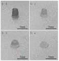
图1为1%的羟乙基纤维素加入GelMA与脱细胞基质(DECM)不同的体积比中形成生物墨水的打印效果。Figure 1 shows the printing effects of bioinks formed by adding 1% hydroxyethyl cellulose to GelMA and decellularized matrix (DECM) in different volume ratios.
具体实施方式Detailed ways
下面结合附图和具体实施例对本发明作进一步说明,以使本领域的技术人员可以更好地理解本发明并能予以实施,但所举实施例不作为对本发明的限定。The present invention will be further described below with reference to the accompanying drawings and specific embodiments, so that those skilled in the art can better understand the present invention and implement it, but the embodiments are not intended to limit the present invention.
除非另有定义,本文所使用的所有的技术和科学术语与属于本发明的技术领域的技术人员通常理解的含义相同。本文中在本发明的说明书中所使用的术语只是为了描述具体的实施例的目的,不是旨在于限制本发明。本文所使用的术语“及/或”包括一个或多个相关的所列项目的任意的和所有的组合。Unless otherwise defined, all technical and scientific terms used herein have the same meaning as commonly understood by one of ordinary skill in the art to which this invention belongs. The terms used herein in the description of the present invention are for the purpose of describing specific embodiments only, and are not intended to limit the present invention. As used herein, the term "and/or" includes any and all combinations of one or more of the associated listed items.
下述实施例中所使用的实验方法如无特殊说明,均为常规方法,所用的材料、试剂等,如无特殊说明,均可从商业途径得到。The experimental methods used in the following examples are conventional methods unless otherwise specified, and the materials, reagents, etc. used can be obtained from commercial sources unless otherwise specified.
实施例1Example 1
一、制备基于肝脏脱细胞基质的复合生物墨水1. Preparation of composite bioink based on liver acellular matrix
(1)肝脏准备:获取新鲜猪肝脏,切为10mm2大小的方块,-80℃冷冻12小时;(1) Liver preparation: Obtain fresh pig liver, cut it into10mm2 squares, and freeze at -80°C for 12 hours;
(2)肝脏脱细胞基质的制备:肝脏常温解冻,进行振荡细胞(振荡速率为60rpm/min)去离子水振荡洗脱2h;0.1%胰酶浸泡3h;1%TritonX-100振荡洗脱1h;4%SDC振荡洗脱1h;80U/ml DNase、5U/ml RNase振荡洗脱30min;2%青霉素-链霉素、2.5μg/ml两性霉素B的PBS振荡洗脱30min。(2) Preparation of liver decellularized matrix: the liver was thawed at room temperature, and the cells were shaken (the shaking rate was 60 rpm/min) and washed with deionized water for 2 hours; 0.1% trypsin was soaked for 3 hours; 1% TritonX-100 was shaken and washed for 1 hour; 4% SDC was shaken for 1h; 80U/ml DNase, 5U/ml RNase were shaken for 30min; 2% penicillin-streptomycin, 2.5μg/ml amphotericin B in PBS was shaken for 30min.
(3)肝脏脱细胞基质的表征:肝脏脱细胞基质经历冷冻干燥后,DNA含量检测以及胶原蛋白等一系列表征,证明脱细胞较完全以后,可以进行下一步;(3) Characterization of the liver acellular matrix: After the liver acellular matrix has undergone freeze-drying, DNA content detection and a series of characterizations such as collagen have proved that after the decellularization is relatively complete, the next step can be performed;
(4)肝脏脱细胞基质溶液的制备与表征(4) Preparation and characterization of liver acellular matrix solution
①肝脏脱细胞基质溶液的制备:将肝脏脱细胞基质冻干,取冻干后的脱细胞基质进行剪碎、研磨,使其易于溶解,研磨至合适颗粒大小,溶于胃蛋白酶中制成溶液;①Preparation of liver acellular matrix solution: freeze-dry the liver acellular matrix, take the freeze-dried acellular matrix, cut it into pieces, grind it to make it easy to dissolve, grind it to a suitable particle size, and dissolve it in pepsin to make a solution ;
②肝脏脱细胞基质溶液的表征:检测溶液中胶原蛋白的浓度,以及溶液中GAG含量,确保检测的含量尽量与未脱细胞处理的天然肝脏对比,没有极为显著的变化,保证制成的生物墨水具有一定的生物活性;②Characterization of liver decellularized matrix solution: Detect the concentration of collagen in the solution and the content of GAG in the solution to ensure that the detected content is compared with the natural liver without decellularization treatment as much as possible, and there is no extremely significant change, to ensure that the prepared bioink have certain biological activity;
(5)生物墨水的制备:将肝脏脱细胞基质与明胶甲基丙烯酰胺(GelMA)1:1混合,并加入1%的羟乙基纤维素,制成生物墨水。(5) Preparation of bio-ink: The liver acellular matrix was mixed with gelatin methacrylamide (GelMA) 1:1, and 1% hydroxyethyl cellulose was added to prepare bio-ink.
二、生物墨水性能的检测2. Detection of bioink performance
(1)生物墨水可打印性的验证:电镜SEM检测生物墨水的孔径,测量杨式模量,检测打印结构和弹性模量;(1) Verification of the printability of the bioink: Electron microscope SEM detects the pore size of the bioink, measures the Young's modulus, and detects the printed structure and elastic modulus;
(2)生物墨水生物相容性的验证:细胞活死染色评估细胞活性,定量尿素和ALB分泌评估肝功能。(2) Verification of biocompatibility of bioink: cell viability was assessed by cell live and dead staining, and liver function was assessed by quantitative urea and ALB secretion.
三、细胞打印检测3. Cell printing detection
(1)肝癌细胞的打印:将生物打印墨水在37℃培养箱预先温育1小时,然后取5mL生物打印墨水与1mL的HepG2细胞悬液在10mL离心管中混合均匀,2000转/分钟,离心2分钟取出放置在生物打印机墨盒中,打印得到组织块,紫外光交联30s得到打印的组织,采用杜氏改良伊格尔培养基-高糖(DMEM高糖)培养液对打印的组织漂洗3次,在37℃培养箱中孵育培养,分别在24、48和72小时用MTT法检测细胞增殖情况,并用Hoechst及PI进行染色检测凝胶中细胞活性。(1) Printing of liver cancer cells: Incubate the bioprinting ink in a 37°C incubator for 1 hour, then take 5mL of the bioprinter ink and 1mL of the HepG2 cell suspension and mix them evenly in a 10mL centrifuge tube, centrifuge at 2000 rpm After 2 minutes, take it out and place it in the bioprinter cartridge, print the tissue block, and cross-link the printed tissue with UV light for 30s. The printed tissue is rinsed 3 times with Duchenne's modified Eagle's medium-high glucose (DMEM high glucose) medium. , incubate in a 37 ℃ incubator, detect cell proliferation by MTT method at 24, 48 and 72 hours respectively, and use Hoechst and PI to stain to detect cell viability in the gel.
(2)正常肝细胞的打印:将生物打印墨水在37℃培养箱预先温育1小时,然后取5mL生物打印墨水与1mL的LO2细胞悬液在10mL离心管中混合均匀,2000转/分钟,离心2分钟取出放置在生物打印机墨盒中,打印得到组织块,紫外光交联30s得到打印的组织,采用杜氏改良伊格尔培养基-高糖(DMEM高糖)培养液对打印的组织漂洗3次,在37℃培养箱中孵育培养,分别在24、48和72小时用MTT法检测细胞增殖情况,并用Hoechst及PI进行染色检测凝胶中细胞活性。(2) Printing of normal hepatocytes: Incubate the bioprinting ink in a 37°C incubator for 1 hour, then take 5mL of the bioprinter ink and 1mL of the LO2 cell suspension and mix them evenly in a 10mL centrifuge tube at 2000 rpm. Centrifuge for 2 minutes, take it out and place it in a bioprinter cartridge, print the tissue block, and cross-link the printed tissue with UV light for 30s. The printed tissue is rinsed with Duchenne's modified Eagle's medium-high glucose (DMEM high glucose) medium for 3 Next, the cells were incubated in a 37°C incubator, and the cell proliferation was detected by MTT method at 24, 48, and 72 hours, and the cell viability in the gel was detected by Hoechst and PI staining.
(3)肝原代细胞的打印:将生物打印墨水在37℃培养箱预先温育1小时,然后取5mL生物打印墨水与1mL的肝原代细胞悬液在10mL离心管中混合均匀,2000转/分钟,离心2分钟取出放置在生物打印机墨盒中,打印得到组织块,紫外光交联30s得到打印的组织,采用杜氏改良伊格尔培养基-高糖(DMEM高糖)培养液对打印的组织漂洗3次,在37℃培养箱中孵育培养,分别在24、48和72小时用MTT法检测细胞增殖情况,并用Hoechst及PI进行染色检测凝胶中细胞活性。(3) Printing of primary liver cells: Incubate the bioprinting ink in a 37°C incubator for 1 hour, then take 5mL of the bioprinter ink and 1mL of the primary liver cell suspension and mix them evenly in a 10mL centrifuge tube at 2000 rpm /min, centrifuge for 2 min, take it out and place it in a bioprinter cartridge, print the tissue block, and cross-link it with UV light for 30s to obtain the printed tissue. The tissue was washed three times and incubated in a 37°C incubator. The cell proliferation was detected by MTT method at 24, 48 and 72 hours, and the cell viability in the gel was detected by Hoechst and PI staining.
附图1示出了1%的羟乙基纤维素加入GelMA与脱细胞基质(DECM)不同的体积比中形成生物墨水的打印效果。Fig. 1 shows the printing effect of bioink formed by adding 1% hydroxyethyl cellulose into GelMA and decellularized matrix (DECM) in different volume ratios.
从图1中可以看出,当GelMA:DECM的比例为1:1~1:2时,得到的生物墨水都显示出了良好的打印性能。当GelMA:DECM的比例上升至1:3时,生物墨水的打印性能出现明显的下降。It can be seen from Figure 1 that when the ratio of GelMA:DECM is 1:1 to 1:2, the obtained bioinks all show good printing performance. When the ratio of GelMA:DECM increased to 1:3, the printing performance of the bioink decreased significantly.
现有技术中,未加羟乙基纤维素时,生物墨水中GelMA:DECM最大比例为1:1,即DECM的上限为50%。本实施例中,通过在生物墨水中引入了1%的羟乙基纤维素,明显的提高生物墨水的打印性能,使得DECM的可打印含量增加至67%,有利于提高打印支架的生物活性。In the prior art, when hydroxyethyl cellulose is not added, the maximum ratio of GelMA:DECM in the bioink is 1:1, that is, the upper limit of DECM is 50%. In this example, by introducing 1% hydroxyethyl cellulose into the bioink, the printing performance of the bioink is significantly improved, so that the printable content of DECM is increased to 67%, which is beneficial to improve the biological activity of the printed scaffold.
实施例2Example 2
一、制备基于肾脏脱细胞基质的复合生物墨水1. Preparation of composite bioink based on kidney acellular matrix
(1)肾脏准备:获取新鲜猪肾脏,切为10mm2大小的方块,-80℃冷冻至少12小时;(1) Kidney preparation: Obtain fresh pig kidneys, cut them into10mm2 cubes, and freeze at -80°C for at least 12 hours;
(2)肾脏脱细胞基质的制备:肾脏常温解冻,进行振荡细胞(振荡速率为120rpm/min)去离子水振荡洗脱2h;0.1%胰酶浸泡3h;1%TritonX-100振荡洗脱1h;4%SDC振荡洗脱1h;80U/ml DNase、5U/ml RNase振荡洗脱30min;2%青霉素-链霉素、2.5μg/ml两性霉素B的PBS振荡洗脱30min。(2) Preparation of kidney acellular matrix: thawed kidneys at room temperature, shaken the cells (oscillation rate of 120 rpm/min) and washed with deionized water for 2 hours; immersed in 0.1% trypsin for 3 hours; shaken and washed with 1% TritonX-100 for 1 hour; 4% SDC was shaken for 1h; 80U/ml DNase, 5U/ml RNase were shaken for 30min; 2% penicillin-streptomycin, 2.5μg/ml amphotericin B in PBS was shaken for 30min.
(3)肾脏脱细胞基质的表征:肾脏脱细胞基质经历冷冻干燥后,DNA含量检测以及胶原蛋白等一系列表征,证明脱细胞较完全以后,可以进行下一步;(3) Characterization of the kidney acellular matrix: After the kidney acellular matrix has undergone freeze-drying, DNA content detection and a series of characterizations such as collagen have proved that after the decellularization is relatively complete, the next step can be performed;
(4)肾脏脱细胞基质溶液的制备与表征(4) Preparation and characterization of kidney acellular matrix solution
①肾脏脱细胞基质溶液的制备:将肾脏脱细胞基质冻干,取冻干后的脱细胞基质进行剪碎、研磨,使其易于溶解,研磨至合适颗粒大小,溶于胃蛋白酶中制成溶液;②肾脏脱细胞基质溶液的表征:检测溶液中胶原蛋白的浓度,以及溶液中GAG含量,确保检测的含量尽量与未脱细胞处理的天然肾脏对比,没有极为显著的变化,保证制成的生物墨水具有一定的生物活性;①Preparation of kidney acellular matrix solution: freeze-dry the kidney acellular matrix, take the freeze-dried acellular matrix for cutting and grinding to make it easy to dissolve, grind to a suitable particle size, dissolve in pepsin to make a solution ;2 Characterization of the kidney acellular matrix solution: Detect the concentration of collagen in the solution and the GAG content in the solution to ensure that the detected content is compared with the natural kidney without decellularization treatment as much as possible, and there is no extremely significant change. The ink has certain biological activity;
(5)生物墨水的制备:将肾脏脱细胞基质与明胶甲基丙烯酰胺(GelMA)1:1混合,并加入1%的羟乙基纤维素,混合均匀,制成生物墨水。(5) Preparation of bio-ink: The kidney acellular matrix was mixed with gelatin methacrylamide (GelMA) at 1:1, and 1% hydroxyethyl cellulose was added, and mixed evenly to prepare a bio-ink.
二、生物墨水性能的检测2. Detection of bioink performance
(1)生物墨水可打印性的验证:电镜SEM检测生物墨水的孔径,测量杨式模量,检测打印结构和弹性模量;(1) Verification of the printability of the bioink: Electron microscope SEM detects the pore size of the bioink, measures the Young's modulus, and detects the printed structure and elastic modulus;
(2)生物墨水生物相容性的验证:细胞活死染色评估细胞活性,定量尿素和ALB分泌评估肾功能。(2) Verification of biocompatibility of bioinks: cell viability was assessed by cell live and dead staining, and renal function was assessed by quantitative urea and ALB secretion.
三、细胞打印检测3. Cell printing detection
(1)肾癌细胞的打印:将生物打印墨水在37℃培养箱预先温育1小时,然后取5mL生物打印墨水与1mL的MPC5细胞悬液在10mL离心管中混合均匀,2000转/分钟,离心2分钟取出放置在生物打印机墨盒中,打印得到组织块,紫外光交联30s得到打印的组织,采用杜氏改良伊格尔培养基-高糖(DMEM高糖)培养液对打印的组织漂洗3次,在37℃培养箱中孵育培养,分别在24、48和72小时用MTT法检测细胞增殖情况,并用Hoechst及PI进行染色检测凝胶中细胞活性。(1) Printing of renal cancer cells: Pre-incubate the bioprinting ink in a 37°C incubator for 1 hour, then mix 5mL of the bioprinter ink and 1mL of the MPC5 cell suspension in a 10mL centrifuge tube at 2000 rpm, Centrifuge for 2 minutes, take it out and place it in a bioprinter cartridge, print the tissue block, and cross-link the printed tissue with UV light for 30s. The printed tissue is rinsed with Duchenne's modified Eagle's medium-high glucose (DMEM high glucose) medium for 3 Next, the cells were incubated in a 37°C incubator, and the cell proliferation was detected by MTT method at 24, 48, and 72 hours, and the cell viability in the gel was detected by Hoechst and PI staining.
(2)正常肾细胞的打印:将生物打印墨水在37℃培养箱预先温育1小时,然后取5mL生物打印墨水与1mL的HK2细胞悬液在10mL离心管中混合均匀,2000转/分钟,离心2分钟取出放置在生物打印机墨盒中,打印得到组织块,紫外光交联30s得到打印的组织,采用杜氏改良伊格尔培养基-高糖(DMEM高糖)培养液对打印的组织漂洗3次,在37℃培养箱中孵育培养,分别在24、48和72小时用MTT法检测细胞增殖情况,并用Hoechst及PI进行染色检测凝胶中细胞活性。(2) Printing of normal kidney cells: Incubate the bioprinting ink in a 37°C incubator for 1 hour, then take 5mL of the bioprinter ink and 1mL of the HK2 cell suspension and mix them evenly in a 10mL centrifuge tube at 2000 rpm. Centrifuge for 2 minutes, take it out and place it in a bioprinter cartridge, print the tissue block, and cross-link the printed tissue with UV light for 30s. The printed tissue is rinsed with Duchenne's modified Eagle's medium-high glucose (DMEM high glucose) medium for 3 Next, the cells were incubated in a 37°C incubator, and the cell proliferation was detected by MTT method at 24, 48, and 72 hours, and the cell viability in the gel was detected by Hoechst and PI staining.
(3)肾原代细胞的打印:将生物打印墨水在37℃培养箱预先温育1小时,然后取5mL生物打印墨水与1mL的肾原代细胞悬液在10mL离心管中混合均匀,2000转/分钟,离心2分钟取出放置在生物打印机墨盒中,打印得到组织块,紫外光交联30s得到打印的组织,采用杜氏改良伊格尔培养基-高糖(DMEM高糖)培养液对打印的组织漂洗3次,在37℃培养箱中孵育培养,分别在24、48和72小时用MTT法检测细胞增殖情况,并用Hoechst及PI进行染色检测凝胶中细胞活性。(3) Printing of primary kidney cells: pre-incubate the bioprinting ink in a 37°C incubator for 1 hour, then mix 5 mL of the bioprinting ink and 1 mL of the primary kidney cell suspension in a 10 mL centrifuge tube, 2000 rpm /min, centrifuge for 2 min, take it out and place it in a bioprinter cartridge, print the tissue block, and cross-link it with UV light for 30s to obtain the printed tissue. The tissue was washed three times and incubated in a 37°C incubator. The cell proliferation was detected by MTT method at 24, 48 and 72 hours, and the cell viability in the gel was detected by Hoechst and PI staining.
以上所述实施例仅是为充分说明本发明而所举的较佳的实施例,本发明的保护范围不限于此。本技术领域的技术人员在本发明基础上所作的等同替代或变换,均在本发明的保护范围之内。本发明的保护范围以权利要求书为准。The above-mentioned embodiments are only preferred embodiments for fully illustrating the present invention, and the protection scope of the present invention is not limited thereto. Equivalent substitutions or transformations made by those skilled in the art on the basis of the present invention are all within the protection scope of the present invention. The protection scope of the present invention is subject to the claims.
Claims (11)
Translated fromChinesePriority Applications (1)
| Application Number | Priority Date | Filing Date | Title |
|---|---|---|---|
| CN202010260081.4ACN111388758A (en) | 2020-04-03 | 2020-04-03 | Composite biological ink based on methacrylated hydrogel/hydroxyethyl cellulose/acellular matrix and preparation method thereof |
Applications Claiming Priority (1)
| Application Number | Priority Date | Filing Date | Title |
|---|---|---|---|
| CN202010260081.4ACN111388758A (en) | 2020-04-03 | 2020-04-03 | Composite biological ink based on methacrylated hydrogel/hydroxyethyl cellulose/acellular matrix and preparation method thereof |
Publications (1)
| Publication Number | Publication Date |
|---|---|
| CN111388758Atrue CN111388758A (en) | 2020-07-10 |
Family
ID=71416680
Family Applications (1)
| Application Number | Title | Priority Date | Filing Date |
|---|---|---|---|
| CN202010260081.4APendingCN111388758A (en) | 2020-04-03 | 2020-04-03 | Composite biological ink based on methacrylated hydrogel/hydroxyethyl cellulose/acellular matrix and preparation method thereof |
Country Status (1)
| Country | Link |
|---|---|
| CN (1) | CN111388758A (en) |
Cited By (4)
| Publication number | Priority date | Publication date | Assignee | Title |
|---|---|---|---|---|
| CN112063224A (en)* | 2020-09-24 | 2020-12-11 | 惠州市至上新材料有限公司 | Biological ink and preparation method thereof |
| CN114712561A (en)* | 2022-03-22 | 2022-07-08 | 苏州大学 | An injectable, photocrosslinked acellular matrix composite hydrogel and its preparation method and application |
| CN118006539A (en)* | 2023-12-27 | 2024-05-10 | 宁波长都生物科技有限责任公司 | Liver-specific biological ink, preparation method thereof and primary hepatocyte 3D biological printing model constructed based on same |
| CN120035452A (en)* | 2024-09-23 | 2025-05-23 | 陕西佰傲再生医学有限公司 | Membrane tissue treatment composition, membrane tissue treatment reagent, tissue repair material and preparation method and application thereof |
Citations (6)
| Publication number | Priority date | Publication date | Assignee | Title |
|---|---|---|---|---|
| WO1999040153A1 (en)* | 1998-02-06 | 1999-08-12 | Monsanto Company | Acid-stable and cationic-compatible cellulose compositions and methods of preparation |
| CN102407332A (en)* | 2011-12-05 | 2012-04-11 | 烟台工程职业技术学院 | Preparation method of porous titanium |
| CN102712699A (en)* | 2009-12-14 | 2012-10-03 | 路博润高级材料公司 | Cassia derivatives |
| CN103599567A (en)* | 2013-11-25 | 2014-02-26 | 四川大学华西医院 | Thermo-sensitive composite material and preparation method and application thereof |
| CN107987287A (en)* | 2017-11-15 | 2018-05-04 | 华东理工大学 | Photic nitroso cross-linked hydrogel material and preparation method and application |
| CN109054503A (en)* | 2018-07-05 | 2018-12-21 | 大连理工大学 | Preparation method of biological printing ink, printing method and application |
- 2020
- 2020-04-03CNCN202010260081.4Apatent/CN111388758A/enactivePending
Patent Citations (6)
| Publication number | Priority date | Publication date | Assignee | Title |
|---|---|---|---|---|
| WO1999040153A1 (en)* | 1998-02-06 | 1999-08-12 | Monsanto Company | Acid-stable and cationic-compatible cellulose compositions and methods of preparation |
| CN102712699A (en)* | 2009-12-14 | 2012-10-03 | 路博润高级材料公司 | Cassia derivatives |
| CN102407332A (en)* | 2011-12-05 | 2012-04-11 | 烟台工程职业技术学院 | Preparation method of porous titanium |
| CN103599567A (en)* | 2013-11-25 | 2014-02-26 | 四川大学华西医院 | Thermo-sensitive composite material and preparation method and application thereof |
| CN107987287A (en)* | 2017-11-15 | 2018-05-04 | 华东理工大学 | Photic nitroso cross-linked hydrogel material and preparation method and application |
| CN109054503A (en)* | 2018-07-05 | 2018-12-21 | 大连理工大学 | Preparation method of biological printing ink, printing method and application |
Non-Patent Citations (3)
| Title |
|---|
| DONALD BEJLERI ET AL: "A Bioprinted Cardiac Patch Composed of Cardiac-Specific Extracellular Matrix and Progenitor Cells for Heart Repair", 《ADVANCED HEALTHCARE MATERIALS》* |
| XIAORUI LI ET AL.: "Hydroxyethyl Cellulose As a Rheological Additive for Tuning the Extrusion Printability and Scaffold Properties", 《3D PRINTING AND ADDITIVE MANUFACTURING》* |
| 马振友: "《中西皮肤外用制剂手册》", 30 September 2019, 河南科学技术出版社* |
Cited By (4)
| Publication number | Priority date | Publication date | Assignee | Title |
|---|---|---|---|---|
| CN112063224A (en)* | 2020-09-24 | 2020-12-11 | 惠州市至上新材料有限公司 | Biological ink and preparation method thereof |
| CN114712561A (en)* | 2022-03-22 | 2022-07-08 | 苏州大学 | An injectable, photocrosslinked acellular matrix composite hydrogel and its preparation method and application |
| CN118006539A (en)* | 2023-12-27 | 2024-05-10 | 宁波长都生物科技有限责任公司 | Liver-specific biological ink, preparation method thereof and primary hepatocyte 3D biological printing model constructed based on same |
| CN120035452A (en)* | 2024-09-23 | 2025-05-23 | 陕西佰傲再生医学有限公司 | Membrane tissue treatment composition, membrane tissue treatment reagent, tissue repair material and preparation method and application thereof |
Similar Documents
| Publication | Publication Date | Title |
|---|---|---|
| CN111388758A (en) | Composite biological ink based on methacrylated hydrogel/hydroxyethyl cellulose/acellular matrix and preparation method thereof | |
| CN110272860B (en) | Construction method and application of three-dimensional cell culture microenvironment | |
| JP7610517B2 (en) | Angioplasty devices and methods for transplant diagnostics and therapy - Patents.com | |
| KR101756935B1 (en) | Hydrogel scaffold having double crosslinking, uses thereof and method for producing the same using two-step crosslinking | |
| CN110790950A (en) | Photo-crosslinking recombinant collagen hydrogel, preparation method and application thereof in 3D bioprinting | |
| CN110818921B (en) | Rapidly-curable double-crosslinked hydrogel and preparation method and application thereof | |
| CN105169483A (en) | Preparation method of acellular matrix gels and acellular matrix gels | |
| Pok et al. | Use of myocardial matrix in a chitosan-based full-thickness heart patch | |
| CN108543116B (en) | Sodium alginate and gelatin composite hydrogel 3D islet scaffold and preparation method thereof | |
| WO2019100454A1 (en) | Decellularized porous scaffold for three-dimensional tumor model, and construction method therefor and applications thereof | |
| CN113577386A (en) | Double-network biological ink and preparation method and application thereof | |
| US20230405186A1 (en) | Preparation method and application of decellularized extracellular matrix tissue paper | |
| CN111748526A (en) | A kind of preparation method of imitating extracellular matrix printable composite bioink | |
| WO2010139209A1 (en) | Kit used for obtaining whole liver scaffold and method for obtaining whole liver scaffold | |
| Wu et al. | Injectable silk fibroin peptide nanofiber hydrogel composite scaffolds for cartilage regeneration | |
| CN113274550A (en) | Vascularized bone bionic multifunctional tissue engineering scaffold with anti-inflammatory effect and preparation method thereof | |
| CN113278587B (en) | Three-dimensional engineered breast cancer lung metastasis model, construction method and application | |
| He et al. | An antibacterial ε-poly-L-lysine-derived bioink for 3D bioprinting applications | |
| CN114904056A (en) | A kind of composite hydrogel based on human placental acellular matrix and preparation method thereof | |
| CN108310463A (en) | 3D printing biological ink and preparation method thereof | |
| CN115216441B (en) | Composite scaffold for three-dimensional culture of stem cells and preparation method thereof | |
| Wang et al. | A decellularized lung extracellular matrix/chondroitin sulfate/gelatin/chitosan-based 3D culture system shapes breast cancer lung metastasis | |
| CN105126171A (en) | Gel biological material having shape memory function and preparation method of gel biological material | |
| CN111420121A (en) | Composite biological ink based on methacrylated hydrogel/nanoclay/acellular matrix and preparation method and application thereof | |
| CN119119709A (en) | A bioactive composite hydrogel, scaffold and application thereof |
Legal Events
| Date | Code | Title | Description |
|---|---|---|---|
| PB01 | Publication | ||
| PB01 | Publication | ||
| SE01 | Entry into force of request for substantive examination | ||
| SE01 | Entry into force of request for substantive examination | ||
| RJ01 | Rejection of invention patent application after publication | ||
| RJ01 | Rejection of invention patent application after publication | Application publication date:20200710 |