CN111242953B - MR image segmentation method and device based on condition generation countermeasure network - Google Patents
MR image segmentation method and device based on condition generation countermeasure networkDownload PDFInfo
- Publication number
- CN111242953B CN111242953BCN202010055252.XACN202010055252ACN111242953BCN 111242953 BCN111242953 BCN 111242953BCN 202010055252 ACN202010055252 ACN 202010055252ACN 111242953 BCN111242953 BCN 111242953B
- Authority
- CN
- China
- Prior art keywords
- image
- segmentation
- network
- net
- cgan
- Prior art date
- Legal status (The legal status is an assumption and is not a legal conclusion. Google has not performed a legal analysis and makes no representation as to the accuracy of the status listed.)
- Active
Links
Images


Classifications
- G—PHYSICS
- G06—COMPUTING OR CALCULATING; COUNTING
- G06T—IMAGE DATA PROCESSING OR GENERATION, IN GENERAL
- G06T7/00—Image analysis
- G06T7/10—Segmentation; Edge detection
- G—PHYSICS
- G06—COMPUTING OR CALCULATING; COUNTING
- G06T—IMAGE DATA PROCESSING OR GENERATION, IN GENERAL
- G06T2207/00—Indexing scheme for image analysis or image enhancement
- G06T2207/10—Image acquisition modality
- G06T2207/10072—Tomographic images
- G06T2207/10088—Magnetic resonance imaging [MRI]
- G—PHYSICS
- G06—COMPUTING OR CALCULATING; COUNTING
- G06T—IMAGE DATA PROCESSING OR GENERATION, IN GENERAL
- G06T2207/00—Indexing scheme for image analysis or image enhancement
- G06T2207/20—Special algorithmic details
- G06T2207/20081—Training; Learning
- G—PHYSICS
- G06—COMPUTING OR CALCULATING; COUNTING
- G06T—IMAGE DATA PROCESSING OR GENERATION, IN GENERAL
- G06T2207/00—Indexing scheme for image analysis or image enhancement
- G06T2207/20—Special algorithmic details
- G06T2207/20084—Artificial neural networks [ANN]
- G—PHYSICS
- G06—COMPUTING OR CALCULATING; COUNTING
- G06T—IMAGE DATA PROCESSING OR GENERATION, IN GENERAL
- G06T2207/00—Indexing scheme for image analysis or image enhancement
- G06T2207/30—Subject of image; Context of image processing
- G06T2207/30004—Biomedical image processing
- G06T2207/30096—Tumor; Lesion
Landscapes
- Engineering & Computer Science (AREA)
- Computer Vision & Pattern Recognition (AREA)
- Physics & Mathematics (AREA)
- General Physics & Mathematics (AREA)
- Theoretical Computer Science (AREA)
- Image Analysis (AREA)
Abstract
Description
Translated fromChinese技术领域technical field
本发明属于图像处理领域,涉及一种基于条件生成对抗网络的MR图像分割方法及装置。The invention belongs to the field of image processing, and relates to an MR image segmentation method and device based on a conditional generation confrontation network.
背景技术Background technique
近年来,图像分割技术在医学影像领域得到了越来越多的应用。例如,弥漫性低级胶质瘤(Lower-Grade Gliomas,LGG)是最常见的中枢神经系统肿瘤。LGG是世界卫生组织(World Health Organization,WHO)规定的II级和III级胶质瘤,包括星形胶质细胞瘤、少突胶质细胞瘤、少突星形胶质细胞瘤。由于II级和III级胶质瘤具有高度的侵袭性,因此,想要通过外科手术完全切除是不可能的,切除后残余的肿瘤通常导致复发并恶化为胶质母细胞瘤(Glioblastoma,GBM)。众所周知,肿瘤发现的越早,病人的生存机率就越高。现有技术针对临床通常使用磁共振成像(Magnetic Resonance Imaging,MRI)来评估脑肿瘤,并且医生在评估脑肿瘤时通常采用人工分割的方法,这种方式既耗时又需要专业知识。目前,深度学习算法在医学图像分析中取得了极大的成功,但是仍普遍存在标记数据不足的问题。介于临床上医生通常使用粗略测量来评估肿瘤,故而实现对肿瘤进行自动分割和定量分析十分必要。In recent years, image segmentation technology has been more and more applied in the field of medical imaging. For example, diffuse low-grade glioma (Lower-Grade Gliomas, LGG) is the most common central nervous system tumor. LGG is a grade II and III glioma defined by the World Health Organization (WHO), including astrocytoma, oligodendroglioma, and oligoastrocytoma. Due to the highly aggressive nature of grade II and III gliomas, complete surgical resection is impossible, and residual tumor after resection usually leads to recurrence and progression to glioblastoma (Glioblastoma, GBM) . It is well known that the earlier a tumor is detected, the higher the patient's chances of survival. In the prior art, Magnetic Resonance Imaging (MRI) is usually used to evaluate brain tumors clinically, and doctors usually use manual segmentation when evaluating brain tumors, which is time-consuming and requires professional knowledge. At present, deep learning algorithms have achieved great success in medical image analysis, but there is still a common problem of insufficient labeled data. Since doctors usually use rough measurements to evaluate tumors clinically, it is necessary to realize automatic segmentation and quantitative analysis of tumors.
目前,该领域相关的研究主要有:Goodfellow等人[1]首先以对抗性方式训练生成模型,生成对抗网络(Generative Adversarial Networks,GANs)由生成器和判别器两部分组成,它们在训练过程中相互竞争以提高各自的性能。为了保证生成图像的真实性,GANs学习真实数据的潜在分布,并从相同的分布中生成新的样本。但是,最初的GANs生成的结果十分不稳定,特别是对大型图像的生成。为了提高生成结果的可控性,条件生成对抗网络(conditional Generative Adversarial Networks,cGAN)在生成器和判别器中同时加入了条件信息。At present, the relevant research in this field mainly includes: Goodfellow et al. [1] first trained the generative model in an adversarial manner, and the Generative Adversarial Networks (GANs) consisted of two parts, the generator and the discriminator, which were trained in the training process. Compete against each other to improve their respective performance. To guarantee the authenticity of generated images, GANs learn the latent distribution of real data and generate new samples from the same distribution. However, the results generated by the original GANs are very unstable, especially for large-scale image generation. In order to improve the controllability of the generated results, conditional Generative Adversarial Networks (cGAN) adds conditional information to both the generator and the discriminator.
现有技术利用GANs生成的医学图像来扩充原始训练集,可以缓解分类任务中由于数据量小而造成的问题。然而,生成的医学图像很少用于分割任务,这主要是因为生成的图像没有相应的精确病变分割掩膜。在大部分已有文献中,生成的样本通常来自一个与原始数据相同的特别稀疏的类,分类标签比分割掩膜更容易获得,因此,人工生成的医学图像一般用于扩充数据集以提高后续的检测和分类结果,不能够解决图像分割中标记数据不足的问题。Existing techniques use medical images generated by GANs to augment the original training set, which can alleviate the problem caused by the small amount of data in classification tasks. However, generated medical images are rarely used for segmentation tasks, mainly because the generated images do not have corresponding accurate lesion segmentation masks. In most of the existing literature, the generated samples usually come from the same extremely sparse class as the original data, and the classification labels are easier to obtain than the segmentation masks. Therefore, artificially generated medical images are generally used to augment the dataset to improve subsequent The detection and classification results cannot solve the problem of insufficient labeled data in image segmentation.
发明内容Contents of the invention
本发明的目的在于针对上述现有技术中图像分割标记数据不足的问题,提供一种基于条件生成对抗网络的MR图像分割方法及装置,使人工生成的图像分割结果更接近真实图像。The purpose of the present invention is to solve the problem of insufficient labeled data for image segmentation in the above-mentioned prior art, and provide an MR image segmentation method and device based on conditional generative adversarial networks, so that the artificially generated image segmentation results are closer to real images.
为了实现上述目的,本发明有如下的技术方案:In order to achieve the above object, the present invention has the following technical solutions:
一种基于条件生成对抗网络的MR图像分割方法,包括以下步骤:A method for segmenting MR images based on conditional generative confrontation networks, comprising the following steps:
通过低级MR图像y和分割掩膜x训练cGAN网络,得到训练好的cGAN模型;Train the cGAN network through the low-level MR image y and the segmentation mask x to obtain the trained cGAN model;
根据输入的分割掩膜x,使用训练好的cGAN模型自动生成人工图像y’;According to the input segmentation mask x, the artificial image y' is automatically generated using the trained cGAN model;
利用人工图像y’和分割掩膜x预训练U-net网络;Pre-train the U-net network using the artificial image y' and the segmentation mask x;
通过低级MR图像y和分割掩膜x继续训练U-net网络,得到训练好的U-net模型;Continue to train the U-net network through the low-level MR image y and the segmentation mask x to obtain the trained U-net model;
利用训练好的U-net模型进行MR图像分割。Use the trained U-net model for MR image segmentation.
作为优选,本发明基于条件生成对抗网络的MR图像分割方法一种实施例中,所述的cGAN网络由生成器和判别器两部分组成,生成器采用U-net架构,判别器采用CNN架构。As a preference, in an embodiment of the MR image segmentation method based on conditional generative adversarial network of the present invention, the cGAN network is composed of a generator and a discriminator, the generator adopts a U-net architecture, and the discriminator adopts a CNN architecture.
作为优选,本发明基于条件生成对抗网络的MR图像分割方法一种实施例中,所述的U-Net架构由编码器和解码器组成,在编码器和解码器堆栈中的镜像层之间设置跳跃连接;所述的跳跃连接用于在输入和输出之间直接通过网络传输低级的图像信息。As a preference, in an embodiment of the MR image segmentation method based on the conditional generation confrontation network of the present invention, the U-Net architecture is composed of an encoder and a decoder, and is set between the mirror layer in the encoder and decoder stack Skip connection; the skip connection is used to directly transmit low-level image information between the input and the output through the network.
作为优选,本发明基于条件生成对抗网络的MR图像分割方法一种实施例中,所述的cGAN网络的损失函数为:As a preference, in one embodiment of the MR image segmentation method based on the conditional generation confrontation network of the present invention, the loss function of the cGAN network is:
LcGAN(G,D)=Ex,y[logD(x,y)]+Ex,z[log(1-D(x,G(x,z)))]LcGAN (G,D)=Ex,y [logD(x,y)]+Ex,z [log(1-D(x,G(x,z)))]
式中,x为分割掩膜,y为低级MR图像,z为随机噪声,D(x,y)表示输入的图像来自于真实图像的概率,D(x,G(x,z))表示输入图像来自生成图像的概率;In the formula, x is the segmentation mask, y is the low-level MR image, z is random noise, D(x,y) represents the probability that the input image comes from a real image, and D(x,G(x,z)) represents the input The probability that the image comes from the generated image;
G(x,z)为分割掩膜x对应的人工生成图像。G(x,z) is the artificially generated image corresponding to the segmentation mask x.
作为优选,本发明基于条件生成对抗网络的MR图像分割方法一种实施例中,所述的随机噪声z为Dropout的形式,应用于生成器的d1-d4层;As a preference, in an embodiment of the MR image segmentation method based on conditional generative confrontation network of the present invention, the random noise z is in the form of Dropout, which is applied to the d1-d4 layers of the generator;
生成器和判别器的损失函数通过将GAN的目标函数与L1距离函数相结合得到;The loss functions of the generator and discriminator are obtained by combining the objective function of GAN with the L1 distance function;
所述的L1距离计算式为LL1(G)=Ex,y[||y-G(x,z)||1];The formula for calculating the L1 distance is LL1 (G)=Ex, y [||yG(x, z)||1 ];
生成器和判别器的损失函数计算式如下:The loss function calculation formula of generator and discriminator is as follows:


上式中,λ为超参数,设定为100;G为生成器,D为判别器。In the above formula, λ is a hyperparameter, set to 100; G is a generator, and D is a discriminator.
作为优选,本发明基于条件生成对抗网络的MR图像分割方法一种实施例中,设ytrue为手工分割掩膜,ypred为U-net网络的分割结果,U-net网络的Dice损失函数为:As a preference, in an embodiment of the MR image segmentation method based on the conditional generation confrontation network of the present invention, ytrue is set as the manual segmentation mask, ypred is the segmentation result of the U-net network, and the Dice loss function of the U-net network is :

训练过程使用敏感性、特异性、总体准确性、Dice相似系数、雅尔卡指数和马修斯相关系数来评估分割性能;所有定量测量均在手工分割掩膜和预测分割结果之间按像素计算;The training process evaluates segmentation performance using sensitivity, specificity, overall accuracy, Dice similarity coefficient, Yalca index, and Mathews correlation coefficient; all quantitative measures are computed per pixel between the manual segmentation mask and the predicted segmentation results ;
上述度量指标分别表示如下:The above metrics are expressed as follows:


良好的分割效果具有较高的敏感性和特异性;利用Dice和Jaccard指标来衡量预测结果和金标准之间的相似性,这些指标代表了精度和灵敏度的调和平均值,最大值为1表示最优结果,0表示最差结果;MCC用来衡量分类质量,包括真阳性和假阳性以及真阴性和假阴性。A good segmentation effect has high sensitivity and specificity; Dice and Jaccard indicators are used to measure the similarity between the prediction results and the gold standard. These indicators represent the harmonic mean of accuracy and sensitivity, and the maximum value is 1. Excellent result, 0 for worst result; MCC is used to measure classification quality, including true positives and false positives and true negatives and false negatives.
作为优选,本发明基于条件生成对抗网络的MR图像分割方法一种实施例中,判别器中使用PatchGAN,图像分割的U-net模型采用改进的U-net架构,改进的U-net架构有以下特点:(1)在卷积过程中填充所有图层以保持图像大小不变,获得一个与输入图像大小相同的分割映射;(2)解码路径中的每一步都包含与编码路径对应的特征映射的拼接;(3)将最后一层的softmax函数替换为sigmoid函数来处理每个像素是黑色还是白色的二分类问题。As a preference, in one embodiment of the MR image segmentation method based on the conditional generation confrontation network of the present invention, PatchGAN is used in the discriminator, and the U-net model of image segmentation adopts an improved U-net architecture, and the improved U-net architecture has the following Features: (1) pad all layers during convolution to keep the image size constant, obtaining a segmentation map of the same size as the input image; (2) each step in the decoding path contains a feature map corresponding to the encoding path (3) Replace the softmax function of the last layer with a sigmoid function to deal with the binary classification problem of whether each pixel is black or white.
本发明还提供了一种基于条件生成对抗网络的MR图像分割装置,包括生成系统和分割系统,生成系统通过低级MR图像y和分割掩膜x训练cGAN网络,得到训练好的cGAN模型,根据输入的分割掩膜x,使用训练好的cGAN模型自动生成人工图像y’;分割系统利用人工图像y’和分割掩膜x预训练U-net网络,通过低级MR图像y和分割掩膜x继续训练U-net网络,得到训练好的U-net模型,利用训练好的U-net模型进行MR图像分割。The present invention also provides an MR image segmentation device based on a conditional generative confrontation network, including a generation system and a segmentation system. The generation system trains the cGAN network through a low-level MR image y and a segmentation mask x to obtain a trained cGAN model. According to the input The segmentation mask x, using the trained cGAN model to automatically generate an artificial image y'; the segmentation system uses the artificial image y' and the segmentation mask x to pre-train the U-net network, and continues to train through the low-level MR image y and the segmentation mask x U-net network, get the trained U-net model, and use the trained U-net model to perform MR image segmentation.
本发明还提供了一种终端设备,包括存储器、处理器以及存储在所述存储器中并可在所述处理器上运行的计算机程序,所述的处理器在执行所述计算机程序时实现本发明上述基于条件生成对抗网络的MR图像分割方法的步骤。The present invention also provides a terminal device, including a memory, a processor, and a computer program stored in the memory and operable on the processor, and the processor implements the present invention when executing the computer program The above-mentioned steps of the MR image segmentation method based on the conditional generative adversarial network.
本发明还提供了一种计算机可读存储介质,所述计算机可读存储介质存储有计算机程序,计算机程序被处理器执行时实现所述基于条件生成对抗网络的MR图像分割方法的步骤。The present invention also provides a computer-readable storage medium, the computer-readable storage medium stores a computer program, and when the computer program is executed by a processor, the steps of the MR image segmentation method based on conditional generative adversarial network are realized.
相较于现有技术,本发明具有如下的有益效果:针对强度不均匀、不同序列强度范围不同以及来自不同采集协议的低级MR图像,本发明生成的图像更接近于真实图像,生成的图像与输入分割掩膜具有相似的形状特征,与真实的MR图像具有相似的纹理特征。输入手工分割掩膜与输出人工图像虽然外观不同,但它们具有相同的底层结构和形状。本发明利用cGAN网络能够从输入的各种掩膜上学习一个给定数据集的分布,利用手工分割掩膜的形状特征生成的图像,扩充和丰富了原始数据集的数据多样性,进而在分割中取得了更好的性能。与常规数据扩充方式相比,生成图像扩充方法在提高数据多样性方面表现的更好。与此同时,本发明提出的图像分割方法在训练过程中收敛速度更快、损失更小且分割精度更高。Compared with the prior art, the present invention has the following beneficial effects: for low-level MR images with inhomogeneous intensity, different intensity ranges of different sequences, and different acquisition protocols, the images generated by the present invention are closer to real images, and the generated images are consistent with The input segmentation mask has similar shape features and similar texture features to the real MR image. The input manual segmentation mask and the output artificial image have the same underlying structure and shape, although they have different appearances. The present invention uses the cGAN network to learn the distribution of a given data set from various input masks, and uses the image generated by the shape feature of the manual segmentation mask to expand and enrich the data diversity of the original data set, and then in the segmentation achieved better performance. Compared with conventional data augmentation methods, generative image augmentation methods perform better in increasing data diversity. At the same time, the image segmentation method proposed by the present invention has faster convergence speed, smaller loss and higher segmentation accuracy in the training process.
附图说明Description of drawings
为了更清楚地说明本发明实施例中的技术方案,下面将对实施例描述中所需要使用的附图作简单地介绍,显而易见地,下面描述中的附图是本发明的一些实施例,对于本领域普通技术人员来讲,在不付出创造性劳动的前提下,还可以根据这些附图获得其他的附图。In order to more clearly illustrate the technical solutions in the embodiments of the present invention, the following will briefly introduce the drawings that need to be used in the description of the embodiments. Obviously, the drawings in the following description are some embodiments of the present invention. For Those of ordinary skill in the art can also obtain other drawings based on these drawings without making creative efforts.
图1本发明的MR图像分割方法针对低级胶质瘤分割的整体框架图;Fig. 1 is an overall frame diagram of the MR image segmentation method of the present invention for low-grade glioma segmentation;
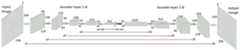
图2(a)本发明条件生成对抗网络的生成器架构示意图;Figure 2 (a) Schematic diagram of the generator architecture of the conditional generative adversarial network of the present invention;
图2(b)本发明条件生成对抗网络的判别器架构示意图;Fig. 2(b) Schematic diagram of the discriminator architecture of the conditional generative confrontation network of the present invention;
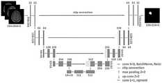
图3本发明用于生成图像的U-net网络针对低级胶质瘤分割的框架图;Figure 3 is a frame diagram of the U-net network used to generate images for low-grade glioma segmentation in the present invention;
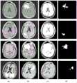
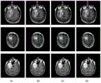
图4TCGA数据集的低级胶质瘤MR图像和分割掩膜示意图:Figure 4 Schematic diagram of the low-grade glioma MR image and segmentation mask of the TCGA dataset:
(a)pre-contrast;(b)FLAIR;(c)post-contrast;(d)ground truth;(a) pre-contrast; (b) FLAIR; (c) post-contrast; (d) ground truth;
图5cGAN生成的人工低级胶质瘤图像:Figure 5 Artificial low-grade glioma image generated by cGAN:
(a)pre-contrast;(b)FLAIR;(c)post-contrast;(d)ground truth;(a) pre-contrast; (b) FLAIR; (c) post-contrast; (d) ground truth;
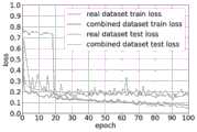
图6(a)在real datasets与real+generated datasets数据集上的训练过程损失函数变化图;Figure 6(a) The change diagram of the training process loss function on the real datasets and real+generated datasets datasets;
图6(b)在real+augmented datasets与all datasets数据集上的训练过程损失函数变化图;Figure 6(b) The change diagram of the training process loss function on the real+augmented datasets and all datasets datasets;
图7在测试集上的局部分割结果图:Figure 7 Local segmentation results on the test set:
(a)real dataset;(b)real+generated datasets;(c)real+augmenteddatasets;(d)all datasets。(a) real dataset; (b) real+generated datasets; (c) real+augmented datasets; (d) all datasets.
具体实施方式Detailed ways
下面将结合本发明实施例中的附图,对本发明实施例中的技术方案进行清楚、完整地描述,显然,所描述的实施例是本发明的一部分实施例,而不是全部的实施例。The following will clearly and completely describe the technical solutions in the embodiments of the present invention with reference to the accompanying drawings in the embodiments of the present invention. Obviously, the described embodiments are part of the embodiments of the present invention, not all of them.
基于本发明中的实施例,本领域普通技术人员在没有作出创造性劳动前提还可以进行若干简单的修改和润饰,所获得的所有其他实施例,都属于本发明保护的范围。Based on the embodiments of the present invention, those skilled in the art can make some simple modifications and embellishments without creative work, and all other obtained embodiments belong to the protection scope of the present invention.
在本发明中提及“实施例”意味着,结合实施例描述的特定特征、结构或特性可以包含在本发明的至少一个实施方案中。在说明书中的各个位置展示该短语并不一定均是指相同的实施例,也不是与其它实施例互斥的独立的或备选的实施例。本领域技术人员显式地和隐式地理解的是,本发明所描述的实施例还可以与其它的实施例相结合。Reference in the present invention to an "example" means that a particular feature, structure, or characteristic described in connection with the example can be included in at least one embodiment of the present invention. The presentation of this phrase in various places in the specification are not necessarily all referring to the same embodiment, nor are independent or alternative embodiments mutually exclusive of other embodiments. It is understood explicitly and implicitly by those skilled in the art that the described embodiments of the present invention can also be combined with other embodiments.
本发明期望得到的生成图像需满足两个要求:(1)人工生成的低级胶质瘤图像尽可能地接近真实的低级胶质瘤图像;(2)生成的低级胶质瘤图像与对应的分割掩膜一一对应。为了解决低级胶质瘤分割中标记数据不足的问题,提出在扩充后的小数据集上分割低级胶质瘤的框架,如图1所示,用cGAN生成标记胶质瘤图像,以扩充标记数据集,提高低级胶质瘤分割结果。提出的框架由生成系统和分割系统两部分组成,如图1所示。生成系统分为条件生成对抗网络训练阶段和人工胶质瘤图像生成阶段。cGAN网络结构如图2(a)和图2(b)所示,由生成器(Generator network,G)和判别器(Discriminator network,D)两部分组成。The generated image expected by the present invention needs to meet two requirements: (1) the artificially generated low-grade glioma image is as close as possible to the real low-grade glioma image; (2) the generated low-grade glioma image and the corresponding segmentation Masks correspond one-to-one. In order to solve the problem of insufficient labeled data in low-grade glioma segmentation, a framework for segmenting low-grade gliomas on the expanded small data set is proposed, as shown in Figure 1, using cGAN to generate labeled glioma images to expand the labeled data set to improve the segmentation results of low-grade gliomas. The proposed framework consists of two parts, a generative system and a segmentation system, as shown in Figure 1. The generation system is divided into a conditional generative adversarial network training stage and an artificial glioma image generation stage. The cGAN network structure is shown in Figure 2(a) and Figure 2(b), which consists of two parts: a generator (Generator network, G) and a discriminator (Discriminator network, D).
本发明提出的基于条件生成对抗网络的MR图像分割装置框架由生成系统和分割系统两部分组成,如图1所示。在生成模型训练阶段,用低级胶质瘤MR图像y和分割掩膜x训练生成模型;在人工胶质瘤图像生成阶段,根据输入的分割掩膜x,使用训练好的cGAN模型自动生成人工胶质瘤图像y’。分割系统基于改进的U-net网络,分为预训练阶段和训练阶段。在预训练阶段,利用人工生成胶质瘤图像y’和分割掩膜x U-net预训练网络;在训练阶段,在预训练模型的基础上,用低级胶质瘤MR图像y和分割掩膜x继续训练U-net网络。The framework of the MR image segmentation device based on the conditional generative confrontation network proposed by the present invention consists of two parts: a generation system and a segmentation system, as shown in FIG. 1 . In the generation model training stage, the low-grade glioma MR image y and the segmentation mask x are used to train the generation model; in the artificial glioma image generation stage, the trained cGAN model is used to automatically generate the artificial glue according to the input segmentation mask x Glioma image y'. The segmentation system is based on the improved U-net network, which is divided into a pre-training stage and a training stage. In the pre-training stage, use the artificially generated glioma image y' and the segmentation mask x U-net pre-training network; in the training stage, based on the pre-trained model, use the low-grade glioma MR image y and the segmentation mask x continues to train the U-net network.
实验使用的影像数据来自美国国家癌症研究所赞助的癌症影像档案(The CancerImaging Archive,TCIA),其中包含癌症基因组图谱(The Cancer Genome Atlas,TCGA)患者对应的MR影像。从TCGA-LGG集合中选取110例患者,其影像数据分别来以下自5个机构:Thomas Jefferson University(TCGA-CS,16例),Henry Ford Hospital(TCGA-DU,45例),UNC(TCGA-EZ,1例),Case Western(TCGA-FG,14例),Case Western St.Joseph’s(TCGA-HT,34例)。所选100例患者包括51例II级胶质瘤,58例III级胶质瘤,1例肿瘤的分级未知。所选数据包括前对比(pre-contrast)、FLAIR和后对比(post-contrast)三个序列,如图4所示,(d)为手工分割掩膜。随机选取10例患者作为测试集,100例患者作为训练集,每例患者的术前影像切片数量在20~88之间不等,包含含瘤切片和不含瘤切片。在同一个病人MR图像集中,含瘤切片数明显低于仅存在背景类的切片。所有切片裁剪到256×256大小,灰度归一化预处理。The image data used in the experiment comes from The Cancer Imaging Archive (TCIA) sponsored by the National Cancer Institute, which contains MR images corresponding to patients from The Cancer Genome Atlas (TCGA). A total of 110 patients were selected from the TCGA-LGG collection, and their image data were obtained from the following 5 institutions: Thomas Jefferson University (TCGA-CS, 16 cases), Henry Ford Hospital (TCGA-DU, 45 cases), UNC (TCGA-CS, 45 cases), UNC (TCGA- EZ, 1 case), Case Western (TCGA-FG, 14 cases), Case Western St. Joseph's (TCGA-HT, 34 cases). The selected 100 patients included 51 grade II gliomas, 58 grade III gliomas, and 1 tumor whose grade was unknown. The selected data includes three sequences of pre-contrast, FLAIR and post-contrast, as shown in Figure 4, (d) is the manual segmentation mask. Ten patients were randomly selected as the test set and 100 patients were used as the training set. The number of preoperative image slices for each patient ranged from 20 to 88, including tumor-containing slices and tumor-free slices. In the same patient MR image set, the number of tumor-containing slices was significantly lower than that of slices with only background classes. All slices were cropped to a size of 256×256, and grayscale normalization was preprocessed.
cGAN学习从输入x(条件)到随机噪声z再到输出y的映射的模型。条件信息的添加使cGAN能够根据输入的分割掩膜(条件)生成满足第二个要求的低级胶质瘤图像(输出)。cGAN learns a model that maps from input x (condition) to random noise z to output y. The addition of conditional information enables cGAN to generate low-grade glioma images (output) satisfying the second requirement based on the input segmentation mask (conditional).
cGAN的损失函数表示为:The loss function of cGAN is expressed as:
LcGAN(G,D)=Ex,y[logD(x,y)]+Ex,z[log(1-D(x,G(x,z)))] (1)LcGAN (G,D)=Ex,y [logD(x,y)]+Ex,z [log(1-D(x,G(x,z)))] (1)
其中x为分割掩膜,y为低级胶质瘤MR图像,z为随机噪声,D(x,y)表示输入的图像来自于真实图像的概率,D(x,G(x,z))表示输入图像来自生成图像的概率。假设x为分割掩膜,y为对应的低级胶质瘤MR图像,则G(x,z)为x对应的人工生成胶质瘤图像,随机噪声z为Dropout的形式,在训练和测试时应用于生成器的d1-d4层,如图2(a)所示。仅使用对抗性损失可能无法将梯度表达为期望的胶质瘤形状,因此,增加L1距离损失以促进学习过程。Where x is the segmentation mask, y is the low-grade glioma MR image, z is random noise, D(x,y) represents the probability that the input image comes from a real image, and D(x,G(x,z)) represents The probability that the input image came from the generated image. Suppose x is the segmentation mask and y is the corresponding low-grade glioma MR image, then G(x,z) is the artificially generated glioma image corresponding to x, and the random noise z is in the form of Dropout, which is applied during training and testing in the d1-d4 layers of the generator, as shown in Figure 2(a). Only using the adversarial loss may fail to express the gradient into the desired glioma shape, therefore, the L1 distance loss is added to facilitate the learning process.
L1距离可以表示为:The L1 distance can be expressed as:
LL1(G)=Ex,y[||y-G(x,z)||1] (2)LL1 (G)=Ex,y [||yG(x,z)||1 ] (2)
通过将GAN的目标函数与L1距离函数相结合,生成器和判别器的损失函数分别定义为:By combining the objective function of GAN with the L1 distance function, the loss functions of the generator and the discriminator are defined as:


λ为超参数,设定为100。λ is a hyperparameter, set to 100.
生成器网络结构:选择U-net架构作为cGAN的生成器网络,U-net网络已经在不同的生物医学图像分割应用中取得了良好的效果。从图2(a)中可以观察到生成器网络的结构设置。可以发现,U-Net架构一般由编码器和解码器组成,编码器和解码器堆栈中的镜像层之间有跳跃连接(skip connection)。跳跃连接的主要作用是在输入和输出之间直接通过网络传输一些低级的图像信息。输入分割掩膜与输出人工胶质瘤图像的虽然外观不同,但它们具有相同的底层结构和形状,若所有信息都经过普通的编码器-解码器网络流通则很容易丢失底层结构和形状。此外,为了使人工生成的图像具有多样性,在解码器的d1-d4层使用了Dropout。Generator network structure: The U-net architecture is selected as the generator network of cGAN. The U-net network has achieved good results in different biomedical image segmentation applications. The structural setup of the generator network can be observed from Fig. 2(a). It can be found that the U-Net architecture generally consists of an encoder and a decoder, and there is a skip connection between the mirror layers in the encoder and decoder stacks. The main function of the skip connection is to transfer some low-level image information directly through the network between the input and output. Although the appearance of the input segmentation mask and the output artificial glioma image are different, they have the same underlying structure and shape, which can be easily lost if all information is circulated through a common encoder-decoder network. Furthermore, in order to make the artificially generated images diverse, dropout is used in the layers d1-d4 of the decoder.
判别器网络结构:判别器网络是典型的CNN结构,图2(b)为详细网络结构。它根据输入的真实胶质瘤MR图像和人工胶质瘤图像,然后输出一个判断:当前输入图像是真还是假?在判别器中使用文献[36]中提出的PatchGAN,判别器只需分辨每个70×70的patch是真还是假,因此,网络运行速度更快,参数更少,可以应用于任意大的图像。patch大小为70×70。Discriminator network structure: The discriminator network is a typical CNN structure, and Figure 2(b) shows the detailed network structure. It outputs a judgment based on the input real glioma MR image and artificial glioma image: Is the current input image real or fake? Using the PatchGAN proposed in the literature [36] in the discriminator, the discriminator only needs to distinguish whether each 70×70 patch is true or false, so the network runs faster, has fewer parameters, and can be applied to arbitrarily large images . The patch size is 70×70.
最终形成的用于图像分割的U-net模型引入一个改进的U-net,它和原始U-net网络有三个主要差异:(1)在卷积过程中填充所有图层以保持图像大小不变,这样我们获得一个与输入图像大小相同的分割映射。(2)在解码路径中的每一步都包含与编码路径对应的特征映射的拼接。裁剪被移除,因为每一个填充卷积的边界像素丢失了。(3)将最后一层的softmax函数替换为sigmoid函数,因为低级胶质瘤分割输出结果的每个像素(黑色或白色)是一个二分类问题。The resulting U-net model for image segmentation introduces an improved U-net, which has three main differences from the original U-net network: (1) All layers are filled during convolution to keep the image size constant , so that we obtain a segmentation map of the same size as the input image. (2) Each step in the decoding path contains a concatenation of feature maps corresponding to the encoding path. Clipping is removed, since the border pixels of each padded convolution are lost. (3) Replace the softmax function of the last layer with a sigmoid function, because each pixel (black or white) of the low-grade glioma segmentation output is a binary classification problem.
设ytrue为手工分割掩膜,ypred为U-net网络的分割结果,分割网络的Dice损失函数为:Let ytrue be the manual segmentation mask, ypred be the segmentation result of the U-net network, and the Dice loss function of the segmentation network is:

对低级胶质瘤图像的分割可以看作是对每个像素的二分类。因此,将TP、TN、FP、FN等二分类问题中的常用概念作为计算评价指标,每个像素的评价指标分别表示真阳性、真阴性、假阳性和假阴性检测的个数。使用敏感性、特异性、总体准确性、Dice相似系数、雅尔卡指数(Jaccard index)和马修斯相关系数(Mattews correlation coefficient,MCC)来评估分割性能。所有定量测量都是在手工分割掩膜和预测分割结果之间按像素计算的。度量指标表示如下:Segmentation of low-grade glioma images can be viewed as a binary classification for each pixel. Therefore, the commonly used concepts in binary classification problems such as TP, TN, FP, and FN are used as calculation evaluation indicators, and the evaluation indicators of each pixel represent the number of true positive, true negative, false positive and false negative detections. Segmentation performance was assessed using sensitivity, specificity, overall accuracy, Dice similarity coefficient, Jaccard index, and Matthews correlation coefficient (MCC). All quantitative measurements are computed per-pixel between the hand-crafted segmentation mask and the predicted segmentation results. The metrics are represented as follows:


良好的分割效果具有较高的敏感性和特异性。利用Dice和Jaccard指标来衡量预测结果和金标准之间的相似性,指标代表了精度和灵敏度的调和平均值,最大值为1表示最优结果,0表示最差结果。MCC用来衡量分类的质量,包括真阳性和假阳性以及真阴性和假阴性。A good segmentation effect has high sensitivity and specificity. The Dice and Jaccard indicators are used to measure the similarity between the prediction results and the gold standard. The indicators represent the harmonic mean of accuracy and sensitivity, with a maximum value of 1 indicating the best result and 0 indicating the worst result. MCC is used to measure the quality of classification, including true positives and false positives and true negatives and false negatives.
用低级胶质瘤训练集训练cGAN网络,利用训练好的网络生成人工胶质瘤图像,如图5所示。脑MR图像存在强度不均匀、不同序列强度范围不同和来自不同的采集协议等问题。结果表明,该生成器生成的图像在L1感觉上接近于真实图像。生成的胶质瘤图像与输入分割掩膜具有相似的形状特征,与真实的胶质瘤MR图像具有相似的纹理特征。输入手工分割掩膜与输出人工胶质瘤图像的虽然外观不同,但它们具有相同的底层结构和形状。生成结果验证了在图像到图像转换环境下cGAN生成各种胶质瘤图像的有效性。The low-grade glioma training set was used to train the cGAN network, and the trained network was used to generate artificial glioma images, as shown in Figure 5. Brain MR images have issues such as inhomogeneous intensity, different intensity ranges for different sequences, and different acquisition protocols. The results show that the images produced by this generator are close to real images in L1 sense. The generated glioma images have similar shape features to the input segmentation mask and similar texture features to real glioma MR images. Although the appearance of the input manual segmentation mask and the output artificial glioma image are different, they have the same underlying structure and shape. The generated results validate the effectiveness of cGAN in generating various glioma images in the context of image-to-image translation.
比较本发明方法与常规数据扩充方式对低级胶质瘤分割的影响,将图3所示的U-net分割网络分别在四种数据集上训练:(1)原始数据集(real datasets),只包含真实数据集,不使用任何数据扩充方式;(2)原始数据集+生成数据集(real+generated datasets),包含人工生成的胶质瘤图像和原始数据集;(3)原始数据集+常规扩充数据集(real+augmented datasets),在原始数据集的基础上采用常规扩充后的数据集;(4)原始数据集+常规扩充数据集+生成数据集(real+augmented+generated dataset,all datsets),人工生成的胶质瘤图像和在原始数据集的基础上采用常规扩充后的数据集。四种训练数据集上的损失函数变化如图6(a)和图6(b)所示,可以看出本发明提出的方法在训练过程中收敛速度更快、损失更小,且分割精度更高。Comparing the influence of the method of the present invention and the conventional data expansion method on the segmentation of low-grade glioma, the U-net segmentation network shown in Figure 3 is trained on four kinds of data sets respectively: (1) original data sets (real datasets), only Contains real datasets without any data augmentation methods; (2) original datasets + generated datasets (real+generated datasets), including artificially generated glioma images and original datasets; (3) original datasets + conventional Augmented datasets (real+augmented datasets), using regular augmented datasets on the basis of the original datasets; (4) Original datasets + regular augmented datasets + generated datasets (real+augmented+generated datasets, all datsets ), artificially generated glioma images and a conventionally augmented dataset based on the original dataset. The changes of the loss functions on the four training data sets are shown in Figure 6(a) and Figure 6(b). It can be seen that the method proposed by the present invention has faster convergence speed, smaller loss and better segmentation accuracy during the training process. high.
脑肿瘤在空间位置和结构上具有高度可变性,上述四种训练模型在测试集上的定量评估结果如表1所示。从表中的实验结果可看出,经过人工生成胶质瘤图像预训练的模型表现出更好的分割性能,而常规数据扩充方式对提高网络分割性能作用有限。Brain tumors are highly variable in spatial location and structure, and the quantitative evaluation results of the above four training models on the test set are shown in Table 1. From the experimental results in the table, it can be seen that the model pre-trained by artificially generated glioma images shows better segmentation performance, while conventional data expansion methods have limited effect on improving network segmentation performance.
在相同的训练参数和测试集下,本发明提出的方法在测试集上明显得到了更好的结果。与原始数据集上训练的分割模型相比,本发明提出的人工生成图像扩充方法在测试集上分割结果的Dice系数提高了5.37%,Jac.提高了5.42%;与常规数据扩充方法相比,本发明提出的人工生成图像扩充方法在测试集上分割结果的Dice系数提高了4.39%,Jac.提高了4.42%;在两个数据集都存在常规数据扩充的情况下,含有本发明提出的人工生成图像扩充的数据集在测试集上分割结果的Dice系数提高了4.01%,Jac.提高了3.89%。Under the same training parameters and test set, the method proposed by the present invention obviously obtains better results on the test set. Compared with the segmentation model trained on the original data set, the Dice coefficient of the segmentation result of the artificially generated image expansion method proposed by the present invention on the test set has increased by 5.37%, and Jac. has increased by 5.42%; compared with the conventional data expansion method, The artificially generated image expansion method proposed by the present invention improves the Dice coefficient of the segmentation result on the test set by 4.39%, and Jac. improves by 4.42%. The dataset that generates image augmentation improves the Dice coefficient by 4.01% and Jac. by 3.89% for segmentation results on the test set.
表1四种训练模型在测试集上的评估结果Table 1 Evaluation results of four training models on the test set

四种训练模型在测试集上的部分分割结果如图7所示。从分割结果中可以看到,在原始数据集和经过传统扩充的数据集上的分割结果存在一些离群点,经过人工生成图像扩充的数据集上的分割结果则消除了这些离群点,使得分割结果更加接近于真实的肿瘤形态。Partial segmentation results of the four training models on the test set are shown in Fig. 7. It can be seen from the segmentation results that there are some outliers in the segmentation results of the original dataset and the traditional augmented dataset, and the segmentation results of the augmented dataset with artificially generated images eliminate these outliers, making The segmentation result is closer to the real tumor shape.
实验结果表明,本发明所提出的低级胶质瘤分割框架能够利用cGAN从输入的各种掩膜上学习一个给定数据集的分布,利用手工分割掩膜的形状特征生成的胶质瘤图像扩充和丰富了原始数据集的数据多样性,进而在胶质瘤分割中取得了更好的性能。与常规数据扩充方式相比,生成图像扩充方法在提高数据多样性方面表现的更好。The experimental results show that the low-grade glioma segmentation framework proposed by the present invention can learn the distribution of a given data set from various input masks by using cGAN, and the glioma image generated by the shape features of the manual segmentation mask is expanded And enrich the data diversity of the original dataset, and then achieve better performance in glioma segmentation. Compared with conventional data augmentation methods, generative image augmentation methods perform better in increasing data diversity.
低级胶质瘤的自动分割对临床诊断和治疗计划的制定有着重要的意义。本发明通过对抗性学习生成具有精确掩膜的胶质瘤图像,用于扩充数据集以提高低级胶质瘤分割网络的性能。评估如何通过对抗性学习来提高特定操作在医学图像分析中的表现。结果表明,所生成的带精确掩膜的低级胶质瘤图像可以成功地应用于计算机辅助诊断的算法中。本发明所提出的方法为低级胶质瘤在医学影像分析中提供了应用于更鲁棒的电脑辅助诊断系统的参考。Automatic segmentation of low-grade glioma is of great significance for clinical diagnosis and treatment planning. The present invention generates glioma images with precise masks through adversarial learning, which is used to expand the data set to improve the performance of the low-grade glioma segmentation network. Evaluating how adversarial learning can improve the performance of specific operations in medical image analysis. The results show that the generated low-grade glioma images with precise masks can be successfully applied in algorithms for computer-aided diagnosis. The method proposed in the present invention provides a reference for a more robust computer-aided diagnosis system for low-grade glioma in medical image analysis.
以上结合具体特征及其实施例对本发明进行了描述,显而易见的,在不脱离本发明的精神和范围的情况下,还可以对其进行各种修改和组合。相应地,本说明书和附图仅仅是所附权利要求所界定的本发明的示例性说明,且视为已覆盖本发明范围内的任意和所有修改、变化、组合或等同物。显然,本领域技术人员可以对本发明进行各种改动和变型,这些不脱离本发明的精神和范围的修改和变型也属于本发明权利要求及其等同技术的范围之内。The present invention has been described above in conjunction with specific features and embodiments thereof, and it is obvious that various modifications and combinations can be made without departing from the spirit and scope of the present invention. Accordingly, the specification and drawings are merely illustrative of the invention as defined by the appended claims and are deemed to cover any and all modifications, variations, combinations or equivalents within the scope of the invention. Obviously, those skilled in the art can make various modifications and variations to the present invention, and these modifications and variations without departing from the spirit and scope of the present invention also fall within the scope of the claims of the present invention and their equivalent technologies.
Claims (10)
Translated fromChinese


Priority Applications (1)
| Application Number | Priority Date | Filing Date | Title |
|---|---|---|---|
| CN202010055252.XACN111242953B (en) | 2020-01-17 | 2020-01-17 | MR image segmentation method and device based on condition generation countermeasure network |
Applications Claiming Priority (1)
| Application Number | Priority Date | Filing Date | Title |
|---|---|---|---|
| CN202010055252.XACN111242953B (en) | 2020-01-17 | 2020-01-17 | MR image segmentation method and device based on condition generation countermeasure network |
Publications (2)
| Publication Number | Publication Date |
|---|---|
| CN111242953A CN111242953A (en) | 2020-06-05 |
| CN111242953Btrue CN111242953B (en) | 2023-02-28 |
Family
ID=70864956
Family Applications (1)
| Application Number | Title | Priority Date | Filing Date |
|---|---|---|---|
| CN202010055252.XAActiveCN111242953B (en) | 2020-01-17 | 2020-01-17 | MR image segmentation method and device based on condition generation countermeasure network |
Country Status (1)
| Country | Link |
|---|---|
| CN (1) | CN111242953B (en) |
Families Citing this family (7)
| Publication number | Priority date | Publication date | Assignee | Title |
|---|---|---|---|---|
| CN112102323B (en)* | 2020-09-17 | 2023-07-07 | 陕西师范大学 | Adhesion cell nucleus segmentation method based on generation of countermeasure network and Caps-Unet network |
| CN112270686B (en)* | 2020-12-24 | 2021-03-16 | 北京达佳互联信息技术有限公司 | Image segmentation model training method, image segmentation device and electronic equipment |
| CN113222811B (en)* | 2021-05-24 | 2022-08-09 | 北京理工大学 | Face attribute migration method based on image mask |
| CN114519719B (en)* | 2022-01-07 | 2024-10-01 | 宁波大学 | Brain tumor MR image segmentation method |
| CN115908446A (en)* | 2022-09-05 | 2023-04-04 | 北京精诊医疗科技有限公司 | Method, apparatus and program product for tumor lesion segmentation |
| CN115359073A (en)* | 2022-10-17 | 2022-11-18 | 湖南自兴智慧医疗科技有限公司 | Chromosome topological structure segmentation method and device based on countermeasure generation network |
| CN118608544A (en)* | 2024-06-20 | 2024-09-06 | 首都医科大学附属北京天坛医院 | A method for segmenting hemorrhage foci in the brain and related products |
Family Cites Families (3)
| Publication number | Priority date | Publication date | Assignee | Title |
|---|---|---|---|---|
| AU2017101166A4 (en)* | 2017-08-25 | 2017-11-02 | Lai, Haodong MR | A Method For Real-Time Image Style Transfer Based On Conditional Generative Adversarial Networks |
| CN107909621A (en)* | 2017-11-16 | 2018-04-13 | 深圳市唯特视科技有限公司 | It is a kind of based on it is twin into confrontation network medical image synthetic method |
| CN109166126B (en)* | 2018-08-13 | 2022-02-18 | 苏州比格威医疗科技有限公司 | Method for segmenting paint cracks on ICGA image based on condition generation type countermeasure network |
- 2020
- 2020-01-17CNCN202010055252.XApatent/CN111242953B/enactiveActive
Also Published As
| Publication number | Publication date |
|---|---|
| CN111242953A (en) | 2020-06-05 |
Similar Documents
| Publication | Publication Date | Title |
|---|---|---|
| CN111242953B (en) | MR image segmentation method and device based on condition generation countermeasure network | |
| CN112419327B (en) | A method, system and device for image segmentation based on generative confrontation network | |
| CN108596882B (en) | The recognition methods of pathological picture and device | |
| Zhang et al. | Automatic segmentation of the cardiac MR images based on nested fully convolutional dense network with dilated convolution | |
| CN110910351B (en) | Ultrasound image modality transfer, classification method and terminal based on generative adversarial network | |
| CN110189308B (en) | Tumor detection method and device based on fusion of BM3D and dense convolution network | |
| CN111325750B (en) | Medical image segmentation method based on multi-scale fusion U-shaped chain neural network | |
| WO2022001623A1 (en) | Image processing method and apparatus based on artificial intelligence, and device and storage medium | |
| CN110120033A (en) | Based on improved U-Net neural network three-dimensional brain tumor image partition method | |
| CN106777953A (en) | The analysis method and system of medical image data | |
| CN112686898B (en) | An automatic segmentation method of radiotherapy target volume based on self-supervised learning | |
| WO2022127500A1 (en) | Multiple neural networks-based mri image segmentation method and apparatus, and device | |
| CN115496720A (en) | Gastrointestinal cancer pathological image segmentation method and related equipment based on ViT mechanism model | |
| Tyagi et al. | An amalgamation of vision transformer with convolutional neural network for automatic lung tumor segmentation | |
| CN115018865A (en) | A medical image segmentation method based on transfer learning | |
| CN111932512A (en) | Intracranial hemorrhage detection algorithm applied to CT image based on CNN and NLSTM neural network | |
| Qi et al. | Automatic lacunae localization in placental ultrasound images via layer aggregation | |
| CN118710912A (en) | A semi-supervised image segmentation method | |
| CN117392468A (en) | Cancer pathology image classification system, media and equipment based on multi-instance learning | |
| Li et al. | Brain tumor MRI segmentation method based on improved Res-UNet | |
| CN119130891A (en) | A small target detection method, system, device and storage medium for medical images | |
| CN117611602A (en) | A fiber bundle segmentation method based on U-NET network dual encoder structure | |
| CN116153530A (en) | Orthodontic treatment monitoring method and device based on oral scanning video | |
| Li et al. | TSRL-Net: Target-aware supervision residual learning for stroke segmentation | |
| Li et al. | A Multi-Category Brain Tumor Classification Method Bases on Improved ResNet50. |
Legal Events
| Date | Code | Title | Description |
|---|---|---|---|
| PB01 | Publication | ||
| PB01 | Publication | ||
| SE01 | Entry into force of request for substantive examination | ||
| SE01 | Entry into force of request for substantive examination | ||
| GR01 | Patent grant | ||
| GR01 | Patent grant |