CN111242850A - A method for improving the resolution of wide-field fundus optical coherence blood flow imaging - Google Patents
A method for improving the resolution of wide-field fundus optical coherence blood flow imagingDownload PDFInfo
- Publication number
- CN111242850A CN111242850ACN202010150103.1ACN202010150103ACN111242850ACN 111242850 ACN111242850 ACN 111242850ACN 202010150103 ACN202010150103 ACN 202010150103ACN 111242850 ACN111242850 ACN 111242850A
- Authority
- CN
- China
- Prior art keywords
- octa
- image
- wide
- area
- output
- Prior art date
- Legal status (The legal status is an assumption and is not a legal conclusion. Google has not performed a legal analysis and makes no representation as to the accuracy of the status listed.)
- Granted
Links
Images


Classifications
- G—PHYSICS
- G06—COMPUTING OR CALCULATING; COUNTING
- G06T—IMAGE DATA PROCESSING OR GENERATION, IN GENERAL
- G06T3/00—Geometric image transformations in the plane of the image
- G06T3/40—Scaling of whole images or parts thereof, e.g. expanding or contracting
- G06T3/4053—Scaling of whole images or parts thereof, e.g. expanding or contracting based on super-resolution, i.e. the output image resolution being higher than the sensor resolution
- G—PHYSICS
- G06—COMPUTING OR CALCULATING; COUNTING
- G06T—IMAGE DATA PROCESSING OR GENERATION, IN GENERAL
- G06T3/00—Geometric image transformations in the plane of the image
- G06T3/40—Scaling of whole images or parts thereof, e.g. expanding or contracting
- G06T3/4038—Image mosaicing, e.g. composing plane images from plane sub-images
- G—PHYSICS
- G06—COMPUTING OR CALCULATING; COUNTING
- G06T—IMAGE DATA PROCESSING OR GENERATION, IN GENERAL
- G06T3/00—Geometric image transformations in the plane of the image
- G06T3/40—Scaling of whole images or parts thereof, e.g. expanding or contracting
- G06T3/4046—Scaling of whole images or parts thereof, e.g. expanding or contracting using neural networks
Landscapes
- Engineering & Computer Science (AREA)
- Physics & Mathematics (AREA)
- General Physics & Mathematics (AREA)
- Theoretical Computer Science (AREA)
- Artificial Intelligence (AREA)
- Evolutionary Computation (AREA)
- Eye Examination Apparatus (AREA)
Abstract
Description
Translated fromChinese技术领域technical field
本发明属于生物医学成像和图像处理技术领域,尤其涉及一种广域眼底光学相干血流成像分辨率提升方法。The invention belongs to the technical field of biomedical imaging and image processing, and in particular relates to a method for improving the resolution of wide-area fundus optical coherent blood flow imaging.
背景技术Background technique
光学相干断层扫描血管造影(Optical Coherence Tomography Angiography,OCTA)是一种低损、高分辨、非侵入性的新型成像方式,在同一位置使用重复B扫描的时间去相关来区分周围组织和血流。该方法有潜力取代传统的荧光素血管造影(FA)和吲哚菁绿色血管造影(ICGA),因此在眼科领域引起了极大的关注。与FA和ICGA等2D成像中脉管系统的重叠深度信息相比,OCTA能够以5~10μm的高轴向分辨率在深度方向上解析血管和毛细血管。它已被广泛用于各种眼部疾病的研究,例如青光眼、年龄相关性黄斑病变和早产儿视网膜病变。另外,人视网膜的OCTA成像还能够指示神经退行性疾病,例如轻度认知障碍和阿尔茨海默氏病。Optical Coherence Tomography Angiography (OCTA) is a new low-loss, high-resolution, non-invasive imaging modality that uses temporal decorrelation of repeated B-scans at the same location to differentiate surrounding tissue and blood flow. This method has the potential to replace traditional fluorescein angiography (FA) and indocyanine green angiography (ICGA), and thus has attracted great attention in the field of ophthalmology. Compared with the overlapping depth information of the vasculature in 2D imaging such as FA and ICGA, OCTA is able to resolve vessels and capillaries in the depth direction with a high axial resolution of 5–10 μm. It has been widely used in the study of various eye diseases such as glaucoma, age-related macular degeneration and retinopathy of prematurity. In addition, OCTA imaging of the human retina can also be indicative of neurodegenerative diseases such as mild cognitive impairment and Alzheimer's disease.
然而,与强调视网膜层结构信息的光学相干断层扫描技术(Optical CoherenceTomography ,OCT)不同,OCTA的重要功能是可视化视网膜和脉络膜毛细血管,因此,OCTA对采集系统的横向分辨率和采样密度有更高的要求。由于一些疾病需要周边的血流成像比如病变主要位于周边的早期糖尿病视网膜病变,小视场的OCTA已经不能满足此要求,需要扩大OCTA对视网膜扫描的成像范围。然而,广域OCTA会牺牲图像的横向分辨率,导致在定量分析中低估血管的生物标记物。保证分辨率的前提下扩大视网膜OCTA成像范围的技术将是增强OCT技术在临床实践中应用中重要的下一步。However, unlike Optical Coherence Tomography (OCT), which emphasizes the structural information of retinal layers, the important function of OCTA is to visualize retinal and choriocapillaris. Therefore, OCTA has higher lateral resolution and sampling density for the acquisition system. requirements. Because some diseases require peripheral blood flow imaging, such as early diabetic retinopathy where the lesions are mainly located in the periphery, OCTA with a small field of view can no longer meet this requirement, and it is necessary to expand the imaging range of OCTA for retinal scanning. However, wide-field OCTA sacrifices the lateral resolution of the images, leading to underestimation of vascular biomarkers in quantitative analysis. The technology to expand the imaging range of retinal OCTA under the premise of ensuring the resolution will be an important next step to enhance the application of OCT technology in clinical practice.
在从单个图像中得到视网膜血管系统的广域和高分辨率图像的OCTA问题上,华盛顿大学提出了一种多帧拼接的方法。通过辅助实时线扫描检眼镜的运动跟踪生成的OCTA,可实现在临床上为患者的功能性视网膜脉管成像,在覆盖视网膜60度以上的同时仍保持高分辨率和高分辨率。但是此项技术十分耗时耗力,且需要病患多次采集,对病患的配合度要求很高。On the OCTA problem of obtaining wide-field and high-resolution images of the retinal vasculature from a single image, the University of Washington proposed a multi-frame stitching method. The OCTA generated by assisting the motion tracking of real-time line scan ophthalmoscopes enables the clinical imaging of functional retinal vasculature in patients, covering more than 60 degrees of the retina while still maintaining high resolution and resolution. However, this technique is very time-consuming and labor-intensive, and requires multiple collections from patients, which requires a high degree of patient cooperation.
发明专利申请CN201910584151.9公开了基于SD-OCT和OCTA视网膜图像的CNV自动检测方法,并具体公开了方法包括步骤1、采集含有CNV病变的SD-OCT视网膜图像、OCTA视网膜图像;步骤2、利用层分割算法分割SD-OCT视网膜图像的ILM、OPL和BM层;步骤3、将三维SD-OCT体数据投影生成CNV显著图;步骤4、将三维OCTA体数据投影生成视网膜内、外层投影图;步骤5、基于视网膜内、外层投影图,去除血流投射伪影获得伪影去除图像;步骤6、通过自适应阈值法将CNV显著图和伪影去除图像二值化;步骤7、对步骤6获得的两个二值图像进行数学形态学处理,获得目标候选区域;步骤8、将步骤7获得的所有目标候选区域合并;步骤9、根据包含种子点数量去除虚假目标候选区域,获得粗略CNV区域;步骤10、对视网膜外层投影图中粗略CNV区域边界内部的像素进行二聚类处理,获得二值图像;步骤11、对步骤10的二值图像进行数学形态学处理获得细化的CNV边界。该方法仅适用于CNV病变的检测。Invention patent application CN201910584151.9 discloses an automatic CNV detection method based on SD-OCT and OCTA retinal images, and specifically discloses that the method includes
发明内容SUMMARY OF THE INVENTION
本发明针对现有技术存在的问题,提出了一种广域眼底光学相干血流成像分辨率提升方法,能同时满足高分辨率和广域的要求,且普适于各类OCTA数据。Aiming at the problems existing in the prior art, the present invention proposes a method for improving the resolution of wide-area fundus optical coherence blood flow imaging, which can meet the requirements of high resolution and wide area at the same time, and is universally applicable to various OCTA data.
本发明是通过以下技术方案得以实现的:The present invention is achieved through the following technical solutions:
本发明一种广域眼底光学相干血流成像分辨率提升方法,包括:The present invention is a method for improving the resolution of wide-area fundus optical coherent blood flow imaging, comprising:
步骤S01,采集两种模式的视网膜OCTA图像,所述两种模式包括广域低清OCTA模式和非广域高清OCTA模式;Step S01, collecting retinal OCTA images of two modes, the two modes include a wide-area low-definition OCTA mode and a non-wide-area high-definition OCTA mode;
步骤S02,将两种模式的视网膜OCTA图像裁剪为成像区域大小一致的子图,并将两种模式的子图统一缩放到大小相同的规格;Step S02, the retinal OCTA images of the two modes are cropped into sub-images with the same size of the imaging area, and the sub-images of the two modes are uniformly scaled to the same size specification;
步骤S03,将属于广域低清OCTA模式的OCTA子图作为源域,记作数据集A,将属于非广域高清OCTA模式的OCTA子图作为目标域,记作数据集B;并将数据集A和数据集B中的部分数据划分为训练集,数据集A和数据集B中的其余部分数据划分为测试集;Step S03, take the OCTA subgraph belonging to the wide-area low-definition OCTA mode as the source domain, denoted as data set A, and take the OCTA subgraph belonging to the non-wide-area high-definition OCTA mode as the target domain, denoted as data set B; Part of the data in set A and data set B is divided into training sets, and the rest of the data in data set A and data set B is divided into test sets;
步骤S04,将步骤S03获得的相同规格的两种模式的视网膜OCTA子图放入深度神经网络进行训练;Step S04, putting the retinal OCTA subgraphs of the two modes of the same specifications obtained in step S03 into a deep neural network for training;
步骤S05,将广域低清OCTA模式的视网膜OCTA子图输入步骤S04训练后的神经网络模型,训练后的神经网络模型输出对广域低清OCTA模式的视网膜OCTA子图进行分辨率增强的测试结果;In step S05, the retinal OCTA sub-image of the wide-area low-definition OCTA mode is input into the neural network model after training in step S04, and the output of the neural network model after training is to perform a resolution enhancement test on the retinal OCTA sub-image of the wide-area low-definition OCTA mode. result;
步骤S06,拼接属于同一张广域图像的重建高清OCTA子图,继而得到广域高清视网膜OCTA图像。Step S06, splicing the reconstructed high-definition OCTA sub-images belonging to the same wide-area image, and then obtaining a wide-area high-definition retinal OCTA image.
该方法是一种端到端的广域低采样OCTA高分辨率重建方法的设计。由于横向采样率低,广域OCTA的横向分辨率差,因此导致血管生物标记物的观察和定量不准确,高采样率和低采样率之间的分辨率差异可以通过模型或数据驱动的方法进行修复。This method is the design of an end-to-end wide-area low-sampling OCTA high-resolution reconstruction method. Wide-field OCTA has poor lateral resolution due to low lateral sampling rates, thus leading to inaccurate observation and quantification of vascular biomarkers, and the difference in resolution between high and low sampling rates can be modelled or data-driven approaches repair.
作为优选,所述步骤S01中的视网膜OCTA图像为表层视网膜血管造影图像。Preferably, the retinal OCTA image in the step S01 is a superficial retinal angiography image.
作为优选,所述步骤S01具体包括:采集广域低清OCTA模式和非广域高清OCTA模式的视网膜OCTA图像,并利用去条纹方法去除采集广域OCTA过程中眼球微动产生的横条纹。Preferably, the step S01 specifically includes: collecting retinal OCTA images in a wide-area low-definition OCTA mode and a non-wide-area high-definition OCTA mode, and using a stripping method to remove horizontal stripes generated by eyeball micro-movements in the process of collecting wide-area OCTA.
作为优选,所述步骤S01中采集的广域低清OCTA模式的视网膜OCTA图像的扫描区域大小,大于所述步骤S01中采集的非广域高清OCTA模式的视网膜OCTA图像的扫描区域大小。Preferably, the scanning area size of the retinal OCTA image in the wide-area low-definition OCTA mode collected in the step S01 is larger than the scanning area size of the retinal OCTA image in the non-wide-area high-definition OCTA mode collected in the step S01.
作为优选,所述步骤S02包括:将两种模式的视网膜OCTA图像裁剪成1x1mm2的子图,统一缩放到340*340个像素点。Preferably, the step S02 includes: cropping the retinal OCTA images of the two modes into sub-images of 1×1 mm2 and uniformly scaling them to 340*340 pixels.
作为优选,所述步骤S04包括:Preferably, the step S04 includes:
步骤S41,将数据集A的输入图像Input A输入生成器GAB,经过生成器网络后,得到输出图像Output B;Step S41, input the input image Input A of the dataset A into the generator GAB , and after passing through the generator network, obtain the output image Output B;
步骤S42,将输出图像Output B输入判别器DB,由判别器DB判定输出图像Output B属于数据集B,则输出1,否则为0;Step S42, input the output image OutputB into the discriminator DB, and the discriminator DB determines that the output image OutputB belongs to the data set B, then outputs 1, otherwise it is 0;
步骤S43,将输出图像Output B输入生成器GBA,经过生成器网络后,得到输出循环图像Rec A;Step S43, the output image Output B is input into the generator GBA , and after passing through the generator network, the output cyclic image Rec A is obtained;
步骤S44,将数据集B的输入图像Input B输入生成器GBA,经过生成器网络后,得到输出图像Output A;Step S44, input the input image Input B of the dataset B into the generator GBA , and after passing through the generator network, obtain the output image Output A;
步骤S45,将输出图像Output A输入判别器DA,由判别器判定DA输出图像Output A属于数据集B,则输出0,否则为1;Step S45, input the output image Output A into the discriminator DA , and the discriminator determines that the output image Output A of DA belongs to the data set B, then outputs 0, otherwise it is 1;
步骤S46,将输出图像Output A输入生成器GAB,经过生成器网络后,得到输出循环图像Rec B;Step S46, the output image Output A is input into the generator GAB , and after passing through the generator network, the output cyclic image Rec B is obtained;
步骤S47,基于损失函数公式Step S47, based on the loss function formula

计算整个训练模型的整体损失函数,继而验证输入图像和来自生成器的合成图像是否处于相同分布,以验证神经网络模型是否训练完毕;Calculate the overall loss function of the entire training model, and then verify that the input image and the synthesized image from the generator are in the same distribution to verify that the neural network model is trained;
其中,生成器GAB用于将源域A中的图像x转换为类似于目标域B中图像的生成图像G(x);生成器GBA用于将目标域B中的图像y转换为类似于源域A中图像的生成图像G(y);β控制损失的比例,L_GAN为判别器将生成图像判别为真实图片的损失,表示对抗性损失;L_cyc为原图像经过生成循环后得到的图像的差异损失,表示周期一致性损失。Among them, the generator GAB is used to convert the image x in the source domain A into a generated image G(x) similar to the image in the target domain B; the generator GBA is used to convert the image y in the target domain B into a similar The generated image G(y) of the image in the source domain A; β controls the ratio of the loss, L_GAN is the loss of the discriminator to distinguish the generated image as a real image, representing the adversarial loss; L_cyc is the original image obtained after the generation cycle. , which represents the cycle-consistency loss.
作为优选,所述步骤S47具体包括:Preferably, the step S47 specifically includes:
依据公式According to the formula

计算输出图像Output B与输入图像Input B之间、输出图像Output A与输入图像InputA之间的对抗损失函数;Calculate the adversarial loss function between the output image Output B and the input image Input B, and between the output image Output A and the input image InputA;
依据公式According to the formula

计算输出循环图像Rec A与输入图像Input A之间、输出循环图像Rec B与输入图像Input B之间的周期一致性损失函数;Calculate the cycle consistency loss function between the output cyclic image Rec A and the input image Input A, and between the output cyclic image Rec B and the input image Input B;
依据公式According to the formula

计算整个训练模型的整体损失函数;Calculate the overall loss function of the entire training model;
验证输出图像Output B与输入图像Input B间、输出图像Output A与输入图像Input A间、输出循环Rec A与输入图像Input A间、输出循环Rec B与输入图像Input B间是否处于相同分布,若是,则神经网络模型训练完成。Verify whether the output image Output B and the input image Input B, between the output image Output A and the input image Input A, between the output cycle Rec A and the input image Input A, and between the output cycle Rec B and the input image Input B are in the same distribution, if so , the neural network model training is completed.
作为优选,所述步骤S04中的深度神经网络采用循环一致对抗性网络框架。Preferably, the deep neural network in the step S04 adopts a cycle-consistent adversarial network framework.
作为优选,所述步骤S06包括拼接属于同一张广域图像的重建高清1x1mm2OCTA子图,继而得到广域高清视网膜8x8mm2OCTA图像。Preferably, the step S06 includes splicing reconstructed high-
作为优选,所述方法获得的广域高清图像用于定量血管生物标志物。Preferably, the wide-field high-definition images obtained by the method are used to quantify vascular biomarkers.
本发明具有以下有益效果:The present invention has the following beneficial effects:
本发明一种广域眼底光学相干血流成像分辨率提升方法:The present invention is a method for improving the resolution of wide-area fundus optical coherent blood flow imaging:
1、本发明采用非成对学习和裁剪子图的方法解决了广域OCTA缺乏成对高清OCTA数据的问题。1. The present invention solves the problem that wide-area OCTA lacks paired high-definition OCTA data by adopting the method of non-paired learning and cropping subgraphs.
2、本发明采用裁剪子图的方法解决了采集OCTA图像数据集小的问题。2. The invention adopts the method of cropping sub-images to solve the problem that the OCTA image data set is small.
3、本发明方法中的模型一经训练,可多次重复使用,免去了在应用时繁琐的图像处理步骤。3. Once the model in the method of the present invention is trained, it can be used repeatedly for many times, thereby eliminating the tedious image processing steps during application.
附图说明Description of drawings
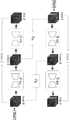
图1为本发明一种广域眼底光学相干血流成像分辨率提升方法的流程图;1 is a flowchart of a method for improving the resolution of a wide-field fundus optical coherent blood flow imaging according to the present invention;
图2为本发明一种广域眼底光学相干血流成像分辨率提升方法的具体示例的流程图;2 is a flowchart of a specific example of a method for improving the resolution of a wide-field fundus optical coherent blood flow imaging according to the present invention;
图3为依据本发明一种广域眼底光学相干血流成像分辨率提升方法重建广域高分辨率OCTA图像的工作流程;3 is a workflow for reconstructing a wide-area high-resolution OCTA image according to a method for improving the resolution of a wide-area fundus optical coherent blood flow imaging according to the present invention;
图4为本发明所使用的循环一致对抗性网络系统框图;4 is a block diagram of a cycle-consistent adversarial network system used in the present invention;
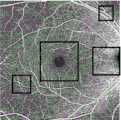
图5a为采用本发明方法示例中原始视网膜OCTA图像;Fig. 5a is the original retinal OCTA image in the example of adopting the method of the present invention;
图5b为采用本发明方法示例中经过深度学习重建的高分辨率OCTA图像;Figure 5b is a high-resolution OCTA image reconstructed by deep learning in an example of the method of the present invention;
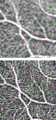
图5c(包括对应图5a中的方框C以及5b中相同位置的两幅对照图)、5d包括对应图5a中的方框D以及5b中相同位置的两幅对照图、5e(包括对应图5a中的方框E以及5b中相同位置的两幅对照图)、5f(包括对应图5a中的方框F以及5b中相同位置的两幅对照图)是图5a中方框部分的放大视图。Figures 5c (including two comparison pictures corresponding to the box C in Figure 5a and the same position in 5b), 5d including two comparison pictures corresponding to the box D in Figure 5a and the same position in 5b, 5e (including the corresponding picture) Box E in 5a and two comparison pictures at the same position in 5b), 5f (including two comparison pictures corresponding to box F in Figure 5a and the same position in 5b) are enlarged views of the box part in Figure 5a.
具体实施方式Detailed ways
以下是本发明的具体实施例并结合附图,对本发明的技术方案作进一步的描述,但本发明并不限于这些实施例。The following are specific embodiments of the present invention and the accompanying drawings to further describe the technical solutions of the present invention, but the present invention is not limited to these embodiments.
如图1,本发明一种广域眼底光学相干血流成像分辨率提升方法,包括:As shown in Figure 1, a method for improving the resolution of wide-field fundus optical coherent blood flow imaging according to the present invention includes:
步骤S01,采集两种模式的视网膜OCTA图像,所述两种模式包括广域低清OCTA模式和非广域高清OCTA模式;Step S01, collecting retinal OCTA images of two modes, the two modes include a wide-area low-definition OCTA mode and a non-wide-area high-definition OCTA mode;
步骤S02,将两种模式的视网膜OCTA图像裁剪为成像区域大小一致的子图,并将两种模式的子图统一缩放到大小相同的规格;Step S02, the retinal OCTA images of the two modes are cropped into sub-images with the same size of the imaging area, and the sub-images of the two modes are uniformly scaled to the same size specification;
步骤S03,将属于广域低清OCTA模式的OCTA子图作为源域,记作数据集A,将属于非广域高清OCTA模式的OCTA子图作为目标域,记作数据集B;并将数据集A和数据集B中的部分数据划分为训练集,数据集A和数据集B中的其余部分数据划分为测试集;Step S03, take the OCTA subgraph belonging to the wide-area low-definition OCTA mode as the source domain, denoted as data set A, and take the OCTA subgraph belonging to the non-wide-area high-definition OCTA mode as the target domain, denoted as data set B; Part of the data in set A and data set B is divided into training sets, and the rest of the data in data set A and data set B is divided into test sets;
步骤S04,将步骤S03获得的相同规格的两种模式的视网膜OCTA子图放入深度神经网络进行训练;Step S04, putting the retinal OCTA subgraphs of the two modes of the same specifications obtained in step S03 into a deep neural network for training;
步骤S05,将广域低清OCTA模式的视网膜OCTA子图输入步骤S04训练后的神经网络模型,训练后的神经网络模型输出对广域低清OCTA模式的视网膜OCTA子图进行分辨率增强的测试结果;In step S05, the retinal OCTA sub-image of the wide-area low-definition OCTA mode is input into the neural network model after training in step S04, and the output of the neural network model after training is to perform a resolution enhancement test on the retinal OCTA sub-image of the wide-area low-definition OCTA mode. result;
步骤S06,拼接属于同一张广域图像的重建高清OCTA子图,继而得到广域高清视网膜OCTA图像。Step S06, splicing the reconstructed high-definition OCTA sub-images belonging to the same wide-area image, and then obtaining a wide-area high-definition retinal OCTA image.
在步骤S01中,步骤S01包括采集广域低清OCTA模式(即广域低横向分辨率,低采样)和非广域高清OCTA模式(即非广域高横向分辨率,高采样)的视网膜OCTA 图像,并利用去条纹方法去除采集广域OCTA过程中眼球微动产生的横条纹。所述步骤S01中采集的广域低清OCTA模式的视网膜OCTA图像的扫描区域大小,大于所述步骤S01中采集的非广域高清OCTA模式的视网膜OCTA图像的扫描区域大小。In step S01, step S01 includes collecting retinal OCTA in a wide-area low-definition OCTA mode (ie, wide-area low lateral resolution, low sampling) and a non-wide-area high-definition OCTA mode (ie, non-wide-area high lateral resolution, high sampling) image, and use the de-striping method to remove the horizontal streaks produced by the micro-movement of the eye during the acquisition of wide-field OCTA. The scanning area size of the retinal OCTA image in the wide-area low-definition OCTA mode collected in the step S01 is larger than the scanning area size of the retinal OCTA image in the non-wide-area high-definition OCTA mode collected in the step S01.
例如,采集20组分别为两种模式的表层视网膜(SVP)血管造影图像。其中广域低清OCTA模式扫描区域大小为8x8mm2,非广域高清OCTA扫描区域大小为3x3mm2,可利用传统去条纹方法去除因采集广域OCTA过程中眼球微动产生的横条纹(参见图2、3)。For example, 20 sets of superficial retinal (SVP) angiography images of two modalities were acquired. Among them, the scanning area size of the wide-area low-definition OCTA mode is 8x8mm2 , and the size of the non-wide-area high-definition OCTA scanning area is 3x3mm2 . The traditional stripping method can be used to remove the horizontal stripes caused by eye movements during the acquisition of wide-area OCTA (see Fig. 2, 3).
在步骤S02中,成像区域大小以及缩放大小根据需要设定。例如,所述步骤S02包括:将两种模式的视网膜OCTA图像裁剪成1x1mm2的子图(参见图2、3),统一缩放到340*340个像素点。In step S02, the size of the imaging area and the zoom size are set as required. For example, the step S02 includes: cropping the retinal OCTA images of the two modes into sub-images of 1×1 mm2 (see FIGS. 2 and 3 ), and uniformly scaling them to 340*340 pixels.
在步骤S03中,对裁剪的子图分类:属于广域低清OCTA的子图作为源域,记作数据集A,属于非广域高清OCTA的子图作为目标域,记作数据集B。对训练集和测试集的划分,可以将数据集A和数据集B图像中的一部分数据(约占90%)划分为训练集,其余部分数据划分为测试集。In step S03, the cropped sub-images are classified: the sub-images belonging to the wide-area low-definition OCTA are taken as the source domain, and denoted as dataset A; For the division of the training set and the test set, a part of the data (about 90%) in the images of the data set A and the data set B can be divided into the training set, and the rest of the data can be divided into the test set.
在步骤S04中,两种模式的OCTA分别作为源域A和目标域B放入深度神经网络进行无监督训练,生成高清的OCTA子图。In step S04, the two modes of OCTA are respectively used as the source domain A and the target domain B into the deep neural network for unsupervised training to generate high-definition OCTA subgraphs.
步骤S04使用非成对的生成对抗网络(GAN, Generative Adversarial Networks ),包含生成器和判别器。我们的深度神经网络采用的是循环一致对抗性网络框架。它的目的是学习一种映射GAB,使用对抗损失,在像素级别和特征级别上,都无法将属于3x3mm2模式中的子图与属于8x8mm2模式中的子图进行区分。然后,我们将其与逆映射GBA耦合,并引入周期一致性损失,以强制生成图像尽可能类似于重建图像(反之亦然)。这样的生成方式不能确保我们的输入图像和输出图像以有意义的方式配对,因此引入了两个鉴别器网络DA和DB以确保输入图像和来自生成器的合成图像处于相同的分布。Step S04 uses an unpaired generative adversarial network (GAN, Generative Adversarial Networks), including a generator and a discriminator. Our deep neural network adopts the recurrent consistent adversarial network framework. Its purpose is to learn a mapping GAB , using an adversarial loss, that is indistinguishable from sub-images belonging to a 3x3mm2 pattern from those belonging to an 8x8mm2 pattern, both at the pixel level and at the feature level. We then couple it with the inverse map GBA and introduce a cycle-consistency loss to force the generated image to be as similar as possible to the reconstructed image (and vice versa). Such generation does not ensure that our input and output images are paired in a meaningful way, so two discriminator networks DA and DB are introduced to ensure that the input image and the synthesized image from the generator are in the same distribution.
将缩放后的参考图像数据集A和数据集B送入循环一致对抗性深度神经网络中进行无监督训练,对低质量的广域OCTA图像进行复原。图4为循环一致对抗性网络系统框图,它的生成器由9个残差模块组成,通过对输入图像的解码和编码转换输入图像的特征得到一张在目标域B中的图像,判别器判别通过卷积网络提取的图像特征是否属于目标域B的类别,由于该模型可以实现A到B,也可以实现B到A,设置了两个生成器(GAB和GBA)和两个判别器(DA和DB)。训练集数据在该模型中的运行过程如下:The scaled reference image datasets A and B are fed into a recurrent consistent adversarial deep neural network for unsupervised training to restore low-quality wide-area OCTA images. Figure 4 is a block diagram of the cyclically consistent adversarial network system. Its generator consists of 9 residual modules. By decoding the input image and transcoding the features of the input image, an image in the target domain B is obtained, and the discriminator discriminates Whether the image features extracted by the convolutional network belong to the category of the target domain B, since the model can achieve A to B or B to A, two generators (GAB and GBA ) and two discriminators are set (DA and DB) . The running process of the training set data in this model is as follows:
所述步骤S04包括:The step S04 includes:
步骤S41,将数据集A (广域低清OCTA子图)的输入图像Input A输入生成器GAB,经过生成器网络后,得到输出图像Output B;Step S41, input the input image Input A of the dataset A (wide-area low-definition OCTA sub-image) into the generator GAB , and after passing through the generator network, obtain the output image Output B;
步骤S42,将输出图像Output B (非广域高清OCTA子图)输入判别器DB,由判别器DB判定输出图像Output B属于数据集B,则输出1,否则为0;Step S42, input the output image Output B (non-wide-area high-definition OCTA sub-image) into the discriminator DB, and the discriminator DB determines that the output image OutputB belongs to the datasetB , then outputs 1, otherwise it is 0;
步骤S43,将输出图像Output B输入生成器GBA,经过生成器网络后,得到输出循环图像Rec A;Step S43, the output image Output B is input into the generator GBA , and after passing through the generator network, the output cyclic image Rec A is obtained;
步骤S44,将数据集B的输入图像Input B输入生成器GBA,经过生成器网络后,得到输出图像Output A;Step S44, input the input image Input B of the dataset B into the generator GBA , and after passing through the generator network, obtain the output image Output A;
步骤S45,将输出图像Output A输入判别器DA,由判别器判定DA输出图像Output A属于数据集B,则输出0,否则为1;Step S45, input the output image Output A into the discriminator DA , and the discriminator determines that the output image Output A of DA belongs to the data set B, then outputs 0, otherwise it is 1;
步骤S46,将输出图像Output A输入生成器GAB,经过生成器网络后,得到输出循环图像Rec B;Step S46, the output image Output A is input into the generator GAB , and after passing through the generator network, the output cyclic image Rec B is obtained;
步骤S47,基于损失函数公式Step S47, based on the loss function formula

计算整个训练模型的整体损失函数,继而验证输入图像和来自生成器的合成图像是否处于相同分布,以验证神经网络模型是否训练完毕;Calculate the overall loss function of the entire training model, and then verify that the input image and the synthesized image from the generator are in the same distribution to verify that the neural network model is trained;
其中,生成器GAB用于将源域A中的图像x转换为类似于目标域B中图像的生成图像G(x),所生成的图像必须保留有原始图像的特性,所以如果我们使用生成器GAB生成一张假图像,那么要能够使用另一个生成器GBA来努力恢复成原始图像;生成器GBA用于将目标域B中的图像y转换为类似于源域A中图像的生成图像G(y);判别器DA用于判断它的输入图像是初始域A中的图像还是生成图像G(y),判别器DB用于判断它的输入图像是目标域B中的图像还是生成图像G(x);此过程必须满足循环一致性;β控制损失的比例,L_GAN为判别器将生成图像判别为真实图片的损失,表示对抗性损失;L_cyc为原图像经过生成循环后得到的图像的差异损失,保证原始图像和循环图像之间的差异应该尽可能小,表示周期一致性损失。Among them, the generator GAB is used to transform the image x in the source domain A into a generated image G(x) similar to the image in the target domain B, the generated image must retain the characteristics of the original image, so if we use the generated image The generator GAB generates a fake image, then another generator GBA can be used to try to restore the original image; the generator GBA is used to transform the image y in the target domain B into a similar image in the source domain A. Generate image G(y); discriminator DA is used to judge whether its input image is an image in initial domain A or generated image G(y), discriminator DB is used to judge whether its input image is in target domain B The image is still the generated image G(x); this process must meet the cycle consistency; β controls the proportion of loss, L_GAN is the loss of the discriminator to distinguish the generated image as a real image, representing the adversarial loss; L_cyc is the original image after the generation cycle The resulting image disparity loss ensures that the disparity between the original image and the cycled image should be as small as possible, representing a cycle consistency loss.
所述步骤S47具体包括:The step S47 specifically includes:
依据公式According to the formula

计算输出图像Output B与输入图像Input B之间、输出图像Output A与输入图像InputA之间的对抗损失函数;Calculate the adversarial loss function between the output image Output B and the input image Input B, and between the output image Output A and the input image InputA;
依据公式According to the formula

计算输出循环图像Rec A与输入图像Input A之间、输出循环图像Rec B与输入图像Input B之间的周期一致性损失函数;Calculate the cycle consistency loss function between the output cyclic image Rec A and the input image Input A, and between the output cyclic image Rec B and the input image Input B;
依据公式According to the formula

计算整个训练模型的整体损失函数;Calculate the overall loss function of the entire training model;
验证输出图像Output B与输入图像Input B间、输出图像Output A与输入图像Input A间、输出循环Rec A与输入图像Input A间、输出循环Rec B与输入图像Input B间是否处于相同分布,若是,则神经网络模型训练完成。Verify whether the output image Output B and the input image Input B, between the output image Output A and the input image Input A, between the output cycle Rec A and the input image Input A, and between the output cycle Rec B and the input image Input B are in the same distribution, if so , the neural network model training is completed.
该模型是基于Ubuntu 16.04 LTS操作系统中的深度学习框架PyTorch实施,模型的训练使用具有12 GB RAM的NVIDIA GeForce GTX 1080 Ti GPU进行。使用Adam优化器从头开始训练生成器和鉴别器,其初始学习率为2*10-4,batch size大小为1。对于这两个函数,我们分别设置β1 = 0.5和β2= 0.999 两个Adam优化器。 在GAN模型的训练中,损失曲线显示在180个时期后收敛,因此我们在200个时期停下来以实现最小的损失,每次训练网络花费了大约4个小时。The model was implemented based on the deep learning framework PyTorch in the Ubuntu 16.04 LTS operating system, and the training of the model was performed using an NVIDIA GeForce GTX 1080 Ti GPU with 12 GB RAM. The generator and discriminator are trained from scratch using the Adam optimizer with an initial learning rate of 2*10-4 and a batch size of 1. For these two functions, we set β1 = 0.5 and β2 = 0.999 for the two Adam optimizers, respectively. In the training of the GAN model, the loss curve showed convergence after 180 epochs, so we stopped at 200 epochs to achieve minimal loss, which took about 4 hours to train the network each time.
在所述步骤S05中,神经网络模型训练完毕后,将低质量的广域OCTA子图输入训练后的神经网络模型,训练后的神经网络模型将输出对低质量OCTA进行分辨率增强的测试结果。In the step S05, after the training of the neural network model is completed, the low-quality wide-area OCTA sub-image is input into the trained neural network model, and the trained neural network model will output the test result of enhancing the resolution of the low-quality OCTA .
在所述步骤S06中,拼接重建结果。例如,拼接属于同一张广域图像的高清1x1mm2OCTA子图得到高分辨率的视网膜8x8mm2 OCTA,该结果即为分辨率增强后的最终图像。In the step S06, the reconstruction results are concatenated. For example, stitching high-definition 1x1mm2 OCTA sub-images belonging to the same wide-area image to obtain a high-resolution retinal 8x8mm2 OCTA, the result is the final image after resolution enhancement.
依据上述方法进行的测试结果如图5a-5f,图5a-5f是广域OCTA图像分辨率增强的结果。图5a是原始视网膜OCTA图像;图5b是经过深度学习重建的高分辨率OCTA图像;图5c-5f是图5a中方框内的放大视图。在整个视野中,可以观察到分辨率的显着提高,本发明提出的方法可以有效地解决分辨率和视场之间的约束,将有助于在广域OCTA上准确定量血管生物标志物,从而有助于诊断和治疗。The test results according to the above method are shown in Figures 5a-5f, and Figures 5a-5f are the results of wide-area OCTA image resolution enhancement. Figure 5a is the original retinal OCTA image; Figure 5b is a high-resolution OCTA image reconstructed by deep learning; Figures 5c-5f are enlarged views inside the box in Figure 5a. In the entire field of view, a significant improvement in resolution can be observed, and the method proposed in the present invention can effectively resolve the constraints between resolution and field of view, which will help to accurately quantify vascular biomarkers on wide-field OCTA, thereby assisting in diagnosis and treatment.
本领域的技术人员应理解,上述描述及附图中所示的本发明的实施例只作为举例而并不限制本发明。本发明的目的已经完整有效地实现。本发明的功能及结构原理已在实施例中展示和说明,在没有背离所述原理下,本发明的实施方式可以有任何变形或修改。It should be understood by those skilled in the art that the embodiments of the present invention shown in the above description and the accompanying drawings are only examples and do not limit the present invention. The objects of the present invention have been fully and effectively achieved. The functional and structural principles of the present invention have been shown and described in the embodiments, and the embodiments of the present invention may be modified or modified in any way without departing from the principles.
Claims (10)


Priority Applications (1)
| Application Number | Priority Date | Filing Date | Title |
|---|---|---|---|
| CN202010150103.1ACN111242850B (en) | 2020-03-06 | 2020-03-06 | Wide-area fundus optical coherence blood flow imaging resolution improving method |
Applications Claiming Priority (1)
| Application Number | Priority Date | Filing Date | Title |
|---|---|---|---|
| CN202010150103.1ACN111242850B (en) | 2020-03-06 | 2020-03-06 | Wide-area fundus optical coherence blood flow imaging resolution improving method |
Publications (2)
| Publication Number | Publication Date |
|---|---|
| CN111242850Atrue CN111242850A (en) | 2020-06-05 |
| CN111242850B CN111242850B (en) | 2023-09-15 |
Family
ID=70880215
Family Applications (1)
| Application Number | Title | Priority Date | Filing Date |
|---|---|---|---|
| CN202010150103.1AActiveCN111242850B (en) | 2020-03-06 | 2020-03-06 | Wide-area fundus optical coherence blood flow imaging resolution improving method |
Country Status (1)
| Country | Link |
|---|---|
| CN (1) | CN111242850B (en) |
Cited By (2)
| Publication number | Priority date | Publication date | Assignee | Title |
|---|---|---|---|---|
| CN112818820A (en)* | 2021-01-28 | 2021-05-18 | 北京达佳互联信息技术有限公司 | Image generation model training method, image generation device and electronic equipment |
| CN113643184A (en)* | 2021-10-18 | 2021-11-12 | 广东唯仁医疗科技有限公司 | Optical coherence tomography-based fundus blood vessel display method, system and medium |
Citations (3)
| Publication number | Priority date | Publication date | Assignee | Title |
|---|---|---|---|---|
| WO2018162690A1 (en)* | 2017-03-10 | 2018-09-13 | Carl Zeiss Meditec, Inc. | Method for analyzing avascular regions in optical coherence tomography angiography images |
| CN110264424A (en)* | 2019-06-20 | 2019-09-20 | 北京理工大学 | A kind of fuzzy retinal fundus images Enhancement Method based on generation confrontation network |
| WO2019240257A1 (en)* | 2018-06-15 | 2019-12-19 | キヤノン株式会社 | Medical image processing device, medical image processing method and program |
- 2020
- 2020-03-06CNCN202010150103.1Apatent/CN111242850B/enactiveActive
Patent Citations (3)
| Publication number | Priority date | Publication date | Assignee | Title |
|---|---|---|---|---|
| WO2018162690A1 (en)* | 2017-03-10 | 2018-09-13 | Carl Zeiss Meditec, Inc. | Method for analyzing avascular regions in optical coherence tomography angiography images |
| WO2019240257A1 (en)* | 2018-06-15 | 2019-12-19 | キヤノン株式会社 | Medical image processing device, medical image processing method and program |
| CN110264424A (en)* | 2019-06-20 | 2019-09-20 | 北京理工大学 | A kind of fuzzy retinal fundus images Enhancement Method based on generation confrontation network |
Non-Patent Citations (1)
| Title |
|---|
| YUKUN GUO等: "Automated segmentation of retinal layer boundaries and capillary plexuses in wide-field optical coherence tomographic angiography"* |
Cited By (4)
| Publication number | Priority date | Publication date | Assignee | Title |
|---|---|---|---|---|
| CN112818820A (en)* | 2021-01-28 | 2021-05-18 | 北京达佳互联信息技术有限公司 | Image generation model training method, image generation device and electronic equipment |
| CN112818820B (en)* | 2021-01-28 | 2024-03-19 | 北京达佳互联信息技术有限公司 | Image generation model training method, image generation device and electronic equipment |
| CN113643184A (en)* | 2021-10-18 | 2021-11-12 | 广东唯仁医疗科技有限公司 | Optical coherence tomography-based fundus blood vessel display method, system and medium |
| CN113643184B (en)* | 2021-10-18 | 2022-02-18 | 广东唯仁医疗科技有限公司 | Optical coherence tomography-based fundus blood vessel display method, system and medium |
Also Published As
| Publication number | Publication date |
|---|---|
| CN111242850B (en) | 2023-09-15 |
Similar Documents
| Publication | Publication Date | Title |
|---|---|---|
| JP7579372B2 (en) | Medical image processing device, medical image processing system, medical image processing method and program | |
| US12307659B2 (en) | Medical image processing apparatus, medical image processing method and computer-readable storage medium | |
| JP7746514B2 (en) | Medical image processing device, medical image processing method and program | |
| Ma et al. | ROSE: a retinal OCT-angiography vessel segmentation dataset and new model | |
| US12040079B2 (en) | Medical image processing apparatus, medical image processing method and computer-readable medium | |
| CN113226153B (en) | Image processing device, image processing method and computer readable storage medium | |
| CN110390650B (en) | OCT image denoising method based on dense connection and generation countermeasure network | |
| US11922601B2 (en) | Medical image processing apparatus, medical image processing method and computer-readable medium | |
| US11887288B2 (en) | Image processing apparatus, image processing method, and storage medium | |
| Abràmoff et al. | Retinal imaging and image analysis | |
| JP2020058800A (en) | Image processing apparatus, image processing method and program | |
| JP2022103221A (en) | Medical image processing equipment, medical image processing system, medical image processing method and program | |
| JP7703349B2 (en) | Image processing device, image processing method, and program | |
| CN111710012B (en) | An OCTA imaging method and device based on two-dimensional composite registration | |
| JP2020513983A (en) | Multiplex en-face angiographic averaging system, method and apparatus for optical coherence tomography | |
| Liu et al. | Disentangled representation learning for OCTA vessel segmentation with limited training data | |
| Lim et al. | Generative data augmentation for diabetic retinopathy classification | |
| JP7344847B2 (en) | Image processing device, image processing method, and program | |
| CN113543695A (en) | Image processing apparatus, image processing method, and program | |
| JP7332463B2 (en) | Control device, optical coherence tomography device, control method for optical coherence tomography device, and program | |
| Yuan et al. | Image enhancement of wide-field retinal optical coherence tomography angiography by super-resolution angiogram reconstruction generative adversarial network | |
| CN117475270B (en) | Lesion identification method and system for diabetic retinopathy image | |
| CN111242850B (en) | Wide-area fundus optical coherence blood flow imaging resolution improving method | |
| CN112562058B (en) | Method for quickly establishing intracranial vascular simulation three-dimensional model based on transfer learning | |
| CN118072921A (en) | A dynamic ultra-wide-angle fluorescein angiography synthesis method based on conditional diffusion model |
Legal Events
| Date | Code | Title | Description |
|---|---|---|---|
| PB01 | Publication | ||
| PB01 | Publication | ||
| SE01 | Entry into force of request for substantive examination | ||
| SE01 | Entry into force of request for substantive examination | ||
| GR01 | Patent grant | ||
| GR01 | Patent grant |