CN111172235B - Biosensor for detecting cathepsin B and detection method and application thereof - Google Patents
Biosensor for detecting cathepsin B and detection method and application thereofDownload PDFInfo
- Publication number
- CN111172235B CN111172235BCN202010042539.9ACN202010042539ACN111172235BCN 111172235 BCN111172235 BCN 111172235BCN 202010042539 ACN202010042539 ACN 202010042539ACN 111172235 BCN111172235 BCN 111172235B
- Authority
- CN
- China
- Prior art keywords
- dna
- cathepsin
- polypeptide
- biosensor
- sequence
- Prior art date
- Legal status (The legal status is an assumption and is not a legal conclusion. Google has not performed a legal analysis and makes no representation as to the accuracy of the status listed.)
- Active
Links
- 108090000712Cathepsin BProteins0.000titleclaimsabstractdescription98
- 102000004225Cathepsin BHuman genes0.000titleclaimsabstractdescription98
- 238000001514detection methodMethods0.000titleclaimsabstractdescription35
- 238000000034methodMethods0.000claimsabstractdescription48
- 230000003321amplificationEffects0.000claimsabstractdescription38
- 238000003199nucleic acid amplification methodMethods0.000claimsabstractdescription38
- 230000000694effectsEffects0.000claimsabstractdescription30
- 238000006243chemical reactionMethods0.000claimsabstractdescription21
- 102100034343IntegraseHuman genes0.000claimsabstractdescription18
- 101710203526IntegraseProteins0.000claimsabstractdescription18
- 108020004707nucleic acidsProteins0.000claimsabstractdescription10
- 102000039446nucleic acidsHuman genes0.000claimsabstractdescription10
- 150000007523nucleic acidsChemical class0.000claimsabstractdescription10
- 239000011324beadSubstances0.000claimsdescription30
- 239000000523sampleSubstances0.000claimsdescription28
- 108090000765processed proteins & peptidesProteins0.000claimsdescription24
- 239000000758substrateSubstances0.000claimsdescription16
- 102000016928DNA-directed DNA polymeraseHuman genes0.000claimsdescription13
- 108010014303DNA-directed DNA polymeraseProteins0.000claimsdescription13
- 108091034117OligonucleotideProteins0.000claimsdescription11
- 238000011534incubationMethods0.000claimsdescription9
- 108091008146restriction endonucleasesProteins0.000claimsdescription9
- 239000007795chemical reaction productSubstances0.000claimsdescription8
- 239000002773nucleotideSubstances0.000claimsdescription8
- 125000003729nucleotide groupChemical group0.000claimsdescription8
- 239000006228supernatantSubstances0.000claimsdescription8
- 238000001502gel electrophoresisMethods0.000claimsdescription7
- YBJHBAHKTGYVGT-ZKWXMUAHSA-N(+)-BiotinChemical groupN1C(=O)N[C@@H]2[C@H](CCCCC(=O)O)SC[C@@H]21YBJHBAHKTGYVGT-ZKWXMUAHSA-N0.000claimsdescription6
- 102000004190EnzymesHuman genes0.000claimsdescription6
- 108090000790EnzymesProteins0.000claimsdescription6
- 229910019142PO4Inorganic materials0.000claimsdescription6
- 238000005259measurementMethods0.000claimsdescription6
- 239000010452phosphateSubstances0.000claimsdescription6
- 239000003795chemical substances by applicationSubstances0.000claimsdescription5
- 238000001917fluorescence detectionMethods0.000claimsdescription5
- 108010090804StreptavidinProteins0.000claimsdescription4
- 238000002189fluorescence spectrumMethods0.000claimsdescription4
- 229960002685biotinDrugs0.000claimsdescription3
- 235000020958biotinNutrition0.000claimsdescription3
- 239000011616biotinSubstances0.000claimsdescription3
- 210000004899c-terminal regionAnatomy0.000claimsdescription2
- 238000006073displacement reactionMethods0.000abstractdescription11
- 125000000539amino acid groupChemical group0.000abstractdescription9
- 230000008569processEffects0.000abstractdescription7
- 230000029087digestionEffects0.000abstractdescription6
- 238000004458analytical methodMethods0.000abstractdescription3
- 108020004414DNAProteins0.000description45
- 210000004027cellAnatomy0.000description20
- 108091032973(ribonucleotides)n+mProteins0.000description11
- 230000000295complement effectEffects0.000description10
- 230000035945sensitivityEffects0.000description10
- 206010028980NeoplasmDiseases0.000description9
- 102000035195PeptidasesHuman genes0.000description9
- 108091005804PeptidasesProteins0.000description9
- 239000004365ProteaseSubstances0.000description9
- FAPWRFPIFSIZLT-UHFFFAOYSA-MSodium chlorideChemical compound[Na+].[Cl-]FAPWRFPIFSIZLT-UHFFFAOYSA-M0.000description8
- 239000000872bufferSubstances0.000description8
- 201000011510cancerDiseases0.000description7
- 238000003776cleavage reactionMethods0.000description7
- 239000000203mixtureSubstances0.000description7
- 239000013615primerSubstances0.000description7
- 239000000243solutionSubstances0.000description7
- 238000002474experimental methodMethods0.000description6
- 102000003908Cathepsin DHuman genes0.000description5
- 108090000258Cathepsin DProteins0.000description5
- 108090000624Cathepsin LProteins0.000description5
- 102000004172Cathepsin LHuman genes0.000description5
- 108090000613Cathepsin SProteins0.000description5
- 102100035654Cathepsin SHuman genes0.000description5
- 206010008342Cervix carcinomaDiseases0.000description5
- WCUXLLCKKVVCTQ-UHFFFAOYSA-MPotassium chlorideChemical compound[Cl-].[K+]WCUXLLCKKVVCTQ-UHFFFAOYSA-M0.000description5
- 208000006105Uterine Cervical NeoplasmsDiseases0.000description5
- 201000010881cervical cancerDiseases0.000description5
- 229940088598enzymeDrugs0.000description5
- PCHJSUWPFVWCPO-UHFFFAOYSA-NgoldChemical compound[Au]PCHJSUWPFVWCPO-UHFFFAOYSA-N0.000description5
- 229910052737goldInorganic materials0.000description5
- 239000010931goldSubstances0.000description5
- VEXZGXHMUGYJMC-UHFFFAOYSA-Nhydrochloric acidSubstancesClVEXZGXHMUGYJMC-UHFFFAOYSA-N0.000description5
- 230000001404mediated effectEffects0.000description5
- 102000004196processed proteins & peptidesHuman genes0.000description5
- 230000007017scissionEffects0.000description5
- XLYOFNOQVPJJNP-UHFFFAOYSA-NwaterSubstancesOXLYOFNOQVPJJNP-UHFFFAOYSA-N0.000description5
- 102000005600CathepsinsHuman genes0.000description4
- 108010084457CathepsinsProteins0.000description4
- KCXVZYZYPLLWCC-UHFFFAOYSA-NEDTAChemical compoundOC(=O)CN(CC(O)=O)CCN(CC(O)=O)CC(O)=OKCXVZYZYPLLWCC-UHFFFAOYSA-N0.000description4
- 239000007983Tris bufferSubstances0.000description4
- JLCPHMBAVCMARE-UHFFFAOYSA-N[3-[[3-[[3-[[3-[[3-[[3-[[3-[[3-[[3-[[3-[[3-[[5-(2-amino-6-oxo-1H-purin-9-yl)-3-[[3-[[3-[[3-[[3-[[3-[[5-(2-amino-6-oxo-1H-purin-9-yl)-3-[[5-(2-amino-6-oxo-1H-purin-9-yl)-3-hydroxyoxolan-2-yl]methoxy-hydroxyphosphoryl]oxyoxolan-2-yl]methoxy-hydroxyphosphoryl]oxy-5-(5-methyl-2,4-dioxopyrimidin-1-yl)oxolan-2-yl]methoxy-hydroxyphosphoryl]oxy-5-(6-aminopurin-9-yl)oxolan-2-yl]methoxy-hydroxyphosphoryl]oxy-5-(6-aminopurin-9-yl)oxolan-2-yl]methoxy-hydroxyphosphoryl]oxy-5-(6-aminopurin-9-yl)oxolan-2-yl]methoxy-hydroxyphosphoryl]oxy-5-(6-aminopurin-9-yl)oxolan-2-yl]methoxy-hydroxyphosphoryl]oxyoxolan-2-yl]methoxy-hydroxyphosphoryl]oxy-5-(5-methyl-2,4-dioxopyrimidin-1-yl)oxolan-2-yl]methoxy-hydroxyphosphoryl]oxy-5-(4-amino-2-oxopyrimidin-1-yl)oxolan-2-yl]methoxy-hydroxyphosphoryl]oxy-5-(5-methyl-2,4-dioxopyrimidin-1-yl)oxolan-2-yl]methoxy-hydroxyphosphoryl]oxy-5-(5-methyl-2,4-dioxopyrimidin-1-yl)oxolan-2-yl]methoxy-hydroxyphosphoryl]oxy-5-(6-aminopurin-9-yl)oxolan-2-yl]methoxy-hydroxyphosphoryl]oxy-5-(6-aminopurin-9-yl)oxolan-2-yl]methoxy-hydroxyphosphoryl]oxy-5-(4-amino-2-oxopyrimidin-1-yl)oxolan-2-yl]methoxy-hydroxyphosphoryl]oxy-5-(4-amino-2-oxopyrimidin-1-yl)oxolan-2-yl]methoxy-hydroxyphosphoryl]oxy-5-(4-amino-2-oxopyrimidin-1-yl)oxolan-2-yl]methoxy-hydroxyphosphoryl]oxy-5-(6-aminopurin-9-yl)oxolan-2-yl]methoxy-hydroxyphosphoryl]oxy-5-(4-amino-2-oxopyrimidin-1-yl)oxolan-2-yl]methyl [5-(6-aminopurin-9-yl)-2-(hydroxymethyl)oxolan-3-yl] hydrogen phosphatePolymersCc1cn(C2CC(OP(O)(=O)OCC3OC(CC3OP(O)(=O)OCC3OC(CC3O)n3cnc4c3nc(N)[nH]c4=O)n3cnc4c3nc(N)[nH]c4=O)C(COP(O)(=O)OC3CC(OC3COP(O)(=O)OC3CC(OC3COP(O)(=O)OC3CC(OC3COP(O)(=O)OC3CC(OC3COP(O)(=O)OC3CC(OC3COP(O)(=O)OC3CC(OC3COP(O)(=O)OC3CC(OC3COP(O)(=O)OC3CC(OC3COP(O)(=O)OC3CC(OC3COP(O)(=O)OC3CC(OC3COP(O)(=O)OC3CC(OC3COP(O)(=O)OC3CC(OC3COP(O)(=O)OC3CC(OC3COP(O)(=O)OC3CC(OC3COP(O)(=O)OC3CC(OC3COP(O)(=O)OC3CC(OC3COP(O)(=O)OC3CC(OC3CO)n3cnc4c(N)ncnc34)n3ccc(N)nc3=O)n3cnc4c(N)ncnc34)n3ccc(N)nc3=O)n3ccc(N)nc3=O)n3ccc(N)nc3=O)n3cnc4c(N)ncnc34)n3cnc4c(N)ncnc34)n3cc(C)c(=O)[nH]c3=O)n3cc(C)c(=O)[nH]c3=O)n3ccc(N)nc3=O)n3cc(C)c(=O)[nH]c3=O)n3cnc4c3nc(N)[nH]c4=O)n3cnc4c(N)ncnc34)n3cnc4c(N)ncnc34)n3cnc4c(N)ncnc34)n3cnc4c(N)ncnc34)O2)c(=O)[nH]c1=OJLCPHMBAVCMARE-UHFFFAOYSA-N0.000description4
- 125000004122cyclic groupChemical group0.000description4
- 239000005549deoxyribonucleosideSubstances0.000description4
- 238000003745diagnosisMethods0.000description4
- 229940079593drugDrugs0.000description4
- 239000003814drugSubstances0.000description4
- 239000003269fluorescent indicatorSubstances0.000description4
- 238000012544monitoring processMethods0.000description4
- 229920001184polypeptidePolymers0.000description4
- 235000018102proteinsNutrition0.000description4
- 102000004169proteins and genesHuman genes0.000description4
- 108090000623proteins and genesProteins0.000description4
- 239000011535reaction bufferSubstances0.000description4
- 239000011780sodium chlorideSubstances0.000description4
- 239000001226triphosphateSubstances0.000description4
- 238000002965ELISAMethods0.000description3
- PEDCQBHIVMGVHV-UHFFFAOYSA-NGlycerineChemical compoundOCC(O)COPEDCQBHIVMGVHV-UHFFFAOYSA-N0.000description3
- TWRXJAOTZQYOKJ-UHFFFAOYSA-LMagnesium chlorideChemical compound[Mg+2].[Cl-].[Cl-]TWRXJAOTZQYOKJ-UHFFFAOYSA-L0.000description3
- 238000003556assayMethods0.000description3
- 238000005516engineering processMethods0.000description3
- 239000012634fragmentSubstances0.000description3
- 230000001939inductive effectEffects0.000description3
- 230000003993interactionEffects0.000description3
- 210000003292kidney cellAnatomy0.000description3
- 239000003161ribonuclease inhibitorSubstances0.000description3
- 238000012216screeningMethods0.000description3
- 230000011664signalingEffects0.000description3
- 210000001519tissueAnatomy0.000description3
- 108091003079Bovine Serum AlbuminProteins0.000description2
- CURLTUGMZLYLDI-UHFFFAOYSA-NCarbon dioxideChemical compoundO=C=OCURLTUGMZLYLDI-UHFFFAOYSA-N0.000description2
- 108020001019DNA PrimersProteins0.000description2
- 239000003155DNA primerSubstances0.000description2
- 239000006144Dulbecco’s modified Eagle's mediumSubstances0.000description2
- GDBQQVLCIARPGH-UHFFFAOYSA-NLeupeptinNatural productsCC(C)CC(NC(C)=O)C(=O)NC(CC(C)C)C(=O)NC(C=O)CCCN=C(N)NGDBQQVLCIARPGH-UHFFFAOYSA-N0.000description2
- CSNNHWWHGAXBCP-UHFFFAOYSA-LMagnesium sulfateChemical compound[Mg+2].[O-][S+2]([O-])([O-])[O-]CSNNHWWHGAXBCP-UHFFFAOYSA-L0.000description2
- 101710163270NucleaseProteins0.000description2
- 239000002202Polyethylene glycolSubstances0.000description2
- 239000013592cell lysateSubstances0.000description2
- 238000004737colorimetric analysisMethods0.000description2
- 235000018417cysteineNutrition0.000description2
- XUJNEKJLAYXESH-UHFFFAOYSA-NcysteineNatural productsSCC(N)C(O)=OXUJNEKJLAYXESH-UHFFFAOYSA-N0.000description2
- FFYPMLJYZAEMQB-UHFFFAOYSA-Ndiethyl pyrocarbonateChemical compoundCCOC(=O)OC(=O)OCCFFYPMLJYZAEMQB-UHFFFAOYSA-N0.000description2
- VHJLVAABSRFDPM-QWWZWVQMSA-NdithiothreitolChemical compoundSC[C@@H](O)[C@H](O)CSVHJLVAABSRFDPM-QWWZWVQMSA-N0.000description2
- 238000002848electrochemical methodMethods0.000description2
- 239000012091fetal bovine serumSubstances0.000description2
- 238000002866fluorescence resonance energy transferMethods0.000description2
- 239000003112inhibitorSubstances0.000description2
- GDBQQVLCIARPGH-ULQDDVLXSA-NleupeptinChemical compoundCC(C)C[C@H](NC(C)=O)C(=O)N[C@@H](CC(C)C)C(=O)N[C@H](C=O)CCCN=C(N)NGDBQQVLCIARPGH-ULQDDVLXSA-N0.000description2
- 108010052968leupeptinProteins0.000description2
- 239000006166lysateSubstances0.000description2
- 239000012139lysis bufferSubstances0.000description2
- -1polybutylene succinatePolymers0.000description2
- 229920002961polybutylene succinatePolymers0.000description2
- 239000004631polybutylene succinateSubstances0.000description2
- 229920001223polyethylene glycolPolymers0.000description2
- 239000001103potassium chlorideSubstances0.000description2
- 235000011164potassium chlorideNutrition0.000description2
- 238000002360preparation methodMethods0.000description2
- 239000000047productSubstances0.000description2
- 238000002331protein detectionMethods0.000description2
- 230000035484reaction timeEffects0.000description2
- 238000011896sensitive detectionMethods0.000description2
- 238000000926separation methodMethods0.000description2
- 229910052709silverInorganic materials0.000description2
- 239000004332silverSubstances0.000description2
- 229910021642ultra pure waterInorganic materials0.000description2
- 239000012498ultrapure waterSubstances0.000description2
- 102000040650(ribonucleotides)n+mHuman genes0.000description1
- RVHSTXJKKZWWDQ-UHFFFAOYSA-N1,1,1,2-tetrabromoethaneChemical compoundBrCC(Br)(Br)BrRVHSTXJKKZWWDQ-UHFFFAOYSA-N0.000description1
- QKNYBSVHEMOAJP-UHFFFAOYSA-N2-amino-2-(hydroxymethyl)propane-1,3-diol;hydron;chlorideChemical compoundCl.OCC(N)(CO)COQKNYBSVHEMOAJP-UHFFFAOYSA-N0.000description1
- MWHLCXQYEVKVFB-UHFFFAOYSA-N4-(2-aminoethyl)benzenesulfonyl chlorideChemical compoundNCCC1=CC=C(S(Cl)(=O)=O)C=C1MWHLCXQYEVKVFB-UHFFFAOYSA-N0.000description1
- NIUDXSFNLBIWOB-DCAQKATOSA-NArg-Leu-CysChemical compoundCC(C)C[C@@H](C(=O)N[C@@H](CS)C(=O)O)NC(=O)[C@H](CCCN=C(N)N)NNIUDXSFNLBIWOB-DCAQKATOSA-N0.000description1
- 241000193738Bacillus anthracisSpecies0.000description1
- 208000006386Bone ResorptionDiseases0.000description1
- 206010006187Breast cancerDiseases0.000description1
- 208000026310Breast neoplasmDiseases0.000description1
- OKTJSMMVPCPJKN-UHFFFAOYSA-NCarbonChemical compound[C]OKTJSMMVPCPJKN-UHFFFAOYSA-N0.000description1
- 102000012406Carcinoembryonic AntigenHuman genes0.000description1
- 108010022366Carcinoembryonic AntigenProteins0.000description1
- 102000003952Caspase 3Human genes0.000description1
- 108090000397Caspase 3Proteins0.000description1
- 208000001333Colorectal NeoplasmsDiseases0.000description1
- 102000053602DNAHuman genes0.000description1
- 102000005593EndopeptidasesHuman genes0.000description1
- 108010059378EndopeptidasesProteins0.000description1
- 102000010834Extracellular Matrix ProteinsHuman genes0.000description1
- 108010037362Extracellular Matrix ProteinsProteins0.000description1
- 108091081406G-quadruplexProteins0.000description1
- PBLLTSKBTAHDNA-KBPBESRZSA-NLys-Gly-PheChemical compound[H]N[C@@H](CCCCN)C(=O)NCC(=O)N[C@@H](CC1=CC=CC=C1)C(O)=OPBLLTSKBTAHDNA-KBPBESRZSA-N0.000description1
- 208000003445Mouth NeoplasmsDiseases0.000description1
- 238000012408PCR amplificationMethods0.000description1
- 101710141795Ribonuclease inhibitorProteins0.000description1
- 229940122208Ribonuclease inhibitorDrugs0.000description1
- 102100037968Ribonuclease inhibitorHuman genes0.000description1
- 102000006382RibonucleasesHuman genes0.000description1
- 108010083644RibonucleasesProteins0.000description1
- 108091028664RibonucleotideProteins0.000description1
- BQCADISMDOOEFD-UHFFFAOYSA-NSilverChemical compound[Ag]BQCADISMDOOEFD-UHFFFAOYSA-N0.000description1
- 108090000190ThrombinProteins0.000description1
- 208000024770Thyroid neoplasmDiseases0.000description1
- 108090000631TrypsinProteins0.000description1
- 102000004142TrypsinHuman genes0.000description1
- 230000009471actionEffects0.000description1
- 230000003213activating effectEffects0.000description1
- 239000012190activatorSubstances0.000description1
- 125000003275alpha amino acid groupChemical group0.000description1
- BFNBIHQBYMNNAN-UHFFFAOYSA-Nammonium sulfateChemical compoundN.N.OS(O)(=O)=OBFNBIHQBYMNNAN-UHFFFAOYSA-N0.000description1
- 229910052921ammonium sulfateInorganic materials0.000description1
- 235000011130ammonium sulphateNutrition0.000description1
- 239000000427antigenSubstances0.000description1
- 230000030741antigen processing and presentationEffects0.000description1
- 102000036639antigensHuman genes0.000description1
- 108091007433antigensProteins0.000description1
- 239000002246antineoplastic agentSubstances0.000description1
- 229940041181antineoplastic drugDrugs0.000description1
- 238000013459approachMethods0.000description1
- 238000003491arrayMethods0.000description1
- 230000009286beneficial effectEffects0.000description1
- 230000008827biological functionEffects0.000description1
- 230000031018biological processes and functionsEffects0.000description1
- 230000015572biosynthetic processEffects0.000description1
- 230000024279bone resorptionEffects0.000description1
- KGBXLFKZBHKPEV-UHFFFAOYSA-Nboric acidChemical compoundOB(O)OKGBXLFKZBHKPEV-UHFFFAOYSA-N0.000description1
- 239000004327boric acidSubstances0.000description1
- 239000012888bovine serumSubstances0.000description1
- 210000000481breastAnatomy0.000description1
- 229910052799carbonInorganic materials0.000description1
- 239000001569carbon dioxideSubstances0.000description1
- 229910002092carbon dioxideInorganic materials0.000description1
- 230000003197catalytic effectEffects0.000description1
- 230000021164cell adhesionEffects0.000description1
- 238000004113cell cultureMethods0.000description1
- 238000003759clinical diagnosisMethods0.000description1
- 230000008878couplingEffects0.000description1
- 238000010168coupling processMethods0.000description1
- 238000005859coupling reactionMethods0.000description1
- 230000009089cytolysisEffects0.000description1
- 238000007405data analysisMethods0.000description1
- 230000007812deficiencyEffects0.000description1
- 238000003936denaturing gel electrophoresisMethods0.000description1
- 229960003964deoxycholic acidDrugs0.000description1
- KXGVEGMKQFWNSR-LLQZFEROSA-Ndeoxycholic acidChemical compoundC([C@H]1CC2)[C@H](O)CC[C@]1(C)[C@@H]1[C@@H]2[C@@H]2CC[C@H]([C@@H](CCC(O)=O)C)[C@@]2(C)[C@@H](O)C1KXGVEGMKQFWNSR-LLQZFEROSA-N0.000description1
- 238000013461designMethods0.000description1
- 238000010586diagramMethods0.000description1
- 238000007865dilutingMethods0.000description1
- 201000010099diseaseDiseases0.000description1
- 208000037265diseases, disorders, signs and symptomsDiseases0.000description1
- 238000010494dissociation reactionMethods0.000description1
- 230000005593dissociationsEffects0.000description1
- 238000004090dissolutionMethods0.000description1
- 230000005782double-strand breakEffects0.000description1
- 238000007877drug screeningMethods0.000description1
- 239000000975dyeSubstances0.000description1
- 238000000835electrochemical detectionMethods0.000description1
- 238000000295emission spectrumMethods0.000description1
- 230000002708enhancing effectEffects0.000description1
- 238000006047enzymatic hydrolysis reactionMethods0.000description1
- CCIVGXIOQKPBKL-UHFFFAOYSA-MethanesulfonateChemical compoundCCS([O-])(=O)=OCCIVGXIOQKPBKL-UHFFFAOYSA-M0.000description1
- 230000005284excitationEffects0.000description1
- 210000002744extracellular matrixAnatomy0.000description1
- 239000000284extractSubstances0.000description1
- 238000001506fluorescence spectroscopyMethods0.000description1
- 239000007850fluorescent dyeSubstances0.000description1
- 230000006870functionEffects0.000description1
- 229910021389grapheneInorganic materials0.000description1
- BTIJJDXEELBZFS-QDUVMHSLSA-KheminChemical compoundCC1=C(CCC(O)=O)C(C=C2C(CCC(O)=O)=C(C)\C(N2[Fe](Cl)N23)=C\4)=N\C1=C/C2=C(C)C(C=C)=C3\C=C/1C(C)=C(C=C)C/4=N\1BTIJJDXEELBZFS-QDUVMHSLSA-K0.000description1
- 229940025294heminDrugs0.000description1
- 238000009396hybridizationMethods0.000description1
- 238000003364immunohistochemistryMethods0.000description1
- 230000002401inhibitory effectEffects0.000description1
- 230000003834intracellular effectEffects0.000description1
- 206010023841laryngeal neoplasmDiseases0.000description1
- 230000002132lysosomal effectEffects0.000description1
- 229910001629magnesium chlorideInorganic materials0.000description1
- 229910052943magnesium sulfateInorganic materials0.000description1
- 235000019341magnesium sulphateNutrition0.000description1
- 238000007885magnetic separationMethods0.000description1
- 239000000463materialSubstances0.000description1
- 239000002609mediumSubstances0.000description1
- 230000001394metastastic effectEffects0.000description1
- 206010061289metastatic neoplasmDiseases0.000description1
- NQMRYBIKMRVZLB-UHFFFAOYSA-Nmethylamine hydrochlorideChemical compound[Cl-].[NH3+]CNQMRYBIKMRVZLB-UHFFFAOYSA-N0.000description1
- 238000001471micro-filtrationMethods0.000description1
- 238000012986modificationMethods0.000description1
- 230000004048modificationEffects0.000description1
- ZPIRTVJRHUMMOI-UHFFFAOYSA-NoctoxybenzeneChemical compoundCCCCCCCCOC1=CC=CC=C1ZPIRTVJRHUMMOI-UHFFFAOYSA-N0.000description1
- 210000000056organAnatomy0.000description1
- 229910052760oxygenInorganic materials0.000description1
- 239000000137peptide hydrolase inhibitorSubstances0.000description1
- 239000012071phaseSubstances0.000description1
- 108010018625phenylalanylarginineProteins0.000description1
- 238000002264polyacrylamide gel electrophoresisMethods0.000description1
- 108010005636polypeptide CProteins0.000description1
- 229910052700potassiumInorganic materials0.000description1
- 230000002797proteolythic effectEffects0.000description1
- 238000011002quantificationMethods0.000description1
- 238000011160researchMethods0.000description1
- 230000004044responseEffects0.000description1
- 239000002336ribonucleotideSubstances0.000description1
- 125000002652ribonucleotide groupChemical group0.000description1
- 210000002966serumAnatomy0.000description1
- 239000007790solid phaseSubstances0.000description1
- 238000002798spectrophotometry methodMethods0.000description1
- 239000011550stock solutionSubstances0.000description1
- 229960005322streptomycinDrugs0.000description1
- 239000000126substanceSubstances0.000description1
- 229910052717sulfurInorganic materials0.000description1
- 238000003786synthesis reactionMethods0.000description1
- 238000012360testing methodMethods0.000description1
- 229960004072thrombinDrugs0.000description1
- 210000001685thyroid glandAnatomy0.000description1
- 238000012546transferMethods0.000description1
- LENZDBCJOHFCAS-UHFFFAOYSA-NtrisChemical compoundOCC(N)(CO)COLENZDBCJOHFCAS-UHFFFAOYSA-N0.000description1
- 239000012588trypsinSubstances0.000description1
- 239000000107tumor biomarkerSubstances0.000description1
- 210000002700urineAnatomy0.000description1
- 229910052720vanadiumInorganic materials0.000description1
- 238000012795verificationMethods0.000description1
Images


Classifications
- C—CHEMISTRY; METALLURGY
- C12—BIOCHEMISTRY; BEER; SPIRITS; WINE; VINEGAR; MICROBIOLOGY; ENZYMOLOGY; MUTATION OR GENETIC ENGINEERING
- C12Q—MEASURING OR TESTING PROCESSES INVOLVING ENZYMES, NUCLEIC ACIDS OR MICROORGANISMS; COMPOSITIONS OR TEST PAPERS THEREFOR; PROCESSES OF PREPARING SUCH COMPOSITIONS; CONDITION-RESPONSIVE CONTROL IN MICROBIOLOGICAL OR ENZYMOLOGICAL PROCESSES
- C12Q1/00—Measuring or testing processes involving enzymes, nucleic acids or microorganisms; Compositions therefor; Processes of preparing such compositions
- C12Q1/34—Measuring or testing processes involving enzymes, nucleic acids or microorganisms; Compositions therefor; Processes of preparing such compositions involving hydrolase
- C12Q1/37—Measuring or testing processes involving enzymes, nucleic acids or microorganisms; Compositions therefor; Processes of preparing such compositions involving hydrolase involving peptidase or proteinase
- C—CHEMISTRY; METALLURGY
- C12—BIOCHEMISTRY; BEER; SPIRITS; WINE; VINEGAR; MICROBIOLOGY; ENZYMOLOGY; MUTATION OR GENETIC ENGINEERING
- C12Q—MEASURING OR TESTING PROCESSES INVOLVING ENZYMES, NUCLEIC ACIDS OR MICROORGANISMS; COMPOSITIONS OR TEST PAPERS THEREFOR; PROCESSES OF PREPARING SUCH COMPOSITIONS; CONDITION-RESPONSIVE CONTROL IN MICROBIOLOGICAL OR ENZYMOLOGICAL PROCESSES
- C12Q1/00—Measuring or testing processes involving enzymes, nucleic acids or microorganisms; Compositions therefor; Processes of preparing such compositions
- C12Q1/68—Measuring or testing processes involving enzymes, nucleic acids or microorganisms; Compositions therefor; Processes of preparing such compositions involving nucleic acids
- C12Q1/6813—Hybridisation assays
- C12Q1/6816—Hybridisation assays characterised by the detection means
- C12Q1/6825—Nucleic acid detection involving sensors
- G—PHYSICS
- G01—MEASURING; TESTING
- G01N—INVESTIGATING OR ANALYSING MATERIALS BY DETERMINING THEIR CHEMICAL OR PHYSICAL PROPERTIES
- G01N2333/00—Assays involving biological materials from specific organisms or of a specific nature
- G01N2333/90—Enzymes; Proenzymes
- G01N2333/914—Hydrolases (3)
- G01N2333/948—Hydrolases (3) acting on peptide bonds (3.4)
- G01N2333/95—Proteinases, i.e. endopeptidases (3.4.21-3.4.99)
- G01N2333/964—Proteinases, i.e. endopeptidases (3.4.21-3.4.99) derived from animal tissue
- G01N2333/96425—Proteinases, i.e. endopeptidases (3.4.21-3.4.99) derived from animal tissue from mammals
- G01N2333/96427—Proteinases, i.e. endopeptidases (3.4.21-3.4.99) derived from animal tissue from mammals in general
- G01N2333/9643—Proteinases, i.e. endopeptidases (3.4.21-3.4.99) derived from animal tissue from mammals in general with EC number
- G01N2333/96466—Cysteine endopeptidases (3.4.22)
Landscapes
- Chemical & Material Sciences (AREA)
- Organic Chemistry (AREA)
- Life Sciences & Earth Sciences (AREA)
- Zoology (AREA)
- Proteomics, Peptides & Aminoacids (AREA)
- Health & Medical Sciences (AREA)
- Engineering & Computer Science (AREA)
- Wood Science & Technology (AREA)
- Analytical Chemistry (AREA)
- Microbiology (AREA)
- Physics & Mathematics (AREA)
- Molecular Biology (AREA)
- Immunology (AREA)
- Biotechnology (AREA)
- Biophysics (AREA)
- Biochemistry (AREA)
- Bioinformatics & Cheminformatics (AREA)
- General Engineering & Computer Science (AREA)
- General Health & Medical Sciences (AREA)
- Genetics & Genomics (AREA)
- Measuring Or Testing Involving Enzymes Or Micro-Organisms (AREA)
Abstract
Description
Translated fromChinese技术领域technical field
本发明属于检测分析技术领域,具体涉及一种检测组织蛋白酶B的生 物传感器及其检测方法和应用。The invention belongs to the technical field of detection and analysis, and in particular relates to a biosensor for detecting cathepsin B, a detection method and an application thereof.
背景技术Background technique
公开该背景技术部分的信息仅仅旨在增加对本发明的总体背景的理解, 而不必然被视为承认或以任何形式暗示该信息构成已经成为本领域一般技 术人员所公知的现有技术。The information disclosed in this background section is only intended to increase the understanding of the general background of the present invention, and is not necessarily regarded as an acknowledgment or any form of suggestion that the information constitutes the prior art already known to those skilled in the art.
半胱氨酸组织蛋白酶是一类位于胞内溶酶体泡内的蛋白酶,其功能是介 导蛋白质的大量溶解。半胱氨酸组织蛋白酶主要根据不同的催化中心分为组 织蛋白酶B、C、F、H、L、K、O、S、V、W、X等11种,在抗原呈递和骨 吸收中发挥重要作用。组织蛋白酶B可裂解特异性肽序列,使细胞粘附蛋白 失活,激活其他蛋白酶(如层粘连蛋白),降解细胞外基质,释放实体瘤转 移细胞,使癌细胞侵袭转移。组织蛋白酶B在多种癌症中过表达,如喉癌、 甲状腺癌、口腔癌、乳腺癌和结肠直肠癌,组织蛋白酶B正成为一种有前途 的肿瘤生物标志物,其浓度和分布可用于指导癌症的诊断和合适的抗癌药物 的选择。因此,准确、灵敏地检测组织蛋白酶B对于早期癌症的诊断和治疗 至关重要。Cysteine cathepsins are a class of proteases located in intracellular lysosomal vesicles that function to mediate bulk dissolution of proteins. Cysteine cathepsins are mainly divided into 11 types of cathepsins B, C, F, H, L, K, O, S, V, W, and X according to different catalytic centers, which play an important role in antigen presentation and bone resorption. effect. Cathepsin B can cleave specific peptide sequences, inactivate cell adhesion proteins, activate other proteases (such as laminin), degrade extracellular matrix, release metastatic cells of solid tumors, and make cancer cells invade and metastasize. Cathepsin B is overexpressed in various cancers, such as laryngeal, thyroid, oral, breast, and colorectal cancers, and cathepsin B is emerging as a promising tumor biomarker whose concentration and distribution can be used to guide Diagnosis of cancer and selection of appropriate anticancer drugs. Therefore, accurate and sensitive detection of cathepsin B is crucial for the diagnosis and treatment of early cancer.
到目前为止,组织蛋白酶B的检测方法多种多样,主要分为亲和法和活 性法。酶联免疫吸附法(ELISA)和免疫组化法可以根据组织蛋白酶B与抗 原的亲和力检测组织蛋白酶B的总浓度,酶联免疫吸附法已被广泛用于检测 组织蛋白酶B在血清、尿液和组织裂解液中的浓度。但这些检测是耗时的, 需要特定的抗体。活性测定法可以检测蛋白酶的活性,而不是检测蛋白酶的 浓度,它更贴近生物学过程,能反映靶标的真实生物学功能,在癌症诊断中 特别有用。采用紫外分光光度法对设计的多肽底物进行催化裂解,检测组织 蛋白酶B的蛋白水解活性,筛选组织蛋白酶B的抑制作用。但该方法耗时长、 样品消耗量大、自动化程度低。电化学检测技术因其易于微型化、简单的直 接电子读出、低成本和利用电极阵列同时检测多个蛋白酶的能力而受到关 注,因此,它有可能成为一种疾病诊断、治疗监测和药物筛选的方法。然而, 这种方法对于研究复杂癌细胞裂解物或组织裂解物中的蛋白酶非常有限。近 年来,基于荧光共振能量转移的蛋白酶传感器被开发出来用于检测组织蛋白 酶B的活性,其中荧光染料和淬灭标记多肽被水解成分离片段后都会发出强 烈的荧光。但这些方法需要设计复杂的荧光共振能量转移染料对,且灵敏度 较差。So far, there are various detection methods for cathepsin B, which are mainly divided into affinity method and activity method. Enzyme-linked immunosorbent assay (ELISA) and immunohistochemistry can detect the total concentration of cathepsin B according to the affinity between cathepsin B and antigen, ELISA has been widely used to detect cathepsin B in serum, urine and concentration in tissue lysates. But these tests are time-consuming and require specific antibodies. Activity assay can detect the activity of protease instead of the concentration of protease, which is closer to the biological process and can reflect the real biological function of the target, which is especially useful in cancer diagnosis. The designed polypeptide substrate was catalytically cleaved by ultraviolet spectrophotometry, the proteolytic activity of cathepsin B was detected, and the inhibitory effect of cathepsin B was screened. However, this method is time-consuming, consumes a large amount of samples, and has a low degree of automation. Electrochemical detection technology has attracted attention due to its ease of miniaturization, simple direct electronic readout, low cost, and ability to simultaneously detect multiple proteases using electrode arrays, thus, it has the potential to become a promising tool for disease diagnosis, treatment monitoring, and drug screening. Methods. However, this approach is very limited for studying proteases in complex cancer cell lysates or tissue lysates. In recent years, protease sensors based on fluorescence resonance energy transfer have been developed to detect cathepsin B activity, in which both fluorescent dyes and quencher-labeled peptides emit strong fluorescence after being hydrolyzed into isolated fragments. However, these methods require the design of complex fluorescence resonance energy transfer dye pairs, and their sensitivity is poor.
发明内容Contents of the invention
针对现有技术的不足,本发明提供一种检测组织蛋白酶B的生物传感 器及其检测方法和应用。本发明通过磁珠分离辅助多肽-DNA共轭物与等温 多路循环信号放大相结合来实现,将DNA聚合酶介导的链延长与核酸酶介 导的单链刻痕相结合,基于多链置换反应的循环信号扩增具有扩增效率高、 反应温度等温、扩增动力学快等优点。利用循环信号放大,磁珠分离效率高, 以及核糖核酸酶H(RNase H)驱动消化的高特异性,可实现对组织蛋白酶 B活性的超灵敏检测,因此具有良好的实际应用之价值。Aiming at the deficiencies in the prior art, the present invention provides a biosensor for detecting cathepsin B and its detection method and application. The present invention is achieved by combining magnetic bead separation-assisted polypeptide-DNA conjugates with isothermal multi-channel signal amplification, combining DNA polymerase-mediated chain elongation with nuclease-mediated single-strand nicking, based on multi-chain The cycle signal amplification of the displacement reaction has the advantages of high amplification efficiency, isothermal reaction temperature, and fast amplification kinetics. The ultra-sensitive detection of cathepsin B activity can be achieved by utilizing the amplification of circulating signals, the high separation efficiency of magnetic beads, and the high specificity of ribonuclease H (RNase H)-driven digestion, so it has good practical application value.
为实现上述技术目的,本发明采用的技术方案如下:For realizing above-mentioned technical purpose, the technical scheme that the present invention adopts is as follows:
本发明的第一个方面,提供一种检测组织蛋白酶B的生物传感器,所 述生物传感器至少包括多肽-DNA偶联物,DNA模板1和DNA模板2;A first aspect of the present invention provides a biosensor for detecting cathepsin B, said biosensor at least comprising a polypeptide-DNA conjugate, a
其中,in,
所述多肽-DNA偶联物含有组织蛋白酶B的生物素化肽底物,其中肽底 物C端修饰有生物素,肽底物N端与DNA 5'端连接;多肽-DNA偶联物的 DNA部分作为核酸扩增反应的引物;The polypeptide-DNA conjugate contains a biotinylated peptide substrate of cathepsin B, wherein the C-terminal of the peptide substrate is modified with biotin, and the N-terminal of the peptide substrate is connected to the 5' end of DNA; the polypeptide-DNA conjugate The DNA portion serves as a primer for nucleic acid amplification reactions;
所述DNA模板1包含三个主要区域X*-Y*-Z*;The
所述DNA模板2包含三个主要区域Z*-Y*-X*;The
其中序列X*与多肽-DNA偶联物结合的DNA部分和生成的触发器X 互补,序列Y*与生成的触发器Y互补,序列Z*与生成的触发器Z互补。The sequence X* is complementary to the DNA portion bound to the polypeptide-DNA conjugate and the generated trigger X, the sequence Y* is complementary to the generated trigger Y, and the sequence Z* is complementary to the generated trigger Z.
进一步的,上述两个DNA模板中,Y*的左右两侧分别设计了限制性核 酸内切酶的识别位点。Further, in the above two DNA templates, recognition sites for restriction endonucleases are respectively designed on the left and right sides of Y*.
所述检测组织蛋白酶B的生物传感器还包括信号探针,所述信号探针 为一个RNA寡核苷酸,在所述RNA寡核苷酸两端分别使用荧光剂和猝灭 剂进行修饰,所述信号探针可以与触发器Z结合,形成RNA-DNA双链。The biosensor for detecting cathepsin B also includes a signal probe, the signal probe is an RNA oligonucleotide, and the two ends of the RNA oligonucleotide are respectively modified with a fluorescent agent and a quencher, so The signal probe can be combined with the trigger Z to form RNA-DNA double strands.
所述检测组织蛋白酶B的生物传感器还包括磁珠、DNA聚合酶和核糖 核酸酶H。The biosensor for detecting cathepsin B also includes magnetic beads, DNA polymerase and ribonuclease H.
其中,所述磁珠为链霉亲和素修饰的磁珠,从而使得生物素化多肽-DNA 偶联物可通过生物素-链霉亲和素相互作用在磁珠表面进行组装;Wherein, the magnetic beads are streptavidin-modified magnetic beads, so that the biotinylated polypeptide-DNA conjugate can be assembled on the surface of the magnetic beads through biotin-streptavidin interaction;
所述DNA聚合酶用于引发链的延伸,产生丰富的X、Y、Z触发器。The DNA polymerase is used to initiate strand extension, producing abundant X, Y, Z triggers.
所述核糖核酸酶H可以选择性地消化信号探针与触发器Z结合形成的 RNA-DNA双链中的RNA寡核苷酸,产生明显的荧光团荧光信号,同时释 放Z触发器。The ribonuclease H can selectively digest the RNA oligonucleotides in the RNA-DNA duplex formed by the combination of the signal probe and the trigger Z to generate an obvious fluorescent signal of the fluorophore while releasing the Z trigger.
本发明的第二个方面,提供一种检测组织蛋白酶B的方法,所述方法 包括:A second aspect of the present invention provides a method for detecting cathepsin B, said method comprising:
1)将多肽-DNA偶联物与磁珠共孵育得多肽-DNA-磁珠轭合物;1) co-incubating the polypeptide-DNA conjugate with magnetic beads to obtain a polypeptide-DNA-magnetic bead conjugate;
2)向步骤1)中加入待测样品共孵育进行切除反应,上清液磁分离待 用;2) Add the sample to be tested to the step 1) and incubate together to perform the excision reaction, and the supernatant is magnetically separated for use;
3)向步骤2)处理后的上清液中加入DNA模板1、DNA模板2和信号 探针共孵育进行酶辅助级联信号放大。3) Add
所述检测组织蛋白酶B的方法还包括对反应产物进行实时荧光检测和/ 或凝胶电泳,以及荧光光谱测量。The method for detecting cathepsin B also includes performing real-time fluorescence detection and/or gel electrophoresis and fluorescence spectrum measurement on the reaction product.
本发明的第三个方面,提供上述生物传感器和/或检测方法在组织蛋白 酶B活性检测和/或筛选组织蛋白酶B相关药物中的应用。A third aspect of the present invention provides the application of the above biosensor and/or detection method in cathepsin B activity detection and/or screening of cathepsin B related drugs.
需要说明的是,本发明虽然以检测组织蛋白酶B为例,提供相关检测 生物传感器以及检测方法,但是基于本发明的构思,如将多肽-DNA偶联物 中的肽底物进行替换用于检测其他组织蛋白酶或者其他酶类等显然也是容 易想到的,因此同样应属于本发明的保护范围。It should be noted that although the present invention takes the detection of cathepsin B as an example to provide related detection biosensors and detection methods, based on the concept of the present invention, such as replacing the peptide substrate in the polypeptide-DNA conjugate for detection Other cathepsins or other enzymes are obviously easy to think of, so they should also belong to the protection scope of the present invention.
本发明的有益技术效果:Beneficial technical effect of the present invention:
1)高灵敏度1) High sensitivity
本发明利用了高效率的恒温指数扩增方法和核糖核酸酶H的特异性循 环消化,将检测信号进行了循环放大,具有超高的灵敏度。这种方法可以检 测到组织蛋白酶B活性的检出限极低,为8.1×10⁻¹²克每毫升,动态范围大, 从1×10-11到1×10⁻⁷克每毫升,可达4个数量级,本发明的灵敏度比电化学方法 (2.4×10⁻⁶克每毫升)提高了5个数量级,比比色法(2.2×10⁻⁷克每毫升)提 高了4个数量级。The invention utilizes a high-efficiency constant-temperature exponential amplification method and specific cyclic digestion of ribonuclease H to amplify detection signals cyclically, and has ultra-high sensitivity. This method can detect cathepsin B activity with an extremely low detection limit of 8.1×10⁻¹² g/ml and a large dynamic range from 1×10-11 to 1×10⁻⁷ g/ml, up to 4 The sensitivity of the invention is 5 orders of magnitude higher than that of the electrochemical method (2.4×10⁻⁶ grams per milliliter), and 4 orders of magnitude higher than that of the colorimetric method (2.2×10⁻⁷ grams per milliliter).
2)高特异性2) High specificity
由于本发明是基于组织蛋白酶B对切割位点的特异性识别,反应十分精 确,保证了该方法的高特异性。核糖核酸酶H只能水解RNA-DNA双链中的 RNA,这也大大减少了该方法的非特异性。Since the present invention is based on the specific recognition of the cleavage site by cathepsin B, the reaction is very precise, which ensures the high specificity of the method. RNase H can only hydrolyze RNA in the RNA-DNA duplex, which also greatly reduces the non-specificity of the method.
3)可以在单个癌细胞中定量3) Can be quantified in single cancer cells
本发明可以进一步扩展为在单个癌细胞中准确地确定组织蛋白酶B活 性的诊断工具。The present invention can be further extended as a diagnostic tool for the accurate determination of cathepsin B activity in single cancer cells.
4)反应时间短4) Short reaction time
本发明检测方法中总反应时间约为2.5小时,优于文献报道的组织蛋白 酶B测定方法。将有氨基酸残基的引物DNA转化成无氨基酸残基的新引物 DNA,可以完全消除核酸扩增的空间位阻效应,扩增反应效率高。The total reaction time in the detection method of the present invention is about 2.5 hours, which is better than the cathepsin B assay method reported in the literature. Converting the primer DNA with amino acid residues into a new primer DNA without amino acid residues can completely eliminate the steric hindrance effect of nucleic acid amplification, and the amplification reaction efficiency is high.
附图说明Description of drawings
构成本发明的一部分的说明书附图用来提供对本发明的进一步理解,本 发明的示意性实施例及其说明用于解释本发明,并不构成对本发明的不当限 定。The accompanying drawings constituting a part of the present invention are used to provide a further understanding of the present invention, and the schematic embodiments of the present invention and their descriptions are used to explain the present invention, and do not constitute improper limitations to the present invention.
图1为多肽-DNA结合多重循环信号扩增用于组织蛋白酶B检测的方 案原理图。Figure 1 is a schematic diagram of the scheme for the detection of cathepsin B by polypeptide-DNA combined with multiple cycle signal amplification.
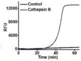
图2A为组织蛋白酶B介导的裂解反应的非变性凝胶电泳监测。条带1, 多肽-DNA结合物;条带2,多肽-DNA结合物+组织蛋白酶B。Figure 2A is a native gel electrophoresis monitoring of cathepsin B-mediated cleavage reactions.
图2B为扩增产物的非变性凝胶电泳分析。条带1,模板1;条带2,模板 2;条带3,多肽-DNA结合+模板1+模板2+Bst DNA聚合酶+限制性核酸内 切酶;条带4,多肽-DNA结合+组织蛋白酶B+模板1+模板2+Bst DNA聚合 酶+限制性核酸内切酶+信号探针+核糖核酸酶H+核糖核酸酶抑制剂。Figure 2B is the native gel electrophoresis analysis of the amplified products.
图2C为实时监测组织蛋白酶B存在和缺失时的荧光强度。组织蛋白酶B 的浓度为1×10⁻⁷克每毫升。Figure 2C is the real-time monitoring of the fluorescence intensity in the presence and absence of cathepsin B. The concentration of cathepsin B was 1×10⁻⁷ g/ml.
图3A为不同浓度组织蛋白酶B的荧光发射光谱。Figure 3A is the fluorescence emission spectra of different concentrations of cathepsin B.
图3B为在1×10-11克每毫升至5×10⁻⁷克每毫升范围内,组织蛋白酶B浓度 对荧光强度的影响。插图显示了组织蛋白酶B浓度在1×10-11到1×10⁻⁷克每毫 升范围内的对数与荧光强度之间的线性关系。Fig. 3B shows the effect of cathepsin B concentration on the fluorescence intensity in the range of 1×10-11 g/ml to 5×10⁻⁷ g/ml. The inset shows the linear relationship between the logarithm of the concentration of cathepsin B and the fluorescence intensity in the range of 1 x 10-11 to 1 x 10⁻⁷ g/ml.
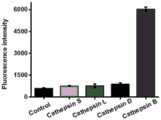
图4为反应缓冲液(对照),1×10-7克每毫升组织蛋白酶S,1×10-7克每毫升 组织蛋白酶L,1×10-7克每毫升组织蛋白酶D,1×10-7克每毫升组织蛋白酶B 的荧光强度测定。Figure 4 is the reaction buffer (control), 1×10⁻⁷ g per ml cathepsin S, 1×10⁻⁷ g per ml cathepsin L, 1×10⁻⁷ g per ml cathepsin D, 1×10- Fluorescence intensity determination of cathepsin B at7 g/mL.
图5A为在分别存在裂解缓冲液,10000个人胚胎肾细胞,10000个人宫 颈癌细胞,10000个人宫颈癌细胞加2微摩尔亮抑酶肽时荧光强度的测量。Figure 5A is the measurement of fluorescence intensity in the presence of lysis buffer, 10,000 human embryonic kidney cells, 10,000 human cervical cancer cells, and 10,000 human cervical cancer cells plus 2 micromolar leupeptin, respectively.
图5B为荧光强度与1到10000个人宫颈癌细胞的数量关系。插图显示了 组织蛋白酶B浓度在1到10000个细胞范围内的对数与荧光强度之间的线性 关系。Figure 5B is the relationship between fluorescence intensity and the number of 1 to 10,000 human cervical cancer cells. The inset shows the linear relationship between the logarithm of cathepsin B concentration and the fluorescence intensity over the range of 1 to 10,000 cells.
具体实施方式Detailed ways
应该指出,以下详细说明都是例示性的,旨在对本发明提供进一步的说 明。除非另有指明,本文使用的所有技术和科学术语具有与本发明所属技术 领域的普通技术人员通常理解的相同含义。It should be noted that the following detailed description is exemplary and intended to provide further explanation of the present invention. Unless defined otherwise, all technical and scientific terms used herein have the same meaning as commonly understood by one of ordinary skill in the art to which this invention belongs.
需要注意的是,这里所使用的术语仅是为了描述具体实施方式,而非意 图限制根据本发明的示例性实施方式。如在这里所使用的,除非上下文另外 明确指出,否则单数形式也意图包括复数形式,此外,还应当理解的是,当 在本说明书中使用术语“包含”和/或“包括”时,其指明存在特征、步骤、操作、 器件、组件和/或它们的组合。应理解,本发明的保护范围不局限于下述特 定的具体实施方案;还应当理解,本发明实施例中使用的术语是为了描述特 定的具体实施方案,而不是为了限制本发明的保护范围。It should be noted that the terminology used here is only for describing specific embodiments, and is not intended to limit exemplary embodiments according to the present invention. As used herein, unless the context clearly dictates otherwise, the singular is intended to include the plural, and it should also be understood that when the terms "comprising" and/or "comprising" are used in this specification, they mean There are features, steps, operations, means, components and/or combinations thereof. It should be understood that the protection scope of the present invention is not limited to the following specific implementations; it should also be understood that the terms used in the examples of the present invention are to describe specific implementations rather than to limit the protection scope of the present invention.
如前所述,现有针对组织蛋白酶B等酶类检测普遍存在耗时长、样品 消耗量大、制备或检测过程复杂繁琐、灵敏度差等缺点。As mentioned above, the existing detection methods for enzymes such as cathepsin B generally have disadvantages such as long time-consuming, large sample consumption, complicated and cumbersome preparation or detection process, and poor sensitivity.
有鉴于此,开发一种简单、灵敏的方法来检测组织蛋白酶B的活性是 非常必要的。将核酸引入蛋白质检测是一个很好的策略。例如,多肽结合的 氯化血红素/G-四联体(hGQ-多肽)结构被用于一系列高效电化学蛋白检测, 如癌胚抗原检测、凝血酶检测、半胱天冬酶3活性检测等。多肽探针还可以 与DNA稳定的银纳米团簇结合,DNA-银纳米团簇-多肽共轭被吸附到氧化 石墨烯表面以检测胰蛋白酶。当目标蛋白含量极低时,可以进一步引入核酸扩增技术,提高检测灵敏度。例如,当DNA-多肽偶联探针附着于固相时, 炭疽内肽酶可裂解肽段,释放DNA,引起聚合酶链反应扩增。但该方法存 在一个缺点:释放的DNA中含有一些氨基酸残基,可能会引起空间位阻效 应,降低进一步的扩增效率。因此,我们开发了一种简单的基于多重循环信 号放大的检测组织蛋白酶B活性的荧光检测方法,将含有氨基酸残基的第 一引物DNA转化成不含氨基酸残基的新引物DNA,可以完全消除核酸扩增 过程中的空间位阻效应,以增加扩增反应效率。同时,多个链置换反应的进 行及核糖核酸酶H的循环消化作用可进一步放大荧光信号。In view of this, it is necessary to develop a simple and sensitive method to detect the activity of cathepsin B. Introducing nucleic acids into protein detection is a good strategy. For example, the peptide-bound hemin/G-quadruplex (hGQ-peptide) structure is used in a series of high-efficiency electrochemical protein detection, such as carcinoembryonic antigen detection, thrombin detection,
具体的,该方法基于多肽-DNA偶合物的多重循环信号扩增。我们设计 了两个具有多个结构域的DNA模板,分别用于DNA聚合酶介导的DNA链延 伸和核酸酶介导的切割,并结合核糖核酸酶H辅助扩增,将组织蛋白酶B活 性转化为可测量的荧光信号。该方法操作简便、快速、易于操作,在放大过 程中不涉及任何热循环,整个测定过程可在2.5小时内完成,优于已有的组 织蛋白酶B测定方法。该方法具有一些不同的特点:(1)将含有氨基酸残基的第一引物DNA转化成不含氨基酸残基的新引物DNA,可以完全消除核酸 扩增过程中的空间位阻效应;(2)多个链置换反应可以诱导循环信号放大; (3)核糖核酸酶H对循环消化信号探测进一步放大荧光信号。本方法灵敏 度高,可定量测定组织蛋白酶B活性,检测限为8.1×10⁻¹²克每毫升,其动态 范围非常大,从1×10-11到1×10⁻⁷克每毫升,甚至可以在单细胞水平准确定量 组织蛋白酶B活性。该方法可用于组织蛋白酶B抑制剂的筛选,在生物医学研究和早期临床诊断中具有巨大的应用潜力。重要的是,通过合理设计相应 的探针,该方法可以作为检测其他类型蛋白酶的通用平台。Specifically, the method is based on multiple cycle signal amplification of polypeptide-DNA conjugates. We designed two DNA templates with multiple domains for DNA polymerase-mediated DNA strand elongation and nuclease-mediated cleavage, respectively, combined with RNase H-assisted amplification to convert cathepsin B activity is a measurable fluorescent signal. The method is simple, fast and easy to operate, does not involve any heat cycle during the amplification process, and the whole determination process can be completed within 2.5 hours, which is superior to the existing cathepsin B determination method. The method has some different characteristics: (1) the first primer DNA containing amino acid residues is converted into a new primer DNA without amino acid residues, which can completely eliminate the steric hindrance effect in the nucleic acid amplification process; (2) Multiple strand displacement reactions can induce circular signal amplification; (3) RNase H further amplifies the fluorescent signal by detecting the circular digestion signal. This method has high sensitivity and can quantitatively measure the activity of cathepsin B. The detection limit is 8.1×10⁻¹² g/ml, and its dynamic range is very large, from 1×10-11 to 1×10⁻⁷ g/ml, even in Accurate quantification of cathepsin B activity at the single-cell level. This method can be used for the screening of cathepsin B inhibitors, and has great application potential in biomedical research and early clinical diagnosis. Importantly, this method can serve as a general platform for detecting other types of proteases by rationally designing corresponding probes.
本发明的一个具体实施方式中,提供一种检测组织蛋白酶B的生物传 感器,所述生物传感器至少包括多肽-DNA偶联物,DNA模板1和DNA模 板2;In a specific embodiment of the present invention, a biosensor for detecting cathepsin B is provided, the biosensor at least includes a polypeptide-DNA conjugate, a
本发明的又一具体实施方式中,所述多肽-DNA偶联物含有组织蛋白酶 B的生物素化肽底物,其中肽底物C端修饰有生物素,肽底物N端与DNA 5'端连接;多肽-DNA偶联物的DNA部分作为核酸扩增反应的引物;In yet another specific embodiment of the present invention, the polypeptide-DNA conjugate contains a biotinylated peptide substrate of cathepsin B, wherein the C-terminus of the peptide substrate is modified with biotin, and the N-terminus of the peptide substrate is connected to the DNA 5' end connection; the DNA part of the polypeptide-DNA conjugate is used as a primer for nucleic acid amplification reaction;
本发明的又一具体实施方式中,所述DNA模板1包含三个主要区域 X*-Y*-Z*;In yet another specific embodiment of the present invention, the
本发明的又一具体实施方式中,所述DNA模板2包含三个主要区域 Z*-Y*-X*;In yet another specific embodiment of the present invention, the
本发明的又一具体实施方式中,其中序列X*与多肽-DNA偶联物结合 的DNA部分和生成的触发器X互补,序列Y*与生成的触发器Y互补,序 列Z*与生成的触发器Z互补。In yet another specific embodiment of the present invention, the sequence X* is complementary to the DNA portion of the polypeptide-DNA conjugate and the generated trigger X, the sequence Y* is complementary to the generated trigger Y, and the sequence Z* is complementary to the generated Flip-flop Z is complementary.
本发明的又一具体实施方式中,上述两个DNA模板中,Y*的左右两侧 分别设计了限制性核酸内切酶的识别位点。In yet another specific embodiment of the present invention, in the above two DNA templates, recognition sites for restriction endonucleases are respectively designed on the left and right sides of Y*.
本发明的又一具体实施方式中,In yet another specific embodiment of the present invention,
所述X*序列为5'-AAC AGA CTC ACT ACG ACC GGG AC-3'(SEQ ID No.1);The X* sequence is 5'-AAC AGA CTC ACT ACG ACC GGG AC-3' (SEQ ID No.1);
所述Y*序列为5'-AAC AGA CTC CAC AAA TTC GAC C-3'(SEQ ID No.2);The Y* sequence is 5'-AAC AGA CTC CAC AAA TTC GAC C-3' (SEQ ID No.2);
所述Z*序列为5'-CGT GAA TAA CTC TAC TAT C-3'(SEQ ID No.3)。The Z* sequence is 5'-CGT GAA TAA CTC TAC TAT C-3' (SEQ ID No.3).
本发明的又一具体实施方式中,In yet another specific embodiment of the present invention,
所述DNA模板1核苷酸序列为:5'-CGT GAA TAA CTC TAC TAT CAACAG AC TCCACA AAT TCG ACCAAC AGACTC ACT ACG ACC GGG AC-磷酸基团-3'(SEQ ID No.4);其中,下划线区域表示Nt.BstNBI的识别 位点;The
所述DNA模板2核苷酸序列为:5'-AAC AGA CTC ACT ACG ACC GGG ACA ACA GACTCC ACA AAT TCG ACCAAC AGA CTC CGT GAA TAA CTC TAC TAT C-磷酸基团-3'(SEQ IDNo.5);其中,下划线区域表示 Nt.BstNBI的识别位点;The
本发明的又一具体实施方式中,所述含有组织蛋白酶B的生物素化肽 底物具体为生物素修饰的KGFRLC(SEQ ID No.6)。In yet another specific embodiment of the present invention, the biotinylated peptide substrate containing cathepsin B is specifically biotin-modified KGFRLC (SEQ ID No. 6).
本发明的又一具体实施方式中,所述多肽-DNA偶联物中DNA的序列 为5'-GTC CCGGTC GTA GTG AGT CT-3'(SEQ ID No.7)。In yet another specific embodiment of the present invention, the sequence of DNA in the polypeptide-DNA conjugate is 5'-GTC CCGGTC GTA GTG AGT CT-3' (SEQ ID No.7).
本发明的又一具体实施方式中,所述多肽-DNA偶联物的序列为:Cterm- 生物素-KGFRLC-Nterm-GTC CCG GTC GTA GTG AGT CT。In yet another specific embodiment of the present invention, the sequence of the polypeptide-DNA conjugate is: Cterm -biotin-KGFRLC-Nterm -GTC CCG GTC GTA GTG AGT CT.
本发明的又一具体实施方式中,所述检测组织蛋白酶B的生物传感器 还包括信号探针,所述信号探针为一个RNA寡核苷酸,在所述RNA寡核 苷酸两端分别使用荧光剂和猝灭剂进行修饰,所述信号探针可以与触发器Z 结合,形成RNA-DNA双链。In yet another specific embodiment of the present invention, the biosensor for detecting cathepsin B also includes a signal probe, and the signal probe is an RNA oligonucleotide, which is used at both ends of the RNA oligonucleotide respectively. The fluorescent agent and the quencher are modified, and the signal probe can be combined with the trigger Z to form an RNA-DNA double strand.
所述荧光剂选自FAM,所述淬灭剂选自BHQ1;The fluorescent agent is selected from FAM, and the quencher is selected from BHQ1;
本发明的又一具体实施方式中,所述信号探针的序列为:In yet another specific embodiment of the present invention, the sequence of the signal probe is:
5'-FAM-rArUrArArCrUrCrUrArCrUrArUrC-BHQ1-3'(SEQ ID No.8)。5'-FAM-rArUrArArCrUrCrUrArCrUrArUrC-BHQ1-3' (SEQ ID No. 8).
本发明的又一具体实施方式中,所述检测组织蛋白酶B的生物传感器 还包括磁珠、DNA聚合酶和核糖核酸酶H。In yet another specific embodiment of the present invention, the biosensor for detecting cathepsin B also includes magnetic beads, DNA polymerase and ribonuclease H.
其中,所述磁珠为链霉亲和素修饰的磁珠,从而使得生物素化多肽-DNA 偶联物可通过生物素-链霉亲和素相互作用在磁珠表面进行组装;Wherein, the magnetic beads are streptavidin-modified magnetic beads, so that the biotinylated polypeptide-DNA conjugate can be assembled on the surface of the magnetic beads through biotin-streptavidin interaction;
所述DNA聚合酶用于引发链的延伸,产生丰富的X、Y、Z触发器。The DNA polymerase is used to initiate strand extension, producing abundant X, Y, Z triggers.
所述核糖核酸酶H可以选择性地消化信号探针与触发器Z结合形成的 RNA-DNA双链中的RNA寡核苷酸,产生明显的荧光团荧光信号,同时释 放Z触发器。The ribonuclease H can selectively digest the RNA oligonucleotides in the RNA-DNA duplex formed by the combination of the signal probe and the trigger Z to generate an obvious fluorescent signal of the fluorophore while releasing the Z trigger.
本发明的又一具体实施方式中,提供一种检测组织蛋白酶B的方法, 所述方法包括:In yet another specific embodiment of the present invention, a method for detecting cathepsin B is provided, the method comprising:
1)将多肽-DNA偶联物与磁珠共孵育得多肽-DNA-磁珠轭合物;1) co-incubating the polypeptide-DNA conjugate with magnetic beads to obtain a polypeptide-DNA-magnetic bead conjugate;
2)向步骤1)中加入待测样品共孵育进行切除反应,上清液磁分离待 用;2) Add the sample to be tested to the step 1) and incubate together to perform the excision reaction, and the supernatant is magnetically separated for use;
3)向步骤2)处理后的上清液中加入DNA模板1、DNA模板2和信号 探针共孵育进行酶辅助级联信号放大。3) Add
其中,in,
所述步骤1)中,所述磁珠为链霉亲和素包覆的磁珠;In the step 1), the magnetic beads are streptavidin-coated magnetic beads;
共孵育处理条件为:室温下处理10-20分钟;Co-incubation treatment conditions are: treatment at room temperature for 10-20 minutes;
所述步骤2)中,共孵育处理条件为:35~40摄氏度孵育0.5~2小时, 优选为37摄氏度孵育1小时;In the step 2), the co-incubation treatment condition is: incubate at 35-40°C for 0.5-2 hours, preferably at 37°C for 1 hour;
所述步骤3)中,共孵育处理添加为35~40摄氏度孵育0.5~2小时,优 选为37摄氏度孵育50分钟。In the step 3), the co-incubation treatment is added to incubate at 35-40 degrees Celsius for 0.5-2 hours, preferably at 37 degrees Celsius for 50 minutes.
所述检测组织蛋白酶B的方法还包括对反应产物进行实时荧光检测和/ 或凝胶电泳,以及荧光光谱测量。The method for detecting cathepsin B also includes performing real-time fluorescence detection and/or gel electrophoresis and fluorescence spectrum measurement on the reaction product.
本发明的又一具体实施方式中,提供上述生物传感器和/或检测方法在 组织蛋白酶B活性检测和/或筛选组织蛋白酶B相关药物中的应用。In yet another specific embodiment of the present invention, the application of the above-mentioned biosensor and/or detection method in cathepsin B activity detection and/or screening of cathepsin B related drugs is provided.
在所述应用中,对组织蛋白酶B活性检测的应用环境可以为外界自然环 境或者生物体体内环境,所述生物体体内环境包括生物个体、器官、组织或 细胞,可以是生物体细胞,尤其值得说明的是,本发明可实现在单个细胞中 对组织蛋白酶B的定量检测。In the application, the application environment for cathepsin B activity detection can be the external natural environment or the internal environment of the organism, and the internal environment of the organism includes a biological individual, an organ, a tissue or a cell, which can be a biological cell, especially worth It is illustrated that the present invention can realize the quantitative detection of cathepsin B in a single cell.
所述组织蛋白酶B相关药物包括但不限于组织蛋白酶B抑制剂或组织蛋 白酶B激活剂。The cathepsin B-related drugs include, but are not limited to, cathepsin B inhibitors or cathepsin B activators.
以下通过实施例对本发明做进一步解释说明,但不构成对本发明的限 制。应理解这些实施例仅用于说明本发明而不用于限制本发明的范围。实施 例1中涉及的核苷酸/氨基酸序列信息如下表所示:Below, the present invention is further explained and illustrated by examples, but does not constitute limitation of the present invention. It should be understood that these examples are only for illustrating the present invention and are not intended to limit the scope of the present invention. The nucleotide/amino acid sequence information involved in Example 1 is shown in the table below:

下划线区域表示Nt.BstNBI的识别位点;信号探针中的小写字母r代表 核糖核苷酸The underlined area indicates the recognition site of Nt.BstNBI; the lowercase letter r in the signal probe represents ribonucleotide
实施例1Example 1
药品和材料。所有寡核苷酸和焦碳酸二乙酯(DEPC)处理水均由Takara 生物技术有限公司(大连,中国)购买,多肽Cterm-生物素-KGFRLC-Nterm由 杭州中肽生化有限公司(杭州,浙江,中国)合成。组织蛋白酶B,组织蛋白 酶L,组织蛋白酶S,抗蛋白酶和亮抑酶肽是从西格玛奥德里奇公司(圣路 易斯,密苏里州,美国)购买,组织蛋白酶D从R&D系统(明尼阿波利斯,明 尼苏达州,美国)购买。链霉亲和素磁珠,Bst DNA聚合酶(大片段),10 ×ThermoPol反应缓冲液(200毫摩尔三(羟甲基)氨基甲烷-盐酸(Tris-HCl), 100毫摩尔硫酸铵((NH4)2SO4),100毫摩尔氯化钾(KCl),20毫摩尔 硫酸镁(MgSO4),1%聚乙二醇辛基苯基醚,PH 8.8),限制性核酸内切 酶(Nt.BstNBI),10×NEB缓冲液3.1(1000毫摩尔氯化钠,500毫摩尔三(羟 甲基)氨基甲烷-盐酸,100毫摩尔氯化镁(MgCl2),1毫克每毫升牛血清 白蛋白(BSA),pH 7.9),核糖核酸酶H,10×核糖核酸酶H反应缓冲液(500 毫摩尔三(羟甲基)氨基甲烷-盐酸,750毫摩尔氯化钾,30毫摩尔氯化镁, 100毫摩尔二硫苏糖醇(DTT),pH 8.3),核糖核酸酶抑制剂,脱氧核糖 核酸苷5’-三磷酸混合物(dNTPs)从新英格兰生物实验室(伊普斯威奇、马 萨诸塞州、美国)获得。SYBR Gold购买于美国生命技术公司(卡尔斯巴德, 加利福尼亚州,美国)。人宫颈癌细胞株(HeLa细胞)和人胚胎肾细胞系 (HEK-293细胞)来自中国科学院上海生物科学研究所细胞库(上海,中国), 其他分析用化学品没有进一步净化。本实验中使用的超纯水来自于微孔过滤 系统,这项涉及肽和RNA的实验使用的是经深度处理的水。本实验中,组 织蛋白酶B和细胞裂解液需要在5毫摩尔二硫苏糖醇和25毫摩尔2-(4-吗啉) 乙烷磺酸(MES)缓冲液(pH 5.0)中在37摄氏度预激活10分钟,酶解反应 才能充分发挥活性。medicines and materials. All oligonucleotides and diethylpyrocarbonate (DEPC) treated water were purchased from Takara Biotechnology Co., Ltd. (Dalian, China), and peptide Cterm -biotin-KGFRLC-Nterm was purchased from Hangzhou Zhongpei Biochemical Co., Ltd. (Hangzhou, China). , Zhejiang, China) synthesis. Cathepsin B, cathepsin L, cathepsin S, antiprotease and leupeptin were purchased from Sigma-Aldrich (St. Louis, MO, USA), and cathepsin D was purchased from R&D Systems (Minneapolis, MN States, United States) to purchase. Streptavidin magnetic beads, Bst DNA polymerase (large fragment), 10 × ThermoPol reaction buffer (200 mmol tris(hydroxymethyl)aminomethane-hydrochloric acid (Tris-HCl), 100 mmol ammonium sulfate (( NH4 )2 SO4 ), 100 mmol potassium chloride (KCl), 20 mmol magnesium sulfate (MgSO4 ), 1% polyethylene glycol octylphenyl ether, pH 8.8), restriction endonuclease (Nt.BstNBI), 10×NEB buffer 3.1 (1000 mmol sodium chloride, 500 mmol tris(hydroxymethyl)aminomethane-hydrochloric acid, 100 mmol magnesium chloride (MgCl2 ), 1 mg per ml bovine serum white protein (BSA), pH 7.9), RNase H, 10× RNase H reaction buffer (500 mmol tris(hydroxymethyl)aminomethane-hydrochloric acid, 750 mmol potassium chloride, 30 mmol magnesium chloride, 100 mmol dithiothreitol (DTT, pH 8.3), ribonuclease inhibitor, deoxyribonucleoside 5'-triphosphate mixture (dNTPs) from New England Biolabs (Ipswich, MA, United States) obtained. SYBR Gold was purchased from American Life Technologies (Carlsbad, CA, USA). Human cervical cancer cell lines (HeLa cells) and human embryonic kidney cell lines (HEK-293 cells) were obtained from the Cell Bank of Shanghai Institute of Biological Sciences, Chinese Academy of Sciences (Shanghai, China), and other analytical chemicals were not further purified. The ultrapure water used in this experiment came from a microfiltration system, and this experiment involving peptides and RNA used highly treated water. In this experiment, cathepsin B and cell lysate need to be pre-treated at 37 °C in 5 mmol dithiothreitol and 25 mmol 2-(4-morpholine)ethanesulfonic acid (MES) buffer (pH 5.0). After activating for 10 minutes, the enzymatic hydrolysis reaction can fully exert its activity.
多肽-DNA偶合物多个循环信号扩增反应。本实验包括三个步骤。首先, 用经焦碳酸二乙酯处理的水稀释多肽-DNA偶联物,制备原液。20微升链霉 亲和素包覆的磁珠溶液(4毫克每毫升)被转移至一个200微升的离心管中, 用1×B&W缓冲液(5毫摩尔三(羟甲基)氨基甲烷-盐酸,pH 7.5,0.5毫摩 尔乙二胺四乙酸(EDTA),1摩尔氯化钠)洗涤两次,然后磁珠在20微升 的2×B&W缓冲液(10毫摩尔三(羟甲基)氨基甲烷-盐酸,pH 7.5,1毫摩尔 乙二胺四乙酸,2摩尔氯化钠)中被重悬,30微升1微摩尔生物素化的多肽 -DNA轭合物被添加到溶液中,室温旋转孵育15分钟。混合物用50微升 1×B&W缓冲液洗涤三次以移除未耦合的多肽-DNA轭合物,剩余的多肽 -DNA-磁珠轭合物被分散在20微升焦碳酸二乙酯处理水中。第二步,5微升 多肽-DNA-磁珠轭合物被添加至20微升的切除反应体系(包括1×组织蛋白酶B反应缓冲液和不同浓度的活性组织蛋白酶B),这个体系在37摄氏度孵育1 小时,进行多肽-DNA偶联裂解,上清液磁分离3分钟待用。第三步,2微升 反应产物添加到20微升循环信号放大反应系统(包括25纳摩尔模板1,25纳 摩尔模板2,500微摩尔脱氧核糖核酸苷5’-三磷酸混合物,2.4个单位的 Bst.DNA聚合酶,10个单位的限制性核酸内切酶,700纳摩尔信号探针,1个 单位的核糖核酸酶H,20个单位的核糖核酸酶抑制剂,2微升10×NEB缓冲液 3.1,2微升10×ThermoPol缓冲液,2微升10×核糖核酸酶缓冲液),然后混合 物在37摄氏度孵育50分钟用于酶辅助级联信号放大。Polypeptide-DNA conjugate multiple cycle signal amplification reaction. This experiment consists of three steps. First, prepare a stock solution by diluting the polypeptide-DNA conjugate with diethylpyrocarbonate-treated water. 20 μl of the streptavidin-coated magnetic bead solution (4 mg/ml) was transferred to a 200 μl centrifuge tube and washed with 1×B&W buffer (5 mmol tris(hydroxymethyl)aminomethane - hydrochloric acid, pH 7.5, 0.5 millimolar ethylenediaminetetraacetic acid (EDTA), 1 mole sodium chloride) washed twice, and then the magnetic beads were washed twice in 20 microliters of 2 × B&W buffer (10 millimolar ) aminomethane-hydrochloric acid, pH 7.5, 1 mmol EDTA, 2 M NaCl), and 30 μl of 1 μM biotinylated polypeptide-DNA conjugate was added to the solution , and incubate with rotation for 15 minutes at room temperature. The mixture was washed three times with 50 μl of 1×B&W buffer to remove uncoupled peptide-DNA conjugates, and the remaining peptide-DNA-magnetic bead conjugates were dispersed in 20 μl of diethylpyrocarbonate-treated water. In the second step, 5 μl of peptide-DNA-magnetic bead conjugate was added to 20 μl of the excision reaction system (including 1× cathepsin B reaction buffer and different concentrations of active cathepsin B), which was activated at 37 Incubate for 1 hour at 100°C for peptide-DNA coupling lysis, and magnetically separate the supernatant for 3 minutes before use. In the third step, 2 microliters of reaction products were added to 20 microliters of cyclic signal amplification reaction system (including 25
实时荧光检测和凝胶电泳。使用BIO-RAD CFX连接TM实时系统(加利 福尼亚,美国)每隔30秒对循环信号放大进行实时荧光监测,荧光指示剂为 1×SYBR Gold。采用14%非变性聚丙烯酰胺凝胶电泳(PAGE)对1×四溴乙 烷缓冲液(9毫摩尔三(羟甲基)氨基甲烷-盐酸,9毫摩尔硼酸,0.2毫摩尔 PH值为7.9的乙二胺四乙酸)中在110伏的恒定电压下,在室温下,在黑暗中 工作60分钟的反应产物进行了分析,荧光指示剂为1×SYBR Gold。Real-time fluorescence detection and gel electrophoresis. The BIO-RAD CFX connection TM real-time system (California, USA) was used for real-time fluorescence monitoring of the cycle signal amplification every 30 seconds, and the fluorescent indicator was 1×SYBR Gold. Using 14% non-denaturing polyacrylamide gel electrophoresis (PAGE) for 1 × tetrabromoethane buffer (9 mmol tris (hydroxymethyl) aminomethane - hydrochloric acid, 9 mmol boric acid, 0.2 mmol pH value of 7.9 EDTA) at a constant voltage of 110 volts, at room temperature, the reaction product was analyzed in the dark for 60 minutes, and the fluorescent indicator was 1×SYBR Gold.
荧光光谱测量。20微升反应产物用超纯水稀释到60微升。荧光信号采 用日立F-7000荧光分光光度计(东京,日本)测定,激发波长为490纳米, 发射光谱扫描范围为500到650纳米,数据分析采用发射波长520纳米处 的荧光强度。Fluorescence spectroscopy measurements. 20 µl of the reaction product was diluted to 60 µl with ultrapure water. The fluorescence signal was measured by a Hitachi F-7000 fluorescence spectrophotometer (Tokyo, Japan), the excitation wavelength was 490 nm, the emission spectrum scanning range was 500 to 650 nm, and the data analysis used the fluorescence intensity at the emission wavelength of 520 nm.
细胞培养及细胞提取物的制备。人类子宫颈癌细胞和人类胚胎肾细胞培 养基为含有10%胎牛血清(FBS)和1%青霉素-链霉素的达尔伯克改良伊格 尔培养基(DMEM),放在含有5%二氧化碳,37摄氏度的培养箱中进行培 养。用胰蛋白酶化法收集生长指数期的细胞。培养基被移除,用PH为7.4的 冷1×聚丁二酸丁二醇酯(PBS)溶液洗涤细胞2次,用1000转每分钟离心5分 钟。100微升裂解缓冲液(10毫摩尔三(羟甲基)氨基甲烷-盐酸,pH 8.0,150毫摩尔氯化钠,1%(质量/体积)乙基苯基聚乙二醇(NP-40),0.25毫 摩尔脱氧胆酸钠,1%(质量/体积)甘油,0.1毫摩尔4-(2-氨基乙基)苯基 磺酰盐酸盐)被用于悬浮细胞,然后将混合物在冰上孵育30分钟,然后在4 摄氏度下用12000转每分钟离心20分钟。将上清液小心地转移到新鲜试管中, pH值小心地调整到5.5。Cell culture and preparation of cell extracts. Human cervical cancer cells and human embryonic kidney cells were cultured in Dulbecco's modified Eagle's medium (DMEM) containing 10% fetal bovine serum (FBS) and 1% penicillin-streptomycin, placed in 5% carbon dioxide , cultured in a 37°C incubator. Cells in exponential phase of growth were harvested by trypsinization. The medium was removed, the cells were washed twice with cold 1X polybutylene succinate (PBS) solution, pH 7.4, and centrifuged at 1000 rpm for 5 minutes. 100 μl of lysis buffer (10 mmol tris(hydroxymethyl)aminomethane-hydrochloric acid, pH 8.0, 150 mmol sodium chloride, 1% (mass/volume) ethylphenyl polyethylene glycol (NP-40 ), 0.25 mmol sodium deoxycholate, 1% (mass/volume) glycerol, 0.1 mmol 4-(2-aminoethyl) phenylsulfonyl hydrochloride) were used to suspend cells, and then the mixture was placed on ice Incubate for 30 minutes, then centrifuge at 12,000 rpm for 20 minutes at 4°C. Carefully transfer the supernatant to a fresh tube and carefully adjust the pH to 5.5.
组织蛋白酶B测定的原理如图1所示。我们设计了一种生物素化多肽 -DNA偶联物,该偶联物含有组织蛋白酶B的生物素化肽底物(生物素 -KGFRLC),N端与5’端DNA连接形成多肽-DNA偶联物。多肽-DNA偶合物 的DNA部分可作为核酸扩增反应的引物。生物素化多肽-DNA偶联物可通过 生物素-链霉亲和素相互作用在链霉亲和素修饰磁珠表面组装。在组织蛋白 酶B的存在下,可以识别并裂解肽KGFRLC,导致DNA部分从磁珠中解离。 经磁性分离后,释放的DNA部分可在两个线性模板(即模板1和模板2)。 模板1包含三个主要区域X*-Y*-Z*,模板2包含三个主要区域Z*-Y*-X*,其 中序列X*与多肽-DNA结合的DNA部分和生成的触发器X互补,序列Y*与生 成的触发器Y互补,序列Z*与生成的触发器Z互补。此外,在两个模板中, Y*的左右两侧分别设计了限制性核酸内切酶的识别位点。所释放的DNA片 段与模板1的杂交可在DNA聚合酶和脱氧核糖核酸苷5’-三磷酸混合物的存 在下引发延伸反应,产生丰富的具有两个识别位点的DNA双链。在DNA聚 合酶和脱氧核糖核酸苷5’-三磷酸混合物的作用下,DNA双链断裂可能引发 新的延伸,形成链置换反应,产生丰富的Y、Z触发器。释放的触发器Z可能 与模板2杂交,引发链位移扩增,产生更多的触发器X和Y;被释放的触发器 Y可以与模板1或模板2杂交,从而使链的位移放大产生更多的触发器X和Z。 然后释放的触发器X、Y、Z可以与它们相应的模板杂交,诱导新的扩展、切 割循环,产生丰富的Z。通过多链置换反应,将第一个带有氨基酸残基的DNA 引物转化成不含任何氨基酸残基的X、Y、Z等新的DNA引物,完全消除了 空间位阻效应,大大提高了扩增效率。所得到的触发器Z可以作为报告探针, 与信号探针结合,信号探针是一个RNA寡核苷酸,在5’和3’端分别用荧光团(FAM)和猝灭剂(BHQ1)修饰,形成RNA-DNA双链。然后核糖核酸酶 H可以选择性地消化RNA-DNA双链的RNA寡核苷酸,产生明显的荧光团荧 光信号,同时释放Z触发器。释放的触发器Z可以进一步与新的信号探针结 合,启动新的消化和释放的循环,从而增强荧光团荧光。而在没有组织蛋白 酶B的情况下,既不会发生多肽底物的裂解反应,也不会发生链位移放大反 应,因此没有荧光增强现象。The principle of cathepsin B assay is shown in Figure 1. We designed a biotinylated peptide-DNA conjugate, which contains a biotinylated peptide substrate of cathepsin B (biotin-KGFRLC), and the N-terminus is connected to the 5'-terminal DNA to form a peptide-DNA conjugate. United things. The DNA portion of the polypeptide-DNA conjugate serves as a primer for nucleic acid amplification reactions. Biotinylated peptide-DNA conjugates can be assembled on the surface of streptavidin-modified magnetic beads through biotin-streptavidin interaction. In the presence of cathepsin B, the peptide KGFRLC can be recognized and cleaved, resulting in partial dissociation of the DNA from the beads. After magnetic separation, the released DNA fraction can be separated between two linear templates (
1、原理的实验验证1. Experimental verification of the principle
为了证明该方法的可行性,我们以SYBR Gold为荧光指示剂,使用14% 非变性凝胶电泳分析组织蛋白酶B的切割过程(图1)。在没有组织蛋白酶B 的情况下,只观察到多肽-DNA结合带(条带1,图2A)。在存在组织蛋白 酶B的情况下,观察到一个较短的条带,因为该肽被组织蛋白酶B裂解(条 带2,图2A),这表明该DNA底物可以被组织蛋白酶B裂解。我们进一步使 用14%的非变性凝胶电泳来分析反应产物(图1)。在限制性核酸内切酶切 开反应产物后,在组织蛋白酶B(条带4,图2B)的存在下,观察到19nt、 22nt、23nt、28nt的几个特征性条带,提示组织蛋白酶B可引发延伸反应,从 而诱导链移位反应。在没有组织蛋白酶B的情况下,观察到两条带分别为64nt 和74nt(条带3,图2B),分别对应于模板1(条带1,图2B)和模板2(条带 2,图2B),但是并没有出现明显的解理产物的特征条带。我们以SYBR Gold为荧光指示剂,实时荧光测量法对扩增反应过程进行监测(图2C)。在组 织蛋白酶B存在的情况下,在520纳米发射波长处检测到明显的荧光信号。 对照组未检测到组织蛋白酶B,未检测到明显的荧光信号。这些结果表明, 组织蛋白酶B可以切割特定的肽序列,诱导基于多链置换反应的循环信号放 大反应。In order to prove the feasibility of this method, we used SYBR Gold as a fluorescent indicator, and used 14% non-denaturing gel electrophoresis to analyze the cleavage process of cathepsin B (Figure 1). In the absence of cathepsin B, only the polypeptide-DNA binding band was observed (
2、灵敏度实验2. Sensitivity experiment
为了研究该方法的敏感性,我们在最佳反应条件下监测了不同浓度的组 织蛋白酶B对荧光强度的影响。如图3所示,随着组织蛋白酶B浓度的增加, 荧光强度从1×10-11克每毫升增加到1×10⁻⁷克每毫升,在5×10⁻⁷克每毫升以上 达到平台,在1×10-11到1×10⁻⁷克每毫升范围内,组织蛋白酶B浓度的对数与 荧光强度呈线性相关(图3B插图)。相关方程为F=1404.5log10 C+16271.2 (R2=0.9940),其中C为组织蛋白酶B的浓度(克每毫升),F为荧光强 度。通过计算平均对照组加3倍标准差,检测限为8.1×10⁻¹²克每毫升。本方 法的灵敏度比电化学方法(2.4×10⁻⁶克每毫升)提高了5个数量级,比比色法 (2.2×10⁻⁷克每毫升)提高了4个数量级。此外,本方法对组织蛋白酶S、组 织蛋白酶L和组织蛋白酶D的响应具有较高的特异性,且无明显信号。To investigate the sensitivity of this method, we monitored the effect of different concentrations of cathepsin B on the fluorescence intensity under optimal reaction conditions. As shown in Figure 3, with the increase of cathepsin B concentration, the fluorescence intensity increased from 1×10-11 g/ml to 1×10⁻⁷ g/ml, and reached a plateau above 5×10⁻⁷ g/ml, In the range of 1 x 10-11 to 1 x 10⁻⁷ g/ml, the logarithm of cathepsin B concentration was linearly related to the fluorescence intensity (Figure 3B inset). The related equation is F=1404.5log10 C+16271.2 (R2 =0.9940), where C is the concentration of cathepsin B (grams per milliliter), and F is the fluorescence intensity. By calculating the mean of the control group plus 3 times the standard deviation, the detection limit was 8.1×10⁻¹² g/ml. The sensitivity of the method is improved by 5 orders of magnitude compared with the electrochemical method (2.4×10⁻⁶ grams per milliliter), and by 4 orders of magnitude compared with the colorimetric method (2.2×10⁻⁷ grams per milliliter). In addition, this method has high specificity in the response to cathepsin S, cathepsin L and cathepsin D, and there is no obvious signal.
3、特异性实验3. Specificity experiment
为了评价该方法的检测特异性,我们使用了组织蛋白酶S、组织蛋白酶 L和组织蛋白酶D作为非特异性蛋白。如图4所示,显著的荧光信号的发射波 长520纳米是观察到的组织蛋白酶B,但没有增强的荧光信号检测的组织蛋 白酶S,组织蛋白酶L和组织蛋白酶D,以上结果表明该方法可以很好的区分 组织蛋白酶B和其它蛋白酶,证明本技术方案具有很好的特异性。To evaluate the detection specificity of this method, we used cathepsin S, cathepsin L and cathepsin D as non-specific proteins. As shown in Figure 4, a significant fluorescence signal at an emission wavelength of 520 nm was observed for cathepsin B, but no enhanced fluorescence signal was detected for cathepsin S, cathepsin L, and cathepsin D. The above results indicate that the method can be well The good distinction between cathepsin B and other proteases proves that the technical solution has good specificity.
应注意的是,以上实例仅用于说明本发明的技术方案而非对其进行限 制。尽管参照所给出的实例对本发明进行了详细说明,但是本领域的普通 技术人员可根据需要对本发明的技术方案进行修改或者等同替换,而不脱 离本发明技术方案的精神和范围。It should be noted that the above examples are only used to illustrate the technical solutions of the present invention and not to limit them. Although the present invention has been described in detail with reference to the given examples, those skilled in the art can modify or equivalently replace the technical solutions of the present invention as required without departing from the spirit and scope of the technical solutions of the present invention.
SEQUENCE LISTINGSEQUENCE LISTING
<110> 山东师范大学<110> Shandong Normal University
<120> 一种检测组织蛋白酶B的生物传感器及其检测方法和应用<120> A Biosensor for Detecting Cathepsin B and Its Detection Method and Application
<130><130>
<160> 8<160> 8
<170> PatentIn version 3.3<170> PatentIn version 3.3
<210> 1<210> 1
<211> 23<211> 23
<212> DNA<212>DNA
<213> 人工序列<213> Artificial sequence
<400> 1<400> 1
aacagactca ctacgaccgg gac 23aacagactca ctacgaccgg gac 23
<210> 2<210> 2
<211> 22<211> 22
<212> DNA<212>DNA
<213> 人工序列<213> Artificial sequence
<400> 2<400> 2
aacagactcc acaaattcga cc 22aacagactcc acaaattcga cc 22
<210> 3<210> 3
<211> 19<211> 19
<212> DNA<212>DNA
<213> 人工序列<213> Artificial sequence
<400> 3<400> 3
cgtgaataac tctactatc 19cgtgaataac tctactatc 19
<210> 4<210> 4
<211> 64<211> 64
<212> DNA<212>DNA
<213> 人工序列<213> Artificial sequence
<400> 4<400> 4
cgtgaataac tctactatca acagactcca caaattcgac caacagactc actacgaccg 60cgtgaataac tctactatca acagactcca caaattcgac caacagactc actacgaccg 60
ggac 64ggac 64
<210> 5<210> 5
<211> 73<211> 73
<212> DNA<212>DNA
<213> 人工序列<213> Artificial sequence
<400> 5<400> 5
aacagactca ctacgaccgg gacaacagac tccacaaatt cgaccaacag actccgtgaa 60aacagactca ctacgaccgg gacaacagac tccacaaatt cgaccaacag actccgtgaa 60
taactctact atc 73taactctact atc 73
<210> 6<210> 6
<211> 6<211> 6
<212> PRT<212> PRT
<213> 人工序列<213> Artificial sequence
<400> 6<400> 6
Lys Gly Phe Arg Leu CysLys Gly Phe Arg Leu Cys
1 51 5
<210> 7<210> 7
<211> 20<211> 20
<212> DNA<212>DNA
<213> 人工序列<213> Artificial sequence
<400> 7<400> 7
gtcccggtcg tagtgagtct 20
<210> 8<210> 8
<211> 14<211> 14
<212> RNA<212> RNA
<213> 人工序列<213> Artificial sequence
<400> 8<400> 8
auaacucuac uauc 14auaacucuac uauc 14
Claims (14)
Translated fromChinesePriority Applications (1)
| Application Number | Priority Date | Filing Date | Title |
|---|---|---|---|
| CN202010042539.9ACN111172235B (en) | 2020-01-15 | 2020-01-15 | Biosensor for detecting cathepsin B and detection method and application thereof |
Applications Claiming Priority (1)
| Application Number | Priority Date | Filing Date | Title |
|---|---|---|---|
| CN202010042539.9ACN111172235B (en) | 2020-01-15 | 2020-01-15 | Biosensor for detecting cathepsin B and detection method and application thereof |
Publications (2)
| Publication Number | Publication Date |
|---|---|
| CN111172235A CN111172235A (en) | 2020-05-19 |
| CN111172235Btrue CN111172235B (en) | 2023-03-21 |
Family
ID=70648027
Family Applications (1)
| Application Number | Title | Priority Date | Filing Date |
|---|---|---|---|
| CN202010042539.9AActiveCN111172235B (en) | 2020-01-15 | 2020-01-15 | Biosensor for detecting cathepsin B and detection method and application thereof |
Country Status (1)
| Country | Link |
|---|---|
| CN (1) | CN111172235B (en) |
Families Citing this family (4)
| Publication number | Priority date | Publication date | Assignee | Title |
|---|---|---|---|---|
| CN111979295B (en)* | 2020-08-13 | 2023-05-09 | 山东师范大学 | A kind of tyrosine phosphatase biosensor and its detection method and application |
| CN112080550B (en)* | 2020-08-21 | 2023-06-06 | 山东师范大学 | A biosensor for detecting matrix metalloproteinases and its application |
| CN112903641A (en)* | 2021-01-19 | 2021-06-04 | 山东师范大学 | Biosensor for detecting histone modification enzyme and detection method and application thereof |
| CN117969625B (en)* | 2024-01-05 | 2025-04-01 | 青岛科技大学 | Photoelectrochemical aptamer sensor based on polypeptide-nucleic acid conjugate and its preparation method and application |
Citations (2)
| Publication number | Priority date | Publication date | Assignee | Title |
|---|---|---|---|---|
| CN101730746A (en)* | 2007-07-06 | 2010-06-09 | 帕普斯特许可有限两合公司 | Determination of the activity of proteases |
| CN107356756A (en)* | 2017-06-12 | 2017-11-17 | 广州医科大学附属第五医院 | A kind of fluorescence probe and its synthetic method and the application in circulating tumor cell detection |
- 2020
- 2020-01-15CNCN202010042539.9Apatent/CN111172235B/enactiveActive
Patent Citations (2)
| Publication number | Priority date | Publication date | Assignee | Title |
|---|---|---|---|---|
| CN101730746A (en)* | 2007-07-06 | 2010-06-09 | 帕普斯特许可有限两合公司 | Determination of the activity of proteases |
| CN107356756A (en)* | 2017-06-12 | 2017-11-17 | 广州医科大学附属第五医院 | A kind of fluorescence probe and its synthetic method and the application in circulating tumor cell detection |
Non-Patent Citations (2)
| Title |
|---|
| Biosensors for epigenetic biomarkers detection A review;Chen-chen Li等;《Biosensors and Bioelectronics》;20190911;第144卷;第1-11页* |
| Lysosome-Targeting Fluorogenic Probe for Cathepsin B Imaging in Living Cells;Yuqi Wang等;《Anal Chem.》;20161220;第88卷(第24期);第12403-12410页* |
Also Published As
| Publication number | Publication date |
|---|---|
| CN111172235A (en) | 2020-05-19 |
Similar Documents
| Publication | Publication Date | Title |
|---|---|---|
| CN111172235B (en) | Biosensor for detecting cathepsin B and detection method and application thereof | |
| US7642056B2 (en) | Method and kit for detecting a target protein using a DNA aptamer | |
| US9297047B2 (en) | Molecular beacon based assay for the detection of biomarkers for breast cancer metastasis | |
| Cao et al. | Naked-eye sensitive detection of nuclease activity using positively-charged gold nanoparticles as colorimetric probes | |
| CN105755101A (en) | Method for detecting DNA (deoxyribonucleic acid) glycosylase activity on basis of single quantum dot level | |
| CN109055498A (en) | MiRNA and/or biological micromolecule detection probe, detection method and kit based on the dendritic rolling ring responsive transcription of hypertree | |
| Yu et al. | An all-in-one telomerase assay based on CRISPR-Cas12a trans-cleavage while telomere synthesis | |
| CN114250272A (en) | A CRISPR-based fluorescent biosensor and its application in DNA glycosylase detection | |
| CN110408680A (en) | Single quantum dot nanosensors based on ligase amplification reaction catalytic assembly for detection of alkaline phosphatase and its applications | |
| CN107389646B (en) | A kind of fluorescence chemical sensor and its detection method detecting transcription factor NF-KB p50 | |
| CN111304298B (en) | Caspase biosensor and application, detection method of caspase activity | |
| Li et al. | Electrophoresis separation assisted G-quadruplex DNAzyme-based chemiluminescence signal amplification strategy on a microchip platform for highly sensitive detection of microRNA | |
| Ashraf et al. | Extension of duplex specific nuclease sensing application with RNA aptamer | |
| Zhou et al. | A conformational switch-based fluorescent biosensor for homogeneous detection of telomerase activity | |
| CN115232865A (en) | Biosensors, compositions, kits and uses based on aptamers and CRISPR/Cas12a systems | |
| Chen et al. | Target-triggered stochastic DNAzyme motors on spherical nucleic acids for simultaneous fluorescence assay of double miRNAs | |
| CN112501257A (en) | Visualization sensor based on nucleic acid self-assembly enzyme-catalysis-free circRNA living cell imaging | |
| Zhang et al. | Target-activated T7 transcription circuit-mediated multiple cycling signal amplification for monitoring of flap endonuclease 1 activity in cancer cells | |
| Zhao et al. | Sensitive detection of MiRNA and CircRNA through DSN enzyme cooperating NEase assisted dual signal amplification | |
| Wen et al. | Construction of a fluorescence sensing platform based on RISC and CRISPR/Cas12a for the assay of Ago2 enzyme activity | |
| Wu et al. | Endoprotein-activating DNAzyme assay for nucleic acid extraction-and amplification-free detection of viable pathogenic bacteria | |
| CN114350853A (en) | Kit and method for detecting virus and mutant thereof | |
| Chen et al. | Sensitive fluorescence detection of pathogens based on target nucleic acid sequence-triggered transcription | |
| Chen et al. | A protein triggering exponential amplification reaction enables label-and wash-free one-pot protein assay with high sensitivity | |
| CN112903641A (en) | Biosensor for detecting histone modification enzyme and detection method and application thereof |
Legal Events
| Date | Code | Title | Description |
|---|---|---|---|
| PB01 | Publication | ||
| PB01 | Publication | ||
| SE01 | Entry into force of request for substantive examination | ||
| SE01 | Entry into force of request for substantive examination | ||
| GR01 | Patent grant | ||
| GR01 | Patent grant | ||
| TR01 | Transfer of patent right | Effective date of registration:20240925 Address after:518000 floors 3 and 4, building B, Yinxing Zhijie phase II, No. 1301-76, sightseeing Road, Guanlan street, Longhua District, Shenzhen, Guangdong Patentee after:Shenzhen Jiyin biomedical Transformation Research Institute Country or region after:China Address before:250014 No. 88, Wenhua East Road, Lixia District, Shandong, Ji'nan Patentee before:SHANDONG NORMAL University Country or region before:China | |
| TR01 | Transfer of patent right |