CN111012318B - Surface focusing array detector and system for photoacoustic breast imaging - Google Patents
Surface focusing array detector and system for photoacoustic breast imagingDownload PDFInfo
- Publication number
- CN111012318B CN111012318BCN202010056116.2ACN202010056116ACN111012318BCN 111012318 BCN111012318 BCN 111012318BCN 202010056116 ACN202010056116 ACN 202010056116ACN 111012318 BCN111012318 BCN 111012318B
- Authority
- CN
- China
- Prior art keywords
- array
- piezoelectric element
- focusing
- shell
- array detector
- Prior art date
- Legal status (The legal status is an assumption and is not a legal conclusion. Google has not performed a legal analysis and makes no representation as to the accuracy of the status listed.)
- Active
Links
- 238000003384imaging methodMethods0.000titleclaimsabstractdescription27
- 210000000481breastAnatomy0.000titleclaimsdescription16
- 239000000523sampleSubstances0.000claimsabstractdescription19
- 238000001514detection methodMethods0.000claimsdescription9
- 238000007789sealingMethods0.000claimsdescription4
- 239000002033PVDF binderSubstances0.000claimsdescription3
- 239000000919ceramicSubstances0.000claimsdescription3
- 229920002981polyvinylidene fluoridePolymers0.000claimsdescription3
- 238000002604ultrasonographyMethods0.000claims1
- 208000026310Breast neoplasmDiseases0.000abstractdescription10
- 206010006187Breast cancerDiseases0.000abstractdescription8
- 238000003325tomographyMethods0.000abstractdescription6
- 238000003745diagnosisMethods0.000abstractdescription5
- 210000005075mammary glandAnatomy0.000abstractdescription5
- 238000004393prognosisMethods0.000abstractdescription5
- 210000001519tissueAnatomy0.000description4
- 208000030270breast diseaseDiseases0.000description2
- 238000000034methodMethods0.000description2
- 206010010356Congenital anomalyDiseases0.000description1
- 206010058314DysplasiaDiseases0.000description1
- ZOKXTWBITQBERF-UHFFFAOYSA-NMolybdenumChemical compound[Mo]ZOKXTWBITQBERF-UHFFFAOYSA-N0.000description1
- 230000009286beneficial effectEffects0.000description1
- 210000004204blood vesselAnatomy0.000description1
- 230000008195breast developmentEffects0.000description1
- 201000011510cancerDiseases0.000description1
- 238000010586diagramMethods0.000description1
- 201000010099diseaseDiseases0.000description1
- 208000037265diseases, disorders, signs and symptomsDiseases0.000description1
- 239000003822epoxy resinSubstances0.000description1
- 210000004907glandAnatomy0.000description1
- 239000011521glassSubstances0.000description1
- 208000027866inflammatory diseaseDiseases0.000description1
- 238000003331infrared imagingMethods0.000description1
- 230000003902lesionEffects0.000description1
- 210000002751lymphAnatomy0.000description1
- 239000000463materialSubstances0.000description1
- 239000007769metal materialSubstances0.000description1
- 238000012986modificationMethods0.000description1
- 230000004048modificationEffects0.000description1
- 229910052750molybdenumInorganic materials0.000description1
- 239000011733molybdenumSubstances0.000description1
- 210000002445nippleAnatomy0.000description1
- 238000013421nuclear magnetic resonance imagingMethods0.000description1
- 230000010355oscillationEffects0.000description1
- 239000004033plasticSubstances0.000description1
- 229920000647polyepoxidePolymers0.000description1
- 239000002861polymer materialSubstances0.000description1
- 230000001681protective effectEffects0.000description1
- 238000007493shaping processMethods0.000description1
- 238000006467substitution reactionMethods0.000description1
Images

Classifications
- A—HUMAN NECESSITIES
- A61—MEDICAL OR VETERINARY SCIENCE; HYGIENE
- A61B—DIAGNOSIS; SURGERY; IDENTIFICATION
- A61B5/00—Measuring for diagnostic purposes; Identification of persons
- A61B5/0093—Detecting, measuring or recording by applying one single type of energy and measuring its conversion into another type of energy
- A61B5/0095—Detecting, measuring or recording by applying one single type of energy and measuring its conversion into another type of energy by applying light and detecting acoustic waves, i.e. photoacoustic measurements
- A—HUMAN NECESSITIES
- A61—MEDICAL OR VETERINARY SCIENCE; HYGIENE
- A61B—DIAGNOSIS; SURGERY; IDENTIFICATION
- A61B5/00—Measuring for diagnostic purposes; Identification of persons
- A61B5/0059—Measuring for diagnostic purposes; Identification of persons using light, e.g. diagnosis by transillumination, diascopy, fluorescence
- A61B5/0082—Measuring for diagnostic purposes; Identification of persons using light, e.g. diagnosis by transillumination, diascopy, fluorescence adapted for particular medical purposes
- A61B5/0091—Measuring for diagnostic purposes; Identification of persons using light, e.g. diagnosis by transillumination, diascopy, fluorescence adapted for particular medical purposes for mammography
- A—HUMAN NECESSITIES
- A61—MEDICAL OR VETERINARY SCIENCE; HYGIENE
- A61B—DIAGNOSIS; SURGERY; IDENTIFICATION
- A61B5/00—Measuring for diagnostic purposes; Identification of persons
- A61B5/43—Detecting, measuring or recording for evaluating the reproductive systems
- A61B5/4306—Detecting, measuring or recording for evaluating the reproductive systems for evaluating the female reproductive systems, e.g. gynaecological evaluations
- A61B5/4312—Breast evaluation or disorder diagnosis
- A—HUMAN NECESSITIES
- A61—MEDICAL OR VETERINARY SCIENCE; HYGIENE
- A61B—DIAGNOSIS; SURGERY; IDENTIFICATION
- A61B8/00—Diagnosis using ultrasonic, sonic or infrasonic waves
- A61B8/08—Clinical applications
- A61B8/0825—Clinical applications for diagnosis of the breast, e.g. mammography
- A—HUMAN NECESSITIES
- A61—MEDICAL OR VETERINARY SCIENCE; HYGIENE
- A61B—DIAGNOSIS; SURGERY; IDENTIFICATION
- A61B8/00—Diagnosis using ultrasonic, sonic or infrasonic waves
- A61B8/44—Constructional features of the ultrasonic, sonic or infrasonic diagnostic device
- A61B8/4444—Constructional features of the ultrasonic, sonic or infrasonic diagnostic device related to the probe
- A—HUMAN NECESSITIES
- A61—MEDICAL OR VETERINARY SCIENCE; HYGIENE
- A61B—DIAGNOSIS; SURGERY; IDENTIFICATION
- A61B8/00—Diagnosis using ultrasonic, sonic or infrasonic waves
- A61B8/44—Constructional features of the ultrasonic, sonic or infrasonic diagnostic device
- A61B8/4483—Constructional features of the ultrasonic, sonic or infrasonic diagnostic device characterised by features of the ultrasound transducer
- A61B8/4488—Constructional features of the ultrasonic, sonic or infrasonic diagnostic device characterised by features of the ultrasound transducer the transducer being a phased array
Landscapes
- Health & Medical Sciences (AREA)
- Life Sciences & Earth Sciences (AREA)
- Physics & Mathematics (AREA)
- Medical Informatics (AREA)
- Surgery (AREA)
- Biophysics (AREA)
- Pathology (AREA)
- Engineering & Computer Science (AREA)
- Biomedical Technology (AREA)
- Heart & Thoracic Surgery (AREA)
- Veterinary Medicine (AREA)
- Molecular Biology (AREA)
- Public Health (AREA)
- Animal Behavior & Ethology (AREA)
- General Health & Medical Sciences (AREA)
- Nuclear Medicine, Radiotherapy & Molecular Imaging (AREA)
- Radiology & Medical Imaging (AREA)
- Gynecology & Obstetrics (AREA)
- Reproductive Health (AREA)
- Acoustics & Sound (AREA)
- Ultra Sonic Daignosis Equipment (AREA)
- Investigating Or Analyzing Materials By The Use Of Ultrasonic Waves (AREA)
Abstract
Description
Technical Field
The invention relates to the technical field of photoacoustic imaging of mammary glands, in particular to a surface focusing array detector and a system for photoacoustic mammary gland imaging.
Background
The breast diseases are diseases derived from related tissues of the breast such as breast glands, fat, lymph, blood vessels, nipples and the like, and comprise inflammatory diseases of the breast, benign lesions of the breast, malignant tumors of the breast, congenital dysplasia, male breast development and the like. There are many methods for examining breast diseases, including molybdenum target X-ray imaging, nuclear magnetic resonance imaging, ultrasonic imaging, infrared imaging, photoacoustic imaging, etc.
The existing method adopts an array sensor to collect photoacoustic information of the mammary gland, and the up-down position of the array sensor is controlled by a lifting control device, so that the collection of different fault information of the mammary gland is realized. When multi-mammary tissue multi-fault information is acquired, the spacing between layers is small, the surface of a single probe of the conventional array detector is a plane, a divergence angle exists in the axial direction, and the received ultrasonic information contains multi-fault information of mammary tissue, so that the axial resolution is low, and the conventional annular array is weak in tomography capacity and low in axial imaging resolution.
Disclosure of Invention
The invention provides an area focusing array detector and an area focusing array system for photoacoustic breast imaging to solve the technical problems.
The invention is realized by the following technical scheme:
a plane focusing array detector for photoacoustic breast imaging comprises a plurality of array elements forming an annular array, wherein the array elements are focusing probes only focusing in the axial direction of the annular array, and the curvature radius of a focusing plane of each focusing probe is the inner radius of the annular array. The scheme is improved on the basis of the existing detector structure, namely, a focusing probe is adopted to realize surface focusing only in the axial direction of the annular array, and the curvature radius of the focusing surface is the inner radius of the annular array, so that the problem of divergence angle is solved, the axial resolution of breast tumor imaging is improved, the tomography capability of the system is improved, and more comprehensive imaging information is provided for the diagnosis and prognosis of breast tumor.
A plane focusing array detection system for photoacoustic breast imaging comprises a laser for generating pulse laser, an array detector for detecting ultrasonic information and a control unit for controlling the laser and the array detector, wherein the array detector is the array detector.
Compared with the prior art, the invention has the following advantages and beneficial effects:
1. the invention adopts the focusing probe to realize surface focusing in the axial direction, solves the problem of divergence angle, improves the axial resolution of breast tumor imaging, improves the tomography capability of the system, and provides more comprehensive imaging information for the diagnosis and prognosis of breast tumor.
Drawings
The accompanying drawings, which are included to provide a further understanding of the embodiments of the invention and are incorporated in and constitute a part of this application, illustrate embodiment(s) of the invention and together with the description serve to explain the principles of the invention.
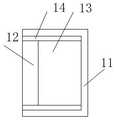
FIG. 1 is a schematic diagram of an array detector according to the present invention.
Fig. 2 isbase:Sub>A sectional viewbase:Sub>A-base:Sub>A of fig. 1.
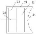
FIG. 3 is a schematic view of another structure of the array detector of the present invention.
Detailed Description
In order to make the objects, technical solutions and advantages of the present invention more apparent, the present invention is further described in detail below with reference to examples and accompanying drawings, and the exemplary embodiments and descriptions thereof are only used for explaining the present invention and are not meant to limit the present invention.
Example 1
A surface focusing array detector for photoacoustic breast imaging comprises a plurality of array elements forming an annular array, wherein the array elements are focusing probes, the focusing probes only realize focusing in the axial direction of the annular array and still keep the original divergence angle in the radial direction; the curvature radius of the focusing surface of the focusing probe is the inner radius of the annular array, so that the position of the focusing surface is limited.
The array elements of the embodiment are axially focused, so that the problem of low axial resolution of breast tumor imaging caused by the existence of the divergence angle of the existing array elements in the axial direction is solved, single fault information is only kept in the axial direction, the tomography capability of the system is improved, and more comprehensive imaging information is provided for diagnosis and prognosis of breast tumors.
The number of array elements in the circular array is not limited, and 128 or more array elements may be used, and is not specifically limited herein. The array element of this embodiment can be implemented in various ways, and the implementation thereof is specifically described in embodiments 2 and 3 below.
Example 2
As shown in fig. 1 and 2, the array element, i.e. the focusing probe, includes afirst housing 11, asealing layer 14, a firstpiezoelectric element 12, and afirst backing layer 13. The first housing as a whole is an external supporting and protecting structure, one end of which is a detection open end, and the firstpiezoelectric element 12 is arranged at the detection open end and is made of a metal material. The two surfaces of the firstpiezoelectric element 12 are respectively covered with a positive electrode and a negative electrode, the front end surface of the firstpiezoelectric element 12, namely the surface covered with the negative electrode, is a focusing surface, the negative electrode is connected with thefirst shell 11, the axial section of the whole firstpiezoelectric element 12 is arc-shaped, and the radial section still keeps a plane; the back end face, namely the face covered with the positive electrode, of the firstpiezoelectric element 12 is poured with afirst backing layer 13 to reduce oscillation and improve signal quality; the positive electrode of the firstpiezoelectric element 12 is led out through afirst lead 15, and thefirst lead 15 sequentially passes through thefirst backing layer 13 and thefirst case 11. Thesealing layer 14 is disposed in the first housing, and covers the firstpiezoelectric element 12 and thefirst backing layer 13 to achieve sealing, and is made of a polymer material such as epoxy resin.
The focusing probe with the structure has certain requirements on the shaping capacity of the first piezoelectric element, and can adopt PVDF piezoelectric film or other shapeable piezoelectric materials. The PVDF piezoelectric film has high piezoelectric coefficient, is easy to shape, and can improve the accuracy of probe signal detection.
Example 3
If the piezoelectric element is not plastic, such as piezoelectric ceramic, a focusing probe structure as shown in fig. 3 can be used, which includes asecond housing 21, a secondpiezoelectric element 22, asecond backing layer 23, and anacoustic lens 24. Likewise, thesecond housing 21 as a whole supports the protective structure externally, with one end being the detection open end. Because the plasticity of the piezoelectric element is not strong, the piezoelectric element can not be made into a focusing arc-shaped structure, so that the secondpiezoelectric element 22 can be made into a strip-shaped structure, the rear end face of the acoustic lens is attached to the secondpiezoelectric element 22, the axial section of the front end face is arc-shaped, and the radial section still keeps a plane. Here, the front face refers to a face closer to the breast tissue in use. The two surfaces of the secondpiezoelectric element 22 are respectively covered with a positive electrode and a negative electrode, the surface covered with the negative electrode is attached to theacoustic lens 24, the negative electrode is connected with thesecond shell 21, the positive electrode is led out of thesecond shell 21 through asecond lead 25, and thesecond lead 25 sequentially penetrates through thesecond backing layer 23 and thesecond shell 21 to be connected with an external radio frequency socket.
Preferably, the secondpiezoelectric element 22 may be a PZT piezoelectric ceramic. Theacoustic lens 24 is made of organic glass.
Example 3
Based on the above embodiments, the present embodiment discloses an application of the array probe, that is, an area focusing array detection system for photoacoustic breast imaging, including a laser for generating pulse laser, an array detector for detecting ultrasonic information, and a control unit for controlling the laser and the array detector, where the array detector is the array detector in any of the above embodiments.
Specifically, the array detector adopts 128 array elements to form an annular array with the diameter of 20 centimeters, the length of a detection surface of a single array element is 15mm, the height of the detection surface of the single array element is 3mm, and the curvature radius of a focal plane of the single array element in the axial direction is the inner radius of the annular array. The laser and the control unit adopt the existing structure. The system can improve the axial resolution of breast tumor imaging and the tomography capability of the system, and provides more comprehensive imaging information for the diagnosis and prognosis of breast tumor. In particular, the tomographic capabilities of the present protocol are described in terms of breast imaging of a patient.
The above-mentioned embodiments are intended to illustrate the objects, technical solutions and advantages of the present invention in further detail, and it should be understood that the above-mentioned embodiments are merely exemplary embodiments of the present invention, and are not intended to limit the scope of the present invention, and any modifications, equivalent substitutions, improvements and the like made within the spirit and principle of the present invention should be included in the scope of the present invention.
Claims (2)
1. An area-focusing array probe for photoacoustic breast imaging comprising a plurality of array elements forming an annular array, characterized in that:
the array elements are focusing probes which focus only in the axial direction of the annular array, and the curvature radius of the focusing surface of each focusing probe is the inner radius of the annular array;
the focusing probe comprises a first shell (11), a sealing layer (14) poured in the first shell (11), a first piezoelectric element (12) with two surfaces respectively coated with a positive electrode and a negative electrode, and a first backing layer (13) poured on the positive electrode surface of the first piezoelectric element (12), wherein the section of the first piezoelectric element (12) is arc-shaped, the positive electrode of the first piezoelectric element (12) is led out of the first shell (11) through a first lead (15), and the negative electrode is connected with the first shell (11); the first piezoelectric element (12) is a PVDF piezoelectric film;
or,
the focusing probe comprises a second shell (21), a second piezoelectric element (22) with positive electrodes and negative electrodes coated on two surfaces, a second backing layer (23) poured on the positive electrode surface of the second piezoelectric element (22) and an acoustic lens (24) arranged on the negative electrode surface of the second piezoelectric element (22), wherein the front end surface of the acoustic lens (24) is arc-shaped, the positive electrodes of the second piezoelectric element (22) are led out of the second shell (21) through a second lead (25), and the negative electrodes are connected with the second shell (21); the second piezoelectric element (22) is PZT piezoelectric ceramic.
2. An area-focused array detection system for photoacoustic breast imaging comprising a laser for generating pulsed laser light, an array detector for detecting ultrasound information and a control unit controlling the laser and the array detector, characterized in that the array detector is the array detector of claim 1.
Priority Applications (1)
| Application Number | Priority Date | Filing Date | Title |
|---|---|---|---|
| CN202010056116.2ACN111012318B (en) | 2020-01-18 | 2020-01-18 | Surface focusing array detector and system for photoacoustic breast imaging |
Applications Claiming Priority (1)
| Application Number | Priority Date | Filing Date | Title |
|---|---|---|---|
| CN202010056116.2ACN111012318B (en) | 2020-01-18 | 2020-01-18 | Surface focusing array detector and system for photoacoustic breast imaging |
Publications (2)
| Publication Number | Publication Date |
|---|---|
| CN111012318A CN111012318A (en) | 2020-04-17 |
| CN111012318Btrue CN111012318B (en) | 2022-10-28 |
Family
ID=70199137
Family Applications (1)
| Application Number | Title | Priority Date | Filing Date |
|---|---|---|---|
| CN202010056116.2AActiveCN111012318B (en) | 2020-01-18 | 2020-01-18 | Surface focusing array detector and system for photoacoustic breast imaging |
Country Status (1)
| Country | Link |
|---|---|
| CN (1) | CN111012318B (en) |
Citations (6)
| Publication number | Priority date | Publication date | Assignee | Title |
|---|---|---|---|---|
| CN102462510A (en)* | 2010-11-12 | 2012-05-23 | 香港理工大学 | Rotary ultrasonic imaging system |
| CN105167808A (en)* | 2015-09-02 | 2015-12-23 | 上海爱声生物医疗科技有限公司 | Transurethral ultrasound prostate detection method, diagnostic apparatus and transducer |
| CN106913314A (en)* | 2015-12-25 | 2017-07-04 | 佳能株式会社 | Information acquisition device and information acquisition method |
| CN107174284A (en)* | 2017-07-08 | 2017-09-19 | 中北大学 | Breast ultrasound imaging system and its detection method based on CMUT annular arrays |
| CN108814555A (en)* | 2018-04-25 | 2018-11-16 | 成都世恩医疗科技有限责任公司 | Finite element fast image reconstruction system and method for optoacoustic mammary gland imager |
| CN110314834A (en)* | 2018-03-28 | 2019-10-11 | 中国科学院深圳先进技术研究院 | A kind of ultrasonic transducer and preparation method thereof |
Family Cites Families (22)
| Publication number | Priority date | Publication date | Assignee | Title |
|---|---|---|---|---|
| DE3931048A1 (en)* | 1989-09-16 | 1991-04-11 | Leica Industrieverwaltung | TAPERED ULTRASONIC DEFLECTING ELEMENT |
| US5713356A (en)* | 1996-10-04 | 1998-02-03 | Optosonics, Inc. | Photoacoustic breast scanner |
| JP2001258879A (en)* | 2000-03-15 | 2001-09-25 | Olympus Optical Co Ltd | Ultrasonic transducer system and ultrasonic transducer |
| US6990170B2 (en)* | 2001-08-09 | 2006-01-24 | Kabushiki Kaisha Toshiba | X-ray computed tomographic imaging apparatus |
| RU2242710C2 (en)* | 2002-06-07 | 2004-12-20 | Геликонов Григорий Валентинович | Method and device for building object image and device for delivering low coherence optical radiation |
| CN1281286C (en)* | 2003-06-19 | 2006-10-25 | 上海交通大学 | Transducer array for high-intersity focusing ultrasonic tumor treatment |
| FR2883190B1 (en)* | 2005-03-15 | 2007-08-10 | Edap S A | ENDO-CAVITARY THERAPEUTIC PROBE COMPRISING AN INTEGRATED IMAGING TRANSDUCER WITHIN THE ULTRASONIC THERAPY TRANSDUCER |
| US8323201B2 (en)* | 2007-08-06 | 2012-12-04 | Orison Corporation | System and method for three-dimensional ultrasound imaging |
| CN102579127B (en)* | 2011-01-14 | 2014-09-03 | 深圳市普罗惠仁医学科技有限公司 | Ultrasonic focusing energy transducer |
| CN102510449B (en)* | 2011-11-18 | 2015-06-10 | 北京理工大学 | Human eye-like image sensor based on non-uniform lens array |
| JP5762995B2 (en)* | 2012-02-28 | 2015-08-12 | 富士フイルム株式会社 | Photoacoustic image generation apparatus and method |
| JP6143390B2 (en)* | 2012-02-29 | 2017-06-07 | 富士フイルム株式会社 | Photoacoustic measuring device |
| CN103202688B (en)* | 2013-04-23 | 2015-11-18 | 华南师范大学 | Ultrashort pulse microwave thermal sound breast imaging checkout gear |
| CN103240220B (en)* | 2013-05-09 | 2015-06-17 | 电子科技大学 | Piezoelectric array ultrasonic transducer |
| CN105595964B (en)* | 2016-01-21 | 2018-08-14 | 曲阜师范大学 | Double focusing ultrasonic probe and thinned array Photoacoustic tomography system |
| CN105988172A (en)* | 2016-01-26 | 2016-10-05 | 安徽蓝盾光电子股份有限公司 | Object distance fine adjustment system used for pipeline online TDLAS gas monitor |
| CN106175677B (en)* | 2016-07-08 | 2018-12-07 | 华南师范大学 | Integrated optoacoustic breast imaging detection device and method based on fiber beam splitting and flexible detector |
| CN106073721A (en)* | 2016-07-26 | 2016-11-09 | 成都世恩医疗科技有限责任公司 | A kind of novel optoacoustic mammary gland imager |
| JP6882100B2 (en)* | 2017-06-30 | 2021-06-02 | キヤノン株式会社 | Acoustic wave probe and acoustic wave device |
| WO2019027697A1 (en)* | 2017-07-31 | 2019-02-07 | Wayne State University | Omnidirectional photoacoustic tomography system |
| CN108310685A (en)* | 2018-03-01 | 2018-07-24 | 西安电子科技大学 | A kind of wearable mouse brain stimulation ultrasonic transducer |
| CN108523847B (en)* | 2018-04-20 | 2021-02-26 | 四川知周光声医疗科技有限公司 | Photoacoustic mammary gland imaging system and imaging method thereof |
- 2020
- 2020-01-18CNCN202010056116.2Apatent/CN111012318B/enactiveActive
Patent Citations (6)
| Publication number | Priority date | Publication date | Assignee | Title |
|---|---|---|---|---|
| CN102462510A (en)* | 2010-11-12 | 2012-05-23 | 香港理工大学 | Rotary ultrasonic imaging system |
| CN105167808A (en)* | 2015-09-02 | 2015-12-23 | 上海爱声生物医疗科技有限公司 | Transurethral ultrasound prostate detection method, diagnostic apparatus and transducer |
| CN106913314A (en)* | 2015-12-25 | 2017-07-04 | 佳能株式会社 | Information acquisition device and information acquisition method |
| CN107174284A (en)* | 2017-07-08 | 2017-09-19 | 中北大学 | Breast ultrasound imaging system and its detection method based on CMUT annular arrays |
| CN110314834A (en)* | 2018-03-28 | 2019-10-11 | 中国科学院深圳先进技术研究院 | A kind of ultrasonic transducer and preparation method thereof |
| CN108814555A (en)* | 2018-04-25 | 2018-11-16 | 成都世恩医疗科技有限责任公司 | Finite element fast image reconstruction system and method for optoacoustic mammary gland imager |
Also Published As
| Publication number | Publication date |
|---|---|
| CN111012318A (en) | 2020-04-17 |
Similar Documents
| Publication | Publication Date | Title |
|---|---|---|
| CN102743191B (en) | Focusing rotary scanning photoacoustic ultrasonic blood vessel endoscope imaging device and focusing rotary scanning photoacoustic ultrasonic blood vessel endoscope imaging method | |
| CN105380586B (en) | A combined stereoscopic scanning optical and acoustic endoscopic imaging device and method thereof | |
| CN108042110B (en) | Multi-mode imaging system | |
| WO2010080991A2 (en) | Miniaturized photoacoustic imaging apparatus including a rotatable reflector | |
| CN215738807U (en) | Endoscope apposition imaging probe for realizing ultrasonic and coherent light tomography and system thereof | |
| WO2010045421A2 (en) | Photoacoustic imaging using a versatile acoustic lens | |
| CN103211620B (en) | Breast carcinoma early-stage detecting instrument based on annular array opto-acoustic sensing technology | |
| CN112450882B (en) | Ultrasound probe, endoscope, endoscopic imaging system, and endoscopic imaging method | |
| US11609326B2 (en) | Transparent ultrasound transducer with light beam shaping and the method for assembling the same | |
| CN106580255A (en) | Electric-control focusing ultrasonic detector used for photoacoustic imaging and electric-control focusing method thereof | |
| CN113848184B (en) | Micro-cavity photoacoustic imaging system based on flexible substrate | |
| EP3259616A1 (en) | Medical imaging detector | |
| KR20220029003A (en) | Ultrasonic-optical multi imaging system based on transpatent ultrasonic sensor | |
| WO2022104701A1 (en) | Ultrasound probe, endoscope, endoscopic imaging system, and endoscopic imaging method | |
| CN103040428B (en) | Optical scanning probe for endoscopic OCT (optical coherence tomography) imaging | |
| CN111012316B (en) | Image reconstruction system of photoacoustic mammary gland | |
| CN203153684U (en) | Optical scanning probe for OCT endoscopic imaging | |
| CN111012318B (en) | Surface focusing array detector and system for photoacoustic breast imaging | |
| CN106377229B (en) | A kind of rotary acoustics and optics merge imaging system | |
| CN115844331A (en) | Multi-angle photoacoustic tomography system and method | |
| CN105125238A (en) | Transurethral bladder ultrasonic detection method, diagnostic apparatus and transducer | |
| CN105167808A (en) | Transurethral ultrasound prostate detection method, diagnostic apparatus and transducer | |
| CN205006919U (en) | Through urethral prostate diasonograph and transducer | |
| Xiao et al. | Lithography of aluminum coated PVDF annular array for photoacoustic endoscopy | |
| JP7598475B2 (en) | Photoacoustic detection system combining transparent ultrasonic sensor |
Legal Events
| Date | Code | Title | Description |
|---|---|---|---|
| PB01 | Publication | ||
| PB01 | Publication | ||
| SE01 | Entry into force of request for substantive examination | ||
| SE01 | Entry into force of request for substantive examination | ||
| TA01 | Transfer of patent application right | Effective date of registration:20200629 Address after:610000 no.4-210401, Section 2, Jianshe North Road, Chenghua District, Chengdu City, Sichuan Province Applicant after:Chen Bingzhang Applicant after:Wang Luguo Address before:No. 1301, building 3, No. 1, Keyuan South Road, hi tech Zone, Chengdu, Sichuan 610000 Applicant before:Sichuan Zhizhou Guangsheng Medical Technology Co.,Ltd. | |
| TA01 | Transfer of patent application right | ||
| TA01 | Transfer of patent application right | Effective date of registration:20200728 Address after:610000, No. 29, Xihe East Road, Pengzhou Industrial Development Zone, Sichuan, Chengdu Applicant after:Zhongchuan Xinmai Technology Co.,Ltd. Address before:610000 no.4-210401, Section 2, Jianshe North Road, Chenghua District, Chengdu City, Sichuan Province Applicant before:Chen Bingzhang Applicant before:Wang Luguo | |
| TA01 | Transfer of patent application right | ||
| GR01 | Patent grant | ||
| GR01 | Patent grant |