CN110991254B - Ultrasound image video classification prediction method and system - Google Patents
Ultrasound image video classification prediction method and systemDownload PDFInfo
- Publication number
- CN110991254B CN110991254BCN201911087843.9ACN201911087843ACN110991254BCN 110991254 BCN110991254 BCN 110991254BCN 201911087843 ACN201911087843 ACN 201911087843ACN 110991254 BCN110991254 BCN 110991254B
- Authority
- CN
- China
- Prior art keywords
- upsampling
- output
- classification prediction
- image
- layer
- Prior art date
- Legal status (The legal status is an assumption and is not a legal conclusion. Google has not performed a legal analysis and makes no representation as to the accuracy of the status listed.)
- Active
Links
Images


Classifications
- G—PHYSICS
- G06—COMPUTING OR CALCULATING; COUNTING
- G06V—IMAGE OR VIDEO RECOGNITION OR UNDERSTANDING
- G06V20/00—Scenes; Scene-specific elements
- G06V20/40—Scenes; Scene-specific elements in video content
- G06V20/41—Higher-level, semantic clustering, classification or understanding of video scenes, e.g. detection, labelling or Markovian modelling of sport events or news items
- G—PHYSICS
- G06—COMPUTING OR CALCULATING; COUNTING
- G06T—IMAGE DATA PROCESSING OR GENERATION, IN GENERAL
- G06T7/00—Image analysis
- G06T7/0002—Inspection of images, e.g. flaw detection
- G06T7/0012—Biomedical image inspection
- G—PHYSICS
- G06—COMPUTING OR CALCULATING; COUNTING
- G06T—IMAGE DATA PROCESSING OR GENERATION, IN GENERAL
- G06T2207/00—Indexing scheme for image analysis or image enhancement
- G06T2207/10—Image acquisition modality
- G06T2207/10132—Ultrasound image
- G—PHYSICS
- G06—COMPUTING OR CALCULATING; COUNTING
- G06T—IMAGE DATA PROCESSING OR GENERATION, IN GENERAL
- G06T2207/00—Indexing scheme for image analysis or image enhancement
- G06T2207/20—Special algorithmic details
- G06T2207/20081—Training; Learning
- G—PHYSICS
- G06—COMPUTING OR CALCULATING; COUNTING
- G06T—IMAGE DATA PROCESSING OR GENERATION, IN GENERAL
- G06T2207/00—Indexing scheme for image analysis or image enhancement
- G06T2207/20—Special algorithmic details
- G06T2207/20084—Artificial neural networks [ANN]
- G—PHYSICS
- G06—COMPUTING OR CALCULATING; COUNTING
- G06T—IMAGE DATA PROCESSING OR GENERATION, IN GENERAL
- G06T2207/00—Indexing scheme for image analysis or image enhancement
- G06T2207/30—Subject of image; Context of image processing
- G06T2207/30004—Biomedical image processing
- G06T2207/30044—Fetus; Embryo
- G—PHYSICS
- G06—COMPUTING OR CALCULATING; COUNTING
- G06V—IMAGE OR VIDEO RECOGNITION OR UNDERSTANDING
- G06V2201/00—Indexing scheme relating to image or video recognition or understanding
- G06V2201/03—Recognition of patterns in medical or anatomical images
Landscapes
- Engineering & Computer Science (AREA)
- General Physics & Mathematics (AREA)
- Theoretical Computer Science (AREA)
- Physics & Mathematics (AREA)
- General Health & Medical Sciences (AREA)
- Multimedia (AREA)
- Software Systems (AREA)
- Health & Medical Sciences (AREA)
- Computational Linguistics (AREA)
- Medical Informatics (AREA)
- Nuclear Medicine, Radiotherapy & Molecular Imaging (AREA)
- Radiology & Medical Imaging (AREA)
- Quality & Reliability (AREA)
- Computer Vision & Pattern Recognition (AREA)
- Ultra Sonic Daignosis Equipment (AREA)
- Image Processing (AREA)
Abstract
Description
Translated fromChinese技术领域technical field
本发明涉及图像处理领域,尤其是涉及一种超声图像视频分类预测方法及系统。The invention relates to the field of image processing, in particular to a method and system for classifying and predicting ultrasound images and videos.
背景技术Background technique
如今利用超声设备获取用户的医学视频图像,并确定诊疗方案越来越普遍,例如通过产前超声进行畸形筛查,保障新生儿健康。通常产前超声检查可大致分为三个过程。首先,医师控制设备扫描胎儿特定的身体区域,然后超声医师需要在操作超声探头的同时从连续扫描视频中搜索标准切面,最后在标准切面上,观察组织结构或测量生物学参数,以确定胎儿是否存在生理异常并评估胎儿的生长发育和健康状况。因此,预测胎儿超声影像标准切面作为产前超声检查的关键步骤,是后续参数测量和异常诊断的前提。Nowadays, it is more and more common to use ultrasound equipment to obtain medical video images of users and determine the diagnosis and treatment plan, such as deformity screening through prenatal ultrasound to ensure the health of newborns. Usually prenatal ultrasonography can be roughly divided into three processes. First, the physician controls the device to scan specific body areas of the fetus, then the sonographer needs to search for standard slices from the continuous scanning video while operating the ultrasound probe, and finally, on the standard slices, observe tissue structures or measure biological parameters to determine whether the fetus is Physiological abnormalities are present and fetal growth and health are evaluated. Therefore, as a key step in prenatal ultrasound examination, predicting the standard section of fetal ultrasound imaging is a prerequisite for subsequent parameter measurement and abnormal diagnosis.
然而,预测标准切面是一项高度专业化的任务,需要深厚的专业知识和临床经验,而且标准切面的筛选耗时且费力,一次完整的产前超声检查通常需要40分钟到一个多小时。由于超声扫描视图的连续性在动态视频中标准切面与相邻帧的非标准切面之间仅存在细微差别。此外,与一般视频分析任务相比,超声成像通常受到噪声和伪影的影响,无论对于人工还是智能算法,都是十分具有挑战性的任务。However, predicting standard cut planes is a highly specialized task that requires deep professional knowledge and clinical experience, and the screening of standard cut planes is time-consuming and laborious. A complete prenatal ultrasound usually takes 40 minutes to more than an hour. Due to the continuity of ultrasound scan views there is only a slight difference between the standard slices in the motion video and the non-standard slices of adjacent frames. In addition, compared with general video analysis tasks, ultrasound imaging is usually affected by noise and artifacts, which is a very challenging task for both artificial and intelligent algorithms.
因此需要提出一种能够对视频图像(例如超声视频图像)进行分类预测的方法。Therefore, it is necessary to propose a method capable of classifying and predicting video images (such as ultrasound video images).
发明内容Contents of the invention
本发明旨在至少解决现有技术中存在的技术问题之一。为此,本发明提出一种超声图像视频分类预测方法,能够对视频的每帧图像进行分类预测并输出分类预测结果。The present invention aims to solve at least one of the technical problems existing in the prior art. To this end, the present invention proposes a video classification and prediction method for ultrasonic images, which can perform classification prediction on each frame of the video and output a classification prediction result.
第一方面,本发明的一个实施例提供了一种超声图像视频分类预测方法,包括:In the first aspect, an embodiment of the present invention provides a method for classifying and predicting ultrasound images and videos, including:
获取原始视频图像;Get the original video image;
利用特征提取网络提取所述原始视频图像的特征;Utilize feature extraction network to extract the feature of described original video image;
对所述特征利用时序上采样输出每帧的分类预测结果;Utilizing temporal upsampling on the feature to output the classification prediction result of each frame;
所述特征提取网络和所述时序上采样构成分类预测网络。The feature extraction network and the time series up-sampling constitute a classification prediction network.
进一步地,所述特征提取网络为三维卷积残差网络,包括:1个卷积层和至少1个残差块。Further, the feature extraction network is a three-dimensional convolutional residual network, including: 1 convolutional layer and at least 1 residual block.
进一步地,所述时序上采样由对应于所述残差块数量的反卷积层进行上采样,同时所述特征提取网络中间层特征经空间最大池化操作后与上采样后的时序特征进行通道融合。Further, the timing upsampling is performed by the deconvolution layer corresponding to the number of residual blocks, and at the same time, the feature extraction network intermediate layer features are subjected to the spatial maximum pooling operation and the upsampled timing features are performed. Channel fusion.
进一步地,还包括对所述原始视频图像进行预处理,所述预处理包括:调整图像大小和图像归一化。Further, it also includes preprocessing the original video image, and the preprocessing includes: adjusting image size and image normalization.
进一步地,所述分类预测网络的损失函数为焦点损失函数,具体表示为:Further, the loss function of the classification prediction network is a focal loss function, specifically expressed as:
FL(pt)=-αt(1-pt)γlog(pt)FL(pt )=-αt (1-pt )γ log(pt )
其中,FL(pt)表示焦点损失值,pt表示输出预测概率,αt表示分类预测结果的权重,γ表示平衡参数,(1-pt)γ表示平衡因子。Among them, FL(pt ) represents the focal loss value,pt represents the output prediction probability, αt represents the weight of the classification prediction result, γ represents the balance parameter, and (1-pt )γ represents the balance factor.
进一步地,所述分类预测网络的优化器为Adam算法优化器。Further, the optimizer of the classification prediction network is an Adam algorithm optimizer.
第二方面,本发明的一个实施例提供了一种超声图像视频分类预测系统,包括:In a second aspect, an embodiment of the present invention provides a system for classifying and predicting ultrasound images and videos, including:
获取模块:用于获取原始视频图像;Obtaining module: used to obtain the original video image;
特征提取模块:用于利用特征提取网络提取所述原始视频图像的特征;Feature extraction module: for utilizing feature extraction network to extract the feature of described original video image;
输出模块:用于对所述特征利用时序上采样输出分类预测结果。An output module: used for outputting classification prediction results by using temporal upsampling on the features.
第三方面,本发明的一个实施例提供了提供一种超声图像视频分类预测设备,包括:In a third aspect, an embodiment of the present invention provides an ultrasonic image video classification prediction device, including:
至少一个处理器,以及与所述至少一个处理器通信连接的存储器;at least one processor, and a memory communicatively coupled to the at least one processor;
其中,所述处理器通过调用所述存储器中存储的计算机程序,用于执行如第一方面任一项所述的方法。Wherein, the processor is used to execute the method according to any one of the first aspect by invoking the computer program stored in the memory.
第四方面,本发明提供一种计算机可读存储介质,所述计算机可读存储介质存储有计算机可执行指令,所述计算机可执行指令用于使计算机执行如第一方面任一项所述的方法。In a fourth aspect, the present invention provides a computer-readable storage medium, the computer-readable storage medium stores computer-executable instructions, and the computer-executable instructions are used to make a computer execute the method described in any one of the first aspect. method.
本发明的有益效果是:The beneficial effects of the present invention are:
本发明通过获取包含时间维度、图像宽度、图像高度的原始视频图像,利用特征提取网络提取原始视频图像的特征,对特征利用时序上采样输出分类预测结果,其分类预测结果包括标准切面和非标准切面。利用特征提取网络高效的学习原始视频图像的时时序信息,通过时序上采样输出视频片段每帧的分类预测结果,提高预测效率,当用于超声视频数据时,能够提高超声诊断效率,减轻医生负担,而且有助于缓解医疗资源不足的问题。可广泛应用于视频图像预测领域。The present invention obtains the original video image including the time dimension, image width, and image height, uses the feature extraction network to extract the features of the original video image, and uses time-series upsampling to output the classification prediction results, and the classification prediction results include standard slices and non-standard slices. section. Using the feature extraction network to efficiently learn the time-series information of the original video image, and output the classification and prediction results of each frame of the video clip through time-series upsampling to improve the prediction efficiency. When used in ultrasound video data, it can improve the efficiency of ultrasound diagnosis and reduce the burden on doctors. , and help alleviate the problem of insufficient medical resources. It can be widely used in the field of video image prediction.
附图说明Description of drawings
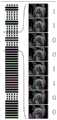
图1~图3为产前超声视频图像示意图;Figures 1 to 3 are schematic diagrams of prenatal ultrasound video images;
图4是产前超声视频图像标注示意图;Fig. 4 is a schematic diagram of prenatal ultrasound video image annotation;
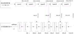
图5是本发明实施例中超声图像视频分类预测方法的一具体实施例实现流程图;Fig. 5 is a flow chart of a specific embodiment of the ultrasonic image video classification prediction method in the embodiment of the present invention;
图6是本发明实施例中超声图像视频分类预测方法的一具体实施例整体示意图;6 is an overall schematic diagram of a specific embodiment of a method for classifying and predicting ultrasound images and videos in an embodiment of the present invention;
图7是本发明实施例中超声图像视频分类预测方法的一具体实施例平衡参数不同取值的损失函数示意图;7 is a schematic diagram of a loss function of a specific embodiment of a method for classifying and predicting ultrasound images and videos in an embodiment of the present invention with different values of balance parameters;
图8是本发明实施例中超声图像视频分类预测系统的一具体实施例结构框图。Fig. 8 is a structural block diagram of a specific embodiment of the ultrasonic image video classification and prediction system in the embodiment of the present invention.
具体实施方式Detailed ways
为了更清楚地说明本发明实施例或现有技术中的技术方案,下面将对照附图说明本发明的具体实施方式。显而易见地,下面描述中的附图仅仅是本发明的一些实施例,对于本领域普通技术人员来讲,在不付出创造性劳动的前提下,还可以根据这些附图获得其他的附图,并获得其他的实施方式。In order to more clearly illustrate the embodiments of the present invention or the technical solutions in the prior art, the specific implementation manners of the present invention will be described below with reference to the accompanying drawings. Obviously, the accompanying drawings in the following description are only some embodiments of the present invention, and those skilled in the art can obtain other accompanying drawings based on these drawings and obtain other implementations.
除非另有定义,本文所使用的所有的技术和科学术语与属于本发明的技术领域的技术人员通常理解的含义相同。本文中在本发明的说明书中所使用的术语只是为了描述具体的实施例的目的,不是旨在于限制本发明。Unless otherwise defined, all technical and scientific terms used herein have the same meaning as commonly understood by one of ordinary skill in the technical field of the invention. The terms used herein in the description of the present invention are for the purpose of describing specific embodiments only, and are not intended to limit the present invention.
实施例一:Embodiment one:
本发明实施例一提供一种超声图像视频分类预测方法,能够广泛应用于视频数据帧级别分类,本实施例以产前超声数据为例说明本实施例的超声图像视频分类预测方法详细预测过程,但并不代表本实施例仅限于超声视频数据。
产前超声主要用于进行新生儿筛查,很多地区由于缺少自动分析超声图像的方法和设备,极大地限制了产前超声检查的效率。特别是在一些欠发达地区,因为缺乏经验丰富的医生,情况尤为严重。Prenatal ultrasound is mainly used for newborn screening. In many areas, the lack of methods and equipment for automatic analysis of ultrasound images greatly limits the efficiency of prenatal ultrasound examinations. Especially in some underdeveloped areas, the situation is particularly serious because of the lack of experienced doctors.
如图1~图3所示,为产前超声视频图像示意图,分别是:图1表示腹围切面,含有胃泡(SB),脐静脉(UV)和脊柱(SP);图2表示双眼球横切面,包含鼻骨,眼球和晶状体;图3表示心脏四腔切面,包含左心房(LA),右心房(RA),左心室(LV),右心室(RV)和降主动脉(DAO),其中第一行是标准切面图像,第二行是对应于不同区域的非标准切面图像,对于非专业人士而言,很难看到它们之间的明显差异。在标准切面定义中,要求画面中必须清楚地看到一些关键结构。例如,在双眼球横切面中,必须清楚地看到鼻骨,双侧眼球以及两侧的晶状体,像晶状体这样细微的结构,在图像中可能只占据几个像素,很难被算法识别出来,但它对于出生缺陷诊断来说又非常重要。As shown in Figures 1 to 3, they are schematic diagrams of prenatal ultrasound video images, respectively: Figure 1 shows the section of the abdominal circumference, including the gastric bubble (SB), umbilical vein (UV) and spine (SP); Figure 2 shows the eyes Transverse section, including nasal bone, eyeball and lens; Figure 3 shows the four-chamber section of the heart, including left atrium (LA), right atrium (RA), left ventricle (LV), right ventricle (RV) and descending aorta (DAO), The first row is a standard section image, and the second row is a non-standard section image corresponding to different regions. For non-professionals, it is difficult to see the obvious difference between them. In the standard aspect definition, it is required that some key structures must be clearly visible in the picture. For example, in the cross-section of both eyeballs, the nasal bone, bilateral eyeballs, and lenses on both sides must be clearly seen. Such subtle structures as the lens may only occupy a few pixels in the image, and it is difficult to be recognized by the algorithm. It is also very important for the diagnosis of birth defects.
图4为产前超声视频图像标注示意图,部分显示了胎儿心脏四腔标准切面分类预测任务的一个视频标注示例,该视频共包含49帧,图中选取显示了其中的8帧超声图像,其中最右边的数字0代表非标准切面,1代表标准切面,虚线代表标准切面,实线代表非标准切面,从图中可以看到标签是不连续的,即标准切面出现的片段很短,在某些情况下,两个标准切面之间也可能出现非标准切面,这主要是因为探头移动、背景噪音以及器官运动的原因,因此视频分析中常用的基于片段候选的预测算法并不适用于标准切面预测任务,本实施例提供一种针对帧级任务的方法来实时生成细粒度和密集的时序分类预测结果。Figure 4 is a schematic diagram of prenatal ultrasound video image labeling, which partially shows an example of video labeling for the four-chamber standard view of the fetal heart. The
图5为本发明实施例提供的一种超声图像视频分类预测方法的实现流程图,如图5所示,该方法包括以下步骤:Fig. 5 is an implementation flow chart of an ultrasonic image video classification and prediction method provided by an embodiment of the present invention. As shown in Fig. 5, the method includes the following steps:
S1:获取原始视频图像,对于输入原始视频图像序列片段,其大小可以表示为L×W×H×T,其中H和W表示每帧图片的高和宽,L表示序列片段的帧数,T则为图片的通道数。S1: Get the original video image. For the input original video image sequence segment, its size can be expressed as L×W×H×T, where H and W represent the height and width of each frame of the picture, L represents the frame number of the sequence segment, and T is the channel number of the image.
S2:对原始视频图像进行预处理,本实施例中,预处理包括:调整图像大小和图像归一化,进行预处理的目的是为了统一输入原始视频图像的格式,提高运算效率。S2: Perform preprocessing on the original video image. In this embodiment, the preprocessing includes: adjusting the image size and normalizing the image. The purpose of the preprocessing is to unify the format of the input original video image and improve computing efficiency.
S3:利用特征提取网络提取原始视频图像的特征。S3: Use the feature extraction network to extract the features of the original video image.
S4:对特征利用时序上采样输出每帧的分类预测结果。S4: Use temporal upsampling to output the classification prediction result of each frame.
如图6所示,为本实施例的超声图像视频分类预测方法整体示意图,从图中可见输入的原始视频图像为包含时序特征的帧图像(L×W×H),经过特征提取网络输出特征,之后进行时序上采样得到分类预测结果。As shown in Figure 6, it is an overall schematic diagram of the ultrasonic image video classification and prediction method in this embodiment. It can be seen from the figure that the input original video image is a frame image (L×W×H) containing time series features, and the feature is output through the feature extraction network. , and then time-series upsampling is performed to obtain classification prediction results.
具体的,步骤S3中,特征提取网络为三维卷积残差网络(本实施例中表示为3DResNet),包括:1个卷积层conv1和至少一个残差块Layer模块,卷积层为1×1的卷积核,Layer模块指的是ResNet中的基本块BasicBlocks,由两个输出通道数相同的3×3卷积组成,进一步地,残差块为4个。Specifically, in step S3, the feature extraction network is a three-dimensional convolutional residual network (represented as 3DResNet in this embodiment), including: 1 convolutional layer conv1 and at least one residual block Layer module, and the convolutional layer is 1× The convolution kernel of 1, the Layer module refers to the basic block BasicBlocks in ResNet, which consists of two 3×3 convolutions with the same number of output channels, and further, there are 4 residual blocks.
其中,ResNet(Residual Network)针对训练卷积神经网络时加深网络导致准确度下降的问题,在已有设计思路的基础上,引入了残差块。每个残差块包含两条路径,其中一条路径是输入特征的直连通路,另一条路径对该特征做两到三次卷积操作得到该特征的残差,最后再将两条路径上的特征相加。Among them, ResNet (Residual Network) introduces a residual block on the basis of existing design ideas to solve the problem of decreasing accuracy caused by deepening the network when training a convolutional neural network. Each residual block contains two paths, one of which is a direct connection path of the input feature, and the other path performs two or three convolution operations on the feature to obtain the residual of the feature, and finally the features on the two paths add up.
在训练时随着网络的加深,特征的时间和空间的维度逐渐缩小,随之通道数量不断增多,低级别的像素信息逐渐提炼为与分类预测标签相关的高级语义信息。当特征每次通过Layer层时,时间维度都会减少一半,最后Layer4输出特征的时间维度减小到原始长度的1/16,在一种具体实施方式中,其变化过程示意为:(1)输入[64,L,112,112]-->(2)Layer1输出[64,L/2,56,56]-->(3)Layer2输出[128,L/4,28,28]-->(4)Layer3输出[256,L/8,14,14]-->(5)Layer4输出[512,L/16,7,7],其中64、128、256和512表示通道数,因此给定输入为时间长度L的视频片段,其输出特征为[512,L/16,7,7]。During training, as the network deepens, the time and space dimensions of features gradually shrink, and the number of channels continues to increase, and the low-level pixel information is gradually refined into high-level semantic information related to classification and prediction labels. When the feature passes through the Layer layer every time, the time dimension will be reduced by half, and finally the time dimension of the Layer4 output feature will be reduced to 1/16 of the original length. In a specific implementation, the change process is shown as follows: (1) input [64,L,112,112]-->(2)Layer1 output [64,L/2,56,56]-->(3)Layer2 output [128,L/4,28,28]-->(4 )Layer3 output [256,L/8,14,14]-->(5)Layer4 output [512,L/16,7,7], where 64, 128, 256 and 512 represent the number of channels, so the given input It is a video clip with a length of time L, and its output feature is [512, L/16, 7, 7].
具体的,步骤S4中,由于在帧级分类任务中,需要对视频中的每帧进行分类,因此,需要通过时序上采样将特征的时序维度重新采样回原始长度。但是直接将特征上采样到完整的尺寸将会失去很多细节信息,因此本实施例中,时序上采样由对应于残差块数量的反卷积层进行上采样,每次做两倍上采样逐渐恢复到原始视频长度,此外,在每次上采样之后,加入来自特征提取网络中间层的特征,将它融合到上采样特征流中,这种特征提取网络中间层和上采样流之间的流动通路结合了来自浅层的低级特征和来自深层的高级特征,这使得时序恢复的过程更加准确和包含更多的细节。Specifically, in step S4, since each frame in the video needs to be classified in the frame-level classification task, it is necessary to resample the timing dimension of the feature back to the original length through timing upsampling. However, directly upsampling the features to the full size will lose a lot of detailed information, so in this embodiment, the timing upsampling is performed by the deconvolution layer corresponding to the number of residual blocks, and each time the upsampling is doubled and gradually Revert to the original video length, in addition, after each upsampling, add features from the middle layer of the feature extraction network, and fuse it into the upsampling feature stream, this flow between the middle layer of the feature extraction network and the upsampling stream The passway combines low-level features from shallow layers with high-level features from deep layers, which makes the process of timing recovery more accurate and more detailed.
参照图6,上采样后的时序特征加入特征提取网络输出的相同通道数的特征进行通道融合,即特征提取网络中间层特征经空间最大池化操作后与上采样后的时序特征进行通道融合,具体的是在每个Layer模块后,使用空间池来压缩三维特征的空间维度并将其提炼为一维时间特征,进一步地,空间池化使用的是最大池化层,其中时间维度的内核大小为1,空间维度的内核大小与输入原始图像数据特征的H和W相同,即可获得不同分辨率L/2,L/4,L/8和L/16的时序特征与上采样后得到的数据进行通道融合。Referring to Figure 6, the upsampled timing features are added to the features of the same number of channels output by the feature extraction network for channel fusion, that is, the features of the middle layer of the feature extraction network are channel-fused with the upsampled timing features after the spatial maximum pooling operation. Specifically, after each Layer module, spatial pooling is used to compress the spatial dimension of the three-dimensional feature and refine it into a one-dimensional temporal feature. Further, the spatial pooling uses the maximum pooling layer, where the kernel size of the temporal dimension is 1, the kernel size of the spatial dimension is the same as the H and W of the input original image data features, and the timing features of different resolutions L/2, L/4, L/8 and L/16 can be obtained after upsampling The data is channel-fused.
由于在特征提取网络中执行了4次向下采样,因此通过四个一维反卷积进行时序上采样,反卷积大小的计算公式表示为:Since 4 times of downsampling are performed in the feature extraction network, sequential upsampling is performed through four one-dimensional deconvolutions, and the calculation formula of the deconvolution size is expressed as:
Nout=(Nin-1)×s+k-2p (1)Nout =(Nin -1)×s+k-2p (1)
其中,s,k和p分别表示步长,内核大小和填充大小。where s, k and p denote the stride size, kernel size and padding size, respectively.
在二倍上采样(即图6中所示的Deconv1~4)中,可选的,设置参数s=2,k=2,p=0。以Deconv1为例,其输入的图像表示为[512,L/16],输出为[256,L/8],其时序维度增加,而通道数减少,将其与来自第Layer3的中间层特征[256,L/8]进行通道融合,这两个特征在通道上堆叠在一起融合后特征形状为[512,L/8]的融合特征,这种融合方式连接了3D特征网络的中间层,而且上采样流可以有效地使用中间层信息,使得网络可以将中间层的时序信息传播到更高分辨率的层。然后以相同的方式,上采样流依次通过每个反卷积层,直到Deconv4的输出形状[256,L]重新回到与输入的原始视频图像相同的长度。In double upsampling (that is, Deconv1˜4 shown in FIG. 6 ), optionally, set parameters s=2, k=2, and p=0. Taking Deconv1 as an example, the input image is expressed as [512, L/16], and the output is [256, L/8], its timing dimension increases, and the number of channels decreases, and it is compared with the middle layer features from Layer3 [ 256, L/8] for channel fusion, these two features are stacked together on the channel and the fusion feature is [512, L/8] after fusion, this fusion method connects the middle layer of the 3D feature network, and The upsampling stream can effectively use the intermediate layer information, so that the network can propagate the timing information of the intermediate layer to the higher resolution layer. Then in the same way, the upsampled stream passes through each deconvolutional layer in turn until the output shape of Deconv4 [256, L] is back to the same length as the input raw video image.
最后是两个一维卷积层,作为分类器用以输出最终分类预测结果,其中第一个卷积层Convk3(可选的,k=3,s=1,p=1),内核大小为3是为了对上采样输出的特征进行进一步学习和细化,填充大小为1是为了使卷积后的特征大小不变,然后是Convk1(可选的,k=1,s=1,p=0),它的作用是减少特征维度和输出最后不同分类的类别得分,本实施例中,将标准切面检测看作是对每一帧的二分类问题,例如,在经过softmax后最后输出的类别得分是[L,1]。Finally, two one-dimensional convolutional layers are used as classifiers to output the final classification prediction results, where the first convolutional layer Convk3 (optional, k=3, s=1, p=1), the kernel size is 3 It is to further study and refine the features of the upsampling output. The padding size is 1 to make the feature size after convolution unchanged, and then Convk1 (optional, k=1, s=1, p=0 ), its role is to reduce the feature dimension and output the category score of the final different classifications. In this embodiment, the standard section detection is regarded as a binary classification problem for each frame. For example, the category score of the final output after softmax is [L,1].
本实施例中,特征提取网络和时序上采样共同构成分类预测网络。In this embodiment, the feature extraction network and time series upsampling together constitute a classification prediction network.
在一种具体实施场景中,使用的所有超声数据均由具有五年以上临床经验的专业超声医师采集和标注,并且所有数据采集程序严格按照产前超声质量控制协议进行,数据集中,受试者的孕龄范围为18~40周,包含了大多数常规产前检查病例的情况。总的来说,数据集总共有1081个视频(共44,457帧),包括三个类别:心脏四腔切面,双眼球横切面和腹围切面,视频长度大约为17~50帧,每段视频中仅包含一种类型的标准平面。数据集的详细组成如下表1所示。In a specific implementation scenario, all ultrasound data used are collected and marked by professional sonographers with more than five years of clinical experience, and all data collection procedures are strictly in accordance with the prenatal ultrasound quality control protocol. Data collection, subjects The range of gestational age is 18 to 40 weeks, including the situation of most cases of routine antenatal care. In general, the data set has a total of 1081 videos (44,457 frames in total), including three categories: heart four-chamber view, double eyeball cross-section and abdominal circumference view. The length of the video is about 17-50 frames. Contains only one type of standard plane. The detailed composition of the dataset is shown in Table 1 below.

表1数据集组成示意Table 1 Schematic diagram of the composition of the data set
表1中显示数据集中样本分布情况如下表2所示。Table 1 shows the distribution of samples in the data set as shown in Table 2 below.
表2表1中数据集样本分布情况Table 2 The distribution of data set samples in Table 1
从表2可以看出标准切面仅占总帧数的约19%,存在严重的类间不平衡问题,这种不平衡会导致分类预测网络训练过程更偏向于数据量大的分类类别(如非标准切面),而忽略具有样本较少的分类类别(如标准切面)。It can be seen from Table 2 that the standard slice only accounts for about 19% of the total number of frames, and there is a serious inter-class imbalance problem. This imbalance will cause the classification prediction network training process to be more biased towards classification categories with a large amount of data (such as non- standard facets), while ignoring taxonomic categories with fewer samples (such as standard facets).
解决类别不平衡问题,以往研究中最常采用的是数据增强或数据欠采样的策略,这些方法通过直接改变输入样本的数据分布来实现平衡,但是在帧级任务中,无论是人为增加少数类的数量或降低多数类的采样率都会直接改变视频数据中原有的时空关联性,破坏原始时序的帧间连续性。To solve the problem of class imbalance, the most commonly used strategies in previous studies are data enhancement or data undersampling. These methods achieve balance by directly changing the data distribution of input samples. However, in frame-level tasks, whether it is artificially increasing the minority The number of samples or the reduction of the sampling rate of the majority class will directly change the original spatio-temporal correlation in the video data and destroy the inter-frame continuity of the original timing.
由于数据增加和数据欠采样等方法直接改变了样本分布,可称这种方法为硬平衡,本实施例中通过改进损失函数(即改变不同分类类别的错误分类成本)用以平衡训练过程,可称之为软平衡,与硬平衡相比,本实施例的软平衡不会改变视频数据中原始帧的分布和关联,因此更适合于帧级任务。Since methods such as data increase and data undersampling directly change the sample distribution, this method can be called hard balancing. In this embodiment, the loss function (that is, changing the misclassification cost of different classification categories) is used to balance the training process. It is called soft balancing. Compared with hard balancing, the soft balancing of this embodiment will not change the distribution and correlation of original frames in video data, so it is more suitable for frame-level tasks.
本实施例中,对于二分类问题,用y∈{±1}表示分类类别标签真值,分类预测网络输出的预测概率pt定义为:In this embodiment, for the binary classification problem, y∈{±1} is used to represent the true value of the classification category label, and the prediction probability pt output by the classification prediction network is defined as:

其中p∈[0,1]表示预测分类类别标签为1的概率,最简单的软平衡方法是在损失函数上添加一个平衡因子α,α∈[0,1]是分类类别标签为1时的权重,而类别为-1时权重等于1-α,表示为αt。Among them, p∈[0,1] represents the probability of predicting the classification category label as 1. The simplest soft balance method is to add a balance factor α to the loss function, and α∈[0,1] is when the classification category label is 1. weight, and when the category is -1, the weight is equal to 1-α, denoted as αt .
当采用交叉熵损失函数时,即使对于分类器来说易分类的样本(如pt>>0.5),虽然它们对函数的损失值影响很小,但在训练过程中后期存在大量简单样本时,这些小的损失值将垄断梯度方向并淹没少数困难样本,使分类器的优化方向偏离想要的方向。例如在标准切面分类检测任务中,标准切面的图像帧前后的几个关键帧是分类的难点,而大量非标准平面可以看作是简单样本。因此本实施例采用焦点损失函数(Focal loss),能够解决物体检测问题中背景和目标样本之间的不平衡问题,焦点损失函数,具体表示为:When the cross-entropy loss function is used, even for samples that are easy to classify for the classifier (such as pt >> 0.5), although they have little effect on the loss value of the function, when there are a large number of simple samples in the later stages of the training process, These small loss values will monopolize the gradient direction and drown out the few hard samples, making the optimization direction of the classifier deviate from the desired direction. For example, in the classification and detection task of standard slices, several key frames before and after the image frames of the standard slices are difficult to classify, while a large number of non-standard planes can be regarded as simple samples. Therefore, this embodiment adopts the focal loss function (Focal loss), which can solve the imbalance problem between the background and the target sample in the object detection problem. The focal loss function is specifically expressed as:
FL(pt)=-αt(1-pt)γlog(pt) (3)FL(pt )=-αt (1-pt )γ log(pt ) (3)
其中,FL(pt)表示焦点损失值,pt表示输出预测概率,αt表示分类预测结果的权重,γ表示平衡参数,γ∈[0,5],(1-pt)γ表示平衡因子。Among them, FL(pt ) represents the focus loss value,pt represents the output prediction probability, αt represents the weight of the classification prediction result, γ represents the balance parameter, γ∈[0,5], (1-pt )γ represents the balance factor.
焦点损失函数在交叉熵损失函数上添加平衡因子(1-pt)γ。如图7所示,为平衡参数不同取值的损失函数示意图,从图中可以看出当γ=0时,焦点损失函数可以等价于交叉熵损失函数,当γ大于0时,可以相对减少简单样品的损失值,用以挖掘困难样本实例,使分类器更加关注错误分类的样品,无论是标准切面还是非标准切面,预测概率pt越大,相应的平衡因子(1-pt)γ就越小,因此简单样本被平衡因子抑制,相应地促使分类器可以正确地识别关键帧。The focal loss function adds a balancing factor (1-pt )γ to the cross-entropy loss function. As shown in Figure 7, it is a schematic diagram of the loss function with different values of the balance parameter. It can be seen from the figure that when γ=0, the focal loss function can be equivalent to the cross-entropy loss function, and when γ is greater than 0, it can be relatively reduced The loss value of simple samples is used to mine difficult sample instances, so that the classifier pays more attention to misclassified samples. Whether it is a standard cut or a non-standard cut, the larger the predicted probability pt is, the corresponding balance factor (1-pt )γ is smaller, so simple samples are suppressed by the balance factor, which in turn encourages the classifier to correctly identify keyframes.
在一种具体实施场景中,采用了时间滑动窗的方式生成输入原始视频图像。因为分类检测网络中不包含全连接层,理论上可以输入任意长度视频,但考虑到GPU显存容量的限制,实际应用中可选的将输出片段长度设置为16帧,这样分类检测网络既具有足够长的时间序列来学习关键时序信息,同时可以在训练阶段使用较大的批处理参数batch_size(例如在12GB显存情况下设置batch_size为8)。对于焦点损失函数的分类预测结果的权重αt(可选的,将负样本和正样本的权重分别设置为[0.2,0.8]),随后固定αt研究γ取不同值时的影响,根据实验结果统计,发现对于标准切面检测任务来说,当γ=1时效果最好,同时使用Adam算法作为优化器,初始学习率设置为0.001,并使用了学习率衰减策略,在验证损失连续10个迭代过程没有降低时学习率自动减小10倍,同时在损失函数上添加了L2正则化项用以抑制过拟合,并将权重衰减系数设置为0.005。In a specific implementation scenario, the input original video image is generated in a time sliding window manner. Because the classification detection network does not contain a fully connected layer, it can theoretically input videos of any length. However, considering the limitation of GPU memory capacity, it is optional to set the length of the output segment to 16 frames in practical applications, so that the classification detection network has sufficient Long time series to learn key timing information, and at the same time, a larger batch parameter batch_size can be used in the training phase (for example, batch_size is set to 8 in the case of 12GB video memory). For the weight αt of the classification prediction result of the focal loss function (optionally, set the weights of negative samples and positive samples to [0.2,0.8] respectively), and then fix αt to study the influence of different values of γ, according to the experimental results According to statistics, it is found that for the standard slice detection task, when γ=1, the effect is the best. At the same time, the Adam algorithm is used as the optimizer, the initial learning rate is set to 0.001, and the learning rate decay strategy is used, and the verification loss is continuously repeated for 10 iterations. When the process is not reduced, the learning rate is automatically reduced by 10 times. At the same time, an L2 regularization item is added to the loss function to suppress overfitting, and the weight decay coefficient is set to 0.005.
本实施例中,当整个视频通过分类预测网络之后,能够得到每一帧图像的预测结果,在训练过程中,将分类预测概率分数较高的类别下标作为每帧的分类预测结果。In this embodiment, after the entire video passes through the classification prediction network, the prediction result of each frame of image can be obtained. During the training process, the category subscript with a higher classification prediction probability score is used as the classification prediction result of each frame.
进一步地,为了给使用者提供更直观的参考,可以设置在超声检查过程中实时显示当前画面是标准切面的预测概率,并通过不同颜色进行标识,例如在最终检测结果中,用橙色线表示网络预测的每帧为标准切面的概率,分类预测标签则用绿线表示等。Further, in order to provide users with a more intuitive reference, it can be set to display in real time the predicted probability that the current picture is a standard section during the ultrasound examination, and mark it with different colors. For example, in the final test result, an orange line is used to represent the network The predicted probability of each frame being a standard slice, the classification prediction label is represented by a green line, etc.
实施例二:Embodiment two:
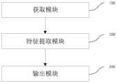
本实施例提供一种超声图像视频分类预测系统,用于执行如实施例所述的方法,如图8所示,为本实施例的超声图像视频分类预测系统结构框图,包括:This embodiment provides a system for classifying and predicting ultrasound images and videos, which is used to execute the method described in the embodiments, as shown in FIG. 8 , which is a structural block diagram of the system for classifying and predicting ultrasound images and videos of this embodiment, including:
获取模块100:用于获取原始视频图像;Obtaining module 100: used to obtain the original video image;
特征提取模块200:用于利用特征提取网络提取原始视频图像的特征;Feature extraction module 200: for utilizing the feature extraction network to extract the features of the original video image;
输出模块300:用于对特征利用时序上采样输出分类预测结果。Output module 300: used for outputting classification prediction results by using temporal upsampling on features.
上述中超声图像视频分类预测系统模块的具体细节已经在实施例一对应的超声图像视频分类预测方法中进行了详细的描述,因此此处不再赘述。The specific details of the above-mentioned ultrasonic image video classification prediction system modules have been described in detail in the ultrasonic image video classification prediction method corresponding to
另外,本发明还提供超声图像视频分类预测设备,包括:In addition, the present invention also provides ultrasonic image video classification prediction equipment, including:
至少一个处理器,以及与所述至少一个处理器通信连接的存储器;at least one processor, and a memory communicatively coupled to the at least one processor;
其中,所述处理器通过调用所述存储器中存储的计算机程序,用于执行如实施例一所述的方法。计算机程序即程序代码,当程序代码在超声图像视频分类预测设备上运行时,程序代码用于使超声图像视频分类预测设备执行本说明书上述实施例一部分描述的超声图像视频分类预测方法中的步骤。Wherein, the processor is used to execute the method described in
另外,本发明还提供一种计算机可读存储介质,计算机可读存储介质存储有计算机可执行指令,其中计算机可执行指令用于使计算机执行如实施例一所述的方法。In addition, the present invention also provides a computer-readable storage medium, where computer-executable instructions are stored in the computer-readable storage medium, where the computer-executable instructions are used to make a computer execute the method as described in
本发明通过获取包含时间维度、图像宽度、图像高度的原始视频图像,利用特征提取网络提取原始视频图像的特征,对特征利用时序上采样输出分类预测结果,其分类预测结果包括标准切面和非标准切面。利用特征提取网络高效的学习原始视频图像的时时序信息,通过时序上采样输出视频片段每帧的分类预测结果,提高预测效率,当用于超声视频数据时,能够提高超声诊断效率,减轻医生负担,而且有助于缓解医疗资源不足的问题。可广泛应用于视频图像预测领域。The present invention obtains the original video image including the time dimension, image width, and image height, uses the feature extraction network to extract the features of the original video image, and uses time-series upsampling to output the classification prediction results, and the classification prediction results include standard slices and non-standard slices. section. Using the feature extraction network to efficiently learn the time-series information of the original video image, and output the classification and prediction results of each frame of the video clip through time-series upsampling to improve the prediction efficiency. When used in ultrasound video data, it can improve the efficiency of ultrasound diagnosis and reduce the burden on doctors. , and help alleviate the problem of insufficient medical resources. It can be widely used in the field of video image prediction.
以上各实施例仅用以说明本发明的技术方案,而非对其限制,尽管参照前述各实施例对本发明进行了详细的说明,本领域的普通技术人员应当理解:其依然可以对前述各实施例所记载的技术方案进行修改,或者对其中部分或者全部技术特征进行等同替换;而这些修改或者替换,并不使相应技术方案的本质脱离本发明各实施例技术方案的范围,其均应涵盖在本发明的权利要求和说明书的范围当中。The above embodiments are only used to illustrate the technical solutions of the present invention, and are not intended to limit them. Although the present invention has been described in detail with reference to the foregoing embodiments, those of ordinary skill in the art should understand that: it can still be used for the foregoing embodiments Modifications to the technical solutions described in the examples, or equivalent replacement of some or all of the technical features; and these modifications or replacements do not make the essence of the corresponding technical solutions depart from the scope of the technical solutions of the embodiments of the present invention, and they shall cover Within the scope of the claims and description of the present invention.
Claims (6)
Translated fromChinese


Priority Applications (1)
| Application Number | Priority Date | Filing Date | Title |
|---|---|---|---|
| CN201911087843.9ACN110991254B (en) | 2019-11-08 | 2019-11-08 | Ultrasound image video classification prediction method and system |
Applications Claiming Priority (1)
| Application Number | Priority Date | Filing Date | Title |
|---|---|---|---|
| CN201911087843.9ACN110991254B (en) | 2019-11-08 | 2019-11-08 | Ultrasound image video classification prediction method and system |
Publications (2)
| Publication Number | Publication Date |
|---|---|
| CN110991254A CN110991254A (en) | 2020-04-10 |
| CN110991254Btrue CN110991254B (en) | 2023-07-04 |
Family
ID=70083586
Family Applications (1)
| Application Number | Title | Priority Date | Filing Date |
|---|---|---|---|
| CN201911087843.9AActiveCN110991254B (en) | 2019-11-08 | 2019-11-08 | Ultrasound image video classification prediction method and system |
Country Status (1)
| Country | Link |
|---|---|
| CN (1) | CN110991254B (en) |
Cited By (1)
| Publication number | Priority date | Publication date | Assignee | Title |
|---|---|---|---|---|
| WO2025063347A1 (en)* | 2023-09-18 | 2025-03-27 | (주)엠큐브테크놀로지 | Bladder volume measuring system and bladder volume measuring method in system |
Families Citing this family (7)
| Publication number | Priority date | Publication date | Assignee | Title |
|---|---|---|---|---|
| CN111317499A (en)* | 2018-12-17 | 2020-06-23 | 天津光电通信技术有限公司 | Heart sound signal processing method based on wavelet technology |
| CN111666852A (en)* | 2020-05-28 | 2020-09-15 | 天津大学 | Micro-expression double-flow network identification method based on convolutional neural network |
| CN112155604B (en)* | 2020-09-24 | 2023-03-31 | 广州爱孕记信息科技有限公司 | Fetal severe deformity detection method and device based on fetal ultrasound image |
| CN116433950A (en)* | 2021-12-31 | 2023-07-14 | 深圳开立生物医疗科技股份有限公司 | Method and device for identifying section type, ultrasonic equipment and storage medium |
| CN114842238B (en)* | 2022-04-01 | 2024-04-16 | 苏州视尚医疗科技有限公司 | Identification method of embedded breast ultrasonic image |
| CN114998835B (en)* | 2022-06-09 | 2024-08-27 | 天津大学 | Target statistical classification method and device for monitoring video |
| CN117934426A (en)* | 2024-01-26 | 2024-04-26 | 浙江省交通运输科学研究院 | Bridge structure health monitoring data anomaly identification method based on image classification |
Citations (2)
| Publication number | Priority date | Publication date | Assignee | Title |
|---|---|---|---|---|
| CN110032926A (en)* | 2019-02-22 | 2019-07-19 | 哈尔滨工业大学(深圳) | A kind of video classification methods and equipment based on deep learning |
| WO2019200753A1 (en)* | 2018-04-17 | 2019-10-24 | 平安科技(深圳)有限公司 | Lesion detection method, device, computer apparatus and storage medium |
Family Cites Families (1)
| Publication number | Priority date | Publication date | Assignee | Title |
|---|---|---|---|---|
| CN110838124B (en)* | 2017-09-12 | 2021-06-18 | 深圳科亚医疗科技有限公司 | Method, system, and medium for segmenting images of objects having sparse distribution |
- 2019
- 2019-11-08CNCN201911087843.9Apatent/CN110991254B/enactiveActive
Patent Citations (2)
| Publication number | Priority date | Publication date | Assignee | Title |
|---|---|---|---|---|
| WO2019200753A1 (en)* | 2018-04-17 | 2019-10-24 | 平安科技(深圳)有限公司 | Lesion detection method, device, computer apparatus and storage medium |
| CN110032926A (en)* | 2019-02-22 | 2019-07-19 | 哈尔滨工业大学(深圳) | A kind of video classification methods and equipment based on deep learning |
Non-Patent Citations (1)
| Title |
|---|
| Peiyao Kong 等.Automatic and Efficient Standard Plane Recognition in Fetal Ultrasound Images via Multi-scale Dense Networks.《Springer Nature Switzerland AG 2018》.2018,第160-168页.* |
Cited By (1)
| Publication number | Priority date | Publication date | Assignee | Title |
|---|---|---|---|---|
| WO2025063347A1 (en)* | 2023-09-18 | 2025-03-27 | (주)엠큐브테크놀로지 | Bladder volume measuring system and bladder volume measuring method in system |
Also Published As
| Publication number | Publication date |
|---|---|
| CN110991254A (en) | 2020-04-10 |
Similar Documents
| Publication | Publication Date | Title |
|---|---|---|
| CN110991254B (en) | Ultrasound image video classification prediction method and system | |
| Al-Fahdawi et al. | Fundus-deepnet: Multi-label deep learning classification system for enhanced detection of multiple ocular diseases through data fusion of fundus images | |
| Nazir et al. | Ecsu-net: an embedded clustering sliced u-net coupled with fusing strategy for efficient intervertebral disc segmentation and classification | |
| Saranya et al. | Blood vessel segmentation in retinal fundus images for proliferative diabetic retinopathy screening using deep learning | |
| Wu et al. | Cascaded fully convolutional networks for automatic prenatal ultrasound image segmentation | |
| US20240185428A1 (en) | Medical Image Analysis Using Neural Networks | |
| CN108898175A (en) | Area of computer aided model building method based on deep learning gastric cancer pathological section | |
| CN110930416A (en) | MRI image prostate segmentation method based on U-shaped network | |
| Zhao et al. | A nested U-shape network with multi-scale upsample attention for robust retinal vascular segmentation | |
| CN116681958B (en) | Fetal lung ultrasonic image maturity prediction method based on machine learning | |
| CN114565620B (en) | Fundus image blood vessel segmentation method based on skeleton prior and contrast loss | |
| Rachmatullah et al. | Convolutional neural network for semantic segmentation of fetal echocardiography based on four-chamber view | |
| CN113796877B (en) | Method, device and storage medium for obtaining stroke prediction value | |
| Lei et al. | Automated detection of retinopathy of prematurity by deep attention network | |
| CN116704305A (en) | Multi-modal and multi-section classification method for echocardiography based on deep learning algorithm | |
| Elmannai et al. | An Improved Deep Learning Framework for Automated Optic Disc Localization and Glaucoma Detection. | |
| Devisri et al. | Fetal growth analysis from ultrasound videos based on different biometrics using optimal segmentation and hybrid classifier | |
| Vo et al. | Multimodal breast lesion classification using cross-attention deep networks | |
| Machado | Mandible-focused osteoporosis risk assessment using dental panoramic radiography and artificial intelligence models | |
| Hsu et al. | A comprehensive study of age-related macular degeneration detection | |
| Wijesinghe et al. | Cardiac MRI Segmentation of Ventricular Structures and Myocardium Using U-Net Variants | |
| Shafi et al. | Improved Transfer Learning based Deep Convolutional Neural Network for Lung Disease Classification using CT image | |
| Tursyngaliyeva | Deep Learning for Medical Image Segmentation: Pneumonia Detection | |
| CN120452803B (en) | Noninvasive diabetic nephropathy grading and predicting method supporting convenient screening | |
| Tabrizi | Semantic segmentation of medical images with deep learning |
Legal Events
| Date | Code | Title | Description |
|---|---|---|---|
| PB01 | Publication | ||
| PB01 | Publication | ||
| SE01 | Entry into force of request for substantive examination | ||
| SE01 | Entry into force of request for substantive examination | ||
| GR01 | Patent grant | ||
| GR01 | Patent grant |