CN110951580B - High-throughput single-cell transcriptome and gene mutation integration analysis integrated device - Google Patents
High-throughput single-cell transcriptome and gene mutation integration analysis integrated deviceDownload PDFInfo
- Publication number
- CN110951580B CN110951580BCN201910932615.0ACN201910932615ACN110951580BCN 110951580 BCN110951580 BCN 110951580BCN 201910932615 ACN201910932615 ACN 201910932615ACN 110951580 BCN110951580 BCN 110951580B
- Authority
- CN
- China
- Prior art keywords
- cell
- fluorescence
- analysis
- gene mutation
- throughput
- Prior art date
- Legal status (The legal status is an assumption and is not a legal conclusion. Google has not performed a legal analysis and makes no representation as to the accuracy of the status listed.)
- Active
Links
- 238000004458analytical methodMethods0.000titleclaimsabstractdescription46
- 206010064571Gene mutationDiseases0.000titleclaimsabstractdescription45
- 230000010354integrationEffects0.000titleclaimsabstractdescription29
- 238000000799fluorescence microscopyMethods0.000claimsabstractdescription25
- 150000007523nucleic acidsChemical class0.000claimsabstractdescription22
- 238000013500data storageMethods0.000claimsabstractdescription21
- 238000007405data analysisMethods0.000claimsabstractdescription20
- 238000012351Integrated analysisMethods0.000claimsabstractdescription19
- 238000003384imaging methodMethods0.000claimsabstractdescription19
- 108090000623proteins and genesProteins0.000claimsabstractdescription17
- 238000002073fluorescence micrographMethods0.000claimsabstractdescription16
- 238000012408PCR amplificationMethods0.000claimsabstractdescription13
- 210000004027cellAnatomy0.000claimsdescription91
- 239000002299complementary DNASubstances0.000claimsdescription17
- 230000005284excitationEffects0.000claimsdescription11
- 102000018697Membrane ProteinsHuman genes0.000claimsdescription10
- 108010052285Membrane ProteinsProteins0.000claimsdescription10
- 108091028043Nucleic acid sequenceProteins0.000claimsdescription9
- 230000035772mutationEffects0.000claimsdescription9
- 230000003321amplificationEffects0.000claimsdescription8
- 238000003199nucleic acid amplification methodMethods0.000claimsdescription8
- 238000011065in-situ storageMethods0.000claimsdescription7
- 238000006243chemical reactionMethods0.000claimsdescription4
- 230000003287optical effectEffects0.000claimsdescription4
- 125000006850spacer groupChemical group0.000claimsdescription4
- 230000009089cytolysisEffects0.000claimsdescription3
- 239000007850fluorescent dyeSubstances0.000claimsdescription3
- 238000001215fluorescent labellingMethods0.000claimsdescription3
- 239000011148porous materialSubstances0.000claimsdescription3
- 238000010839reverse transcriptionMethods0.000claimsdescription2
- 238000005382thermal cyclingMethods0.000claims1
- 102000004169proteins and genesHuman genes0.000abstractdescription7
- 108020004707nucleic acidsProteins0.000abstractdescription6
- 102000039446nucleic acidsHuman genes0.000abstractdescription6
- 230000006870functionEffects0.000abstractdescription3
- 206010028980NeoplasmDiseases0.000description22
- 108091032973(ribonucleotides)n+mProteins0.000description20
- 239000000523sampleSubstances0.000description18
- 238000005516engineering processMethods0.000description15
- 238000012163sequencing techniqueMethods0.000description9
- 238000010586diagramMethods0.000description7
- 206010059866Drug resistanceDiseases0.000description4
- 238000001514detection methodMethods0.000description4
- 238000012165high-throughput sequencingMethods0.000description4
- 210000004881tumor cellAnatomy0.000description4
- 239000003153chemical reaction reagentSubstances0.000description3
- 238000000034methodMethods0.000description3
- 102000040650(ribonucleotides)n+mHuman genes0.000description2
- 108020004635Complementary DNAProteins0.000description2
- XUIMIQQOPSSXEZ-UHFFFAOYSA-NSiliconChemical compound[Si]XUIMIQQOPSSXEZ-UHFFFAOYSA-N0.000description2
- 201000011510cancerDiseases0.000description2
- 238000012512characterization methodMethods0.000description2
- 230000017525heat dissipationEffects0.000description2
- 238000002372labellingMethods0.000description2
- 208000032839leukemiaDiseases0.000description2
- 230000037361pathwayEffects0.000description2
- 230000008261resistance mechanismEffects0.000description2
- 229910052710siliconInorganic materials0.000description2
- 239000010703siliconSubstances0.000description2
- 238000003786synthesis reactionMethods0.000description2
- 238000007671third-generation sequencingMethods0.000description2
- 238000011222transcriptome analysisMethods0.000description2
- 108700028369AllelesProteins0.000description1
- 108010077544ChromatinProteins0.000description1
- VYZAMTAEIAYCRO-UHFFFAOYSA-NChromiumChemical compound[Cr]VYZAMTAEIAYCRO-UHFFFAOYSA-N0.000description1
- 108091034117OligonucleotideProteins0.000description1
- 108010026552ProteomeProteins0.000description1
- JLCPHMBAVCMARE-UHFFFAOYSA-N[3-[[3-[[3-[[3-[[3-[[3-[[3-[[3-[[3-[[3-[[3-[[5-(2-amino-6-oxo-1H-purin-9-yl)-3-[[3-[[3-[[3-[[3-[[3-[[5-(2-amino-6-oxo-1H-purin-9-yl)-3-[[5-(2-amino-6-oxo-1H-purin-9-yl)-3-hydroxyoxolan-2-yl]methoxy-hydroxyphosphoryl]oxyoxolan-2-yl]methoxy-hydroxyphosphoryl]oxy-5-(5-methyl-2,4-dioxopyrimidin-1-yl)oxolan-2-yl]methoxy-hydroxyphosphoryl]oxy-5-(6-aminopurin-9-yl)oxolan-2-yl]methoxy-hydroxyphosphoryl]oxy-5-(6-aminopurin-9-yl)oxolan-2-yl]methoxy-hydroxyphosphoryl]oxy-5-(6-aminopurin-9-yl)oxolan-2-yl]methoxy-hydroxyphosphoryl]oxy-5-(6-aminopurin-9-yl)oxolan-2-yl]methoxy-hydroxyphosphoryl]oxyoxolan-2-yl]methoxy-hydroxyphosphoryl]oxy-5-(5-methyl-2,4-dioxopyrimidin-1-yl)oxolan-2-yl]methoxy-hydroxyphosphoryl]oxy-5-(4-amino-2-oxopyrimidin-1-yl)oxolan-2-yl]methoxy-hydroxyphosphoryl]oxy-5-(5-methyl-2,4-dioxopyrimidin-1-yl)oxolan-2-yl]methoxy-hydroxyphosphoryl]oxy-5-(5-methyl-2,4-dioxopyrimidin-1-yl)oxolan-2-yl]methoxy-hydroxyphosphoryl]oxy-5-(6-aminopurin-9-yl)oxolan-2-yl]methoxy-hydroxyphosphoryl]oxy-5-(6-aminopurin-9-yl)oxolan-2-yl]methoxy-hydroxyphosphoryl]oxy-5-(4-amino-2-oxopyrimidin-1-yl)oxolan-2-yl]methoxy-hydroxyphosphoryl]oxy-5-(4-amino-2-oxopyrimidin-1-yl)oxolan-2-yl]methoxy-hydroxyphosphoryl]oxy-5-(4-amino-2-oxopyrimidin-1-yl)oxolan-2-yl]methoxy-hydroxyphosphoryl]oxy-5-(6-aminopurin-9-yl)oxolan-2-yl]methoxy-hydroxyphosphoryl]oxy-5-(4-amino-2-oxopyrimidin-1-yl)oxolan-2-yl]methyl [5-(6-aminopurin-9-yl)-2-(hydroxymethyl)oxolan-3-yl] hydrogen phosphatePolymersCc1cn(C2CC(OP(O)(=O)OCC3OC(CC3OP(O)(=O)OCC3OC(CC3O)n3cnc4c3nc(N)[nH]c4=O)n3cnc4c3nc(N)[nH]c4=O)C(COP(O)(=O)OC3CC(OC3COP(O)(=O)OC3CC(OC3COP(O)(=O)OC3CC(OC3COP(O)(=O)OC3CC(OC3COP(O)(=O)OC3CC(OC3COP(O)(=O)OC3CC(OC3COP(O)(=O)OC3CC(OC3COP(O)(=O)OC3CC(OC3COP(O)(=O)OC3CC(OC3COP(O)(=O)OC3CC(OC3COP(O)(=O)OC3CC(OC3COP(O)(=O)OC3CC(OC3COP(O)(=O)OC3CC(OC3COP(O)(=O)OC3CC(OC3COP(O)(=O)OC3CC(OC3COP(O)(=O)OC3CC(OC3COP(O)(=O)OC3CC(OC3CO)n3cnc4c(N)ncnc34)n3ccc(N)nc3=O)n3cnc4c(N)ncnc34)n3ccc(N)nc3=O)n3ccc(N)nc3=O)n3ccc(N)nc3=O)n3cnc4c(N)ncnc34)n3cnc4c(N)ncnc34)n3cc(C)c(=O)[nH]c3=O)n3cc(C)c(=O)[nH]c3=O)n3ccc(N)nc3=O)n3cc(C)c(=O)[nH]c3=O)n3cnc4c3nc(N)[nH]c4=O)n3cnc4c(N)ncnc34)n3cnc4c(N)ncnc34)n3cnc4c(N)ncnc34)n3cnc4c(N)ncnc34)O2)c(=O)[nH]c1=OJLCPHMBAVCMARE-UHFFFAOYSA-N0.000description1
- 230000002159abnormal effectEffects0.000description1
- 230000009286beneficial effectEffects0.000description1
- 230000015572biosynthetic processEffects0.000description1
- 238000004422calculation algorithmMethods0.000description1
- 230000001413cellular effectEffects0.000description1
- 210000003483chromatinAnatomy0.000description1
- 229910052804chromiumInorganic materials0.000description1
- 239000011651chromiumSubstances0.000description1
- 238000003759clinical diagnosisMethods0.000description1
- 230000000295complement effectEffects0.000description1
- 238000012350deep sequencingMethods0.000description1
- 230000007812deficiencyEffects0.000description1
- 238000003745diagnosisMethods0.000description1
- 201000010099diseaseDiseases0.000description1
- 208000037265diseases, disorders, signs and symptomsDiseases0.000description1
- 238000013399early diagnosisMethods0.000description1
- 238000005530etchingMethods0.000description1
- 238000001506fluorescence spectroscopyMethods0.000description1
- 230000007614genetic variationEffects0.000description1
- 238000012268genome sequencingMethods0.000description1
- 238000003205genotyping methodMethods0.000description1
- PCHJSUWPFVWCPO-UHFFFAOYSA-NgoldChemical compound[Au]PCHJSUWPFVWCPO-UHFFFAOYSA-N0.000description1
- 230000036541healthEffects0.000description1
- 238000010438heat treatmentMethods0.000description1
- 238000010166immunofluorescenceMethods0.000description1
- 238000007641inkjet printingMethods0.000description1
- 239000006166lysateSubstances0.000description1
- 238000010801machine learningMethods0.000description1
- 230000036210malignancyEffects0.000description1
- 238000004519manufacturing processMethods0.000description1
- 239000003550markerSubstances0.000description1
- 230000007246mechanismEffects0.000description1
- 238000005457optimizationMethods0.000description1
- 230000008506pathogenesisEffects0.000description1
- 238000000206photolithographyMethods0.000description1
- 238000010837poor prognosisMethods0.000description1
- 238000002360preparation methodMethods0.000description1
- 238000007637random forest analysisMethods0.000description1
- 238000000926separation methodMethods0.000description1
- 239000000758substrateSubstances0.000description1
- 238000002560therapeutic procedureMethods0.000description1
- 238000013518transcriptionMethods0.000description1
- 230000035897transcriptionEffects0.000description1
- 238000012070whole genome sequencing analysisMethods0.000description1
Images


Classifications
- C—CHEMISTRY; METALLURGY
- C12—BIOCHEMISTRY; BEER; SPIRITS; WINE; VINEGAR; MICROBIOLOGY; ENZYMOLOGY; MUTATION OR GENETIC ENGINEERING
- C12Q—MEASURING OR TESTING PROCESSES INVOLVING ENZYMES, NUCLEIC ACIDS OR MICROORGANISMS; COMPOSITIONS OR TEST PAPERS THEREFOR; PROCESSES OF PREPARING SUCH COMPOSITIONS; CONDITION-RESPONSIVE CONTROL IN MICROBIOLOGICAL OR ENZYMOLOGICAL PROCESSES
- C12Q1/00—Measuring or testing processes involving enzymes, nucleic acids or microorganisms; Compositions therefor; Processes of preparing such compositions
- C12Q1/68—Measuring or testing processes involving enzymes, nucleic acids or microorganisms; Compositions therefor; Processes of preparing such compositions involving nucleic acids
- C12Q1/6844—Nucleic acid amplification reactions
- C12Q1/6858—Allele-specific amplification
Landscapes
- Chemical & Material Sciences (AREA)
- Life Sciences & Earth Sciences (AREA)
- Organic Chemistry (AREA)
- Engineering & Computer Science (AREA)
- Zoology (AREA)
- Wood Science & Technology (AREA)
- Proteomics, Peptides & Aminoacids (AREA)
- Health & Medical Sciences (AREA)
- Biophysics (AREA)
- Chemical Kinetics & Catalysis (AREA)
- Immunology (AREA)
- Microbiology (AREA)
- Molecular Biology (AREA)
- Analytical Chemistry (AREA)
- Physics & Mathematics (AREA)
- Biotechnology (AREA)
- Biochemistry (AREA)
- Bioinformatics & Cheminformatics (AREA)
- General Engineering & Computer Science (AREA)
- General Health & Medical Sciences (AREA)
- Genetics & Genomics (AREA)
- Measuring Or Testing Involving Enzymes Or Micro-Organisms (AREA)
- Investigating, Analyzing Materials By Fluorescence Or Luminescence (AREA)
Abstract
Description
Translated fromChinese技术领域technical field
本发明涉及生物检测领域,特别涉及一种高通量单细胞转录组与基因突变整合分析一体化装置。The invention relates to the field of biological detection, in particular to a high-throughput single-cell transcriptome and gene mutation integration analysis integrated device.
背景技术Background technique
肿瘤是严重影响人类健康的重大疾病之一,肿瘤细胞从基因型到表型上存在极大的差异(肿瘤的高度异质性),而这种高度异质性与肿瘤的恶性程度、耐药性、复发转移等都密切相关,是造成肿瘤早期诊断困难、临床诊治复杂、耐药复发和预后差的根源之一。全面解析肿瘤异质性是实现肿瘤精准治疗的关键。Tumor is one of the major diseases that seriously affects human health. There are great differences in tumor cells from genotype to phenotype (high heterogeneity of tumors), and this high heterogeneity is related to the degree of malignancy and drug resistance of tumors. It is one of the root causes of difficult early diagnosis of tumors, complicated clinical diagnosis and treatment, drug resistance recurrence and poor prognosis. Comprehensive analysis of tumor heterogeneity is the key to realizing precise tumor treatment.
高通量测序技术的发展为解析异质性肿瘤群体带来希望。目前各种组学水平的常规高通量测序成为肿瘤异质性群体研究的常用手段,用来发现新的遗传变异或异常通路,探索新的发病或耐药机制等。然而目前基于bulk(混合群体)的常规高通量测序技术无法克服肿瘤细胞高度异质性的难题,仅能通过大样本人群研究发现关键主克隆变异及通路改变,难以实现对单个患者异质性克隆群体的全面解析,成为实现肿瘤精准治疗的瓶颈。近年来新兴的单细胞测序技术为解析肿瘤异质性、鉴别不同功能亚群提供了可能。单细胞测序能够获得每个细胞的基因组变异图谱及转录组表达图谱,通过单个细胞的图谱精确划分克隆归属,实现对异质性克隆群体的全面解析。Timothy A.Graubert团队将一例已通过全基因组测序与靶向深度测序进行全面刻画的继发白血病样本进行了单细胞基因组分型测序,仅通过12个细胞DNA测序数据即发现了之前被认为是一个亚克隆的群体其实是由两个互斥的亚克隆构成,充分说明单细胞测序的优势和其在多克隆研究中的必要性。然而早期的单细胞测序技术往往通量低,成本高,一定程度上限制了精确分析并追踪异质性群体变化的分析。2016年10x Genomics公司推出的10x Chromium Single Cell Gene ExpressionSolution平台实现了高通量的单细胞转录组测序,具有周期短、成本低、细胞捕获率高等优势,在发育生物学及肿瘤异质性群体研究中应用广泛,在转录组水平实现对异质性肿瘤群体的全面刻画。The development of high-throughput sequencing technologies has brought hope for dissecting heterogeneous tumor populations. At present, conventional high-throughput sequencing at various omics levels has become a common method for studying tumor heterogeneity groups, which is used to discover new genetic variations or abnormal pathways, and explore new pathogenesis or drug resistance mechanisms. However, at present, conventional high-throughput sequencing technology based on bulk (mixed population) cannot overcome the problem of high heterogeneity of tumor cells. It is only possible to discover key main clonal variants and pathway changes through large sample population studies, and it is difficult to realize the heterogeneity of individual patients. The comprehensive analysis of the clonal population has become a bottleneck for the realization of precise tumor therapy. In recent years, emerging single-cell sequencing technologies have provided the possibility to analyze tumor heterogeneity and identify different functional subgroups. Single-cell sequencing can obtain the genome variation map and transcriptome expression map of each cell, and accurately divide the clone attribution through the map of a single cell to achieve a comprehensive analysis of the heterogeneous clonal population. Timothy A. Graubert's team performed single-cell genotyping sequencing of a secondary leukemia sample that had been fully characterized by whole genome sequencing and targeted deep sequencing, and found that it was previously thought to be a The subclone population is actually composed of two mutually exclusive subclones, which fully demonstrates the advantages of single-cell sequencing and its necessity in polyclonal research. However, early single-cell sequencing technologies tended to be low-throughput and high-cost, which limited to a certain extent accurate analysis and tracking of changes in heterogeneous populations. The 10x Chromium Single Cell Gene ExpressionSolution platform launched by 10x Genomics in 2016 realizes high-throughput single-cell transcriptome sequencing. It has the advantages of short cycle, low cost, and high cell capture rate. It is used in developmental biology and tumor heterogeneous population research. It has a wide range of applications and achieves a comprehensive characterization of heterogeneous tumor populations at the transcriptome level.
然而对于基因组变异驱动的恶性肿瘤群体,仅从转录组水平无法实现对肿瘤群体的鉴定以及功能异质性的解析。研究者开始着眼于基于单细胞水平的多组学研究平台,10x和BD公司分别实现单细胞转录组与单细胞染色质开放性(ATAC-seq)或单细胞蛋白质组的结合。然而,对于肿瘤异质性研究中最需要的单细胞转录组与基因组信息的整合平台,目前尚无成熟技术。对此,来自不同实验室的研究者进行了大量尝试,目前大部分技术仍然依赖同时将单个细胞中的转录组与基因组进行分离而分别测序,操作繁琐且通量较小。对转录组和基因组同时测序的技术又面临扩增效率低下或等位基因扩增偏好等难题,近期新提出的Target-seq技术针对肿瘤群体设计同时检测转录组及特异基因突变的技术,也说明了肿瘤研究中对该技术的需求,但该技术仍处于实验室水平,仍然无法实现一次上千细胞数的分析。另外,Peter Van Galen等人在Cell发表文献,通过单细胞转录本与三代测序技术相结合,首次实现对白血病患者肿瘤群体(以基因组变异为金标准)中转录组异质性的解析,发现肿瘤群体存在于表达谱不同的多种谱系中,明确了基因组异质性与转录组异质性相互独立又相互影响的关系,也表明在单细胞转录组水平进一步明确细胞的基因组变异的重要性。然而,该研究中使用的三代测序检测突变的技术具有很大局限性,突变检出率受到具体突变位点的限制,单个突变检出率最高仅23%,平均可以检测到突变的细胞不超过5%,作者最终采用随机森林的机器学习算法预测肿瘤群体,无法实现对肿瘤细胞群体的直接鉴定,也没有将基因组与转录组异质性很好的对应。且该技术操作繁琐,样本需求量高,花费大,不适于全面推广。However, for the malignant tumor population driven by genomic variation, the identification of tumor population and the analysis of functional heterogeneity cannot be achieved only from the transcriptome level. Researchers began to focus on multi-omics research platforms based on the single-cell level, and 10x and BD companies realized the combination of single-cell transcriptome and single-cell chromatin openness (ATAC-seq) or single-cell proteome, respectively. However, there is currently no mature technology for the integration of single-cell transcriptomic and genomic information, which is most needed in tumor heterogeneity studies. In this regard, researchers from different laboratories have made a lot of attempts. Most of the current technologies still rely on the simultaneous separation of the transcriptome and the genome in a single cell for separate sequencing, which is cumbersome and has low throughput. The technology of simultaneous transcriptome and genome sequencing is faced with problems such as low amplification efficiency or allele amplification bias. Recently, the newly proposed Target-seq technology is designed for the tumor population to simultaneously detect transcriptome and specific gene mutations. It also shows that The demand for this technology in tumor research has increased, but the technology is still at the laboratory level, and it is still impossible to analyze thousands of cells at a time. In addition, Peter Van Galen et al. published a paper in Cell. Through the combination of single-cell transcripts and third-generation sequencing technology, the analysis of transcriptome heterogeneity in the tumor population of leukemia patients (with genomic variation as the gold standard) was achieved for the first time. Populations exist in a variety of lineages with different expression profiles, which clarifies the relationship between genomic heterogeneity and transcriptome heterogeneity that are independent and mutually influencing, and also shows the importance of further clarifying the genomic variation of cells at the single-cell transcriptome level. However, the third-generation sequencing technology used in this study to detect mutations has great limitations. The mutation detection rate is limited by the specific mutation site. The highest single mutation detection rate is only 23%, and the average number of cells that can detect mutations is not more than 23%. 5%, the authors finally used the random forest machine learning algorithm to predict the tumor population, which could not realize the direct identification of the tumor cell population, nor did the genome and transcriptome heterogeneity correspond well. Moreover, this technique is cumbersome to operate, requires high samples, and costs a lot, so it is not suitable for comprehensive promotion.
因此,在肿瘤研究中实现单细胞水平基因组与转录组异质性的整合分析具有重要性与迫切性。Therefore, it is important and urgent to realize the integrated analysis of genomic and transcriptomic heterogeneity at the single-cell level in tumor research.
发明内容SUMMARY OF THE INVENTION
本发明所要解决的技术问题在于针对上述现有技术中的不足,提供一种高通量单细胞转录组与基因突变整合分析一体化装置。The technical problem to be solved by the present invention is to provide a high-throughput single-cell transcriptome and gene mutation integration analysis integrated device in view of the above-mentioned deficiencies in the prior art.
为解决上述技术问题,本发明采用的技术方案是:一种高通量单细胞转录组与基因突变整合分析一体化装置,包括高通量单细胞编码芯片和整合分析装置;In order to solve the above technical problems, the technical solution adopted in the present invention is: a high-throughput single-cell transcriptome and gene mutation integrated analysis device, including a high-throughput single-cell coding chip and an integrated analysis device;
所述高通量单细胞编码芯片具有用于捕获单细胞的微孔,每个所述微孔具有唯一的空间坐标编码,且所述微孔内修饰有若干条用于捕获目标RNA的核酸序列,所述核酸序列包括用于标示RNA源自的细胞的细胞标签和用于标示结合的RNA的分子标签,每个微孔的细胞标签与空间坐标编码一一对应;The high-throughput single-cell coding chip has micropores for capturing single cells, each of the micropores has a unique spatial coordinate code, and several nucleic acid sequences for capturing target RNA are modified in the micropores , the nucleic acid sequence includes a cell tag for indicating the cell from which the RNA is derived and a molecular tag for indicating the combined RNA, and the cell tag of each microwell corresponds to the spatial coordinate code one-to-one;
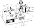
所述整合分析装置包括壳体以及设置在所述壳体内的温控热循环模块、荧光成像模块和数据存储分析模块,所述荧光成像模块包括光源组件、显微物镜、荧光分光组件和成像探测器;The integrated analysis device includes a housing and a temperature-controlled thermal cycle module, a fluorescence imaging module, and a data storage and analysis module arranged in the housing. The fluorescence imaging module includes a light source assembly, a microscope objective lens, a fluorescence spectroscopic assembly, and an imaging detector. device;
所述温控热循环组件上设置有用于安置所述高通量单细胞编码芯片的载物热台,所述温控热循环组件用于提供PCR扩增反应所需的温度环境;The temperature-controlled thermal cycle assembly is provided with a thermal stage for placing the high-throughput single-cell encoding chip, and the temperature-controlled thermal cycle assembly is used to provide a temperature environment required for the PCR amplification reaction;
所述光源组件发出的激发光经所述荧光分光组件后,再经过所述显微物镜后到达所述温控热循环组件上的所述高通量单细胞编码芯片,其中的样品被激发产生的荧光原路返回经过所述显微物镜和荧光分光模块后,再进入所述成像探测器,进行荧光成像;After the excitation light emitted by the light source assembly passes through the fluorescence spectroscopic assembly, and then passes through the microscope objective lens, it reaches the high-throughput single-cell encoding chip on the temperature-controlled thermal cycle assembly, where the sample is excited to generate After returning to the original path of the fluorescence through the microscope objective lens and the fluorescence spectroscopic module, it enters the imaging detector for fluorescence imaging;
所述数据存储分析模块用于存储所述成像探测器采集的荧光图像信息,并进行单细胞转录组与基因突变整合分析。The data storage and analysis module is used for storing the fluorescence image information collected by the imaging detector, and performing integrated analysis of single-cell transcriptome and gene mutation.
优选的是,所述光源组件包括第一LED光源、第二LED光源、第三LED光源、第一二向色镜、第二二向色镜和扩束透镜组,Preferably, the light source assembly includes a first LED light source, a second LED light source, a third LED light source, a first dichroic mirror, a second dichroic mirror and a beam expander lens group,
所述第一LED光源发出的光依次透射所述第一二向色镜、第二二向色镜后到达所述扩束透镜组;The light emitted by the first LED light source sequentially transmits the first dichroic mirror and the second dichroic mirror to reach the beam expander lens group;
所述第二LED光源发出的光经第一二向色镜反射、第二二向色镜透射后到达所述扩束透镜组;The light emitted by the second LED light source is reflected by the first dichroic mirror and transmitted by the second dichroic mirror and then reaches the beam expander lens group;
所述第三LED光源发出的光经第二二向色镜反射后到达所述扩束透镜组;The light emitted by the third LED light source is reflected by the second dichroic mirror and reaches the beam expander lens group;
所述第一LED光源、第二LED光源、第三LED光源发出三种不同波长的光,且三种光的波长范围覆盖400nm-700nm。The first LED light source, the second LED light source, and the third LED light source emit light of three different wavelengths, and the wavelength ranges of the three kinds of light cover 400nm-700nm.
优选的是,所述荧光分光组件包括支架、可转动设置在所述支架上的旋转台、用于驱动所述旋转台转动的电机以及均匀间隔设置在所述旋转台上的若干个荧光分光模块,Preferably, the fluorescence spectroscopic assembly includes a bracket, a rotary table rotatably arranged on the bracket, a motor for driving the rotary table to rotate, and a plurality of fluorescence spectroscopic modules arranged on the rotary table at regular intervals ,
所述荧光分光模块包括激发光滤光片、样品光滤光片和第四二向色镜;The fluorescence spectroscopic module includes an excitation light filter, a sample light filter and a fourth dichroic mirror;
所述旋转台用于将若干个所述荧光分光模块中的一个切换进入光路,所述扩束透镜组出射的激发光经过所述激发光滤光片后被所述第四二向色镜反射,然后经过所述显微物镜到达安置在所述载物热台上的高通量单细胞编码芯片上;所述高通量单细胞编码芯片中的样品产生的荧光经过所述显微物镜后透射所述第四二向色镜,再经过所述样品光滤光片后到达所述成像探测器,所述成像探测器采集的荧光图像信息传输至所述数据存储分析模块。The rotating stage is used to switch one of the several fluorescence splitting modules into the optical path, and the excitation light emitted by the beam expander lens group is reflected by the fourth dichroic mirror after passing through the excitation light filter. , and then through the microscope objective lens to the high-throughput single-cell coding chip arranged on the thermal stage of the object; the fluorescence generated by the sample in the high-throughput single-cell coding chip passes through the microscope objective lens. The fourth dichroic mirror is transmitted through the sample light filter to reach the imaging detector, and the fluorescence image information collected by the imaging detector is transmitted to the data storage and analysis module.
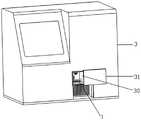
优选的是,所述壳体上设置有放样窗口,所述放样窗口上设置有滑盖。Preferably, a stakeout window is provided on the housing, and a slide cover is provided on the stakeout window.
优选的是,所述温控热循环组件包括温控盒体、设置在所述温控盒体内的散热器、设置在所述散热器上的帕尔贴以及设置在所述散热器侧部的风扇,所述载物热台设置在所述帕尔贴上,所述载物热台上设置有透明盖板。Preferably, the temperature control thermal cycle assembly includes a temperature control box body, a radiator arranged in the temperature control box body, a Peltier arranged on the radiator, and a radiator arranged on the side of the radiator A fan, the object carrier heat stage is arranged on the Peltier, and a transparent cover plate is arranged on the object carrier heat stage.
优选的是,所述核酸序列还包括Spacer序列、作为PCR扩增时的引物结合区域的通用引物序列以及Ploy T。Preferably, the nucleic acid sequence further includes a Spacer sequence, a universal primer sequence used as a primer binding region during PCR amplification, and Ploy T.
优选的是,单个所述微孔内的所有细胞标签具有相同的序列,不同所述微孔内的细胞标签的序列均不相同,从而通过所述细胞标签标识RNA源自的细胞;Preferably, all cell tags in a single microwell have the same sequence, and the sequences of cell tags in different microwells are different, so that the cell tag is used to identify the cell from which the RNA is derived;
单个所述微孔内的所有分子标签具有不同的序列,从而通过所述分子标签标识单个细胞中的RNA。All molecular tags within a single said microwell have different sequences, thereby identifying RNA in a single cell by said molecular tags.
优选的是,所述微孔具有在一个微孔中只能容纳单个细胞的尺寸和形状。Preferably, the microwells are of a size and shape that can accommodate only a single cell in a microwell.
优选的是,所述微孔为正六边形,且呈蜂窝状排列,其数量为102-106个;Preferably, the micropores are regular hexagons and arranged in a honeycomb shape, and the number thereof is 102 -106 ;
所述微孔的外接圆的直径为30-60μm,深度为20-300μm,孔间的间距为10-30μm。The diameter of the circumscribed circle of the micro-holes is 30-60 μm, the depth is 20-300 μm, and the spacing between the holes is 10-30 μm.
优选的是,所述高通量单细胞转录组与基因突变整合分析一体化装置分析步骤包括:Preferably, the analysis step of the integrated device for high-throughput single-cell transcriptome and gene mutation integration analysis includes:
1)将样品加入所述高通量单细胞编码芯片中,通过其上的微孔捕获单细胞,将所述高通量单细胞编码芯片置于所述载物热台上,通过所述荧光成像模块对所述高通量单细胞编码芯片进行荧光成像,利用所述数据存储分析模块进行各个微孔位置的单细胞表面蛋白分型分析;1) Add the sample into the high-throughput single-cell coding chip, capture single cells through the micropores on it, place the high-throughput single-cell coding chip on the thermal stage of the carrier, and pass the fluorescence The imaging module performs fluorescence imaging on the high-throughput single-cell encoding chip, and uses the data storage and analysis module to perform single-cell surface protein typing analysis at each microwell position;
2)对所述高通量单细胞编码芯片的微孔中的单细胞进行原位裂解扩增,逆转录合成携带细胞标签、分子标签的cDNA,游离cDNA收集后用于单细胞转录组分析,固定在微孔内的cDNA序列用于基因突变分析;2) performing in-situ lysis amplification on the single cells in the micropores of the high-throughput single-cell coding chip, reverse-transcribed to synthesize cDNAs carrying cell tags and molecular tags, and the free cDNAs are collected for single-cell transcriptome analysis, cDNA sequences immobilized in microwells for gene mutation analysis;
3)启动所述温控热循环组件,针对固定在所述高通量单细胞编码芯片的微孔内的cDNA进行PCR扩增,并对目的基因的野生型和突变型进行双色荧光标记,通过所述荧光成像模块采集双色荧光图像,然后通过所述数据存储分析模块对双色荧光图像进行分析,计算各微孔位置的野生型与突变型的比例,得到单细胞的基因突变表达信息;3) Start the temperature-controlled thermal cycle assembly, carry out PCR amplification for the cDNA fixed in the micropores of the high-throughput single-cell coding chip, and carry out double-color fluorescent labeling on the wild type and mutant type of the target gene, and pass The fluorescence imaging module collects a dual-color fluorescence image, and then analyzes the dual-color fluorescence image through the data storage and analysis module, calculates the ratio of the wild type to the mutant type at each microwell position, and obtains the gene mutation expression information of a single cell;
4)通过所述数据存储分析模块将相同位置的单细胞表面蛋白分型信息与扩增后的单细胞基因突变表达信息结合,建立单细胞表面蛋白分型与突变整合分析的数据库。4) The single cell surface protein typing information at the same position is combined with the amplified single cell gene mutation expression information through the data storage and analysis module to establish a database for single cell surface protein typing and mutation integration analysis.
本发明的有益效果是:本发明的高通量单细胞转录组与基因突变整合分析一体化装置,通过设计具有微孔空间坐标、细胞核酸标签和分子核酸标签的三重编码功能的高通量单细胞编码芯片,可将单细胞的基因突变、转录组和蛋白表达信息一一对应起来;再通过温控热循环模块可实现PCR扩增,通过荧光成像模块采集样品的荧光图像,通过数据存储分析模块对荧光图像进行存储于分析,能实现单细胞表面蛋白分型与突变整合分析的数据库、高通量单细胞转录组与基因突变整合分析的完整数据库的建立,实现单细胞转录组与基因突变整合分析;The beneficial effects of the present invention are as follows: the high-throughput single-cell transcriptome and gene mutation integration analysis integrated device of the present invention, by designing a high-throughput single cell with triple encoding functions of micropore space coordinates, cell nucleic acid tags and molecular nucleic acid tags The cell coding chip can match the gene mutation, transcriptome and protein expression information of a single cell one by one; then PCR amplification can be realized through the temperature-controlled thermal cycle module, and the fluorescence image of the sample is collected through the fluorescence imaging module, and the data is stored and analyzed through data storage and analysis. The module stores and analyzes fluorescence images, and can realize the establishment of a database for single-cell surface protein typing and mutation integration analysis, a complete database for high-throughput single-cell transcriptome and gene mutation integration analysis, and realize single-cell transcriptome and gene mutation. integrated analysis;
本发明在明确单个细胞中携带的基因组变异信息后,结合单细胞转录组甚至蛋白表达信息,可实现对肿瘤细胞多组学的全面认识和对肿瘤异质性群体的全面刻画,为肿瘤的早期诊断、耐药机制、新靶点探索和治疗方案优化提供基础。After the genome variation information carried in a single cell is clarified, the invention can realize a comprehensive understanding of the multi-omics of tumor cells and a comprehensive characterization of the tumor heterogeneity group by combining the single cell transcriptome and even the protein expression information, which is an early stage of the tumor. It provides the basis for diagnosis, drug resistance mechanism, new target exploration and treatment optimization.
附图说明Description of drawings
图1为本发明的高通量单细胞转录组与基因突变整合分析一体化装置的原理框图;Fig. 1 is the principle block diagram of the integrated device for high-throughput single-cell transcriptome and gene mutation integration analysis of the present invention;
图2为本发明的高通量单细胞转录组与基因突变整合分析一体化装置的内部结构示意图;2 is a schematic diagram of the internal structure of the integrated device for high-throughput single-cell transcriptome and gene mutation integration analysis of the present invention;
图3为本发明的高通量单细胞转录组与基因突变整合分析一体化装置的外部结构示意图;3 is a schematic diagram of the external structure of the integrated device for high-throughput single-cell transcriptome and gene mutation integration analysis of the present invention;
图4为本发明的荧光分光组件的支架的结构示意图;Fig. 4 is the structural schematic diagram of the bracket of the fluorescence spectroscopic assembly of the present invention;
图5为本发明的荧光分光模块的结构示意图;5 is a schematic structural diagram of a fluorescence spectroscopic module of the present invention;
图6为本发明的荧光分光模块的剖视结构示意图;6 is a schematic cross-sectional structural diagram of a fluorescence spectroscopic module of the present invention;
图7为本发明的温控热循环模块的分解图;7 is an exploded view of the temperature control thermal cycle module of the present invention;
图8为本发明的高通量单细胞转录组与基因突变整合分析一体化装置的光路图。FIG. 8 is an optical path diagram of the integrated device for high-throughput single-cell transcriptome and gene mutation integration analysis according to the present invention.
附图标记说明:Description of reference numbers:
1—高通量单细胞编码芯片;2—整合分析装置;3—壳体;4—温控热循环模块;5—荧光成像模块;6—光源组件;7—显微物镜;8—荧光分光组件;9—成像探测器;30—放样窗口;31—滑盖;40—载物热台;41—温控盒体;42—散热器;43—帕尔贴;44—风扇;60—第一LED光源;61—第二LED光源;62—第三LED光源;63—第一二向色镜;64—第二二向色镜;65—扩束透镜组;70—升降台;80—支架;81—旋转台;82—电机;83—荧光分光模块;810—安装槽;830—镜片安装块;831—激发光滤光片;832—样品光滤光片;833—第四二向色镜。1—High-throughput single-cell coding chip; 2—Integrated analysis device; 3—Housing; 4—Temperature-controlled thermal cycle module; 5—Fluorescence imaging module; 6—Light source assembly; 7—Microscope objective lens; 8—Fluorescence spectroscopy Component; 9—imaging detector; 30—setout window; 31—slide cover; 40—stage heating stage; 41—temperature control box; 42—radiator; 43—peltier; 44—fan; 60—paragraph One LED light source; 61—second LED light source; 62—third LED light source; 63—first dichroic mirror; 64—second dichroic mirror; 65—beam expanding lens group; 70—lifting platform; 80— Bracket; 81—Rotary Stage; 82—Motor; 83—Fluorescence Spectroscopic Module; 810—Mounting Slot; 830—Lens Mounting Block; 831—Excitation Filter; 832—Sample Filter; 833—Fourth Two Directions color mirror.
具体实施方式Detailed ways
下面结合实施例对本发明做进一步的详细说明,以令本领域技术人员参照说明书文字能够据以实施。The present invention will be further described in detail below with reference to the embodiments, so that those skilled in the art can implement according to the description.
应当理解,本文所使用的诸如“具有”、“包含”以及“包括”术语并不排除一个或多个其它元件或其组合的存在或添加。It should be understood that terms such as "having", "comprising" and "including" as used herein do not exclude the presence or addition of one or more other elements or combinations thereof.
如图1所示,本实施例的一种高通量单细胞转录组与基因突变整合分析一体化装置,包括高通量单细胞编码芯片1和整合分析装置2;As shown in FIG. 1 , a high-throughput single-cell transcriptome and gene mutation integration analysis integrated device of this embodiment includes a high-throughput single-
高通量单细胞编码芯片1具有用于捕获单细胞的微孔,每个微孔具有唯一的空间坐标编码,且微孔内修饰有若干条用于捕获目标RNA的核酸序列,核酸序列包括用于标示RNA源自的细胞的细胞标签和用于标示结合的RNA的分子标签,每个微孔的细胞标签与空间坐标编码一一对应;The high-throughput single-
整合分析装置2包括壳体3以及设置在壳体3内的温控热循环模块4、荧光成像模块5和数据存储分析模块,荧光成像模块5包括光源组件6、显微物镜7、荧光分光组件8和成像探测器9;The
温控热循环组件上设置有用于安置高通量单细胞编码芯片1的载物热台40,温控热循环组件用于提供PCR扩增反应所需的温度环境;The temperature-controlled thermal cycle assembly is provided with a
光源组件6发出的激发光经荧光分光组件8后,再经过显微物镜7后到达温控热循环组件上的高通量单细胞编码芯片1,其中的样品被激发产生的荧光原路返回经过显微物镜7和荧光分光模块83后,再进入成像探测器9,进行荧光成像;The excitation light emitted by the
数据存储分析模块用于存储成像探测器9采集的荧光图像信息,并进行单细胞转录组与基因突变整合分析。The data storage and analysis module is used to store the fluorescence image information collected by the imaging detector 9, and perform integrated analysis of single-cell transcriptome and gene mutation.
实施例1Example 1
在以上基础上,本实施例中提供一种具体的整合分析装置2。Based on the above, a specific
参照图2-8,其中,光源组件6包括第一LED光源60、第二LED光源61、第三LED光源62、第一二向色镜63、第二二向色镜64和扩束透镜组65,第一LED光源60发出的光依次透射第一二向色镜63、第二二向色镜64后到达扩束透镜组65;第二LED光源61发出的光经第一二向色镜63反射、第二二向色镜64透射后到达扩束透镜组65;第三LED光源62发出的光经第二二向色镜64反射后到达扩束透镜组65;第一LED光源60、第二LED光源61、第三LED光源62发出三种不同波长的光,且三种光的波长范围覆盖400nm-700nm。2-8, the
其中,荧光分光组件8包括支架80、可转动设置在支架80上的旋转台81、用于驱动旋转台81转动的电机82以及均匀间隔设置在旋转台81上的若干个荧光分光模块83,荧光分光模块83包括镜片安装块830以及设置在其中的激发光滤光片831、样品光滤光片832和第四二向色镜833;镜片安装块830上设置3个开口,底部开口供激发光和样品光直接通过,左侧开口安装激发光滤光片831,上部开口安装样品光滤光片832。The fluorescence spectroscopic assembly 8 includes a
旋转台81用于将若干个荧光分光模块83中的一个切换进入光路,扩束透镜组65出射的激发光经过激发光滤光片831后被第四二向色镜833反射,然后经过显微物镜7到达安置在载物热台40上的高通量单细胞编码芯片1上;高通量单细胞编码芯片1中的样品产生的荧光经过显微物镜7后透射第四二向色镜833,再经过样品光滤光片832后到达成像探测器9,成像探测器9采集的荧光图像信息传输至数据存储分析模块。本实施例中包括5个不同的荧光分光模块83,分别设置在旋转台81上开设的5个安装槽810内,从而实现多种荧光的分光。The
其中,壳体3上设置有放样窗口30,放样窗口30上设置有滑盖31。通过放样窗口30方便将高通量单细胞编码芯片1放入到载物热台40上。The
其中,温控热循环组件包括温控盒体41、设置在温控盒体41内的散热器42、设置在散热器42上的帕尔贴43以及设置在散热器42侧部的风扇44,载物热台40设置在帕尔贴43上,载物热台40上设置有透明盖板,通过透明盖板密封,且不影响荧光成像。通过温控热循环组件实现PCR扩增反应过程中的温度控制。载物热台40具有很好的导热性能,帕尔贴43对载物热台40进行加热,散热器42具有多个散热鳍片,配合风扇44实现快速散热,从而实现温度升降控制。The temperature control thermal cycle assembly includes a
其中,显微物镜7安装在升降台70上,可上下移动,方便调节,配合电机实现对焦功能。整个整合分析装置2可通过上位机进行集中控制。Among them, the microscope objective lens 7 is installed on the
实施例2Example 2
在上述基础上,提供一种高通量单细胞编码芯片1。Based on the above, a high-throughput single-
其中,芯片在其基板上设有多个微孔,每个微孔具有唯一的空间坐标编码,且微孔内修饰有若干条用于捕获目标RNA的已知的核酸序列,核酸序列包括用于标示RNA源自的细胞的细胞标签和用于标示结合的RNA的分子标签,每个微孔的细胞标签与空间坐标编码一一对应。Among them, the chip is provided with a plurality of micropores on its substrate, each micropore has a unique spatial coordinate code, and several known nucleic acid sequences for capturing target RNA are modified in the micropores, and the nucleic acid sequences include The cell label indicating the cell from which the RNA originates and the molecular label used to indicate the bound RNA are in a one-to-one correspondence with the spatial coordinate code of each microwell.
其中,核酸序列还包括Spacer序列、作为PCR扩增时的引物结合区域的通用引物序列以及Ploy T。在进一步优选的实施例中,每个微孔内修饰的核酸序列不小于106条。分子标签为一段已知的随机核酸序列。Wherein, the nucleic acid sequence also includes the Spacer sequence, the universal primer sequence used as the primer binding region during PCR amplification, and the Ploy T. In a further preferred embodiment, the modified nucleic acid sequences in each microwell are not less than 106 . A molecular tag is a known random nucleic acid sequence.
其中,微孔具有在一个微孔中只能容纳单个细胞的尺寸和形状。在优选的实施例中,微孔为正六边形,且呈蜂窝状排列,其数量为102-106个。微孔的外接圆的直径为30-60μm,深度为20-300μm,孔间的间距为10-30μm。Among them, the microwell has a size and shape that can only accommodate a single cell in one microwell. In a preferred embodiment, the micropores are regular hexagons and arranged in a honeycomb shape, and the number thereof is 102 -106 . The diameter of the circumscribed circle of the microhole is 30-60 μm, the depth is 20-300 μm, and the spacing between the holes is 10-30 μm.
当该微孔阵列装载细胞后,针对每一个特定微孔,一个细胞就携带了该微孔空间坐标编码,这个微孔空间坐标同时对应一个已知的细胞标签(核酸序列)和一组已知的分子标签(随机序列)。装载的单细胞可以进行免疫荧光标记,通过高通量多色荧光成像获取蛋白表达信息。When the microwell array is loaded with cells, for each specific microwell, a cell carries the spatial coordinate code of the microwell, and the spatial coordinate of the microwell corresponds to a known cell label (nucleic acid sequence) and a set of known of molecular tags (random sequences). The loaded single cells can be labeled with immunofluorescence, and protein expression information can be obtained by high-throughput multicolor fluorescence imaging.
其中,单个微孔内的所有细胞标签具有相同的序列,不同微孔内的细胞标签的序列均不相同,从而通过细胞标签标识RNA源自的细胞;所以,在最后测序数据中可以通过细胞标签知道序列来源与哪个细胞,区分哪些序列是来自同一个细胞,哪些是来自不同的细胞。Among them, all cell tags in a single microwell have the same sequence, and the sequences of cell tags in different microwells are different, so that the cell tag is used to identify the cell from which the RNA originates; therefore, in the final sequencing data, the cell tag can be identified by the cell tag. Know which cell the sequence comes from, and distinguish which sequences are from the same cell and which are from different cells.
单个微孔内的所有分子标签具有不同的序列,从而通过分子标签标识单个细胞中的RNA。分子标签标识只负责针对同一个细胞内的RNA进行标记,而不管不同细胞之间的RNA。对于单个细胞来说,通过分子标签可区别每一条RNA。所以,对于最后得到的检测数据,通过细胞标签区分不同的细胞,并且一个细胞标签对应一个唯一的微孔空间坐标编码,从而知道RNA源自的细胞以及微孔坐标位置,然后再通过分子标签区分每一条RNA。从而能将每一条RNA源自的细胞、位置坐标信息对应起来,在单细胞转录组与基因突变整合分析中,通过本发明的芯片所采用的微孔空间坐标、细胞核酸标签和分子核酸标签的三重编码技术,可将单细胞的基因突变、转录组和蛋白表达信息一一对应起来。All molecular tags within a single microwell have different sequences, thereby identifying RNA in a single cell by molecular tags. Molecular labeling is only responsible for labeling RNAs within the same cell, regardless of RNAs between different cells. For individual cells, each RNA can be distinguished by molecular tags. Therefore, for the final detection data, different cells are distinguished by cell labels, and a cell label corresponds to a unique micropore spatial coordinate code, so as to know the cell from which the RNA originates and the micropore coordinate position, and then distinguish by molecular labels. each RNA. In this way, the cell and location coordinate information from which each RNA is derived can be corresponded. In the integrated analysis of single-cell transcriptome and gene mutation, the micropore space coordinates, cell nucleic acid tags and molecular nucleic acid tags adopted by the chip of the present invention can be determined. The triple coding technology can map the gene mutation, transcriptome and protein expression information of single cells one by one.
当细胞在孔内原位裂解后,释放RNA被孔内的核酸序列捕获,通过碱基互补配对的方式,为检测目标标志物接上了细胞标签和分子标签。并且,通过扩增在孔壁和孔内同时形成了cDNA。针对游离的cDNA通过进行高通量测序,可以获取单细胞的转录组信息,这一组学信息会与单细胞的微孔空间坐标编码进行对应。针对固定在孔壁上的cDNA,进行原位的荧光PCR,可以获取单细胞额基因突变信息。通过这样的三重编码技术,结合本发明的分析方法,即可将单细胞的基因突变、转录组和蛋白表达信息一一对应起来。After the cells are in situ lysed in the well, the released RNA is captured by the nucleic acid sequence in the well, and the target marker is detected with a cell label and a molecular label by means of base complementary pairing. Also, cDNA is simultaneously formed on the pore walls and inside the pore by amplification. Through high-throughput sequencing for free cDNA, the transcriptome information of a single cell can be obtained, and this omics information will correspond to the spatial coordinate encoding of the microwell of the single cell. For the cDNA immobilized on the well wall, in situ fluorescent PCR can be performed to obtain single-cell frontal gene mutation information. Through such triple encoding technology, combined with the analysis method of the present invention, the gene mutation, transcriptome and protein expression information of a single cell can be mapped one-to-one.
在一种进一步的实施例中,上述芯片可通过以下制作工艺得到:In a further embodiment, the above-mentioned chip can be obtained by the following manufacturing process:
1)制备微孔阵列芯片:1) Preparation of microwell array chip:
通过MEMS技术,在硅上通过光刻和深硅刻蚀直接形成微孔,微孔可以是盲孔或通孔;Through MEMS technology, micro-holes are directly formed on silicon by photolithography and deep silicon etching, and the micro-holes can be blind holes or through holes;
2)在微孔内修饰核酸序列,获得高通量单细胞分析芯片:2) Modify the nucleic acid sequence in the micropore to obtain a high-throughput single-cell analysis chip:
利用喷墨打印的方式,结合寡核苷酸原位化学合成方法,在微孔内合成spacer、通用引物、细胞标签序列和延伸接头;然后通过核酸扩增方法,以分子标签和PolyA为模板,将原位合成的序列延伸形成分子标签序列段,从而得到最终的核酸序列。Using inkjet printing, combined with in situ chemical synthesis of oligonucleotides, spacers, universal primers, cell tag sequences and extension linkers were synthesized in micropores; The in situ synthesized sequence is extended to form a molecular tag sequence segment to obtain the final nucleic acid sequence.
实施例3Example 3
提供一种高通量单细胞转录组与基因突变整合分析一体化装置,结合实施例1的整合分析装置2和实施例2的高通量单细胞编码芯片1获得。A high-throughput single-cell transcriptome and gene mutation integrated analysis device is provided, which is obtained by combining the
本实施例中的高通量单细胞转录组与基因突变整合分析一体化装置的其分析步骤包括:The analysis steps of the integrated high-throughput single-cell transcriptome and gene mutation integrated analysis device in this embodiment include:
1)预先对细胞的目的基因进行荧光标记,再将样品加入高通量单细胞编码芯片1中,通过其上的微孔捕获单细胞,将高通量单细胞编码芯片1置于载物热台40上,启动光源组件6、显微物镜7、荧光分光组件8和成像探测器9,通过荧光成像模块5对高通量单细胞编码芯片1进行荧光成像,然后利用数据存储分析模块进行各个微孔位置的单细胞表面蛋白分型分析;其中,向高通量单细胞编码芯片1中加样、加试剂等操作可先将高通量单细胞编码芯片1从载物热台40上取出后加入,也可通过加样机构直接在载物热台40上进行加入操作;1) Fluorescently label the target gene of the cells in advance, and then add the sample to the high-throughput single-
2)向高通量单细胞编码芯片1再加入裂解液和扩增试剂,对高通量单细胞编码芯片1的微孔中的单细胞进行原位裂解扩增,逆转录合成携带细胞标签、分子标签的cDNA,游离cDNA收集后用于单细胞转录组分析,固定在微孔内的cDNA序列用于基因突变分析;2) Add lysate and amplification reagents to the high-throughput single-
3)向高通量单细胞编码芯片1中加入PCR扩增试剂,启动温控热循环组件,针对固定在高通量单细胞编码芯片1的微孔内的cDNA进行PCR扩增,并对目的基因的野生型和突变型进行双色荧光标记(加入预先设计的修饰有不同荧光基团的两种引物探针,其中一种用于与野生型目的基因结合,另一种用于与突变型目的基因结合,扩增后野生型目的基因和突变型目的基因均分别带有不同的荧光分子),通过荧光成像模块5采集双色荧光图像,然后通过数据存储分析模块对双色荧光图像进行分析,计算各微孔位置的野生型与突变型的比例,得到单细胞的基因突变表达信息;3) Add a PCR amplification reagent to the high-throughput single-
4)通过数据存储分析模块将相同位置的单细胞表面蛋白分型信息与扩增后的单细胞基因突变表达信息结合,建立单细胞表面蛋白分型与突变整合分析的数据库。4) The single-cell surface protein typing information at the same location is combined with the amplified single-cell gene mutation expression information through the data storage and analysis module to establish a database for single-cell surface protein typing and mutation integration analysis.
5)针对步骤2)中收集的游离cDNA,通过基因测序分析cDNA,获取单细胞转录谱及亚型信息,由于cDNA上接上了细胞标签和分子标签,从而能获知每一条cDNA来源的细胞和微孔位置,从而将单细胞的基因突变、转录组和蛋白表达信息一一对应起来,形成高通量单细胞转录组与基因突变整合分析的完整数据库,建立多组学整合分析模型,实现单细胞转录组与基因突变整合分析。5) For the free cDNA collected in step 2), analyze the cDNA by gene sequencing to obtain the single-cell transcription profile and subtype information. Since the cDNA is connected with cell tags and molecular tags, it is possible to know the cell and the source of each cDNA. The position of the micropores, so as to map the gene mutation, transcriptome and protein expression information of single cells one by one, form a complete database of high-throughput single-cell transcriptome and gene mutation integration analysis, establish a multi-omics integration analysis model, and realize single-cell integration analysis. Integrative analysis of cellular transcriptomes and gene mutations.
尽管本发明的实施方案已公开如上,但其并不仅仅限于说明书和实施方式中所列运用,它完全可以被适用于各种适合本发明的领域,对于熟悉本领域的人员而言,可容易地实现另外的修改,因此在不背离权利要求及等同范围所限定的一般概念下,本发明并不限于特定的细节。Although the embodiment of the present invention has been disclosed as above, it is not limited to the application listed in the description and the embodiment, and it can be applied to various fields suitable for the present invention. For those skilled in the art, it can be easily Therefore, the invention is not limited to the specific details without departing from the general concept defined by the appended claims and the scope of equivalents.
Claims (5)
Priority Applications (2)
| Application Number | Priority Date | Filing Date | Title |
|---|---|---|---|
| CN201910932615.0ACN110951580B (en) | 2019-09-29 | 2019-09-29 | High-throughput single-cell transcriptome and gene mutation integration analysis integrated device |
| PCT/CN2019/112969WO2021056653A1 (en) | 2019-09-29 | 2019-10-24 | Encoded chip, method and device for high-throughput integrative analysis of single-cell transcriptome and gene mutation |
Applications Claiming Priority (1)
| Application Number | Priority Date | Filing Date | Title |
|---|---|---|---|
| CN201910932615.0ACN110951580B (en) | 2019-09-29 | 2019-09-29 | High-throughput single-cell transcriptome and gene mutation integration analysis integrated device |
Publications (2)
| Publication Number | Publication Date |
|---|---|
| CN110951580A CN110951580A (en) | 2020-04-03 |
| CN110951580Btrue CN110951580B (en) | 2022-05-20 |
Family
ID=69976332
Family Applications (1)
| Application Number | Title | Priority Date | Filing Date |
|---|---|---|---|
| CN201910932615.0AActiveCN110951580B (en) | 2019-09-29 | 2019-09-29 | High-throughput single-cell transcriptome and gene mutation integration analysis integrated device |
Country Status (1)
| Country | Link |
|---|---|
| CN (1) | CN110951580B (en) |
Families Citing this family (2)
| Publication number | Priority date | Publication date | Assignee | Title |
|---|---|---|---|---|
| CN111524550B (en)* | 2020-04-27 | 2022-09-20 | 华中科技大学 | Method for integrating single cell morphology and single cell transcriptome information of cerebral neurons |
| CN113448363B (en)* | 2021-07-08 | 2022-05-20 | 中国科学院苏州生物医学工程技术研究所 | Automatic control system of Raman optical equipment |
Citations (3)
| Publication number | Priority date | Publication date | Assignee | Title |
|---|---|---|---|---|
| CN1521269A (en)* | 2003-01-28 | 2004-08-18 | 国家质量监督检验检疫总局动植物检疫 | Method for detecting nucleic acid based on hybridization trapping in single tube |
| CN206459960U (en)* | 2017-01-20 | 2017-09-01 | 华东医药(杭州)基因科技有限公司 | A kind of droplet type digital pcr chip analyzer |
| WO2019113457A1 (en)* | 2017-12-07 | 2019-06-13 | Massachusetts Institute Of Technology | Single cell analyses |
Family Cites Families (10)
| Publication number | Priority date | Publication date | Assignee | Title |
|---|---|---|---|---|
| EP2024513B1 (en)* | 2006-06-14 | 2016-10-19 | Verinata Health, Inc | Rare cell analysis using sample splitting and dna tags |
| KR101423936B1 (en)* | 2009-03-11 | 2014-07-29 | (주)바이오니아 | Universal automatic apparatus for real time monitoring of products of nucleic acid amplification reaction and method thereof |
| WO2010151609A1 (en)* | 2009-06-24 | 2010-12-29 | Photoswitch Biosciences Inc. | Photoswitch-enabled ion channel assay system |
| GB2525104B (en)* | 2013-08-28 | 2016-09-28 | Cellular Res Inc | Massively Parallel Single Cell Nucleic Acid Analysis |
| CN104614353B (en)* | 2015-01-28 | 2017-05-10 | 中国科学院半导体研究所 | Two channel-based multi-spectrum fluorescent imaging microscopic system and method |
| EP3259602B9 (en)* | 2015-02-20 | 2021-05-19 | Takara Bio USA, Inc. | Method for rapid accurate dispensing, visualization and analysis of single cells |
| CN105738331B (en)* | 2016-01-29 | 2019-07-23 | 山东师范大学 | A kind of bidifly light induced fluorescence polychrome detector for Single-cell electrophoresis chip |
| CN106498040B (en)* | 2016-10-12 | 2018-10-23 | 浙江大学 | A kind of molecular labeling microballon and the unicellular sequencing approach of high throughput based on the molecular labeling microballon |
| EP3580354A4 (en)* | 2017-02-13 | 2021-03-31 | Yale University | HIGH THROUGHPUT SINGLE CELL POLYOMICS |
| CN112255206B (en)* | 2020-09-11 | 2023-06-09 | 中国科学院苏州生物医学工程技术研究所 | Spectroscopic detection unit, particle detection device and method |
- 2019
- 2019-09-29CNCN201910932615.0Apatent/CN110951580B/enactiveActive
Patent Citations (3)
| Publication number | Priority date | Publication date | Assignee | Title |
|---|---|---|---|---|
| CN1521269A (en)* | 2003-01-28 | 2004-08-18 | 国家质量监督检验检疫总局动植物检疫 | Method for detecting nucleic acid based on hybridization trapping in single tube |
| CN206459960U (en)* | 2017-01-20 | 2017-09-01 | 华东医药(杭州)基因科技有限公司 | A kind of droplet type digital pcr chip analyzer |
| WO2019113457A1 (en)* | 2017-12-07 | 2019-06-13 | Massachusetts Institute Of Technology | Single cell analyses |
Also Published As
| Publication number | Publication date |
|---|---|
| CN110951580A (en) | 2020-04-03 |
Similar Documents
| Publication | Publication Date | Title |
|---|---|---|
| Moffitt et al. | The emerging landscape of spatial profiling technologies | |
| CN110577983A (en) | High-throughput single-cell transcriptome and gene mutation integration analysis method | |
| US12400733B2 (en) | In situ code design methods for minimizing optical crowding | |
| Pillai et al. | Review of sequencing platforms and their applications in phaeochromocytoma and paragangliomas | |
| Petrik | Diagnostic applications of microarrays | |
| JP2010516281A (en) | High-throughput chemical reaction equipment | |
| WO2007002490A2 (en) | Massively parallel 2-dimensional capillary electrophoresis | |
| CN110951580B (en) | High-throughput single-cell transcriptome and gene mutation integration analysis integrated device | |
| Deng et al. | Microtechnologies for single-cell and spatial multi-omics | |
| WO2021011895A1 (en) | Methods and devices for single cell barcoding | |
| CN112689751A (en) | Flow cell with one or more barrier features | |
| US20240060954A1 (en) | Obtaining information from a biological sample in a flow cell | |
| US20210147833A1 (en) | Systems and methods for information storage and retrieval using flow cells | |
| EP3942281A1 (en) | Multi-function analytic devices | |
| CN117616474A (en) | Intensity extraction with interpolation and adaptation for base detection | |
| CN110678558A (en) | Methods for the holistic detection of non-destructive measurement information and genome-related information of individual cells | |
| Hardiman | Microarray technologies–an overview | |
| WO2021056653A1 (en) | Encoded chip, method and device for high-throughput integrative analysis of single-cell transcriptome and gene mutation | |
| WO2016015037A1 (en) | Apparatus and methods for detecting multiple labelled biopolymers | |
| Zhou et al. | A fuzzy sequencer for rapid DNA fragment counting and genotyping | |
| Cornelis | Forensic Lab-on-a-Chip DNA analysis | |
| JP3944576B2 (en) | Aptamer acquisition method using microarray | |
| Dasari et al. | Microarray based genotyping: a review | |
| KR100429967B1 (en) | Method of analysing one or more gene by using a dna chip | |
| Li et al. | Representative Products |
Legal Events
| Date | Code | Title | Description |
|---|---|---|---|
| PB01 | Publication | ||
| PB01 | Publication | ||
| SE01 | Entry into force of request for substantive examination | ||
| SE01 | Entry into force of request for substantive examination | ||
| GR01 | Patent grant | ||
| GR01 | Patent grant |