CN110891488B - sagittal rotation determined - Google Patents
sagittal rotation determinedDownload PDFInfo
- Publication number
- CN110891488B CN110891488BCN201880016818.9ACN201880016818ACN110891488BCN 110891488 BCN110891488 BCN 110891488BCN 201880016818 ACN201880016818 ACN 201880016818ACN 110891488 BCN110891488 BCN 110891488B
- Authority
- CN
- China
- Prior art keywords
- distance
- data
- plane
- pelvic
- pelvis
- Prior art date
- Legal status (The legal status is an assumption and is not a legal conclusion. Google has not performed a legal analysis and makes no representation as to the accuracy of the status listed.)
- Active
Links
Images


Classifications
- A—HUMAN NECESSITIES
- A61—MEDICAL OR VETERINARY SCIENCE; HYGIENE
- A61B—DIAGNOSIS; SURGERY; IDENTIFICATION
- A61B34/00—Computer-aided surgery; Manipulators or robots specially adapted for use in surgery
- A61B34/10—Computer-aided planning, simulation or modelling of surgical operations
- A—HUMAN NECESSITIES
- A61—MEDICAL OR VETERINARY SCIENCE; HYGIENE
- A61B—DIAGNOSIS; SURGERY; IDENTIFICATION
- A61B6/00—Apparatus or devices for radiation diagnosis; Apparatus or devices for radiation diagnosis combined with radiation therapy equipment
- A61B6/50—Apparatus or devices for radiation diagnosis; Apparatus or devices for radiation diagnosis combined with radiation therapy equipment specially adapted for specific body parts; specially adapted for specific clinical applications
- A61B6/505—Apparatus or devices for radiation diagnosis; Apparatus or devices for radiation diagnosis combined with radiation therapy equipment specially adapted for specific body parts; specially adapted for specific clinical applications for diagnosis of bone
- A—HUMAN NECESSITIES
- A61—MEDICAL OR VETERINARY SCIENCE; HYGIENE
- A61B—DIAGNOSIS; SURGERY; IDENTIFICATION
- A61B17/00—Surgical instruments, devices or methods
- A61B17/16—Instruments for performing osteoclasis; Drills or chisels for bones; Trepans
- A61B17/1662—Instruments for performing osteoclasis; Drills or chisels for bones; Trepans for particular parts of the body
- A61B17/1664—Instruments for performing osteoclasis; Drills or chisels for bones; Trepans for particular parts of the body for the hip
- A61B17/1666—Instruments for performing osteoclasis; Drills or chisels for bones; Trepans for particular parts of the body for the hip for the acetabulum
- A—HUMAN NECESSITIES
- A61—MEDICAL OR VETERINARY SCIENCE; HYGIENE
- A61B—DIAGNOSIS; SURGERY; IDENTIFICATION
- A61B17/00—Surgical instruments, devices or methods
- A61B17/16—Instruments for performing osteoclasis; Drills or chisels for bones; Trepans
- A61B17/17—Guides or aligning means for drills, mills, pins or wires
- A61B17/1703—Guides or aligning means for drills, mills, pins or wires using imaging means, e.g. by X-rays
- A—HUMAN NECESSITIES
- A61—MEDICAL OR VETERINARY SCIENCE; HYGIENE
- A61B—DIAGNOSIS; SURGERY; IDENTIFICATION
- A61B17/00—Surgical instruments, devices or methods
- A61B17/16—Instruments for performing osteoclasis; Drills or chisels for bones; Trepans
- A61B17/17—Guides or aligning means for drills, mills, pins or wires
- A61B17/1739—Guides or aligning means for drills, mills, pins or wires specially adapted for particular parts of the body
- A61B17/1742—Guides or aligning means for drills, mills, pins or wires specially adapted for particular parts of the body for the hip
- A61B17/1746—Guides or aligning means for drills, mills, pins or wires specially adapted for particular parts of the body for the hip for the acetabulum
- A—HUMAN NECESSITIES
- A61—MEDICAL OR VETERINARY SCIENCE; HYGIENE
- A61B—DIAGNOSIS; SURGERY; IDENTIFICATION
- A61B5/00—Measuring for diagnostic purposes; Identification of persons
- A61B5/0059—Measuring for diagnostic purposes; Identification of persons using light, e.g. diagnosis by transillumination, diascopy, fluorescence
- A—HUMAN NECESSITIES
- A61—MEDICAL OR VETERINARY SCIENCE; HYGIENE
- A61B—DIAGNOSIS; SURGERY; IDENTIFICATION
- A61B5/00—Measuring for diagnostic purposes; Identification of persons
- A61B5/103—Measuring devices for testing the shape, pattern, colour, size or movement of the body or parts thereof, for diagnostic purposes
- A61B5/107—Measuring physical dimensions, e.g. size of the entire body or parts thereof
- A61B5/1072—Measuring physical dimensions, e.g. size of the entire body or parts thereof measuring distances on the body, e.g. measuring length, height or thickness
- A—HUMAN NECESSITIES
- A61—MEDICAL OR VETERINARY SCIENCE; HYGIENE
- A61B—DIAGNOSIS; SURGERY; IDENTIFICATION
- A61B5/00—Measuring for diagnostic purposes; Identification of persons
- A61B5/103—Measuring devices for testing the shape, pattern, colour, size or movement of the body or parts thereof, for diagnostic purposes
- A61B5/11—Measuring movement of the entire body or parts thereof, e.g. head or hand tremor or mobility of a limb
- A61B5/1121—Determining geometric values, e.g. centre of rotation or angular range of movement
- A—HUMAN NECESSITIES
- A61—MEDICAL OR VETERINARY SCIENCE; HYGIENE
- A61B—DIAGNOSIS; SURGERY; IDENTIFICATION
- A61B6/00—Apparatus or devices for radiation diagnosis; Apparatus or devices for radiation diagnosis combined with radiation therapy equipment
- A—HUMAN NECESSITIES
- A61—MEDICAL OR VETERINARY SCIENCE; HYGIENE
- A61B—DIAGNOSIS; SURGERY; IDENTIFICATION
- A61B6/00—Apparatus or devices for radiation diagnosis; Apparatus or devices for radiation diagnosis combined with radiation therapy equipment
- A61B6/52—Devices using data or image processing specially adapted for radiation diagnosis
- A61B6/5211—Devices using data or image processing specially adapted for radiation diagnosis involving processing of medical diagnostic data
- A61B6/5217—Devices using data or image processing specially adapted for radiation diagnosis involving processing of medical diagnostic data extracting a diagnostic or physiological parameter from medical diagnostic data
- A—HUMAN NECESSITIES
- A61—MEDICAL OR VETERINARY SCIENCE; HYGIENE
- A61B—DIAGNOSIS; SURGERY; IDENTIFICATION
- A61B6/00—Apparatus or devices for radiation diagnosis; Apparatus or devices for radiation diagnosis combined with radiation therapy equipment
- A61B6/52—Devices using data or image processing specially adapted for radiation diagnosis
- A61B6/5211—Devices using data or image processing specially adapted for radiation diagnosis involving processing of medical diagnostic data
- A61B6/5223—Devices using data or image processing specially adapted for radiation diagnosis involving processing of medical diagnostic data generating planar views from image data, e.g. extracting a coronal view from a 3D image
- A—HUMAN NECESSITIES
- A61—MEDICAL OR VETERINARY SCIENCE; HYGIENE
- A61B—DIAGNOSIS; SURGERY; IDENTIFICATION
- A61B6/00—Apparatus or devices for radiation diagnosis; Apparatus or devices for radiation diagnosis combined with radiation therapy equipment
- A61B6/52—Devices using data or image processing specially adapted for radiation diagnosis
- A61B6/5294—Devices using data or image processing specially adapted for radiation diagnosis involving using additional data, e.g. patient information, image labeling, acquisition parameters
- A—HUMAN NECESSITIES
- A61—MEDICAL OR VETERINARY SCIENCE; HYGIENE
- A61B—DIAGNOSIS; SURGERY; IDENTIFICATION
- A61B6/00—Apparatus or devices for radiation diagnosis; Apparatus or devices for radiation diagnosis combined with radiation therapy equipment
- A61B6/58—Testing, adjusting or calibrating thereof
- A61B6/582—Calibration
- G—PHYSICS
- G06—COMPUTING OR CALCULATING; COUNTING
- G06T—IMAGE DATA PROCESSING OR GENERATION, IN GENERAL
- G06T11/00—2D [Two Dimensional] image generation
- G06T11/003—Reconstruction from projections, e.g. tomography
- G06T11/005—Specific pre-processing for tomographic reconstruction, e.g. calibration, source positioning, rebinning, scatter correction, retrospective gating
- G—PHYSICS
- G06—COMPUTING OR CALCULATING; COUNTING
- G06T—IMAGE DATA PROCESSING OR GENERATION, IN GENERAL
- G06T3/00—Geometric image transformations in the plane of the image
- G06T3/06—Topological mapping of higher dimensional structures onto lower dimensional surfaces
- G06T3/073—Transforming surfaces of revolution to planar images, e.g. cylindrical surfaces to planar images
- G—PHYSICS
- G06—COMPUTING OR CALCULATING; COUNTING
- G06T—IMAGE DATA PROCESSING OR GENERATION, IN GENERAL
- G06T3/00—Geometric image transformations in the plane of the image
- G06T3/18—Image warping, e.g. rearranging pixels individually
- G—PHYSICS
- G06—COMPUTING OR CALCULATING; COUNTING
- G06T—IMAGE DATA PROCESSING OR GENERATION, IN GENERAL
- G06T3/00—Geometric image transformations in the plane of the image
- G06T3/60—Rotation of whole images or parts thereof
- G—PHYSICS
- G06—COMPUTING OR CALCULATING; COUNTING
- G06T—IMAGE DATA PROCESSING OR GENERATION, IN GENERAL
- G06T7/00—Image analysis
- G06T7/0002—Inspection of images, e.g. flaw detection
- G06T7/0012—Biomedical image inspection
- G06T7/0014—Biomedical image inspection using an image reference approach
- G—PHYSICS
- G06—COMPUTING OR CALCULATING; COUNTING
- G06T—IMAGE DATA PROCESSING OR GENERATION, IN GENERAL
- G06T7/00—Image analysis
- G06T7/80—Analysis of captured images to determine intrinsic or extrinsic camera parameters, i.e. camera calibration
- G—PHYSICS
- G16—INFORMATION AND COMMUNICATION TECHNOLOGY [ICT] SPECIALLY ADAPTED FOR SPECIFIC APPLICATION FIELDS
- G16H—HEALTHCARE INFORMATICS, i.e. INFORMATION AND COMMUNICATION TECHNOLOGY [ICT] SPECIALLY ADAPTED FOR THE HANDLING OR PROCESSING OF MEDICAL OR HEALTHCARE DATA
- G16H50/00—ICT specially adapted for medical diagnosis, medical simulation or medical data mining; ICT specially adapted for detecting, monitoring or modelling epidemics or pandemics
- G16H50/30—ICT specially adapted for medical diagnosis, medical simulation or medical data mining; ICT specially adapted for detecting, monitoring or modelling epidemics or pandemics for calculating health indices; for individual health risk assessment
- A—HUMAN NECESSITIES
- A61—MEDICAL OR VETERINARY SCIENCE; HYGIENE
- A61B—DIAGNOSIS; SURGERY; IDENTIFICATION
- A61B17/00—Surgical instruments, devices or methods
- A61B2017/00681—Aspects not otherwise provided for
- A61B2017/00725—Calibration or performance testing
- A—HUMAN NECESSITIES
- A61—MEDICAL OR VETERINARY SCIENCE; HYGIENE
- A61B—DIAGNOSIS; SURGERY; IDENTIFICATION
- A61B34/00—Computer-aided surgery; Manipulators or robots specially adapted for use in surgery
- A61B34/10—Computer-aided planning, simulation or modelling of surgical operations
- A61B2034/101—Computer-aided simulation of surgical operations
- A61B2034/102—Modelling of surgical devices, implants or prosthesis
- A—HUMAN NECESSITIES
- A61—MEDICAL OR VETERINARY SCIENCE; HYGIENE
- A61B—DIAGNOSIS; SURGERY; IDENTIFICATION
- A61B34/00—Computer-aided surgery; Manipulators or robots specially adapted for use in surgery
- A61B34/10—Computer-aided planning, simulation or modelling of surgical operations
- A61B2034/101—Computer-aided simulation of surgical operations
- A61B2034/105—Modelling of the patient, e.g. for ligaments or bones
- A—HUMAN NECESSITIES
- A61—MEDICAL OR VETERINARY SCIENCE; HYGIENE
- A61B—DIAGNOSIS; SURGERY; IDENTIFICATION
- A61B34/00—Computer-aided surgery; Manipulators or robots specially adapted for use in surgery
- A61B34/10—Computer-aided planning, simulation or modelling of surgical operations
- A61B2034/107—Visualisation of planned trajectories or target regions
- A—HUMAN NECESSITIES
- A61—MEDICAL OR VETERINARY SCIENCE; HYGIENE
- A61B—DIAGNOSIS; SURGERY; IDENTIFICATION
- A61B90/00—Instruments, implements or accessories specially adapted for surgery or diagnosis and not covered by any of the groups A61B1/00 - A61B50/00, e.g. for luxation treatment or for protecting wound edges
- A61B90/06—Measuring instruments not otherwise provided for
- A61B2090/061—Measuring instruments not otherwise provided for for measuring dimensions, e.g. length
- A—HUMAN NECESSITIES
- A61—MEDICAL OR VETERINARY SCIENCE; HYGIENE
- A61B—DIAGNOSIS; SURGERY; IDENTIFICATION
- A61B90/00—Instruments, implements or accessories specially adapted for surgery or diagnosis and not covered by any of the groups A61B1/00 - A61B50/00, e.g. for luxation treatment or for protecting wound edges
- A61B90/36—Image-producing devices or illumination devices not otherwise provided for
- A61B90/37—Surgical systems with images on a monitor during operation
- A61B2090/376—Surgical systems with images on a monitor during operation using X-rays, e.g. fluoroscopy
- A—HUMAN NECESSITIES
- A61—MEDICAL OR VETERINARY SCIENCE; HYGIENE
- A61F—FILTERS IMPLANTABLE INTO BLOOD VESSELS; PROSTHESES; DEVICES PROVIDING PATENCY TO, OR PREVENTING COLLAPSING OF, TUBULAR STRUCTURES OF THE BODY, e.g. STENTS; ORTHOPAEDIC, NURSING OR CONTRACEPTIVE DEVICES; FOMENTATION; TREATMENT OR PROTECTION OF EYES OR EARS; BANDAGES, DRESSINGS OR ABSORBENT PADS; FIRST-AID KITS
- A61F2/00—Filters implantable into blood vessels; Prostheses, i.e. artificial substitutes or replacements for parts of the body; Appliances for connecting them with the body; Devices providing patency to, or preventing collapsing of, tubular structures of the body, e.g. stents
- A61F2/02—Prostheses implantable into the body
- A61F2/30—Joints
- A61F2/46—Special tools for implanting artificial joints
- A61F2/4657—Measuring instruments used for implanting artificial joints
- A61F2002/4668—Measuring instruments used for implanting artificial joints for measuring angles
- G—PHYSICS
- G06—COMPUTING OR CALCULATING; COUNTING
- G06T—IMAGE DATA PROCESSING OR GENERATION, IN GENERAL
- G06T2207/00—Indexing scheme for image analysis or image enhancement
- G06T2207/10—Image acquisition modality
- G06T2207/10064—Fluorescence image
- G—PHYSICS
- G06—COMPUTING OR CALCULATING; COUNTING
- G06T—IMAGE DATA PROCESSING OR GENERATION, IN GENERAL
- G06T2207/00—Indexing scheme for image analysis or image enhancement
- G06T2207/10—Image acquisition modality
- G06T2207/10072—Tomographic images
- G06T2207/10081—Computed x-ray tomography [CT]
- G—PHYSICS
- G06—COMPUTING OR CALCULATING; COUNTING
- G06T—IMAGE DATA PROCESSING OR GENERATION, IN GENERAL
- G06T2207/00—Indexing scheme for image analysis or image enhancement
- G06T2207/10—Image acquisition modality
- G06T2207/10116—X-ray image
- G—PHYSICS
- G06—COMPUTING OR CALCULATING; COUNTING
- G06T—IMAGE DATA PROCESSING OR GENERATION, IN GENERAL
- G06T2207/00—Indexing scheme for image analysis or image enhancement
- G06T2207/20—Special algorithmic details
- G06T2207/20092—Interactive image processing based on input by user
- G06T2207/20101—Interactive definition of point of interest, landmark or seed
- G—PHYSICS
- G06—COMPUTING OR CALCULATING; COUNTING
- G06T—IMAGE DATA PROCESSING OR GENERATION, IN GENERAL
- G06T2207/00—Indexing scheme for image analysis or image enhancement
- G06T2207/30—Subject of image; Context of image processing
- G06T2207/30004—Biomedical image processing
- G06T2207/30008—Bone
- G—PHYSICS
- G06—COMPUTING OR CALCULATING; COUNTING
- G06T—IMAGE DATA PROCESSING OR GENERATION, IN GENERAL
- G06T2219/00—Indexing scheme for manipulating 3D models or images for computer graphics
- G06T2219/20—Indexing scheme for editing of 3D models
- G06T2219/2016—Rotation, translation, scaling
Landscapes
- Health & Medical Sciences (AREA)
- Life Sciences & Earth Sciences (AREA)
- Engineering & Computer Science (AREA)
- Medical Informatics (AREA)
- Surgery (AREA)
- General Health & Medical Sciences (AREA)
- Public Health (AREA)
- Physics & Mathematics (AREA)
- Biomedical Technology (AREA)
- Heart & Thoracic Surgery (AREA)
- Molecular Biology (AREA)
- Veterinary Medicine (AREA)
- Animal Behavior & Ethology (AREA)
- Nuclear Medicine, Radiotherapy & Molecular Imaging (AREA)
- Pathology (AREA)
- Biophysics (AREA)
- Radiology & Medical Imaging (AREA)
- Optics & Photonics (AREA)
- High Energy & Nuclear Physics (AREA)
- Dentistry (AREA)
- Oral & Maxillofacial Surgery (AREA)
- General Physics & Mathematics (AREA)
- Theoretical Computer Science (AREA)
- Orthopedic Medicine & Surgery (AREA)
- Computer Vision & Pattern Recognition (AREA)
- Physiology (AREA)
- Robotics (AREA)
- Quality & Reliability (AREA)
- Geometry (AREA)
- Data Mining & Analysis (AREA)
- Databases & Information Systems (AREA)
- Epidemiology (AREA)
- Primary Health Care (AREA)
- Apparatus For Radiation Diagnosis (AREA)
- Measurement Of The Respiration, Hearing Ability, Form, And Blood Characteristics Of Living Organisms (AREA)
Abstract
Translated fromChinese
Description
Translated fromChinese技术领域technical field
本发明涉及一种用于确定患者骨盆的矢状旋转的计算机实现的方法,以及对应的计算机程序和系统。The present invention relates to a computer-implemented method for determining the sagittal rotation of a patient's pelvis, and a corresponding computer program and system.
发明内容Contents of the invention
在涉及患者骨盆的医疗程序中,例如髋关节置换手术,希望知道骨盆如何相对于其它解剖结构(如特定患者的股骨)定向,以及不同的姿势中的定向,特别是对于患者的站立姿势。在髋关节置换手术的特定情况下,最终的目标是使人造髋关节部件相对于骨盆定向,使得患者可在手术后以通常的方式移动,而髋关节的运动范围相对于术前的运动范围未受到不利约束。In medical procedures involving a patient's pelvis, such as hip replacement surgery, it is desirable to know how the pelvis is oriented relative to other anatomy, such as a particular patient's femur, and in different postures, especially for the patient's standing posture. In the specific case of hip replacement surgery, the ultimate goal is to orient the artificial hip component relative to the pelvis so that the patient can move in the usual manner post-surgery, with the range of motion of the hip joint unchanged relative to the pre-operative range of motion. subject to adverse constraints.
用于确定植入物定向的现有技术途径是确定骨盆倾斜角度,即患者站立姿势中前骨盆平面(APP)与患者冠状面之间的角度,于是前骨盆平面用作用于对准人造关节部件的参考平面。然而,为了确定骨盆倾斜,沿前后方向拍摄的X射线图像可能仅提供倾斜角度的非常粗略的估计。为了更精确地确定骨盆倾斜,因此需要在横向方向上获取站立患者的骨盆的附加术前X射线图像,或甚至执行术前CT扫描。然而,这些途径引起对患者增加并因此不需要的辐射暴露。A prior art approach for determining implant orientation is to determine the pelvic tilt angle, i.e. the angle between the anterior pelvic plane (APP) and the patient's coronal plane in the patient's standing posture, whereupon the anterior pelvic plane is used for aligning the artificial joint components the reference plane. However, to determine pelvic tilt, x-ray images taken in an anterior-posterior direction may only provide a very rough estimate of the tilt angle. For a more precise determination of pelvic tilt, it is therefore necessary to acquire additional preoperative X-ray images of the pelvis of a standing patient in lateral direction, or even to perform a preoperative CT scan. However, these routes result in increased and thus unwanted radiation exposure for the patient.
本发明允许精确确定患者骨盆的矢状旋转,而不对患者施加额外辐射暴露。The present invention allows precise determination of the sagittal rotation of the patient's pelvis without imposing additional radiation exposure on the patient.
该方法、程序和系统由所附的独立权利要求限定。本发明的优点、有利特征、有利实施例和有利方面在下文中公开并且包含在从属权利要求的主题中。在技术上有利和可行的任何地方,根据本发明可组合不同的有利特征。确切地说,具有与另一个实施例的另一个特征相同或相似的功能的一个实施例的特征可与所述其它特征交换,并且将额外功能添加到另一个实施例的一个实施例的特征可具体添加到所述其它实施例。The method, program and system are defined by the appended independent claims. Advantages, advantageous features, advantageous embodiments and advantageous aspects of the invention are disclosed hereinafter and are contained in the subject-matter of the dependent claims. Wherever technically advantageous and feasible, different advantageous features may be combined according to the invention. Rather, a feature of one embodiment that has the same or similar function as another feature of another embodiment may be exchanged with said other feature, and a feature of one embodiment that adds additional functionality to another embodiment may be Add specifically to the other embodiments described.
根据本发明的第一方面,提供了一种用于确定患者骨盆的矢状旋转的计算机实现的方法,其中该方法包括以下步骤:According to a first aspect of the present invention there is provided a computer-implemented method for determining the sagittal rotation of a patient's pelvis, wherein the method comprises the steps of:
-获取描述沿前后方向制作的患者骨盆的二维X射线图像的图像数据;- acquiring image data describing a two-dimensional x-ray image of the patient's pelvis made in an anterior-posterior direction;
-基于图像数据确定描述在二维X射线图像中再现或可从二维X射线图像导出的多个骨盆标志的位置的位置数据;- determining position data describing the position of a plurality of pelvic landmarks reproducible in or derivable from a two-dimensional x-ray image based on the image data;
-获取描述校准平面相对于患者骨盆的位置的校准数据,校准平面垂直于前后方向;- acquiring calibration data describing the position of a calibration plane relative to the patient's pelvis, the calibration plane being perpendicular to the anterior-posterior direction;
-基于位置数据和校准数据,确定描述二维投影的校准的投影数据,该二维投影表示沿前后方向投射到校准平面中的实际患者骨盆的多个骨盆标志;- based on the position data and the calibration data, determining calibrated projection data describing a two-dimensional projection representing a plurality of pelvic landmarks of the actual patient's pelvis projected into the calibration plane in an anterior-posterior direction;
-获取距离数据,该距离数据描述校准的二维投影中的至少一个第一骨盆标志和至少一个第二骨盆标志之间的内侧-外侧距离和颅-尾距离中的至少一个;- obtaining distance data describing at least one of a medial-lateral distance and a cranio-caudal distance between at least one first pelvic landmark and at least one second pelvic landmark in the calibrated two-dimensional projection;
-从数据库获取描述对应关系的回归数据,特别是所述内侧-外侧距离和颅-尾距离中的至少一个和骨盆矢状旋转之间的线性对应关系;- obtaining from a database regression data describing a correspondence, in particular a linear correspondence between at least one of said medial-lateral distance and cranio-caudal distance and sagittal pelvic rotation;
-基于距离数据和回归数据确定患者骨盆的矢状旋转。- Determine the sagittal rotation of the patient's pelvis based on distance data and regression data.
换句话说,在前后方向上获取患者骨盆的X射线图像,且在X射线图像中识别至少两个标志。例如,此类标志是患者骨盆的显著特征。附加地或备选地,标志也可从这些显著的特征中计算出;例如,髋关节的旋转中心可根据髋臼表面上的点的位置来计算,这可以在X射线图像中识别。In other words, an X-ray image of the patient's pelvis is acquired in an anterior-posterior direction, and at least two landmarks are identified in the X-ray image. For example, such landmarks are distinctive features of the patient's pelvis. Additionally or alternatively, landmarks can also be calculated from these salient features; for example, the center of rotation of the hip joint can be calculated from the position of a point on the acetabular surface, which can be identified in an x-ray image.
此外,校准平面相对于骨盆限定,使得标志可投影到该校准平面中。在优选的实施例,校准平面的位置限定为使得其包括至少一个髋臼的旋转中心。假设用于获取X射线图像的辐射源由点状源表示,显然通过这种源获取的骨盆图像略微“失真”,这取决于辐射源和所描绘的骨盆的特征之间的距离。为了补偿这种图像失真,后续程序相关的的标志虚拟地投射到校准平面中,好似(实际)骨盆的标志仅在前后方向上投射到校准平面中。换句话说,确定三维骨盆的“标志阵列”在前后方向上进入校准平面的“平行投影”。Furthermore, a calibration plane is defined relative to the pelvis such that the landmarks can be projected into this calibration plane. In a preferred embodiment, the alignment plane is positioned such that it includes the center of rotation of at least one acetabulum. Assuming that the radiation source used to acquire the X-ray images is represented by a point-like source, it is clear that the image of the pelvis acquired by such a source is slightly "distorted" depending on the distance between the radiation source and the features of the pelvis depicted. In order to compensate for this image distortion, subsequent procedure-related landmarks are virtually projected into the calibration plane, as if the (real) landmarks of the pelvis were only projected into the calibration plane in the anterior-posterior direction. In other words, determine the "parallel projection" of the "landmark array" of the three-dimensional pelvis into the calibration plane in the anterior-posterior direction.
在确定准平行投影的情况下,投影图像用于校准平面内和至少两个标志之间的距离测量。In the case of determining quasi-parallel projections, the projected images are used for distance measurements in the calibration plane and between at least two markers.
此外,提供了一种数据库,其包括多个参考患者数据集,其不仅包括针对多个参考患者的相同距离测量值,而且还包括用于每个参考患者的骨盆的矢状旋转的确定值。因此,数据库提供了每个参考患者的校准平面内的标志间距离与骨盆的矢状旋转之间的相关性。Furthermore, a database is provided which comprises a plurality of reference patient data sets which not only comprise the same distance measurements for the plurality of reference patients but also comprise a determined value for the sagittal rotation of the pelvis for each reference patient. Thus, the database provides a correlation between the interlandmark distance in the calibration plane and the sagittal rotation of the pelvis for each reference patient.
针对当前患者测量的距离与针对参考患者确定的相应距离之间的比较最终允许从参考数据库提供的统计数据导出当前患者的矢状旋转角度。The comparison between the distances measured for the current patient and the corresponding distances determined for the reference patient ultimately allows deriving the sagittal rotation angle of the current patient from the statistics provided by the reference database.
根据本发明的第一备选实施例,校准平面内的骨盆标志之间的“有意义的”距离测量仅在确定校准平面和X射线源之间的精确距离时才是可能的。为了确定校准平面相对于辐射源的位置,可使用在X射线图像中复制的预定尺寸和/或几何形状的至少一个校准特征。辐射源和图像平面之间的已知距离以及此校准特征的已知尺寸和/或几何形状允许根据X射线图像中的特征描绘来计算这些特征相对于三维空间中的辐射源的位置,并因此限定了三维空间中的校准平面的位置。然而,也可在相对于骨盆的前部和后部位置提供此特征。此校准特征例如描述于US 8,075,184中。设置在骨盆前面的校准特征可限定第一前参考平面,而设置在骨盆后面的校准特征可限定第二后参考平面。参考平面的两个位置在三维空间中是已知的。知道从校准平面到前平面的AP距离与两个参考平面之间的AP距离的比率,可限定两个参考平面之间的校准平面的相对位置,并且因此校准平面限定在三维空间中。参考平面与校准平面的比率从数据库获取。According to a first alternative embodiment of the invention, a "meaningful" distance measurement between pelvic landmarks in the calibration plane is only possible if the precise distance between the calibration plane and the X-ray source is determined. For determining the position of the calibration plane relative to the radiation source, at least one calibration feature of predetermined size and/or geometry reproduced in the X-ray image may be used. The known distance between the radiation source and the image plane and the known size and/or geometry of this calibration feature allows the calculation of the position of these features relative to the radiation source in three-dimensional space from their delineation in the x-ray image, and thus The location of the calibration plane in three-dimensional space is defined. However, this feature may also be provided in an anterior and posterior position relative to the pelvis. This calibration feature is eg described in US 8,075,184. Alignment features disposed on the front of the pelvis may define a first anterior reference plane, while alignment features disposed on the back of the pelvis may define a second posterior reference plane. The two positions of the reference plane are known in three-dimensional space. Knowing the ratio of the AP distance from the calibration plane to the front plane to the AP distance between the two reference planes, the relative position of the calibration plane between the two reference planes can be defined and thus defined in three-dimensional space. The ratio of the reference plane to the calibration plane is obtained from a database.
然而,还必须知道每个解剖学标志相对于校准平面在前后方向上的距离。每个标志相对于校准平面的距离可从包含多个骨盆标志的前后距离的预定义值的数据库获取。在备选方案中,可通过将可从解剖图谱获取的三维骨盆模型与二维X射线图像匹配来获取A-P距离。该骨盆模型指定多个骨盆标志的位置,并因此还可指定骨盆标志中的每一个相对于预定平面的A-P距离。因此,可从该数据库提供的统计值中识别当前患者的标志的必要A-P距离。However, the anteroposterior distance of each anatomical landmark relative to the calibration plane must also be known. The distance of each landmark relative to the calibration plane may be obtained from a database containing predefined values of anterior-posterior distances of a plurality of pelvic landmarks. In an alternative, the A-P distance can be obtained by matching a 3D pelvic model, available from an anatomical atlas, to a 2D X-ray image. The pelvic model specifies the positions of a plurality of pelvic landmarks, and thus also specifies the A-P distance of each of the pelvic landmarks relative to a predetermined plane. Thus, the necessary A-P distances for the landmarks of the current patient can be identified from the statistics provided by this database.
对于上文所描述的实施例,X射线源、骨盆标志和图像平面之间的设置几何形状是已知的。根据这种已知的设置几何形状,可计算骨盆“平行投影”到与图像平面平行的任何任意校准平面。这种“平行投影”现在没有由“锥形”X射线投影引起的任何偏移或失真,并且最终允许在校准平面内的骨盆标志之间进行有意义的距离测量。For the embodiments described above, the setup geometry between the X-ray source, the pelvic landmarks and the image plane is known. From this known setup geometry, a "parallel projection" of the pelvis can be calculated to any arbitrary calibration plane parallel to the image plane. This "parallel projection" is now free of any offset or distortion caused by "conical" X-ray projections, and finally allows meaningful distance measurements between pelvic landmarks within the calibration plane.
虽然上述第一实施例要求确定X射线源和探测器平面之间的距离,但是下面的备选第二实施例不需要。如上面已经进一步描述的,设置在骨盆前面的校准特征可限定第一前参考平面,而设置在骨盆后面的校准特征可限定第二后参考平面。对于两个参考平面,由于校准特征的已知尺寸,放大因子/失真因子是已知的。同样,每个标志限定定位在两个参考平面之间的标志平面。While the first embodiment described above requires determining the distance between the X-ray source and the detector plane, the alternative second embodiment below does not. As has been further described above, alignment features disposed on the front of the pelvis may define a first anterior reference plane, while alignment features disposed on the back of the pelvis may define a second posterior reference plane. For both reference planes, the magnification/distortion factors are known due to the known dimensions of the calibration features. Likewise, each marker defines a marker plane positioned between two reference planes.
知道从校准特征之一到标志之一的A-P距离与两个校准特征之间的A-P距离的比率,可限定参考平面之间的每个标志平面的相对位置。从数据库获取标志和校准球体的比率。Knowing the ratio of the A-P distance from one of the calibration features to one of the markers to the A-P distance between the two calibration features, the relative position of each marker plane between the reference planes can be defined. Get the ratio of the flag and the calibration sphere from the database.
由于X射线的射影性质,放大因子在点状源和探测器平面之间增加线性,并因此它也增加了前参考平面和后参考平面之间的线性。因为前参考平面和后参考平面之间的每个标志平面的相对位置是已知的并且相对位置与放大因子线性相关,所以可限定每个标志平面的放大因子。Due to the projective nature of X-rays, the magnification factor increases the linearity between the point source and detector planes, and thus it also increases the linearity between the front and rear reference planes. Since the relative position of each marker plane between the front and rear reference planes is known and the relative position is linearly related to the magnification factor, the magnification factor for each marker plane can be defined.
一般来说,通过每个标志,可绘制平面,该平面平行于参考平面之一,并且对于这些平面中的每一个,已知如何放大该平面中的对象。因此,已知位于这些平面之一中的对象的真实尺寸,其允许在骨盆标志之间进行有意义的距离测量。In general, by each marker a plane can be drawn, parallel to one of the reference planes, and for each of these planes it is known how to zoom in on objects in that plane. Thus, the true size of an object lying in one of these planes is known, which allows meaningful distance measurements between pelvic landmarks.
可手动识别标志,例如通过练习者触摸原始X射线图像内的标志或已经“校准”的图像。在备选方案中,通过将从解剖图谱获取的三维骨盆模型与二维图像匹配,可自动识别标志。The landmarks can be identified manually, for example by a practitioner touching the landmarks within the raw X-ray image or an image that has been "calibrated". In an alternative, landmarks can be automatically identified by matching a 3D pelvic model obtained from an anatomical atlas to a 2D image.
然后可从解剖图谱导出图像内的标志的位置,其指示骨盆模型内的标志的位置。The locations of landmarks within the image can then be derived from the anatomical atlas, which indicates the locations of landmarks within the pelvic model.
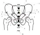
根据本发明的优选的实施例,骨盆标志选自:According to a preferred embodiment of the present invention, the pelvic landmarks are selected from:
-左髂前上棘,右髂前上棘;- left anterior superior iliac spine, right anterior superior iliac spine;
-左髂骶关节,右左髂骶;- Left iliac joint, right left iliac;
-左侧孔点,右侧孔点;- Hole point on the left, hole point on the right;
-耻骨联合的颅边缘;以及- the cranial margin of the pubic symphysis; and
-左髋臼旋转中心,右髋臼旋转中心。- Left acetabular center of rotation, right acetabular center of rotation.
当然,也可采集适合于距离测量的任何其它骨盆标志。Of course, any other pelvic landmarks suitable for distance measurement may also be acquired.
根据本发明的另一优选实施例,解剖学标志之间的距离在颅-尾/垂直方向和/或内侧-外侧/水平方向上测量。距离测量值可选自:According to another preferred embodiment of the invention, the distances between anatomical landmarks are measured in cranio-caudal/vertical direction and/or medial-lateral/horizontal direction. Distance measurements can be selected from:
-左髂前上棘与右髂前上棘之间的内侧-外侧距离;- the medial-lateral distance between the left anterior superior iliac spine and the right anterior superior iliac spine;
-髂骶关节与连接髂前上棘的线之间的颅-尾距离;- the cranio-caudal distance between the iliac joint and the line joining the anterior superior iliac spine;
-左髋臼或右髋臼旋转中心与连接髂前上棘的线之间的颅-尾距离;- Cranio-caudal distance between the center of rotation of the left or right acetabulum and the line joining the anterior superior iliac spine;
-左髋臼或右髋臼旋转中心与连接左侧孔点和右侧孔点的线之间的颅-尾距离;- the cranial-caudal distance between the center of rotation of the left or right acetabulum and the line connecting the left and right foramen points;
-左髋臼或右髋臼旋转中心与患者正中矢状平面之间的内侧-外侧距离;以及- the medial-lateral distance between the center of rotation of the left or right acetabulum and the patient's midsagittal plane; and
-左髋臼或右髋臼旋转中心与耻骨联合的颅缘之间的颅-尾距离。- Cranial-caudal distance between the center of rotation of the left or right acetabulum and the cranial border of the pubic symphysis.
同样,可在本发明的背景下执行所识别的标志之间的任何其它合适的距离测量。Likewise, any other suitable distance measurement between identified landmarks may be performed in the context of the present invention.
如上面已经进一步阐述的,当患者采取站立姿势时,希望知道患者骨盆的矢状旋转。即使可针对患者的任何姿势执行本发明,但是最理想的是站立姿势。As already explained further above, it is desirable to know the sagittal rotation of the patient's pelvis when the patient assumes a standing position. Even though the invention can be performed with any posture of the patient, the most ideal is the standing posture.
利用针对当前患者获取的必要距离测量值,可将每个测量值的确定值与从对多个参考患者执行的相应测量值获得的统计分布进行比较。根据在内侧-外侧方向上进行的测量,可能得出用于比较多个骨盆骨的共同基础,而不管骨的实际尺寸和由此产生的测量的颅-尾距离。然后在颅-尾方向上进行的测量直接指示骨盆在正中矢状平面中旋转了多少。因为,根据本发明,数据库还提供了在颅-尾方向上测量的距离与骨盆的矢状旋转之间的相关性,当前患者的骨盆的矢状旋转可最终从数据库导出。With the necessary distance measurements taken for the current patient, the determined value for each measurement can be compared to a statistical distribution obtained from corresponding measurements performed on a plurality of reference patients. From measurements made in the medial-lateral direction, it is possible to derive a common basis for comparing multiple pelvic bones, regardless of the actual size of the bones and the resulting measured cranial-caudal distance. Measurements taken in the cranio-caudal direction then directly indicate how much the pelvis is rotated in the midsagittal plane. Since, according to the invention, the database also provides a correlation between distances measured in the cranio-caudal direction and the sagittal rotation of the pelvis, the sagittal rotation of the pelvis for the current patient can finally be derived from the database.
具体而言,骨盆矢状旋转可限定患者的冠状面与包含左髂前上棘、右髂前上棘,以及左髋臼的旋转中心和右髋臼的旋转中心中的至少一个的AAC平面之间的角度。Specifically, the sagittal pelvic rotation can define a relationship between the coronal plane of the patient and the AAC plane that includes the left anterior superior iliac spine, the right anterior superior iliac spine, and at least one of the centers of rotation of the left and right acetabulum. angle between.
此外,确定的AAC平面可以作为用于手术的配准程序的参考基础,所述配准程序特别用于髋关节置换手术。Furthermore, the determined AAC plane can serve as a reference basis for registration procedures for surgery, in particular for hip replacement surgery.
定义definition
根据本发明的方法是例如计算机实现的方法。例如,根据本发明的方法的所有步骤或仅仅一些步骤(即,小于步骤的总数)可由计算机(例如,至少一个计算机)执行。计算机实现的方法的实施例是计算机用于执行数据处理方法的用途。计算机实现的方法的实施例是涉及计算机的操作的方法,使得计算机操作成执行该方法的一个、多个或所有步骤。The method according to the invention is eg a computer-implemented method. For example, all steps or only some steps (ie less than the total number of steps) of the method according to the invention may be performed by a computer (eg at least one computer). An embodiment of a computer-implemented method is the use of a computer to perform a data processing method. An embodiment of a computer-implemented method is a method involving the operation of a computer such that the computer is operative to perform one, more or all steps of the method.
例如,计算机包括至少一个处理器和例如至少一个存储器,以便(技术上)处理数据,例如电子地和/或光学地处理数据。处理器例如由作为半导体的物质或组合物制成,例如至少部分n和/或p掺杂的半导体,例如II-,III-,IV-,V-,VI-半导体材料中的至少一种,例如(掺杂的)硅和/或砷化镓。所描述的计算步骤例如由计算机执行。确定步骤或计算步骤是例如在技术方法的框架内确定数据的步骤,例如在程序的框架内。计算机例如是任何种类的数据处理装置,例如电子数据处理装置。计算机可为通常认为是这样的装置,例如台式PC、笔记本计算机、上网本等,但也可为任何可编程设备,例如,如移动电话或嵌入式处理器。计算机可例如包括“子计算机”的系统(网络),其中每个子计算机本身代表计算机。术语“计算机”包括云计算机,例如云服务器。术语“云计算机”包括云计算机系统,其例如包括至少一个云计算机的系统,并且例如包括多个可操作地互连的云计算机,如服务器群。这种云计算机优选连接到诸如万维网(WWW)的广域网,并且位于所有连接到万维网的所谓计算机云中。这种基础设施用于“云计算”,其描述了计算、软件、数据访问和存储服务,其不需要终端用户知道提供特定服务的计算机的物理位置和/或配置。例如,术语“云”在这方面用作因特网(万维网)的隐喻。例如,云将计算基础设施提供为服务(IaaS)。云计算机可用作用于执行本发明方法的操作系统和/或数据处理应用程序的虚拟主机。云计算机例如是由AmazonWeb ServicesTM提供的弹性计算云(EC2)。计算机例如包括接口,以便接收或输出数据和/或执行模数转换。该数据例如是表示物理特性和/或从技术信号生成的数据。技术信号例如通过(技术)检测装置(例如,如用于检测标记装置的装置)和/或(技术)分析装置(例如,如用于执行(医疗)成像方法的装置)产生,其中技术信号例如是电信号或光信号。技术信号例如表示计算机接收或输出的数据。计算机优选地可操作地联接到显示装置,该显示装置允许计算机输出的信息被显示,例如显示给用户。显示装置的一个实例是增强现实装置(也称为增强现实眼镜),其可以用作用于导航的“护目镜”。这种增强现实眼镜的具体实例是GoogleGlass(Google,Inc.的商标)。增强现实装置既可用于通过用户交互将信息输入到计算机中,并且也可以用于显示由计算机输出的信息。显示装置的另一个实例将是标准计算机监视器,包括例如可操作地联接到计算机的液晶显示器,用于从计算机接收显示控制数据,用于产生用于在显示装置上显示图像信息内容的信号。此计算机监视器的特定实施例是数字灯箱。监视器还可为便携式监视器,例如手持式装置,如智能电话或个人数字助理或数字媒体播放器。For example, a computer comprises at least one processor and eg at least one memory in order to (technically) process data, eg electronically and/or optically. The processor is for example made of a substance or composition which is a semiconductor, for example an at least partially n- and/or p-doped semiconductor, for example at least one of II-, III-, IV-, V-, VI-semiconductor materials, For example (doped) silicon and/or gallium arsenide. The described calculation steps are performed, for example, by a computer. A determination step or calculation step is a step of determining data, eg within the framework of a technical method, eg within the framework of a program. A computer is, for example, any kind of data processing device, such as an electronic data processing device. A computer may be what is commonly thought of as such, such as a desktop PC, notebook computer, netbook, etc., but may also be any programmable device such as, for example, a mobile phone or an embedded processor. A computer may, for example, comprise a system (network) of "subcomputers", each of which represents a computer itself. The term "computer" includes cloud computers, such as cloud servers. The term "cloud computer" includes cloud computer systems, such as a system comprising at least one cloud computer, and, for example, a plurality of operably interconnected cloud computers, such as server farms. Such cloud computers are preferably connected to a wide area network such as the World Wide Web (WWW) and are located in all so-called computer clouds connected to the World Wide Web. This infrastructure is used in "cloud computing," which describes computing, software, data access, and storage services that do not require the end user to know the physical location and/or configuration of the computers providing a particular service. For example, the term "cloud" is used in this context as a metaphor for the Internet (World Wide Web). For example, the cloud provides computing infrastructure as a service (IaaS). A cloud computer can be used as a virtual host for an operating system and/or data processing application for carrying out the method of the present invention. A cloud computer is, for example, the Elastic Compute Cloud (EC2) provided by Amazon Web Services™ . A computer includes, for example, an interface to receive or output data and/or to perform analog-to-digital conversion. The data are, for example, data representing physical properties and/or generated from technical signals. A technical signal is generated, for example, by a (technical) detection device (eg, as a device for detecting a marking device) and/or a (technical) analysis device (eg, as a device for performing a (medical) imaging method), wherein the technical signal is eg be electrical or optical signals. Technical signals represent, for example, data received or output by a computer. The computer is preferably operatively coupled to a display device that allows information output by the computer to be displayed, for example to a user. One example of a display device is an augmented reality device (also known as augmented reality glasses), which can be used as "goggles" for navigation. A specific example of such augmented reality glasses is Google Glass (trademark of Google, Inc.). Augmented reality devices can be used both to input information into a computer through user interaction and to display information output by the computer. Another example of a display device would be a standard computer monitor comprising, for example, a liquid crystal display operatively coupled to a computer for receiving display control data from the computer for generating signals for displaying image information content on the display device. A particular embodiment of this computer monitor is a digital light box. The monitor may also be a portable monitor such as a handheld device such as a smart phone or a personal digital assistant or digital media player.
表述“获取数据”例如包括(在计算机实现的方法的框架内)其中数据由计算机实现的方法或程序确定的场景。确定数据例如涵盖测量物理量并将测量值转换成数据,例如数字数据,和/或借助于计算机且例如在根据本发明的方法的框架内计算数据。“获取数据”的含义还包括例如通过计算机实现的方法或程序接收或检索数据的场景,例如来自另一程序,先前的方法步骤或数据存储介质,例如用于通过计算机实现的方法或程序进一步处理。生成待获取的数据可以,但不一定根据本发明的方法的一部分。因此,表述“获取数据”也可以例如意味着等待接收数据和/或接收数据。接收的数据可例如通过接口输入。表述“获取数据”还可表示计算机实现的方法或程序执行步骤以便(主动地)从数据源接收或检索数据,例如数据存储介质(例如,如ROM、RAM、数据库、硬盘驱动器等),或通过接口(例如,从另一台计算机或网络)。分别由所公开的方法或装置获取的数据可从位于数据存储装置中的数据库获取,该数据存储装置对于计算机是可操作的,用于数据库和计算机之间的数据传输,例如从数据库到计算机。计算机获取数据以用作确定数据的步骤的输入。所确定的数据可再次输出到相同或另一数据库以供稍后使用。该数据库或用于实现所公开方法的数据库可位于网络数据存储装置或网络服务器(例如,云数据存储装置或云服务器)或本地数据存储装置(如可操作地连接到执行所公开方法的至少一个计算机的大容量存储装置)上。通过在获取步骤之前执行附加步骤,可使数据“准备好使用”。根据此附加步骤,生成数据以便获取。例如检测或捕获数据(例如,通过分析装置)。备选地或附加地,例如经由接口根据额外步骤输入数据。可例如输入所生成的数据(例如,进入计算机)。根据附加步骤(在获取步骤之前),还可通过执行将数据存储在数据存储介质(例如ROM、RAM、CD和/或硬盘驱动器)中的附加步骤来提供数据,使得它们准备好在根据本发明的方法或程序的框架内使用。因此,“获取数据”的步骤还可以涉及命令装置获得和/或提供要获取的数据。具体而言,获取步骤不涉及侵入性步骤,侵入性步骤将代表对身体的实质性物理干扰,需要执行专业医学专业知识并且即使在具有所需专业护理和专业知识的情况下进行也会带来巨大的健康风险。具体而言,获取数据(例如确定数据)的步骤不涉及外科手术步骤,并且特别是不涉及使用手术或治疗来治疗人体或动物体的步骤。为了区分本方法使用的不同数据,将数据表示(即称为)“XY数据”等,并根据它们描述的信息来限定,然后优选地将其称为“XY信息“等。The expression "obtaining data" includes for example (within the framework of a computer-implemented method) a scenario in which data are determined by a computer-implemented method or a program. Determining data encompasses, for example, the measurement of physical quantities and the conversion of measured values into data, for example digital data, and/or the calculation of data by means of a computer, for example within the framework of the method according to the invention. The meaning of "obtaining data" also includes scenarios where data are received or retrieved, e.g. by a computer-implemented method or program, e.g. from another program, a previous method step or a data storage medium, e.g. for further processing by a computer-implemented method or program . Generating the data to be acquired can, but need not be part of the method according to the invention. Thus, the expression "acquiring data" can also mean, for example, waiting to receive data and/or receiving data. The received data can be entered, for example, via an interface. The expression "obtaining data" may also denote a computer-implemented method or program performing steps to (actively) receive or retrieve data from a data source, such as a data storage medium (eg, such as ROM, RAM, database, hard drive, etc.), or via interface (for example, from another computer or network). Data respectively obtained by a disclosed method or apparatus may be obtained from a database located in a data storage device operable to a computer for data transfer between the database and the computer, eg from the database to the computer. The computer obtains the data for use as input to the step of determining the data. The determined data can be exported again to the same or another database for later use. The database or the database used to implement the disclosed methods may be located in a network data storage device or a network server (for example, a cloud data storage device or cloud server) or a local data storage device (such as operatively connected to at least one computer's mass storage device). Data can be made "ready to use" by performing additional steps prior to the acquisition step. According to this additional step, data is generated for acquisition. For example, detecting or capturing data (eg, by an analysis device). Alternatively or additionally, data is entered according to additional steps, for example via an interface. The generated data can be imported (eg, into a computer), for example. According to an additional step (before the acquisition step), the data can also be provided by performing an additional step of storing the data in a data storage medium (such as ROM, RAM, CD and/or hard drive) so that they are ready to be stored in accordance with the invention used within the framework of a method or procedure. Thus, the step of "obtaining data" may also involve instructing the device to obtain and/or provide data to be retrieved. Specifically, the acquisition step does not involve invasive steps that would represent a substantial physical disturbance to the body, require specialized medical expertise to be performed and would bring huge health risk. In particular, the step of obtaining data (eg determining data) does not involve surgical steps, and in particular does not involve steps of treating the human or animal body using surgery or therapy. In order to distinguish the different data used by the method, the data are represented (ie referred to as) "XY data" etc., and defined in terms of the information they describe, and then preferably referred to as "XY information" etc.
本发明还涉及一种程序,当在计算机上运行时,该程序使计算机执行这里描述的一个或多个或所有方法步骤,和/或存储程序的程序存储介质(特别是在非暂时性形式),和/或包括所述程序存储介质的计算机,和/或(物理的,例如电的,例如技术上生成的)信号波,例如数字信号波,其携带代表该程序的信息,例如,上述程序,例如其包括适于执行本文所述的任何或所有方法步骤的代码装置。The invention also relates to a program which, when run on a computer, causes the computer to carry out one or more or all of the method steps described herein, and/or a program storage medium storing the program (especially in a non-transitory form) , and/or a computer comprising said program storage medium, and/or a (physical, e.g. electrical, e.g. technically generated) signal wave, e.g. a digital signal wave, which carries information representing the program, e.g. the above-mentioned program , for example comprising code means adapted to perform any or all of the method steps described herein.
本发明还涉及一种用于计算机辅助手术的导航系统,包括:前述权利要求的计算机,用于处理绝对点数据和相对点数据;The invention also relates to a navigation system for computer-assisted surgery, comprising: a computer according to the preceding claims, for processing absolute point data and relative point data;
检测装置,用于检测主点和辅助点的位置,以产生绝对点数据并将绝对点数据提供给计算机;A detection device for detecting the positions of the main point and the auxiliary point to generate absolute point data and provide the absolute point data to the computer;
数据接口,用于接收相对点数据并用于将相对点数据供应给计算机;以及a data interface for receiving the relative point data and for supplying the relative point data to the computer; and
用户界面,用于从计算机接收数据以便向用户提供信息,其中所接收的数据是由计算机基于计算机执行的处理结果生成的。A user interface for receiving data from a computer to provide information to a user, wherein the received data is generated by the computer based on the results of processing performed by the computer.
在本发明的框架内,计算机程序元素可由硬件和/或软件体现(这包括固件、驻留软件、微代码等)。在本发明的框架内,计算机程序单元可采用计算机程序产品的形式,该计算机程序产品可由计算机可用(例如,计算机可读)数据存储介质来体现,其包括体现在所述数据存储介质中的计算机可用(例如,计算机可读)程序指令、“代码”或“计算机程序”,以用在指令执行系统上或结合指令执行系统使用。此系统可为计算机;计算机可为数据处理装置,包括用于执行根据本发明的计算机程序元素和/或程序的装置,例如数据处理装置,其包括执行计算机程序元素的数字处理器(中央处理单元或CPU),以及用于存储用于执行计算机程序元素和/或通过执行计算机程序元素产生的数据的可选的易失性存储器(例如随机存取存储器或RAM)。在本发明的框架内,计算机可用的(例如计算机可读)数据存储介质可为任何数据存储介质,其可以包括、存储、通信、传播或传输程序以在指令执行系统、设备或装置上使用或结合它们使用。计算机可用的(例如计算机可读)数据存储介质可为例如但不限于电子、磁、光、电磁、红外或半导体系统、设备或装置或传播介质,例如,如互联网。计算机可用或计算机可读数据存储介质甚至可为例如纸或其上印刷有程序的其它合适的介质,因为程序可被电子地捕获,例如通过光学扫描纸或其它合适的介质,并然后以合适的方式编辑、转译或另外处理。数据存储介质优选是非易失性数据存储介质。这里描述的计算机程序产品和任何软件和/或硬件形成用于在示例实施例中执行本发明的功能的各种装置。计算机和/或数据处理装置可例如包括引导信息装置,该引导信息装置包括用于输出引导信息的装置。引导信息可例如通过视觉指示装置(例如,监视器和/或灯)在视觉上,和/或通过声学指示装置(例如,扬声器和/或数字语音输出装置)在声学上,和/或通过触觉指示装置(例如,振动元件或结合到仪器中的振动元件)在触觉上输出给用户。出于本文献的目的,计算机是技术计算机,其例如包括技术(例如有形)部件,例如机械和/或电子部件。本文献中提到的任何装置都是技术性的,并例如是有形装置。Within the framework of the invention, computer program elements may be embodied by hardware and/or software (this includes firmware, resident software, microcode, etc.). Within the framework of the invention, a computer program element may take the form of a computer program product embodied by a computer-usable (for example, computer-readable) data storage medium comprising a computer program embodied in said data storage medium. Program instructions, "code" or "computer program" are available (eg, computer readable) for use on or in connection with an instruction execution system. This system may be a computer; the computer may be a data processing device comprising means for executing computer program elements and/or programs according to the invention, for example a data processing device comprising a digital processor (central processing unit) executing computer program elements or CPU), and optional volatile memory (such as random access memory or RAM) for storing data used for and/or generated by the execution of computer program elements. Within the framework of the present invention, a computer usable (e.g. computer readable) data storage medium may be any data storage medium which may include, store, communicate, propagate or transport a program for use on an instruction execution system, device or device or Use them in combination. A computer usable (eg computer readable) data storage medium may be, for example but not limited to, an electronic, magnetic, optical, electromagnetic, infrared or semiconductor system, device or device or a communication medium such as for example the Internet. The computer-usable or computer-readable data storage medium may even be, for example, paper or other suitable medium having the program printed thereon, as the program may be captured electronically, for example by optically scanning the paper or other suitable medium, and then displaying it in a suitable edited, translated or otherwise processed. The data storage medium is preferably a non-volatile data storage medium. The computer program product and any software and/or hardware described herein form the various means for carrying out the functions of the invention in example embodiments. The computer and/or the data processing device may for example comprise guidance information means comprising means for outputting guidance information. Guidance information may be visual, for example, through visual indicators (e.g., monitors and/or lights), and/or acoustically through acoustic indicator devices (e.g., speakers and/or digital voice output devices), and/or through tactile A pointing device (eg, a vibrating element or a vibrating element incorporated into the instrument) provides tactile output to the user. For the purposes of this document, a computer is a technical computer, which eg comprises technical (eg physical) components, eg mechanical and/or electronic components. Any means mentioned in this document are technical and are, for example, physical means.
标志是解剖学身体部分的限定元素,其在多个患者的相同解剖学身体部分中始终相同或以高度相似性再现。典型的标志是例如股骨的上髁或椎骨的横突和/或背侧突的尖端。点(主要点或辅助点)可表示此类标志。位于身体部分的特征解剖结构(例如在其表面上)的标志也可表示所述结构。标志可表示作为整体或仅其点或部分的解剖结构。标志还可例如位于解剖结构上,例如突出结构。这种解剖结构的一个实例是髂嵴的后部。标志的另一个实例是由髋臼的边沿限定的实例,例如由所述边沿的中心。在另一实例中,标志表示来源于众多检测点的髋臼的底部或最深点。因此,一个标志可例如代表大量检测点。如上所述,标志可表示基于身体部分的特征结构限定的解剖学特征。另外,标志还可表示由两个身体部分的相对移动限定的解剖学特征,如当相对于髋臼移动时股骨的旋转中心。A landmark is a defined element of an anatomical body part which is always the same or reproduces with a high degree of similarity in the same anatomical body part of multiple patients. Typical landmarks are eg the epicondyle of the femur or the tips of the transverse and/or dorsal processes of the vertebrae. Points (primary or secondary) may represent such markers. Landmarks on characteristic anatomical structures located on the body part (eg on the surface thereof) may also represent said structures. A landmark may represent an anatomical structure as a whole or only a point or part thereof. Landmarks may also be located, for example, on anatomical structures, such as protruding structures. An example of such an anatomical structure is the posterior portion of the iliac crest. Another example of a landmark is one defined by the rim of the acetabulum, eg by the center of the rim. In another example, the landmarks represent the floor or deepest point of the acetabulum from a plurality of detection points. Thus, one marker may for example represent a large number of detection points. As mentioned above, landmarks may represent anatomical features defined based on the characteristic structure of the body part. Additionally, the landmarks may also represent anatomical features defined by the relative movement of the two body parts, such as the center of rotation of the femur when moving relative to the acetabulum.
如果要分析的分析对象是已知的,则关于成像几何形状的信息优选包括允许计算分析图像(x射线图像)的信息,给定成像几何形状分析设备和要由x-射线辐射分析的分析对象(解剖学身体部分)之间的已知相对位置,其中“已知”意味着分析对象的空间几何形状(大小和形状)是已知的。这意味着例如关于分析对象(解剖学身体部分)和分析辐射(x射线辐射)之间的相互作用的三维“空间分辨”信息是已知的,其中“相互作用”意味着例如分析辐射被分析对象阻挡或部分或完全允许通过分析对象。成像几何形状的位置和特定定向例如由X射线设备的位置限定,例如由X射线源和X射线探测器的位置,和/或例如由X射线束的多重(歧管)的位置,其穿过分析对象并由X射线探测器探测。成像几何形状例如描述了所述多重(歧管)的位置(即地点,且具体是定向)和形状(例如,呈现特定倾斜角的圆锥形状)。该位置可例如由穿过所述多重的中心的x射线束的位置或由表示x射线束的多重(歧管)的几何对象(如截头圆锥)的位置表示。关于上述相互作用的信息优选地以三维方式(例如来自三维CT的三维)已知,并且描述对于分析物体的点和/或区域的空间分辨方式的相互作用,例如对于分析对象的所有点和/或区域。例如,对成像几何结构的了解允许相对于图像平面(例如,x射线检测器的平面)计算辐射源(例如,x射线源)的位置。关于由成像几何限定的三维分析对象和二维分析图像之间的连接,例如参考以下出版物:If the analysis object to be analyzed is known, the information about the imaging geometry preferably includes information allowing the calculation of an analysis image (x-ray image), given the imaging geometry analysis device and the analysis object to be analyzed by x-ray radiation Known relative position between (anatomical body parts), where "known" means that the spatial geometry (size and shape) of the object of analysis is known. This means that e.g. three-dimensional "spatially resolved" information is known about the interaction between the analysis object (anatomical body part) and the analysis radiation (x-ray radiation), where "interaction" means e.g. the analysis radiation is analyzed Objects block and either partially or fully allow through analysis objects. The position and specific orientation of the imaging geometry is defined for example by the position of the X-ray apparatus, for example by the position of the X-ray source and the X-ray detector, and/or for example by the position of the multiplex (manifold) of the X-ray beam, which passes through The object is analyzed and detected by an X-ray detector. The imaging geometry describes, for example, the position (ie location, and in particular orientation) and shape (eg, conical shape exhibiting a specific tilt angle) of the multiplex (manifold). This position may eg be represented by the position of the x-ray beam passing through the center of the multiplicity or by the position of a geometric object such as a truncated cone representing the multiplicity (manifold) of the x-ray beam. Information about the above-mentioned interactions is preferably known in three dimensions (for example three dimensions from three-dimensional CT) and describes the interaction in a spatially resolved manner for points and/or regions of the analysis object, for example for all points and/or areas of the analysis object or area. For example, knowledge of the imaging geometry allows calculation of the position of a radiation source (eg, x-ray source) relative to the image plane (eg, the plane of the x-ray detector). With regard to the connection between the 3D analysis object defined by the imaging geometry and the 2D analysis image, reference is made, for example, to the following publications:
1.“An Efficient and Accurate Camera Calibration Technique for 3DMachine Vision”,Roger Y.Tsai,Proceedings of the IEEE Conference on ComputerVision and Pattern Recognition。Miami Beach,Florida,1986,第364-374页1. “An Efficient and Accurate Camera Calibration Technique for 3DMachine Vision,” Roger Y. Tsai, Proceedings of the IEEE Conference on ComputerVision and Pattern Recognition. Miami Beach, Florida, 1986, pp. 364-374
2.“A Versatile Camera Calibration Technique for High-Accuracy 3DMachine Vision Metrology Using Off-the-Shelf TV Cameras and Lenses”,RogerY.Tsai,IEEE Journal of Robotics and Automation,第RA-3卷,第4期,1987年8月,第323-344页。2. "A Versatile Camera Calibration Technique for High-Accuracy 3DMachine Vision Metrology Using Off-the-Shelf TV Cameras and Lenses", Roger Y. Tsai, IEEE Journal of Robotics and Automation, Vol. RA-3, No. 4, 1987 August, pp. 323-344.
3.“Fluoroscopic X-ray Image Processing and Registration for Computer-Aided Orthopedic Surgery”,Ziv Yaniv3. "Fluoroscopic X-ray Image Processing and Registration for Computer-Aided Orthopedic Surgery," Ziv Yaniv
4.EP 08 156 293.64. EP 08 156 293.6
5.US 61/054,1875. US 61/054,187
优选地,获取图谱数据,其描述(例如,限定,更具体地表示和/或是)解剖学身体部分的一般三维形状。因此,图谱数据代表解剖学身体部分的图谱。图谱通常由多个通用对象模型组成,其中通用对象模型一起形成复杂结构。例如,图谱构成了从多个人体收集的解剖信息生成的患者身体(例如,身体的一部分)的统计模型,例如来自包含这些人体图像的医学图像数据。原则上,图谱数据因此代表对多个人体的这种医学图像数据的统计分析的结果。该结果可作为图像输出,因此图谱数据包含医学图像数据或与医学图像数据相当。这种比较可例如通过应用图像融合算法来执行,该算法在图谱数据和医学图像数据之间进行图像融合。比较的结果可为图谱数据和医学图像数据之间的相似性的度量。图谱数据包括位置信息,该位置信息可匹配(例如通过应用弹性或刚性图像融合算法)例如包含在医学图像数据中的位置信息,以便例如将图谱数据与医学图像数据进行比较,以便确定医学图像数据中的解剖结构的位置,其对应于由图谱数据限定的解剖结构。Preferably, atlas data is acquired which describes (eg defines, more specifically represents and/or is) the general three-dimensional shape of the anatomical body part. Thus, atlas data represent an atlas of anatomical body parts. Graphs often consist of multiple generic object models, where the generic object models together form complex structures. For example, an atlas constitutes a statistical model of a patient's body (eg, part of a body) generated from anatomical information collected from multiple bodies, such as from medical image data containing images of those bodies. In principle, the atlas data thus represent the result of a statistical analysis of such medical image data of multiple human bodies. The result can be output as an image, so the atlas data contains or is equivalent to medical image data. Such a comparison can be performed, for example, by applying an image fusion algorithm that performs image fusion between the atlas data and the medical image data. The result of the comparison may be a measure of similarity between the atlas data and the medical image data. The atlas data includes location information that can be matched (e.g. by applying elastic or rigid image fusion algorithms) to location information contained, for example, in medical image data, for example, to compare the atlas data with the medical image data in order to determine the medical image data The locations of the anatomical structures in , which correspond to the anatomical structures defined by the atlas data.
人体,其解剖结构用作产生图谱数据的输入,有利地共享共同特征,如性别、年龄、种族、身体测量(例如尺寸和/或质量)和病理状态中的至少一个。解剖学信息例如描述了人体的解剖结构,并且例如从关于人体的医学图像信息中提取。例如,股骨的图谱可包括头部、颈部、身体、大转子、小转子和下肢作为共同构成完整结构的对象。例如,大脑的图谱可包括端脑、小脑、间脑、脑桥、中脑和髓质作为共同构成复杂结构的对象。这种图谱的一个应用是在医学图像的分割中,其中图谱与医学图像数据匹配,并且将图像数据与匹配的图谱进行比较以便分配图像数据的点(像素或体素)到匹配的图谱的对象,从而将图像数据分割成对象。The human body, whose anatomy is used as input to generate the atlas data, advantageously shares common characteristics such as at least one of gender, age, ethnicity, anthropometric (eg size and/or mass) and pathological status. Anatomical information describes, for example, the anatomy of the human body and is extracted, for example, from medical image information about the human body. For example, an atlas of the femur may include the head, neck, body, greater trochanter, lesser trochanter, and lower extremity as objects that together form a complete structure. For example, an atlas of the brain may include the telencephalon, cerebellum, diencephalon, pons, midbrain, and medulla as objects that together constitute a complex structure. One application of such an atlas is in the segmentation of medical images, where the atlas is matched to medical image data, and the image data is compared with the matched atlas in order to assign points (pixels or voxels) of the image data to objects of the matched atlas , thereby splitting the image data into objects.
具体而言,本发明不涉及或特别包括或包含侵入性步骤,该侵入性步骤将代表对需要专业医学专业知识的身体的实质性物理干扰,并且即使在进行所需的专业护理和专业知识时也具有实质性的健康风险。例如,本发明不包括定位医用植入物以将其紧固到解剖结构的步骤或将医用植入物紧固到解剖结构的步骤或制备解剖结构以使医用植入物固定到其的步骤。更具体地,本发明不涉及或具体包含任何手术或治疗活动。相反,本发明适用于相对于医疗植入物定位工具,该医疗植入物可在患者体外。仅仅由于这个原因,通过实施本发明不需要或未暗示任何手术或治疗活动,并且特别是手术或治疗步骤。In particular, the present invention does not relate to or specifically include or include invasive steps that would represent a substantial physical disturbance to the body requiring specialized medical expertise and, even while performing the required specialized care and expertise There are also substantial health risks. For example, the present invention does not include the steps of positioning a medical implant for securing it to an anatomical structure or of securing a medical implant to an anatomical structure or of preparing an anatomical structure for securing a medical implant thereto. More specifically, the present invention does not relate to or specifically encompass any surgical or therapeutic activity. Rather, the invention is applicable to positioning the tool relative to a medical implant, which may be outside the patient's body. For this reason alone, no surgical or therapeutic activity, and in particular surgical or therapeutic steps, is required or implied by the practice of the invention.
例如,可以在用于髋部手术的导航软件中实现本发明。因此,该软件可能导入X射线数据集,并在此基础上,计算X射线图像平面和AAC平面之间的上述矢状旋转,这最终将允许在由术前X射线数据集限定的功能平面上导航。For example, the invention may be implemented in navigation software for hip surgery. Thus, the software may import the X-ray data set and, based on this, calculate the above-mentioned sagittal rotation between the X-ray image plane and the AAC plane, which will eventually allow the functional plane defined by the preoperative X-ray data set navigation.
附图说明Description of drawings
在下文中,参考代表本发明优选实施例的附图描述了本发明。然而,本发明的范围不限于图中公开的具体特征,附图示出:Hereinafter, the present invention is described with reference to the accompanying drawings representing preferred embodiments of the invention. However, the scope of the invention is not limited to the specific features disclosed in the drawings, which show:
图1X射线成像的几何布置;Fig. 1 Geometric arrangement of X-ray imaging;
图2具有校准特征的前后X射线图像;Figure 2 Before and after X-ray images with calibration features;
图3显示了图2的图像,其中指示了骨盆标志;Figure 3 shows the image of Figure 2 with the pelvic landmarks indicated;
图4X射线平面与AAC平面之间的矢状旋转角;Fig. 4 The sagittal rotation angle between the X-ray plane and the AAC plane;
图5AAC平面的配准;Figure 5 Registration of the AAC plane;
图6本发明计算机实现的方法的基本步骤。Figure 6 shows the basic steps of the computer-implemented method of the present invention.
具体实施方式Detailed ways
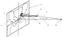
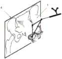
常规X射线成像方法的几何设置可在图1中看到。准点辐射源12以球形方式发射辐射,并朝向骨盆(未示出)发射。当发射的辐射由物质吸附到不同程度时,骨盆1的图像5在图像平面(未示出)中形成在骨盆1后面。然而,由于发射的辐射的球形传播,该图像失真。因此,将在图像中的位置处描绘示例性标志3,该位置不同于如果整个骨盆1沿前后方向2投射到图像平面中将获得的位置。The geometric setup of a conventional X-ray imaging method can be seen in Figure 1. The
根据本发明的一个实施例,通过确定包括X射线源12和校准平面4之间的距离,以及骨盆标志3中的每一个到校准平面4在前后方向2的距离13的几何设置来补偿该位置偏移或失真。According to one embodiment of the invention, this position is compensated by determining a geometrical setup comprising the distance between the
在患者骨盆的X射线图像连同多个校准特征9、10已经沿前后方向2(图1中所示)拍摄之后,然后可在X射线图像内手动或自动识别多个骨盆标志3、14、15,如图3所示。After an X-ray image of the patient's pelvis has been taken in the anterior-posterior direction 2 (shown in FIG. 1 ) along with a plurality of calibration features 9, 10, a plurality of
在X射线图像已通过执行图6中概述的本发明方法的步骤2、3、4和5进行的“校准”后,骨盆标志3、14、15之间在内侧-外侧方向和颅-尾方向上的距离测量是可能的。然后可将确定的距离6、7与从多个参考患者获得的统计值进行比较。After the X-ray image has been "calibrated" by performing
由于存储在存储介质上的数据库提供了距离测量值与骨盆1的矢状旋转8的确定值之间的直接相关性,因此可从该数据库导出当前患者骨盆的矢状旋转8。Since the database stored on the storage medium provides a direct correlation between the distance measurements and the determined value of the
根据本发明的优选实施例,矢状旋转8限定冠状面4和AAC平面16(图4中所示)之间的角度。在实际骨盆1已经在真实空间中配准之后,AAC平面16可用作遵循本发明方法的手续的参考平面,例如通过确定左和右前髂上棘的空间位置和左髋臼或右髋臼的旋转中心之一,如图5所示。According to a preferred embodiment of the invention, the
Claims (15)
Translated fromChineseApplications Claiming Priority (3)
| Application Number | Priority Date | Filing Date | Title |
|---|---|---|---|
| EP2017055583 | 2017-03-09 | ||
| EPPCT/EP2017/055583 | 2017-03-09 | ||
| PCT/EP2018/055067WO2018162322A1 (en) | 2017-03-09 | 2018-03-01 | Sagittal rotation determination |
Publications (2)
| Publication Number | Publication Date |
|---|---|
| CN110891488A CN110891488A (en) | 2020-03-17 |
| CN110891488Btrue CN110891488B (en) | 2023-05-23 |
Family
ID=61569254
Family Applications (1)
| Application Number | Title | Priority Date | Filing Date |
|---|---|---|---|
| CN201880016818.9AActiveCN110891488B (en) | 2017-03-09 | 2018-03-01 | sagittal rotation determined |
Country Status (5)
| Country | Link |
|---|---|
| US (3) | US10869724B2 (en) |
| EP (2) | EP4008258B8 (en) |
| CN (1) | CN110891488B (en) |
| AU (1) | AU2018232490B2 (en) |
| WO (1) | WO2018162322A1 (en) |
Families Citing this family (9)
| Publication number | Priority date | Publication date | Assignee | Title |
|---|---|---|---|---|
| CN110891488B (en)* | 2017-03-09 | 2023-05-23 | 史密夫和内修有限公司 | sagittal rotation determined |
| CN111179350B (en)* | 2020-02-13 | 2022-04-08 | 张逸凌 | Hip joint image processing system |
| US11107586B1 (en) | 2020-06-24 | 2021-08-31 | Cuptimize, Inc. | System and method for analyzing acetabular cup position |
| CN112754664B (en)* | 2021-01-10 | 2022-01-28 | 杭州素问九州医疗科技有限公司 | Orthopedic surgery system for finding hip joint center |
| CN115105201B (en)* | 2021-05-08 | 2025-01-10 | 北京仁馨医疗科技有限公司 | Sacral nerve foramen positioning method and positioning system based on holographic image and application thereof |
| KR102619164B1 (en)* | 2021-11-26 | 2023-12-29 | 경희대학교 산학협력단 | Real-time measuring method and apparatus for 3D direction of anterior pelvic plane |
| CN115429247B (en)* | 2022-09-23 | 2024-10-22 | 中国科学院长春光学精密机械与物理研究所 | Pelvis measurement method, device, electronic equipment and storage medium |
| CN117159145B (en)* | 2023-10-24 | 2024-02-23 | 北京昭衍新药研究中心股份有限公司 | Brain stereotactic method for non-human primate experimental animals |
| CN119655876A (en)* | 2024-11-26 | 2025-03-21 | 江苏省人民医院 | Pelvis fracture reduction method, system, computer device and readable storage medium |
Citations (10)
| Publication number | Priority date | Publication date | Assignee | Title |
|---|---|---|---|---|
| WO2002062248A1 (en)* | 2001-02-06 | 2002-08-15 | Cedara Software Corp. | Computer-assisted surgical positioning method and system |
| CN1509156A (en)* | 2001-03-13 | 2004-06-30 | 比约恩・弗朗克・艾弗森 | Method and device for providing information after insertion of a prosthesis in a hip joint |
| JP2009195490A (en)* | 2008-02-21 | 2009-09-03 | Lexi:Kk | Program for preoperative plan of artificial hip joint replacement, and instrument for supporting the replacement |
| CN101779223A (en)* | 2007-06-21 | 2010-07-14 | 苏尔吉克斯有限公司 | A system for measuring the true dimensions and orientation of objects in a two dimensional image |
| WO2011092531A1 (en)* | 2010-01-28 | 2011-08-04 | Pécsi Tudományegyetem | A method and a system for multi-dimensional visualization of the spinal column by vertebra vectors, sacrum vector, sacrum plateau vector and pelvis vectors |
| WO2014069553A1 (en)* | 2012-10-31 | 2014-05-08 | 株式会社上島電興社 | Pelvic rotation angle calculation device, pelvic rotation angle calculation method, device for estimating three-dimensional shape of acetabular coverage, method for estimating three-dimensional shape of acetabular coverage, pelvic rotation angle calculation program, and program for estimating three-dimensional shape of acetabular coverage |
| WO2014127354A1 (en)* | 2013-02-18 | 2014-08-21 | Orthogrid Systems, Llc | Alignment plate apparatus and system and method of use |
| WO2015120892A2 (en)* | 2014-02-13 | 2015-08-20 | Brainlab Ag | Method for assisting the positioning of a medical structure on the basis of two-dimensional image data |
| WO2016186969A1 (en)* | 2015-05-20 | 2016-11-24 | Radlink, Inc. | System and method for precision position detection and reproduction during surgery |
| CN106447764A (en)* | 2016-09-08 | 2017-02-22 | 福州大学 | Three-dimensional visualization automatic measuring method for human body pelvis parameters |
Family Cites Families (11)
| Publication number | Priority date | Publication date | Assignee | Title |
|---|---|---|---|---|
| EP1563799B2 (en) | 2004-02-11 | 2012-11-28 | BrainLAB AG | Adjustable marker arrangement |
| DE102005012708A1 (en)* | 2005-03-11 | 2006-09-21 | Eberhard-Karls-Universität Tübingen | Method for determining body orientations in space based on two x-ray images |
| EP1925256A1 (en)* | 2006-11-24 | 2008-05-28 | BrainLAB AG | Method and device for registering an anatomical structure with markers |
| EP2119397B1 (en) | 2008-05-15 | 2013-12-18 | Brainlab AG | Determining calibration information for an x-ray machine |
| US8075184B2 (en) | 2008-11-26 | 2011-12-13 | Richard King | X-ray calibration |
| EP2667813B1 (en) | 2011-01-26 | 2016-04-20 | Brainlab AG | Computer program for planning the positioning of an implant |
| DE102012111385B4 (en) | 2012-11-23 | 2018-05-03 | Diers Engineering Gmbh | Determining the spatial position and orientation of the vertebral bodies of the spine |
| FR3031665B1 (en) | 2015-01-19 | 2017-01-13 | Univ Bretagne Occidentale | PORTABLE ECHOGRAPHIC MEASUREMENT DEVICE ADAPTED TO MEASURE OF PELVIC VERSION. |
| US10991070B2 (en)* | 2015-12-18 | 2021-04-27 | OrthoGrid Systems, Inc | Method of providing surgical guidance |
| CN110177492A (en)* | 2016-11-18 | 2019-08-27 | 斯特赖克公司 | Method and apparatus for treating joint hits the treatment that the clamp type femur acetabular bone in disease and hip joint hits disease including the cam type femur acetabular bone in hip joint |
| CN110891488B (en)* | 2017-03-09 | 2023-05-23 | 史密夫和内修有限公司 | sagittal rotation determined |
- 2018
- 2018-03-01CNCN201880016818.9Apatent/CN110891488B/enactiveActive
- 2018-03-01WOPCT/EP2018/055067patent/WO2018162322A1/ennot_activeCeased
- 2018-03-01EPEP21216493.3Apatent/EP4008258B8/enactiveActive
- 2018-03-01EPEP18708970.1Apatent/EP3432798B1/enactiveActive
- 2018-03-01USUS16/306,991patent/US10869724B2/enactiveActive
- 2018-03-01AUAU2018232490Apatent/AU2018232490B2/enactiveActive
- 2020
- 2020-12-10USUS17/117,480patent/US11801094B2/enactiveActive
- 2023
- 2023-10-06USUS18/377,509patent/US12310667B2/enactiveActive
Patent Citations (10)
| Publication number | Priority date | Publication date | Assignee | Title |
|---|---|---|---|---|
| WO2002062248A1 (en)* | 2001-02-06 | 2002-08-15 | Cedara Software Corp. | Computer-assisted surgical positioning method and system |
| CN1509156A (en)* | 2001-03-13 | 2004-06-30 | 比约恩・弗朗克・艾弗森 | Method and device for providing information after insertion of a prosthesis in a hip joint |
| CN101779223A (en)* | 2007-06-21 | 2010-07-14 | 苏尔吉克斯有限公司 | A system for measuring the true dimensions and orientation of objects in a two dimensional image |
| JP2009195490A (en)* | 2008-02-21 | 2009-09-03 | Lexi:Kk | Program for preoperative plan of artificial hip joint replacement, and instrument for supporting the replacement |
| WO2011092531A1 (en)* | 2010-01-28 | 2011-08-04 | Pécsi Tudományegyetem | A method and a system for multi-dimensional visualization of the spinal column by vertebra vectors, sacrum vector, sacrum plateau vector and pelvis vectors |
| WO2014069553A1 (en)* | 2012-10-31 | 2014-05-08 | 株式会社上島電興社 | Pelvic rotation angle calculation device, pelvic rotation angle calculation method, device for estimating three-dimensional shape of acetabular coverage, method for estimating three-dimensional shape of acetabular coverage, pelvic rotation angle calculation program, and program for estimating three-dimensional shape of acetabular coverage |
| WO2014127354A1 (en)* | 2013-02-18 | 2014-08-21 | Orthogrid Systems, Llc | Alignment plate apparatus and system and method of use |
| WO2015120892A2 (en)* | 2014-02-13 | 2015-08-20 | Brainlab Ag | Method for assisting the positioning of a medical structure on the basis of two-dimensional image data |
| WO2016186969A1 (en)* | 2015-05-20 | 2016-11-24 | Radlink, Inc. | System and method for precision position detection and reproduction during surgery |
| CN106447764A (en)* | 2016-09-08 | 2017-02-22 | 福州大学 | Three-dimensional visualization automatic measuring method for human body pelvis parameters |
Also Published As
| Publication number | Publication date |
|---|---|
| US20240033007A1 (en) | 2024-02-01 |
| US20210161597A1 (en) | 2021-06-03 |
| AU2018232490A1 (en) | 2018-11-29 |
| EP4008258B8 (en) | 2023-08-23 |
| CN110891488A (en) | 2020-03-17 |
| US20200305976A1 (en) | 2020-10-01 |
| EP4008258A1 (en) | 2022-06-08 |
| EP4008258B1 (en) | 2023-07-19 |
| US10869724B2 (en) | 2020-12-22 |
| EP3432798A1 (en) | 2019-01-30 |
| US11801094B2 (en) | 2023-10-31 |
| AU2018232490B2 (en) | 2019-07-25 |
| WO2018162322A1 (en) | 2018-09-13 |
| EP3432798B1 (en) | 2021-12-29 |
| US12310667B2 (en) | 2025-05-27 |
Similar Documents
| Publication | Publication Date | Title |
|---|---|---|
| CN110891488B (en) | sagittal rotation determined | |
| JP2024504482A (en) | Computer-implemented method for augmented reality spinal rod planning and bending for navigational spine surgery | |
| US10667864B2 (en) | Inline-view determination | |
| US10561345B2 (en) | Determination of center of rotation of a bone | |
| US11826112B2 (en) | Method for registering articulated anatomical structures | |
| US20190357982A1 (en) | Microscope Tracking Based on Video Analysis | |
| US20200390412A1 (en) | Determining a target position of an x-ray device | |
| US12263031B2 (en) | Determining a configuration of a medical x-ray imaging system for detecting a marker device | |
| WO2022069510A1 (en) | Method of calibrating a cage | |
| US20230298186A1 (en) | Combining angiographic information with fluoroscopic images | |
| US12112437B2 (en) | Positioning medical views in augmented reality | |
| US20250078418A1 (en) | Conjunction of 2d and 3d visualisations in augmented reality | |
| US20250040993A1 (en) | Detection of positional deviations in patient registration | |
| WO2023179875A1 (en) | Method for registration of a virtual image in an augmented reality system |
Legal Events
| Date | Code | Title | Description |
|---|---|---|---|
| PB01 | Publication | ||
| PB01 | Publication | ||
| SE01 | Entry into force of request for substantive examination | ||
| SE01 | Entry into force of request for substantive examination | ||
| CB02 | Change of applicant information | Address after:Tennessee Applicant after:SMITH & NEPHEW, Inc. Applicant after:SMITH & NEPHEW ORTHOPAEDICS AG Applicant after:Singapore Smith & nephew Asia Pacific Ltd. Address before:Tennessee Applicant before:SMITH & NEPHEW, Inc. Applicant before:SMITH & NEPHEW ORTHOPAEDICS AG Applicant before:Smith & Repair Co.,Ltd. | |
| CB02 | Change of applicant information | ||
| GR01 | Patent grant | ||
| GR01 | Patent grant |