CN110866923A - Computer-aided fibrosis analysis method and electronic device - Google Patents
Computer-aided fibrosis analysis method and electronic deviceDownload PDFInfo
- Publication number
- CN110866923A CN110866923ACN201910789554.7ACN201910789554ACN110866923ACN 110866923 ACN110866923 ACN 110866923ACN 201910789554 ACN201910789554 ACN 201910789554ACN 110866923 ACN110866923 ACN 110866923A
- Authority
- CN
- China
- Prior art keywords
- fraction
- portal
- nodes
- fiber
- electronic device
- Prior art date
- Legal status (The legal status is an assumption and is not a legal conclusion. Google has not performed a legal analysis and makes no representation as to the accuracy of the status listed.)
- Granted
Links
Images


Classifications
- G—PHYSICS
- G16—INFORMATION AND COMMUNICATION TECHNOLOGY [ICT] SPECIALLY ADAPTED FOR SPECIFIC APPLICATION FIELDS
- G16H—HEALTHCARE INFORMATICS, i.e. INFORMATION AND COMMUNICATION TECHNOLOGY [ICT] SPECIALLY ADAPTED FOR THE HANDLING OR PROCESSING OF MEDICAL OR HEALTHCARE DATA
- G16H50/00—ICT specially adapted for medical diagnosis, medical simulation or medical data mining; ICT specially adapted for detecting, monitoring or modelling epidemics or pandemics
- G16H50/20—ICT specially adapted for medical diagnosis, medical simulation or medical data mining; ICT specially adapted for detecting, monitoring or modelling epidemics or pandemics for computer-aided diagnosis, e.g. based on medical expert systems
- G—PHYSICS
- G06—COMPUTING OR CALCULATING; COUNTING
- G06N—COMPUTING ARRANGEMENTS BASED ON SPECIFIC COMPUTATIONAL MODELS
- G06N20/00—Machine learning
- G—PHYSICS
- G06—COMPUTING OR CALCULATING; COUNTING
- G06N—COMPUTING ARRANGEMENTS BASED ON SPECIFIC COMPUTATIONAL MODELS
- G06N3/00—Computing arrangements based on biological models
- G06N3/02—Neural networks
- G06N3/04—Architecture, e.g. interconnection topology
- G06N3/0464—Convolutional networks [CNN, ConvNet]
- G—PHYSICS
- G06—COMPUTING OR CALCULATING; COUNTING
- G06N—COMPUTING ARRANGEMENTS BASED ON SPECIFIC COMPUTATIONAL MODELS
- G06N3/00—Computing arrangements based on biological models
- G06N3/02—Neural networks
- G06N3/08—Learning methods
- G—PHYSICS
- G06—COMPUTING OR CALCULATING; COUNTING
- G06N—COMPUTING ARRANGEMENTS BASED ON SPECIFIC COMPUTATIONAL MODELS
- G06N3/00—Computing arrangements based on biological models
- G06N3/02—Neural networks
- G06N3/08—Learning methods
- G06N3/09—Supervised learning
- G—PHYSICS
- G06—COMPUTING OR CALCULATING; COUNTING
- G06T—IMAGE DATA PROCESSING OR GENERATION, IN GENERAL
- G06T7/00—Image analysis
- G06T7/0002—Inspection of images, e.g. flaw detection
- G06T7/0012—Biomedical image inspection
- G—PHYSICS
- G06—COMPUTING OR CALCULATING; COUNTING
- G06T—IMAGE DATA PROCESSING OR GENERATION, IN GENERAL
- G06T7/00—Image analysis
- G06T7/10—Segmentation; Edge detection
- G—PHYSICS
- G06—COMPUTING OR CALCULATING; COUNTING
- G06T—IMAGE DATA PROCESSING OR GENERATION, IN GENERAL
- G06T7/00—Image analysis
- G06T7/10—Segmentation; Edge detection
- G06T7/11—Region-based segmentation
- G—PHYSICS
- G06—COMPUTING OR CALCULATING; COUNTING
- G06V—IMAGE OR VIDEO RECOGNITION OR UNDERSTANDING
- G06V10/00—Arrangements for image or video recognition or understanding
- G06V10/20—Image preprocessing
- G06V10/255—Detecting or recognising potential candidate objects based on visual cues, e.g. shapes
- G—PHYSICS
- G06—COMPUTING OR CALCULATING; COUNTING
- G06V—IMAGE OR VIDEO RECOGNITION OR UNDERSTANDING
- G06V10/00—Arrangements for image or video recognition or understanding
- G06V10/40—Extraction of image or video features
- G06V10/44—Local feature extraction by analysis of parts of the pattern, e.g. by detecting edges, contours, loops, corners, strokes or intersections; Connectivity analysis, e.g. of connected components
- G06V10/443—Local feature extraction by analysis of parts of the pattern, e.g. by detecting edges, contours, loops, corners, strokes or intersections; Connectivity analysis, e.g. of connected components by matching or filtering
- G06V10/449—Biologically inspired filters, e.g. difference of Gaussians [DoG] or Gabor filters
- G06V10/451—Biologically inspired filters, e.g. difference of Gaussians [DoG] or Gabor filters with interaction between the filter responses, e.g. cortical complex cells
- G06V10/454—Integrating the filters into a hierarchical structure, e.g. convolutional neural networks [CNN]
- G—PHYSICS
- G06—COMPUTING OR CALCULATING; COUNTING
- G06V—IMAGE OR VIDEO RECOGNITION OR UNDERSTANDING
- G06V10/00—Arrangements for image or video recognition or understanding
- G06V10/70—Arrangements for image or video recognition or understanding using pattern recognition or machine learning
- G06V10/764—Arrangements for image or video recognition or understanding using pattern recognition or machine learning using classification, e.g. of video objects
- G—PHYSICS
- G06—COMPUTING OR CALCULATING; COUNTING
- G06V—IMAGE OR VIDEO RECOGNITION OR UNDERSTANDING
- G06V10/00—Arrangements for image or video recognition or understanding
- G06V10/70—Arrangements for image or video recognition or understanding using pattern recognition or machine learning
- G06V10/82—Arrangements for image or video recognition or understanding using pattern recognition or machine learning using neural networks
- G—PHYSICS
- G16—INFORMATION AND COMMUNICATION TECHNOLOGY [ICT] SPECIALLY ADAPTED FOR SPECIFIC APPLICATION FIELDS
- G16H—HEALTHCARE INFORMATICS, i.e. INFORMATION AND COMMUNICATION TECHNOLOGY [ICT] SPECIALLY ADAPTED FOR THE HANDLING OR PROCESSING OF MEDICAL OR HEALTHCARE DATA
- G16H30/00—ICT specially adapted for the handling or processing of medical images
- G16H30/40—ICT specially adapted for the handling or processing of medical images for processing medical images, e.g. editing
- G—PHYSICS
- G16—INFORMATION AND COMMUNICATION TECHNOLOGY [ICT] SPECIALLY ADAPTED FOR SPECIFIC APPLICATION FIELDS
- G16H—HEALTHCARE INFORMATICS, i.e. INFORMATION AND COMMUNICATION TECHNOLOGY [ICT] SPECIALLY ADAPTED FOR THE HANDLING OR PROCESSING OF MEDICAL OR HEALTHCARE DATA
- G16H50/00—ICT specially adapted for medical diagnosis, medical simulation or medical data mining; ICT specially adapted for detecting, monitoring or modelling epidemics or pandemics
- G16H50/30—ICT specially adapted for medical diagnosis, medical simulation or medical data mining; ICT specially adapted for detecting, monitoring or modelling epidemics or pandemics for calculating health indices; for individual health risk assessment
- G—PHYSICS
- G06—COMPUTING OR CALCULATING; COUNTING
- G06F—ELECTRIC DIGITAL DATA PROCESSING
- G06F18/00—Pattern recognition
- G06F18/20—Analysing
- G06F18/25—Fusion techniques
- G06F18/254—Fusion techniques of classification results, e.g. of results related to same input data
- G—PHYSICS
- G06—COMPUTING OR CALCULATING; COUNTING
- G06N—COMPUTING ARRANGEMENTS BASED ON SPECIFIC COMPUTATIONAL MODELS
- G06N3/00—Computing arrangements based on biological models
- G06N3/02—Neural networks
- G06N3/04—Architecture, e.g. interconnection topology
- G06N3/045—Combinations of networks
- G—PHYSICS
- G06—COMPUTING OR CALCULATING; COUNTING
- G06T—IMAGE DATA PROCESSING OR GENERATION, IN GENERAL
- G06T2207/00—Indexing scheme for image analysis or image enhancement
- G06T2207/20—Special algorithmic details
- G06T2207/20084—Artificial neural networks [ANN]
- G—PHYSICS
- G06—COMPUTING OR CALCULATING; COUNTING
- G06T—IMAGE DATA PROCESSING OR GENERATION, IN GENERAL
- G06T2207/00—Indexing scheme for image analysis or image enhancement
- G06T2207/30—Subject of image; Context of image processing
- G06T2207/30004—Biomedical image processing
- G06T2207/30056—Liver; Hepatic
- G—PHYSICS
- G06—COMPUTING OR CALCULATING; COUNTING
- G06V—IMAGE OR VIDEO RECOGNITION OR UNDERSTANDING
- G06V2201/00—Indexing scheme relating to image or video recognition or understanding
- G06V2201/03—Recognition of patterns in medical or anatomical images
Landscapes
- Engineering & Computer Science (AREA)
- Theoretical Computer Science (AREA)
- Health & Medical Sciences (AREA)
- Physics & Mathematics (AREA)
- General Physics & Mathematics (AREA)
- Medical Informatics (AREA)
- General Health & Medical Sciences (AREA)
- Computer Vision & Pattern Recognition (AREA)
- Evolutionary Computation (AREA)
- Biomedical Technology (AREA)
- Artificial Intelligence (AREA)
- Software Systems (AREA)
- Public Health (AREA)
- Data Mining & Analysis (AREA)
- Computing Systems (AREA)
- Databases & Information Systems (AREA)
- Multimedia (AREA)
- Epidemiology (AREA)
- Life Sciences & Earth Sciences (AREA)
- Molecular Biology (AREA)
- Primary Health Care (AREA)
- Mathematical Physics (AREA)
- General Engineering & Computer Science (AREA)
- Pathology (AREA)
- Nuclear Medicine, Radiotherapy & Molecular Imaging (AREA)
- Biophysics (AREA)
- Computational Linguistics (AREA)
- Radiology & Medical Imaging (AREA)
- Quality & Reliability (AREA)
- Biodiversity & Conservation Biology (AREA)
- Image Analysis (AREA)
- Apparatus For Radiation Diagnosis (AREA)
- Investigating Or Analysing Biological Materials (AREA)
- Measuring And Recording Apparatus For Diagnosis (AREA)
Abstract
Description
Translated fromChinese技术领域technical field
本发明是有关于一种纤维化的计算机辅助分析方法,可以客观的计算出一级分。The present invention relates to a computer-aided analysis method of fibrosis, which can objectively calculate the first grade.
背景技术Background technique
肝脏纤维化是慢性B肝或C肝患者的常见变化,并会逐渐进展成肝硬化以及肝癌,如何早期诊断出肝纤维化的程度并给予适当治疗,是预防疾病恶化的重要课题。然而,习知的纤维化分级方法仰赖医生的主观判断,因此如何提供一个客观的计算机辅助分级方法,为此领域技术人员所关心的议题。Liver fibrosis is a common change in patients with chronic hepatitis B or C, and it will gradually progress to liver cirrhosis and liver cancer. How to diagnose the degree of liver fibrosis early and give appropriate treatment is an important issue to prevent the deterioration of the disease. However, the conventional fibrosis grading method relies on the subjective judgment of doctors, so how to provide an objective computer-aided grading method is a topic of concern to those skilled in the art.
发明内容SUMMARY OF THE INVENTION
本发明的实施例提出一种电子装置,包括记忆体与处理器。记忆体储存有多个指令,处理器用以执行这些指令以完成多个步骤:取得医学影像,并对医学影像执行一分割演算法以取得一分割影像,其中分割影像包括纤维部分与细胞部分;根据分割影像侦测医学影像中的环形纤维;以及根据环形纤维的大小来决定一级分。An embodiment of the present invention provides an electronic device including a memory and a processor. The memory stores a plurality of instructions, and the processor is used for executing the instructions to complete a plurality of steps: acquiring a medical image, and executing a segmentation algorithm on the medical image to acquire a segmented image, wherein the segmented image includes a fiber part and a cell part; Segment the image to detect ring fibers in medical images; and determine a grade based on the size of the ring fibers.
在一些实施例中,根据分割影像侦测医学影像中的环形纤维的步骤包括:排除纤维部分,对细胞部分执行侵蚀程序以取得被纤维包围区域;计算被包围区域的被包围程度;以及如果被包围区域的程度大于第一临界值,判断被包围区域为环形纤维。In some embodiments, the step of detecting annular fibers in the medical image from the segmented image includes: excluding the fiber portion, performing an erosion procedure on the cellular portion to obtain an area surrounded by fibers; calculating the degree of enclosure of the surrounded area; If the degree of the enclosed area is greater than the first critical value, it is determined that the enclosed area is an annular fiber.
在一些实施例中,上述的环形纤维判断也可以利用圆度来去除可能误判。圆度是根据以下方程式(1)所计算,其中fcirc为圆度,A为被包围区域的面积,P为被包围区域的周长。In some embodiments, the above-mentioned annular fiber judgment may also utilize roundness to eliminate possible false judgments. The circularity is calculated according to the following equation (1), wherefcirc is the circularity, A is the area of the enclosed area, and P is the perimeter of the enclosed area.
fcirc=4πAP2…(1)fcirc = 4πAP2 …(1)
在一些实施例中,上述根据环形纤维的大小来决定级分的步骤包括:将环形纤维的面积的总和除以细胞部分的面积以得到一比值;以及若此比值大于等于第二临界值,设定级分为第一级分,否则设定级分为第二级分,其中第一级分大于第二级分。In some embodiments, the step of determining the fraction according to the size of the annular fibers includes: dividing the sum of the areas of the annular fibers by the area of the cell portion to obtain a ratio; and if the ratio is greater than or equal to a second threshold, set The rating is divided into the first grade, otherwise the set grade is divided into the second grade, wherein the first grade is greater than the second grade.
在一些实施例中,上述的步骤还包括:侦测医学影像中的多个门脉区与多个中央静脉;根据分割影像计算门脉区与中央静脉的桥接数目;以及根据桥接数目设定级分为第三级分或第四级分。In some embodiments, the above-mentioned steps further include: detecting a plurality of portal regions and a plurality of central veins in the medical image; calculating the number of bridges between the portal region and the central vein according to the segmented image; and setting the level according to the number of bridges Divided into the third grade or the fourth grade.
在一些实施例中,上述根据分割影像计算门脉区与中央静脉的桥接数目的步骤包括:将门脉区与中央静脉作为多个节点;对节点执行三角化演算法以判断每一个节点的相邻节点;对于每一个节点,根据分割影像判断节点是否透过纤维部分相连于对应的相邻节点,借此计算桥接数目。上述根据桥接数目设定级分为第三级分或第四级分的步骤包括:判断桥接数目与边数目之间的比值是否大于第三临界值,若是则设定级分为第三级分,否则设定级分为第四级分,其中第三级分大于第四级分,边数目为(3n-3-k),n为节点的数目,k为节点所形成的一凸壳(convex hull)上节点的数目。In some embodiments, the above-mentioned step of calculating the number of bridges between the portal area and the central vein according to the segmented image includes: taking the portal area and the central vein as multiple nodes; performing a triangulation algorithm on the nodes to determine the phase of each node Neighboring node; for each node, determine whether the node is connected to the corresponding adjacent node through the fiber part according to the segmented image, thereby calculating the number of bridges. The above-mentioned step of setting the stage according to the number of bridges into the third stage or the fourth stage includes: judging whether the ratio between the number of bridges and the number of sides is greater than the third critical value, and if so, the setting stage is divided into the third stage. , otherwise set the grade to the fourth grade, where the third grade is greater than the fourth grade, the number of edges is (3n-3-k), n is the number of nodes, and k is a convex hull formed by the nodes ( The number of nodes on convex hull).
在一些实施例中,计算桥接数目的步骤包括:将门脉区与中央静脉作为多个节点,以节点之间的距离作为边以形成一图,根据此图建立一树状结构,用以指示每一个节点对应的相邻节点;对于每一个节点,根据分割影像判断此节点是否透过纤维部分相连于对应的相邻节点以计算出桥接数目。在一些实施例中,上述根据桥接数目设定级分为第三级分或第四级分的步骤还包括:计算有透过纤维部分与其他节点相连的节点数目与所有节点的数目之间的比值是否大于一临界值,若是则设定级分为第三级分,否则设定级分为第四级分。In some embodiments, the step of calculating the number of bridges includes: taking the portal region and the central vein as a plurality of nodes, using the distance between the nodes as edges to form a graph, and establishing a tree structure according to the graph to indicate The adjacent nodes corresponding to each node; for each node, the number of bridges is calculated by judging whether the node is connected to the corresponding adjacent node through the fiber part according to the segmented image. In some embodiments, the above-mentioned step of setting the grade into the third grade or the fourth grade according to the number of bridges further includes: calculating the difference between the number of nodes connected to other nodes through the fiber portion and the number of all nodes Whether the ratio is greater than a critical value, if so, the set grade is divided into the third grade, otherwise the set grade is divided into the fourth grade.
在一些实施例中,上述的步骤还包括:对于每一门脉区,根据分割影像判断门脉区中属于纤维部分的面积与门脉区的面积之间的比值是否大于第四临界值,若是则判断门脉区为门脉扩张,其中扩张数目表示门脉区中有几个门脉区为门脉扩张;以及根据扩张数目来设定级分为第五级分、第六级分或第七级分。In some embodiments, the above-mentioned steps further include: for each portal region, judging whether the ratio between the area belonging to the fiber portion in the portal region and the area of the portal region is greater than a fourth critical value according to the segmented image, and if so Then it is judged that the portal vein area is portal vein dilation, wherein the number of dilation indicates how many portal vein areas in the portal vein area are portal vein dilatation; Seven grades.
在一些实施例中,上述的步骤还包括:若扩张数目与门脉区的数目之间的比值大于第五临界值,设定级分为第五级分;若扩张数目与门脉区的数目之间的比值小于等于第五临界值且大于零,设定级分为第六级分;以及若扩张数目为零,设定级分为第七级分。In some embodiments, the above-mentioned steps further include: if the ratio between the number of dilations and the number of portal regions is greater than a fifth threshold, setting the level to be the fifth level; if the number of dilations and the number of portal regions If the ratio is less than or equal to the fifth critical value and greater than zero, the set level is the sixth level; and if the number of expansions is zero, the set level is the seventh level.
以另一个角度来说,本发明的实施例提出一种纤维化的计算机辅助分析方法,适用于一电子装置。此计算机辅助分析方法包括:取得医学影像,并对医学影像执行一分割演算法以取得一分割影像,其中分割影像包括纤维部分与细胞部分;根据分割影像侦测医学影像中的环形纤维;以及根据环形纤维的大小来决定一级分。From another perspective, an embodiment of the present invention provides a computer-aided analysis method for fibrosis, which is applicable to an electronic device. The computer-aided analysis method includes: obtaining a medical image, and executing a segmentation algorithm on the medical image to obtain a segmented image, wherein the segmented image includes a fiber part and a cell part; detecting annular fibers in the medical image according to the segmented image; and The size of the annular fibers determines a fraction.
为让本发明的上述特征和优点能更明显易懂,下文特举实施例,并配合所附附图作详细说明如下。In order to make the above-mentioned features and advantages of the present invention more obvious and easy to understand, the following embodiments are given and described in detail with the accompanying drawings as follows.
附图说明Description of drawings
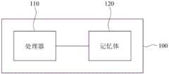
图1是根据一实施例绘示电子装置的示意图;FIG. 1 is a schematic diagram illustrating an electronic device according to an embodiment;
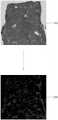
图2A与图2B是根据一些实施例绘示医学影像与分割影像的示意图;2A and 2B are schematic diagrams illustrating medical images and segmented images according to some embodiments;
图3是根据一实施例绘示判断环形纤维的示意图;3 is a schematic diagram illustrating determination of annular fibers according to an embodiment;
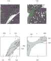
图4至图6是根据一实施例绘示局部医学影像的示意图;4 to 6 are schematic diagrams illustrating partial medical images according to an embodiment;
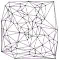
图7是根据一实施例绘示三角化后的示意图;7 is a schematic diagram illustrating triangulation according to an embodiment;
图8是根据一实施例绘示分割影像上节点的示意图;8 is a schematic diagram illustrating a node on a segmented image according to an embodiment;
图9是根据一实施例绘示三角化后一些节点所形成的一凸壳的示意图;9 is a schematic diagram illustrating a convex hull formed by some nodes after triangulation according to an embodiment;
图10是根据一实施例绘示建立连接矩阵的示意图;10 is a schematic diagram illustrating establishing a connection matrix according to an embodiment;
图11是根据一实施例绘示删除环的示意图;11 is a schematic diagram illustrating a deletion ring according to an embodiment;
图12是根据一实施例绘示纤维化的计算机辅助分析方法的流程图。12 is a flow chart illustrating a computer-aided analysis method of fibrosis according to an embodiment.
【符号说明】【Symbol Description】
100:电子装置100: Electronics
110:处理器110: Processor
120:记忆体120: memory
210、310、330:医学影像210, 310, 330: Medical Imaging
220、320、340、350:分割影像220, 320, 340, 350: Split image
321、341、351:细胞部分321, 341, 351: Cell Parts
322、342:血管部分322, 342: Vascular part
323:胆管部分323: Bile Duct Section
324、343、352:纤维部分324, 343, 352: Fiber part
360、370:影像360, 370: Video
371~374:环形纤维371-374: Ring fibers
410:医学影像410: Medical Imaging
411~413:门脉区411-413: Portal area
510、610:纤维化部分510, 610: Fibrotic part
520、620:非纤维化部分520, 620: Non-fibrous parts
801~808:节点801~808: Node
910:凸壳910: Convex Hull
1010:图1010: Figures
1020:连接矩阵1020: Connection Matrix
1110~1112:环1110~1112: Ring
1201~1207:步骤1201~1207: Steps
具体实施方式Detailed ways
关于本文中所使用的“第一”、“第二”、…等,并非特别指次序或顺位的意思,其仅为了区别以相同技术用语描述的元件或操作。The "first", "second", .
图1是根据一实施例绘示电子装置的示意图。请参照图1,电子装置100可以是智能手机、平板计算机、个人计算机、笔记型计算机、工业计算机或具有计算能力的各种电子装置或医疗器材等,本发明并不在此限。电子装置100包括了处理器110与记忆体120,其中处理器110可为中央处理器、微处理器、微控制器、数字信号处理器、影像处理芯片、特殊应用集成电路等,记忆体120可为挥发性记忆体或非挥发性记忆体,其中储存有多个指令,处理器110会执行这些指令来完成一纤维化分级方法。首先介绍依沙克(Ishak)纤维化级分,请参照以下表1。FIG. 1 is a schematic diagram illustrating an electronic device according to an embodiment. Referring to FIG. 1 , the


表1Table 1
表1的纤维化级分仰赖着医生的主观判断,以下提出一个客观的纤维化分级方法。首先取得一医学影像,此医学影像可以是肝脏组织切片扫描影像,在图2A的例子中,医学影像210是肝脏组织的切片影像,但在其他实施例中也可以是其他器官的医学影像,本发明并不在此限。接下来,对医学影像210执行分割(segmentation)演算法以取得分割影像220,此分割影像220为二值化影像,用以表示每个像素是否为纤维。在一些实施例中,上述的分割演算法是采用卷积神经网路,但在其他实施例中也可以采用任意的分割演算法,本发明并不在此限。举例来说,在训练阶段,卷积神经网路的输入是医学影像,而输出是经过人工标示出纤维的二值化影像;在测试阶段则可以将医学影像210输入至训练好的卷积神经网路,此卷积神经网路会输出分割影像220。在上述实施例中分割影像220为二值化影像,但在其他实施例中所产生的分割影像中也可以具有更多类别。举例来说,请参照图2B,医学影像310与医学影像330都是肝脏组织的切片扫描影像,分割影像320是对医学影像310进行分割的结果,分割影像340是对医学影像330进行分割的结果。分割影像320包括了细胞部分321、血管部分322、胆管部分323与纤维部分324。分割影像340包括了细胞部分341、血管部分342与纤维部分343。在其他实施例中也可以分割出其他类别,本发明并不在此限。The fibrosis grading in Table 1 relies on the subjective judgment of doctors, and an objective fibrosis grading method is proposed below. First, a medical image is obtained. The medical image can be a sliced image of liver tissue. In the example of FIG. 2A, the
接下来,根据分割影像来侦测医学影像中的环形纤维,此环形纤维指的是被纤维包围的肝细胞,环形纤维亦可被称为结核(nodule)。由于在分割影像中已经分辨出细胞部分与纤维部分,如果将纤维部分排除,则环形纤维会留下肝细胞,其大部分形状会接近圆形。但这样的作法只能找出被纤维完全包围的肝细胞,比较实际的做法是只要肝细胞有一比率(例如75%)的部分被包围就可以判断为环形纤维。因此,在此可以先排除分割影像中的纤维部分,对剩余的细胞部分做影像处理中的侵蚀(erosion)程序以取得被包围区域,如果一区域的肝细胞有75%以上的部分都被纤维包围,则侵蚀程序会移除没有被包围的肝细胞,使得剩余的肝细胞是独立的(没有与其他肝细胞相连)。请参照图3,分割影像350包括了细胞部分351与纤维部分352,在排除纤维部分352并对细胞部分351做侵蚀程序以后可以得到影像360,其中用不同的灰阶来表示各个独立的被包围区域,例如被包围区域361~365。Next, according to the segmented images, annular fibers in medical images are detected. The annular fibers refer to liver cells surrounded by fibers. The annular fibers can also be called nodules. Since the cellular part and the fibrous part have been distinguished in the segmented image, if the fibrous part is excluded, the ring-shaped fiber will leave the hepatocytes, and most of the shape will be close to a circle. However, in such a method, only hepatocytes completely surrounded by fibers can be found. As a practical method, as long as a proportion (eg, 75%) of hepatocytes are partially surrounded, it can be judged as annular fibers. Therefore, the fiber part in the segmented image can be excluded first, and the erosion procedure in image processing can be performed on the remaining cell part to obtain the surrounded area. If more than 75% of the liver cells in a region are covered by fibers Surrounded, the erosion procedure removes hepatocytes that are not surrounded, so that the remaining hepatocytes are independent (not connected to other hepatocytes). Please refer to FIG. 3 , the
接下来,计算每个被包围区域的圆度,如果圆度大于一临界值则可以判断为环形纤维。在一些实施例中,上述的圆度可以采用以下方程式(1)。Next, the circularity of each enclosed area is calculated, and if the circularity is greater than a critical value, it can be judged as an annular fiber. In some embodiments, the aforementioned roundness may employ the following equation (1).
fcirc=4πAP2…(1)fcirc = 4πAP2 …(1)
其中fcirc为圆度,A为被包围区域的面积,P为被包围区域的周长,当圆度越大,表示被包圆区域越接近圆形。在此实施例中,当被包围区域的长轴(区域中最远的两点之间的距离)大于等于1厘米且圆度大于等于0.3时会被判断为环形纤维。例如,影像370中绘示了环形纤维371~374,其余的被包围区域则不是环形纤维。在一些实施例中,在排除纤维部分352以后也可以对细胞部分351做多次侵蚀程序,每次侵蚀程序都采用不同的核心,在每次侵蚀程序后都会执行上述圆度的计算并判断是否为环形纤维,这些判断的结果会取联集,也就是说一个被包围区域只要在任何一次侵蚀程序被判断为环形纤维,此被包围区域就是环形纤维。执行多次侵蚀程序的目的是因为当使用核心较小的侵蚀程序时可能会无法切断被包围区域361~365之间相连的肝细胞,因此会找不到被包围区域361~365。where fcirc is the circularity, A is the area of the enclosed area, and P is the perimeter of the enclosed area. The larger the circularity, the closer the enclosed area is to a circle. In this embodiment, when the long axis of the enclosed area (the distance between the two farthest points in the area) is greater than or equal to 1 cm and the circularity is greater than or equal to 0.3, it will be judged as an annular fiber. For example,
在判断出环形纤维以后,根据这些环形纤维的大小可来决定级分为5分或6分。具体来说,可以将所有环形纤维371~374的面积总和除以分割影像中所有细胞部分351的面积以得到一比值,如果此比值大于等于一临界值,则设定级分为6分,否则设定级分为5分。After judging the annular fibers, the grades of 5 or 6 can be determined according to the size of these annular fibers. Specifically, the sum of the areas of all the annular fibers 371-374 can be divided by the area of all the
如果医学影像中并没有环形纤维,则表示级分为0~4分的其中之一。当纤维化比较严重时,门脉区与中央静脉会具有纤维桥接,因此级分为3分或4分。如果没有纤维桥接,则是0~2分的其中之一。因此接下来,侦测医学影像210中的门脉区(portal area)与中央静脉(central vein),所侦测出的门脉区与中央静脉如图4被包围盒(bounding box)所围绕,局部放大的医学影像410中具有门脉区411~413。上述侦测的步骤可以采用影像分析或任意的机器学习演算法,如卷积神经网路、支持向量机等,本发明并不在此限。在一些实施例中,可以训练一个机器学习模型来侦测门脉区与中央静脉,或者也可以训练两个机器学习模型来分别侦测门脉区与中央静脉。If there are no annular fibers in the medical image, it means that the grade is one of 0 to 4 points. When the fibrosis is more severe, the portal area and the central vein will have fibrous bridging, so the grade is 3 or 4. One of 0 to 2 points if there is no fiber bridging. Therefore, next, the portal area and the central vein in the
在此先说明没有纤维桥接的情形,在此情况要判断门脉区是否有纤维扩张的情形。具体来说,对于每一个门脉区都可以判断纤维部分的面积与该门脉区的面积之间的比值是否大于一临界值(例如50%),若是则可以判断此门脉区具有门脉扩张。此门脉区的面积是指纤维部分的面积加上血管部分或细胞部分的面积,这些面积都可以根据上述的分割影像来取得。举例来说,在图5的例子中,门脉区具有纤维化部分510与非纤维化部分520,在图5的例子中,纤维化部分510的面积与整个门脉区的面积之间的比值并没有大于50%,因此这个门脉区并没有门脉扩张。在图6的例子中,门脉区具有纤维化部分610与非纤维化部分620,但纤维化部分610的面积与整个门脉区的面积之间的比值已经大于50%,因此这个门脉区具有门脉扩张。First, the situation without fiber bridging will be described. In this case, it is necessary to determine whether there is fiber expansion in the portal vein region. Specifically, for each portal vein region, it can be judged whether the ratio between the area of the fiber portion and the area of the portal vein region is greater than a critical value (for example, 50%), and if so, it can be judged that the portal vein region has a portal vein expansion. The area of the portal region refers to the area of the fiber part plus the area of the blood vessel part or the cell part, and these areas can be obtained from the above-mentioned segmented images. For example, in the example of Fig. 5, the portal region has a
在此,扩张数目表示医学影像中有几个门脉区具有门脉扩张。如果扩张数目与所有门脉区的数目之间的比值大于一临界值(例如50%),设定级分为2级分。若扩张数目与所有门脉区的数目之间的比值小于等于50%且大于一个临界值(例如0、1、2或其他合适的数值),设定级分为1级分;若扩张数目小于等于此临界值(例如0),设定级分为0级分。这些比值也可以因应所搭配演算法而有调整,均属于本专利范围。Here, the number of dilations indicates how many portal regions in the medical image have portal dilations. If the ratio between the number of dilations and the number of all portal regions is greater than a critical value (eg, 50%), the grade is set as 2 grades. If the ratio between the number of dilations and the number of all portal regions is less than or equal to 50% and greater than a critical value (such as 0, 1, 2 or other suitable values), set the grade to 1; if the number of dilations is less than Equal to this critical value (eg 0), the set level is 0 level. These ratios can also be adjusted according to the matching algorithm, which all belong to the scope of this patent.
在此说明有纤维桥接的情形,当纤维化的程度较严重时,则门脉区或中央静脉的纤维有可能向四周相近的门脉区或中央静脉相连接形成桥接纤维,因此必须要计算门脉区与中央静脉的桥间数目。值得注意的是,此桥接数目包括了两个门脉区之间的桥间、两个中央静脉之间的桥接、以及门脉区与中央静脉之间的桥接。具体来说,可以将医学影像中所有的门脉区与中央静脉当作多个节点,对这些节点执行三角化演算法,执行的结果为一个图(graph),包含了复数节点与复数边,例如图7所示,此三角化演算法例如为DelaunayTriangulation,但本发明并不在此限。在三角化以后,每个节点都会透过三角形的边连接至附近的多个节点(亦称相邻节点),对于三角化后的每一个节点,都可根据上述的分割影像判断此节点是否透过纤维相连于对应的相邻节点。举例来说,请参照Here, it is explained that there is fiber bridging. When the degree of fibrosis is more serious, the fibers in the portal vein area or central vein may connect to the surrounding portal vein area or central vein to form bridging fibers. Therefore, it is necessary to calculate the portal vein area or central vein. The number of bridges between the vein area and the central vein. Notably, this number of bridges includes bridges between two portal areas, bridges between two central veins, and bridges between the portal area and the central vein. Specifically, all portal areas and central veins in medical images can be regarded as multiple nodes, and a triangulation algorithm can be performed on these nodes. The result of the execution is a graph, including complex nodes and complex edges. For example, as shown in FIG. 7 , the triangulation algorithm is, for example, Delaunay Triangulation, but the present invention is not limited thereto. After triangulation, each node will be connected to multiple nearby nodes (also called adjacent nodes) through the edges of the triangle. For each node after triangulation, it can be judged whether the node is transparent or not according to the above segmented image. The fibers are connected to the corresponding adjacent nodes. For example, see
图8,在图8中是将节点801~808绘示于分割影像中,借此看出节点801~808之间是否有桥接纤维。如上所述,每个节点801~808都对应至一个门脉或中央静脉,在一些实施例中节点801~808的座标为对应的包围盒的中心点。以节点804为例,依照三角化的结果,节点804是相邻于节点801、806、808与805,但节点804与节点801之间并没有桥接纤维,节点804与节点805、806、808之间有桥接纤维(桥接数目可加上3),也就是说,如果桥接数目越大,则表示纤维化的程度越严重。上述是否有桥接的判断可以采用广度优先搜寻(breadthfirst search),借此判断两个节点之间是否有连续的纤维连接着,但在其他实施例中也可采用其他合适的演算法来判断两个节点之间是否由纤维连接,本发明并不在此限。FIG. 8 , in FIG. 8 ,
图9是根据一实施例绘示三角化后一些节点所形成的一凸壳的示意图。请参照图9,在上述的三角化以后,共会形成(2n-2-k)个三角形以及(3n-3-k)个边,其中n为所有节点的数目,而k为这些节点所形成的凸壳910(convex hull)上节点的数目,(3n-3-k)亦称为边数目,而(2n-2-k)亦称为三角形数目。上述判断节点之间是否透过纤维彼此连接的步骤便是要检查这(3n-3-k)个边是否为桥接纤维。在一些实施例中,如果上述的桥接数目与边数目(3n-3-k)的比值小于一临界值(例如50%),则判定级分为3级分,否则判定级分为4级分。9 is a schematic diagram illustrating a convex hull formed by some nodes after triangulation according to an embodiment. Please refer to Figure 9, after the above triangulation, a total of (2n-2-k) triangles and (3n-3-k) edges will be formed, where n is the number of all nodes, and k is formed by these nodes. The number of nodes on the
在一些实施例中,在将门脉区与中央静脉当作节点以后,可以把节点之间的距离当作边以形成一个完全连接图,然后将此完全连接图转换为一个树状结构(例如最小权重生成树或其他树状结构),此树状结构用以指示每一个节点所对应的相邻节点。对于每一个节点,判断此节点是否透过纤维部分相连于对应的相邻节点,借此可以计算出桥接数目。在一些实施例中,也可以将桥接数目除以所有节点的个数(即正整数n)以得到一比值,若此比值大于一临界值则判定为4级分,否则判定为3级分。在一些实施例中,也可以计算有透过纤维部分相连接的节点的数目与所有节点的数目之间的比值是否大于一临界值,若是判定为4级分,否则判定为3级分。In some embodiments, after treating the portal region and the central vein as nodes, the distances between the nodes can be treated as edges to form a fully connected graph, which is then converted into a tree-like structure (eg Minimum weight spanning tree or other tree structure), this tree structure is used to indicate the adjacent nodes corresponding to each node. For each node, it is judged whether the node is connected to the corresponding adjacent node through the fiber part, so that the number of bridges can be calculated. In some embodiments, the number of bridges can also be divided by the number of all nodes (ie, a positive integer n) to obtain a ratio. If the ratio is greater than a critical value, it is judged as 4 grades, otherwise it is judged as 3 grades. In some embodiments, it is also possible to calculate whether the ratio between the number of nodes connected through fiber parts and the number of all nodes is greater than a critical value.
上述的0~6级分仅是范例,在其他实施例中可以采用其他数值来作为级分,或者可以用任意的符号或文字来表示级分,本发明并不在此限。换言之,上述的0~6级分亦可被称为第七至第一级分,但本发明并不限制第七至第一级分包含了什么数字、符号或文字。The above-mentioned 0-6 grades are only examples. In other embodiments, other numerical values may be used as the grades, or any symbols or characters may be used to represent the grades, and the present invention is not limited thereto. In other words, the above-mentioned grades 0-6 may also be referred to as the seventh to first grades, but the present invention does not limit what numbers, symbols or characters are included in the seventh to first grades.
在一些实施例中,上述的环形纤维也可以根据桥接的情况来判断,这是因为节点之间会透过纤维彼此相连且形成一个环(circle),例如图8的节点804、806、808之间便形成一个环。为了计算出所有环的数目,可以根据节点与节点之间的相邻关系建立一连接矩阵,举例来说,图10中绘示了图(graph)1010,图1010中的节点便是上述的门脉区与中央静脉,而图1010中的每个边表示桥接纤维,根据图1010的节点与边可以建立连接矩阵1020,例如节点A与节点B之间有边(即这两个门脉区/中央静脉之间有桥接纤维),但节点B与节点D之间没有边,以此类推。根据此连接矩阵1020可以判断出多个环,例如采用论文“Finding Allthe Elementary Circuits of a Directed Graph”by D.B.Johnson,1975揭露的演算法,但本发明并不在此限。In some embodiments, the above-mentioned annular fibers can also be judged according to the bridging situation, because the nodes are connected to each other through the fibers and form a circle, such as the
图11是根据一实施例绘示多个环的示意图。在图11的例子中共有3个环1110~1112,其中环1110具有四个节点,环1111与环1112都具有三个节点,以另一个角度来说,环1111与环1112的联集会组成环1110。上述Johnson演算法的结果会指出共有4个环,但这样会重复计算纤维化的情形,因此必须排除重复计算的环。具体来说,对于每一个环,判断此环是否为其他环的联集,此判断步骤可以写为以下方程式(2)。FIG. 11 is a schematic diagram illustrating a plurality of rings according to an embodiment. In the example of FIG. 11 , there are three
Ci=Uj,j≠iCj…(2)Ci =Uj,j≠i Cj ...(2)
其中Ci、Cj表示环。若方程式(2)成立,则删除环Ci,在对所有的环都进行上述方程式(2)的判断以后,剩余的环便不是其他环的联集,并不会重复计算,而剩余环的总数便称为环数目。例如,在图11的实施例中环1110会被删除,剩下环1111、1112会被计数到。以另外一个角度来说,上述找出环并删除重复环的步骤便是要从(2n-2-k)个三角形中找到由桥接纤维形成的三角形。接下来,判断此环数目与上述三角形数目(2n-2-k)之间的比值是否小于一临界值(例如50%),若是则设定级分为5级分,否则设定级分为6级分。wherein Ci and Cj represent rings. If equation (2) is established, then delete the ring Ci . After the above equation (2) is judged for all rings, the remaining rings are not the union of other rings, and the calculation will not be repeated. The total is called the number of rings. For example, in the embodiment of FIG. 11, the
图12是根据一实施例绘示纤维化的计算机辅助分析方法的流程图。请参照图12,在步骤1201,对医学影像执行一分割演算法以取得分割影像。在步骤1202,判断是否有环形纤维。若有环形纤维,在步骤1203中,根据环形纤维的大小来决定级分5或6。若没有环形纤维,在步骤1204中侦测门脉区与中央静脉。在步骤1205中,判断是否有桥接。如果门脉区与中央静脉具有桥接,在步骤1206中根据桥接数目决定级分3或4。若没有桥接,在步骤1207中根据门脉扩张的情形决定级分0~2。然而,图12中各步骤已详细说明如上,在此便不再赘述。值得注意的是,图12中各步骤可以实作为多个程式码或是电路,本发明并不在此限。此外,图12的方法可以搭配以上实施例使用,也可以单独使用。换言之,图12的各步骤之间也可以加入其他的步骤。12 is a flow chart illustrating a computer-aided analysis method of fibrosis according to an embodiment. Referring to FIG. 12, in
虽然本发明已以实施例揭露如上,然其并非用以限定本发明,任何所属技术领域中具有通常知识者,在不脱离本发明的精神和范围内,当可作些许的更动与润饰,故本发明的保护范围当视所附的权利要求书所界定的范围为准。Although the present invention has been disclosed as above with examples, it is not intended to limit the present invention. Anyone with ordinary knowledge in the technical field can make some changes and modifications without departing from the spirit and scope of the present invention. Therefore, the protection scope of the present invention should be based on the scope defined by the appended claims.
Claims (10)
Applications Claiming Priority (4)
| Application Number | Priority Date | Filing Date | Title |
|---|---|---|---|
| US201862723460P | 2018-08-27 | 2018-08-27 | |
| US62/723,460 | 2018-08-27 | ||
| TW108127947ATWI714200B (en) | 2018-08-27 | 2019-08-06 | Computer aided method, electrical device and computer program product for analyzing fibrosis |
| TW108127947 | 2019-08-06 |
Publications (2)
| Publication Number | Publication Date |
|---|---|
| CN110866923Atrue CN110866923A (en) | 2020-03-06 |
| CN110866923B CN110866923B (en) | 2022-11-01 |
Family
ID=69583492
Family Applications (1)
| Application Number | Title | Priority Date | Filing Date |
|---|---|---|---|
| CN201910789554.7AActiveCN110866923B (en) | 2018-08-27 | 2019-08-26 | Computer-aided fibrosis analysis method and electronic device |
Country Status (3)
| Country | Link |
|---|---|
| US (1) | US11508060B2 (en) |
| JP (1) | JP7071744B2 (en) |
| CN (1) | CN110866923B (en) |
Citations (8)
| Publication number | Priority date | Publication date | Assignee | Title |
|---|---|---|---|---|
| US6738499B1 (en)* | 1998-11-13 | 2004-05-18 | Arch Development Corporation | System for detection of malignancy in pulmonary nodules |
| WO2006042864A1 (en)* | 2004-10-22 | 2006-04-27 | Institut Pasteur | Method of processing an image identifying fibrosis in an organic tissue |
| TW200922627A (en)* | 2007-11-30 | 2009-06-01 | Iner Aec Executive Yuan | Glyco-molecular imaging method for grade classification of liver fibrosis and its glyco-molecular imaging agent thereof |
| CN101540051A (en)* | 2008-03-21 | 2009-09-23 | 国立大学法人神户大学 | Diagnostic imaging support processing apparatus and diagnostic imaging support processing program product |
| US20150148658A1 (en)* | 2012-07-11 | 2015-05-28 | University Of Mississippi Medical Center | Method for the detection and staging of liver fibrosis from image acquired data |
| CN107103187A (en)* | 2017-04-10 | 2017-08-29 | 四川省肿瘤医院 | The method and system of Lung neoplasm detection classification and management based on deep learning |
| CN107895368A (en)* | 2017-11-24 | 2018-04-10 | 北京大学人民医院 | Application of the parameter as the characteristic parameter by stages of the liver fibrosis of adult or children in SHG/TPEF images |
| CN108426862A (en)* | 2018-04-28 | 2018-08-21 | 福建师范大学 | A method of differentiating that liver cancer breaks up grade using multi-photon imaging technique |
Family Cites Families (2)
| Publication number | Priority date | Publication date | Assignee | Title |
|---|---|---|---|---|
| CN100486600C (en)* | 2005-12-30 | 2009-05-13 | 安徽医科大学 | Application of spanishneedles herb flavonid in preparing medicine for preventing and treating liver fibrosis |
| WO2015054597A2 (en)* | 2013-10-12 | 2015-04-16 | H. Lee Moffitt Cancer Center And Research Institute, Inc. | Systems and methods for diagnosing tumors in a subject by performing a quantitative analysis of texture-based features of a tumor object in a radiological image |
- 2019
- 2019-08-26CNCN201910789554.7Apatent/CN110866923B/enactiveActive
- 2019-08-26USUS16/551,703patent/US11508060B2/enactiveActive
- 2019-08-26JPJP2019153511Apatent/JP7071744B2/enactiveActive
Patent Citations (8)
| Publication number | Priority date | Publication date | Assignee | Title |
|---|---|---|---|---|
| US6738499B1 (en)* | 1998-11-13 | 2004-05-18 | Arch Development Corporation | System for detection of malignancy in pulmonary nodules |
| WO2006042864A1 (en)* | 2004-10-22 | 2006-04-27 | Institut Pasteur | Method of processing an image identifying fibrosis in an organic tissue |
| TW200922627A (en)* | 2007-11-30 | 2009-06-01 | Iner Aec Executive Yuan | Glyco-molecular imaging method for grade classification of liver fibrosis and its glyco-molecular imaging agent thereof |
| CN101540051A (en)* | 2008-03-21 | 2009-09-23 | 国立大学法人神户大学 | Diagnostic imaging support processing apparatus and diagnostic imaging support processing program product |
| US20150148658A1 (en)* | 2012-07-11 | 2015-05-28 | University Of Mississippi Medical Center | Method for the detection and staging of liver fibrosis from image acquired data |
| CN107103187A (en)* | 2017-04-10 | 2017-08-29 | 四川省肿瘤医院 | The method and system of Lung neoplasm detection classification and management based on deep learning |
| CN107895368A (en)* | 2017-11-24 | 2018-04-10 | 北京大学人民医院 | Application of the parameter as the characteristic parameter by stages of the liver fibrosis of adult or children in SHG/TPEF images |
| CN108426862A (en)* | 2018-04-28 | 2018-08-21 | 福建师范大学 | A method of differentiating that liver cancer breaks up grade using multi-photon imaging technique |
Non-Patent Citations (2)
| Title |
|---|
| 贾同,赵大哲,杨金柱,王旭: "一种基于HRCT影像的肺结节计算机辅助检测方法", 《系统仿真学报》, vol. 20, 31 July 2008 (2008-07-31), pages 1* |
| 陆中臣: "《流域地貌系统》", 31 May 1991, pages: 16* |
Also Published As
| Publication number | Publication date |
|---|---|
| US11508060B2 (en) | 2022-11-22 |
| JP7071744B2 (en) | 2022-05-19 |
| JP2020035444A (en) | 2020-03-05 |
| CN110866923B (en) | 2022-11-01 |
| US20200065965A1 (en) | 2020-02-27 |
Similar Documents
| Publication | Publication Date | Title |
|---|---|---|
| WO2023179122A1 (en) | Defect detection method and apparatus, electronic device, and readable storage medium | |
| JPWO2019026104A1 (en) | Information processing apparatus, information processing program, and information processing method | |
| CN111369623B (en) | Lung CT image identification method based on deep learning 3D target detection | |
| Ren et al. | An unsupervised semi-automated pulmonary nodule segmentation method based on enhanced region growing | |
| Liu et al. | NCRNet: Neighborhood context refinement network for skin lesion segmentation | |
| Bang et al. | Computer-aided diagnosis of gastrointestinal ulcer and hemorrhage using wireless capsule endoscopy: systematic review and diagnostic test accuracy meta-analysis | |
| CN111709914A (en) | A No-reference Image Quality Evaluation Method Based on HVS Characteristics | |
| WO2024001747A1 (en) | Pulmonary blood vessel model establishment method and apparatus, and server | |
| CN117392464A (en) | Method and system for image anomaly detection based on multi-scale denoising probability model | |
| Wang et al. | Accurate airway tree segmentation in ct scans via anatomy-aware multi-class segmentation and topology-guided iterative learning | |
| CN113887677B (en) | Classification method, apparatus, apparatus and medium for images of intrapapillary capillaries | |
| Lin et al. | Improving sensitivity and connectivity of retinal vessel segmentation via error discrimination network | |
| CN110866923B (en) | Computer-aided fibrosis analysis method and electronic device | |
| Abdullah et al. | Preliminary study of pneumonia symptoms detection method using cellular neural network | |
| JP6177649B2 (en) | Data processing apparatus, length measurement system, defect inspection system, image tracking system, and data processing method | |
| CN114708271A (en) | A medical image segmentation method based on highlight removal | |
| TWI714200B (en) | Computer aided method, electrical device and computer program product for analyzing fibrosis | |
| CN114581467A (en) | An Image Segmentation Method Based on Residual Dilated Spatial Pyramid Network Algorithm | |
| Wang et al. | Detection of quality visualization of appendiceal orifices using local edge cross-section profile features and near pause detection | |
| CN118470049A (en) | Classification and segmentation of ultrasound images of dialysis access and calculation method and medium of vascular stenosis rate | |
| CN115631194B (en) | Method, device, equipment and medium for identifying and detecting intracranial aneurysm | |
| CN115601335B (en) | Multi-scale fusion full convolution network lymph node detection method based on CT image | |
| CN114723660B (en) | Cloth defect detection method and system | |
| CN118541728A (en) | Image generation device, image generation method, image generation program, learning device, and learning data | |
| Fukano et al. | Recognition method of lung nodules using blood vessel extraction techniques and 3D object models |
Legal Events
| Date | Code | Title | Description |
|---|---|---|---|
| PB01 | Publication | ||
| PB01 | Publication | ||
| SE01 | Entry into force of request for substantive examination | ||
| SE01 | Entry into force of request for substantive examination | ||
| GR01 | Patent grant | ||
| GR01 | Patent grant |