CN110859991B - Preparation method of double-layer composite scaffold for inducing regeneration of dental pulp dentin tissues - Google Patents
Preparation method of double-layer composite scaffold for inducing regeneration of dental pulp dentin tissuesDownload PDFInfo
- Publication number
- CN110859991B CN110859991BCN201911075077.4ACN201911075077ACN110859991BCN 110859991 BCN110859991 BCN 110859991BCN 201911075077 ACN201911075077 ACN 201911075077ACN 110859991 BCN110859991 BCN 110859991B
- Authority
- CN
- China
- Prior art keywords
- scaffold
- psc
- double
- dentin
- pulp
- Prior art date
- Legal status (The legal status is an assumption and is not a legal conclusion. Google has not performed a legal analysis and makes no representation as to the accuracy of the status listed.)
- Active
Links
- 210000004268dentinAnatomy0.000titleclaimsabstractdescription26
- 239000002131composite materialSubstances0.000titleclaimsabstractdescription21
- 230000001939inductive effectEffects0.000titleclaimsabstractdescription15
- 238000002360preparation methodMethods0.000titleclaimsabstractdescription14
- 230000008929regenerationEffects0.000titleclaimsabstractdescription13
- 238000011069regeneration methodMethods0.000titleclaimsabstractdescription13
- 210000003074dental pulpAnatomy0.000titleabstractdescription9
- 239000000463materialSubstances0.000claimsabstractdescription26
- 239000005313bioactive glassSubstances0.000claimsabstractdescription25
- 210000001519tissueAnatomy0.000claimsabstractdescription23
- 108090000765processed proteins & peptidesProteins0.000claimsabstractdescription20
- 229920001184polypeptidePolymers0.000claimsabstractdescription19
- 102000004196processed proteins & peptidesHuman genes0.000claimsabstractdescription19
- 210000000130stem cellAnatomy0.000claimsabstractdescription9
- IMQLKJBTEOYOSI-GPIVLXJGSA-NInositol-hexakisphosphateChemical compoundOP(O)(=O)O[C@H]1[C@H](OP(O)(O)=O)[C@@H](OP(O)(O)=O)[C@H](OP(O)(O)=O)[C@H](OP(O)(O)=O)[C@@H]1OP(O)(O)=OIMQLKJBTEOYOSI-GPIVLXJGSA-N0.000claimsabstractdescription8
- IMQLKJBTEOYOSI-UHFFFAOYSA-NPhytic acidNatural productsOP(O)(=O)OC1C(OP(O)(O)=O)C(OP(O)(O)=O)C(OP(O)(O)=O)C(OP(O)(O)=O)C1OP(O)(O)=OIMQLKJBTEOYOSI-UHFFFAOYSA-N0.000claimsabstractdescription8
- 229940068041phytic acidDrugs0.000claimsabstractdescription8
- 235000002949phytic acidNutrition0.000claimsabstractdescription8
- 239000000467phytic acidSubstances0.000claimsabstractdescription8
- 210000004872soft tissueAnatomy0.000claimsabstractdescription6
- 230000017423tissue regenerationEffects0.000claimsabstractdescription6
- 238000004132cross linkingMethods0.000claimsabstractdescription5
- 239000003431cross linking reagentSubstances0.000claimsabstractdescription5
- 230000004048modificationEffects0.000claimsabstractdescription5
- 238000012986modificationMethods0.000claimsabstractdescription5
- 239000002243precursorSubstances0.000claimsabstractdescription5
- SXRSQZLOMIGNAQ-UHFFFAOYSA-NGlutaraldehydeChemical compoundO=CCCCC=OSXRSQZLOMIGNAQ-UHFFFAOYSA-N0.000claimsabstractdescription4
- 239000000017hydrogelSubstances0.000claimsdescription13
- YXFVVABEGXRONW-UHFFFAOYSA-NTolueneChemical compoundCC1=CC=CC=C1YXFVVABEGXRONW-UHFFFAOYSA-N0.000claimsdescription12
- 239000002245particleSubstances0.000claimsdescription12
- LFQSCWFLJHTTHZ-UHFFFAOYSA-NEthanolChemical compoundCCOLFQSCWFLJHTTHZ-UHFFFAOYSA-N0.000claimsdescription11
- XLYOFNOQVPJJNP-UHFFFAOYSA-NwaterSubstancesOXLYOFNOQVPJJNP-UHFFFAOYSA-N0.000claimsdescription11
- 230000004069differentiationEffects0.000claimsdescription8
- 108010010803GelatinProteins0.000claimsdescription7
- 230000015572biosynthetic processEffects0.000claimsdescription7
- 239000008273gelatinSubstances0.000claimsdescription7
- 229920000159gelatinPolymers0.000claimsdescription7
- 235000019322gelatineNutrition0.000claimsdescription7
- 235000011852gelatine dessertsNutrition0.000claimsdescription7
- IJGRMHOSHXDMSA-UHFFFAOYSA-NAtomic nitrogenChemical compoundN#NIJGRMHOSHXDMSA-UHFFFAOYSA-N0.000claimsdescription6
- BOTDANWDWHJENH-UHFFFAOYSA-NTetraethyl orthosilicateChemical compoundCCO[Si](OCC)(OCC)OCCBOTDANWDWHJENH-UHFFFAOYSA-N0.000claimsdescription6
- 238000000034methodMethods0.000claimsdescription6
- WYTZZXDRDKSJID-UHFFFAOYSA-N(3-aminopropyl)triethoxysilaneChemical compoundCCO[Si](OCC)(OCC)CCCNWYTZZXDRDKSJID-UHFFFAOYSA-N0.000claimsdescription5
- HNXGGWNCFXZSAI-UHFFFAOYSA-N2-morpholin-2-ylethanesulfonic acidChemical compoundOS(=O)(=O)CCC1CNCCO1HNXGGWNCFXZSAI-UHFFFAOYSA-N0.000claimsdescription5
- 239000008367deionised waterSubstances0.000claimsdescription5
- 229910021641deionized waterInorganic materials0.000claimsdescription5
- 238000003760magnetic stirringMethods0.000claimsdescription5
- 239000011148porous materialSubstances0.000claimsdescription5
- NNRFRJQMBSBXGO-CIUDSAMLSA-N(3s)-3-[[2-[[(2s)-2-amino-5-(diaminomethylideneamino)pentanoyl]amino]acetyl]amino]-4-[[(1s)-1-carboxy-2-hydroxyethyl]amino]-4-oxobutanoic acidChemical compoundNC(N)=NCCC[C@H](N)C(=O)NCC(=O)N[C@@H](CC(O)=O)C(=O)N[C@@H](CO)C(O)=ONNRFRJQMBSBXGO-CIUDSAMLSA-N0.000claimsdescription4
- 102000008186CollagenHuman genes0.000claimsdescription4
- 108010035532CollagenProteins0.000claimsdescription4
- 101000829980Homo sapiens Ral guanine nucleotide dissociation stimulatorProteins0.000claimsdescription4
- 102100023320Ral guanine nucleotide dissociation stimulatorHuman genes0.000claimsdescription4
- VYPSYNLAJGMNEJ-UHFFFAOYSA-NSilicium dioxideChemical compoundO=[Si]=OVYPSYNLAJGMNEJ-UHFFFAOYSA-N0.000claimsdescription4
- 229920001436collagenPolymers0.000claimsdescription4
- 239000000203mixtureSubstances0.000claimsdescription4
- 239000011240wet gelSubstances0.000claimsdescription4
- FPQQSJJWHUJYPU-UHFFFAOYSA-N3-(dimethylamino)propyliminomethylidene-ethylazanium;chlorideChemical groupCl.CCN=C=NCCCN(C)CFPQQSJJWHUJYPU-UHFFFAOYSA-N0.000claimsdescription3
- NQTADLQHYWFPDB-UHFFFAOYSA-NN-HydroxysuccinimideChemical groupON1C(=O)CCC1=ONQTADLQHYWFPDB-UHFFFAOYSA-N0.000claimsdescription3
- 239000002270dispersing agentSubstances0.000claimsdescription3
- 239000006185dispersionSubstances0.000claimsdescription3
- 239000000499gelSubstances0.000claimsdescription3
- 239000011521glassSubstances0.000claimsdescription3
- 229910052757nitrogenInorganic materials0.000claimsdescription3
- 239000000843powderSubstances0.000claimsdescription3
- 238000010992refluxMethods0.000claimsdescription3
- 229920001661ChitosanPolymers0.000claimsdescription2
- HTTJABKRGRZYRN-UHFFFAOYSA-NHeparinChemical compoundOC1C(NC(=O)C)C(O)OC(COS(O)(=O)=O)C1OC1C(OS(O)(=O)=O)C(O)C(OC2C(C(OS(O)(=O)=O)C(OC3C(C(O)C(O)C(O3)C(O)=O)OS(O)(=O)=O)C(CO)O2)NS(O)(=O)=O)C(C(O)=O)O1HTTJABKRGRZYRN-UHFFFAOYSA-N0.000claimsdescription2
- 102000004887Transforming Growth Factor betaHuman genes0.000claimsdescription2
- 108090001012Transforming Growth Factor betaProteins0.000claimsdescription2
- 108010009583Transforming Growth FactorsProteins0.000claimsdescription2
- 102000009618Transforming Growth FactorsHuman genes0.000claimsdescription2
- 229910052681coesiteInorganic materials0.000claimsdescription2
- 150000001875compoundsChemical class0.000claimsdescription2
- 210000002808connective tissueAnatomy0.000claimsdescription2
- 229910052906cristobaliteInorganic materials0.000claimsdescription2
- 230000007646directional migrationEffects0.000claimsdescription2
- 238000004108freeze dryingMethods0.000claimsdescription2
- 238000010438heat treatmentMethods0.000claimsdescription2
- 229960002897heparinDrugs0.000claimsdescription2
- 229920000669heparinPolymers0.000claimsdescription2
- 238000002156mixingMethods0.000claimsdescription2
- 230000000877morphologic effectEffects0.000claimsdescription2
- 239000000377silicon dioxideSubstances0.000claimsdescription2
- 235000012239silicon dioxideNutrition0.000claimsdescription2
- 238000003756stirringMethods0.000claimsdescription2
- 229910052682stishoviteInorganic materials0.000claimsdescription2
- ZRKFYGHZFMAOKI-QMGMOQQFSA-NtgfbetaChemical compoundC([C@H](NC(=O)[C@H](C(C)C)NC(=O)CNC(=O)[C@H](CCC(O)=O)NC(=O)[C@H](CCCNC(N)=N)NC(=O)[C@H](CC(N)=O)NC(=O)[C@H](CC(C)C)NC(=O)[C@H]([C@@H](C)O)NC(=O)[C@H](CCC(O)=O)NC(=O)[C@H]([C@@H](C)O)NC(=O)[C@H](CC(C)C)NC(=O)CNC(=O)[C@H](C)NC(=O)[C@H](CO)NC(=O)[C@H](CCC(N)=O)NC(=O)[C@@H](NC(=O)[C@H](C)NC(=O)[C@H](C)NC(=O)[C@@H](NC(=O)[C@H](CC(C)C)NC(=O)[C@@H](N)CCSC)C(C)C)[C@@H](C)CC)C(=O)N[C@@H]([C@@H](C)O)C(=O)N[C@@H](C(C)C)C(=O)N[C@@H](CC=1C=CC=CC=1)C(=O)N[C@@H](C)C(=O)N1[C@@H](CCC1)C(=O)N[C@@H]([C@@H](C)O)C(=O)N[C@@H](CC(N)=O)C(=O)N[C@@H](CCC(O)=O)C(=O)N[C@@H](C)C(=O)N[C@@H](CC=1C=CC=CC=1)C(=O)N[C@@H](CCCNC(N)=N)C(=O)N[C@@H](C)C(=O)N[C@@H](CC(C)C)C(=O)N1[C@@H](CCC1)C(=O)N1[C@@H](CCC1)C(=O)N[C@@H](CCCNC(N)=N)C(=O)N[C@@H](CCC(O)=O)C(=O)N[C@@H](CCCNC(N)=N)C(=O)N[C@@H](CO)C(=O)N[C@@H](CCCNC(N)=N)C(=O)N[C@@H](CC(C)C)C(=O)N[C@@H](CC(C)C)C(O)=O)C1=CC=C(O)C=C1ZRKFYGHZFMAOKI-QMGMOQQFSA-N0.000claimsdescription2
- 229910052905tridymiteInorganic materials0.000claimsdescription2
- 238000009777vacuum freeze-dryingMethods0.000claimsdescription2
- ZCCIPPOKBCJFDN-UHFFFAOYSA-Ncalcium nitrateChemical compound[Ca+2].[O-][N+]([O-])=O.[O-][N+]([O-])=OZCCIPPOKBCJFDN-UHFFFAOYSA-N0.000claims2
- 230000004071biological effectEffects0.000claims1
- 239000007853buffer solutionSubstances0.000claims1
- 238000002242deionisation methodMethods0.000claims1
- 230000032724odontogenesisEffects0.000claims1
- 239000012688phosphorus precursorSubstances0.000claims1
- 238000005406washingMethods0.000claims1
- 210000004262dental pulp cavityAnatomy0.000abstractdescription10
- 230000000975bioactive effectEffects0.000abstractdescription4
- 230000006698inductionEffects0.000abstractdescription3
- 230000005012migrationEffects0.000abstractdescription3
- 238000013508migrationMethods0.000abstractdescription3
- 230000009286beneficial effectEffects0.000abstractdescription2
- 230000024245cell differentiationEffects0.000abstractdescription2
- 230000003399chemotactic effectEffects0.000abstractdescription2
- 238000002791soakingMethods0.000abstract1
- 210000004027cellAnatomy0.000description8
- 210000004416odontoblastAnatomy0.000description5
- 239000012620biological materialSubstances0.000description4
- 210000001968dental pulp cellAnatomy0.000description4
- 238000010186stainingMethods0.000description4
- ZHJGWYRLJUCMRT-UHFFFAOYSA-N5-[6-[(4-methylpiperazin-1-yl)methyl]benzimidazol-1-yl]-3-[1-[2-(trifluoromethyl)phenyl]ethoxy]thiophene-2-carboxamideChemical compoundC=1C=CC=C(C(F)(F)F)C=1C(C)OC(=C(S1)C(N)=O)C=C1N(C1=C2)C=NC1=CC=C2CN1CCN(C)CC1ZHJGWYRLJUCMRT-UHFFFAOYSA-N0.000description3
- 229920000954PolyglycolidePolymers0.000description3
- 230000033558biomineral tissue developmentEffects0.000description3
- 230000006378damageEffects0.000description3
- 238000001804debridementMethods0.000description3
- 230000000694effectsEffects0.000description3
- 229920001606poly(lactic acid-co-glycolic acid)Polymers0.000description3
- 229920001610polycaprolactonePolymers0.000description3
- 108090000623proteins and genesProteins0.000description3
- 238000011160researchMethods0.000description3
- 230000000250revascularizationEffects0.000description3
- 229910004298SiO 2Inorganic materials0.000description2
- 230000004791biological behaviorEffects0.000description2
- 229910010293ceramic materialInorganic materials0.000description2
- 238000006243chemical reactionMethods0.000description2
- 238000011161developmentMethods0.000description2
- 230000018109developmental processEffects0.000description2
- 239000012634fragmentSubstances0.000description2
- 238000003384imaging methodMethods0.000description2
- 208000014674injuryDiseases0.000description2
- 229920000747poly(lactic acid)Polymers0.000description2
- 239000004632polycaprolactoneSubstances0.000description2
- 239000004626polylactic acidSubstances0.000description2
- 230000001105regulatory effectEffects0.000description2
- 238000001878scanning electron micrographMethods0.000description2
- 239000008279solSubstances0.000description2
- 230000008733traumaEffects0.000description2
- 102100022375Dentin matrix acidic phosphoprotein 1Human genes0.000description1
- 101710105839Dentin matrix acidic phosphoprotein 1Proteins0.000description1
- 102100029792Dentin sialophosphoproteinHuman genes0.000description1
- 108010037362Extracellular Matrix ProteinsProteins0.000description1
- 102000010834Extracellular Matrix ProteinsHuman genes0.000description1
- 230000005526G1 to G0 transitionEffects0.000description1
- AEMRFAOFKBGASW-UHFFFAOYSA-NGlycolic acidPolymersOCC(O)=OAEMRFAOFKBGASW-UHFFFAOYSA-N0.000description1
- 101000865404Homo sapiens Dentin sialophosphoproteinProteins0.000description1
- -1N-hydroxysuccinimide AmineChemical class0.000description1
- OAICVXFJPJFONN-UHFFFAOYSA-NPhosphorusChemical compound[P]OAICVXFJPJFONN-UHFFFAOYSA-N0.000description1
- 239000002253acidSubstances0.000description1
- 239000011149active materialSubstances0.000description1
- 238000005576amination reactionMethods0.000description1
- 210000003484anatomyAnatomy0.000description1
- 230000033115angiogenesisEffects0.000description1
- 229910052586apatiteInorganic materials0.000description1
- 230000006399behaviorEffects0.000description1
- 239000003462bioceramicSubstances0.000description1
- 238000012925biological evaluationMethods0.000description1
- 239000008280bloodSubstances0.000description1
- 210000004369bloodAnatomy0.000description1
- 210000000988bone and boneAnatomy0.000description1
- 210000001185bone marrowAnatomy0.000description1
- 239000011575calciumSubstances0.000description1
- 238000005266castingMethods0.000description1
- 230000021164cell adhesionEffects0.000description1
- 230000012292cell migrationEffects0.000description1
- 230000009194climbingEffects0.000description1
- 238000010276constructionMethods0.000description1
- 230000032798delaminationEffects0.000description1
- 208000002925dental cariesDiseases0.000description1
- 238000010586diagramMethods0.000description1
- 239000003814drugSubstances0.000description1
- 229940079593drugDrugs0.000description1
- 238000005516engineering processMethods0.000description1
- 125000004494ethyl ester groupChemical group0.000description1
- 210000002744extracellular matrixAnatomy0.000description1
- 238000000227grindingMethods0.000description1
- 239000003102growth factorSubstances0.000description1
- 229910052588hydroxylapatiteInorganic materials0.000description1
- 230000003902lesionEffects0.000description1
- 239000011159matrix materialSubstances0.000description1
- 230000017074necrotic cell deathEffects0.000description1
- 210000005036nerveAnatomy0.000description1
- 230000008520organizationEffects0.000description1
- VSIIXMUUUJUKCM-UHFFFAOYSA-Dpentacalcium;fluoride;triphosphateChemical compound[F-].[Ca+2].[Ca+2].[Ca+2].[Ca+2].[Ca+2].[O-]P([O-])([O-])=O.[O-]P([O-])([O-])=O.[O-]P([O-])([O-])=OVSIIXMUUUJUKCM-UHFFFAOYSA-D0.000description1
- XYJRXVWERLGGKC-UHFFFAOYSA-Dpentacalcium;hydroxide;triphosphateChemical compound[OH-].[Ca+2].[Ca+2].[Ca+2].[Ca+2].[Ca+2].[O-]P([O-])([O-])=O.[O-]P([O-])([O-])=O.[O-]P([O-])([O-])=OXYJRXVWERLGGKC-UHFFFAOYSA-D0.000description1
- 210000004053periapical tissueAnatomy0.000description1
- 230000003239periodontal effectEffects0.000description1
- 229910052698phosphorusInorganic materials0.000description1
- 239000011574phosphorusSubstances0.000description1
- 239000004633polyglycolic acidSubstances0.000description1
- 229950008885polyglycolic acidDrugs0.000description1
- 239000002861polymer materialSubstances0.000description1
- 230000035755proliferationEffects0.000description1
- 230000009993protective functionEffects0.000description1
- 102000004169proteins and genesHuman genes0.000description1
- 230000004044responseEffects0.000description1
- 210000004499secondary dentinAnatomy0.000description1
- 238000007873sievingMethods0.000description1
- RMAQACBXLXPBSY-UHFFFAOYSA-Nsilicic acidChemical compoundO[Si](O)(O)ORMAQACBXLXPBSY-UHFFFAOYSA-N0.000description1
- 239000002904solventSubstances0.000description1
- 239000000126substanceSubstances0.000description1
- 238000002560therapeutic procedureMethods0.000description1
- 230000008719thickeningEffects0.000description1
- 230000007704transitionEffects0.000description1
- 238000002054transplantationMethods0.000description1
- 210000005239tubuleAnatomy0.000description1
Images


Classifications
- A—HUMAN NECESSITIES
- A61—MEDICAL OR VETERINARY SCIENCE; HYGIENE
- A61L—METHODS OR APPARATUS FOR STERILISING MATERIALS OR OBJECTS IN GENERAL; DISINFECTION, STERILISATION OR DEODORISATION OF AIR; CHEMICAL ASPECTS OF BANDAGES, DRESSINGS, ABSORBENT PADS OR SURGICAL ARTICLES; MATERIALS FOR BANDAGES, DRESSINGS, ABSORBENT PADS OR SURGICAL ARTICLES
- A61L27/00—Materials for grafts or prostheses or for coating grafts or prostheses
- A61L27/02—Inorganic materials
- A61L27/10—Ceramics or glasses
- A—HUMAN NECESSITIES
- A61—MEDICAL OR VETERINARY SCIENCE; HYGIENE
- A61L—METHODS OR APPARATUS FOR STERILISING MATERIALS OR OBJECTS IN GENERAL; DISINFECTION, STERILISATION OR DEODORISATION OF AIR; CHEMICAL ASPECTS OF BANDAGES, DRESSINGS, ABSORBENT PADS OR SURGICAL ARTICLES; MATERIALS FOR BANDAGES, DRESSINGS, ABSORBENT PADS OR SURGICAL ARTICLES
- A61L27/00—Materials for grafts or prostheses or for coating grafts or prostheses
- A61L27/02—Inorganic materials
- A61L27/12—Phosphorus-containing materials, e.g. apatite
- A—HUMAN NECESSITIES
- A61—MEDICAL OR VETERINARY SCIENCE; HYGIENE
- A61L—METHODS OR APPARATUS FOR STERILISING MATERIALS OR OBJECTS IN GENERAL; DISINFECTION, STERILISATION OR DEODORISATION OF AIR; CHEMICAL ASPECTS OF BANDAGES, DRESSINGS, ABSORBENT PADS OR SURGICAL ARTICLES; MATERIALS FOR BANDAGES, DRESSINGS, ABSORBENT PADS OR SURGICAL ARTICLES
- A61L27/00—Materials for grafts or prostheses or for coating grafts or prostheses
- A61L27/14—Macromolecular materials
- A61L27/22—Polypeptides or derivatives thereof, e.g. degradation products
- A61L27/227—Other specific proteins or polypeptides not covered by A61L27/222, A61L27/225 or A61L27/24
- A—HUMAN NECESSITIES
- A61—MEDICAL OR VETERINARY SCIENCE; HYGIENE
- A61L—METHODS OR APPARATUS FOR STERILISING MATERIALS OR OBJECTS IN GENERAL; DISINFECTION, STERILISATION OR DEODORISATION OF AIR; CHEMICAL ASPECTS OF BANDAGES, DRESSINGS, ABSORBENT PADS OR SURGICAL ARTICLES; MATERIALS FOR BANDAGES, DRESSINGS, ABSORBENT PADS OR SURGICAL ARTICLES
- A61L27/00—Materials for grafts or prostheses or for coating grafts or prostheses
- A61L27/50—Materials characterised by their function or physical properties, e.g. injectable or lubricating compositions, shape-memory materials, surface modified materials
- A61L27/56—Porous materials, e.g. foams or sponges
- A—HUMAN NECESSITIES
- A61—MEDICAL OR VETERINARY SCIENCE; HYGIENE
- A61L—METHODS OR APPARATUS FOR STERILISING MATERIALS OR OBJECTS IN GENERAL; DISINFECTION, STERILISATION OR DEODORISATION OF AIR; CHEMICAL ASPECTS OF BANDAGES, DRESSINGS, ABSORBENT PADS OR SURGICAL ARTICLES; MATERIALS FOR BANDAGES, DRESSINGS, ABSORBENT PADS OR SURGICAL ARTICLES
- A61L2430/00—Materials or treatment for tissue regeneration
- A61L2430/12—Materials or treatment for tissue regeneration for dental implants or prostheses
Landscapes
- Health & Medical Sciences (AREA)
- Chemical & Material Sciences (AREA)
- Transplantation (AREA)
- Epidemiology (AREA)
- Veterinary Medicine (AREA)
- Dermatology (AREA)
- Medicinal Chemistry (AREA)
- Oral & Maxillofacial Surgery (AREA)
- Public Health (AREA)
- General Health & Medical Sciences (AREA)
- Life Sciences & Earth Sciences (AREA)
- Animal Behavior & Ethology (AREA)
- Inorganic Chemistry (AREA)
- Engineering & Computer Science (AREA)
- Ceramic Engineering (AREA)
- Dispersion Chemistry (AREA)
- Materials For Medical Uses (AREA)
Abstract
Description
Translated fromChinese技术领域technical field
本发明涉及生物医用材料技术领域,具体涉及一种诱导牙髓牙本质组织再生的双层复合支架制备方法。The invention relates to the technical field of biomedical materials, in particular to a method for preparing a double-layer composite scaffold for inducing regeneration of dental pulp and dentin tissue.
背景技术Background technique
人类牙齿易受到龋病、外伤或咬合创伤等侵袭,引发牙髓和牙本质的损伤。牙髓损伤难以逆转,易发生坏死,目前临床治疗的主要方法是根管治疗,是清创之后,使用惰性材料严密充填根管。受根管系统复杂解剖结构的限制,根管治疗技术复杂,耗费大量的社会医疗成本;需使用机械和化学药物清创,对根尖周组织有潜在的危害。重要的是,失去牙髓后牙齿无法继续形成继发性牙本质和第三期牙本质,丧失了感受外界刺激和自我保护的能力,也使年轻恒牙的牙根无法继续发育。因此,牙髓牙本质再生一直是牙髓病学的理想方法和发展方向。Human teeth are susceptible to dental caries, trauma or occlusal trauma, resulting in damage to the pulp and dentin. Pulp damage is difficult to reverse and prone to necrosis. Currently, the main method of clinical treatment is root canal therapy. After debridement, the root canal is tightly filled with inert materials. Restricted by the complex anatomical structure of the root canal system, the root canal treatment technology is complex, which consumes a lot of social medical costs; mechanical and chemical debridement is required, which has potential harm to the periapical tissue. Importantly, after the pulp is lost, the teeth cannot continue to form secondary dentin and tertiary dentin, lose the ability to sense external stimuli and self-protection, and also prevent the roots of young permanent teeth from continuing to develop. Therefore, pulp and dentin regeneration has always been the ideal method and development direction of endodontics.
对于年轻恒牙的牙髓根尖周病变,现有研究及临床病例尝试使用牙髓血运重建术(revascularization),即使血液充盈清创后的根管,试图恢复牙髓活力,促进牙根继续发育。虽然影像学显示部分经过治疗的患牙牙根长度增加,根管壁增厚,甚至牙髓活力恢复。但组织学研究发现牙髓血运重建术治疗后的患牙根管内形成的矿化组织与固有牙本质存在间隙,多为牙骨质样或骨样、以及牙周膜样组织,而非牙本质,更未见到牙髓牙本质复合体样结构。牙髓牙本质复合体是维持牙齿感知和防护功能的重要结构,其内层为血管和神经的牙髓软组织,外层是由牙髓中的成牙本质细胞分泌形成的牙本质硬组织。精确调控再生组织的类型和形态,实现牙髓牙本质复合组织的再生是牙髓病治疗需要突破的难点。For the pulp and periapical lesions of young permanent teeth, existing research and clinical cases have tried to use pulp revascularization (revascularization), even if the blood fills the debridement of the root canal, trying to restore the pulp vitality and promote the continued development of the root. . Although some of the treated teeth showed increased root length, thickening of the root canal wall, and even restoration of pulp vitality. However, histological studies have found that there is a gap between the mineralized tissue formed in the root canal of the tooth after pulp revascularization and the inherent dentin, which is mostly cementum-like or bone-like, and periodontal ligament-like tissue, not dentin. , and no pulp-dentin complex-like structure was seen. The dental pulp-dentin complex is an important structure to maintain the sensing and protective functions of teeth. Precisely regulating the type and shape of the regenerated tissue and realizing the regeneration of the pulp-dentin composite tissue are the difficulties that need to be broken through in the treatment of endodontics.
现在关于牙髓牙本质再生的支架材料的研究主要包括生物基质成分如胶原、明胶等,高分子聚合材料如PLA(poly(lactic)acid,聚乳酸)、PCL(polycaprolactone,聚己内酯)、PGA(poly glycolic acid,聚乙交酯)、PLGA[poly(lactide-co-glycolide),聚丙交酯乙交酯等和生物陶瓷类材料如生物活性玻璃、羟基磷灰石等等。但是上述材料效果单一,无法实现分层诱导牙髓牙本质软硬两种组织的目的。因此,构建具有诱导分层诱导能力的多功能复合支架是解决牙髓牙本质复合组织的一种可行途径。At present, the research on the scaffold materials for pulp and dentin regeneration mainly includes biological matrix components such as collagen, gelatin, etc., polymer materials such as PLA (poly(lactic)acid, polylactic acid), PCL (polycaprolactone, polycaprolactone), PGA (poly glycolic acid, polyglycolide), PLGA [poly (lactide-co-glycolide), poly(lactide-co-glycolide), etc. and bioceramic materials such as bioactive glass, hydroxyapatite and so on. However, the above-mentioned materials have a single effect and cannot achieve the purpose of stratified induction of both soft and hard tissues of the pulp and dentin. Therefore, the construction of multifunctional composite scaffolds with the ability to induce delamination is a feasible way to solve the composite organization of dental pulp and dentin.
从20世纪90年代后期,学者们开始研究在分子水平上刺激细胞产生特殊应答反应的第三代生物材料,即组织诱导生物材料。组织诱导生物材料具有生物活性、可控降解、并能够在基因或分子水平上调控细胞的生物学行为。Hench教授1969年成功研制生物活性玻璃(bioactive glasses,BG),并观察到BG可与骨组织直接结合,提出了生物活性的概念。BG是一种生物活性陶瓷材料,其基本组成体系为SiO2-CaO-P2O5,它可在体内先产生快速的离子交换反应,形成具有生物活性的羟基磷灰石(hydroxyl-carbonate-apatite,HCA),HCA和天然矿化组织非常相似,有利于细胞的黏附和组织的生物结合。申请人前期的研究发现,BG能诱导人牙髓细胞迁移,在BG颗粒表面较好贴附。此外,一定浓度的BG能够促使细胞由静止期向分裂期转变,提高牙髓细胞的增殖活性,增强牙髓细胞成牙本质相关的一些功能蛋白的表达,如DSPP,DMP-1等等。异位移植BG能够诱导成纤维状的牙髓细胞分化形成高柱状的类成牙本质细胞,在BG表面呈极性整齐排列,诱导生成的牙本质与天然的牙本质结构非常相似,可见到牙本质小管在上述类成牙本质细胞下方整齐平行排列,二者构成牙髓牙本质复合体。因此BG可作为诱导细胞成牙本质向分化,促进牙本质硬组织生成的理想材料之一。此外,对BG表面进行氨基化修饰,通过接枝具有调节干细胞行为的特定生物活性因子,如生长因子、多肽或基因片段,能进一步实现对干细胞的生物学行为的调控,使干细胞的定向迁移和成牙向分化具有可行性。From the late 1990s, scholars began to study the third-generation biomaterials that stimulate cells to produce specific responses at the molecular level, namely tissue-induced biomaterials. Tissue-inducing biomaterials are biologically active, controllably degraded, and can modulate the biological behavior of cells at the gene or molecular level. Professor Hench successfully developed bioactive glasses (BG) in 1969, and observed that BG can be directly combined with bone tissue, and proposed the concept of bioactivity. BG is a bioactive ceramic material whose basic composition system is SiO2 -CaO-P2 O5 . apatite, HCA), HCA is very similar to the natural mineralized tissue, which is conducive to the adhesion of cells and the biological binding of tissue. The applicant's previous research found that BG can induce the migration of human dental pulp cells and better adhere to the surface of BG particles. In addition, a certain concentration of BG can promote the transition of cells from stationary phase to division phase, improve the proliferation activity of dental pulp cells, and enhance the expression of some functional proteins related to odontoblasts in dental pulp cells, such as DSPP, DMP-1 and so on. Ectopic transplantation of BG can induce the differentiation of fibroblast-like dental pulp cells into tall columnar odontoblast-like cells, which are arranged in orderly polarity on the surface of BG, and the induced dentin is very similar to the natural dentin structure. Essence tubules are arranged neatly and parallel under the above-mentioned odontoblast-like cells, and the two constitute the pulp-dentin complex. Therefore, BG can be used as one of the ideal materials to induce odontoblast differentiation and promote the formation of dentin hard tissue. In addition, amination modification on the surface of BG can further regulate the biological behavior of stem cells by grafting specific biologically active factors that regulate the behavior of stem cells, such as growth factors, polypeptides or gene fragments. Odontogenic differentiation is feasible.
发明内容SUMMARY OF THE INVENTION
本发明的目的是提供一种诱导牙髓牙本质组织再生的双层复合支架制备方法,具有分层诱导能力且顺应牙的根管形态,用于诱导牙髓牙本质复合组织再生的生物活性支架材料;支架内层能够趋化干细胞的迁移爬入,利于牙髓样软组织的生成;支架外层能够进一步诱导细胞成牙本质向分化,促进牙本质样硬组织的生成;最终形成具有类似天然牙齿的特殊软硬组织。The purpose of the present invention is to provide a preparation method of a double-layer composite scaffold for inducing the regeneration of dental pulp and dentin, which has the ability to induce layering and conforms to the root canal shape of the tooth, and is a biologically active scaffold for inducing the regeneration of dental pulp and dentin. Material: The inner layer of the scaffold can chemotactic the migration and climbing of stem cells, which is conducive to the formation of pulp-like soft tissue; the outer layer of the scaffold can further induce the odontoblast differentiation of cells and promote the formation of dentin-like hard tissue; the final formation has similar natural teeth. special soft and hard tissues.
为了达到上述目的,本发明有如下技术方案:In order to achieve the above object, the present invention has the following technical solutions:
本发明的一种诱导牙髓牙本质组织再生的双层复合支架制备方法,包括以下步骤:A preparation method of a double-layer composite scaffold for inducing pulp and dentin tissue regeneration of the present invention comprises the following steps:
(1)以植酸为前驱体合成的生物活性玻璃(PSC)的制备:PSC以植酸作为磷的前驱体,加入乙醇和水中混匀;在磁力搅拌下,加入正硅酸乙酯(Si(OC2H5)4,tetraethylorthosilicate,TEOS),四水硝酸钙(Ca(NO3)2·4H2O,calcium nitrate tetrahydrate,CN),继续搅拌直至形成透明、均一的溶胶;10-35℃室温下静置10-50天形成湿凝胶;湿凝胶置于25-60℃烘箱中陈化2-7天,然后在70-120℃下再干燥1-2周,形成干凝胶;将干凝胶研磨后置入高温炉400-700℃热处理1-5小时,过筛后得到即为植酸制备的生物活性玻璃,即PSC;所得磷硅酸基玻璃中的P2O5的摩尔百分含量为0.5-68%,SiO2的摩尔百分含量为13.2-99%,CaO的摩尔百分含量为0.5-80%,颗粒大小为100-500微米;(1) Preparation of bioactive glass (PSC) synthesized with phytic acid as the precursor: PSC uses phytic acid as the precursor of phosphorus, and is mixed with ethanol and water; under magnetic stirring, ethyl orthosilicate (Si (OC2 H5 )4 , tetraethylorthosilicate, TEOS), calcium nitrate tetrahydrate (Ca(NO3 )2 ·4H2 O, calcium nitrate tetrahydrate, CN), continue stirring until a transparent, homogeneous sol is formed; 10-35°C Let stand for 10-50 days at room temperature to form a wet gel; place the wet gel in an oven at 25-60°C for 2-7 days, and then dry it at 70-120°C for another 1-2 weeks to form a dry gel; After grinding the xerogel, put it into a high-temperature furnace for heat treatment at 400-700 ° C for 1-5 hours, and after sieving, obtain a bioactive glass prepared from phytic acid, namely PSC; the P2 O5 in the obtained phosphosilicate-based glass is The mole percentage is 0.5-68%, the mole percentage ofSiO2 is 13.2-99%, the mole percentage of CaO is 0.5-80%, and the particle size is 100-500 microns;
(2)PSC的多肽改性:取PSC粉末,以无水甲苯作为分散剂,二者混合后超声震荡辅助分散,加入3-氨丙基三乙氧基硅烷(APTES),于氮气保护下80℃加热回流反应24h,离心,依次使用甲苯、无水乙醇、去离子水清洗,低温冻干后制得PSC-NH2;多肽序列枝接:配置2-吗啉乙磺酸(MES)缓冲液,调整pH至5.5,依次加入1-(3-二甲氨基丙基)-3-乙基碳二亚胺盐酸盐(EDC)、N-羟基琥珀酰亚胺(NHS)、多肽序列、PSC-NH2,磁力搅拌下室温反应24h,使用去离子水清洗,低温冻干后制得多肽改性PSC;(2) Polypeptide modification of PSC: take PSC powder, use anhydrous toluene as dispersant, mix the two with ultrasonic vibration to assist dispersion, add 3-aminopropyltriethoxysilane (APTES), and under nitrogen protection for 80 The reaction was heated to reflux at ℃ for 24h, centrifuged, washed with toluene, anhydrous ethanol, and deionized water in turn, and freeze-dried at low temperature to obtain PSC-NH2 ; polypeptide sequence grafting: configure 2-morpholine ethanesulfonic acid (MES) buffer , adjust the pH to 5.5, add 1-(3-dimethylaminopropyl)-3-ethylcarbodiimide hydrochloride (EDC), N-hydroxysuccinimide (NHS), polypeptide sequence, PSC in turn -NH2 , reacted at room temperature for 24 h under magnetic stirring, washed with deionized water, and freeze-dried at low temperature to obtain polypeptide-modified PSC;
(3)支架外层部分的制备:将步骤(2)的多肽改性PSC颗粒与水凝胶材料复合,水凝胶浓度为1-5mg/ml,多肽改性PSC与水凝胶的质量体积比范围是0.5-5mg/ml,超声震荡混匀后注入模具中,得到锥形中空的外层支架,尖端外径为0.6-1.0mm,内径0.3-0.5mm,锥度0.04-0.06,长度10-15mm;真空冷冻干获得外层支架;(3) Preparation of the outer part of the scaffold: compound the polypeptide-modified PSC particles of step (2) with the hydrogel material, the hydrogel concentration is 1-5 mg/ml, and the mass volume of the polypeptide-modified PSC and the hydrogel is The ratio range is 0.5-5mg/ml. After ultrasonic vibration and mixing, it is injected into the mold to obtain a conical hollow outer scaffold. The outer diameter of the tip is 0.6-1.0mm, the inner diameter is 0.3-0.5mm, the taper is 0.04-0.06, and the length is 10- 15mm; vacuum freeze-dried to obtain outer scaffold;
(4)支架内层部分的制备:将浓度范围为1-5mg/ml水凝胶材料注入步骤(3)中获得的外层支架内,真空冷冻干;(4) Preparation of the inner layer part of the stent: inject the hydrogel material with a concentration range of 1-5 mg/ml into the outer layer stent obtained in step (3), and freeze-dry it in a vacuum;
(5)双层支架的交联完成:将步骤(4)中获得的支架浸泡于0.1%-0.5%戊二醛交联剂中,使内外层支架交联形成具有双层结构的复合支架。(5) Completion of cross-linking of the double-layer scaffold: soak the scaffold obtained in step (4) in 0.1%-0.5% glutaraldehyde cross-linking agent to cross-link the inner and outer scaffolds to form a composite scaffold with a double-layer structure.
其中,所述步骤(2)的多肽序列为RGDS多肽或转化生长因子TGF-β结合多肽或肝素结合多肽。Wherein, the polypeptide sequence of the step (2) is RGDS polypeptide or transforming growth factor TGF-β binding polypeptide or heparin binding polypeptide.
其中,所述步骤(3)的水凝胶材料为明胶、胶原或壳聚糖。水凝类材料,如胶原、明胶等,拥有较好的生物相容性,制备简单,能够模拟天然细胞外基质,且来源丰富而被广泛应用于组织工程。水凝胶类材料无生物矿化诱导作用,能为牙髓软组织的再生提供支持作用,不会进一步诱导再生的牙髓组织分化形成功能性牙本质。冷冻铸造和冷冻干燥法制得的多孔水凝胶支架其孔径和孔隙率可控,通过对交联剂的种类和交联时间的调控能够进一步控制材料的弹性模量,不仅利于神经、血管生成,还能够进一步的药物装载等,可作为诱导牙髓软组织再生的合适材料。Wherein, the hydrogel material of the step (3) is gelatin, collagen or chitosan. Hydraulic materials, such as collagen, gelatin, etc., have good biocompatibility, are simple to prepare, can simulate natural extracellular matrix, and are widely used in tissue engineering due to their abundant sources. Hydrogel materials have no biomineralization induction effect, can provide support for the regeneration of dental pulp soft tissue, and will not further induce the differentiation of regenerated dental pulp tissue to form functional dentin. Porous hydrogel scaffolds prepared by freeze-casting and freeze-drying have controllable pore size and porosity, and the elastic modulus of the material can be further controlled by regulating the type of cross-linking agent and cross-linking time, which is not only beneficial to nerve and angiogenesis, It can also be further loaded with drugs, etc., and can be used as a suitable material for inducing pulp soft tissue regeneration.
其中,所述步骤5)获得的支架上有若干个孔隙,孔隙大小为100-500微米。Wherein, the scaffold obtained in the step 5) has several pores, and the size of the pores is 100-500 microns.
本发明的优点在于:The advantages of the present invention are:
(1)本发明制得的双层支架材料具有分层诱导能力,分别诱导软硬组织的形成,符合牙髓牙本质复合结构的特点;内层结构可诱导根尖周来源的干细胞定向迁移,增强髓腔中央新生结缔组织的血管化甚至神经化,促进牙髓样软组织的生成;外层结构可进一步诱导干细胞的成牙本质向定向分化,诱导牙本质样硬组织的生成;这种利用双层结构,诱导不同组织形态特点的牙体复合结构的再生的支架材料,是现有单一性状材料无法实现的。(1) The double-layer scaffold material prepared by the present invention has the ability to induce layering, induces the formation of soft and hard tissues respectively, and conforms to the characteristics of the composite structure of dental pulp and dentin; the inner layer structure can induce the directional migration of stem cells derived from the apical period, Enhance the vascularization and even neuralization of the new connective tissue in the central pulp cavity, and promote the generation of pulp-like soft tissue; the outer layer structure can further induce the odontoblast-oriented differentiation of stem cells and induce the generation of dentin-like hard tissue; The layered structure is a scaffold material for inducing the regeneration of tooth composite structures with different tissue morphological characteristics, which cannot be achieved by existing single-character materials.
(2)外层含有的多肽改性生物活性陶瓷材料将是针对牙本质再生设计制备的生物活性诱导材料:在前期研究已经证实生物活性玻璃是具有成牙本质向分化和矿化作用的良好生物活性材料,对生物活性玻璃颗粒表面进行氨基化修饰,以枝接具有诱导干细胞迁移、黏附的多肽片段,或诱导成牙本质向分化的诱导因子,制备出针对牙本质再生需求,具有诱导干细胞成牙本质向分化的生物活性玻璃颗粒;(2) The polypeptide-modified bioactive ceramic material contained in the outer layer will be a bioactive inducing material designed and prepared for dentin regeneration: in previous studies, it has been confirmed that bioactive glass is a good biological material with odontoblast differentiation and mineralization. Active material, the surface of bioactive glass particles is aminated to graft polypeptide fragments that induce stem cell migration and adhesion, or induce odontoblast differentiation, to prepare dentin regeneration needs. Dentinotropically differentiated bioactive glass particles;
(3)该复合支架具有合适锥形的预成3D支架材料,能够良好顺应根管形态,方便应用,具有临床应用前景。(3) The composite stent has a suitable conical pre-formed 3D stent material, which can well conform to the root canal shape, is convenient for application, and has clinical application prospects.
附图说明Description of drawings
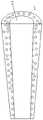
图1为双层复合支架的结构图;Fig. 1 is the structure diagram of double-layer composite stent;
图2为图1的纵向剖视图;Fig. 2 is the longitudinal sectional view of Fig. 1;
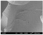
图3-1为实例1支架内层部分SEM图(电子扫描显像图);Figure 3-1 is a SEM image (scanning electron imaging image) of the inner layer part of the stent of Example 1;
图3-2为实例1支架外层部分SEM图(电子扫描显像图)Figure 3-2 is the SEM image (scanning electron imaging image) of the outer layer of the stent of Example 1
图4-1为实例1支架内层部分细胞矿化结节染色(冯科萨染色图);Figure 4-1 shows the staining of mineralized nodules in the inner layer of the scaffold of Example 1 (Von Kossa staining);
图4-2为实例1支架外层部分细胞矿化结节染色(冯科萨染色图)。Figure 4-2 shows the staining of mineralized nodules of cells in the outer layer of the scaffold of Example 1 (von Kossa's stained image).
图中,1、生物活性玻璃;2、水凝胶;In the figure, 1, bioactive glass; 2, hydrogel;
具体实施方式Detailed ways
以下实施例用于说明本发明,但不用来限制本发明的范围。The following examples are intended to illustrate the present invention, but not to limit the scope of the present invention.
参见图1-4,See Figure 1-4,
实施例1:Example 1:
(1)制备生物活性玻璃PSC颗粒:该实例中制备的生物活性玻璃成分为:P2O510.8mol%,SiO254.2mol%和CaO35mol%;将植酸(50wt%)3ml,正硅酸乙酯10.95ml,四水硝酸钙7.47g,n水/正硅酸乙酯=4,n乙醇/正硅酸乙酯=8混合制成前驱体溶胶溶液,放置到30℃下凝胶25天,55℃陈化5天除去其中的乙醇和少量水;100℃烧结1周去除剩余的溶剂;600℃烘箱1小时后得到生物活性玻璃PSC颗粒;(1) Preparation of bioactive glass PSC particles: The components of bioactive glass prepared in this example are: P2 O5 10.8 mol%, SiO2 54.2 mol % and CaO 35 mol %; phytic acid (50wt%) 3ml, orthosilicic acid 10.95ml of ethyl ester, 7.47g of calcium nitrate tetrahydrate, n water/ethyl orthosilicate = 4, n ethanol/ethyl orthosilicate = 8 mixed to prepare a precursor sol solution, placed at 30 °C for 25 days , aged at 55°C for 5 days to remove ethanol and a small amount of water; sintered at 100°C for 1 week to remove the remaining solvent; oven at 600°C for 1 hour to obtain bioactive glass PSC particles;
(2)RGDS多肽改性生物活性玻璃:取200mg PSC粉末,以100ml无水甲苯作为分散剂,二者混合后超声震荡15min辅助分散,加入3ml3-氨丙基三乙氧基硅烷(APTES),于氮气保护下80℃加热回流反应24h,离心,依次使用甲苯、无水乙醇、去离子水清洗,低温冻干后制得PSC-NH2;多肽序列枝接:配置2-吗啉乙磺酸(MES)缓冲液,调整pH至5.5,依次加入76.68mg 1-(3-二甲氨基丙基)-3-乙基碳二亚胺盐酸盐(EDC)、11.51mg N-羟基琥珀酰亚胺(NHS)、20mg RGDS多肽序列、200mg PSC-NH2,磁力搅拌下室温反应24h,使用去离子水清洗,低温冻干后制得多肽改性PSC;(2) RGDS polypeptide modified bioactive glass: take 200mg PSC powder, use 100ml anhydrous toluene as a dispersant, mix the two with ultrasonic vibration for 15min to assist dispersion, add 3ml 3-aminopropyltriethoxysilane (APTES), Under the protection of nitrogen, the reaction was heated under reflux at 80°C for 24h, centrifuged, washed with toluene, anhydrous ethanol, and deionized water in turn, and freeze-dried at low temperature to obtain PSC-NH2 ; polypeptide sequence grafting: configure 2-morpholineethanesulfonic acid (MES) buffer, adjust the pH to 5.5, then add 76.68 mg 1-(3-dimethylaminopropyl)-3-ethylcarbodiimide hydrochloride (EDC), 11.51 mg N-hydroxysuccinimide Amine (NHS), 20mg RGDS polypeptide sequence, 200mg PSC-NH2, reacted at room temperature for 24h under magnetic stirring, washed with deionized water, and freeze-dried at low temperature to obtain polypeptide-modified PSC;
(3)外支架外层部分的制备:将改性后PSC颗粒与浓度为3mg/ml的明胶胶溶液复合,PSC颗粒与明胶溶液的质量体积比是0.5mg/ml,超声震荡混匀后注入模具中,得到锥形中空的外层支架,尖端外径为1.0mm,内径0.5mm,锥度0.06,长度15mm;真空冷冻干获得支架外层部分;(3) Preparation of the outer layer part of the outer support: the modified PSC particles are compounded with a gelatin solution with a concentration of 3 mg/ml, and the mass-volume ratio of the PSC particles to the gelatin solution is 0.5 mg/ml. In the mold, a conical hollow outer stent is obtained, the outer diameter of the tip is 1.0 mm, the inner diameter is 0.5 mm, the taper is 0.06, and the length is 15 mm; the outer part of the stent is obtained by vacuum freeze-drying;
(4)支架内层部分的制备:将浓度为3mg/ml明胶材料注入外层支架内,真空冷冻干;(4) Preparation of the inner part of the stent: the gelatin material with a concentration of 3 mg/ml was injected into the outer stent, and vacuum freeze-dried;
(5)双层支架的交联完成:将步骤(4)中获得的支架浸泡于0.1%戊二醛交联剂中交联24小时,使内外层支架交联形成具有双层结构的复合支架。(5) The cross-linking of the double-layer scaffold is completed: soak the scaffold obtained in step (4) in 0.1% glutaraldehyde cross-linking agent for 24 hours to cross-link the inner and outer scaffolds to form a composite scaffold with a double-layer structure .
实施例1的生物学评价:Biological evaluation of Example 1:
将实施例1中得到的支架与人骨髓间充质细胞共培养,2周后进行扫描电镜和矿化结节冯科萨染色观察,结果见图3和图4。可见材料支架内层及外层均具有较好的细胞相容性,添加有生物活性玻璃PSC的支架外层能够显著促进矿化的生成。The scaffold obtained in Example 1 was co-cultured with human bone marrow mesenchymal cells, and 2 weeks later, scanning electron microscope and mineralized nodules were observed by von Kossa staining. The results are shown in Figures 3 and 4 . It can be seen that the inner and outer layers of the material scaffold have good cytocompatibility, and the outer layer of the scaffold added with bioactive glass PSC can significantly promote the formation of mineralization.
如上所述,便可较为充分的实现本发明。以上所述仅为本发明的较为合理的实施实例,本发明的保护范围包括但并不局限于此,本领域的技术人员任何基于本发明技术方案上非实质性变性变更均包括在本发明包括范围之内。As described above, the present invention can be fully realized. The above is only a reasonable implementation example of the present invention, and the protection scope of the present invention includes but is not limited to this. Any non-substantial modification changes based on the technical solution of the present invention by those skilled in the art are included in the present invention. within the range.
Claims (3)
Translated fromChinesePriority Applications (1)
| Application Number | Priority Date | Filing Date | Title |
|---|---|---|---|
| CN201911075077.4ACN110859991B (en) | 2019-11-06 | 2019-11-06 | Preparation method of double-layer composite scaffold for inducing regeneration of dental pulp dentin tissues |
Applications Claiming Priority (1)
| Application Number | Priority Date | Filing Date | Title |
|---|---|---|---|
| CN201911075077.4ACN110859991B (en) | 2019-11-06 | 2019-11-06 | Preparation method of double-layer composite scaffold for inducing regeneration of dental pulp dentin tissues |
Publications (2)
| Publication Number | Publication Date |
|---|---|
| CN110859991A CN110859991A (en) | 2020-03-06 |
| CN110859991Btrue CN110859991B (en) | 2022-05-03 |
Family
ID=69654886
Family Applications (1)
| Application Number | Title | Priority Date | Filing Date |
|---|---|---|---|
| CN201911075077.4AActiveCN110859991B (en) | 2019-11-06 | 2019-11-06 | Preparation method of double-layer composite scaffold for inducing regeneration of dental pulp dentin tissues |
Country Status (1)
| Country | Link |
|---|---|
| CN (1) | CN110859991B (en) |
Families Citing this family (5)
| Publication number | Priority date | Publication date | Assignee | Title |
|---|---|---|---|---|
| CN112755251B (en)* | 2021-01-21 | 2022-05-20 | 中怡(深圳)医疗科技集团有限公司 | Bionic biological joint based on freeze casting technology and preparation method thereof |
| CN112957451B (en)* | 2021-02-26 | 2021-12-17 | 四川大学 | Bioactive glass/biomimetic functional polypeptide complex and preparation method and application |
| CN114259412B (en)* | 2022-02-23 | 2023-05-26 | 北京大学口腔医学院 | Preparation method of photocurable bioactive marrow covering material |
| CN115068692B (en)* | 2022-06-30 | 2023-07-25 | 南京智皓领先科技有限公司 | Method for constructing biological tooth root based on stem cell technology |
| CN117379598B (en)* | 2023-07-27 | 2024-03-22 | 天津医科大学口腔医院 | Polymer hydrogel and preparation method and application thereof |
Citations (2)
| Publication number | Priority date | Publication date | Assignee | Title |
|---|---|---|---|---|
| CN101014297A (en)* | 2004-08-06 | 2007-08-08 | 彭特恩临床科技有限公司 | Dental filling material |
| CN105687145A (en)* | 2015-12-31 | 2016-06-22 | 江苏昌吉永生物科技股份有限公司 | Preparation method of protein and polypeptide sustained release microspheres of biologically active glass composite polylactic acid |
Family Cites Families (2)
| Publication number | Priority date | Publication date | Assignee | Title |
|---|---|---|---|---|
| CA2637616A1 (en)* | 2006-01-19 | 2007-07-26 | Osteotech, Inc. | Injectable and moldable bone substitute materials |
| WO2018197946A1 (en)* | 2017-04-26 | 2018-11-01 | Meital Zilberman | Hydrogel compositions including fibers and methods of use thereof |
- 2019
- 2019-11-06CNCN201911075077.4Apatent/CN110859991B/enactiveActive
Patent Citations (2)
| Publication number | Priority date | Publication date | Assignee | Title |
|---|---|---|---|---|
| CN101014297A (en)* | 2004-08-06 | 2007-08-08 | 彭特恩临床科技有限公司 | Dental filling material |
| CN105687145A (en)* | 2015-12-31 | 2016-06-22 | 江苏昌吉永生物科技股份有限公司 | Preparation method of protein and polypeptide sustained release microspheres of biologically active glass composite polylactic acid |
Also Published As
| Publication number | Publication date |
|---|---|
| CN110859991A (en) | 2020-03-06 |
Similar Documents
| Publication | Publication Date | Title |
|---|---|---|
| CN110859991B (en) | Preparation method of double-layer composite scaffold for inducing regeneration of dental pulp dentin tissues | |
| Farag | Recent trends on biomaterials for tissue regeneration applications | |
| Sun et al. | Hydroxyapatite nanowire@ magnesium silicate core–shell hierarchical nanocomposite: Synthesis and application in bone regeneration | |
| Kerativitayanan et al. | Nanoengineered osteoinductive and elastomeric scaffolds for bone tissue engineering | |
| Sowmya et al. | Role of nanostructured biopolymers and bioceramics in enamel, dentin and periodontal tissue regeneration | |
| CN109381749A (en) | Bone tissue reparation ink, composition, bracket and preparation method and kit | |
| CN103055352B (en) | Calcium phosphate/collagen composite biologic ceramic material and preparation method thereof | |
| CN101141987B (en) | Medical material, artificial tooth root and manufacturing method of medical material | |
| Xu et al. | Accurately shaped tooth bud cell–derived mineralized tissue formation on silk scaffolds | |
| CN107456607A (en) | Guide Periodontal Tissue Regeneration film of new " sandwich " structure a kind of of difunctionalization and its preparation method and application | |
| CN101970023A (en) | 3-dimensional silk hydroxyapatite compositions | |
| Sadeghian et al. | Dentin extracellular matrix loaded bioactive glass/GelMA support rapid bone mineralization for potential pulp regeneration | |
| CN102302804A (en) | Hydroxyapatite-based biological composite scaffold and tissue engineered bone | |
| CN104288830A (en) | Micro-nano rod-shaped bioactive glass and preparation method and application thereof | |
| Yang et al. | A xonotlite nanofiber bioactive 3D-printed hydrogel scaffold based on osteo-/angiogenesis and osteoimmune microenvironment remodeling accelerates vascularized bone regeneration | |
| He et al. | Rational design and fabrication of porous calcium–magnesium silicate constructs that enhance angiogenesis and improve orbital implantation | |
| CN107235721A (en) | A kind of porous Bredigites biological ceramic support of 3 D-printing and preparation method and application | |
| CN102008752A (en) | Porous biphasic calcium phosphate biological scaffold with nano hydroxyapatite coating and preparation method thereof | |
| CN106267342A (en) | A kind of dentistry implant and preparation method thereof | |
| CN103598919B (en) | A kind of preparation method of dental implant surface biological gradient coating | |
| CN103566411A (en) | Mesoporous bioactive glass-modified porous ceramic ball prosthetic eye holder and preparation method thereof | |
| Zhang et al. | Advancing collagen-based biomaterials for oral and craniofacial tissue regeneration | |
| Miao et al. | Multi-stage controllable degradation of strontium-doped calcium sulfate hemihydrate-tricalcium phosphate microsphere composite as a substitute for osteoporotic bone defect repairing: degradation behavior and bone response | |
| CN117547659A (en) | A collagen composite membrane for guiding bone regeneration and its preparation method and application | |
| Wang et al. | 3D-printed PCL@ BG scaffold integrated with SDF-1α-loaded hydrogel for enhancing local treatment of bone defects |
Legal Events
| Date | Code | Title | Description |
|---|---|---|---|
| PB01 | Publication | ||
| PB01 | Publication | ||
| SE01 | Entry into force of request for substantive examination | ||
| SE01 | Entry into force of request for substantive examination | ||
| GR01 | Patent grant | ||
| GR01 | Patent grant | ||
| TR01 | Transfer of patent right | Effective date of registration:20250109 Address after:Unit A1805, Phase IV, International Science and Technology Park, 1355 Jinji Lake Avenue, Suzhou Industrial Park, Jiangsu Province 215000 Patentee after:Dengtingkang (Suzhou) Medical Equipment Co.,Ltd. Country or region after:China Address before:100081 dental pulp department, third floor, outpatient department of Peking University stomatological hospital, No. 22, Zhongguancun South Street, Haidian District, Beijing Patentee before:PEKING University SCHOOL OF STOMATOLOGY Country or region before:China | |
| TR01 | Transfer of patent right |