CN110804621A - A kind of preparation method of Escherichia coli extracellular vesicles with endogenous high expression of miRNA - Google Patents
A kind of preparation method of Escherichia coli extracellular vesicles with endogenous high expression of miRNADownload PDFInfo
- Publication number
- CN110804621A CN110804621ACN201911054070.4ACN201911054070ACN110804621ACN 110804621 ACN110804621 ACN 110804621ACN 201911054070 ACN201911054070 ACN 201911054070ACN 110804621 ACN110804621 ACN 110804621A
- Authority
- CN
- China
- Prior art keywords
- mirna
- evs
- mir
- trna
- cells
- Prior art date
- Legal status (The legal status is an assumption and is not a legal conclusion. Google has not performed a legal analysis and makes no representation as to the accuracy of the status listed.)
- Pending
Links
Images


Classifications
- C—CHEMISTRY; METALLURGY
- C12—BIOCHEMISTRY; BEER; SPIRITS; WINE; VINEGAR; MICROBIOLOGY; ENZYMOLOGY; MUTATION OR GENETIC ENGINEERING
- C12N—MICROORGANISMS OR ENZYMES; COMPOSITIONS THEREOF; PROPAGATING, PRESERVING, OR MAINTAINING MICROORGANISMS; MUTATION OR GENETIC ENGINEERING; CULTURE MEDIA
- C12N15/00—Mutation or genetic engineering; DNA or RNA concerning genetic engineering, vectors, e.g. plasmids, or their isolation, preparation or purification; Use of hosts therefor
- C12N15/09—Recombinant DNA-technology
- C12N15/63—Introduction of foreign genetic material using vectors; Vectors; Use of hosts therefor; Regulation of expression
- C12N15/70—Vectors or expression systems specially adapted for E. coli
- A—HUMAN NECESSITIES
- A61—MEDICAL OR VETERINARY SCIENCE; HYGIENE
- A61K—PREPARATIONS FOR MEDICAL, DENTAL OR TOILETRY PURPOSES
- A61K31/00—Medicinal preparations containing organic active ingredients
- A61K31/70—Carbohydrates; Sugars; Derivatives thereof
- A61K31/7088—Compounds having three or more nucleosides or nucleotides
- A61K31/7105—Natural ribonucleic acids, i.e. containing only riboses attached to adenine, guanine, cytosine or uracil and having 3'-5' phosphodiester links
- A—HUMAN NECESSITIES
- A61—MEDICAL OR VETERINARY SCIENCE; HYGIENE
- A61K—PREPARATIONS FOR MEDICAL, DENTAL OR TOILETRY PURPOSES
- A61K9/00—Medicinal preparations characterised by special physical form
- A61K9/48—Preparations in capsules, e.g. of gelatin, of chocolate
- A61K9/50—Microcapsules having a gas, liquid or semi-solid filling; Solid microparticles or pellets surrounded by a distinct coating layer, e.g. coated microspheres, coated drug crystals
- A61K9/5005—Wall or coating material
- A61K9/5063—Compounds of unknown constitution, e.g. material from plants or animals
- A61K9/5068—Cell membranes or bacterial membranes enclosing drugs
- A—HUMAN NECESSITIES
- A61—MEDICAL OR VETERINARY SCIENCE; HYGIENE
- A61P—SPECIFIC THERAPEUTIC ACTIVITY OF CHEMICAL COMPOUNDS OR MEDICINAL PREPARATIONS
- A61P35/00—Antineoplastic agents
- C—CHEMISTRY; METALLURGY
- C12—BIOCHEMISTRY; BEER; SPIRITS; WINE; VINEGAR; MICROBIOLOGY; ENZYMOLOGY; MUTATION OR GENETIC ENGINEERING
- C12N—MICROORGANISMS OR ENZYMES; COMPOSITIONS THEREOF; PROPAGATING, PRESERVING, OR MAINTAINING MICROORGANISMS; MUTATION OR GENETIC ENGINEERING; CULTURE MEDIA
- C12N15/00—Mutation or genetic engineering; DNA or RNA concerning genetic engineering, vectors, e.g. plasmids, or their isolation, preparation or purification; Use of hosts therefor
- C12N15/09—Recombinant DNA-technology
- C12N15/11—DNA or RNA fragments; Modified forms thereof; Non-coding nucleic acids having a biological activity
- C12N15/113—Non-coding nucleic acids modulating the expression of genes, e.g. antisense oligonucleotides; Antisense DNA or RNA; Triplex- forming oligonucleotides; Catalytic nucleic acids, e.g. ribozymes; Nucleic acids used in co-suppression or gene silencing
- C12N15/1135—Non-coding nucleic acids modulating the expression of genes, e.g. antisense oligonucleotides; Antisense DNA or RNA; Triplex- forming oligonucleotides; Catalytic nucleic acids, e.g. ribozymes; Nucleic acids used in co-suppression or gene silencing against oncogenes or tumor suppressor genes
Landscapes
- Health & Medical Sciences (AREA)
- Life Sciences & Earth Sciences (AREA)
- Engineering & Computer Science (AREA)
- Genetics & Genomics (AREA)
- Chemical & Material Sciences (AREA)
- Bioinformatics & Cheminformatics (AREA)
- Organic Chemistry (AREA)
- General Health & Medical Sciences (AREA)
- Molecular Biology (AREA)
- Zoology (AREA)
- Biomedical Technology (AREA)
- Wood Science & Technology (AREA)
- General Engineering & Computer Science (AREA)
- Biotechnology (AREA)
- Public Health (AREA)
- Animal Behavior & Ethology (AREA)
- Biochemistry (AREA)
- Medicinal Chemistry (AREA)
- Pharmacology & Pharmacy (AREA)
- Biophysics (AREA)
- Veterinary Medicine (AREA)
- Physics & Mathematics (AREA)
- Epidemiology (AREA)
- Plant Pathology (AREA)
- Microbiology (AREA)
- Oncology (AREA)
- Virology (AREA)
- Botany (AREA)
- General Chemical & Material Sciences (AREA)
- Chemical Kinetics & Catalysis (AREA)
- Nuclear Medicine, Radiotherapy & Molecular Imaging (AREA)
- Cell Biology (AREA)
- Micro-Organisms Or Cultivation Processes Thereof (AREA)
Abstract
Description
Translated fromChinese技术领域technical field
本申请属于细胞生物学技术领域,具体涉及一种内源性高表达miRNA的大肠杆菌胞外囊泡的制备方法。The application belongs to the technical field of cell biology, and in particular relates to a preparation method of Escherichia coli extracellular vesicles with endogenous high expression of miRNA.
背景技术Background technique
细菌胞外囊泡(extracellular vesicles,EVs)是细菌在生长过程中自发产生的一种纳米尺寸的球形磷脂双分子层蛋白。EVs的尺寸一般在20~300nm之间,其中包含有脂多糖(LPS)、蛋白质、脂质、DNA、RNA、毒力因子等物质。由于细菌分为革兰氏阳性细菌和革兰氏阴性细菌两大类,其中革兰氏阳性细菌是单独的细胞壁结构,其胞外囊泡被称为EVs;而革兰氏阴性细菌有两层膜,外膜跟内膜,而革兰氏阴性细菌的EVs起源于外膜,因此也叫作(outer membrane vesicles,OMVs)。Bacterial extracellular vesicles (EVs) are nano-sized spherical phospholipid bilayer proteins spontaneously produced by bacteria during growth. EVs are generally between 20 and 300 nm in size and contain lipopolysaccharide (LPS), proteins, lipids, DNA, RNA, virulence factors and other substances. Since bacteria are divided into two categories: Gram-positive bacteria and Gram-negative bacteria, Gram-positive bacteria are separate cell wall structures whose extracellular vesicles are called EVs; while Gram-negative bacteria have two layers Membrane, outer membrane and inner membrane, and EVs of Gram-negative bacteria originate from the outer membrane, so they are also called (outer membrane vesicles, OMVs).
由于细菌胞外囊泡的形成过程的特殊性,部分应用研究研究表明,利用细菌纳米胞外囊泡载药时,表现出较好的:获取成本低、容易通过基因工程的技术进行改造、磷脂双分子层的结构十分稳定、可以通过发酵的技术大量获得、可以穿过多重生理屏障等优点,因此以细菌胞外囊泡(EVs)作为药物递送系统的载体受到了越来越多研究和重视。Due to the particularity of the formation process of bacterial extracellular vesicles, some applied research studies have shown that when bacterial nano-sized extracellular vesicles are used for drug delivery, it exhibits better performance: low cost of acquisition, easy transformation through genetic engineering technology, phospholipid The structure of the bilayer is very stable, can be obtained in large quantities by fermentation technology, and can pass through multiple physiological barriers. Therefore, bacterial extracellular vesicles (EVs) as carriers of drug delivery systems have received more and more research and attention. .
miRNA又称MicroRNA,是真核生物体内的一段非常短的内源性的非编码单链RNA分子,大小约为20-25个核苷酸。miRNA通过与mRNA的3’端非翻译区配对结合抑制mRNA翻译成蛋白质,或者诱导mRNA降解,从而在转录后调节很多基因的表达,参与生物体细胞增殖分化等多个重要的生物过程,是真核生物基因表达的重要调控者。当miRNA的靶基因是致癌基因或抑癌基因时,miRNA的表达可影响肿瘤细胞的生长和增殖,从而影响肿瘤的发生发展,越来越多的研究表明miRNA可以影响癌症的进程。但实际应用中,受限于miRNA的合成成本及生物体内分布、和吸收利用等特点,其应用前景尚不够明朗。miRNA, also known as MicroRNA, is a very short endogenous non-coding single-stranded RNA molecule in eukaryotes, with a size of about 20-25 nucleotides. miRNA inhibits the translation of mRNA into protein by pairing with the untranslated region at the 3' end of mRNA, or induces mRNA degradation, thereby regulating the expression of many genes after transcription, and participating in many important biological processes such as cell proliferation and differentiation of organisms. Important regulator of nuclear gene expression. When the target genes of miRNAs are oncogenes or tumor suppressor genes, the expression of miRNAs can affect the growth and proliferation of tumor cells, thereby affecting the occurrence and development of tumors. More and more studies have shown that miRNAs can affect the process of cancer. However, in practical application, the application prospect of miRNA is still not clear due to the characteristics of synthesis cost, in vivo distribution, and absorption and utilization of miRNA.
基于上述缺陷,发明人认为,如果能够在细菌细胞内部进行高效的内源性高表达miRNA,同时利用细菌胞外囊泡的低成本载药特点,则对于相关疾病的治疗和预防是具有十分重要的技术意义的。Based on the above-mentioned defects, the inventors believe that if the high-efficiency endogenous high expression of miRNAs can be carried out inside bacterial cells, and the low-cost drug-loading characteristics of bacterial extracellular vesicles can be used at the same time, it is very important for the treatment and prevention of related diseases. of technical significance.
发明内容SUMMARY OF THE INVENTION
以部分特定miRNA为例,本申请目的在于提供一种将内源性高表达miRNA与大肠杆菌胞外囊泡进行技术结合的方法,从而为相关疾病的治疗和预防提供一定技术参考。Taking some specific miRNAs as an example, the purpose of this application is to provide a method for technically combining endogenous highly expressed miRNAs with E. coli extracellular vesicles, so as to provide certain technical references for the treatment and prevention of related diseases.
本申请所采取的技术方案详述如下。The technical solution adopted in this application is described in detail as follows.
一种内源性高表达miRNA的大肠杆菌胞外囊泡的制备方法,具体包括如下步骤:A preparation method of Escherichia coli extracellular vesicles with endogenous high expression of miRNA, specifically comprising the following steps:
(一)设计引物及PCR扩增(1) Design primers and PCR amplification
以Lys-tRNA的序列和pET-31b(+)载体序列为基础,设计PCR扩增用引物序列如下:Based on the sequence of Lys-tRNA and the sequence of pET-31b(+) vector, the primer sequences for PCR amplification were designed as follows:
pET-31b(+)-F:pET-31b(+)-F:
5’- AACTCGAGCATATGAATTCATCTCCTTCTTAAAGTTAAACAAAATTATT-3’,5'- AACTCGAGCATATGAATTCATCTCCTTCTTAAAGTTAAACAAAATTATT-3',
pET-31b(+)-R:5’- TTCTCGAGGATCC GCAATAACTAGCATAACCCCTTG-3’;pET-31b(+)-R: 5'- TTCTCGAGGATCC GCAATAACTAGCATAACCCCTTG-3';
tRNA-F:5’- TTGAATTCTGGCTGGGGTACCTGGATTCGAACCAG-3’,tRNA-F: 5'-TTGAATTCTGGCTGGGGTACCTGGATTCGAACCAG-3',
tRNA-R:5’- AAGGATCCGGGTCCAGGGTTCAAGTCCCTGTTC-3’;tRNA-R: 5'- AAGGATCCGGGTCCAGGGTTCAAGTCCCTGTTC-3';
所述Lys-tRNA序列(85bp,如SEQ ID NO.1所示)为:GAATTCGCCCGGATAGCTCAGTCGGTAGAGCAGCGGCCGCGGCCGCGGGTCCAGGGTTCAAGTCCCTGTTCGGGCGCCAAAGCTTThe Lys-tRNA sequence (85bp, as shown in SEQ ID NO. 1) is: GAATTCGCCCGGATAGCTCAGTCGGTAGAGCAGCGGCCGCGGCCGCGGGTCCAGGGTTCAAGTCCCTGTTCGGGCGCCAAAGCTT
PCR扩增时,以大肠杆菌DNA为模板,利用上述引物进行PCR扩增,扩增完成后,提取备用;During PCR amplification, using Escherichia coli DNA as a template, the above primers are used for PCR amplification, and after the amplification is completed, extraction is used for subsequent use;
(二)酶切、连接(2) Enzyme cleavage and ligation
利用EcoR 1、BamH 1限制性内切酶分别对步骤(一)中所回收PCR扩增产物(即pET-31b(+)载体和tRNA)进行双酶切,回收酶切片段后使用T4 DNA连接酶进行连接;The PCR amplification products recovered in step (1) (i.e. pET-31b(+) vector and tRNA) were double digested with
(三)转化、筛选和鉴定(3) Transformation, screening and identification
将步骤(二)中的连接产物转化DH5a感受态细胞,并进行抗性筛选、菌落PCR鉴定和测序鉴定;Transform the ligation product in step (2) into DH5a competent cells, and carry out resistance screening, colony PCR identification and sequencing identification;
(四)重组载体的自连(4) Self-ligation of recombinant vector
对步骤(三)中测序正确的质粒,采用Xho I限制性内切酶酶切(37℃酶切过夜),回收酶切片段后,再利用T4 DNA连接酶进行连接(4℃连接过夜),然后再将连接产物转化DH5a感受态细胞,并进行菌落PCR鉴定,并将鉴定正确的阳性克隆接种到含有氨苄青霉素的LB培养基中进行培养(37℃、220rpm过夜培养),此即为含有pET-31b(+)-tRNA载体的转化细菌,收集细菌并且保存菌种于40%的甘油中;或者直接提取质粒应用、或者将质粒-20℃保存;The plasmids sequenced correctly in step (3) were digested with Xho I restriction endonuclease (37°C overnight), and the digested fragments were recovered, and then ligated with T4 DNA ligase (4°C overnight), Then the ligation product was transformed into DH5a competent cells, and colony PCR was carried out to identify the correct positive clones. Inoculate the correctly identified positive clones into LB medium containing ampicillin for cultivation (37°C, 220rpm overnight cultivation), which is the pET-containing -31b(+)-tRNA vector transformed bacteria, collect bacteria and store the strains in 40% glycerol; or directly extract the plasmid for application, or store the plasmid at -20°C;
需要解释的是,此操作步骤构建自连载体的目的,是为了后续使用该自连载体构建其他连接miRNA的载体,如果不构建该自连载体,后续在构建连接miRNA的重组载体时,需要再次进行上述(一)、(二)、(三)的操作,从而增加操作的复杂度;It should be explained that the purpose of constructing the self-linking vector in this operation step is to use the self-linking vector to construct other miRNA-linked vectors. Carry out the operations of (1), (2) and (3) above, thereby increasing the complexity of the operation;
(五)针对目的miRNA,设计引物并进行PCR扩增(5) Design primers and perform PCR amplification for the target miRNA
所述目的miRNA,以miR-34a、miR-124、miR-126为例,具体PCR扩增时引物序列设计为:For the target miRNA, take miR-34a, miR-124, and miR-126 as examples, and the primer sequences during PCR amplification are designed as follows:
miR-34a-F:5’-AACTCGAGGCCAGCTGTGAGTGTTTCTTTGGC-3’,miR-34a-F: 5'-AACTCGAGGCCAGCTGTGAGTGTTTCTTTGGC-3',
miR-34a-R:5’-AACATATGGGCCCCACAACGTGCAGCAC-3’;miR-34a-R: 5'-AACATATGGGCCCCACAACGTGCAGCAC-3';
miR-124-F:5’-AACTCGAGGTCTGCAGAAACCGTCGAACGA-3’,miR-124-F: 5'-AACTCGAGGTCTGCAGAAAACCGTCGAACGA-3',
miR-124-R:5’-AACATATGCATTCCGATCCTTACAAC-3’;miR-124-R: 5'-AACATATGCATTCCGATCCTTACAAC-3';
miR-126-F:5’-AACTCGAGCGCTGGCGACGGGACATTATTA-3’,miR-126-F: 5'-AACTCGAGCGCTGGCGACGGGACATTATTA-3',
miR-126-R:5’-AACATATGTGCCGTGGACGGCGCATTA-3’;miR-126-R: 5'-AACATATGTGCCGTGGACGGCGCATTA-3';
然后以人基因组DNA为模板,分别利用上述引物对进行PCR扩增,电泳检测PCR扩增产物,并回收、纯化扩增产物获得;Then, using the human genomic DNA as a template, the above primer pairs are used for PCR amplification respectively, and the PCR amplification products are detected by electrophoresis, and the amplification products are recovered and purified to obtain;
(六)酶切、连接(6) Enzyme cleavage and ligation
利用Xho I、Nde I限制性内切酶,对步骤(四)中最终重新自连的pET-31b(+)-tRNA载体进行双酶切(37℃双酶切过夜),同时对步骤(五)中目的miRNA产物分别进行双酶切(37℃双酶切过夜),分别回收目的片段,并利用T4 DNA重新进行连接;Using Xho I and Nde I restriction enzymes, the pET-31b(+)-tRNA vector that was finally self-ligated in step (4) was double digested (double digested at 37°C overnight), and the step (5) was double digested at the same time. ), the target miRNA products were subjected to double-enzyme digestion (double-enzyme digestion at 37 °C overnight), respectively, and the target fragments were recovered and re-ligated with T4 DNA;
(七)转化、诱导目的miRNA表达和提取EVs(VII) Transformation, induction of target miRNA expression and extraction of EVs
将步骤(六)中的连接产物转化大肠杆菌BL21感受态细胞,利用IPTG作为诱导剂来诱导重组载体表达miRNA,诱导表达完成后,提取EVs,经过鉴定,所提取的EVs中包含有内源性表达的miRNA。The ligation product in step (6) was transformed into E. coli BL21 competent cells, and IPTG was used as an inducer to induce the recombinant vector to express miRNA. After the induction and expression, EVs were extracted. After identification, the extracted EVs contained endogenous expressed miRNA.
现有技术中,即使不考虑miRNA合成成本,在将外源性的将小RNA导入细菌Evs时,虽然有电穿孔法,诱导法等可以进行操作,但是这些方法都存在操作复杂、处理过程繁琐、依靠昂贵的仪器设备等弊端,因此进一步限定了Evs在作为miRNA载体中的应用。本申请中,为使人源miRNA能够在大肠杆菌中高效表达,同时为避免所表达RNA的降解,发明人以特定miRNA(miR-34a、miR-124、miR-126)为例,通过利用tRNA、rRNA的支架保护作用,结合相关基因工程技术,重组构建了相关表达载体。初步实验结果表明,改造细菌较好实现了内源性、高效、大量表达功能性的miRNA的目的,同时较好实现了包含有miRNA的大肠杆菌Evs的高效收集,也即,较好实现了以Evs作为miRNA载体的技术目标,进而可为相关疾病的预防或治疗奠定良好的技术基础。In the prior art, even if the cost of miRNA synthesis is not considered, when exogenous small RNA is introduced into bacterial Evs, although electroporation, induction, etc. can be operated, these methods are complicated in operation and tedious in processing. , relying on expensive equipment and other drawbacks, thus further limiting the application of Evs as a miRNA carrier. In this application, in order to enable high-efficiency expression of human miRNAs in E. coli, and at the same time to avoid the degradation of the expressed RNAs, the inventors take specific miRNAs (miR-34a, miR-124, miR-126) as examples, by using tRNA , rRNA scaffold protection, combined with related genetic engineering technology, recombination and construction of related expression vectors. Preliminary experimental results show that the modified bacteria better achieve the purpose of endogenous, efficient and large-scale expression of functional miRNAs, and at the same time better achieve efficient collection of E. coli Evs containing miRNAs, that is, better achieve As the technical target of miRNA carrier, Evs can lay a good technical foundation for the prevention or treatment of related diseases.
附图说明Description of drawings
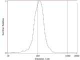
图1为大肠杆菌EVs粒径分析;Fig. 1 is the particle size analysis of Escherichia coli EVs;
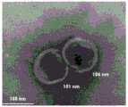
图2为大肠杆菌EVs投射电镜图;Figure 2 is a TEM image of Escherichia coli EVs;
图3为菌液与EVs中tRNA表达量情况;Figure 3 shows the expression of tRNA in bacterial fluid and EVs;
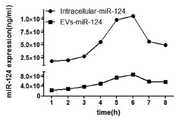
图4为miR-34a在菌体以及EVs中的表达量;Figure 4 shows the expression of miR-34a in bacteria and EVs;
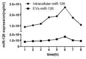
图5为miR-124在菌体以及EVs中的表达量;Figure 5 is the expression level of miR-124 in bacteria and EVs;
图6为miR-126在菌体以及EVs中的表达量;Figure 6 is the expression level of miR-126 in bacteria and EVs;
图7为EVs侵染MBA-MD-231细胞;Figure 7 shows EVs infecting MBA-MD-231 cells;
图8 为EVs侵染A549细胞;Figure 8 shows EVs infecting A549 cells;
图9 为EVs侵染H22细胞;Figure 9 shows EVs infecting H22 cells;
图10为MBA-MD-231细胞增殖曲线;Figure 10 is the proliferation curve of MBA-MD-231 cells;
图11为 MBA-MD-231细胞增殖曲线;Figure 11 is the MBA-MD-231 cell proliferation curve;
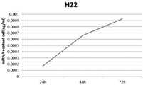
图12为 H22细胞增殖曲线;Figure 12 is the H22 cell proliferation curve;
图13为MBA-MD-231细胞中的tRNA和pei-miR-126的变化量;Figure 13 shows the changes of tRNA and pei-miR-126 in MBA-MD-231 cells;
图14为A549细胞中的tRNA和pei-miR-124的变化量;Figure 14 shows the changes of tRNA and pei-miR-124 in A549 cells;
图15 为H22细胞中的tRNA和pei-miR-34a的变化量;Figure 15 shows the changes of tRNA and pei-miR-34a in H22 cells;
图16为MBA-MD-231细胞中的成熟的miR-126的变化量;Figure 16 is the variation of mature miR-126 in MBA-MD-231 cells;
图17为A549细胞中的成熟的miR-124的变化量;Figure 17 is the variation of mature miR-124 in A549 cells;
图18为H22细胞中的成熟的miR-34a的变化量;Figure 18 is the variation of mature miR-34a in H22 cells;
图19为AS1411-EVs和DZ-AS1411-EVs对杀伤效果对比;Figure 19 shows the comparison of the killing effect of AS1411-EVs and DZ-AS1411-EVs;
图20为不同组之间荷瘤肿瘤体积的变化;Figure 20 is the change in tumor-bearing tumor volume between different groups;
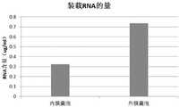
图21为内膜囊泡与外膜囊泡内源性装载RNA的量比较;Figure 21 is a comparison of the amount of endogenously loaded RNA in inner membrane vesicles and outer membrane vesicles;
图22为内膜囊泡载药与外膜囊泡载药杀伤癌细胞的效果比较。Figure 22 is a comparison of the effect of drug loading in inner membrane vesicles and outer membrane vesicles in killing cancer cells.
具体实施方式Detailed ways
下面结合实施例对本申请做进一步的解释说明。在介绍具体实施例前,就下述实施例中部分实验背景情况简要介绍说明如下。The application will be further explained below in conjunction with the examples. Before introducing specific embodiments, a brief introduction and description of some experimental backgrounds in the following embodiments are as follows.
实验仪器:laboratory apparatus:
纳米颗粒跟踪分析仪,德国,ZetaView;Nanoparticle Tracking Analyzer, Germany, ZetaView;
透射电子显微镜,日立,T7700;Transmission electron microscope, Hitachi, T7700;
PCR仪D-8707,TAKARA BIO INC;PCR machine D-8707, TAKARA BIO INC;
荧光定量PCR仪,Roche,Light Cycle96;Fluorescence quantitative PCR instrument, Roche, Light Cycle96;
倒置荧光显微镜,OLYMPUS, CX31;Inverted fluorescence microscope, OLYMPUS, CX31;
激光共聚焦显微镜,OLYMPUS,FV1000;Laser confocal microscope, OLYMPUS, FV1000;
实验试剂:Experimental reagents:
氨苄青霉素、BCA蛋白浓度测定试剂盒、DNA marker、PCR高保真酶mix、1640培养基、胰酶、BL21感受态细胞、K12大肠杆菌菌株,北京索莱宝公司产品;Ampicillin, BCA protein concentration determination kit, DNA marker, PCR high fidelity enzyme mix, 1640 medium, trypsin, BL21 competent cells, K12 Escherichia coli strain, products of Beijing Soleibo Company;
质粒小提试剂盒、PCR产物纯化试剂盒、琼脂糖凝胶DNA回收试剂盒、RNA提取试剂盒、反转录试剂盒、荧光量PCR试剂盒,郑州贝贝生物科技有限公司产品;Plasmid extraction kit, PCR product purification kit, agarose gel DNA recovery kit, RNA extraction kit, reverse transcription kit, fluorescence PCR kit, products of Zhengzhou Beibei Biotechnology Co., Ltd.;
细胞膜绿色荧光探针,碧云天公司产品;Cell membrane green fluorescent probe, a product of Biyuntian;
胎牛血清,Gibco公司产品;Fetal bovine serum, a product of Gibco;
A549细胞(腺癌细胞)、MBA-MD-231细胞(人乳腺癌细胞)、H22细胞(肝癌细胞),普诺赛公司;A549 cells (adenocarcinoma cells), MBA-MD-231 cells (human breast cancer cells), H22 cells (liver cancer cells), Proceeds;
CCK8试剂盒,日本同仁;CCK8 kit, Japanese colleagues;
Hind III、Sal I、EcoR I、BamH I限制性内切酶、T4 DNA连接酶,北京NEB公司产品;Hind III, Sal I, EcoR I, BamH I restriction endonuclease, T4 DNA ligase, products of Beijing NEB Company;
LB培养基、完全培养基(90ml 1640培养基中加入10ml胎牛血清)、PBS(pH=7.4)、氨苄青霉素(100mg/ml)母液、1mol/L IPTG、1×TAE、40%甘油等,常规配置备用。LB medium, complete medium (10ml fetal bovine serum added to 90ml 1640 medium), PBS (pH=7.4), ampicillin (100mg/ml) stock solution, 1mol/L IPTG, 1×TAE, 40% glycerol, etc., General configuration backup.
大肠杆菌EVs的提取和鉴定:Extraction and identification of E. coli EVs:
就本申请中所涉及的大肠杆菌Evs的提取和鉴定过程简要介绍说明如下:A brief introduction to the extraction and identification process of Escherichia coli Evs involved in this application is as follows:
首先,将LB平板(100mg/ml氨苄青霉素)、37℃ 恒温培养12小时的菌株,接种于LB液体培养基中(100mg/ml氨苄青霉素),37℃、220rmp的条件下震荡培养12小时;First, inoculate the LB plate (100mg/ml ampicillin) at 37°C constant temperature for 12 hours, inoculate it in LB liquid medium (100mg/ml ampicillin), and shake for 12 hours at 37°C and 220rmp;
然后,取50ml菌液,l00000g 离心30min,弃沉淀;再用100000g离心60分钟;Then, take 50ml of bacterial liquid, centrifuge at 100000g for 30min, discard the precipitation; then centrifuge at 100000g for 60min;
最后,用10ml的PBS溶液(pH=7.4)重悬沉淀物,之后再次100000g离心60分钟,最终用1ml的PBS溶液(pH=7.4重悬,即得到大肠杆菌EVs溶液。Finally, the pellet was resuspended with 10ml of PBS solution (pH=7.4), then centrifuged again at 100,000g for 60 minutes, and finally resuspended with 1ml of PBS solution (pH=7.4) to obtain Escherichia coli EVs solution.
对于所制备大肠杆菌Evs的粒径进行分析时,采用纳米颗粒跟踪分析进行检测分析,结果如图1所示,可以看出,所制备大肠杆菌Evs的平均粒径在112.8nm左右。When analyzing the particle size of the prepared E. coli Evs, nanoparticle tracking analysis was used for detection and analysis. The results are shown in Figure 1. It can be seen that the average particle size of the prepared E. coli Evs is about 112.8 nm.
对于所制备大肠杆菌Evs的形态鉴定,主要通过透射电镜(TME)进行鉴定,结果如图2所示,可以看出,其呈典型的“杯托”状形态。The morphological identification of the prepared E. coli Evs was mainly carried out by transmission electron microscopy (TME). The results are shown in Figure 2. It can be seen that it has a typical "cup holder" shape.
实施例1Example 1
需要说明的是,通过基因工程直接表达RNA时,由于RNA结构不够稳定,如果直接将RNA序列插入到载体中,RNA可能会被随处可见的RNA酶直接降解,从而造成不会在大肠杆菌胞内表达,为解决这一技术问题,需要设计一段支架来保护RNA不被内源性的RNA酶降解。本申请即设计利用特定的“tRNA支架”,从而避免后续表达过程中的降解作用。本实施例即就含有“tRNA支架”的pET-31b(+)-tRNA载体的构建过程简要介绍说明如下。It should be noted that when RNA is directly expressed by genetic engineering, since the RNA structure is not stable enough, if the RNA sequence is directly inserted into the vector, the RNA may be directly degraded by RNases that can be seen everywhere, resulting in no intracellular expression of E. coli. Expression, in order to solve this technical problem, a scaffold needs to be designed to protect RNA from degradation by endogenous RNases. The present application is designed to utilize a specific "tRNA scaffold" to avoid degradation during subsequent expression. In this example, the construction process of the pET-31b(+)-tRNA vector containing the "tRNA scaffold" is briefly described as follows.
(一)设计引物及PCR扩增(1) Design primers and PCR amplification
以大肠杆菌Lys-tRNA的序列和pET-31b(+)载体序列为基础,设计PCR扩增用引物。Based on the sequence of Escherichia coli Lys-tRNA and pET-31b(+) vector sequence, primers for PCR amplification were designed.
Lys-tRNA序列(85bp)为:Lys-tRNA sequence (85bp) is:
GAATTCGCCCGGATAGCTCAGTCGGTAGAGCAGCGGCCGCGGCCGCGGGTCCAGGGTTCAAGTCCCTGTTCGGGCGCCAAAGCTT。GAATTCGCCCGGATAGCTCAGTCGGTAGAGCAGCGGCCGCGGCCGCGGGTCCAGGGTTCAAGTCCCTGTTCGGGCGCCAAAGCTT.
具体引物序列设计如下:The specific primer sequences are designed as follows:
pET-31b(+)-F:pET-31b(+)-F:
5’- AACTCGAGCATATGAATTCATCTCCTTCTTAAAGTTAAACAAAATTATT-3’,5'- AACTCGAGCATATGAATTCATCTCCTTCTTAAAGTTAAACAAAATTATT-3',
pET-31b(+)-R:5’- TTCTCGAGGATCC GCAATAACTAGCATAACCCCTTG-3’;pET-31b(+)-R: 5'- TTCTCGAGGATCC GCAATAACTAGCATAACCCCTTG-3';
tRNA-F:5’- TTGAATTCTGGCTGGGGTACCTGGATTCGAACCAG-3’,tRNA-F: 5'-TTGAATTCTGGCTGGGGTACCTGGATTCGAACCAG-3',
tRNA-R:5’- AAGGATCCGGGTCCAGGGTTCAAGTCCCTGTTC-3’。tRNA-R: 5'-AAGGATCCGGGTCCAGGGTTCAAGTCCCTGTTC-3'.
PCR扩增时,以大肠杆菌DNA为模板,利用上述引物进行PCR扩增,20 ul扩增体系设计如下:During PCR amplification, take Escherichia coli DNA as template, utilize above-mentioned primers to carry out PCR amplification, 20 ul amplification system is designed as follows:
E.coli DNA(或者人基因组DNA),1ul;E.coli DNA (or human genomic DNA), 1ul;
Fow primer,0.5ul;Fow primer, 0.5ul;
Rew primer,0.5ul;Rew primer, 0.5ul;
2×mix,10ul2×mix, 10ul
ddH2O,8ul;ddH2 O, 8ul;
扩增程序如下:95℃、5min;94℃、30s,60℃、30s,72℃、5min,30个循环,72℃、 7min。The amplification program was as follows: 95°C, 5 min; 94°C, 30s, 60°C, 30s, 72°C, 5min, 30 cycles, 72°C, 7min.
对PCR产物4℃保存备用,或者电泳检测后直接进行提取、纯化。The PCR products were stored at 4°C for later use, or extracted and purified directly after electrophoresis detection.
(二)酶切、连接(2) Enzyme cleavage and ligation
利用EcoR 1、BamH 1限制性内切酶分别对步骤(一)中所回收PCR产物(即pET-31b(+)载体和tRNA)进行双酶切,50 ul酶切体系设计如下:The PCR products recovered in step (1) (i.e. pET-31b(+) vector and tRNA) were double digested with
pET-31b(+)(或者tRNA),25ul;pET-31b(+) (or tRNA), 25ul;
EcoR 1,1ul;
BamH 1,1ul;
10×buffer,5ul;10×buffer, 5ul;
ddH2O,18ul;ddH2 O, 18ul;
37℃双酶切过夜。Double digestion at 37°C overnight.
对双酶切后双酶切产物进行琼脂糖凝胶电泳(tRNA插入片段很小,采用2%琼脂糖凝胶电泳,质粒载体采用1%琼脂糖凝胶电泳),然后利用胶回收试剂盒进行回收、纯化。Perform agarose gel electrophoresis on the double-enzyme-digested products after double-enzyme digestion (the tRNA insert is very small, using 2% agarose gel electrophoresis, and the plasmid vector using 1% agarose gel electrophoresis), and then using a gel recovery kit to carry out recovery and purification.
将回收得到的载体和tRNA目的片段(插入片段)使用T4 DNA连接酶进行连接,15ul连接体系设计如下:The recovered vector and tRNA target fragment (insert fragment) were connected using T4 DNA ligase, and the 15ul ligation system was designed as follows:
pET-31b(+)载体,4ul;pET-31b(+) vector, 4ul;
tRNA片段,7.5ul;tRNA fragment, 7.5ul;
T4连接酶,1ul;T4 ligase, 1ul;
10×buffer,1.5ul;10×buffer, 1.5ul;
ddH2O,1ul;ddH2 O, 1ul;
4℃连接过夜。Ligation overnight at 4°C.
(三)转化、筛选和鉴定(3) Transformation, screening and identification
将步骤(二)中的连接产物转化DH5a感受态细胞,并进行抗性筛选、菌落PCR鉴定和测序鉴定。The ligation product in step (2) was transformed into DH5a competent cells, and subjected to resistance screening, colony PCR identification and sequencing identification.
转化操作时,具体操作参考如下:When converting operations, the specific operations refer to the following:
将50ul的DH5a感受态细胞放于冰上2min,待其融化后,将15ul连接产物加入其中,轻轻混匀,冰上放置30min,42℃热激90s,冰上放置2min,加入500ul无抗LB培养基,放置恒温震荡培养箱于37℃、150rpm的条件下培养1h,之后吸取200ul菌液均匀涂到加入抗生素的LB固定培养板上,37℃倒置培养过夜。Put 50 ul of DH5a competent cells on ice for 2 min. After thawing, add 15 ul of ligation product to it, mix gently, place on ice for 30 min, heat shock at 42°C for 90 s, place on ice for 2 min, add 500 ul of antibody-free LB medium was placed in a constant temperature shaking incubator for 1 hour at 37°C and 150 rpm, and then 200ul of bacterial liquid was drawn and evenly spread on the LB fixed culture plate with antibiotics added, and incubated overnight at 37°C upside down.
菌落PCR鉴定时,挑取转化操作时的阳性单克隆菌落,然后将菌落稀释液加入到PCR管中作为模板,进行PCR反应,PCR反应时,具体用引物如下:During colony PCR identification, pick the positive monoclonal colonies during the transformation operation, and then add the colony dilution into the PCR tube as a template to carry out the PCR reaction. The specific primers used in the PCR reaction are as follows:
T-1 F:5’-TTGAATTCGCCCGGATAGCTCAGTC-3’,T-1 F: 5'-TTGAATTCGCCCGGATAGCTCAGTC-3',
R:5’-AAGGATCCTGGCGCCCGAACAGG-3’;R: 5'-AAGGATCCTGGCGCCCGAACAGG-3';
具体PCR反应体系及PCR反应过程参考前述内容即可。The specific PCR reaction system and PCR reaction process can refer to the foregoing content.
对于菌落PCR鉴定正确的阳性克隆,进一步接种到含有氨苄青霉素的LB培养基中,37℃、220rpm过夜培养,然后提取质粒并进行测序鉴定,确保片段插入正确。Correct positive clones identified by colony PCR were further inoculated into LB medium containing ampicillin, cultured overnight at 37°C and 220 rpm, and then the plasmids were extracted and sequenced to ensure that the fragments were inserted correctly.
(四)重组载体的自连(4) Self-ligation of recombinant vector
对步骤(三)中测序正确的质粒,采用Xho I限制性内切酶37℃酶切过夜,回收酶切片段后,再利用T4 DNA连接酶4℃连接过夜,然后再将连接产物转化DH5a感受态细胞,并进行菌落PCR鉴定,并将鉴定正确的阳性克隆接种到含有氨苄青霉素的LB培养基中,37℃、220rpm过夜培养,此即为含有pET-31b(+)-tRNA载体的转化细菌,收集细菌并且保存菌种于40%的甘油中,同时提取质粒,将质粒保存在-20℃或者立即使用。For the correctly sequenced plasmid in step (3), use Xho I restriction endonuclease to digest overnight at 37 °C, and after recovering the digested fragments, use T4 DNA ligase to ligate overnight at 4 °C, and then transform the ligated product into DH5a receptors. The positive clones with correct identification were inoculated into LB medium containing ampicillin and cultured overnight at 37°C and 220 rpm, which is the transformed bacteria containing pET-31b(+)-tRNA vector , collect the bacteria and store the strain in 40% glycerol, extract the plasmid at the same time, store the plasmid at -20°C or use it immediately.
相关酶切、连接、PCR鉴定体系及操作参考前述操作即可,具体菌落PCR鉴定时采用如下引物序列:Relevant enzyme digestion, ligation, PCR identification system and operation can refer to the aforementioned operations, and the following primer sequences are used in the specific colony PCR identification:
Z-1 F:5’-AA TCTAGATAATCTGAGGGTCCAGGGTTCAAGTC-3’,Z-1 F: 5'-AA TCTAGATAATCTGAGGGTCCAGGGTTCAAGTC-3',
R:5’-AACTCGAGAAAGTCTGATGCTCTACCGACTGAGCTAT-3’。R: 5'-AACTCGAGAAAGTCTGATGCTCTACCGACTGAGCTAT-3'.
实施例2Example 2
进一步以现有miRNA (miR-126、miR-124、miR-34a)为例,在实施例1所构建的自连重组载体基础上,利用基因工程,重组了相关表达载体,从而可以在大肠杆菌内进行高效内源表达,具体过程简介如下。Further taking the existing miRNAs (miR-126, miR-124, miR-34a) as examples, on the basis of the self-linked recombinant vector constructed in Example 1, the relevant expression vectors were recombined by genetic engineering, so that they can be expressed in Escherichia coli. Efficient endogenous expression is carried out in vivo, and the specific process is briefly described as follows.
(一)引物设计及PCR扩增(1) Primer design and PCR amplification
针对现有miR-34a、miR-124、miR-126序列,设计PCR扩增用引物序列如下:For the existing miR-34a, miR-124, and miR-126 sequences, the primer sequences for PCR amplification are designed as follows:
miR-34a-F:5’-AACTCGAGGCCAGCTGTGAGTGTTTCTTTGGC-3’,miR-34a-F: 5'-AACTCGAGGCCAGCTGTGAGTGTTTCTTTGGC-3',
miR-34a-R:5’-AACATATGGGCCCCACAACGTGCAGCAC-3’;miR-34a-R: 5'-AACATATGGGCCCCACAACGTGCAGCAC-3';
miR-124-F:5’-AACTCGAGGTCTGCAGAAACCGTCGAACGA-3’,miR-124-F: 5'-AACTCGAGGTCTGCAGAAAACCGTCGAACGA-3',
miR-124-R:5’-AACATATGCATTCCGATCCTTACAAC-3’;miR-124-R: 5'-AACATATGCATTCCGATCCTTACAAC-3';
miR-126-F:5’-AACTCGAGCGCTGGCGACGGGACATTATTA-3’,miR-126-F: 5'-AACTCGAGCGCTGGCGACGGGACATTATTA-3',
miR-126-R:5’-AACATATGTGCCGTGGACGGCGCATTA-3’;miR-126-R: 5'-AACATATGTGCCGTGGACGGCGCATTA-3';
然后以人基因组DNA为模板,分别利用上述引物对进行PCR扩增,电泳检测PCR扩增产物,并回收、纯化扩增产物获得,具体操作参考前述内容即可。Then, using the human genomic DNA as a template, PCR amplification is carried out using the above primer pairs respectively, and the PCR amplification products are detected by electrophoresis, and the amplification products are recovered and purified.
(二)酶切、连接(2) Enzyme cleavage and ligation
利用Xho I、Nde I限制性内切酶,对实施例1中最终重新自连的pET-31b(+)-tRNA载体进行双酶切(37℃双酶切过夜),同时对步骤(一)中的扩增所得miR-34a、miR-124 、miR-126产物分别进行双酶切(37℃双酶切过夜),分别回收目的片段,并利用T4 DNA重新进行连接,具体操作参考前述操作即可。Using Xho I and Nde I restriction enzymes, the pET-31b(+)-tRNA vector that was finally self-ligated in Example 1 was double digested (double digested overnight at 37°C), and at the same time, step (1) The miR-34a, miR-124, and miR-126 products amplified in 2 were subjected to double-enzyme digestion respectively (double-enzyme digestion at 37°C overnight), respectively, and the target fragments were recovered and re-ligated with T4 DNA. Can.
(三)转化、诱导鉴定(3) Transformation and induction identification
为检测所构建重组载体是否可以成功内源表达以及EVs中是否含有miRNA,进一步地,发明人将步骤(二)中的连接产物转化了大肠杆菌BL21感受态细胞,而由于前述所构建载体中含有诱导性的T7启动子,因此,发明人以IPTG作为诱导剂来诱导重组载体表达miRNA。具体转化操作参考前述操作即可。In order to test whether the constructed recombinant vector can be successfully expressed endogenously and whether EVs contain miRNA, further, the inventor transformed the ligation product in step (2) into E. coli BL21 competent cells, and since the aforementioned constructed vector contains Inducible T7 promoter, therefore, the inventors used IPTG as an inducer to induce the recombinant vector to express miRNA. For specific conversion operations, refer to the aforementioned operations.
具体诱导过程和表达量检测情况简要介绍如下。The specific induction process and the detection of expression level are briefly introduced as follows.
取转化、筛选后,重组正确的含有转化子的1mL菌液,转接于100ml加有氨苄青霉素的LB液体培养基中,加入40ul 1mol/L的IPTG,37℃、220rpm震荡培养,以诱导大肠杆菌表达含有tRNA骨架的miRNA。After the transformation and screening, 1 mL of bacterial liquid containing the transformant was reconstituted correctly, transferred to 100 mL of LB liquid medium with ampicillin, 40 μl of 1 mol/L IPTG was added, and 37 ° C, 220 rpm was shaken and cultivated to induce the large intestine. Bacillus expresses miRNAs containing tRNA backbones.
在加入IPTG后第1~8小时,每隔一小时,取1ml菌液,提取菌体中的总RNA,并反转录为cDNA,以16sRNA作为内参,进行荧光定量PCR检测(具体操作参考试剂盒说明书或现有技术即可),以检测菌体的菌体细胞内部含有tRNA骨架的miRNA的表达情况。From 1 to 8 hours after the addition of IPTG, every hour, take 1 ml of bacterial solution, extract the total RNA in the bacterial body, and reverse-transcribe it into cDNA, use 16sRNA as an internal reference, and perform fluorescence quantitative PCR detection (refer to reagents for specific operations). Box instructions or prior art) to detect the expression of miRNA containing tRNA backbone inside the bacterial cell.
具体情况如图3所示。从图中可以看出,前六个小时,菌体细胞内的tRNA量随着时间的增加逐渐增多,而从第六小时开始,菌体细胞内的tRNA量开始降低;EVs中tRNA变化量同菌体细胞中tRNA的变化量。The specific situation is shown in Figure 3. It can be seen from the figure that in the first six hours, the amount of tRNA in the cells gradually increased with time, and from the sixth hour, the amount of tRNA in the cells began to decrease; the amount of tRNA in EVs was the same Variation of tRNA in bacterial cells.
同时,在加入IPTG后第1~8小时,每隔一小时,取10ml菌液,提取大肠杆菌EVs,并进行荧光定量PCR检测,以检测含有tRNA骨架的miRNA在囊泡内含量情况。At the same time, from 1 to 8 hours after adding IPTG, every one hour, 10 ml of bacterial solution was taken to extract E. coli EVs, and quantitative PCR was performed to detect the content of miRNA containing tRNA backbone in vesicles.
由于提取体系略有不同,因此最终将换算成同一单位大小的细菌细胞内部的miRNA表(miR-126、miR-124、miR-34a)达情况与EVs中表达情况进行绘图,分别如图4、图5、图6所示。总体结果表明:在诱导第6小时,胞内和EVs中的含有tRNA骨架的miRNA的表达量最高,在第1-6小时,表达量逐渐升高,第6小时后,表达量逐渐降低。Because the extraction system is slightly different, the expression of the miRNA tables (miR-126, miR-124, miR-34a) inside the bacterial cells converted into the same unit size and the expression in EVs are finally plotted, as shown in Figure 4, Figure 5, Figure 6. The overall results showed that the expression levels of miRNAs containing tRNA backbones in cells and EVs were the highest at the 6th hour of induction, gradually increased from 1 to 6 hours, and gradually decreased after the 6th hour.
实施例3Example 3
为验证所提取EVs中所包含的miRNA活性情况,发明人进一步提取了大肠杆菌EVs进行细胞侵染实验(空白对照)和细胞增殖实验,同时对miRNA表达量情况进行了检测,并结合其他检测情况进行了动物实验。具体实验情况简要介绍如下。In order to verify the activity of miRNA contained in the extracted EVs, the inventors further extracted E. coli EVs for cell infection experiment (blank control) and cell proliferation experiment, and at the same time, the expression of miRNA was detected, combined with other detection conditions. Animal experiments were carried out. The specific experimental situation is briefly introduced as follows.
(一)侵染细胞实验(1) Infection cell experiment
首先,参考前述操作,提取制备1ml大肠杆菌(未转染原始BL21菌株)EVs;First, referring to the previous operation, extract and prepare 1 ml of E. coli (untransfected original BL21 strain) EVs;
其次,加入10ul稀释后的细胞膜绿色荧光探针,37℃水浴30min;然后用10ml PBS溶液重悬EVs和染料,之后再次100000g离心60分钟,并用1ml的PBS溶液重悬,即得到染色后的大肠杆菌EVs溶液;Next, add 10ul of diluted cell membrane green fluorescent probe, water bath at 37°C for 30min; then resuspend EVs and dye with 10ml PBS solution, then centrifuge again at 100,000g for 60 minutes, and resuspend with 1ml PBS solution to obtain the stained large intestine Bacillus EVs solution;
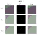
最后,将预先培养好的MBA-MD-231细胞 、A549细胞、H22细胞分别接种到激光共聚焦培养皿中,每个培养皿中接种6×10⁵个细胞;接种完2~3小时后(或者待细胞贴壁后),,每个培养皿中加入200ug上述染色后的EVs,轻轻混匀,将培养皿放入37℃、CO2培养箱中培养,分别在培养24h、48h、72h后,拍摄激光共聚焦图片。Finally, pre-cultured MBA-MD-231 cells, A549 cells, and H22 cells were seeded into laser confocal culture dishes, and 6×10⁵ cells were seeded in each culture dish; 2~3 hours after seeding ( Or after the cells adhered), add 200ug of the above-stained EVs to each petri dish, mix gently, and place the petri dish in a 37°C, CO2 incubator for 24h, 48h, and 72h respectively. After that, take a laser confocal picture.
结果如图7、图8、图9所示。可以看出:针对不同细胞株(MBA-MD-231、A549、H22细胞),24h后EVs进入细胞,细胞内出现荧光;48h后荧光强度稍微增强,说明EVs仍在进入细胞;72h后荧光出现最强,说明随着时间的积累,EVs不断地进入细胞。The results are shown in Figure 7, Figure 8, and Figure 9. It can be seen that: for different cell lines (MBA-MD-231, A549, H22 cells), EVs entered the cells after 24 hours, and fluorescence appeared in the cells; after 48 hours, the fluorescence intensity increased slightly, indicating that EVs were still entering the cells; after 72 hours, fluorescence appeared The strongest, indicating that EVs continue to enter cells over time.
(二)细胞增殖实验(2) Cell proliferation experiment
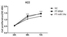
首先,以实施例2所制备转染并IPTG诱导表达6小时后BL21菌株为基础,提取EVs;First, EVs were extracted based on the BL21 strain prepared in Example 2 after transfection and induction of IPTG expression for 6 hours;
其次,将细胞(MBA-MD-231细胞 、A549细胞、H22细胞)接种到96孔板中,每个孔接种量为1000个细胞;每个时间段的细胞铺到一个板子上,每个样品重复6次;每个孔加入200ugEVs,对照组加入相同体积的PBS,放置37℃培养箱,24h、48h、72h后,分别在酶标仪下测定450nm波长下的吸光度值,统计数据。Second, cells (MBA-MD-231 cells, A549 cells, H22 cells) were seeded into 96-well plates, and each well was seeded at 1,000 cells; cells from each time period were plated on one plate, and each sample was plated on one plate.
具体结果分别如图10、图11、图12所示。结果表明,在MBA-MD-231、A549、H22三个不同的细胞系中,PT-tRNA对细胞没有任何毒性影响,同NC对照组的细胞增殖情况没有太大差别,而在加入PT-miR-126后,MBA-MD-231细胞的增殖受到了抑制,加入PT-miR-124后,A549细胞的增殖受到了抑制,加入PT-miR-34a后,H22细胞的增殖受到了抑制。The specific results are shown in Figure 10, Figure 11, and Figure 12, respectively. The results showed that in the three different cell lines of MBA-MD-231, A549 and H22, PT-tRNA did not have any toxic effect on the cells, and the cell proliferation was not much different from the NC control group. The proliferation of MBA-MD-231 cells was inhibited after -126, the proliferation of A549 cells was inhibited by the addition of PT-miR-124, and the proliferation of H22 cells was inhibited by the addition of PT-miR-34a.
上述结果表明,在使用“PT-tRNA”支架连接miRNA杀伤癌细胞的过程中,不同的miRNA针对不同癌细胞都表现出了杀伤作用,而“PT-tRNA”支架本身对细胞无任何毒性影响。The above results show that in the process of using the "PT-tRNA" scaffold to connect miRNAs to kill cancer cells, different miRNAs have shown killing effects on different cancer cells, while the "PT-tRNA" scaffold itself has no toxic effect on cells.
(三)细胞内miRNA的量以及被细胞切割为成熟miRNA的量情况(3) The amount of intracellular miRNA and the amount of mature miRNA cleaved by cells
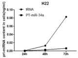
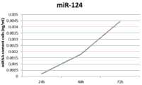
上述EVs侵染细胞的实验验证了EVs能进入到细胞中,因此pri-miRNA会随着EVs也一起进入到了细胞中。而进入到细胞后,pri-miRNA会被Drosha进一步加工切割成为成熟的miRNA。本实验检测了0-72h内进入到细胞中的pri-miRNA的量,以及pri-miRNA在细胞内被加工为成熟的miRNA的量。具体实验检测过程为:The above experiments of EVs infecting cells verified that EVs could enter cells, so pri-miRNAs would enter cells together with EVs. After entering the cell, pri-miRNA will be further processed and cut into mature miRNA by Drosha. In this experiment, the amount of pri-miRNA entered into cells within 0-72h and the amount of pri-miRNA processed into mature miRNA in cells were detected. The specific experimental detection process is as follows:
提取100ng EVs,加入到预先培养的细胞(MBA-MD-231细胞 、A549细胞、H22细胞)中,分别在24h、48h、72h后收集细胞,提取细胞的RNA,反转录成cDNA,最后进行荧光定量PCR。统计数据,并进行绘图。100ng of EVs were extracted and added to pre-cultured cells (MBA-MD-231 cells, A549 cells, H22 cells), and the cells were collected after 24h, 48h, and 72h, respectively, and the RNA of the cells was extracted, reverse transcribed into cDNA, and finally carried out Fluorescence quantitative PCR. Statistics and plotting.
细胞中pri-miRNA量统计结果如图13、图14、图15所示。可以看出,进入到不同细胞中(MBA-MD-231、A549、H22细胞)的pri-miRNA(miR-126、miR-124、miR-34a)量都随着时间的变化而逐渐增加,第三天进入到细胞中的pei-miRNA的量最多。The statistical results of the amount of pri-miRNA in the cells are shown in Figure 13, Figure 14, and Figure 15. It can be seen that the amount of pri-miRNA (miR-126, miR-124, miR-34a) entering into different cells (MBA-MD-231, A549, H22 cells) gradually increased with time. The highest amount of pei-miRNA entered the cells on three days.
细胞内成熟的miRNA的量统计结果如图16、图17、图18所示。可以看出,不同细胞中(MBA-MD-231、A549、H22细胞)的pri-miRNA(miR-126、miR-124、miR-34a)被加工为成熟的miRNA的量都随着时间的变化而逐渐增加,第三天细胞内被加工为成熟的miRNA的量最多。Figure 16, Figure 17, and Figure 18 show the statistical results of the amount of mature miRNA in cells. It can be seen that the amount of pri-miRNAs (miR-126, miR-124, miR-34a) processed into mature miRNAs in different cells (MBA-MD-231, A549, H22 cells) varies with time While gradually increasing, the amount of miRNA that was processed into mature cells was the highest on the third day.
(四)细胞中靶基因的变化量(4) Changes in target genes in cells
为了验证对癌细胞起杀伤作用的是EVs中的miRNA,进一步研究细胞中靶基因的变化量。针对现有技术中部分靶基因设计引物序列(具体引物序列可从Primerbank查询,不再详述),进行荧光定量PCR检测(以16S RNA为内参基因)。In order to verify that it is the miRNAs in EVs that kill cancer cells, the changes of target genes in cells were further studied. Primer sequences were designed for some target genes in the prior art (the specific primer sequences can be queried from Primerbank, which will not be described in detail), and quantitative PCR was performed (with 16S RNA as the internal reference gene).
实验检测时,提取相应EVs加入到细胞中,48h后收集细胞,提取细胞的RNA并反转录成cDNA作为模板,然后进行荧光定量检测。During the experimental detection, the corresponding EVs were extracted and added to the cells. After 48 hours, the cells were collected, and the RNA of the cells was extracted and reverse transcribed into cDNA as a template, and then fluorescence quantitative detection was performed.
具体靶基因在不同细胞中变化结果如下表所示。The changes of specific target genes in different cells are shown in the table below.

分析可以看出,A549细胞中STAT3基因明显降低;MBA-MD-231细胞中ADAM9、AKT1、RAF1、CXCR4基因降低,CXCR4基因降低的最明显,而Egfl-7、CXCL12基因变化量不明显;H22细胞中SATB2、E2f3、SIRT1、CCND1基因降低,SIRT4基因降低最明显,而E2f1、Bcl2基因变化量不明显;4T1细胞中CXCR4、CXCL12基因降低,而CXCR4基因降低最明显。The analysis showed that STAT3 gene was significantly decreased in A549 cells; ADAM9, AKT1, RAF1, and CXCR4 genes were decreased in MBA-MD-231 cells, and CXCR4 gene was most significantly decreased, while Egfl-7 and CXCL12 genes did not change significantly; H22 The genes of SATB2, E2f3, SIRT1 and CCND1 were decreased in the cells, and the gene of SIRT4 was decreased most obviously, while the gene of E2f1 and Bcl2 was not significantly changed.
(五)EVs偶联核酸适配体实现肿瘤靶向(5) EVs-conjugated nucleic acid aptamers to achieve tumor targeting
为了更好的实现EVs靶向肿瘤,提高EVs杀伤癌细胞的效率,发明人以胆固醇修饰的AS1411核酸适配体偶联EVs,从而来提高EVs的靶向性。In order to better target EVs to tumors and improve the efficiency of EVs in killing cancer cells, the inventors coupled EVs with cholesterol-modified AS1411 nucleic acid aptamer to improve the targeting of EVs.
所述AS1411具体序列如下:The specific sequence of the AS1411 is as follows:
5'-Cholesteryl-TTTTTGGTGGTGGTGGTTGTGGTGGTGGTGG-3'。5'-Cholesteryl-TTTTTGGTGGTGGTGGTTGTGGTGGTGGGTGG-3'.
作为对照,以合成的随机序列DZ-AS1411作为对照,所合成的随机序列为:As a control, the synthesized random sequence DZ-AS1411 is used as a control, and the synthesized random sequence is:
5'-Cholesteryl-TTTTTTATAATAATAATTATAATAATAATTTAAAAT-3'。5'-Cholesteryl-TTTTTTATAATAATAATTATAATAATAATTTAAAAT-3'.
具体实验时:During the specific experiment:
首先将5uM的AS1411与100ug EVs(miR-126)在4℃的PBS中孵育一夜,收集偶联上AS1411的Evs;加入到相同量1000个MBA-MD-231细胞中,细胞铺板在96孔板中,每个样品重复6次;24h之后在酶标仪下测定450nm波长下的吸光度值,统计数据。First, 5uM of AS1411 was incubated with 100ug EVs (miR-126) in PBS at 4°C overnight, and the EVs coupled with AS1411 were collected; added to the same amount of 1000 MBA-MD-231 cells, and the cells were plated in 96-well plates , each sample was repeated 6 times; after 24h, the absorbance value at 450nm wavelength was measured under the microplate reader, and the statistical data was obtained.
比较AS1411-EVs和DZ-AS1411-EVs对MBA-MD-231细胞的杀伤能力。The killing ability of AS1411-EVs and DZ-AS1411-EVs on MBA-MD-231 cells was compared.
结果如图19所示。可以看出,没有靶向功能的DZ-AH1411-EVs只能杀伤10%的MBA-MD-231细胞,而AS1411- EVs能杀伤74%的MBA-MD-231细胞。这一结果表明,使用适配体偶联EVs,可以进一步提高EVs杀伤癌细胞的能力。The results are shown in Figure 19. It can be seen that DZ-AH1411-EVs without targeting function can only kill 10% of MBA-MD-231 cells, while AS1411-EVs can kill 74% of MBA-MD-231 cells. This result suggests that the ability of EVs to kill cancer cells can be further enhanced by using aptamers to couple EVs.
(六)动物实验(6) Animal experiments
上述系列实验表明EVs在体外细胞中具有杀伤作用,进一步地,发明人在动物体内验证了EVs的抗肿瘤功效。具体过程为:The above series of experiments show that EVs have a killing effect on cells in vitro. Further, the inventors have verified the anti-tumor efficacy of EVs in animals. The specific process is:
从北京维通利华公司购买六周龄的裸鼠,待小鼠长到八周龄时,开始给小鼠荷瘤,每只小鼠注射106个MBA-MD-231细胞,两周后小鼠长瘤。Six-week-old nude mice were purchased from Beijing Weitong Lihua Co., Ltd. When the mice were eight weeks old, they were given tumor-bearing mice. Each mouse was injected with 106 MBA-MD-231 cells. Two weeks later Tumors in mice.
将荷瘤小鼠平均分成三组,PBS组、AS1411-EVs-tRNA组、AS1411-EVs-miR-126组。通过尾静注射,每周三次,连续注射21天。The tumor-bearing mice were equally divided into three groups, PBS group, AS1411-EVs-tRNA group and AS1411-EVs-miR-126 group. By tail vein injection, three times a week, continuous injection for 21 days.
给药结束,将小鼠麻醉后处死,收集肿瘤,比较各组之间的肿瘤差异。At the end of the administration, the mice were anesthetized and sacrificed, the tumors were collected, and the tumor differences among the groups were compared.
实验结果如图20所示。可以看出,注射AS1411-EVs-tRNA后小鼠的肿瘤体积与注射PBS的小鼠肿瘤相比没有太大的差异,说明AS1411-EVs-tRNA对荷瘤小鼠没有治疗效果,而注射AS1411-EVs-miR-126后小鼠的肿瘤体积明显的减小,说明AS1411-EVs-miR-126对荷瘤小鼠有治疗功效。The experimental results are shown in Figure 20. It can be seen that the tumor volume of mice injected with AS1411-EVs-tRNA is not much different from that of mice injected with PBS, indicating that AS1411-EVs-tRNA has no therapeutic effect on tumor-bearing mice, while injection of AS1411- The tumor volume of mice after EVs-miR-126 was significantly reduced, indicating that AS1411-EVs-miR-126 has therapeutic effect on tumor-bearing mice.
(七)内源性载药与外源性载药的效率比较(VII) Comparison of the efficiency of endogenous drug loading and exogenous drug loading
进一步地,发明人针对现有技术中(专利CN 110037996A《内源性高表达miRNA的大肠杆菌内膜囊泡的制备方法及其在制备抗肿瘤药物中的应用》)的大肠杆菌内膜囊泡的制备方法,比较了外膜囊泡与内源囊泡装载miRNA的量以及杀伤肿瘤的效果。Further, the inventors aimed at the Escherichia coli endomembrane vesicles in the prior art (Patent CN 110037996A "The preparation method of Escherichia coli inner membrane vesicles with high endogenous expression of miRNA and its application in the preparation of antitumor drugs"). The preparation method of , compared the amount of miRNA loaded in outer membrane vesicles and endogenous vesicles and the effect of killing tumors.
(1)miRNA装载效率区别(1) Differences in miRNA loading efficiency
根据发明专利CN 110037996A《内源性高表达miRNA的大肠杆菌内膜囊泡的制备方法及其在制备抗肿瘤药物中的应用》中的方法提取大肠杆菌内膜EVs(内源性表达miR-126),同时根据本发明提供的方法提取大肠杆菌外膜EVs。提取RNA并反转录为cDNA后,使用荧光定量PCR依次检测内膜囊泡与外膜中miR-126(内源性表达miR-126)的含量。According to the method in the invention patent CN 110037996A "Preparation of Escherichia coli Inner Membrane Vesicles with Endogenous High Expression of miRNA and Its Application in the Preparation of Antitumor Drugs", Escherichia coli inner membrane EVs (endogenous expression of miR-126 ), and at the same time, the outer membrane EVs of Escherichia coli were extracted according to the method provided by the present invention. After RNA was extracted and reverse transcribed into cDNA, the content of miR-126 (endogenously expressed miR-126) in inner membrane vesicles and outer membrane was detected by real-time quantitative PCR.
结果如图21所示,可以看出,相同量的外膜囊泡装载miRNA的量比内膜囊泡要高的多,说明外膜囊泡装载miRNA的效率要比内膜囊泡装载miRNA的效率高的多。The results are shown in Figure 21. It can be seen that the amount of miRNA loaded in outer membrane vesicles with the same amount is much higher than that in inner membrane vesicles, indicating that the efficiency of miRNA loading in outer membrane vesicles is higher than that in inner membrane vesicles. Much more efficient.
(2)肿瘤杀伤效果(2) Tumor killing effect
分别取100ng的外膜囊泡与100ng的内膜囊泡加入到预先铺在96孔中的MBA-MD-231细胞中,铺板量为1000个细胞,24h后在酶标仪下测定450nm波长下的吸光度值,统计数据。Take 100ng of outer membrane vesicles and 100ng of inner membrane vesicles and add them to MBA-MD-231 cells pre-plated in 96 wells, and the plating amount is 1000 cells. The absorbance value, statistics.
结果如图22所示。可以看出,相同条件的内膜囊泡与外膜囊泡加入到相同量的MBA-MD-231细胞中,内膜囊泡杀伤癌细胞的效率为14%,而外膜囊泡杀伤癌细胞的效率为72%,说明外膜囊泡对癌细胞的杀伤效率更高。The results are shown in Figure 22. It can be seen that when the inner membrane vesicles and outer membrane vesicles under the same conditions are added to the same amount of MBA-MD-231 cells, the efficiency of the inner membrane vesicles to kill cancer cells is 14%, while the outer membrane vesicles kill cancer cells. The efficiency was 72%, indicating that the outer membrane vesicles were more efficient in killing cancer cells.
SEQUENCE LISTING SEQUENCE LISTING
<110> 郑州大学<110> Zhengzhou University
<120> 一种内源性高表达miRNA的大肠杆菌胞外囊泡的制备方法<120> A kind of preparation method of Escherichia coli extracellular vesicles with endogenous high expression of miRNA
<130> none<130> none
<160> 1<160> 1
<170> PatentIn version 3.5<170> PatentIn version 3.5
<210> 1<210> 1
<211> 85<211> 85
<212> DNA<212> DNA
<213> 人工设计<213> Manual Design
<400> 1<400> 1
gaattcgccc ggatagctca gtcggtagag cagcggccgc ggccgcgggt ccagggttca 60gaattcgccc ggatagctca gtcggtagag cagcggccgc ggccgcgggt ccagggttca 60
agtccctgtt cgggcgccaa agctt 85agtccctgtt cgggcgccaa agctt 85
Claims (7)
Priority Applications (1)
| Application Number | Priority Date | Filing Date | Title |
|---|---|---|---|
| CN201911054070.4ACN110804621A (en) | 2019-10-31 | 2019-10-31 | A kind of preparation method of Escherichia coli extracellular vesicles with endogenous high expression of miRNA |
Applications Claiming Priority (1)
| Application Number | Priority Date | Filing Date | Title |
|---|---|---|---|
| CN201911054070.4ACN110804621A (en) | 2019-10-31 | 2019-10-31 | A kind of preparation method of Escherichia coli extracellular vesicles with endogenous high expression of miRNA |
Publications (1)
| Publication Number | Publication Date |
|---|---|
| CN110804621Atrue CN110804621A (en) | 2020-02-18 |
Family
ID=69489903
Family Applications (1)
| Application Number | Title | Priority Date | Filing Date |
|---|---|---|---|
| CN201911054070.4APendingCN110804621A (en) | 2019-10-31 | 2019-10-31 | A kind of preparation method of Escherichia coli extracellular vesicles with endogenous high expression of miRNA |
Country Status (1)
| Country | Link |
|---|---|
| CN (1) | CN110804621A (en) |
Cited By (3)
| Publication number | Priority date | Publication date | Assignee | Title |
|---|---|---|---|---|
| CN114410636A (en)* | 2021-12-31 | 2022-04-29 | 郑州大学 | Improved AS1411 aptamer and its conjugated EVs |
| CN114990038A (en)* | 2022-05-24 | 2022-09-02 | 郑州大学 | Bacterial outer membrane vesicle and application thereof in preparation of preeclampsia treatment medicine |
| CN119716044A (en)* | 2024-12-27 | 2025-03-28 | 浙江工业大学 | Encoding microsphere and application thereof in simultaneous detection of extracellular vesicle multiple markers |
Citations (6)
| Publication number | Priority date | Publication date | Assignee | Title |
|---|---|---|---|---|
| US20160331686A1 (en)* | 2015-05-12 | 2016-11-17 | Clsn Laboratories, Inc. | Compositions and Methods for Yeast Extracellular Vesicles as Delivery Systems |
| CN107142228A (en)* | 2017-04-18 | 2017-09-08 | 浙江大学 | A kind of preparation of Escherichia coli outer membrane vesicles, medicine-carrying method and its application in antitumor |
| CN108904468A (en)* | 2017-08-31 | 2018-11-30 | 李牧青 | A method of building targets extracellular vesica |
| TW201919673A (en)* | 2017-09-08 | 2019-06-01 | 美商艾弗洛生物科技股份有限公司 | Bacterial extracellular vesicles |
| CN109837306A (en)* | 2019-03-20 | 2019-06-04 | 江南大学附属医院(无锡市第四人民医院) | Contain the excretion body and its preparation method and application of miRNA-204-5p |
| CN110037996A (en)* | 2019-04-30 | 2019-07-23 | 郑州大学 | Endogenous height expresses the preparation method and its application in preparation of anti-tumor drugs of membrane vesicle in the Escherichia coli of miRNA |
- 2019
- 2019-10-31CNCN201911054070.4Apatent/CN110804621A/enactivePending
Patent Citations (6)
| Publication number | Priority date | Publication date | Assignee | Title |
|---|---|---|---|---|
| US20160331686A1 (en)* | 2015-05-12 | 2016-11-17 | Clsn Laboratories, Inc. | Compositions and Methods for Yeast Extracellular Vesicles as Delivery Systems |
| CN107142228A (en)* | 2017-04-18 | 2017-09-08 | 浙江大学 | A kind of preparation of Escherichia coli outer membrane vesicles, medicine-carrying method and its application in antitumor |
| CN108904468A (en)* | 2017-08-31 | 2018-11-30 | 李牧青 | A method of building targets extracellular vesica |
| TW201919673A (en)* | 2017-09-08 | 2019-06-01 | 美商艾弗洛生物科技股份有限公司 | Bacterial extracellular vesicles |
| CN109837306A (en)* | 2019-03-20 | 2019-06-04 | 江南大学附属医院(无锡市第四人民医院) | Contain the excretion body and its preparation method and application of miRNA-204-5p |
| CN110037996A (en)* | 2019-04-30 | 2019-07-23 | 郑州大学 | Endogenous height expresses the preparation method and its application in preparation of anti-tumor drugs of membrane vesicle in the Escherichia coli of miRNA |
Cited By (4)
| Publication number | Priority date | Publication date | Assignee | Title |
|---|---|---|---|---|
| CN114410636A (en)* | 2021-12-31 | 2022-04-29 | 郑州大学 | Improved AS1411 aptamer and its conjugated EVs |
| CN114990038A (en)* | 2022-05-24 | 2022-09-02 | 郑州大学 | Bacterial outer membrane vesicle and application thereof in preparation of preeclampsia treatment medicine |
| CN114990038B (en)* | 2022-05-24 | 2023-06-27 | 郑州大学 | A bacterial outer membrane vesicle and its application in the preparation of drugs for the treatment of preeclampsia |
| CN119716044A (en)* | 2024-12-27 | 2025-03-28 | 浙江工业大学 | Encoding microsphere and application thereof in simultaneous detection of extracellular vesicle multiple markers |
Similar Documents
| Publication | Publication Date | Title |
|---|---|---|
| CN110804621A (en) | A kind of preparation method of Escherichia coli extracellular vesicles with endogenous high expression of miRNA | |
| CN101255438A (en) | Construction method of transgenic Chlamydomonas reinhardtii expressing human tissue kallikrein | |
| CN108753726A (en) | A kind of excretion body and its preparation method and application containing ECRG4 mRNA | |
| CN116987603A (en) | Recombinant saccharomyces cerevisiae strain for high yield of cannabigerolic acid as well as construction method and application thereof | |
| CN114410636A (en) | Improved AS1411 aptamer and its conjugated EVs | |
| CN105861551B (en) | The carrier and its construction method of Combined expression microRNAs inhibition Cells Proliferation of Human Breast Cancer and application | |
| CN116875558A (en) | Lung cancer cell strain for stably expressing VGLL3 gene, construction method and application thereof | |
| CN113416768B (en) | Application of PRKRA gene as target in inhibiting replication of peste des petits ruminants virus | |
| CN110075122B (en) | Liver cancer therapeutic exosome medicine | |
| CN114181937B (en) | shRNA molecule for silencing human LINC01614 expression and application thereof | |
| CN105734075B (en) | A vector that interferes with ABCB5 gene expression and its application in cancer stem cell therapy | |
| CN118652889B (en) | A circular ribozyme system and its application | |
| CN118879638A (en) | A multi-gene co-expression non-small cell lung cancer model and its construction method | |
| CN114540422A (en) | Preparation and application of mesenchymal stem cell exosome for delivering RNA (ribonucleic acid) medicament to damaged part in targeted manner | |
| CN104531760B (en) | The short hairpin RNA interference plasmid and its application process of Dp71 albumen | |
| CN115812699B (en) | A carbon-based nanomaterial for delivering dsRNA as a nucleic acid carrier and its preparation method and application | |
| CN105087418B (en) | Salmonella strain for RNA interference in mammalian cells, preparation method and application thereof | |
| WO2015085903A1 (en) | Non-coded rna of in-vivo infected microorganisms, parasitic microorganisms, symbiotic microorganisms and identification and application thereof | |
| Xia et al. | Microvesicles containing microRNA-21 induce myocardial fibrosis via AKT pathway | |
| CN101948544A (en) | FAT10 gene siRNA recombination analogue virus as well as preparation method and application thereof | |
| CN116370652B (en) | Construction and application of nano-carrier system for efficiently delivering exogenous nucleic acid to silence bacterial gene | |
| CN117417432B (en) | Fugu rubripes growth hormone and application thereof | |
| CN103937745B (en) | People A375 stable cell strain and the construction method of long-chain non-coding GAS5 defect | |
| CN114657181B (en) | A sgRNA targeting H1.4 and H1.4 gene editing method | |
| CN113181374B (en) | Spherical microRNA and preparation method and application thereof |
Legal Events
| Date | Code | Title | Description |
|---|---|---|---|
| PB01 | Publication | ||
| PB01 | Publication | ||
| SE01 | Entry into force of request for substantive examination | ||
| SE01 | Entry into force of request for substantive examination |