CN110799102B - Transcatheter atrial sealing skirt, anchor, tether implant assembly - Google Patents
Transcatheter atrial sealing skirt, anchor, tether implant assemblyDownload PDFInfo
- Publication number
- CN110799102B CN110799102BCN201880033804.8ACN201880033804ACN110799102BCN 110799102 BCN110799102 BCN 110799102BCN 201880033804 ACN201880033804 ACN 201880033804ACN 110799102 BCN110799102 BCN 110799102B
- Authority
- CN
- China
- Prior art keywords
- anchor
- tether
- sealing skirt
- atrial
- delivery
- Prior art date
- Legal status (The legal status is an assumption and is not a legal conclusion. Google has not performed a legal analysis and makes no representation as to the accuracy of the status listed.)
- Active
Links
Images


Classifications
- A—HUMAN NECESSITIES
- A61—MEDICAL OR VETERINARY SCIENCE; HYGIENE
- A61F—FILTERS IMPLANTABLE INTO BLOOD VESSELS; PROSTHESES; DEVICES PROVIDING PATENCY TO, OR PREVENTING COLLAPSING OF, TUBULAR STRUCTURES OF THE BODY, e.g. STENTS; ORTHOPAEDIC, NURSING OR CONTRACEPTIVE DEVICES; FOMENTATION; TREATMENT OR PROTECTION OF EYES OR EARS; BANDAGES, DRESSINGS OR ABSORBENT PADS; FIRST-AID KITS
- A61F2/00—Filters implantable into blood vessels; Prostheses, i.e. artificial substitutes or replacements for parts of the body; Appliances for connecting them with the body; Devices providing patency to, or preventing collapsing of, tubular structures of the body, e.g. stents
- A61F2/02—Prostheses implantable into the body
- A61F2/24—Heart valves ; Vascular valves, e.g. venous valves; Heart implants, e.g. passive devices for improving the function of the native valve or the heart muscle; Transmyocardial revascularisation [TMR] devices; Valves implantable in the body
- A61F2/2412—Heart valves ; Vascular valves, e.g. venous valves; Heart implants, e.g. passive devices for improving the function of the native valve or the heart muscle; Transmyocardial revascularisation [TMR] devices; Valves implantable in the body with soft flexible valve members, e.g. tissue valves shaped like natural valves
- A61F2/2418—Scaffolds therefor, e.g. support stents
- A—HUMAN NECESSITIES
- A61—MEDICAL OR VETERINARY SCIENCE; HYGIENE
- A61F—FILTERS IMPLANTABLE INTO BLOOD VESSELS; PROSTHESES; DEVICES PROVIDING PATENCY TO, OR PREVENTING COLLAPSING OF, TUBULAR STRUCTURES OF THE BODY, e.g. STENTS; ORTHOPAEDIC, NURSING OR CONTRACEPTIVE DEVICES; FOMENTATION; TREATMENT OR PROTECTION OF EYES OR EARS; BANDAGES, DRESSINGS OR ABSORBENT PADS; FIRST-AID KITS
- A61F2/00—Filters implantable into blood vessels; Prostheses, i.e. artificial substitutes or replacements for parts of the body; Appliances for connecting them with the body; Devices providing patency to, or preventing collapsing of, tubular structures of the body, e.g. stents
- A61F2/02—Prostheses implantable into the body
- A61F2/24—Heart valves ; Vascular valves, e.g. venous valves; Heart implants, e.g. passive devices for improving the function of the native valve or the heart muscle; Transmyocardial revascularisation [TMR] devices; Valves implantable in the body
- A61F2/2412—Heart valves ; Vascular valves, e.g. venous valves; Heart implants, e.g. passive devices for improving the function of the native valve or the heart muscle; Transmyocardial revascularisation [TMR] devices; Valves implantable in the body with soft flexible valve members, e.g. tissue valves shaped like natural valves
- A—HUMAN NECESSITIES
- A61—MEDICAL OR VETERINARY SCIENCE; HYGIENE
- A61F—FILTERS IMPLANTABLE INTO BLOOD VESSELS; PROSTHESES; DEVICES PROVIDING PATENCY TO, OR PREVENTING COLLAPSING OF, TUBULAR STRUCTURES OF THE BODY, e.g. STENTS; ORTHOPAEDIC, NURSING OR CONTRACEPTIVE DEVICES; FOMENTATION; TREATMENT OR PROTECTION OF EYES OR EARS; BANDAGES, DRESSINGS OR ABSORBENT PADS; FIRST-AID KITS
- A61F2/00—Filters implantable into blood vessels; Prostheses, i.e. artificial substitutes or replacements for parts of the body; Appliances for connecting them with the body; Devices providing patency to, or preventing collapsing of, tubular structures of the body, e.g. stents
- A61F2/02—Prostheses implantable into the body
- A61F2/24—Heart valves ; Vascular valves, e.g. venous valves; Heart implants, e.g. passive devices for improving the function of the native valve or the heart muscle; Transmyocardial revascularisation [TMR] devices; Valves implantable in the body
- A61F2/2427—Devices for manipulating or deploying heart valves during implantation
- A—HUMAN NECESSITIES
- A61—MEDICAL OR VETERINARY SCIENCE; HYGIENE
- A61F—FILTERS IMPLANTABLE INTO BLOOD VESSELS; PROSTHESES; DEVICES PROVIDING PATENCY TO, OR PREVENTING COLLAPSING OF, TUBULAR STRUCTURES OF THE BODY, e.g. STENTS; ORTHOPAEDIC, NURSING OR CONTRACEPTIVE DEVICES; FOMENTATION; TREATMENT OR PROTECTION OF EYES OR EARS; BANDAGES, DRESSINGS OR ABSORBENT PADS; FIRST-AID KITS
- A61F2/00—Filters implantable into blood vessels; Prostheses, i.e. artificial substitutes or replacements for parts of the body; Appliances for connecting them with the body; Devices providing patency to, or preventing collapsing of, tubular structures of the body, e.g. stents
- A61F2/02—Prostheses implantable into the body
- A61F2/24—Heart valves ; Vascular valves, e.g. venous valves; Heart implants, e.g. passive devices for improving the function of the native valve or the heart muscle; Transmyocardial revascularisation [TMR] devices; Valves implantable in the body
- A61F2/2442—Annuloplasty rings or inserts for correcting the valve shape; Implants for improving the function of a native heart valve
- A61F2/2454—Means for preventing inversion of the valve leaflets, e.g. chordae tendineae prostheses
- A61F2/2457—Chordae tendineae prostheses
- A—HUMAN NECESSITIES
- A61—MEDICAL OR VETERINARY SCIENCE; HYGIENE
- A61F—FILTERS IMPLANTABLE INTO BLOOD VESSELS; PROSTHESES; DEVICES PROVIDING PATENCY TO, OR PREVENTING COLLAPSING OF, TUBULAR STRUCTURES OF THE BODY, e.g. STENTS; ORTHOPAEDIC, NURSING OR CONTRACEPTIVE DEVICES; FOMENTATION; TREATMENT OR PROTECTION OF EYES OR EARS; BANDAGES, DRESSINGS OR ABSORBENT PADS; FIRST-AID KITS
- A61F2/00—Filters implantable into blood vessels; Prostheses, i.e. artificial substitutes or replacements for parts of the body; Appliances for connecting them with the body; Devices providing patency to, or preventing collapsing of, tubular structures of the body, e.g. stents
- A61F2/02—Prostheses implantable into the body
- A61F2/24—Heart valves ; Vascular valves, e.g. venous valves; Heart implants, e.g. passive devices for improving the function of the native valve or the heart muscle; Transmyocardial revascularisation [TMR] devices; Valves implantable in the body
- A61F2/2442—Annuloplasty rings or inserts for correcting the valve shape; Implants for improving the function of a native heart valve
- A61F2/246—Devices for obstructing a leak through a native valve in a closed condition
- A—HUMAN NECESSITIES
- A61—MEDICAL OR VETERINARY SCIENCE; HYGIENE
- A61F—FILTERS IMPLANTABLE INTO BLOOD VESSELS; PROSTHESES; DEVICES PROVIDING PATENCY TO, OR PREVENTING COLLAPSING OF, TUBULAR STRUCTURES OF THE BODY, e.g. STENTS; ORTHOPAEDIC, NURSING OR CONTRACEPTIVE DEVICES; FOMENTATION; TREATMENT OR PROTECTION OF EYES OR EARS; BANDAGES, DRESSINGS OR ABSORBENT PADS; FIRST-AID KITS
- A61F2/00—Filters implantable into blood vessels; Prostheses, i.e. artificial substitutes or replacements for parts of the body; Appliances for connecting them with the body; Devices providing patency to, or preventing collapsing of, tubular structures of the body, e.g. stents
- A61F2/02—Prostheses implantable into the body
- A61F2/24—Heart valves ; Vascular valves, e.g. venous valves; Heart implants, e.g. passive devices for improving the function of the native valve or the heart muscle; Transmyocardial revascularisation [TMR] devices; Valves implantable in the body
- A61F2/2442—Annuloplasty rings or inserts for correcting the valve shape; Implants for improving the function of a native heart valve
- A61F2/2466—Delivery devices therefor
- A—HUMAN NECESSITIES
- A61—MEDICAL OR VETERINARY SCIENCE; HYGIENE
- A61F—FILTERS IMPLANTABLE INTO BLOOD VESSELS; PROSTHESES; DEVICES PROVIDING PATENCY TO, OR PREVENTING COLLAPSING OF, TUBULAR STRUCTURES OF THE BODY, e.g. STENTS; ORTHOPAEDIC, NURSING OR CONTRACEPTIVE DEVICES; FOMENTATION; TREATMENT OR PROTECTION OF EYES OR EARS; BANDAGES, DRESSINGS OR ABSORBENT PADS; FIRST-AID KITS
- A61F2/00—Filters implantable into blood vessels; Prostheses, i.e. artificial substitutes or replacements for parts of the body; Appliances for connecting them with the body; Devices providing patency to, or preventing collapsing of, tubular structures of the body, e.g. stents
- A61F2/02—Prostheses implantable into the body
- A61F2/24—Heart valves ; Vascular valves, e.g. venous valves; Heart implants, e.g. passive devices for improving the function of the native valve or the heart muscle; Transmyocardial revascularisation [TMR] devices; Valves implantable in the body
- A61F2/2427—Devices for manipulating or deploying heart valves during implantation
- A61F2/2436—Deployment by retracting a sheath
- A—HUMAN NECESSITIES
- A61—MEDICAL OR VETERINARY SCIENCE; HYGIENE
- A61F—FILTERS IMPLANTABLE INTO BLOOD VESSELS; PROSTHESES; DEVICES PROVIDING PATENCY TO, OR PREVENTING COLLAPSING OF, TUBULAR STRUCTURES OF THE BODY, e.g. STENTS; ORTHOPAEDIC, NURSING OR CONTRACEPTIVE DEVICES; FOMENTATION; TREATMENT OR PROTECTION OF EYES OR EARS; BANDAGES, DRESSINGS OR ABSORBENT PADS; FIRST-AID KITS
- A61F2210/00—Particular material properties of prostheses classified in groups A61F2/00 - A61F2/26 or A61F2/82 or A61F9/00 or A61F11/00 or subgroups thereof
- A61F2210/0014—Particular material properties of prostheses classified in groups A61F2/00 - A61F2/26 or A61F2/82 or A61F9/00 or A61F11/00 or subgroups thereof using shape memory or superelastic materials, e.g. nitinol
- A—HUMAN NECESSITIES
- A61—MEDICAL OR VETERINARY SCIENCE; HYGIENE
- A61F—FILTERS IMPLANTABLE INTO BLOOD VESSELS; PROSTHESES; DEVICES PROVIDING PATENCY TO, OR PREVENTING COLLAPSING OF, TUBULAR STRUCTURES OF THE BODY, e.g. STENTS; ORTHOPAEDIC, NURSING OR CONTRACEPTIVE DEVICES; FOMENTATION; TREATMENT OR PROTECTION OF EYES OR EARS; BANDAGES, DRESSINGS OR ABSORBENT PADS; FIRST-AID KITS
- A61F2210/00—Particular material properties of prostheses classified in groups A61F2/00 - A61F2/26 or A61F2/82 or A61F9/00 or A61F11/00 or subgroups thereof
- A61F2210/0061—Particular material properties of prostheses classified in groups A61F2/00 - A61F2/26 or A61F2/82 or A61F9/00 or A61F11/00 or subgroups thereof swellable
- A—HUMAN NECESSITIES
- A61—MEDICAL OR VETERINARY SCIENCE; HYGIENE
- A61F—FILTERS IMPLANTABLE INTO BLOOD VESSELS; PROSTHESES; DEVICES PROVIDING PATENCY TO, OR PREVENTING COLLAPSING OF, TUBULAR STRUCTURES OF THE BODY, e.g. STENTS; ORTHOPAEDIC, NURSING OR CONTRACEPTIVE DEVICES; FOMENTATION; TREATMENT OR PROTECTION OF EYES OR EARS; BANDAGES, DRESSINGS OR ABSORBENT PADS; FIRST-AID KITS
- A61F2220/00—Fixations or connections for prostheses classified in groups A61F2/00 - A61F2/26 or A61F2/82 or A61F9/00 or A61F11/00 or subgroups thereof
- A61F2220/0008—Fixation appliances for connecting prostheses to the body
- A61F2220/0016—Fixation appliances for connecting prostheses to the body with sharp anchoring protrusions, e.g. barbs, pins, spikes
- A—HUMAN NECESSITIES
- A61—MEDICAL OR VETERINARY SCIENCE; HYGIENE
- A61F—FILTERS IMPLANTABLE INTO BLOOD VESSELS; PROSTHESES; DEVICES PROVIDING PATENCY TO, OR PREVENTING COLLAPSING OF, TUBULAR STRUCTURES OF THE BODY, e.g. STENTS; ORTHOPAEDIC, NURSING OR CONTRACEPTIVE DEVICES; FOMENTATION; TREATMENT OR PROTECTION OF EYES OR EARS; BANDAGES, DRESSINGS OR ABSORBENT PADS; FIRST-AID KITS
- A61F2220/00—Fixations or connections for prostheses classified in groups A61F2/00 - A61F2/26 or A61F2/82 or A61F9/00 or A61F11/00 or subgroups thereof
- A61F2220/0025—Connections or couplings between prosthetic parts, e.g. between modular parts; Connecting elements
- A61F2220/0075—Connections or couplings between prosthetic parts, e.g. between modular parts; Connecting elements sutured, ligatured or stitched, retained or tied with a rope, string, thread, wire or cable
- A—HUMAN NECESSITIES
- A61—MEDICAL OR VETERINARY SCIENCE; HYGIENE
- A61F—FILTERS IMPLANTABLE INTO BLOOD VESSELS; PROSTHESES; DEVICES PROVIDING PATENCY TO, OR PREVENTING COLLAPSING OF, TUBULAR STRUCTURES OF THE BODY, e.g. STENTS; ORTHOPAEDIC, NURSING OR CONTRACEPTIVE DEVICES; FOMENTATION; TREATMENT OR PROTECTION OF EYES OR EARS; BANDAGES, DRESSINGS OR ABSORBENT PADS; FIRST-AID KITS
- A61F2250/00—Special features of prostheses classified in groups A61F2/00 - A61F2/26 or A61F2/82 or A61F9/00 or A61F11/00 or subgroups thereof
- A61F2250/0004—Special features of prostheses classified in groups A61F2/00 - A61F2/26 or A61F2/82 or A61F9/00 or A61F11/00 or subgroups thereof adjustable
- A—HUMAN NECESSITIES
- A61—MEDICAL OR VETERINARY SCIENCE; HYGIENE
- A61F—FILTERS IMPLANTABLE INTO BLOOD VESSELS; PROSTHESES; DEVICES PROVIDING PATENCY TO, OR PREVENTING COLLAPSING OF, TUBULAR STRUCTURES OF THE BODY, e.g. STENTS; ORTHOPAEDIC, NURSING OR CONTRACEPTIVE DEVICES; FOMENTATION; TREATMENT OR PROTECTION OF EYES OR EARS; BANDAGES, DRESSINGS OR ABSORBENT PADS; FIRST-AID KITS
- A61F2250/00—Special features of prostheses classified in groups A61F2/00 - A61F2/26 or A61F2/82 or A61F9/00 or A61F11/00 or subgroups thereof
- A61F2250/0058—Additional features; Implant or prostheses properties not otherwise provided for
- A61F2250/0069—Sealing means
Landscapes
- Health & Medical Sciences (AREA)
- Cardiology (AREA)
- Engineering & Computer Science (AREA)
- Biomedical Technology (AREA)
- Life Sciences & Earth Sciences (AREA)
- Transplantation (AREA)
- Heart & Thoracic Surgery (AREA)
- Vascular Medicine (AREA)
- Oral & Maxillofacial Surgery (AREA)
- Animal Behavior & Ethology (AREA)
- General Health & Medical Sciences (AREA)
- Public Health (AREA)
- Veterinary Medicine (AREA)
- Prostheses (AREA)
- Surgical Instruments (AREA)
- Media Introduction/Drainage Providing Device (AREA)
- Structures Of Non-Positive Displacement Pumps (AREA)
Abstract
Description
Translated fromChinese技术领域technical field
本发明大致涉及以微创方式植入心脏的医疗装置和系统,以及这些装置和系统的植入方法。更具体地,本发明涉及使用一或多种锚固件(其拴系经导管的瓣膜或其他心脏装置在心脏内)以微创方式植入心脏任何壁部的医疗装置和系统。本发明还涉及一种在有或没有心内导线(intracardiac leads)的情况下,心房裙部密封的经导管的瓣膜,以减少瓣周反流(paravalvular regurgitation)。The present invention generally relates to medical devices and systems for minimally invasive implantation of the heart, and methods of implanting such devices and systems. More specifically, the present invention relates to medical devices and systems that are minimally invasively implanted into any wall of the heart using one or more anchors that tether a transcatheter valve or other cardiac device within the heart. The invention also relates to a transcatheter valve with atrial skirt sealing with or without intracardiac leads to reduce paravalvular regurgitation.
背景技术Background technique
经导管的(transcatheter)瓣膜已被证明可安全有效地替代原生心脏瓣膜。这些瓣膜已经过广泛实验,可以替代主动脉、二尖瓣和肺动脉瓣膜,但是鉴于假体必须附着复杂而脆弱的解剖结构,置换三尖瓣仍然具有挑战性。通常锚固经导管的瓣膜仍然很困难,尤其是经导管的三尖瓣,因为这样做无论是在心脏瓣膜的原位位置(in-situ position)还是在其他体腔中,都需要与心脏瓣膜环或其他体腔的多种形状和尺寸相互作用。在这方面,通过减少在假体的确切位置上固定的需求,拴系经导管的瓣膜至一个或多个固定到心内壁上的锚固件的能力将为所述经导管的假体提供更大的安全性和灵活性。特别是有利于锚固经导管的三尖瓣假体。Transcatheter valves have been shown to be safe and effective replacements for native heart valves. These valves have been tested extensively and can replace the aortic, mitral, and pulmonary valves, but replacing the tricuspid valve remains challenging given the complex and delicate anatomy to which the prosthesis must be attached. Anchoring transcatheter valvesin general remains difficult, especially transcatheter tricuspid valves, because doing so requires contact with the heart valve annulus or The various shapes and sizes of other body cavities interact. In this regard, the ability to tether a transcatheter valve to one or more anchors fixed to the endocardial wall will provide greater flexibility for the transcatheter prosthesis by reducing the need for fixation in the exact position of the prosthesis. security and flexibility. In particular, it facilitates the anchoring of transcatheter tricuspid valve prostheses.
几个组别已经描述心内锚固件和系绳,但是这些系统在其应用中具有局限性。例如Vidlund(1)和Lutter(2)描述一种使用单一系绳将人工瓣膜锚固的系统,所述系绳将瓣膜的远端连接到“心外膜系绳固定装置”(Vidlund专利)(其也可被描述为一锚固件)。由于固定装置位于心脏的心尖之外(心外膜),因此所述系统需要通过胸壁进入心脏的心尖。对于泵血功能下降(射血分数)的患者或组织脆弱的患者,需要通过胸部进入心脏可能会增加手术风险,尤其是涉及三尖瓣的应用。Several groups have described intracardiac anchors and tethers, but these systems have limitations in their application. For example, Vidlund (1) and Lutter (2) describe a system for anchoring a prosthetic valve using a single tether that connects the distal end of the valve to an "epicardial tether fixation device" (Vidlund patent) (the can also be described as an anchor). Since the fixation device is located outside the apex of the heart (epicardium), the system requires access to the apex of the heart through the chest wall. In patients with reduced pumping capacity (ejection fraction) or tissue fragility, the need to access the heart through the chest may increase procedural risk, especially in applications involving the tricuspid valve.
相反,Rowe及其同事(3)描述一种三尖瓣反流的接合装置(coaptation device),所述装置使用血管内方法锚固在右心室尖,而无需通过胸壁进入心脏。Rowe指出:“一柔性锚固轨道连接到锚固件,一导管上的一接合元件坐落于锚固轨道上。最后,有一近端的锚固特征可将接合导管的近端以皮下的方式固定在锁骨下静脉附近。”此应用的第一个限制是在错误的位置或非最佳的配置的情况下无法取回和重新定位锚固件。另一个限制是将锚固轨道固定到锚固件上。与此相反,能够首先配置锚固件,然后递送锁定到锚固件中的各种直径或材料的缆线或系绳才是有利的。通过使系绳独立于锚固件,可以轻松配置系绳/锚固元件的多个排列,从而使根据特定的临床需求定制应用的能力最大化。最后,所述申请描述了锚固件、远端的锚固轨道、接合装置和近端的锚固轨道之间的一固定连接,这需要大量锚固轨道保留在上腔静脉和无名/锁骨下静脉中。近端的锚固轨道连接到皮下口袋(subcutaneous pocket),所有这些都会带来永久性静脉导线的风险,特别是血栓形成、感染和静脉狭窄。In contrast, Rowe and colleagues (3) describe a tricuspid regurgitation coaptation device that uses an endovascular approach to anchor the right ventricular apex without accessing the heart through the chest wall. "A flexible anchoring track is attached to the anchor, and an engaging element on a catheter sits on the anchoring track," Rowe states. Finally, a proximal anchoring feature secures the proximal end of the engaging catheter subcutaneously in the subclavian vein. nearby.” The first limitation of this application is the inability to retrieve and reposition the anchors in case of wrong positions or non-optimal configurations. Another limitation is securing the anchor rail to the anchor. In contrast, it would be advantageous to be able to deploy the anchor first and then deliver cables or tethers of various diameters or materials that lock into the anchor. By making the tether independent of the anchor, multiple permutations of the tether/anchor elements can be easily configured, maximizing the ability to tailor the application to specific clinical needs. Finally, said application describes a fixed connection between the anchor, the distal anchoring track, the engagement means and the proximal anchoring track, which requires a large number of anchoring tracks to remain in the superior vena cava and innominate/subclavian vein. The proximal anchoring track connects to a subcutaneous pocket, all of which pose risks for permanent venous leads, especially thrombosis, infection, and venous stenosis.
Solem及其同事(4)克服了其应用中的一些局限性,其描述两阶段的过程的可能性,即首先可以配置锚固件,然后将“细长的主体部分”连接到锚固件,且细长的主体部分连接到系绳和血流控制装置。“对于用户希望更换罩部(canopy)或装置的其他部分的情形,...如果...可膨胀的瓣膜部分不是最佳尺寸或与锚固件之间不是最佳的距离”,则采用两阶段的过程可能会很有用。锚固件可以由“当锚固部...暴露时向外扩张的臂件或钩件”所组成。细长的主体可以通过多种机构连接到锚固件,包括“卡扣式或快速连接的连接”。在一实施例中,细长的主体可以具有“环状结构...配置用以围绕...锚固件大致呈管状的突起而前进”。为了便于连接,“锚固件近端...包括一连接构件...呈大致管状突出物形式的...具有向外延伸的锁定夹...可响应于向内压力而向内弯曲...但一旦释放向内压力,其将迅速向外弹回。”Some limitations in its application were overcome by Solem and colleagues (4) who described the possibility of a two-stage process whereby first the anchor could be deployed and then an "elongated body part" was attached to the anchor, and the thin The long body section is connected to the tether and blood flow control device. "For the situation where the user wishes to replace the canopy or other parts of the device, ... if ... the inflatable valve portion is not optimally sized or at an optimal distance from the anchor", two A process of stages may be useful. The anchor may consist of "arms or hooks that expand outward when the anchor ... is exposed". The elongated body can be attached to the anchor by a variety of mechanisms, including "snap-on or quick-connect connections." In an embodiment, the elongated body may have "annular structure... configured to advance around... the generally tubular protrusion of the anchor." To facilitate attachment, the "proximal end of the anchor...comprises a connecting member...in the form of a generally tubular protrusion...with outwardly extending locking clips...bendable inwardly in response to inward pressure. . . but once the inward pressure is released, it snaps back outward quickly."
尽管Solem的应用具有某些优点,但所述应用也受到限制。首先递送锚固件的优选方法是通过经由胸壁切口推进锚固件递送导管,然后通过心脏中一腔室进入一切口。甚至更希望能够通过真正的血管内方法(例如通过颈内或股静脉)递送锚固件,而不需要胸部和心脏腔室切口。接下来,锚固件由可扩张的臂件或钩件所组成,这些臂件或钩件挖入心脏壁,然后向后弯曲,类似于鱼钩。Solem认为可以收回锚固件的可扩张的钩件:“当锚固部...被拉回到远端护套...叉件(prongs)上的向内压力...来自远端护套...将导致叉件…锚固部的…向内塌陷……从而使锚固部塌陷……回到其递送的(即预配置)状态……”然而以这种方式可容易地取回向后弯曲的叉件而不会在过程中损坏组织是被怀疑的,因此需要一种更安全的可收回的锚固件。此外,叉件在心脏的一位置可能是安全的,但是如果需要将其移动到另一位置,则可能会造成损伤(即穿孔),因为臂长不可调节。因此需要改变锚固件的组织深度的能力(例如与心室自由壁相比,心室间隔可耐受更大的深度)。接下来,即使经由分离的步骤,细长的构件与锚固件的连接也被固定,使得细长的构件不能围绕锚固件的轴线旋转。当在细长的构件上调节经导管的瓣膜时,可能有必要旋转瓣膜以更好地安置在原生心脏瓣环中,而Solem的细长的构件一旦固定到锚固件上就无法旋转。最后,Solem的应用要求系绳通过细长的构件连接到锚固件。能够将系绳直接连接到锚固件并使系绳围具有绕锚固件的轴线旋转的能力,才是特别有利的,因为这将最紧密地模仿腱索(心脏本身的系绳)的功能,它们直接从心脏壁连接到心脏瓣膜。Although the application of Solem has certain advantages, said application is also limited. A preferred method of delivering the anchor first is by advancing the anchor delivery catheter through an incision in the chest wall, then through a chamber in the heart and into the incision. It would be even more desirable to be able to deliver the anchor by a true intravascular approach, such as through the internal jugular or femoral vein, without the need for chest and heart chamber incisions. Next, the anchors consist of expandable arms, or hooks, that dig into the heart wall and then bend back, similar to fishhooks. Solem argues that the anchor's expandable hooks can be retracted: "When the anchor...is drawn back into the distal sheath...the inward pressure on the prongs...comes from the distal sheath. ..will cause the inward collapse of the fork...anchor...thereby collapsing the anchor...back to its delivered (i.e. pre-configured) state..." However, the backward bend is easily retrieved in this manner It is doubtful that the prongs can be used without damaging tissue during the procedure, so a safer retractable anchor is needed. Furthermore, the fork may be secure in one location on the heart, but if it needs to be moved to another location, injury (ie, perforation) may result because the arm length is not adjustable. There is therefore a need for the ability to vary the tissue depth of the anchor (eg, the ventricular septum can tolerate a greater depth than the ventricular free wall). Next, even via the step of separation, the connection of the elongate member to the anchor is secured such that the elongate member cannot rotate about the axis of the anchor. When adjusting a transcatheter valve on the elongated member, it may be necessary to rotate the valve to better seat in the native heart annulus, whereas Solem's elongated member cannot be rotated once secured to the anchor. Finally, the Solem application requires the tether to be connected to the anchor by an elongated member. It would be particularly advantageous to be able to attach a tether directly to the anchor and to have the ability to rotate the tether about the axis of the anchor, as this would most closely mimic the function of the chordae (the heart's own tether), which Attaches directly from the heart wall to the heart valves.
此外限制经导管的二尖瓣和三尖瓣的瓣周反流(paravalvular regurgitation)是具有挑战性的,因为二尖瓣和三尖瓣是复杂的鞍形结构(saddle-shaped structures),其在心动周期中是高度动态的。具有严重三尖瓣反流(tricuspid regurgitation,TR)的患者的心内导线(intracardiac leads)频繁出现加剧了三尖瓣膜的困难。由于心室导线从右心房横穿瓣环到右心室,因此经导管的三尖瓣必须同时密封瓣环和导线,以限制这些患者的反流。Furthermore, limiting paravalvular regurgitation of the transcatheter mitral and tricuspid valves is challenging because the mitral and tricuspid valves are complex saddle-shaped structures that The cardiac cycle is highly dynamic. Tricuspid valve difficulties are exacerbated by frequent intracardiac leads in patients with severe tricuspid regurgitation (TR). Because the ventricular wire traverses the annulus from the right atrium to the right ventricle, a transcatheter tricuspid valve must seal both the annulus and the wire to limit regurgitation in these patients.
在接受经导管的主动脉瓣置换术(transcatheter aortic valvereplacements,TAVR)的患者中,研究人员开发了减轻瓣周反流的技术,但是这些方法存在局限性,特别是在存在心内导线的情况下。具体地,球囊膨胀式、机械膨胀式和自膨胀式TAVR在瓣环处围绕其支架框架结合了密封膜,以减少瓣周反流。密封膜由聚对苯二甲酸乙二醇酯(polyethylene terephthalate,PET或Dacron)或猪心包组织包裹物所组成。这些密封膜通过填充TAVR外侧和主动脉瓣环之间的间隙而起作用,但这需要将瓣膜直接与瓣环(annulus)并置。对于经导管的三尖瓣,将瓣膜框架直接与三尖瓣环并置可能是不想要的或不可行的,因为与主动脉瓣环不同,三尖瓣环是可扩张的,具有最少的外部支撑,且容易损伤。另外,通过将心内导线捕获在瓣膜框架和瓣环之间来密封它会增加导线损伤的风险,这是不想要的。In patients undergoing transcatheter aortic valve replacements (TAVR), researchers have developed techniques to alleviate paravalvular regurgitation, but these methods have limitations, especially in the presence of intracardiac leads . Specifically, balloon-expandable, mechanically expandable, and self-expanding TAVR incorporate a sealing membrane around its stent frame at the annulus to reduce paravalvular regurgitation. The sealing film is composed of polyethylene terephthalate (polyethylene terephthalate, PET or Dacron) or porcine pericardial tissue wrapping. These sealing membranes work by filling the space between the outside of the TAVR and the aortic annulus, but this requires apposition of the valve directly to the annulus. For transcatheter tricuspid valves, apposition of the valve frame directly to the tricuspid annulus may not be desirable or feasible because, unlike the aortic annulus, the tricuspid annulus is expandable with minimal external Supportive and easily damaged. Additionally, sealing the intracardiac lead by trapping it between the valve frame and annulus increases the risk of lead damage, which is undesirable.
大多数经导管的二尖瓣置换术(transcatheter mitral valve replacements,TMVR)已经使用类似的机制(通过将二尖瓣叶的基部捕获在瓣膜框架和瓣环之间)来限制瓣周反流。因此,像TAVR方法一样,TMVR减轻瓣周反流的方法可能会损坏脆弱的三尖瓣环,或将其捕获在瓣膜框架和瓣环之间,从而损坏心内导线。例如,Medtronic Intrepid和NSCINavigate瓣膜通过径向力抵靠在瓣环(Intrepid)或通过环形“小翼(winglets)”或钩件(Navigate)进行锚固。CardiAQ-Edwards TMVR使用亚环形(sub-annular)夹持机制直接与瓣环相互作用,而Neovasc Tiara瓣膜通过纤维三角(fibrous trigones)间接相互作用,还利用原生瓣叶接合(这两种机制都可能捕获和损伤导线)。三种TMVR装置(Caisson,HighLife和Mvalve)将一环形锚固件用作为TMVR装置的对接系统,这会挤压并可能损坏锚固件与TMVR装置之间的任何心内导线。Most transcatheter mitral valve replacements (TMVR) have used a similar mechanism (by trapping the base of the mitral valve leaflets between the valve frame and annulus) to limit paravalvular regurgitation. Therefore, like the TAVR approach, the TMVR approach to paravalvular regurgitation mitigation may damage the delicate tricuspid annulus or trap it between the valve frame and annulus, thereby damaging the intracardiac lead. For example, the Medtronic Intrepid and NSCINavigate valves are anchored by radial force against the annulus (Intrepid) or by annular "winglets" or hooks (Navigate). The CardiAQ-Edwards TMVR interacts directly with the annulus using a sub-annular clamping mechanism, while the Neovasc Tiara valve interacts indirectly through fibrous trigones and also utilizes native leaflet coaptation (both mechanisms are possible capture and damage the wire). Three TMVR devices (Caisson, HighLife, and Mvalve) use a loop anchor as the docking system for the TMVR device, which can pinch and potentially damage any intracardiac leads between the anchor and the TMVR device.
心内导线的损坏并不是TMVR装置减轻瓣周反流的唯一问题。因为大多数TMVR装置通过直接锚固到瓣环来密封二尖瓣环来减少反流,所以这些装置在不同程度上限制了二尖瓣环运动的自由度。限制这种自由度可能会导致左心室功能障碍。例如,一项比较经导管的二尖瓣修复术(使用Abbott Vascular的MitraClip装置)与开心手术(open heartsurgery)的研究报导表明,与经导管的修复术相比,开心手术的二尖瓣环运动明显降低,作者认为这是在开心手术之后,左室射血分数(LVEF)降低的一因素。同样,一项比较柔性二尖瓣环和刚性二尖瓣环成形术环的研究报导发现,刚性环的LVEF明显较低,这比柔性环对二尖瓣环运动的约束更大。因此,为了限制瓣周反流,当前的TMVR装置必须锚固并约束二尖瓣环,这可能对左心室功能产生有害影响。Damage to the intracardiac lead is not the only problem with TMVR devices mitigating paravalvular regurgitation. Because most TMVR devices reduce regurgitation by anchoring directly to the annulus to seal the mitral annulus, these devices restrict the freedom of movement of the mitral annulus to varying degrees. Restricting this degree of freedom may lead to left ventricular dysfunction. For example, a study comparing transcatheter mitral valve repair (using Abbott Vascular's MitraClip device) with open heart surgery reported that mitral annulus movement was less in open heart surgery than in transcatheter repair Significantly lower, the authors believe that this is after open heart surgery, left ventricular ejection fraction (LVEF) decreased as a factor. Similarly, a study comparing a flexible mitral annulus with a rigid mitral annuloplasty ring reported that the LVEF was significantly lower for the rigid ring, which constrains the motion of the mitral annulus more than the flexible ring. Therefore, to limit paravalvular regurgitation, current TMVR devices must anchor and constrain the mitral annulus, which can have detrimental effects on left ventricular function.
为了在避免二尖瓣环的约束的同时限制瓣周反流,TMVR心房裙部本身可以减轻瓣周反流和在心内导线周围密封;例如Medtronic Intrepid、Neovasc Tiara和HighlifeTMVR装置的心房裙部可减少瓣周反流,并有助于心内导线周围的密封。但是,如果这些TMVR装置没有使用二尖瓣环锚固,所有这些裙部都会受到重要限制。Neovasc Tiara心房裙部受到的最大限制是要不对称以顺应“D形”二尖瓣环和主动脉-二尖瓣幕帘(aorto-mitralcurtai)。这种不对称与右心房底和三尖瓣环不兼容。其他TMVR的裙部是对称的,并且可能与右心房底和三尖瓣环兼容,但是这些裙部缺少向下的作用力和柔韧性(沿着瓣环的垂直轴),而向下的作用力和柔韧性在减少瓣周反流或密封心内导线是需要的。尽管AbbottVascular Tendyne的瓣膜使用心外膜瓣膜系绳避免了瓣环锚固,但其裙部仍缺乏将心内导线密封的作用力和柔韧性。与其他TMVR装置的裙部一样,Tendyne瓣膜裙部由覆盖有PET的柔性互连线材环所组成,并且所有这些裙部呈漏斗形,其心房顶部宽而瓣环底部窄。这些漏斗形的裙部容易向内弯曲,并且没有任何机构来差异地增加心房裙部的向外和向下的作用力。例如在裙部与导线相互作用的地方增加这些作用力的机构将允许裙部以控制和约束导线。前述的心房裙部不具有这样的机构。因此,穿过右心房进入心室的导线不会受到裙部顶部的约束;相反,所述导线可能会使裙部向内弯曲,以在所述心房底的裙部建立不连续性,允许瓣周反流。To limit paravalvular regurgitation while avoiding restriction of the mitral annulus, the TMVR atrial skirt itself relieves paravalvular regurgitation and seals around the intracardiac lead; for example, the atrial skirts of the Medtronic Intrepid, Neovasc Tiara, and HighlifeTMVR devices reduce Paravalvular regurgitation and helps seal around intracardiac leads. However, all of these skirts suffer from important limitations if these TMVR devices are not anchored using the mitral annulus. The greatest limitation of the Neovasc Tiara atrial skirt is asymmetry to conform to the "D-shaped" mitral annulus and aorto-mitral curtai. This asymmetry is incompatible with the right atrial floor and tricuspid annulus. Other TMVR skirts are symmetrical and may be compatible with the right atrial floor and tricuspid annulus, but these skirts lack downward force and flexibility (along the vertical axis of the annulus), whereas downward Strength and flexibility are needed in reducing paravalvular regurgitation or sealing intracardiac leads. Although Abbott Vascular Tendyne's valve uses epicardial valve tethers to avoid annulus anchorage, its skirt still lacks the force and flexibility to seal against an intracardiac lead. Like the skirts of other TMVR devices, the Tendyne valve skirt is composed of rings of flexible interconnected wire covered with PET, and all of these skirts are funnel-shaped, with the atrium wide at the top and the annulus narrow at the bottom. These funnel-shaped skirts tend to bend inwardly, and there is no mechanism to differentially increase the outward and downward forces of the atrial skirts. Mechanisms that increase these forces, such as where the skirt interacts with the wire, would allow the skirt to control and restrain the wire. The aforementioned atrial skirts do not have such a mechanism. Thus, a lead passing through the right atrium into the ventricle will not be constrained by the top of the skirt; instead, the lead may bend the skirt inwardly to create a discontinuity in the skirt at the floor of the atrium, allowing paravalvular reflux.
最后,当前心房裙部的另一局限性是它们固定到其相关联的TMVR装置。能够将心房裙部与瓣膜分离是有利的,也就是说,先放置心房裙部,然后在二尖瓣或三尖瓣空间内配置经导管的瓣膜。这样做将允许心房裙部和瓣膜的多种组合,这将使根据解剖结构的心房、瓣环和心室的变化定制经导管的瓣膜放置和密封的能力最大化。Finally, another limitation of current atrial skirts is their fixation to their associated TMVR devices. It would be advantageous to be able to separate the atrial skirt from the valve, that is, place the atrial skirt first and then deploy the transcatheter valve in the mitral or tricuspid space. Doing so would allow for multiple combinations of atrial skirts and valves, which would maximize the ability to tailor transcatheter valve placement and sealing to anatomically varied atrial, annulus, and ventricles.
因此非常需要制造一种具有几个不同特征的经导管的瓣膜裙部。第一,其功效应独立于二尖瓣或三尖瓣环锚固,以避免损伤瓣环解剖结构或损害心室功能。其次,裙部应能够以不同的柔韧性和作用力向下弯曲,以顺应心房底的局部形貌,并顺应和密封心内导线。最后,开发一种心房裙部是有利的,所述裙部既可以作为经导管的瓣膜的一集成部件而放置,也可以独立于经导管的瓣膜而放置,以利于将预先存在的经导管的瓣膜对接和密封至二尖瓣环或三尖瓣环。创建一可以独立放置并用作为一对接系统的裙部大大扩展了治疗二尖瓣或三尖瓣疾病患者的可能性。It is therefore highly desirable to manufacture a transcatheter valve skirt with several distinct features. First, its efficacy should be independent of mitral or tricuspid annular anchoring to avoid damage to annular anatomy or impairment of ventricular function. Second, the skirt should be able to bend downward with varying flexibility and force to conform to the local topography of the atrial floor and to conform and seal the intracardiac lead. Finally, it would be advantageous to develop an atrial skirt that can be placed either as an integral part of the transcatheter valve or separately from the transcatheter valve to facilitate replacement of pre-existing transcatheter The valve abuts and seals to the mitral or tricuspid annulus. Creating a skirt that can be placed independently and used as a docking system greatly expands the possibilities for treating patients with mitral or tricuspid valve disease.
申请人的美国专利申请(申请号15/943,792)公开一种经导管的锚固件和系绳装置、系统和植入方法,包括:一锚固件递送系统,用于引入与锚固件连接的系绳,以及一瓣膜递送系统,用于递送、定位和密封瓣膜。根据以下描述的公开,锚固件递送系统包括锚固件,所述锚固件被植入并且在最初不与系绳连接。Applicant's U.S. Patent Application (Application No. 15/943,792) discloses a transcatheter anchor and tether device, system and method of implantation comprising: an anchor delivery system for introducing a tether connected to the anchor , and a valve delivery system for delivering, positioning and sealing the valve. According to the disclosure described below, an anchor delivery system includes an anchor that is implanted and initially not connected to a tether.
发明内容Contents of the invention
本文提出了微创植入的医疗装置和系统,用于将一个或多个锚固件植入心脏壁,以将系绳从锚固件连接到心内装置,特别是经导管的瓣膜。另外,提出医疗装置和系统,其有或没有心内导线的情况下都能以微创方式植入而密封经导管的瓣膜,以减少瓣周反流(paravalvular regurgitation)。一方面,使用一锚固件递送导管,将所述锚固件完全在血管内递送,而不需要经过胸部或心脏切口。Presented herein are minimally invasively implantable medical devices and systems for implanting one or more anchors into the heart wall to connect tethers from the anchors to intracardiac devices, particularly transcatheter valves. Additionally, medical devices and systems are proposed that can be minimally invasively implanted with or without an intracardiac lead to seal transcatheter valves to reduce paravalvular regurgitation. In one aspect, using an anchor delivery catheter, the anchor is delivered entirely intravascularly without the need for a chest or cardiac incision.
在一方面,所述系统包括一锚固件引入护套、一锚固件递送导管和一锚固螺钉,所述锚固螺钉连附到被配置用以接收一系绳的一锚固帽。所述系绳配置用以连附到一或多根绳索,并且所述系绳经由所述绳索连附到所述心内装置,诸如一经导管的瓣膜等。In one aspect, the system includes an anchor introduction sheath, an anchor delivery catheter, and an anchor screw attached to an anchor cap configured to receive a tether. The tether is configured to attach to one or more cords, and the tether is attached to the intracardiac device, such as a transcatheter valve, via the cords.
根据各个方面,所述锚固螺钉可以是包覆在一钉状头周围的一倾斜平面,或者锚固螺钉可以是诸如阿基米德型螺钉之类的任何螺旋装置,并且可以是“右旋的”或“左旋的”。锚固螺钉由任何金属合金所组成,例如但不限于镍钛诺、不锈钢、钛或钴铬合金。According to various aspects, the anchor screw may be an inclined plane wrapped around a spike-like head, or the anchor screw may be any helical device such as an Archimedes screw, and may be "right-handed" or "left-handed". The anchor screw is composed of any metal alloy such as but not limited to Nitinol, stainless steel, titanium or cobalt chrome.
所述锚固帽也由任何金属合金组成,并且连接至所述锚固螺钉的近端部分。所述锚固帽的一近端限定一内部的“母”螺纹,所述“母”螺纹接收递送缆线远端的“公”螺纹。一方面,在将锚固帽固定到心脏壁的过程中(这通过将锚固帽旋转而发生),递送缆线保持附接到锚固帽,从而将锚固螺钉驱动到心脏壁中。在另一方面,在将锚固件固定到壁部上之后,使用递送缆线来引导系绳,直到系绳的对接环与锚固件连接。最后可以从锚固帽上将递送缆线拧下并移除。The anchor cap is also composed of any metal alloy and is attached to the proximal portion of the anchor screw. A proximal end of the anchor cap defines an internal "female" thread that receives the "male" thread of the distal end of the delivery cable. In one aspect, during the process of securing the anchor cap to the heart wall, which occurs by rotating the anchor cap, the delivery cable remains attached to the anchor cap, thereby driving the anchor screw into the heart wall. In another aspect, after the anchor is secured to the wall, the delivery cable is used to guide the tether until the docking loop of the tether is connected to the anchor. Finally the delivery cable can be unscrewed and removed from the anchor cap.
一方面,所述系绳具有连附到至少一对接环臂上的对接环,所述对接环臂在对接环臂的近端处限定有孔眼。每个孔眼都连接到一系绳杆的远端,所述系绳杆通过一钩件连接到所述孔眼。系绳杆由任何金属合金组成,并且系绳杆的近端连接至一绳索。一方面,系绳的对接环在锚固帽上前进,从而压下锚固帽的突出的锁定臂。在另一方面,对接环到达锚固帽的端部,允许突出的锁定臂推出,从而将对接环锁定到位,因此也将系绳锁定到位。在另一方面,即使在锁定就位之后,系绳也可绕着锚固帽的纵轴自由旋转,而不会影响锚固帽或锚固螺钉的位置。为了取回,锚固件递送导管在锁定臂上反转,从而使锁定臂下压,并使系绳的对接环缩回。根据一方面,心房密封裙部与经导管的瓣膜集成在一起,并且完全在血管内递送,而不需要经过胸部或心脏切口。可替代地,心房密封裙部独立于经导管的瓣膜,并且完全在血管内递送,而不需要经过胸部或心脏切口。通过独立于经导管的瓣膜而放置心房裙部,心房裙部作为任何经导管的瓣膜的对接系统。In one aspect, the tether has a docking ring attached to at least one docking ring arm defining an eyelet at a proximal end of the docking ring arm. Each eyelet is connected to the distal end of a tether rod connected to the eyelet by a hook. The tether rod is composed of any metal alloy, and the proximal end of the tether rod is connected to a rope. In one aspect, the docking loop of the tether is advanced over the anchor cap, thereby depressing the protruding locking arm of the anchor cap. On the other hand, the docking ring reaches the end of the anchor cap, allowing the protruding locking arm to push out, thereby locking the docking ring, and thus the tether, in place. On the other hand, even after being locked in place, the tether is free to rotate about the longitudinal axis of the anchor cap without affecting the position of the anchor cap or the anchor screw. To retrieve, the anchor delivery catheter is inverted on the locking arm, thereby depressing the locking arm and retracting the docking loop of the tether. According to one aspect, the atrial sealing skirt is integrated with the transcatheter valve and delivered entirely intravascularly without the need for a chest or cardiac incision. Alternatively, the atrial sealing skirt is separate from the transcatheter valve and delivered entirely intravascularly without the need for a chest or cardiac incision. By placing the atrial skirt independently of the transcatheter valve, the atrial skirt serves as a docking system for any transcatheter valve.
在一方面,无论是否将心房裙部与瓣膜集成在一起,所述系统包括一配置用以固定至心房底的心房密封裙部、以及至少一系绳,所述系绳配置用以通过系绳与锚固件的相互作用将心房密封裙部连接和/或固定至任何心内壁。In one aspect, whether or not the atrial skirt is integrated with the valve, the system includes an atrial sealing skirt configured to be secured to the atrial floor, and at least one tether configured to pass the tether Interaction with the anchors connects and/or secures the atrial sealing skirt to any endocardial wall.
在一方面,心房密封裙部是自膨胀的,并且由镍钛合金组成,并且被合成材料覆盖,合成材料例如但不限于,聚四氟乙烯(polytetrafluoroethylene,PTFE)或聚对苯二甲酸乙二酯(polyethylene terephthalate,PET)、或生物膜材料,生物膜材料例如但不限于牛或猪的心包组织。In one aspect, the atrial sealing skirt is self-expanding and is composed of Nitinol and is covered with a synthetic material such as, but not limited to, polytetrafluoroethylene (PTFE) or polyethylene terephthalate. PET (polyethylene terephthalate, PET), or biofilm material, such as but not limited to bovine or porcine pericardial tissue.
在一方面,覆盖心房裙部的膜的直径大于并置位置(site of apposition)的瓣环的直径,使得在使用中,膜基本上覆盖二尖瓣或三尖瓣瓣环。In one aspect, the diameter of the membrane covering the atrial skirt is greater than the diameter of the annulus at a site of apposition such that, in use, the membrane substantially covers the mitral or tricuspid annulus.
心房裙部的框架开始为圆柱体状,圆柱体的底部位于或低于瓣膜环的高度,圆柱体的顶部延伸到心房。从圆柱体的顶部延伸出上缘,所述上缘由一个或多个由激光切割或镍钛合金制成的金属丝延伸部组成。这些延伸部形成为(例如但不限于)直线、弧形、钩形、圆形、椭圆形、正弦曲线或三边或更多边的多边形形状。上缘的延伸部(如裙部本体)被合成或生物膜覆盖和/或连接。所述上缘垂直于心房裙部本体,或者可以向心房底弯曲,如呈凸曲线或凹曲线。为了在上缘向心房底弯曲时便于密封,覆盖的织物(fabric)由编织(braided)或针织(knit)织物组成,这可实现“可拉伸性”,从而提高顺应心房底形貌并包裹任意心内导线的能力。The frame of the atrial skirt begins as a cylinder with the base at or below the level of the annulus and the top of the cylinder extending into the atria. Extending from the top of the cylinder is an upper rim consisting of one or more wire extensions laser cut or nitinol. These extensions are formed in the shape of, for example and without limitation, straight lines, arcs, hooks, circles, ellipses, sinusoids, or polygons of three or more sides. Extensions of the upper edge (eg, skirt body) are covered and/or connected by synthetic or biological membranes. The upper edge is perpendicular to the main body of the atrium skirt, or can be curved toward the bottom of the atrium, such as in a convex curve or a concave curve. To facilitate sealing as the superior edge bends toward the atrial floor, the covering fabric consists of a braided or knitted fabric, which allows for "stretchability" that improves compliance with the topography of the atrial floor and wraps Capability of arbitrary intracardiac leads.
一方面,一个或多个管体与上缘相邻,管体沿着裙部本体的内部或外部纵向延伸,其呈圆柱体的形状,其横截面为圆、椭圆形、抛 物线形或双曲线形的任何部分,或采用底部和顶部平坦并呈三边或更多边的多边形的多面体形状。这些管体由覆盖裙部的膜构造而成,或者可以由但不限于不锈钢、镍钛合金或其他金属合金制成。一个或多个管体是中空的,并容纳至少一绳索,所述绳索与至少一系绳相连,并且各管体都连附到裙部的心房表面附近的可拆卸的锁定件。In one aspect, adjacent to the upper edge, one or more tubes extending longitudinally along the interior or exterior of the skirt body are in the shape of a cylinder with a circular, elliptical, parabolic or hyperbolic cross-section any part of a shape, or a polyhedron in the form of a polygon with a flat base and top and three or more sides. These tubes are constructed from a membrane covering the skirt, or may be made of, but not limited to, stainless steel, nitinol, or other metal alloys. The one or more tubes are hollow and accommodate at least one cord connected to at least one tether, and each tube is attached to a removable lock near the atrial surface of the skirt.
所述至少一锚固系统包括一锚固螺钉,所述锚固螺钉配置用以拧入或以其他方式牢固地连附到任何心内壁。一方面,一系绳经由锚固帽连接至锚固螺钉,且至少一绳索自系绳延伸穿过裙部本体的至少一管体。因此所述密封裙部经由管体连接到绳索上,从而使密封裙部独立地或集成到瓣膜上,密封裙部与绳索滑动地相互作用。在另一方面,绳索的近端连附到缝线,所述缝线延伸到心脏的外部以供用户使用。The at least one anchoring system includes an anchor screw configured to screw into or otherwise securely attach to any endocardial wall. In one aspect, a tether is connected to the anchor screw via the anchor cap, and at least one cord extends from the tether through at least one tube of the skirt body. The sealing skirt is thus connected via the tube to the cord, so that the sealing skirt interacts slidingly with the cord, independently or integrated into the valve. In another aspect, the proximal end of the cord is attached to a suture that extends to the outside of the heart for use by the user.
所述系统还包括至少一心房定位杆,心房定位杆的近端连附到递送系统,并且心房定位杆的远端可逆地连接到可拆卸的锁定件,所述可拆卸的锁定件连附到心房裙部的管体的近端。通过定位杆的内腔穿过缝线和/或绳索,从而使定位杆推动或拉动心房裙部,从而对相关的上缘施加差异性的作用力和挠曲,从而并置在心房底和/或顺应在心内导线周围。在另一方面,旋转定位杆和/或推动或拉动定位杆的内部元件导致可拆卸的锁定件与绳索和/或缝线接合,从而将绳索和/或缝线固定至心房裙部,保持心房裙部到心房底和/或心内导线的作用力和/或挠曲。The system also includes at least one atrial positioning rod, the proximal end of the atrial positioning rod is attached to the delivery system, and the distal end of the atrial positioning rod is reversibly connected to a removable lock attached to the The proximal end of the tube of the atrium skirt. Sutures and/or cords are threaded through the lumen of the positioning rod so that the positioning rod pushes or pulls on the atrial skirt, thereby applying differential force and deflection to the associated superior border, apposition to the atrial floor and/or Or conform around the intracardiac lead. In another aspect, rotating the positioning rod and/or pushing or pulling an inner element of the positioning rod causes the removable lock to engage the cord and/or suture, thereby securing the cord and/or suture to the atrial skirt, maintaining the Force and/or deflection of the skirt to the atrial floor and/or intracardiac lead.
还提供了相关的操作方法。通过检视以下附图和详细说明,对于本领域技术人员而言,以微创方式植入心脏中的医疗装置和系统的其他装置、方法、系统、特征和优点将是清楚的。旨在将所有这样的额外的装置、方法、系统、特征和优点都包括在本说明书中,在所附权利要求所保护的以微创方式植入心脏的医疗装置和系统的范围内。Related methods of operation are also provided. Other devices, methods, systems, features and advantages of medical devices and systems for minimally invasive implantation of the heart will be apparent to one of ordinary skill in the art upon inspection of the following figures and detailed description. It is intended that all such additional devices, methods, systems, features and advantages be included within this specification, be within the scope of medical devices and systems for minimally invasive cardiac implantation as claimed in the appended claims.
附图说明Description of drawings
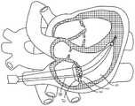
图1是心脏的剖视图,显示跨心脏三尖瓣定位的经导管的心房密封裙部系统。Figure 1 is a cross-sectional view of a heart showing a transcatheter atrial sealing skirt system positioned across the tricuspid valve of the heart.
图2是心脏的剖视图,显示跨心脏二尖瓣定位的经导管的心房密封裙部系统。2 is a cross-sectional view of a heart showing the transcatheter atrial sealing skirt system positioned across the mitral valve of the heart.
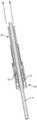
图3是一用于将系绳锚固至心脏壁的锚固件递送装置的递送缆线的立体图。3 is a perspective view of a delivery cable of an anchor delivery device for anchoring a tether to a heart wall.
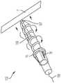
图4是一用于将系绳锚固至心脏壁的锚固件的立体图。Figure 4 is a perspective view of an anchor for anchoring the tether to the heart wall.
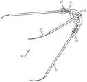
图5是一用于将心房密封裙部锚固至锚固件的系绳的立体图。5 is a perspective view of a tether used to anchor the atrial sealing skirt to the anchor.
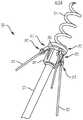
图6是包括系绳的锚固组件的立体图,系绳用于将心房密封裙部连接至锚固件,系绳与锚固件连接,锚固件用于将系绳锚固至心脏壁。6 is a perspective view of an anchor assembly including a tether for connecting the atrial sealing skirt to an anchor connected to the anchor for anchoring the tether to the heart wall.
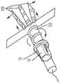
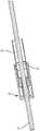
图7A至图7C是根据本发明另一个方面的具有分开的锚固螺钉的锚固件的立体图。7A-7C are perspective views of an anchor with split anchor screws according to another aspect of the invention.
图8A是锚固件递送装置的侧视图。8A is a side view of an anchor delivery device.
图8B是示出的锚固件递送装置在递送护套内的侧视图。8B is a side view of the anchor delivery device shown within the delivery sheath.
图8C是锚固件递送装置的端视图。Figure 8C is an end view of the anchor delivery device.
图9A是定位在右心室中的锚固件递送装置的透视图。9A is a perspective view of the anchor delivery device positioned in the right ventricle.
图9B是植入到心内壁中的锚固件的透视图。Figure 9B is a perspective view of an anchor implanted in the endocardial wall.
图10A和图10B显示锚固件递送被移除。Figures 10A and 10B show anchor delivery removed.
图11A是在植入的锚固件上的系绳递送组件的透视图。11A is a perspective view of a tether delivery assembly on an implanted anchor.
图11B和图11C是被移除的系绳递送组件的透视图。11B and 11C are perspective views of the tether delivery assembly removed.
图11D是绳索与缝线的融合的放大图。Figure 1 ID is an enlarged view of the fusion of the cord and suture.
图12A是密封裙部递送装置的透视图,其中心房密封裙部的递送系统位于右心室中。12A is a perspective view of the sealing skirt delivery device with the delivery system of the atrial sealing skirt positioned in the right ventricle.
图12B是心房密封裙部的递送装置的透视图,其中递送引导件被部分地抽出并且密封裙部膨胀。12B is a perspective view of the delivery device of the atrial sealing skirt with the delivery guide partially withdrawn and the sealing skirt inflated.
图12C是心房密封裙部的端视图。Figure 12C is an end view of the atrial sealing skirt.
图13A是通过心房定位杆将心房密封裙部定位在右心房底上的透视图。13A is a perspective view of the positioning of the atrial sealing skirt on the floor of the right atrium by the atrial positioning rod.
图13B是通过心房锁定件将心房密封裙部锁定在三尖瓣环中的位置中的立体图,其中定位杆被部分地抽出;13B is a perspective view of the atrial sealing skirt locked in position in the tricuspid annulus by the atrial lock with the retaining rod partially withdrawn;
图14A和图14B是心房密封裙部的侧视图和俯视透视图。14A and 14B are side and top perspective views of an atrial sealing skirt.
图15A是心脏的剖切透视图,显示被定位的心房密封裙部。Figure 15A is a cut away perspective view of a heart showing the atrial sealing skirt in position.
图15B是心脏的剖切透视图,显示定位在心房底上并与之相顺应的心房密封裙部。Figure 15B is a cutaway perspective view of the heart showing the atrial sealing skirt positioned on and conforming to the atrial floor.
图16A是心脏的剖切立体图,显示与心房底相顺应并围绕心内导线密封的心房密封裙部。Figure 16A is a cutaway perspective view of a heart showing the atrial sealing skirt conforming to the atrial floor and sealing around an intracardiac lead.
图16B是顺应并密封在心内导线周围的心房裙部的放大立体图。Figure 16B is an enlarged perspective view of the atrial skirt conforming and sealing around an intracardiac lead.
图17A是连接至心房定位杆和绳索的心房密封裙部的放大侧视图。Figure 17A is an enlarged side view of the atrial sealing skirt connected to the atrial positioning rod and tether.
图17B是锁定系统的放大侧视图。Figure 17B is an enlarged side view of the locking system.
图18A是锁定系统的立体图。Figure 18A is a perspective view of the locking system.
图18B是锁定系统的立体剖视图。Figure 18B is a perspective cross-sectional view of the locking system.
图19A是用于锁定在未锁定位置的心房密封裙部的锁定系统的截面侧视图。19A is a cross-sectional side view of a locking system for locking an atrial sealing skirt in an unlocked position.
图19B是锁定系统的截面侧视图,锁定系统用于将心房密封裙部定位在锁定位置中。19B is a cross-sectional side view of a locking system for positioning the atrial sealing skirt in a locked position.
图20A是处于锁定位置的锁定系统的局部剖视图。20A is a partial cross-sectional view of the locking system in the locked position.
图20B是锁定系统的立体图。Figure 20B is a perspective view of the locking system.
图21A是心脏的剖视图,其中心房裙部位于心脏内并且所有递送装置被移除。Figure 21A is a cross-sectional view of a heart with the atrial skirt inside the heart and all delivery devices removed.
图21B是心脏的剖视图,其中心房裙部位于心脏内并显示缝线切割器。Figure 21B is a cross-sectional view of the heart with the atrium skirt within the heart and showing the suture cutter.
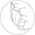
图22是密封裙部的侧视图,其中一边缘从凹形构造过渡到凸形构造。Figure 22 is a side view of a sealing skirt with one edge transitioning from a concave configuration to a convex configuration.
图23A和图23B是心室密封裙部的立体图和俯视图,其带有集成到裙部中的由瓣叶组成的瓣膜。23A and 23B are perspective and top views of a ventricular sealing skirt with a valve composed of leaflets integrated into the skirt.
图24A和图24B是用于接收瓣膜的心房密封裙部的立体图和俯视图。24A and 24B are perspective and top views of an atrial sealing skirt for receiving a valve.
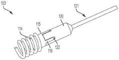
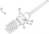
图25A至图25F是锚固件的立体图,所述锚固件具有锚固螺钉和锚固帽,锚固配置用以接收连接环和系绳系统,其按顺序步骤显示。25A-25F are perspective views of an anchor having an anchor screw and an anchor cap configured to receive a connecting ring and tether system, shown in sequential steps.
具体实施方式Detailed ways
通过参考以下详细描述、实施例和权利要求以及其先前和以下描述,可以更容易地理解本发明。在公开和描述本系统、装置和/或方法之前,应当理解,除非另外指明,否则本发明不限于所公开的特定系统、装置和/或方法,因此当然可以加以变化。还应理解,本文所使用的术语仅出于描述特定方面的目的,而无意于进行限制。The present invention can be understood more readily by reference to the following detailed description, examples and claims, together with previous and following descriptions thereof. Before the present systems, devices and/or methods are disclosed and described, it is to be understood that unless otherwise indicated, this invention is not limited to the particular systems, devices and/or methods disclosed as such may, of course, vary. It is also to be understood that terminology used herein is for the purpose of describing particular aspects only and is not intended to be limiting.
提供本发明的以下描述作为本发明当前已知最佳的方面的可行的教导。相关领域的技术人员将认识到,可以对所描述的方面进行许多改变,同时仍然获得本发明的有益结果。还将清楚的是,通过选择本发明的一些特征而不利用其他特征,可以获得本发明的一些期望的益处。因此本领域技术人员将认识到,对本发明的许多修改和调整是可能的,并且在某些情况下甚至可能是想要的,并且是本发明的一部分。因此提供以下描述作为对本发明原理的说明,而不是对本发明的限制。The following description of the invention is provided as a practical teaching of the best currently known aspects of the invention. Those skilled in the relevant art will recognize that many changes may be made to the aspects described while still obtaining the beneficial results of the invention. It will also be apparent that some of the desired benefits of the invention may be obtained by selecting some of the features of the invention without utilizing other features. Those skilled in the art will therefore recognize that many modifications and adaptations to the present invention are possible and can even be desirable in certain circumstances and are a part of the present invention. The following description is therefore provided as an illustration of the principles of the invention, not as a limitation of the invention.
如本文所用,单数形式的“一(a,an)”和“所述(the)”包括复数指示物,除非上下文另外明确指出。因此,例如,除非上下文另外明确指出,否则对“一系绳”的提及包括具有两条或更多条系绳的方案。As used herein, the singular forms "a, an" and "the" include plural referents unless the context clearly dictates otherwise. Thus, for example, reference to "a tether" includes reference to having two or more tethers unless the context clearly dictates otherwise.
本文的范围可以表示为从“大约(about)”一特定值和/或至“大约”另一特定值。当表达这样的范围时,另一方面包括从一特定值和/或至另一特定值。类似地,当将值表示为近似值时,使用先行词“大约(about)”,将理解特定值形成另一方面。将进一步理解,每个范围的端点相对于另一端点以及独立于另一端点都是显著的。Ranges can be expressed herein as from "about" one particular value, and/or to "about" another particular value. When expressing such a range, another aspect includes from the one particular value and/or to the other particular value. Similarly, when values are expressed as approximations, by use of the antecedent "about," it will be understood that the particular value forms another aspect. It will be further understood that the endpoints of each range are significant relative to the other endpoints as well as independently of the other endpoints.
如本文所使用的,术语“可选的”或“可选地”是指随后描述的情形或情况可能发生或可能不发生,并且所述描述包括所述情况或情形发生的情况和未发生的情况。如本文所用,“流体”是指自由流动的任何物质,包括液体、气体和等离子体。如本文所用,“流体连通”是指允许物质在相关部件之间自由流动的任何连接或相对定位。As used herein, the term "optional" or "optionally" means that the subsequently described circumstance or circumstance may or may not occur, and that the description includes instances where said circumstance or circumstance occurs and instances where it does not occur Condition. As used herein, "fluid" refers to any substance that flows freely, including liquids, gases, and plasmas. As used herein, "fluid communication" refers to any connection or relative positioning that permits the free flow of substances between related components.
本申请涉及微创地植入心脏中的医疗装置和系统,以及这些装置和系统的植入方法。更具体地,本申请涉及用于在血管内将一锚固件75引入和锚固到心脏壁上,并且将瓣膜100(参见图23以及图14A和图14B)植入心脏(所述瓣膜100被系到所述锚固件75)以取代原生瓣膜的装置、方法和系统。并且,一系绳组件与锚固件75配合,所述系绳组件将所述瓣膜100连接到锚固件75。此外,所述瓣膜100包括一密封裙部46,用于与所述瓣膜100配合以顺应相应的心房底,以防止假体的瓣周反流(paravalvular regurgitation ofprosthesis)。申请人的美国专利(申请号15/943,792)涉及一种心房密封裙部和锚固件,其相互系在一起并整体被植入。根据本文的公开内容,所述锚固件是独立于所述系绳和所述心房密封裙部地被植入的。然而应当理解,本申请和申请人先前提交的公开内容的各部件是可互换的。例如,申请人的先前公开的锚固件可以在锚固件的递送期间不带有连接的系绳地被植入。相反,本文描述的系绳系统也可以与其中公开的多个锚固件结合使用。The present application relates to medical devices and systems for minimally invasive implantation in the heart, and methods of implanting these devices and systems. More specifically, the present application relates to methods for intravascularly introducing and anchoring an
锚固组件:Anchor components :
图3至图6中所示的锚固组件101的各部件包括一锚固件75(所述锚固件75具有一锚固螺钉17)、一锚固帽16和允许递送一系绳18的一递送缆线12。所述锚固帽16连接到所述锚固螺钉17。所述递送缆线12可移除地连接到锚固帽16。如图所示,锚固螺钉17被尺寸化和构造为一螺旋螺钉,以固定到心内壁上。但是,可选地,锚固螺钉17的尺寸可以不同(取决于其所附着的心脏壁的长度,可以更长或更短),并且可以构造为一倾斜平面、钉状头或本领域技术人员已知的任何其他类型的螺钉。在一方面,所述螺钉由任何已知的金属合金组成,包括但不限于镍钛诺(nitinol)、钛(titanium)或钴铬(cobalt-chromium)。在另一方面,螺钉17的金属合金可以涂覆有生物组织,例如牛、羊、猪或马的心包,或涂覆有可能促进愈合并限制炎症的抗炎药的任何组合。锚固螺钉17的尖端76可选地由与锚固螺钉17相同或不同的材料构成和/或涂覆,并且可以制成钝的或锋利的尖端。The components of the
在使用中,通过旋转锚固螺钉17直到尖端76在心脏壁中处于期望的深度,以将锚固件75固定至心脏壁。锚固螺钉17拧入的深度可以根据心脏内的位置进行调节。例如,可以将锚固螺钉17更深地植入心室间隔(interventricular septim)中,以进行更大的固定,这与心室自由壁相反,即心外膜壁(epicardial wall),较浅地植入心室自由壁是较安全的。通过反转锚固螺钉17的旋转,将锚固件75安全地从心脏壁移出,或者重新定位,或者完全移出。In use, the
锚固帽16包括至少一从锚固帽16径向向外延伸的锁定臂78。锁定臂78被尺寸化和构造用于将系绳18的一部分(如下所述)可释放地固定到锚固帽16上。所述至少一锁定臂78在第一锁定位置和第二解锁位置之间移动,在第一锁定位置中锁定构件78远离锚固帽16的主体延伸一第一距离,在第二解锁位置中锁定构件78远离锚固帽16延伸一第二距离,所述第二距离小于所述第一距离。锚固帽16包括至少一偏压构件(未显示),例如弹簧,其配置用以将各锁定臂78推向第一锁定位置。如图所示,提供多个锁定臂78,多个锁定臂78绕着锚固帽16的圆周等距分布,然而也可以想到,多个锁定臂78不必等距分布。The
现在参考图3,递送缆线12包括柔性递送线材13,所述柔性递送线材13具有位于或形成在递送线材13的远端上的一远端螺纹端部14。所述递送线材由不锈钢、镍钛诺或其他金属合金构成,但不限于此,所述递送线材具有或不具有亲水涂层,或者具有或不具有聚合物涂层,例如聚四氟乙烯(polytetrafluoroethylene,PTFE)。所述远端螺纹端部14被尺寸化和构造为选择性地啮合一互补螺纹,所述互补螺纹形成在锚固帽16的近端77中的一腔室中。参见图4和图6,在使用中,远端螺纹端部14前进(例如拧进)所述锚固帽16的近端77,以将锚固帽16连接到柔性线材13的远端。如下面更充分地描述的,远端螺纹端部14从锚固件75的近端拧开,从而将柔性线材13从锚固件75分离。Referring now to FIG. 3 , delivery cable 12 includes a
根据本发明的另一方面,在图7A至图7C中显示扩张的锚固组件102。如图所示,所述锚固组件102是一心室间的,例如跨心室间隔(interventricular septum)的锚固件。锚固组件102包括锚固帽16和如上所述的用于与系绳18配合的锁定臂78。锚固组件102还包括一锚固轴105,锚固轴105具有配置用以穿透心内壁的一远侧尖端107。锚固轴105和锚固螺钉17至少由两个(如图所示由三个)轴和锚固区段108所组成。所述区段108在植入和心内壁穿透过程中通过一内部拉紧装置(例如拉紧线109)所固定,拉紧线109分为至少两条或所示的三条线109,终止于各区段108的远侧尖端107。一旦锚固轴105的远侧尖端107进入心内壁(例如心室间隔),内部拉紧线109释放并松弛,从而允许各轴区段108通过各内部偏压构件(未显示)的作用而分离。内部偏压构件例如为但不限于一个或多个弹簧,其沿着各轴区段108的一个或多个内壁而定位。According to another aspect of the present invention, an expanded
根据本发明的另一方面,如图25A至图25F所示,显示一锚固组件103。所述锚固组件103包括一锚固轴112和一锚固螺钉114。如图所示,锚固螺钉114具有螺旋构造并且从锚固螺钉基部115向远侧延伸。锚固螺钉基部115限定至少一个或多个(如图所示)锚固凸缘116,以及位于它们之间的凹入区域117。锚固轴112包括至少一个或多个(如图25B所示)锁定构件118。锁定构件118例如由弹簧(未显示)从锚固轴112径向向外偏压。一锚固件连接器120和连接器杆121与锚固轴112配合,以旋转锚固螺钉114。锚固件连接器120限定至少一个或多个(如图所示)孔122,所述孔122配置用以容纳锚固凸缘116。相应地,锚固件连接器120和连接器杆121与锚固轴112配合连接,从而向内推动锁定构件118。孔122和凸缘116的配合使锚固件连接器112和锚固螺钉基部115成为一体。连接器杆121的旋转从而使用于心室间或心外膜植入到心内壁的锚固螺钉114旋转。According to another aspect of the present invention, as shown in FIGS. 25A-25F , an anchoring
在植入锚固螺钉114之后,将系绳环125施加在连接器杆121和锚固件连接器120上,并邻接锚固螺钉114的近端。系绳环125包括大致圆柱形的第一远侧部分126和第二近端部分127,所述第二近端部分127的直径大于第一部分126的直径。第二部分127限定至少一个或多个(如图所示)孔129,孔129配置用以接收系绳杆130,如图25E和图25F所示。如图25D所示,锚固件连接器120和连接器杆121被移除。多个锁定构件18被径向向外推动,以与系绳环125的第二部分127接合,以将系绳环125锁定在锚固螺钉基部115上。系绳杆130如上所述地操作以与心房密封裙部46配合。After
系绳组件:Tether components :
当柔性线材13连接至锚固件75时,柔性线材用作为一导轨(guide rail)以将所述系绳18推进至锚固件75。系绳18包括一个或多个系绳杆19,系绳杆19可旋转地连接到对接环20。如图5所示,系绳杆19连接到由对接环臂71所限定的孔眼70。系绳18在递送缆线12的柔性线材13上前进,并且系绳18的对接环20将锚固帽16的至少一锁定臂78压到第二解锁位置。在锁定臂78处于第二位置的情况下,系绳18越过锚固帽16上的锁定臂78,直到对接环20抵靠和/或邻近锚固帽16的远端79。在此,锚固帽16的偏压构件将至少一锁定臂78推至第一锁定位置,从而将对接环20以及系绳18的其余部分可释放地连接到锚固件75。When the
在一方面,当系绳18连接至锚固件75时,系绳18围绕锚固件的纵轴旋转完整的360度。可选地,在另一方面,通过系绳18的一部分与至少一锁定臂78的相互作用,可以将系绳18限制在较小的旋转角度。In one aspect, when
如图6所示,在一方面,系绳18包括至少一与对接环20连接的对接环臂71,以及与对接环臂71连接的至少一系绳杆19。如图所示,对接环臂71的远端牢固地连接到对接环20或与其一体地形成。如图所示,至少一个对接环臂包括多个对接环臂71。如图所示,多个对接环臂78围绕对接环的圆周等距分布,但是可以设想多个对接环臂71不需要等间隔。对接环臂71限定一孔眼70。系绳杆19包括配置用以与孔眼70配合的一系绳杆钩件72。As shown in FIG. 6 , in one aspect, the
各对接环臂71的一近端可旋转地连接到相应的系绳杆19的远端。如图所示,系绳杆钩件72由系绳杆19所限定,并与各系绳杆19的远端连接或一体形成。在另一方面,孔眼70和系绳杆钩件72的尺寸和配置使得系绳杆钩件72被插入孔眼70中以将系绳杆19牢固地、可旋转地连接至对接环20。在使用中,每个系绳杆钩件72围绕孔眼70的圆周旋转。如图5所示,每一系绳杆的近端连接到一绳索21。系绳杆19和系绳杆钩件72可以由任何金属合金构成。A proximal end of each
系绳18配置用以与任何心内锚固件配合,心内锚固件包括但不限于本文公开的心室内(interventricular)和心外(epicardial)锚固件,以及本申请人通过引用并入本文的申请人先前公开的心室内和心外锚固件。
锚固件递送装置:Anchor Delivery Device :
现在参考图8A至图8C和图9A至图9B,显示用于将锚固帽16定位和配置在期望位置的锚固件递送装置23,锚固件递送装置23与图3至图6中所示的锚固组件101、以及图7A至图7C所示的锚固组件102的各部件有关。递送装置23包括一锚固件递送引导件25和一锚固件递送杆29。锚固件递送引导件具有远端28和内部的引导件管腔,所述内部的引导件管腔的大小和配置使得锚固件递送杆29的至少一部分穿过其中延伸。锚固件递送引导件25的至少一部分是柔性的,使得锚固件递送引导件25的远端28定位在心内壁7处或附近。Referring now to FIGS. 8A to 8C and FIGS. 9A to 9B , an
锚固件递送杆29配置用以将锚固螺钉17牢固地连附到心内壁7。锚固件递送杆29具有一远端31、一相对的近侧旋转手柄30以及在其之间延伸的一内部的杆管腔。所述内部的杆管腔的尺寸和配置使得递送缆线12的至少一部分从中延伸穿过。锚固件递送杆29的至少一部分是柔性的,使得锚固件递送杆29的远端处的一杆尖端31可以定位在心内壁7处或附近。The
锚固帽16的一部分(如图所示,靠近锚固帽远端79的部分)由锚固件杆尖端31接收并在其中延伸。锚固帽16的近侧部分的外部构造包括一第一表面构造,并且所述锚固件杆29的远侧部分的内壁构造具有一第二构造,其中第一构造和第二构造彼此相配合。因此当锚固帽16定位在锚固件杆尖端31中并与之接合时,锚固件递送杆29的旋转使锚固帽16旋转。在此位置,锚固螺钉17从锚固件递送杆29向远侧延伸,如图8B所示,递送缆线12延伸穿过锚固件递送杆29的内部的杆管腔。A portion of the anchor cap 16 (as shown, the portion near the anchor cap distal end 79 ) is received by and extends from the
锚固件递送装置23还包括引导件手柄26,所述引导件手柄26具有连接至锚固件递送引导件25的偏转旋钮27。引导件手柄26和偏转旋钮27被配置并用于帮助将锚固件递送引导件25的远端28引导至心内壁7。一杆手柄30连接至锚固件递送杆29,其中当锚固帽定位在锚固件杆尖端31中时,所述杆手柄的旋转使所述杆尖端31和锚固帽16旋转。The
如图8A所示,护套24配置用以接收锚固件递送引导件25。护套24与锚固件递送引导件流体连通,使得诸如肝素化生理盐水(heparinized saline)之类的流体通过护套24围绕锚固件递送引导件。一中央护套通道33(图9B)限定在护套24中,以提供与锚固件递送引导件25的内部的引导件管腔的连通,用于锚固件递送杆29和其他系统部件穿过中央护套通道33延伸。As shown in FIG. 8A ,
植入锚固件的方法:Methods of implanting anchors :
如图9A所示,在三尖瓣环(tricuspid annulus)中,例如J形线材34被用户在血管内引导至心内壁7。然后将锚固件递送装置23引导越过J形线材,直到锚固件递送引导件25的远端28位于心内壁7处或附近。图9至图11显示图3至图6的锚固组件10植入到一心内壁(intracardiac wall),即心外膜壁(epicardial wall)。锚固组件101也可以植入到一心室内壁(interventricular wall)中。J形线材是例如但不限于0.025”或0.035”J形线材。当然可以考虑具有其他直径的J形线材。锚固帽16连接至锚固件递送杆29的远端31。然后将锚固件递送杆29通过锚固件递送引导件25的内部的引导件管腔而插入,直到锚固帽16和所述向远侧延伸的锚固螺钉17位于心内壁7处或附近。As shown in FIG. 9A , in the tricuspid annulus, for example, a J-shaped
图7A至图7C的锚固组件102也可以通过J形线材34植入并引导以,例如进入心室内壁(intraventricular wall),如图所示的心内壁7。图25A至图25F的锚固组件103也可以被J形线材34植入并引导到心内壁7中,例如心室内壁或心外膜壁。The
当锚固件系统101、102或103的锚固螺钉17位于心内壁7附近时,锚固件递送杆29或121的旋转手柄30旋转,以引起所述锚固帽16的相应旋转,如图9B所示。例如旋转手柄30沿第一方向旋转以引起锚固帽16的相应旋转。连接至锚固帽16的锚固螺钉17也旋转并旋入心内壁的一部分,直到锚固帽16邻近心尖壁(apex wall)。应该注意,锚固螺钉17可能会或可能不会完全穿过任何心内壁而延伸,但不需要经心尖通路(trans-apical access)。在将锚固帽16放置在期望位置时,将锚固件递送杆29和锚固件递送引导件25从心脏2收回,如图10A所示。如图10B所示,在放置锚固帽16之后,递送缆线12的柔性线材13从锚固帽16延伸穿过三尖瓣环并穿过右心房3。When the
心房密封裙部:Atrial Sealing Skirt :
如图14A、图14B、图23A、图23B、图24A和图24B所示,系统1包括具有心房密封裙部46的心脏瓣膜100,心房密封裙部46具有一裙部上缘47,裙部上缘47沿着瓣膜100的上端周向地延伸。心房密封裙部46包括基本呈圆柱形的一心房裙部本体48以及构造成顺应心房底4(例如如图所示的右心房底)的所述心房裙部上缘47。如本文所述,心房密封裙部46通过系绳18连接至锚固件75。当锚固件75固定到心内壁7上时,绳索21(其以熔接或以其他方式连接到系绳18的系绳杆19)将瓣膜100连接到锚固件75。As shown in Figure 14A, Figure 14B, Figure 23A, Figure 23B, Figure 24A and Figure 24B, system 1 comprises the
如图1所示,所述经导管的心房密封裙部46的尺寸和配置适于坐落在右心房3和右心室6之间的三尖瓣(在所示的示例中)。密封裙部46可以与瓣膜瓣叶110预先组装在一起,作为一体的瓣膜100(图23A,图23B),或是密封裙部46可以构成为不带有瓣膜瓣叶,并用作为一用于一单独的经导管的瓣膜(图24A,图24B)的对接系统。这是示例性的。但是可选地,如图2所示,瓣膜的尺寸和配置略有不同,其可以定位在左心房8和左心室11之间的二尖瓣环中。因此,这些装置、系统、方法略有不同,可用于三尖瓣或二尖瓣,并可通过静脉结构在血管内地放置,静脉结构包括但不限于颈内静脉,锁骨下静脉、锁骨下静脉或股静脉。As shown in FIG. 1 , the transcatheter
心房密封裙部46是自膨胀的(即裙部是可压缩的,以使其适合通过系统1的导管)并由镍钛合金组成,但也可以包含,但不限于,不锈钢、镍钛合金或其他金属合金制成的元件。在另一方面,心房密封裙部的下直径小于或近似等于配置位置5处的环(三尖瓣环)或配置位置10处的环(二尖瓣环)的直径,从而防止或减少与脆弱的三尖瓣环的并置(apposition),并防止或减少二尖瓣环的约束。The
如图12C、图14A和图14B,图23A和图23B以及图24A和图24B所示,在心房密封裙部46的外壁中限定至少一管体53。各管体的尺寸和形状使得绳索21的一部分(如图12A和图12B所示,在近端连接到缝线45)延伸穿过管体53,从而将系绳18连接到心房密封裙部46,允许自由移动,直到裙部46被锁定就位。在另一方面,心房密封裙部46具有沿其外径定位的锚固元件(未显示)。这些锚固元件允许固定到三尖瓣或二尖瓣环和/或瓣叶上,但是不一定用于主要的固定机构。As shown in FIGS. 12C , 14A and 14B , 23A and 23B , and 24A and 24B , at least one
至少一绳索21连接到系绳18的系绳杆19,并且绳索21的近端部分连接到缝线45。在一方面,所述绳索可以是结实而柔韧的绳索例如,但不限于,膨体聚四氟乙烯(expandedpolytetrafluoroethylene,ePTFE)或超高分子量聚乙烯(ultra- high-molecular-weightpolyethylene,(UHMWPE, UHMW)绳索。在使用中,以下将更全面地描述,绳索21的中央部分(在远端和近端之间)延伸穿过和/或连接到心房密封裙部46,以将所述裙部相对于三尖瓣环或二尖瓣环保持在期望的位置。At least one
图23A和图23B还显示心房密封裙部46。密封裙部46是一体的瓣膜100,由从密封裙部本体48径向向内延伸的瓣叶110组成。瓣叶110由牛、马或猪心包瓣叶所组成。心房密封裙部46可以用作为任何常规瓣膜的对接系统,或者可以预先组装成包括由瓣叶110组成的瓣膜100。如果心房密封裙部46包含瓣膜100,而所述瓣膜100由缝到密封裙部本体48的内部的多个瓣叶110所组成,这种构造将起到任何常规瓣膜的作用,瓣叶110在舒张期(心脏松弛)期间打开,从而允许血液从右心房3进入右心室6,或从左心房8进入左心室11,并在收缩期关闭(心脏收缩),从而防止血液分别从右心室或左心室回流到右心房或左心房。23A and 23B also show the
如图14A和图14B、图23A和图23B以及图24A和图24B所示,由心房裙部本体48和心房裙部上缘47所限定的心房密封裙部46包括膜状材料,所述密封裙部46的直径大于所述配置位置处的环的直径。例如,心房密封裙部46的裙部直径可以大于三尖瓣环或二尖瓣环的直径。在另一方面,心房裙由但不限于选自以下的合成材料形成:聚碳酸酯(polycarbonate)、聚氨酯(polyurethane)、聚酯(polyester)、膨体聚四氟乙烯(expandedpolytetrafluoroethylene,ePTFE)、聚对苯二甲酸乙二醇酯(PET)、硅树脂(silicone)、天然或合成橡胶或它们的组合。心房裙部46还可以覆盖有成年或幼年的牛、羊、马或猪心包。可选地,心房密封裙部46的至少一部分可以由替代材料形成,例如但不限于聚氨酯泡沫(polyurethane foam)或其他聚合物。As shown in Figures 14A and 14B, Figures 23A and 23B, and Figures 24A and 24B, the
在另一方面,心房密封裙部46的至少一部分沿其长度具有一个或多个固定构件(未示出),从而允许进一步锚固到右心房底和/或三尖瓣环的心房侧的其他部分,防止心房密封裙部46迁移到近侧的右心房3中,从而防止假体的不稳定(例如摇摆)和瓣周反流(paravalvular regurgitation)。可选地,通过稍作修改,这些固定构件允许将心房密封裙部46进一步锚固到左心房底和/或二尖瓣环的心房侧的部分,从而防止心房密封裙部46迁移到近侧的左心房8,也防止假体的不稳定(例如摇摆)和瓣周反流。In another aspect, at least a portion of the
心房密封裙部46至少包括心房裙部本体48和心房裙部上缘47。如图所示,心房裙部本体48是圆柱状,并且具有可变的长度和直径,其选择性地由激光切割或模制的镍钛合金组成,但也可以包含任何其他金属合金元素,并且可以沿其圆周或长度的任何部分覆盖有上述生物膜材料或合成材料。如图所示,上缘47从裙部本体48径向向外延伸并向下延伸,从而形成大致上凹陷的上缘,该上缘具有面向右心房底4或左心房底10的凹陷部。缘部47围绕裙部本体48的上端周向地延伸。The
提供至少一个或所显示的多个柔性延伸构件49,并且所述柔性延伸构件49例如可以由但不限于激光切割或模制的镍钛合金制成,所述柔性延伸构件49通过延伸构件基部50连附到裙部本体的顶部并终止于延伸构件尖端51。一弹性密封膜52在一个或多个延伸构件49之间垂直于相邻延伸构件49而延伸。在图14A和图14B中,延伸构件49可以径向向外并且大体上线性地延伸,但这是示例性的。如图23A、图23B、图24A和图24B所示,延伸构件可以是非线性的并且大致为U形。如图所示,密封膜52围绕裙部缘部47周向地延伸,其也可以仅延伸圆周的一部分。密封裙部本体48包括多个支撑件114,其类似于上缘47的延伸构件49,例如可以由(但不限于)激光切割或模制的镍钛诺构成。如图所示,支撑件114形成格子状构造,但是可以构想其他构造,包括但不限于垂直延伸的支撑件。At least one, or as shown, a plurality of
如上所述,密封构件52由生物组织或合成纤维织物构成。一方面,合成纤维织物是编织或针织的(braided or knit),从而允许顺应心房底形状所需的“可拉伸性”,包括覆盖和密封心内导线(例如永久性起搏器导线(pacemaker leads)66)的能力,如图16A和图16B所示。如图16A所示,根据一方面,心房裙部上缘47顺应右心房底4并在心内导线66周围密封。在图16B中,延伸构件49经由延伸构件基部50连附到心房裙部本体48,并且延伸构件尖端51向下弯曲,从而允许弹性密封膜52缠绕在心内导线66的顶部周围,从而防止导线周围的反流。这种构造需要通过一个或多个心房定位杆44所施加的向下的作用力,所述心房定位杆44连附到一个或多个管体53,并通过一个或多个可拆卸的锁定件56(图17B)而被锁定就位,所述可拆卸的锁定件56集成在如本文所述的管体53的心房端的内部。As described above, the sealing
系绳和心房密封裙部的递送组件:Delivery Components of Tether and Atrial Seal Skirt :
根据上述方法,锚固件75由锚固件递送装置23引入并固定到心内壁,并且包括锚固螺钉17的锚固件75已植入心内壁。锚固帽16和递送缆线13保留在心脏内,并准备接收上述的系绳18。According to the method described above, the
现在参考图11A、图11B、图11C和图11D,并且如上所述,系绳18通过如图所示的护套137形式的心房裙部的递送系统,在递送缆线12的柔性线材13上前进。通过将对接环20连接到锚固帽16,从而将系绳18锁定到锚固帽16上。从系绳杆19延伸的至少一绳索21连接到至少一系绳杆19上。至少一绳索21在近侧连接到至少一缝线45,缝线45通过递送护套137的中央管腔33在体外延伸。一旦系绳被锁定到锚固帽16,则如图11C所示,取回护套137,使植入的锚固件16、系绳18、绳索21和缝线从植入位置延伸。在图11C所示的方面,提供一系绳递送护套137作为第二递送引导件,并且与心房密封裙部的递送引导件护套38有所不同,所述心房密封裙部的递送引导件护套38为第三递送引导件,如下面所描述的。因此移除系绳护套137,并且使用心房密封裙部的递送引导件护套38。然而这可以在单一步骤中实现,如图12A至图12B所示,其中用相同的护套38递送系绳18和心房密封裙部46并构成第二递送步骤。Referring now to FIGS. 11A, 11B, 11C and 11D, and as described above, the
现在参考图12A和图12B,显示用于将心房密封裙部46定位和配置在期望的配置位置5或10处的心房密封裙部的递送系统37。心房密封裙部的递送系统37包括心房密封裙部的递送引导件38、鼻锥43、心房密封裙部的配置旋钮39和至少一心房定位杆44。心房密封裙部的递送引导件38具有一远端41、一相对的近侧的心房密封裙部的配置旋钮39、以及在其间延伸的一内部的引导件管腔40。所述内部的引导件管腔40的尺寸和配置适于使得心房密封裙部46和其他系统部件被选择性地且可移除地插入其中。心房密封裙部的递送引导件38的至少一部分是柔性的,从而使锚固件递送引导件25远端的尖端41越过所述配置位置5并进入右心室6。可替代地,定位远侧的尖端41以越过配置位置10并进入左心室11。Referring now to FIGS. 12A and 12B , there is shown an atrial sealing
心房密封裙部的配置旋钮39连接到心房密封裙部的递送引导件38的近端。心房密封裙部的配置旋钮限定了与内部的引导件管腔40流体连通的中央通道60。因此,心房定位杆44、引导线材13和/或至少一缝线45可以延伸穿过中央通道60进入内部的引导件管腔40。如图所示,心房密封裙部的配置旋钮39可旋转并配置为使得旋钮39沿第一方向的旋转引起在心房密封裙部46附近的心房密封裙部的递送引导件38的远侧的尖端41被取回,从而使心房密封裙部46膨胀。鼻锥43可以是任何常规鼻锥,其与心房密封裙部的递送引导件38相连,并配置用以将心房密封裙部46引导至所述配置位置5。The atrial sealing
锁定系统:Locking system :
参考图13A和图13B,至少一心房定位杆44具有远端54、近端61和在其间延伸的内部的杆管腔62,内部的杆管腔的尺寸和配置使得缝线45和/或绳索21的一部分插入其中。心房定位杆44的至少一部分是柔性的,使得心房定位杆的远端54可以定位在配置位置5处或附近。13A and 13B, at least one
至少一定位杆44连接至管体53。如图13A和图13B所示,每一管体53包含一可拆卸的锁定件56(图17A和图17B),其配置用以牢固地连附至少一绳索21。因此所述绳索21被牢固地连附到系绳18的系绳杆19,系绳杆19被连接到锚固帽16,锚固帽16通过锚固螺钉17被固定到心内壁7,并且例如可拆卸的锁定件56牢固地将绳索21附接在右心房中。At least one
参照图17A、图17B、图18A、图18B、图19A和图19B,锁定系统55包括可拆卸的锁定件56,锁定件56集成在管体53内,管体53连附到第一通道海波管57(gateway hypotube)和第二收回的海波管58(retracting hypotube)。在可拆卸的锁定件56的内部是锁定夹59。现在参照图21B,系统1还包括一缝线切割器65,缝线切割器65的尺寸和配置适于穿过递送护套24以切割至少一缝线45(如图21B所示)。Referring to Fig. 17A, Fig. 17B, Fig. 18A, Fig. 18B, Fig. 19A and Fig. 19B, the locking
植入、定位和锁定心房裙部的方法:Methods of implanting, positioning and locking the atrial skirt :
在使用中,系统1通过放置右或左心室锚固件75并将系绳18对接至锚固件75,利用经导管的方法植入心房密封裙部46(具有集成的瓣膜)。如图12A所示,将心房密封裙部的递送系统37放置在递送缆线12的柔性线材13上并插入心脏2的一部分。当将心房密封裙部的递送引导件38以及预加载到其远端41的心房密封裙部46和集成的瓣膜100插入心脏时,至少缝线45的一部分穿过心房密封裙部46的壁部所限定的至少一管体53中,如图12B和图12C所示,并且随着心房密封裙部的递送引导件38的前进,缝线45和绳索21的至少一部分沿着心房密封裙部的递送引导件38的内部的引导件管腔40并向近侧延伸超出内部的引导件管腔40。因此至少一绳索21的一部分延伸穿过并超过心房密封裙部的递送引导件38的远端41,并且至少一缝线45的一部分延伸穿过并超过心房密封裙部的递送引导件38。定位心房密封裙部的递送引导件38,使得心房密封裙部的递送引导件38以及预加载到其远端41的心房密封裙部46和集成的瓣膜100经过配置位置5并进入右心室6。In use, the system 1 implants the atrial sealing skirt 46 (with integrated valve) using a transcatheter approach by placing a right or
心房密封裙部46和瓣膜100预加载到心房密封裙部的递送引导件38的远端41中,以定位在配置位置5上。如图所示,缝线45与瓣膜100预组装,使得各缝线45穿过心房密封裙部46的壁部所限定的至少一管体53中,如图12B和图12C所示。当心房密封裙部46和心房密封裙部的递送引导件38的远端41作为一单元前进并接近配置位置时,缝线45的端部和绳索21的一部分将变成穿过心房密封裙部46中所限定的管体53。这样,心房密封裙部46可沿着至少一绳索的长度移动,直到达到期望的配置位置5。即心房密封裙部自由地浮动在绳索21上,直到被可拆卸的锁定件56锁定到位。The
当心房密封裙部46处于期望的配置位置5时,心房密封裙部的配置旋钮39用于至少部分地收回围绕心房密封裙部46的递送引导件38。在引导件38没有密封裙部46的情况下,裙部46膨胀至其完整的、不受约束的尺寸。可选地,因为心房密封裙部的位置是可调节的,所以心房密封裙部的配置旋钮39用于在期望的配置位置附近膨胀密封裙部46。When the
然后将心房定位杆44插在各缝线45上,使得各缝线45的一部分在内部的杆管腔62内延伸,并且各缝线的一部分延伸超出定位杆44的近端61。参照图13A和图13B,然后将定位杆44插入通过心房密封裙部的引导件38,并且绳索21的一部分被杆44的内部的杆管腔62所接收,并且定位杆的远端54(具有连附于其上的可拆卸的锁定件56)与心房密封裙部46相邻。用户将多个定位杆44向下推,直到密封裙部相对于三尖瓣环处于一期望位置。
心房密封裙部46的位置不需要通过心室尖心脏(ventricular apex heart)2牵拉系绳18,因为心房密封裙部46在系绳18上自由移动,直到达到所期望的裙部46位置为止。在达到想要的瓣膜位置之后,至少一心房定位杆44将心房密封裙部46推入就位,并通过嵌套在各管体53内并连接到各定位杆44端部的可拆卸的锁定件56而被锁定就位。心房密封裙部46可以重新定位或收回,直到释放延伸穿过各心房定位杆44的缝线45。The position of the
如图15A和图15B所示,心房密封裙部46在右心房3内的定位使得心房裙部上缘47顺应右心房底4的形状。经由心房密封裙部的递送系统端部41,医师使一个或多个心房定位杆44前进,以使心房密封裙部46在一个或多个绳索21上平移,所述一个或多个绳索21延伸穿过心房裙部本体48所限定的一个或多个管体53。如图15B所示,随着心房密封裙部46向右心室6前进,心房裙部上缘47接触心房底4,并且一个或多个延伸构件49根据局部解剖结构不同地挠曲。由于各心房定位杆44通过不同的力推动,因此可以获得精确的张紧量,并且因此延伸构件49或多或少地弯曲以利于心房裙部上缘47顺应围绕心房底4的整个圆周,从而限制通过三尖瓣口的反流。As shown in FIGS. 15A and 15B , the positioning of the
图22显示心房裙部上缘47从凹面到凸面的转换。当瓣膜通过附在延伸构件49的延伸构件基部50上的心房定位杆44(图15A和图15B)被向下推到心房底4时,延伸构件尖端51向上弯曲,以顺应心房底解剖结构。瓣膜100的进一步向远侧的运动(在图22中从左到右显示)进一步改变了密封裙部46的形状,因为密封裙部46顺应心房底,并且延伸构件基部50被心房定位杆44向下推动(图13A、图15A和图15B)。仅作为示例,如上所述,对心房密封裙部46的定位和顺应的描述是指将心房密封裙部46定位在左心房底9上,从而限制通过二尖瓣口10的反流。Figure 22 shows the transition of the
现在参考图19A和图19B,拉动所述收回的海波管58导致锁定夹59收回,所述锁定夹59向下推压锁定舌片63,以与绳索21接合。更具体地,第二海波管58被收回,并且由于其与锁定夹59的连接,其也使锁定夹59收回。锁定夹59在收回时接触第一通道海波管57的接触点64,断开锁定夹59以允许第二海波管58被移除。一旦收回的海波管58被拉动,通道海波管57的内臂向内弹起,从而允许通道海波管被移除。第一通道海波管57是有益的,因为它使得在第二海波管58被收回时能够锁定绳索21。然后移除通道海波管57,将锁定夹59留在心房密封裙部46的管体53内。图20A显示一完全接合的锁的剖视图。根据一方面,定位杆44可与通道海波管集成在一起或可移除地连接至通道海波管。图20B显示一完全接合的锁的一完整视图。应当理解,可以采用本申请人的美国专利(申请号15/943,792)的锁定系统代替在此描述的锁定系统。在不脱离本发明的精神和范围的情况下,所述锁定系统可以在任一系统中使用。Referring now to FIGS. 19A and 19B , pulling the retracted hypotube 58 causes retraction of the locking
如图21A和图21B所示,在心房密封裙部牢固地顺应心房底4的情况下,缝线切割器65在缝线上移动45,然后到心房裙部上缘47。缝线切割器65在可拆卸的锁定件56上方切割并释放各缝线45的远端。然后将缝线45和缝线切割器65从心脏2移除。With the atrial sealing skirt firmly conforming to the atrial floor 4, the suture cutter 65 is moved 45 over the suture and then to the
一方面,在切断缝线45之前,心房密封裙部46可以被取回或重新定位。例如,如果确定要移除或重新定位心房密封裙部,则将一心房定位杆44定位在各缝线上,以使缝线的一部分位于内部的杆管腔62中。当定位杆的远端54与可拆卸的锁定件56相邻或与之接触,使通道海波管57前进,而所述收回的海波管58将可拆卸的锁定件连附到定位杆的远端,从而解除绳索21的锁定。随着各绳索的解锁,可以将瓣膜从配置位置5中移除和/或重新定位在配置位置5中。In one aspect, the
在另一方面,可在瓣膜配置后数天至数周将心房密封裙部46重新定位和/或移除。在这方面,缝线不被切割,而是缠绕在一线轴或其他缠绕装置上。然后将所述装置连接到心房裙部上缘47上的瓣膜。在配置瓣膜并完成手术几天后,可以重新捕获线轴/缠绕装置,从而可以解开并取回缝线。然后将心房定位杆44定位在各缝线上,以使缝线的一部分位于内部的杆管腔62内。当定位杆的远端54与可拆卸的锁定件56相邻或与其接触时,使通道海波管57前进,而所述收回的海波管58将可拆卸的锁定件连附到定位杆的远端,从而解除绳索21的锁定。随着各绳索的解锁,可以将瓣膜从配置位置5中移除和/或重新定位在配置位置5中。In another aspect, the
尽管在以上说明书中公开了本发明的几个方面,但是本领域技术人员可以理解,受益于前述说明书和相关附图中呈现的教示,许多修改和其他方面都包括在本发明中。因此应当理解,本发明不限于以上公开的特定方面,并且许多修改和其他方面旨在被包括在所附权利要求的范围内。此外,尽管本文以及随后的权利要求书中采用了特定术语,但是它们仅以一般性和描述性意义使用,而不是为了限制所描述的发明。Although several aspects of the invention have been disclosed in the foregoing specification, those skilled in the art will appreciate that many modifications and other aspects are encompassed in the present invention having the benefit of the teachings presented in the foregoing specification and the associated drawings. It is therefore to be understood that the inventions are not to be limited to the particular aspects disclosed above and that modifications and other aspects are intended to be included within the scope of the appended claims. Furthermore, although specific terms are employed herein and in the claims that follow, they are used in a generic and descriptive sense only and not for limitation of the invention as described.
Claims (34)
Priority Applications (4)
| Application Number | Priority Date | Filing Date | Title |
|---|---|---|---|
| CN202310004290.6ACN116059008A (en) | 2017-04-05 | 2018-04-04 | Atrial sealing skirt |
| CN202310004287.4ACN116059007A (en) | 2017-04-05 | 2018-04-04 | Intracardiac anchor assembly |
| CN202310004292.5ACN116059009A (en) | 2017-04-05 | 2018-04-04 | Intracardiac anchor assembly and epicardial anchor assembly |
| CN202310004288.9ACN116077238A (en) | 2017-04-05 | 2018-04-04 | Medical assembly |
Applications Claiming Priority (9)
| Application Number | Priority Date | Filing Date | Title |
|---|---|---|---|
| US201762481846P | 2017-04-05 | 2017-04-05 | |
| US62/481,846 | 2017-04-05 | ||
| US201762509587P | 2017-05-22 | 2017-05-22 | |
| US62/509,587 | 2017-05-22 | ||
| US201762558315P | 2017-09-13 | 2017-09-13 | |
| US62/558,315 | 2017-09-13 | ||
| US15/943,792 | 2018-04-03 | ||
| US15/943,792US10820991B2 (en) | 2017-04-05 | 2018-04-03 | Transcatheter atrial sealing skirt, anchor, and tether and methods of implantation |
| PCT/US2018/026118WO2018187495A1 (en) | 2017-04-05 | 2018-04-04 | Transcatherer atrial sealing skirt, anchor, and tether and methods of implantation |
Related Child Applications (4)
| Application Number | Title | Priority Date | Filing Date |
|---|---|---|---|
| CN202310004292.5ADivisionCN116059009A (en) | 2017-04-05 | 2018-04-04 | Intracardiac anchor assembly and epicardial anchor assembly |
| CN202310004288.9ADivisionCN116077238A (en) | 2017-04-05 | 2018-04-04 | Medical assembly |
| CN202310004290.6ADivisionCN116059008A (en) | 2017-04-05 | 2018-04-04 | Atrial sealing skirt |
| CN202310004287.4ADivisionCN116059007A (en) | 2017-04-05 | 2018-04-04 | Intracardiac anchor assembly |
Publications (2)
| Publication Number | Publication Date |
|---|---|
| CN110799102A CN110799102A (en) | 2020-02-14 |
| CN110799102Btrue CN110799102B (en) | 2023-01-20 |
Family
ID=63710530
Family Applications (7)
| Application Number | Title | Priority Date | Filing Date |
|---|---|---|---|
| CN202310004269.6APendingCN116115390A (en) | 2017-04-05 | 2018-04-03 | Medical assembly |
| CN201880034100.2AActiveCN110730633B (en) | 2017-04-05 | 2018-04-03 | Transcatheter valve, anchor, tether implant assembly |
| CN202310004290.6APendingCN116059008A (en) | 2017-04-05 | 2018-04-04 | Atrial sealing skirt |
| CN201880033804.8AActiveCN110799102B (en) | 2017-04-05 | 2018-04-04 | Transcatheter atrial sealing skirt, anchor, tether implant assembly |
| CN202310004288.9APendingCN116077238A (en) | 2017-04-05 | 2018-04-04 | Medical assembly |
| CN202310004287.4APendingCN116059007A (en) | 2017-04-05 | 2018-04-04 | Intracardiac anchor assembly |
| CN202310004292.5APendingCN116059009A (en) | 2017-04-05 | 2018-04-04 | Intracardiac anchor assembly and epicardial anchor assembly |
Family Applications Before (3)
| Application Number | Title | Priority Date | Filing Date |
|---|---|---|---|
| CN202310004269.6APendingCN116115390A (en) | 2017-04-05 | 2018-04-03 | Medical assembly |
| CN201880034100.2AActiveCN110730633B (en) | 2017-04-05 | 2018-04-03 | Transcatheter valve, anchor, tether implant assembly |
| CN202310004290.6APendingCN116059008A (en) | 2017-04-05 | 2018-04-04 | Atrial sealing skirt |
Family Applications After (3)
| Application Number | Title | Priority Date | Filing Date |
|---|---|---|---|
| CN202310004288.9APendingCN116077238A (en) | 2017-04-05 | 2018-04-04 | Medical assembly |
| CN202310004287.4APendingCN116059007A (en) | 2017-04-05 | 2018-04-04 | Intracardiac anchor assembly |
| CN202310004292.5APendingCN116059009A (en) | 2017-04-05 | 2018-04-04 | Intracardiac anchor assembly and epicardial anchor assembly |
Country Status (13)
| Country | Link |
|---|---|
| US (4) | US10820991B2 (en) |
| EP (4) | EP3606444B1 (en) |
| JP (6) | JP7179012B2 (en) |
| KR (4) | KR102339027B1 (en) |
| CN (7) | CN116115390A (en) |
| AU (7) | AU2018248410B2 (en) |
| BR (4) | BR122020023074B1 (en) |
| CA (3) | CA3059102C (en) |
| IL (3) | IL297339A (en) |
| SG (2) | SG11201909246YA (en) |
| UY (2) | UY37667A (en) |
| WO (2) | WO2018187390A1 (en) |
| ZA (2) | ZA201907132B (en) |
Families Citing this family (66)
| Publication number | Priority date | Publication date | Assignee | Title |
|---|---|---|---|---|
| US20090276040A1 (en)* | 2008-05-01 | 2009-11-05 | Edwards Lifesciences Corporation | Device and method for replacing mitral valve |
| US9554897B2 (en) | 2011-04-28 | 2017-01-31 | Neovasc Tiara Inc. | Methods and apparatus for engaging a valve prosthesis with tissue |
| US9308087B2 (en) | 2011-04-28 | 2016-04-12 | Neovasc Tiara Inc. | Sequentially deployed transcatheter mitral valve prosthesis |
| CA3007660A1 (en) | 2015-12-15 | 2017-06-22 | Neovasc Tiara Inc. | Transseptal delivery system |
| US11833034B2 (en) | 2016-01-13 | 2023-12-05 | Shifamed Holdings, Llc | Prosthetic cardiac valve devices, systems, and methods |
| US10433952B2 (en) | 2016-01-29 | 2019-10-08 | Neovasc Tiara Inc. | Prosthetic valve for avoiding obstruction of outflow |
| CA3042588A1 (en) | 2016-11-21 | 2018-05-24 | Neovasc Tiara Inc. | Methods and systems for rapid retraction of a transcatheter heart valve delivery system |
| CN110337280A (en)* | 2017-02-17 | 2019-10-15 | 株式会社田端心脏医学研究 | Artificial heart valve |
| DE102017002974B4 (en)* | 2017-03-28 | 2024-08-08 | Immanuel Albertinen Diakonie Ggmbh | Heart valve implant, suitable for use in minimally invasive surgery to repair a heart valve and/or a heart valve leaflet on the beating heart and heart valve implant system |
| US11337685B2 (en) | 2017-04-05 | 2022-05-24 | Opus Medical Therapies, LLC | Transcatheter anchoring assembly for a mitral valve, a mitral valve, and related methods |
| US10716668B2 (en)* | 2017-04-05 | 2020-07-21 | Medtronic, Inc. | Delivery system with anchoring nosecone and method of delivery |
| US11103351B2 (en) | 2017-04-05 | 2021-08-31 | Opus Medical Therapies, LLC | Transcatheter atrial sealing skirt and related method |
| US11123187B2 (en) | 2017-04-05 | 2021-09-21 | Opus Medical Therapies, LLC | Transcatheter atrial anchors and methods of implantation |
| CN116115390A (en)* | 2017-04-05 | 2023-05-16 | 欧普斯医疗疗法有限公司 | Medical assembly |
| US10820992B2 (en)* | 2017-04-05 | 2020-11-03 | Opus Medical Therapies, LLC | Transcatheter atrial sealing skirt, anchor, and tether and methods of implantation |
| CA3073834A1 (en) | 2017-08-25 | 2019-02-28 | Neovasc Tiara Inc. | Sequentially deployed transcatheter mitral valve prosthesis |
| WO2019195860A2 (en) | 2018-04-04 | 2019-10-10 | Vdyne, Llc | Devices and methods for anchoring transcatheter heart valve |
| US12121775B2 (en) | 2018-06-27 | 2024-10-22 | Dhiren Rajagopal | Brace for preventing finger injuries |
| AU2019325548B2 (en) | 2018-08-21 | 2025-06-26 | Shifamed Holdings, Llc | Prosthetic cardiac valve devices, systems, and methods |
| US10595994B1 (en) | 2018-09-20 | 2020-03-24 | Vdyne, Llc | Side-delivered transcatheter heart valve replacement |
| US11278437B2 (en) | 2018-12-08 | 2022-03-22 | Vdyne, Inc. | Compression capable annular frames for side delivery of transcatheter heart valve replacement |
| US11344413B2 (en) | 2018-09-20 | 2022-05-31 | Vdyne, Inc. | Transcatheter deliverable prosthetic heart valves and methods of delivery |
| US11071627B2 (en) | 2018-10-18 | 2021-07-27 | Vdyne, Inc. | Orthogonally delivered transcatheter heart valve frame for valve in valve prosthesis |
| US12186187B2 (en) | 2018-09-20 | 2025-01-07 | Vdyne, Inc. | Transcatheter deliverable prosthetic heart valves and methods of delivery |
| US10321995B1 (en) | 2018-09-20 | 2019-06-18 | Vdyne, Llc | Orthogonally delivered transcatheter heart valve replacement |
| CN113260337A (en) | 2018-10-05 | 2021-08-13 | 施菲姆德控股有限责任公司 | Prosthetic heart valve devices, systems, and methods |
| CN113056302B (en) | 2018-10-19 | 2023-03-28 | 施菲姆德控股有限责任公司 | Adjustable medical device |
| US11109969B2 (en) | 2018-10-22 | 2021-09-07 | Vdyne, Inc. | Guidewire delivery of transcatheter heart valve |
| CN113271890B (en) | 2018-11-08 | 2024-08-30 | 内奥瓦斯克迪亚拉公司 | Ventricular deployment of transcatheter mitral valve prosthesis |
| US11253359B2 (en) | 2018-12-20 | 2022-02-22 | Vdyne, Inc. | Proximal tab for side-delivered transcatheter heart valves and methods of delivery |
| WO2020146842A1 (en) | 2019-01-10 | 2020-07-16 | Vdyne, Llc | Anchor hook for side-delivery transcatheter heart valve prosthesis |
| JP2022523456A (en)* | 2019-01-14 | 2022-04-25 | エムテックス カーディオ アーゲー | Heart valve apex coapter |
| US11185409B2 (en) | 2019-01-26 | 2021-11-30 | Vdyne, Inc. | Collapsible inner flow control component for side-delivered transcatheter heart valve prosthesis |
| US11273032B2 (en) | 2019-01-26 | 2022-03-15 | Vdyne, Inc. | Collapsible inner flow control component for side-deliverable transcatheter heart valve prosthesis |
| WO2020154797A1 (en)* | 2019-01-28 | 2020-08-06 | Vesalius Cardiovascular Inc. | Apparatus for use in repairing mitral valves and method of use thereof |
| WO2020181154A2 (en) | 2019-03-05 | 2020-09-10 | Vdyne, Inc. | Tricuspid regurgitation control devices for orthogonal transcatheter heart valve prosthesis |
| CA3132873A1 (en) | 2019-03-08 | 2020-09-17 | Neovasc Tiara Inc. | Retrievable prosthesis delivery system |
| US11173027B2 (en) | 2019-03-14 | 2021-11-16 | Vdyne, Inc. | Side-deliverable transcatheter prosthetic valves and methods for delivering and anchoring the same |
| US11076956B2 (en) | 2019-03-14 | 2021-08-03 | Vdyne, Inc. | Proximal, distal, and anterior anchoring tabs for side-delivered transcatheter mitral valve prosthesis |
| EP3941391B1 (en) | 2019-03-19 | 2024-12-04 | Shifamed Holdings, LLC | Prosthetic cardiac valve devices, systems |
| US11491006B2 (en) | 2019-04-10 | 2022-11-08 | Neovasc Tiara Inc. | Prosthetic valve with natural blood flow |
| CA3138875A1 (en) | 2019-05-04 | 2020-11-12 | Vdyne, Inc. | Cinch device and method for deployment of a side-delivered prosthetic heart valve in a native annulus |
| US11779742B2 (en) | 2019-05-20 | 2023-10-10 | Neovasc Tiara Inc. | Introducer with hemostasis mechanism |
| JP7520897B2 (en) | 2019-06-20 | 2024-07-23 | ニオバスク ティアラ インコーポレイテッド | Thin prosthetic mitral valve |
| EP4480458A3 (en) | 2019-08-20 | 2025-04-09 | Vdyne, Inc. | Delivery devices for side-deliverable transcatheter prosthetic valves |
| CN120531525A (en) | 2019-08-26 | 2025-08-26 | 维迪内股份有限公司 | Laterally deliverable transcatheter prosthetic valve and method for its delivery and anchoring |
| CN114786593B (en) | 2019-10-11 | 2023-12-12 | 欧普斯医疗疗法有限公司 | Transcatheter chordae implantation device and implantation method |
| US11234813B2 (en) | 2020-01-17 | 2022-02-01 | Vdyne, Inc. | Ventricular stability elements for side-deliverable prosthetic heart valves and methods of delivery |
| CA3162885A1 (en) | 2020-01-22 | 2021-07-29 | Vivek RAJAGOPAL | Transcatheter anchor support, systems and methods of implantation |
| US20210369454A1 (en)* | 2020-02-10 | 2021-12-02 | Synedcor LLC | System and Method for Percutaneously Delivering a Tricuspid Valve |
| JP7624749B2 (en)* | 2020-02-20 | 2025-01-31 | エンリケス-サラノ、モーリス | Transcatheter valve leads and valve elements |
| EP4114313B1 (en)* | 2020-03-03 | 2025-10-01 | Shifamed Holdings, LLC | Prosthetic cardiac valve devices, systems |
| US11951002B2 (en)* | 2020-03-30 | 2024-04-09 | Tendyne Holdings, Inc. | Apparatus and methods for valve and tether fixation |
| CN116456937A (en) | 2020-08-31 | 2023-07-18 | 施菲姆德控股有限责任公司 | Prosthetic Valve Delivery System |
| US11173028B1 (en)* | 2020-09-09 | 2021-11-16 | Cardiac Implants Llc | Positioning a medical device in the right atrium or right ventricle using a non-flexible catheter |
| CN116916855A (en)* | 2020-09-14 | 2023-10-20 | Nvt亚洲运营中心 | Anchor delivery systems and related methods |
| CA3194445A1 (en)* | 2020-10-01 | 2022-04-07 | Vivek RAJAGOPAL | Transcatheter anchor support and methods of implantation |
| EP4029477A4 (en)* | 2020-11-23 | 2022-11-02 | Jiangsu Trulive Medtech Co., Ltd | MITRAL VALVE DEVICE IMPLANTED THROUGH ATRIAL SEPTUM AND IMPLANTATION PROCEDURE |
| US12329635B2 (en) | 2020-12-04 | 2025-06-17 | Shifamed Holdings, Llc | Flared prosthetic cardiac valve delivery devices and systems |
| US11957578B2 (en) | 2020-12-14 | 2024-04-16 | Synecor Llc | Device and method for percutaneously delivering a tricuspid valve |
| CN112891024B (en)* | 2021-01-14 | 2025-08-08 | 上海汇禾医疗科技有限公司 | Anchoring devices |
| US12201521B2 (en) | 2021-03-22 | 2025-01-21 | Shifamed Holdings, Llc | Anchor position verification for prosthetic cardiac valve devices |
| CN112932742A (en)* | 2021-03-31 | 2021-06-11 | 宁波健世生物科技有限公司 | Implantation instrument system capable of reducing loading pipe diameter |
| CN114041872B (en)* | 2021-09-28 | 2023-07-18 | 四川大学华西医院 | A leaflet segmentation device |
| CN114145885B (en)* | 2021-12-06 | 2023-06-16 | 上海汇禾医疗科技股份有限公司 | Integrated medical instrument |
| CN114795588B (en)* | 2022-04-24 | 2025-03-25 | 鑫依医疗科技(北京)有限公司 | Retrievable artificial valve system |
Citations (2)
| Publication number | Priority date | Publication date | Assignee | Title |
|---|---|---|---|---|
| CN102869318A (en)* | 2010-01-22 | 2013-01-09 | 4科技有限公司 | Tricuspid valve repair using tension |
| CN105744915A (en)* | 2013-11-20 | 2016-07-06 | 詹姆斯·E·科尔曼 | Adjustable heart valve implant |
Family Cites Families (181)
| Publication number | Priority date | Publication date | Assignee | Title |
|---|---|---|---|---|
| US3489165A (en) | 1968-10-30 | 1970-01-13 | Vapor Corp | Inline pressure regulator |
| JPS5549049Y2 (en) | 1977-06-27 | 1980-11-15 | ||
| US4242715A (en) | 1978-08-10 | 1980-12-30 | Ultradyne, Inc. | Self-defense apparatus |
| US4337496A (en) | 1978-08-10 | 1982-06-29 | Ultradyne, Inc. | Self-defense apparatus |
| US4441489A (en) | 1981-03-10 | 1984-04-10 | National Research Development Corporation | Orthopaedic splints |
| US4657000A (en) | 1981-07-23 | 1987-04-14 | Dynasplints Systems, Inc. | Adjustable splint and securing means therefor |
| US4441890A (en) | 1982-09-29 | 1984-04-10 | Exxon Research And Engineering Co. | Method for improving stability of residual fuel oils |
| US4549175A (en) | 1982-10-06 | 1985-10-22 | Dainichi-Nippon Cables, Ltd. | Image transmission apparatus using a random arrangement of optical fibers |
| BR8407183A (en) | 1983-11-21 | 1985-11-05 | Univ Southampton | INSECTICIDE COMPOSITION |
| US4677971A (en) | 1985-12-26 | 1987-07-07 | Rolyan Manufacturing Co. Inc | Adjustable wrist splint |
| US4746057A (en) | 1986-03-25 | 1988-05-24 | Wagner Otto W | Finger-stabilized eating plate |
| US4665905A (en) | 1986-06-09 | 1987-05-19 | Brown Charles S | Dynamic elbow and knee extension brace |
| US4830360A (en) | 1987-07-24 | 1989-05-16 | Carr Jr Earnest F | Orthopedic exercise glove |
| US5020524A (en) | 1989-08-03 | 1991-06-04 | Donohue Patrick T | Modular digital traction system |
| US5079776A (en) | 1989-12-07 | 1992-01-14 | Crawford David H | Glove for rock climbing |
| US5038764A (en) | 1989-12-20 | 1991-08-13 | Camp International, Inc. | Orthotic splint |
| EP0503182A1 (en) | 1991-03-08 | 1992-09-16 | Patrick T. Donohue | Modular digital traction system |
| US5230699A (en) | 1992-02-18 | 1993-07-27 | Grasinger John E | Phalanx splint |
| WO1994020049A1 (en) | 1993-03-08 | 1994-09-15 | Fingease Pty. Ltd. | Dynamic traction splint |
| US5312438A (en)* | 1993-05-03 | 1994-05-17 | Lanny L. Johnson | Suture anchor and method of use |
| US5683451A (en)* | 1994-06-08 | 1997-11-04 | Cardiovascular Concepts, Inc. | Apparatus and methods for deployment release of intraluminal prostheses |
| US5569306A (en)* | 1995-06-06 | 1996-10-29 | Thal; Raymond | Knotless suture anchor assembly |
| US6042583A (en)* | 1995-06-14 | 2000-03-28 | Medworks Corporation | Bone anchor-insertion tool and surgical method employing same |
| US5706520A (en) | 1995-08-15 | 1998-01-13 | The United States Of America As Represented By The Department Of Health And Human Services | Hand puncture protector |
| US5662704A (en)* | 1995-12-01 | 1997-09-02 | Medtronic, Inc. | Physiologic mitral valve bioprosthesis |
| US5849004A (en)* | 1996-07-17 | 1998-12-15 | Bramlet; Dale G. | Surgical anchor |
| US6093162A (en) | 1998-11-16 | 2000-07-25 | Fairleigh; Angela | Dynamic splint for post-operative treatment of flexible implant arthroplasty of the fingers |
| US6752813B2 (en)* | 1999-04-09 | 2004-06-22 | Evalve, Inc. | Methods and devices for capturing and fixing leaflets in valve repair |
| US20060106279A1 (en)* | 2004-05-14 | 2006-05-18 | Ample Medical, Inc. | Devices, systems, and methods for reshaping a heart valve annulus, including the use of a bridge implant having an adjustable bridge stop |
| GB0125925D0 (en)* | 2001-10-29 | 2001-12-19 | Univ Glasgow | Mitral valve prosthesis |
| US20060241656A1 (en)* | 2002-06-13 | 2006-10-26 | Starksen Niel F | Delivery devices and methods for heart valve repair |
| US20050119735A1 (en)* | 2002-10-21 | 2005-06-02 | Spence Paul A. | Tissue fastening systems and methods utilizing magnetic guidance |
| US7404824B1 (en) | 2002-11-15 | 2008-07-29 | Advanced Cardiovascular Systems, Inc. | Valve aptation assist device |
| US20040138707A1 (en)* | 2003-01-14 | 2004-07-15 | Greenhalgh E. Skott | Anchor removable from a substrate |
| EP1462880A3 (en) | 2003-03-24 | 2005-04-06 | Fila Luxembourg S.a.r.l. | Housing for electronic device wearable on user's finger |
| US20040190383A1 (en) | 2003-03-24 | 2004-09-30 | Fila Luxembourg S.A.R.L. | Housing for electronic device wearable on user's finger |
| US7530995B2 (en) | 2003-04-17 | 2009-05-12 | 3F Therapeutics, Inc. | Device for reduction of pressure effects of cardiac tricuspid valve regurgitation |
| US20050247320A1 (en)* | 2003-10-10 | 2005-11-10 | Stack Richard S | Devices and methods for retaining a gastro-esophageal implant |
| US7780725B2 (en) | 2004-06-16 | 2010-08-24 | Sadra Medical, Inc. | Everting heart valve |
| US7748389B2 (en) | 2003-12-23 | 2010-07-06 | Sadra Medical, Inc. | Leaflet engagement elements and methods for use thereof |
| EP2529699B1 (en) | 2003-12-23 | 2014-01-29 | Sadra Medical, Inc. | Repositionable heart valve |
| EP2926767B2 (en) | 2003-12-23 | 2023-03-08 | Boston Scientific Scimed, Inc. | Repositionable heart valve |
| US8343213B2 (en)* | 2003-12-23 | 2013-01-01 | Sadra Medical, Inc. | Leaflet engagement elements and methods for use thereof |
| WO2005094711A2 (en) | 2004-03-30 | 2005-10-13 | Green Alan J | Flowable agent dispenser |
| US8005084B2 (en) | 2004-11-30 | 2011-08-23 | Broadcom Corporation | Mirroring in a network device |
| WO2011034628A1 (en)* | 2005-02-07 | 2011-03-24 | Evalve, Inc. | Methods, systems and devices for cardiac valve repair |
| CA2597066C (en) | 2005-02-07 | 2014-04-15 | Evalve, Inc. | Methods, systems and devices for cardiac valve repair |
| US7722666B2 (en)* | 2005-04-15 | 2010-05-25 | Boston Scientific Scimed, Inc. | Valve apparatus, system and method |
| SE531468C2 (en) | 2005-04-21 | 2009-04-14 | Edwards Lifesciences Ag | An apparatus for controlling blood flow |
| US9492277B2 (en)* | 2005-08-30 | 2016-11-15 | Mayo Foundation For Medical Education And Research | Soft body tissue remodeling methods and apparatus |
| US20070118151A1 (en)* | 2005-11-21 | 2007-05-24 | The Brigham And Women's Hospital, Inc. | Percutaneous cardiac valve repair with adjustable artificial chordae |
| US20070142838A1 (en)* | 2005-12-19 | 2007-06-21 | Christopher Jordan | Surgical suture staple and attachment device for securing a soft tissue to a bone |
| US8932348B2 (en) | 2006-05-18 | 2015-01-13 | Edwards Lifesciences Corporation | Device and method for improving heart valve function |
| US20070277279A1 (en) | 2006-05-19 | 2007-12-06 | David Battat | Finger mounted rock climbing implement |
| US20080044847A1 (en) | 2006-06-23 | 2008-02-21 | Shusta Eric V | Blood-Brain Barrier Model |
| US8333155B2 (en) | 2006-11-28 | 2012-12-18 | Zipholdings, Llc. | Trim controlled, dynamically controllable, trolley brake |
| US20080228266A1 (en)* | 2007-03-13 | 2008-09-18 | Mitralign, Inc. | Plication assistance devices and methods |
| US8034033B2 (en) | 2007-04-13 | 2011-10-11 | Yair Grinberg | Hypodermic syringe with vial attachment |
| DE102007043830A1 (en)* | 2007-09-13 | 2009-04-02 | Lozonschi, Lucian, Madison | Heart valve stent |
| WO2009055952A1 (en)* | 2007-10-30 | 2009-05-07 | Woodwelding Ag | Method and device for producing an anchorage in human or animal tissue |
| US8382829B1 (en)* | 2008-03-10 | 2013-02-26 | Mitralign, Inc. | Method to reduce mitral regurgitation by cinching the commissure of the mitral valve |
| MX2010011789A (en) | 2008-04-29 | 2011-04-21 | Things 4 Strings Llc | Bow hold training device. |
| US20090276040A1 (en)* | 2008-05-01 | 2009-11-05 | Edwards Lifesciences Corporation | Device and method for replacing mitral valve |
| US8323335B2 (en) | 2008-06-20 | 2012-12-04 | Edwards Lifesciences Corporation | Retaining mechanisms for prosthetic valves and methods for using |
| US8425402B2 (en)* | 2008-07-21 | 2013-04-23 | Bioventrix, Inc. | Cardiac anchor structures, methods, and systems for treatment of congestive heart failure and other conditions |
| US8652202B2 (en)* | 2008-08-22 | 2014-02-18 | Edwards Lifesciences Corporation | Prosthetic heart valve and delivery apparatus |
| CN102292053A (en) | 2008-09-29 | 2011-12-21 | 卡迪尔克阀门技术公司 | Heart valve |
| JP2012505049A (en)* | 2008-10-10 | 2012-03-01 | ガイデッド デリバリー システムズ, インコーポレイテッド | Tether tensioning device and related methods |
| US8489165B2 (en) | 2008-10-29 | 2013-07-16 | Cnoga Medical Ltd. | Finger deployed device for measuring blood and physiological characteristics |
| EP3613383B1 (en)* | 2008-11-21 | 2023-08-30 | Percutaneous Cardiovascular Solutions Pty Limited | Heart valve prosthesis |
| US8911494B2 (en)* | 2009-05-04 | 2014-12-16 | Valtech Cardio, Ltd. | Deployment techniques for annuloplasty ring |
| US8808368B2 (en)* | 2008-12-22 | 2014-08-19 | Valtech Cardio, Ltd. | Implantation of repair chords in the heart |
| US8147542B2 (en)* | 2008-12-22 | 2012-04-03 | Valtech Cardio, Ltd. | Adjustable repair chords and spool mechanism therefor |
| US20110011917A1 (en)* | 2008-12-31 | 2011-01-20 | Hansen Medical, Inc. | Methods, devices, and kits for treating valve prolapse |
| US9402720B2 (en) | 2009-01-12 | 2016-08-02 | Valve Medical Ltd. | Modular percutaneous valve structure and delivery method |
| US9011522B2 (en)* | 2009-04-10 | 2015-04-21 | Lon Sutherland ANNEST | Device and method for temporary or permanent suspension of an implantable scaffolding containing an orifice for placement of a prosthetic or bio-prosthetic valve |
| US8549175B2 (en) | 2009-06-09 | 2013-10-01 | Qualcomm Incorporated | Methods and apparatus for adaptively scheduling a finger stabilization algorithm |
| EP2470119B1 (en) | 2009-08-27 | 2017-05-10 | Medtronic Inc. | Transcatheter valve delivery systems |
| US8690939B2 (en) | 2009-10-29 | 2014-04-08 | Valtech Cardio, Ltd. | Method for guide-wire based advancement of a rotation assembly |
| US8615349B2 (en) | 2009-11-11 | 2013-12-24 | GM Global Technology Operations LLC | Method of detecting filling of hydraulic clutch |
| US8449599B2 (en) | 2009-12-04 | 2013-05-28 | Edwards Lifesciences Corporation | Prosthetic valve for replacing mitral valve |
| US20130190861A1 (en) | 2012-01-23 | 2013-07-25 | Tendyne Holdings, Inc. | Prosthetic Valve for Replacing Mitral Valve |
| EP3300695B1 (en)* | 2009-12-08 | 2023-05-24 | Avalon Medical Ltd. | Device and system for transcatheter mitral valve replacement |
| US9441832B2 (en) | 2010-03-02 | 2016-09-13 | Glenn Bushee | Compact lighting system with infrared indicator |
| US20110224785A1 (en)* | 2010-03-10 | 2011-09-15 | Hacohen Gil | Prosthetic mitral valve with tissue anchors |
| SE535140C2 (en) | 2010-03-25 | 2012-04-24 | Jan Otto Solem | An implantable device, kit and system for improving cardiac function, including means for generating longitudinal movement of the mitral valve |
| US8579964B2 (en)* | 2010-05-05 | 2013-11-12 | Neovasc Inc. | Transcatheter mitral valve prosthesis |
| US10856978B2 (en)* | 2010-05-20 | 2020-12-08 | Jenavalve Technology, Inc. | Catheter system |
| US8790394B2 (en)* | 2010-05-24 | 2014-07-29 | Valtech Cardio, Ltd. | Adjustable artificial chordeae tendineae with suture loops |
| CN101917638B (en) | 2010-07-07 | 2012-10-31 | 深圳超多维光电子有限公司 | Stereo display device, mobile terminal and stereo display tracking method |
| AU2011275468B2 (en) | 2010-07-09 | 2014-02-06 | Highlife Sas | Transcatheter atrio-ventricular valve prosthesis |
| EP2600798B1 (en) | 2010-08-03 | 2015-10-28 | Cook Medical Technologies LLC | Two valve caval stent for functional replacement of incompetent tricuspid valve |
| US10105224B2 (en) | 2010-09-01 | 2018-10-23 | Mvalve Technologies Ltd. | Cardiac valve support structure |
| US8641757B2 (en) | 2010-09-10 | 2014-02-04 | Edwards Lifesciences Corporation | Systems for rapidly deploying surgical heart valves |
| US20120078360A1 (en) | 2010-09-23 | 2012-03-29 | Nasser Rafiee | Prosthetic devices, systems and methods for replacing heart valves |
| US10321998B2 (en) | 2010-09-23 | 2019-06-18 | Transmural Systems Llc | Methods and systems for delivering prostheses using rail techniques |
| US9072872B2 (en) | 2010-10-29 | 2015-07-07 | Medtronic, Inc. | Telescoping catheter delivery system for left heart endocardial device placement |
| EP2478868A1 (en)* | 2011-01-25 | 2012-07-25 | The Provost, Fellows, Foundation Scholars, and the other Members of Board, of the College of the Holy and Undivided Trinity of Queen Elizabeth | Implant device |
| US8888843B2 (en)* | 2011-01-28 | 2014-11-18 | Middle Peak Medical, Inc. | Device, system, and method for transcatheter treatment of valve regurgitation |
| EP2688516B1 (en) | 2011-03-21 | 2022-08-17 | Cephea Valve Technologies, Inc. | Disk-based valve apparatus |
| CN103814128A (en) | 2011-04-04 | 2014-05-21 | 加利福尼亚大学董事会 | Enhanced cellulose degradation |
| CN103491901B (en)* | 2011-04-04 | 2016-04-20 | 特拉维夫医学中心医学研究,基础设施及健康服务基金 | Device and method for heart valve repair |
| US9308087B2 (en) | 2011-04-28 | 2016-04-12 | Neovasc Tiara Inc. | Sequentially deployed transcatheter mitral valve prosthesis |
| US8702635B2 (en) | 2011-05-13 | 2014-04-22 | June M. Hegland | Phalangeal deformity ring splint |
| CN103813757A (en) | 2011-06-01 | 2014-05-21 | 尼奥绰德有限公司 | Minimally Invasive Repair of heart valve leaflets |
| US9331855B2 (en) | 2011-07-01 | 2016-05-03 | Intel Corporation | Apparatus, system, and method for providing attribute identity control associated with a processor |
| US8998976B2 (en) | 2011-07-12 | 2015-04-07 | Boston Scientific Scimed, Inc. | Coupling system for medical devices |
| SI2734307T1 (en) | 2011-07-21 | 2020-10-30 | Menarini Silicon Biosystems S.P.A. | Assay to capture and detect circulating multiple myeloma cells from blood |
| EP4289398A3 (en)* | 2011-08-11 | 2024-03-13 | Tendyne Holdings, Inc. | Improvements for prosthetic valves and related inventions |
| KR20130027807A (en) | 2011-09-08 | 2013-03-18 | 현대자동차주식회사 | System for supporting movement for disabled person's drive |
| US20130331929A1 (en) | 2011-09-09 | 2013-12-12 | Endoluminal Sciences Pty Ltd. | Means for Controlled Sealing of Endovascular Devices |
| US8900295B2 (en)* | 2011-09-26 | 2014-12-02 | Edwards Lifesciences Corporation | Prosthetic valve with ventricular tethers |
| EA201400478A1 (en) | 2011-10-19 | 2014-10-30 | Твелв, Инк. | DEVICES, SYSTEMS AND METHODS OF PROTESIZING THE HEART VALVE |
| US9039757B2 (en)* | 2011-10-19 | 2015-05-26 | Twelve, Inc. | Prosthetic heart valve devices, prosthetic mitral valves and associated systems and methods |
| US8858623B2 (en) | 2011-11-04 | 2014-10-14 | Valtech Cardio, Ltd. | Implant having multiple rotational assemblies |
| US9827092B2 (en) | 2011-12-16 | 2017-11-28 | Tendyne Holdings, Inc. | Tethers for prosthetic mitral valve |
| WO2013103612A1 (en) | 2012-01-04 | 2013-07-11 | Tendyne Holdings, Inc. | Improved multi-component cuff designs for transcatheter mitral valve replacement, subvalvular sealing apparatus for transcatheter mitral valves and wire framed leaflet assembly |
| US20130184811A1 (en)* | 2012-01-13 | 2013-07-18 | Tendyne Holdings, Inc. | Device and Method for Replacing Mitral Valve |
| US9005084B2 (en) | 2012-01-26 | 2015-04-14 | Cognatus Innovations Llc | Apparatus and systems for finger exercise |
| DE102012002785B4 (en) | 2012-02-15 | 2017-06-01 | Festo Ag & Co. Kg | orthosis |
| US10420645B2 (en)* | 2012-02-15 | 2019-09-24 | Children's Medical Center Corporation | Right ventricular papillary approximation |
| US20130304197A1 (en) | 2012-02-28 | 2013-11-14 | Mvalve Technologies Ltd. | Cardiac valve modification device |
| US9033393B2 (en) | 2012-03-21 | 2015-05-19 | Everest DAMSI | Convertible tarp system |
| US9427315B2 (en)* | 2012-04-19 | 2016-08-30 | Caisson Interventional, LLC | Valve replacement systems and methods |
| US9474605B2 (en) | 2012-05-16 | 2016-10-25 | Edwards Lifesciences Corporation | Devices and methods for reducing cardiac valve regurgitation |
| US9345573B2 (en)* | 2012-05-30 | 2016-05-24 | Neovasc Tiara Inc. | Methods and apparatus for loading a prosthesis onto a delivery system |
| US9033383B2 (en) | 2012-07-11 | 2015-05-19 | Kenrick Rampersad | Disposable finger tongs for handling a food product |
| US9578982B2 (en) | 2012-07-11 | 2017-02-28 | Kenrick Rampersad | Disposable finger tongs for handling a food product |
| WO2016179427A1 (en) | 2015-05-07 | 2016-11-10 | Kenrick Rampersad | Disposable finger tongs for handling food product |
| WO2014022124A1 (en) | 2012-07-28 | 2014-02-06 | Tendyne Holdings, Inc. | Improved multi-component designs for heart valve retrieval device, sealing structures and stent assembly |
| WO2014021905A1 (en) | 2012-07-30 | 2014-02-06 | Tendyne Holdings, Inc. | Improved delivery systems and methods for transcatheter prosthetic valves |
| US10499941B2 (en)* | 2012-12-14 | 2019-12-10 | Mayo Foundation For Medical Education And Research | Mitral valve repair devices |
| US20150351906A1 (en)* | 2013-01-24 | 2015-12-10 | Mitraltech Ltd. | Ventricularly-anchored prosthetic valves |
| US9439763B2 (en) | 2013-02-04 | 2016-09-13 | Edwards Lifesciences Corporation | Prosthetic valve for replacing mitral valve |
| DE102013102609A1 (en) | 2013-03-14 | 2014-09-18 | Technische Universität Dresden | Fingerling |
| US9486306B2 (en) | 2013-04-02 | 2016-11-08 | Tendyne Holdings, Inc. | Inflatable annular sealing device for prosthetic mitral valve |
| US20140296972A1 (en) | 2013-04-02 | 2014-10-02 | Tendyne Holdings | Deployment Compensator for Transcatheter Valve Delivery |
| WO2014201452A1 (en)* | 2013-06-14 | 2014-12-18 | Cardiosolutions, Inc. | Mitral valve spacer and system and method for implanting the same |
| CN105658178B (en) | 2013-06-25 | 2018-05-08 | 坦迪尼控股股份有限公司 | Thrombus management and structural compliance features for prosthetic heart valves |
| US20140379076A1 (en) | 2013-06-25 | 2014-12-25 | Tendyne Holdings, Inc. | Halo Wire Fluid Seal Device for Prosthetic Mitral Valves |
| WO2015020682A1 (en) | 2013-08-07 | 2015-02-12 | The United States Of America, As Represented By The Secretary, Department Of Health And Human Service | Transvascular and transcameral device access and closure |
| US20150196363A1 (en) | 2013-12-07 | 2015-07-16 | Insurgical Inc. | Limited-use tool disposable enclosure |
| EP2896387A1 (en) | 2014-01-20 | 2015-07-22 | Mitricares | Heart valve anchoring device |
| WO2015120122A2 (en) | 2014-02-05 | 2015-08-13 | Robert Vidlund | Apparatus and methods for transfemoral delivery of prosthetic mitral valve |
| US9986993B2 (en) | 2014-02-11 | 2018-06-05 | Tendyne Holdings, Inc. | Adjustable tether and epicardial pad system for prosthetic heart valve |
| US20150250590A1 (en) | 2014-03-10 | 2015-09-10 | St. Jude Medical, Cardiology Division, Inc. | Transapical mitral chordae replacement |
| GB2524326B (en) | 2014-03-21 | 2017-12-20 | Tim Wong Leung | Novel freight container |
| EP3157469B2 (en) | 2014-06-18 | 2024-10-02 | Polares Medical Inc. | Mitral valve implants for the treatment of valvular regurgitation |
| WO2015193728A2 (en)* | 2014-06-19 | 2015-12-23 | 4Tech Inc. | Cardiac tissue cinching |
| CA2958065C (en)* | 2014-06-24 | 2023-10-31 | Middle Peak Medical, Inc. | Systems and methods for anchoring an implant |
| US9180005B1 (en)* | 2014-07-17 | 2015-11-10 | Millipede, Inc. | Adjustable endolumenal mitral valve ring |
| US10383729B2 (en) | 2014-09-29 | 2019-08-20 | The Provost, Fellows Foundation Scholars, and The Other Members of the Board, of the College of The Holy and Undivided Trinity of Queen Elizabeth Near Dublin (TCD) | Heart valve treatment device and method |
| US10213307B2 (en) | 2014-11-05 | 2019-02-26 | Medtronic Vascular, Inc. | Transcatheter valve prosthesis having an external skirt for sealing and preventing paravalvular leakage |
| EP4344676A1 (en) | 2014-12-14 | 2024-04-03 | Trisol Medical Ltd. | Prosthetic valve and deployment system |
| US9861477B2 (en) | 2015-01-26 | 2018-01-09 | Boston Scientific Scimed Inc. | Prosthetic heart valve square leaflet-leaflet stitch |
| WO2016126739A1 (en) | 2015-02-03 | 2016-08-11 | RCM Enterprise, LLC | Biomechanical finger brace assembly |
| US20160245616A1 (en) | 2015-02-23 | 2016-08-25 | Damon William Derousse | Vertical forward grip |
| US10080652B2 (en) | 2015-03-13 | 2018-09-25 | Boston Scientific Scimed, Inc. | Prosthetic heart valve having an improved tubular seal |
| CA2983002C (en)* | 2015-04-16 | 2023-07-04 | Tendyne Holdings, Inc. | Apparatus and methods for delivery, repositioning, and retrieval of transcatheter prosthetic valves |
| US10441416B2 (en)* | 2015-04-21 | 2019-10-15 | Edwards Lifesciences Corporation | Percutaneous mitral valve replacement device |
| AU2016202264B2 (en) | 2015-04-23 | 2017-12-07 | T.J Preston & D.L Taylor | Climbing tool |
| US10232564B2 (en) | 2015-04-29 | 2019-03-19 | Edwards Lifesciences Corporation | Laminated sealing member for prosthetic heart valve |
| US10779936B2 (en) | 2015-05-18 | 2020-09-22 | Mayo Foundation For Medical Education And Research | Percutaneously-deployable prosthetic tricuspid valve |
| CA2990872C (en)* | 2015-06-22 | 2022-03-22 | Edwards Lifescience Cardiaq Llc | Actively controllable heart valve implant and methods of controlling same |
| EP3370649B1 (en) | 2015-11-02 | 2023-03-15 | Edwards Lifesciences Corporation | Devices for reducing cardiac valve regurgitation |
| PL3184081T3 (en) | 2015-12-22 | 2021-10-04 | Medira Ag | Prosthetic mitral valve coaptation enhancement device |
| CN108778187A (en) | 2015-12-30 | 2018-11-09 | 管道医疗技术股份有限公司 | tethered mitral valve leaflets |
| US10188541B2 (en) | 2016-01-21 | 2019-01-29 | William Miles Combs | Immobilizing splints |
| WO2017189276A1 (en) | 2016-04-29 | 2017-11-02 | Medtronic Vascular Inc. | Prosthetic heart valve devices with tethered anchors and associated systems and methods |
| WO2017201196A1 (en) | 2016-05-17 | 2017-11-23 | Boston Scientific Scimed, Inc. | Replacement heart valve implant with inflow stitching |
| US20170340443A1 (en)* | 2016-05-24 | 2017-11-30 | Edwards Lifesciences Corporation | Posterior mitral valve leaflet approximation |
| EP3311774B1 (en) | 2016-10-19 | 2022-05-11 | P+F Products + Features Vertriebs GmbH | Self-expandable atrioventricular valve and system of cardiac valves |
| CN106618798B (en) | 2016-10-24 | 2019-10-11 | 宁波健世生物科技有限公司 | A kind of heart valve prosthesis and its conveying and method for releasing fixed by interventricular septum |
| US9877833B1 (en) | 2016-12-30 | 2018-01-30 | Pipeline Medical Technologies, Inc. | Method and apparatus for transvascular implantation of neo chordae tendinae |
| CN116115390A (en) | 2017-04-05 | 2023-05-16 | 欧普斯医疗疗法有限公司 | Medical assembly |
| US10820992B2 (en)* | 2017-04-05 | 2020-11-03 | Opus Medical Therapies, LLC | Transcatheter atrial sealing skirt, anchor, and tether and methods of implantation |
| US11103351B2 (en)* | 2017-04-05 | 2021-08-31 | Opus Medical Therapies, LLC | Transcatheter atrial sealing skirt and related method |
| US11123187B2 (en)* | 2017-04-05 | 2021-09-21 | Opus Medical Therapies, LLC | Transcatheter atrial anchors and methods of implantation |
| US10327895B2 (en) | 2017-05-05 | 2019-06-25 | Vdyne, Llc | Pressure differential actuated prosthetic medical device |
| US12121775B2 (en) | 2018-06-27 | 2024-10-22 | Dhiren Rajagopal | Brace for preventing finger injuries |
| CN114786593B (en) | 2019-10-11 | 2023-12-12 | 欧普斯医疗疗法有限公司 | Transcatheter chordae implantation device and implantation method |
- 2018
- 2018-04-03CNCN202310004269.6Apatent/CN116115390A/enactivePending
- 2018-04-03CACA3059102Apatent/CA3059102C/enactiveActive
- 2018-04-03BRBR122020023074-6Apatent/BR122020023074B1/enactiveIP Right Grant
- 2018-04-03KRKR1020197032772Apatent/KR102339027B1/ennot_activeExpired - Fee Related
- 2018-04-03EPEP18781785.3Apatent/EP3606444B1/enactiveActive
- 2018-04-03WOPCT/US2018/025971patent/WO2018187390A1/ennot_activeCeased
- 2018-04-03KRKR1020217039390Apatent/KR102493649B1/enactiveActive
- 2018-04-03AUAU2018248410Apatent/AU2018248410B2/enactiveActive
- 2018-04-03JPJP2019555113Apatent/JP7179012B2/enactiveActive
- 2018-04-03CNCN201880034100.2Apatent/CN110730633B/enactiveActive
- 2018-04-03BRBR112019020867-4Apatent/BR112019020867B1/ennot_activeIP Right Cessation
- 2018-04-03USUS15/943,792patent/US10820991B2/enactiveActive
- 2018-04-03CACA3178029Apatent/CA3178029A1/enactivePending
- 2018-04-03SGSG11201909246Ypatent/SG11201909246YA/enunknown
- 2018-04-04EPEP23152358.0Apatent/EP4190280A1/ennot_activeWithdrawn
- 2018-04-04CACA3059106Apatent/CA3059106C/enactiveActive
- 2018-04-04CNCN202310004290.6Apatent/CN116059008A/enactivePending
- 2018-04-04ILIL297339Apatent/IL297339A/enunknown
- 2018-04-04CNCN201880033804.8Apatent/CN110799102B/enactiveActive
- 2018-04-04EPEP23152356.4Apatent/EP4186475A1/ennot_activeWithdrawn
- 2018-04-04KRKR1020217040132Apatent/KR20210152032A/ennot_activeCeased
- 2018-04-04JPJP2019555194Apatent/JP7166283B2/enactiveActive
- 2018-04-04BRBR112019020864-0Apatent/BR112019020864B1/ennot_activeIP Right Cessation
- 2018-04-04AUAU2018248292Apatent/AU2018248292B2/ennot_activeCeased
- 2018-04-04CNCN202310004288.9Apatent/CN116077238A/enactivePending
- 2018-04-04EPEP18780758.1Apatent/EP3606443B1/enactiveActive
- 2018-04-04KRKR1020197032776Apatent/KR102339045B1/ennot_activeExpired - Fee Related
- 2018-04-04CNCN202310004287.4Apatent/CN116059007A/enactivePending
- 2018-04-04CNCN202310004292.5Apatent/CN116059009A/enactivePending
- 2018-04-04WOPCT/US2018/026118patent/WO2018187495A1/ennot_activeCeased
- 2018-04-04SGSG11201909247Upatent/SG11201909247UA/enunknown
- 2018-04-04BRBR122020025165-4Apatent/BR122020025165B1/enactiveIP Right Grant
- 2018-04-05UYUY0001037667Apatent/UY37667A/ennot_activeApplication Discontinuation
- 2018-04-05UYUY0001037668Apatent/UY37668A/ennot_activeApplication Discontinuation
- 2019
- 2019-10-03ILIL26980519Apatent/IL269805A/enunknown
- 2019-10-03ILIL269802Apatent/IL269802B2/enunknown
- 2019-10-29ZAZA2019/07132Apatent/ZA201907132B/enunknown
- 2019-10-29ZAZA2019/07131Apatent/ZA201907131B/enunknown
- 2020
- 2020-09-03USUS17/011,364patent/US11911266B2/enactiveActive
- 2020-09-23USUS17/029,462patent/US20210030538A1/ennot_activeAbandoned
- 2020-09-23USUS17/029,447patent/US20210000595A1/ennot_activeAbandoned
- 2021
- 2021-07-07AUAU2021204745Apatent/AU2021204745B2/ennot_activeCeased
- 2021-10-11AUAU2021250836Apatent/AU2021250836A1/ennot_activeAbandoned
- 2021-10-13AUAU2021250884Apatent/AU2021250884B2/ennot_activeCeased
- 2021-11-17AUAU2021269345Apatent/AU2021269345A1/ennot_activeAbandoned
- 2022
- 2022-03-08AUAU2022201586Apatent/AU2022201586A1/ennot_activeAbandoned
- 2022-07-25JPJP2022117984Apatent/JP2022136164A/enactivePending
- 2022-07-25JPJP2022117990Apatent/JP7291278B2/enactiveActive
- 2022-07-25JPJP2022117985Apatent/JP2022136165A/enactivePending
- 2022-07-25JPJP2022117991Apatent/JP7336006B2/enactiveActive
Patent Citations (2)
| Publication number | Priority date | Publication date | Assignee | Title |
|---|---|---|---|---|
| CN102869318A (en)* | 2010-01-22 | 2013-01-09 | 4科技有限公司 | Tricuspid valve repair using tension |
| CN105744915A (en)* | 2013-11-20 | 2016-07-06 | 詹姆斯·E·科尔曼 | Adjustable heart valve implant |
Also Published As
Similar Documents
| Publication | Publication Date | Title |
|---|---|---|
| CN110799102B (en) | Transcatheter atrial sealing skirt, anchor, tether implant assembly | |
| US11992402B2 (en) | Methods of implantation for transcatheter atrial sealing skirt, anchor, and tether | |
| JP7193655B2 (en) | Transcatheter anchoring assembly for mitral valve, mitral valve, and related methods | |
| HK40015236B (en) | Implantational components of transcatheter atrial sealing skirt, anchor and tether | |
| HK40015236A (en) | Transcatherer atrial sealing skirt, anchor, and tether and methods of implantation | |
| CA3178031A1 (en) | Transcatherer atrial sealing skirt, anchor, and tether and methods of implantation | |
| NZ785961A (en) | Transcatheter valve devices and systems | |
| NZ786184A (en) | Transcatherer atrial sealing skirt, anchor, and tether | |
| NZ758114A (en) | Method and system for controlling and/or regulating the treatment of heat-sensitive liquid food products | |
| NZ758114B2 (en) | Patient interface and method for making same |
Legal Events
| Date | Code | Title | Description |
|---|---|---|---|
| PB01 | Publication | ||
| PB01 | Publication | ||
| SE01 | Entry into force of request for substantive examination | ||
| SE01 | Entry into force of request for substantive examination | ||
| REG | Reference to a national code | Ref country code:HK Ref legal event code:DE Ref document number:40015236 Country of ref document:HK | |
| GR01 | Patent grant | ||
| GR01 | Patent grant |